development, sensibility and reliability of a new case ... · mbbs, ccd, frcpc, facp, facr a ......
TRANSCRIPT
Development, Sensibility and Reliability of a New
Case-finding Questionnaire: the Toronto Axial
Spondyloarthritis Questionnaire (TASQ) in
Inflammatory Bowel Disease
by
Khalid Abdalla Ali Bin Yarouf Alnaqbi
MBBS, CCD, FRCPC, FACP, FACR
A thesis submitted in conformity with the requirements for the degree of
Master of Science
Graduate Department of The Institute of Medical Science University of
Toronto
©Copyright by Khalid Abdalla Ali Bin Yarouf Alnaqbi, 2012

ii
ii
Development, Sensibility and Reliability of a New Case-
finding Questionnaire: the Toronto Axial Spondyloarthritis
Questionnaire (TASQ) in Inflammatory Bowel Disease
Khalid Abdalla Ali Bin Yarouf Alnaqbi
Master of Science
Institute of Medical Science
University of Toronto
2012
Abstract
Background: There is an unacceptable delay in diagnosis of axial Spondyloarthritis (axSpA)
especially in its early stages among patients with inflammatory bowel disease (IBD).
Objective: to develop a sensible and reliable questionnaire to identify undetected axSpA among
IBD patients.
Methods: Candidate items for the questionnaire were selected on 3 domains (IBD, inflammatory
back symptoms, and extra-axial features). Sensibility of the Toronto axSpA Questionnaire
(TASQ) was assessed leading to drafting 18 items. Test-retest reliability study was conducted
among 77 patients with established IBD and axSpA and kappa agreement coefficients were
calculated for items.
Results: The TASQ was developed using multiple steps of sensibility assessment resulting in 16
items. Kappa coefficients ranged from 0.81 to 1.00 for all items indicating almost perfect
agreement.

iii
iii
Conclusion: TASQ is a newly developed, sensible and reliable questionnaire that should
facilitate identification and referral of IBD patients to rheumatologists and should avoid delay in
diagnosis of axSpA.
iv
iv
Acknowledgments
I am indebted to my parents, Mariam and Abdalla Alnaqbi, for their love and support since I was
born. I am also deeply indebted to my wife, Tamador Alnaqbi, for her continuous support
especially during the completion of this thesis. Caring for our lovely 3 children (Mahra, Mayed,
and Bader) in a faraway country from home is a great testimony that behind every great work is a
woman!
I would like to express my sincere gratitude to my supervisor, Prof. Robert Inman, whose
enthusiasm and commitment made a great impact in the achievement of this project. I am blessed
to have him as a world expert and a mentor who takes pride in the achievements of his co-
workers. This was partly why I pursued a Clinical Research Fellowship in the Spondyloarthritis
Program at the University of Toronto.
A debt of my gratitude must go to the members of my thesis committee, Profs. Dafna Gladman,
and Brian Feldman. They kindly guided me while my knowledge in research was growing.
Thank you for being proud of me. Special thanks go to Dr. George Tomlinson for his guidance
on statistical analysis of this project.
I am grateful for the Spondyloarthritis team for their valuable feedback including Laura Passalent
(advanced physiotherapist), Joan Blair (clinical trial manager), Donna Young (infusion nurse
coordinator), Phyllis McGee (biologic nurse coordinator), and Adele Carty (research analyst). I
am also grateful for Drs. Zahi Touma and Sindhu Johnson for their valuable contributions. A
great appreciation must be given to my oral defense examiners, Drs. Muhammad Asim Khan and
Cheryl Rosen for their insightful comments.
Last but not least, I would like to thank the patients of the Spondylitis Clinic who took time to
participate in this project and provided their constructive feedback.

v
v
Table of Contents
Acknowledgments .......................................................................................................................... iv
Table of Contents ............................................................................................................................ v
List of Abbreviations ..................................................................................................................... ix
List of Tables .................................................................................................................................. x
List of Figures ................................................................................................................................ xi
Chapter 1 Introduction and Literature Review ............................................................................... 1
1 Spondyloarthritis ...................................................................................................................... 2
1.1 Epidemiology and Classification Criteria for AS .............................................................. 2
1.2 Clinical Manifestations of AS ........................................................................................... 4
1.3 Outcome Measures in AS .................................................................................................. 6
1.3.1 Bath Ankylosing Spondylitis Disease Activity Index (BASDAI) .............................. 6
1.3.2 Bath Ankylosing Spondylitis Functional Index (BASFI) ........................................... 7
1.3.3 Patient Global Assessment of Disease Activity .......................................................... 7
1.3.4 Ankylosing Spondylitis Disease Activity Score (ASDAS) ......................................... 7
1.3.5 Total Back Pain Score ................................................................................................. 8
1.3.6 Nocturnal Back Pain Score.......................................................................................... 8
1.3.7 Ankylosing Spondylitis Quality of Life (ASQoL) ...................................................... 8
1.3.8 Duration of Morning Stiffness of the Back over Last Week ....................................... 8
1.3.9 Fatigue ......................................................................................................................... 8
1.3.10 Outcome Measures Requiring Physical Examination ............................................... 9
1.3.11 Acute Phase Reactants (ESR or CRP) ....................................................................... 9
1.4 Primary AS and secondary AS .......................................................................................... 9
1.5 Axial Spondyloarthritis (axSpA) ....................................................................................... 9
1.6 Management of axSpA .................................................................................................... 10
2 axSpA in Inflammatory Bowel Disease (IBD) ...................................................................... 11
2.1 Epidemiology of IBD ...................................................................................................... 11
2.2 History and Epidemiology of IBD-Associated Arthritis ................................................. 12
2.3 Clinical Manifestations of IBD-Associated Arthritis ...................................................... 13
2.4 Management of IBD-Associated Arthritis ....................................................................... 16

vi
vi
3 Literature Review about Previous Attempts for Earlier Detection of axSpA ........................ 17
3.1 Early Descriptive Studies of Inflammatory Back pain .................................................... 17
3.2 Development of Rome Classification Criteria of AS (1961) ........................................... 18
3.3 New York Criteria (1966) ................................................................................................ 20
3.4 Studies of Calin and Colleagues (1977) .......................................................................... 22
3.5 Modification of New York Criteria (1984) ..................................................................... 23
3.6 Mau Criteria for Early AS (1985) .................................................................................... 25
3.7 Proposal for Diagnosis of AS by Cats and Colleagues (1987) ........................................ 27
3.8 Amor Criteria and ESSG Criteria for Classification of SpA (1990-1991) ...................... 28
3.9 Studies of Rudwaleit and Colleagues (2004-2006) ......................................................... 32
3.10 Development of the ASAS Criteria for axSpA (2009) .................................................. 36
3.11 Development of Case Ascertainment Questionnaire (2010) ......................................... 39
3.12 Summary ........................................................................................................................ 41
Chapter 2 Rationale, Hypothesis and Aims .................................................................................. 42
1 Rationale ................................................................................................................................ 43
2 Hypothesis .............................................................................................................................. 43
3 Aims ....................................................................................................................................... 43
Chapter 3 Methods ........................................................................................................................ 44
1 Development of Questionnaire Items .................................................................................... 45
1.1 Phase 1: Conceptualization .............................................................................................. 47
1.2 Phase 2: Item Pool Generation ........................................................................................ 47
1.3 Phase 3: Scaling Responses and Instrument Format ....................................................... 48
1.4 Phase 4: Selection of Items .............................................................................................. 48
1.4.1 Sensibility Assessment of Questionnaire Items ........................................................ 49
1.5 Phase 5: Pilot Study ......................................................................................................... 54
1.6 Phase 6: Sensibility Assessment of Post-Piloted TASQ Completed by the Committee
Members ................................................................................................................................... 55
2 Reliability study ..................................................................................................................... 55
2.1 Concept ............................................................................................................................ 55
2.2 Population of Interest and Sampling Method .................................................................. 56
2.3 Sample Size Calculations for Test-Retest Reliability Study ........................................... 57
3 Statistical Analyses ................................................................................................................ 58

vii
vii
3.1 Statistical Analysis for the Sensibility Assessment ......................................................... 58
3.2 Statistical Analysis for Patients in the Pilot Study .......................................................... 58
3.3 Statistical Analyses for Responders and Non-Responders .............................................. 59
3.4 Coding the Questionnaire’s Responses ........................................................................... 59
3.5 Statistical Analysis for Test-Retest Reliability ................................................................ 60
Chapter 4 Results .......................................................................................................................... 61
1 Selection of Items Using Sensibility Assessment .................................................................. 62
2 Pilot study .............................................................................................................................. 65
2.1 Stage 1 ............................................................................................................................. 67
2.2 Stage 2 ............................................................................................................................. 68
2.3 Stage 3 ............................................................................................................................. 70
3 Second Sensibility Assessment Completed by the Committee Members ............................. 70
4 Version 4 of the Questionnaire .............................................................................................. 72
5 Description of the Items in the Final Questionnaire .............................................................. 72
5.1 Inflammatory Bowel Disease (IBD) ................................................................................ 74
5.2 Inflammatory Back Symptoms ........................................................................................ 74
5.3 Extra-axial manifestations ............................................................................................... 77
6 Reliability study ..................................................................................................................... 78
6.1 Response Rate of Reliability Questionnaires .................................................................. 78
6.2 Missing Answers on the Questionnaire ........................................................................... 78
6.3 Comparison between Responders and Non-Responders ................................................. 79
6.4 Descriptive Statistics of the Items ................................................................................... 81
6.5 Test-Retest (Intra-rater) Reliability ................................................................................. 86
Chapter 5 Discussion .................................................................................................................... 88
1 Questionnaire Development ................................................................................................... 89
2 Clinimetric Measures in the Literature .................................................................................. 90
3 Sensibility Assessment of TASQ ........................................................................................... 92
3.1 Comprehensibility (Transparency) .................................................................................. 92
3.2 Content Validity ............................................................................................................... 93
3.3 Feasibility ........................................................................................................................ 94
3.3.1 Acceptability ............................................................................................................. 94
3.3.2 Readability................................................................................................................. 94

viii
viii
4 Reliability ............................................................................................................................... 97
4.1 Rating Responses of the Questionnaire Items ................................................................. 97
4.2 Test-Retest Reliability ..................................................................................................... 98
5 Recent Questionnaire for Back Pain (April 2012) ................................................................. 99
Chapter 6 Conclusions ................................................................................................................ 101
Chapter 7 Limitations and Future Directions .............................................................................. 103
1 Development ........................................................................................................................ 104
2 Future Validation Studies .................................................................................................... 105
Bibliography ............................................................................................................................... 107
Appendix A: Consent to participate in a research study ............................................................. 117

ix
ix
List of Abbreviations
AS: Ankylosing Spondylitis
ASAS: Assessment of SpondyloArthritis international Society
axSpA: axial SpondyloArthritis
BASDAI: Bath Ankylosing Spondylitis Disease Activity Index
BASFI: Bath Ankylosing Spondylitis Disease Functional Index
CI: Confidence Interval
CRP: C-Reactive Protein
ESR: Erythrocyte Sedimentation rate
ESSG: European Spondyloarthropathy Study Group
IBD: Inflammatory Bowel Disease
IBP: Inflammatory Back Pain
LR: Likelihood Ratio
MLBP: Mechanical Low Back Pain
MRI: Magnetic Resonance Imaging
NSAIDs: Non-Steroidal Anti-Inflammatory Drugs
OR: Odds Ratio
SpA: Spondyloarthritis
SPARCC: Spondyloarthritis Research Consortium of Canada Registry for Spondyloarthritis
TASQ: Toronto Axial Spondyloarthritis Questionnaire
TNF: Tumor Necrosis Factor
TWH: Toronto Western Hospital

x
x
List of Tables
Table 1. Summary of the characteristics of articular involvement in IBD patients ...................... 15
Table 2. Mau criteria for early diagnosis of AS (1985). ............................................................... 26
Table 3. Proposal for diagnostic criteria of AS (1987) ................................................................. 27
Table 4. Summary of diagnostic tests for individual parameters used for the diagnosis of early
axSpA in patients with chronic back pain lasting ≥ 3 months ........................................ 33
Table 5. Frequencies and positive likelihood ratios of IBP features in AS and MLBP patients .. 35
Table 6. Checklist of sensibility assessment with definitions ....................................................... 51
Table 7. Questionnaire for sensibility assessment ........................................................................ 53
Table 8. Sensibility assessment for the pre-piloted TASQ completed by the committee members
........................................................................................................................................ 63
Table 9. Stages of sensibility assessment of TASQ by patients ................................................... 66
Table 10. Comparison of sensibility assessments completed by the committee members for the
pre-piloted and post-piloted TASQ .............................................................................. 71
Table 11. Demographic and clinical characteristics of responders and non-responders. Values are
means (SD) unless otherwise indicated ........................................................................ 80
Table 12. Summary of the responses for features of axSpA among patients with axSpA and IBD
...................................................................................................................................... 82
Table 13. Results of the test-retest reliability analysis ................................................................. 87

xi
xi
List of Figures
Figure 1. Rome criteria for AS (1961) .......................................................................................... 19
Figure 2. New York criteria for AS (1966) ................................................................................... 21
Figure 3. Modified New York criteria for AS (1984) ................................................................... 24
Figure 4. Amor classification criteria for spondyloarthritis (1990) .............................................. 29
Figure 5. The European Spondyloarthropathy Study Group (ESSG) classification criteria for
spondyloarthritis (1991) ............................................................................................... 31
Figure 6. ASAS classification criteria for axial Spondyloarthritis (axSpA) ................................. 38
Figure 7. Stages of development of the TASQ ............................................................................. 46
Figure 8. Description of the domains of the TASQ ...................................................................... 73
Figure 9. The Toronto Axial Spondyloarthritis Questionnaire (TASQ) ....................................... 85

1
Chapter 1
Introduction and Literature
Review
2
1 Spondyloarthritis
Spondyloarthritis (SpA) encompasses a heterogeneous group of idiopathic inflammatory
disorders that includes ankylosing spondylitis (AS), psoriatic arthritis, reactive arthritis,
inflammatory bowel disease-associated arthritis (or enteropathic arthritis) and
undifferentiated spondyloarthritis (SpA). They share common features including age of
symptom onset in young or middle age, characteristic clinical features (inflammatory
back pain, peripheral arthritis, enthesitis), familial aggregation, sero-negativity (negative
rheumatoid factor), and genetic predisposition (such as HLA-B27). While the disease
pathogenesis remains unknown, the inflammatory processes in these disorders likely
involve tumor necrosis factor (TNF)- in a central role. The prevalence of SpA as a
group as defined by the European Spondyloarthropathy Study Group (ESSG) criteria has
been reported in the ranges of 0.3 – 1.4% in Europe and the United States (1, 2).
1.1 Epidemiology and Classification Criteria for AS
AS is the most common disease among the SpA clinical subsets. It is characterized by
progressive structural damage of the spine (spondylitis, spondylodiscitis, and facet joints
arthritis) and the sacroiliac joints. Establishing the true prevalence of AS is challenging
due to the use of different criteria to define the disease in many studies, the characteristics
of the target population, and the variability in the prevalence of HLA-B27 amongst
different racial groups. In addition, differing methodologies in various studies contributes
to the difficulty in making cross-study comparisons. The most widely used classification
criteria in the recent literature for establishing definitive AS are the modified New York
1984 criteria (3).
Classic AS often starts in the third decade of life, with a male predominance (male to
female ratio approximating 2:1). Late-onset AS has been described, but is uncommon. It
has been found that 5% of patients who present to primary care physicians with chronic
back pain have inflammatory back pain (IBP) secondary to SpA (4). It is estimated that

3
3.8% of patients with IBP who present to chiropractors have AS (5). The higher
prevalence generally reflects populations with a higher prevalence of HLA-B27. In AS
patients, HLA-B27 can be found in 80-95%, but is observed less often than other SpA
diseases. The prevalence of HLA-B27 ranges from 8% in Caucasians of Western
European extraction to 10-16% of Scandinavian and Eastern European countries (1). The
prevalence of AS, as defined by the modified New York criteria, ranges from 0.24% to
0.55% (1, 6-9). The highest prevalence of HLA-B27 has been found in 53% of Pawai
tribe in the highlands of Papua New Guinea, in 50% of Canadian Haida Indians of the
Queen Charlotte in British Columbia, and in 40% of the Chukotka Eskimos in Siberia
(Eastern Russia) (10-12). Population studies on the incidence of AS are uncommon. A
Norwegian study found an annual incidence of primary AS (as defined by the modified
New York criteria) of 7.26 per 100,000, while an age-adjusted mean annual incidence
rate of northwest Greece was 1.5 per 100,000 (13, 14). The frequency of AS is very low
in sub-Saharan Africans, Arabs, East Asians (e.g. Japan and China), and Australian
unmixed aboriginals reflecting the rarity of HLA-B27 in these populations (1).
There is an unacceptable delay in diagnosis of AS of 8 to 11 years (15) and this is partly
due to poor recognition of the disease in its early stages. In fact, definitive radiographic
sacroiliitis may not appear until approximately 8 years after symptom onset, and this may
represent a late finding in chronic relapsing or continuous inflammatory phase of the
disease (15, 16). Risk factors for radiographic progression are male gender, smoking,
active sacroiliitis on magnetic resonance imaging (MRI), and high C-reactive protein
(CRP) level (17). New classification criteria (from the Assessment of SpondyloArthritis
international Society or ASAS) have been recently established and validated to identify
patients with early axial SpA (axSpA).
Mortality in AS was recently shown to be increased in men with chronic disease, and the
most common cause of death is cardiovascular disease which, in ascending order,
includes coronary disease, valvular disease, thoracic aortic aneurysm. Other causes of
mortality were equally distributed among the following: chronic renal failure,
cardiomyopathy, stroke, atrioventricular conduction abnormality, pseudomembranous
colitis, and chronic alcoholism (18).
4
1.2 Clinical Manifestations of AS
The most common manifestation of AS is inflammatory back pain (IBP) especially
before the age of 40. Details on characteristics of IBP will be discussed in Section 1.3.
The pain can be accompanied by variable degrees of other features such as peripheral
arthritis, dactylitis, and enthesitis.
Some expert rheumatologists consider the hip and shoulder included in the axial joints.
Hip involvement can be the presenting symptoms in children and adolescents. It is
usually a marker of severe disease that may be a harbinger of a future arthroplasty.
Peripheral arthritis is generally uncommon in early-onset AS, but is more common in
women, late-onset AS (> 50 years) and juvenile-onset AS (< 16 years) (19-21). This type
of arthritis is often seronegative (negative rheumatoid factor), asymmetrical with
predominant lower limb involvement, non-erosive and non-deforming. However, it can
rarely be chronic and erosive.
Dactylitis is a characteristic finding in SpA. It occurs secondary to flexor tenosynovitis
of the finger or toe which is usually associated with joint inflammation. It often appears
like a “sausage-like” swelling which can be painful.
Enthesitis is inflammation at the site of insertion of a tendon, fascia or ligament to bone.
It is considered another hallmark feature of SpA. It can affect any enthesis site but most
commonly in the lower limbs. The most common site is the heel where Achilles tendon
and plantar fascia are inserted to the calcaneus. Other affected sites can be the insertion
site of patellar tendon into the tibial tubercle, insertion of quadriceps tendon into the
superior pole of the patella, insertions on the ischial tuberosities, the femoral greater
trochanteric areas, the iliac crests, the humeral epicondyles, and supraspinatus insertion
(22).

5
Extra-articular manifestations can affect different organs including the eye (uveitis),
bowel (macroscopic and microscopic colitis), skin (psoriasis), lung, heart, kidney, and
bone.
Uveitis is inflammation of the middle layer of the eye (uveal tract). It is estimated that
15% of all types of uveitis are associated with AS, and this likelihood increases to 30-
50% in acute anterior uveitis or iritis. If the patient is HLA-B27 positive, this increases
the likelihood even further to 84–90% (23). Uveitis, particularly iritis, is considered to be
the most common extra-axial manifestation of AS. A recent systematic review found a
prevalence of uveitis in AS to be 33.2% for mean disease duration of 17.7 years. It should
be noted that all the included AS patients that study were diagnosed by the Rome 1961
criteria, the New York 1966 criteria, or the modified New York 1984 criteria (24). Iritis is
usually acute, anterior, unilateral, and often recurrent especially in HLA-B27 positive AS
patients. Patients often complain of painful red eye with photophobia, tearing, and
blurred vision. If treated immediately, the episode generally resolves over 2 to 3 months
without major complications.
Subclinical gut inflammation as confirmed previously by ileocolonoscopy
demonstrating inflammation on biopsy, was detected in 24-49% of AS patients, in whom
50-60% had evidence of abnormal histological analysis. There is a correlation between
gut inflammation and peripheral arthritis, which is typically an oligoarthritis. Subclinical
gut inflammation, especially in chronic lesions, can progress to overt inflammatory bowel
disease (IBD) in 6.5% of patients with SpA despite absence of bowel symptoms at the
initial ileocolonoscopy. The prevalence of IBD, as an extra-axial manifestation of AS, is
5-10% (25). Therefore, there is a low threshold for screening ileocolonoscopy in AS
patients complaining of bowel symptoms.
Psoriasis can occur concurrently in 10-25% of AS patients. Cardiac involvement
including aortitis, aortic regurgitation and conduction disturbances occurs in up to 9% of
long-standing AS disease. It is largely due to post-inflammatory fibrotic reaction of the
aortic wall and myocardial wall. The incidence of renal abnormalities has been reported
in 10-35% of AS patients with amyloidosis being the most common feature. Lung

6
abnormalities are uncommon with an incidence of 1% in AS patients. These can manifest
as progressive bilateral apical fibrosis, restrictive lung disease secondary to rigid chest
wall, bronchiectasis, interstitial changes, and ground-glass opacities. Diffuse
osteoporosis and vertebral fractures are the most common bone manifestations of AS
(26, 27).
A multi-center Spanish national cohort recently found a 3.5% prevalence of late-onset AS
(≥ 50 years) which did not differ from early-onset AS in terms of sex distribution, family
history of SpA, HLA-B27 positive status or back pain. Late-onset AS patients had more
involvement of cervical spine and arthritis of upper and lower limbs at onset of the
disease. In terms of extra-axial manifestations, those patients were significantly different
with less uveitis but more cardiac complications compared to early-onset AS patients (19).
Structural damage in AS is defined as new bone formation which can progress to
complete or incomplete ankylosis (fusion) of the joints, and can best be visualized on X-
ray or computed tomography (CT) scan. Late-stage AS can manifest with severe
restriction in spinal mobility, postural changes as a result of spinal deformity, and
functional disability.
1.3 Outcome Measures in AS
1.3.1 Bath Ankylosing Spondylitis Disease Activity Index (BASDAI)
BASDAI is a validated self-administered questionnaire that consists of 6 items on a 10-
cm visual analog scale for each item. It assesses level of fatigue, spinal and peripheral
joint pain, localized tenderness, level and duration of morning stiffness. An average score
of all the items is calculated, and generally the higher the score, the more severe the
disease is (28). A score ≥ 4 has been arbitrarily recognized in clinical trials as an
indicator of active disease.

7
1.3.2 Bath Ankylosing Spondylitis Functional Index (BASFI)
BASFI is a validated self-administered questionnaire that consists of 10 items that yields
a composite score ranging from 0 to 10 using 10-cm visual analog scale for each question.
It assesses the degree of mobility and functional ability. A higher BASFI score correlates
with reduced functional ability (29).
1.3.3 Patient Global Assessment of Disease Activity
The patient global assessment of disease activity is the patients’ assessment of how active
their spondylitis was on average during last week. It is marked on a 10-cm visual analog
scale with 0 representing “not active” on the left-hand box and 10 representing “very
active” on the right-hand box (30).
1.3.4 Ankylosing Spondylitis Disease Activity Score (ASDAS)
ASDAS is a newly developed and validated outcome measure in axSpA trials (31, 32). It
combines patient-reported symptoms (back pain, duration of morning stiffness during last
week, patient global disease activity during last week, and peripheral pain/ swelling on a
visual analog scale from 0 to 10 cm or on a 10-point numerical rating scale) with an
inflammatory serum marker i.e. CRP or Erythrocyte Sedimentation rate (ESR). ASDAS-
ESR is a continuous measure with a score range between 0 and 6.9. Inactive disease is
defined as a score of < 1.3, a moderate disease activity score is 1.3 – 2.0, a high disease
activity score is 2.1 – 3.5, and very high disease activity is > 3.5. ASDAS-CRP is also a
continuous measure with a score range between 0 and 6.7. The definition of disease
activity in ASDAS-CRP is similar to the ASDAS-ESR.

8
1.3.5 Total Back Pain Score
Total back pain score is the participant’s assessment of how much pain they have in their
back they have due to AS at any time. It is marked on a 10-cm visual analog scale with 0
representing “no pain” on the left-hand box and 10 representing “most severe pain” on
the right-hand box (30).
1.3.6 Nocturnal Back Pain Score
It is the subject’s assessment of how much pain they have in their back they have due to
AS at night. It is marked on a 10-cm visual analog scale with 0 representing “no pain” on
the left-hand box and 10 representing “most severe pain” on the right-hand box (30).
1.3.7 Ankylosing Spondylitis Quality of Life (ASQoL)
The ASQoL is a validated questionnaire to measure the impact of AS on the quality of
life. Its score ranges from 0 to 18, and the higher the overall score, the greater impact on
QoL (30).
1.3.8 Duration of Morning Stiffness of the Back over Last Week
1.3.9 Fatigue
Fatigue can be marked on a 10-cm visual analog scale or numerical rating scale with 0
representing “no fatigue” on the left-hand side and 10 representing “worse fatigue” on the
right-hand side (33).

9
1.3.10 Outcome Measures Requiring Physical Examination
These outcome measures include measurement of spinal mobility (chest expansion,
modified Schober’s test, occiput-to-wall distance, and lumbar lateral flexion), swollen
joint count (44 joints) and enthesitis score (e.g. the Maastricht AS Enthesitis Score,
Berlin Enthesitis Score, and San Francisco Enthesitis Score) (33).
1.3.11 Acute Phase Reactants (ESR or CRP)
1.4 Primary AS and secondary AS
AS can occur in isolation (primary or idiopathic AS), or associated with other types of
SpA (secondary AS) including reactive arthritis, psoriatic arthritis, and enteropathic
arthritis. It has been estimated that 12 - 26% of patients with reactive arthritis develop AS
depending on the triggering infection and the length of follow up (27). Axial arthritis
occurs in 25 - 70% of psoriatic arthritis depending on the definition used, with severe
peripheral arthritis and HLA-B27 being risk factors (34, 35). A recent large, multi-center
multi-ethnic study found that axial psoriatic arthritis was significantly associated with
arthritis of upper and lower limbs, dactylitis, and enthesitis (36). Detailed discussion on
IBD-associated AS will follow shortly.
1.5 Axial Spondyloarthritis (axSpA)
AS is considered the prototype of SpA. The newly coined and more inclusive term “axial
Spondyloarthritis (axSpA)” comprises non-radiographic axSpA with predominant axial
involvement but lacking the diagnostic changes in the sacroiliac joints or spine seen in
classic AS. axSpA captures patients with early diagnosis that can be aided with positive

10
HLA-B27 and/or sacroiliitis on MRI. It should be emphasized that not all patients with
non-radiographic axSpA develop end-stage AS with joint ankylosis, although they may
experience the same symptomatic burden of disease as their counterparts who have
classic AS. In fact, a recent observational prospective study in 769 patients with AS
found complete spinal fusion occurred in 28% of patients after more than 30 years and in
43% of patients after more than 40 years (37). Using the new ASAS classification criteria,
axSpA has an estimated point prevalence of 21.5% in primary care patients aged 19-45
years who present with chronic low back pain (38). This emphasizes the importance of
the new term axSpA.
1.6 Management of axSpA
The 2010 ASAS/ EULAR (European League against Rheumatism) guidelines for the
management of axSpA recommend an initial non-pharmacological approach with patient
education and exercise. The recommended pharmacological approach considers full
doses of non-steroidal anti-inflammatory drugs (NSAIDs) as the first-line therapy for
pain and stiffness. These drugs (especially celecoxib) can improve the symptoms and
reduce the structural changes of the spine when taken regularly. There is no evidence that
NSAIDs accelerate the progression of subclinical gut inflammation found in AS patients
to overt IBD (25). A recent study in axSpA patients did not find a correlation between
NSAIDs use and bowel symptoms or elevation of fecal calprotectin (a marker in
intestinal inflammation) (39).
The literature does not support the use of systemic glucocorticosteroids and disease-
modifying antirheumatic drugs (DMARDs) for axial symptoms. The latter, especially
sulfasalazine and methotrexate, can be used for peripheral arthritis.
TNF inhibitors are novel biological therapies that have revolutionized the management of
many autoimmune inflammatory diseases including axSpA and also IBD. They should be
used in patients who fail or have contraindications to NSAIDs and have persistently

11
active disease. The most widely used TNF inhibitors are infliximab (Remicade),
etanercept (Enbrel), adalimumab (Humira), and golimumab (Simponi). Switching
to a second TNF inhibitor due to primary non-response, secondary loss of efficacy, and
intolerance has been shown to be beneficial. Fortunately, patients who fail 3 TNF
inhibitors are very rare (40-43). Although these recommendations were focused on
classic AS, randomized controlled trials have demonstrated the efficacy of TNF inhibitors
in non-radiographic axSpA. However, the ability to retard structural damage in the spine
has yet to be proven (44-46).
2 axSpA in Inflammatory Bowel Disease (IBD)
2.1 Epidemiology of IBD
IBD encompasses a group of idiopathic inflammatory diseases that affect the
gastrointestinal (GI) system and comprises predominantly ulcerative colitis and Crohn’s
disease, and to a lesser extent, indeterminate colitis (47). Crohn’s disease (also known as
terminal ileitis or regional enteritis) can affect any part of the GI tract “from mouth to
perianal area”, predominantly the ileum and colon. Ulcerative colitis often affects the
rectum and any part of the colon. Unclassified (indeterminate) colitis has a prevalence of
10-15% of all IBD depending on the definition used. It may evolve to ulcerative colitis or
Crohn’s disease, or may remain unclassified (48). In a population-based Canadian study,
the peak incidence of Crohn’s disease occurred between 20-29 years of age, with an
overall incidence rate of 13.4 per 100,000. This incidence rate is amongst the highest in
the world. The peak age of incidence of ulcerative colitis was different across the
provinces but appeared to be bimodal, with an overall incidence rate of 11.8 per 100,000
(49). The prevalence of HLA-B27 in IBD patients is similar to that of general population
(50, 51). However, the association of HLA-B27 with AS-associated IBD is still higher
than the IBD population without AS, and therefore, HLA-B27 in IBD patients seems to
increase the risk of AS development (52).
12
2.2 History and Epidemiology of IBD-Associated Arthritis
The first description of arthritis in IBD was published in 1929. Prior to 1950s, AS,
psoriatic arthritis, reactive arthritis, and IBD-associated AS were lumped together into a
unifying disease entity called “rheumatoid spondylitis” as they were considered part of
the spectrum of rheumatoid arthritis. In the 1950s, the peripheral arthritis of IBD was
finally separated from rheumatoid arthritis, and in 1960, the concept of IBD-associated
AS was born (53, 54).
Articular involvement is the most common extra-intestinal manifestation of IBD with a
prevalence rate of 16-33%. It generally occurs more commonly in patients with colonic
inflammation (55, 56). Articular involvement in IBD encompasses arthralgias and
arthritis (peripheral arthritis, IBP, asymptomatic radiographic sacroiliitis, and axSpA).
Arthralgias, as defined by pain in any joint without swelling, occur in 8 to 30% of IBD
patients (55).
Peripheral arthritis is more common and has been reported in 2.8 – 30.6% of IBD patients
(57). The prevalence of IBP as defined by the Calin criteria (for detailed description, see
section 3.4 Studies of Calin and Colleagues (1977) in IBD ranges between 5.2 to 30% (50,
58-61). Isolated asymptomatic sacroiliitis in IBD has been reported in 2–18% on pelvic
X-ray (50, 58, 62, 63), and in 13.6–33.3% on CT scan especially in Crohn’s disease (63-
66). This unique type of axial abnormality is weakly associated with HLA-B27. However,
it is strongly associated with CARD15 polymorphism which is a susceptibility gene
associated with Crohn’s patients (51). Gastroenterologists regard AS as a significant
extra-intestinal manifestation of IBD. The prevalence of IBD-associated AS (i.e. AS
diagnosed in IBD patients) using the modified New York criteria ranges from 3.1% to
10% depending on whether the study was population- or hospital-based (50, 58, 60, 67).
The 4-year prevalence of IBD-associated AS in the Spondylo-Arthritis Research
Consortium of Canada (SPARCC) registry is 6.7% by the modified New York criteria

13
(68). The association between HLA-B27 and IBD-associated AS is not as strong as in
primary AS patients (60% and 85-90% respectively) (26, 69).
The age of onset of IBD-associated AS is similar to that of primary AS (14, 15). In
contrast, a large multi-center population-based study has recently found an earlier age of
onset in patients with primary AS compared to IBD-associated AS (36). Population-based
studies have found no association between AS and the type of IBD. Studies confirmed
the male predominance in this disease (50, 60, 63). These findings were also
demonstrated in a population-based Canadian study of patients with IBD for more than
10 years who made at least 5 clinic visits, when the diagnosis of AS was established
using administrative data based on the International Classification of Diseases, 9th
Revision, Clinical Modification (ICD-9-CM) code although the diagnostic criteria were
not mentioned in this study (70). The delay in diagnosis of IBD-associated AS is similar
to that of primary AS (14, 50). In both diseases, patients equally suffered with respect to
work disability, reduced quality of life, and presence of active disease (36).
2.3 Clinical Manifestations of IBD-Associated Arthritis
Axial symptoms in IBD-associated arthritis are not different from those in primary AS
with IBP being the predominant feature. These symptoms generally do not run in parallel
to bowel activity.
Symptoms of axSpA can precede IBD symptoms in 31 to 50% while symptoms of IBD
and axSpA can occur simultaneously in 15 to 40% (50, 59, 71, 72). In a preliminary IBD
cohort study conducted at University Health Network in Toronto, 39% of IBD patients
experienced IBD symptoms prior to the onset of axSpA symptoms, 52% experienced
bowel symptoms after the onset of AS symptoms, and 9% had symptoms of IBD and AS
concurrently (73).
Extra-axial manifestations of IBD-associated arthritis include peripheral arthritis,
enthesitis, dactylitis, uveitis, and psoriasis. Peripheral arthritis occurs more commonly
in AS-associated IBD than among IBD patients without AS (50, 51). The Oxford IBD

14
group led by Orchard has classified the peripheral arthritis without axial involvement into
oligo- or pauci-articular (type I) and poly-articular (type II). Both types share common
characteristics with the peripheral arthritis of AS in that the arthritis is often sero-negative,
non-erosive and non-deforming which progresses in only a minority of cases (10%) to
chronic, erosive arthritis. This classification has been circulating in the gastroenterology
literature but less in the rheumatology literature (59). Table 1 summarizes the
characteristics of the articular involvement in IBD patients.

15
Table 1. Summary of the characteristics of articular involvement in IBD patients
Oligo-articular
arthritis (Type I)
Poly-articular
arthritis
(Type II)
Axial (AS) Axial
(asymptomatic
sacroiliitis)
Prevalence 4 – 8%, mostly in
Crohn’s disease
3% 3.1 – 10% X-ray: 2 – 18%
CT scan: 13.6-
33.3%
Sex distribution males = females males = females males >females males = females
HLA genotype –DRB1*03, -B35,
and –B*27
HLA–B44
–B*27 and
–DRB1*01
–
Affected joints < 5 large weight-
bearing joints (e.g.
knees, ankles)
≥ 5 small joints
especially of the
hands and feet
Sacroiliac joints
and spine
Sacroiliac joints
Symmetry of joint
involvement
Asymmetrical Symmetrical Symmetrical or
asymmetrical
Symmetrical
Natural course Acute, self-limiting
(< 10 weeks)
10-20% develop
chronic symptoms
Insidious onset.
Symptoms can
last for months or
years.
Often
progressive
Often
asymptomatic
Association with
bowel activity
Yes No No No
Association with
other extra-
intestinal
manifestations
Erythema nodosum
uveitis
Uveitis Uveitis,
peripheral
arthritis,
enthesitis
Erythema
nodosum, uveitis
Adapted from the following references (27, 50, 51, 55, 58, 59, 64, 74, 75).

16
Enthesitis is the most frequently affected site of peripheral inflammation in IBD patients,
with Achilles tendon presenting as heel pain. Furthermore, enthesitis occurs more
commonly in AS-associated IBD than amongst IBD patients without AS (50). The
association between AS and uveitis has been also observed in IBD patients. Uveitis
occurs more commonly in AS-associated IBD than among IBD patients without AS (50,
51). In addition, in a population-based Canadian study, iritis was the most prevalent
extra-intestinal manifestation in IBD over a 10-year period, especially in women with
ulcerative colitis although this study utilized exclusively an administrative database and
excluded patients with diagnosis of peripheral arthritis (70).
2.4 Management of IBD-Associated Arthritis
Some studies have found that NSAIDs use may exacerbate IBD symptoms and may
result in ulceration of the GI tract (76-78). However, their use is not absolutely
contraindicated in IBD patients. This was evident from 2 large randomized controlled
trials which concluded that cyclooxygenase-2 (COX-2) inhibitors use might actually be
safe in some patients with IBD (79, 80). A recent large retrospective observational study
of 629 IBD patients, who were followed for 1315 clinic visits, has found no correlation
between low-dose NSAIDs and reactivation of IBD. Although high-dose NSAID use was
associated with a higher disease activity index score among Crohn’s disease with colonic
inflammation, this was not reflected by a significant increase in disease activity (81). In
experimentally-induced colitis, COX-2 inhibitors improved the severity of colitis (82). In
summary, a subset of IBD patients may tolerate these drugs and perhaps even benefit
from these drugs. The general recommendation is to use caution when prescribing
NSAIDs in IBD with close monitoring for possible GI adverse events. DMARDs are used
mainly for managing IBD symptoms and peripheral arthritis, but not for axial symptoms.
The main biologic agents used in IBD are infliximab (Remicade) and adalimumab
(Humira). They have been found to induce and maintain remission in moderate to
severely active Crohn’s disease and ulcerative colitis (83-85). They have been also used

17
in treating both active axSpA and IBD, primarily infliximab (44, 86, 87). Although
etanercept (Enbrel) can treat IBD-associated arthritis, its efficacy is marginal in treating
IBD. Furthermore, etanercept-related exacerbation of IBD has been increasingly reported.
Therefore, the general recommendation is to avoid etanercept in the management of IBD.
If IBD patients have undiagnosed axSpA which has been masked by the use of these
agents, axSpA symptoms can be expected to be significantly improved or even resolve.
3 Literature Review about Previous Attempts for
Earlier Detection of axSpA
3.1 Early Descriptive Studies of Inflammatory Back pain
It proves a challenge to separate previous attempts to define the characteristics of IBP
from the history of the classification criteria for AS and SpA. This section will provide
essential historical background in order to understand the inherent challenge in the proper
definition of IBP.
The earliest description of the clinical features of AS was reported in 1691 by an Irish
physician, Dr. Bernard Conner. I am not aware of an English translation of his MD thesis.
In the 19th
century, publications in the medical literature enhanced knowledge of AS with
more detailed clinicopathologic description of AS from such authors as Charles Fagge, A.
Strumpell, Vladimir Bechterew, Pierre Marie, and Bernard Sachs. Prior to 1950s, AS was
thought to be a variant of rheumatoid arthritis, hence it was called “rheumatoid
spondylitis”. This term encompassed AS, psoriatic arthritis, reactive arthritis and IBD-
associated AS. With the discovery of rheumatoid factor, the realization that AS has
specific clinical features that distinguished it from rheumatoid arthritis, and the utilization
of X-rays, it was evident that AS was not related to rheumatoid arthritis. The American
Association of Rheumatism changed the nomenclature to AS in 1963 (88).

18
There have been several attempts to increase the sensitivity of questions for IBP in order
to capture patients with early axSpA disease. Hart and colleagues provided the earliest
descriptive features of IBP in 1949: pain first occurring most commonly in the lumbo-
sacral area, and less commonly in mid-lumbar, mid-thoracic, cervical spine, or buttocks.
This was associated with morning stiffness that improved during the day and/or with
frequent exercise but was aggravated with immobility. In contrast, some patients
perceived rest as an alleviating factor for their back pain. Some had to wake at night to
exercise their back to avoid morning stiffness. Some patients complained of chest
tightness that resulted in dyspnea. A young patient used to practice playing bagpipes to
overcome his thoracic stiffness. These symptoms led many patients to suffer from
chronic anxiety. The diagnosis up to this point in time was made based on clinical
background and radiological criteria (sacroiliitis on X-ray) without meeting specific
criteria (89, 90).
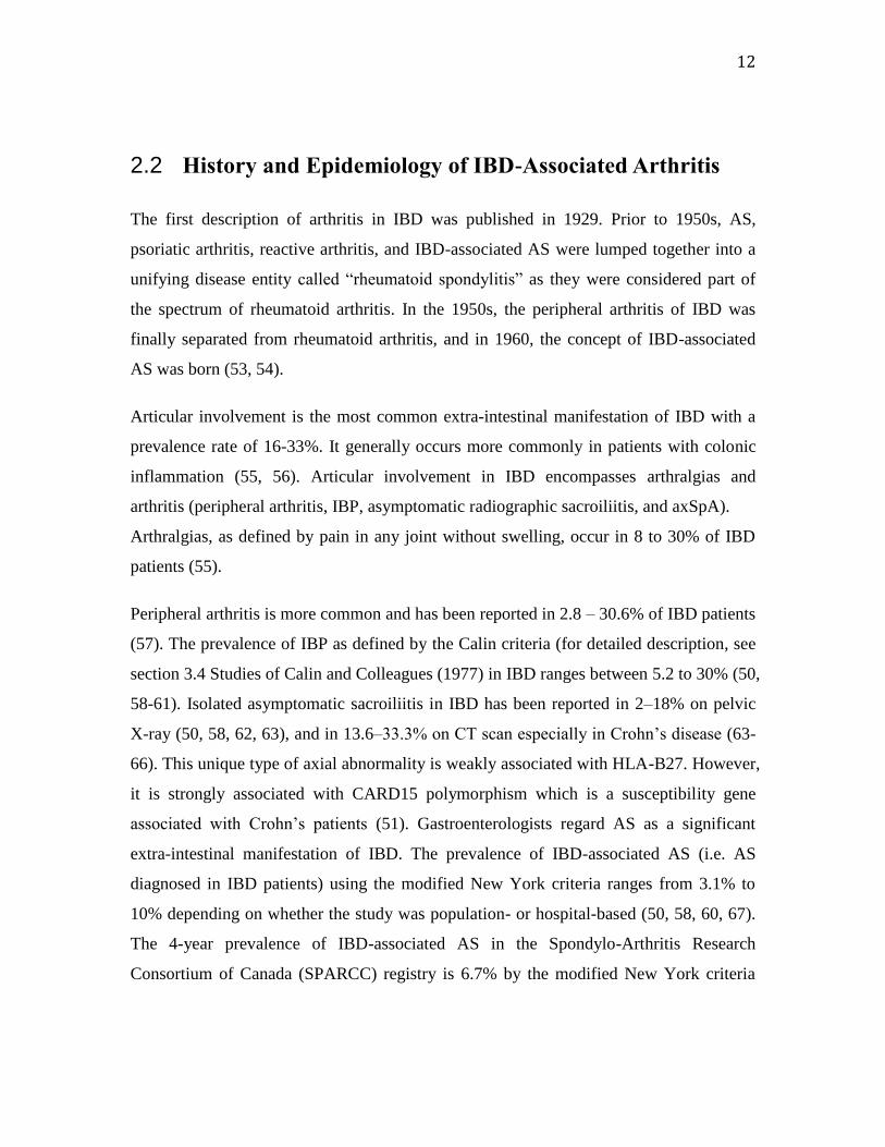
3.2 Development of Rome Classification Criteria of AS (1961)
As the literature about AS started to grow, a group of rheumatologists convened in Rome
in 1961 and agreed to formulate new criteria for AS, the Rome criteria, which were based
on clinical data that relied on history (low back pain and stiffness for more than 3 months
not relieved by rest, pain and stiffness in the thoracic region, history or evidence of iritis
or its sequelae), physical examination (limited motion in lumbar spine and limited chest
expansion), and radiographic sacroiliitis (without grading). The diagnosis of definitive
AS was based on bilateral sacroiliitis and any clinical criteria, or, fulfillment of 4 out of 5
clinical criteria (Figure 1). Some historical items were removed from the final criteria
including buttock pain that awakens patients at night, and good response to
phenylbutazone (an NSAID) which was found to improve IBP (91).
19
Figure 1. Rome criteria for AS (1961)
Clinical:
1. Low back pain and stiffness > 3 months not relieved by rest.
2. Pain and stiffness in thoracic region
3. Limited motion in lumbar spine
4. Limited chest expansion
5. History or evidence of iritis or its sequelae.
6. Radiograph showing bilateral sacroiliac changes characteristic of AS (this would
exclude bilateral osteoarthrosis of sacroiliac joints)
Definitive AS = Bilateral sacroiliitis (radiograph) and ≥ 1 clinical criteria, or, 4/5 clinical
criteria.
Reproduced from Kellgren JH, Jeffrey MR, Ball J: The epidemiology of chronic rheumatism,
Oxford: Blackwell Scientific Publications; 1963:326-327.
20
The Rome criteria were validated in Pima Indians with definitive AS, but some items
have been found to have poor diagnostic utility. For example, the item of historical iritis
was found to be infrequent with low sensitivity (4%) and difficult to be elicited with
certainty on history. Another historical item “low back pain lasting 3 months or more and
not relieved by rest” was difficult to apply and had low sensitivity (30%). Similarly, the
item on “pain and stiffness in the thoracic region” had low sensitivity (15%).
3.3 New York Criteria (1966)
The group of rheumatologists convened again in New York in 1966 and modified these
criteria (Figure 2). The iritis was removed, the description of back pain was changed to (a
history or the presence of pain at the dorsolumbar junction or in the lumbar spine), items
on physical examination were modified, and radiographical description of sacroiliitis was
clarified and graded. This has led to formulation of the New York criteria for AS (1966).
In 1973, Moll and Wright validated the New York criteria but found focusing only on the
item of historical back pain to be too sensitive and too non-specific, with the concern that
this might lead to including more false positive patients (92).

21
Figure 2. New York criteria for AS (1966)
Clinical criteria for AS:
1. A history or the presence of pain at the dorsolumbar junction or in the lumbar spine.
2. Limitation of motion of the lumbar spine in all 3 planes: anterior flexion, lateral
flexion, and extension.
3. Limitation of chest expansion to 1 inch (2.5 cm) or less, measured at the level of the
4th
intercostal space.
X-ray grading:
0 = normal.
1 = suspicious.
2 = abnormal with erosions or sclerosis.
3 = unequivocal abnormal, moderate or advanced sacroiliitis showing one or more:
erosions, sclerosis, widening, narrowing, and partial ankylosis.
4 = total ankylosis.
Application of these criteria:
Definitive AS:
o Grade 3-4 bilateral sacroiliitis AND 1 or more clinical criteria, OR
o Grade 3-4 unilateral sacroiliitis (or grade 2 bilateral sacroiliitis) AND clinical
criterion 1 (or both clinical criteria 2 and 3).
Probable AS: grade 3 or 4 bilateral sacroiliitis without a clinical criterion.
* When sacroiliitis is present, the following variants of AS should be designated
individually: rheumatoid arthritis, psoriasis, ulcerative colitis or regional ileitis, Reiter’s
syndrome, and juvenile RA.
* Individuals with conditions e.g. fluorosis, hypophosphatemic osteomalacia, brucellosis,
and familial Mediterranean fever, which may confuse the picture of AS, should be
identified in tabulations for AS and should be listed separately.
Reproduced from Bennett PH, Burch TA: Population studies of the rheumatic
diseases. Amsterdam, Excerpta Medica Foundation, 1968. 456-457.

22
3.4 Studies of Calin and Colleagues (1977)
Calin and colleagues pursued the quest in 1977 to identify AS patients earlier and
developed the first screening questionnaire which was administered to patients with
known AS, orthopedic patients (with normal sacroiliac joints and negative HLA-B27),
and healthy individuals. The aim of this symptom-based questionnaire was to identify
characteristics of IBP in AS. It was developed in English and had 17 items with binary
response options except for one item asking about the age of onset of back pain. The final
questionnaire had 5 items which were thought to represent the characteristics of IBP
(excluding neck pain) in AS patients compared to mechanical or non-specific back pain:
1) age of onset of back pain occurring less than 40 years, 2) insidious onset of pain, 3)
persistent pain for 3 months or more, 4) association with morning stiffness, and 5)
improvement with exercise. In this study, the item “no improvement with rest” was
significantly associated with AS compared to non-specific back pain, but was not
included in the final Calin criteria. Similarly, the item “awakening at night for back pain”
was significantly associated with AS compared to controls of healthy people, but was not
included in the final criteria. The specificity of fulfilling 4 out of 5 criteria was 85% and
the sensitivity was 95%. However, certain limitations deserve to be mentioned.
First, the optimal diagnostic parameters could not be validated in 2 subsequent studies
and the specificity was shown to be moderate (75%) with the sensitivity low (23-38%). In
one of these studies, a self-administered questionnaire (in German) contained 11 items in
which 9 items were related to back pain, and 2 items were related to pain in the
interscapular area and anterior chest. Some of the items were translated from Calin
criteria. Second, the term “insidious onset” remained ambiguous. In fact, a recent study
found that this term differentiates AS patients from those with mechanical back pain in
only one fourth of those with AS if the term indicates an onset of back pain within a year
(93, 94). Third, the Calin criteria alone cannot be used to classify or diagnose patients
with radiographic or non-radiographic axSpA. In fact, a population-based study
conducted by Calin’s group validated their questionnaire in 10,150 patients with back
pain who worked in an industrial complex. Of the 1880 respondents, 367 (20%) fulfilled
Calin criteria of whom only 16 (4.4%) had AS.

23
In 1983, Mller and colleagues found that 73% of AS patients had insidious onset of
back pain, 15% had sudden onset of back pain (minutes – hours), and 12% started with
back pain developing over a few days to weeks (95). Another study published in the same
year showed that the presence of morning stiffness of the back had a sensitivity of 93%
and a specificity of 43% for early AS (96).
3.5 Modification of New York Criteria (1984)
In 1984, a modification of the New York criteria for AS was published (Figure 3). The
IBP was defined as low back pain and stiffness for more than 3 months, which improves
with exercise but not with rest. The items on physical examination were modified again.
Diagnosis of definitive AS requires at least 1 clinical feature and 1 radiographic
abnormality of the anteroposterior pelvic X-ray. The clinical features include IBP,
restriction in lumbar spine motion, and restriction in chest expansion. The radiological
criteria include either bilateral sacroiliitis grade ≥ 2 or unilateral sacroiliitis grade ≥ 3 (3).

24
Figure 3. Modified New York criteria for AS (1984)
From the ASAS slide collection, http://www.asas-group.org, open access.

25
In 1985, Gran proposed addition of new characteristics to further refine IBP: back pain
that awakens at night (specificity 53%, sensitivity 71%) with getting out of bed
(specificity 79%, sensitivity 65%), not relieved by lying down (specificity 49%,
sensitivity 80%), and associated with morning stiffness for 30 minutes or more
(specificity 59%, sensitivity 64%) (97).
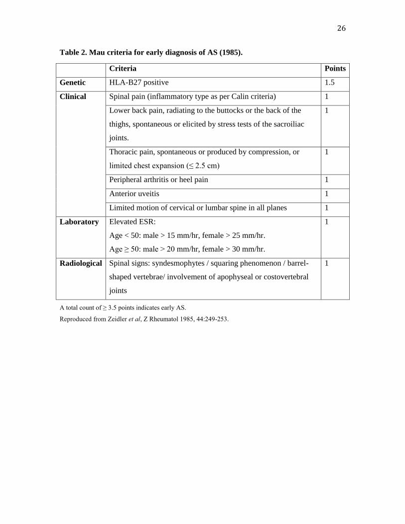
3.6 Mau Criteria for Early AS (1985)
In the French literature, the criteria of Sèze and Lequesne were formulated in 1961.
Baudoin and Landureau modified these criteria in 1979. A third modification was
proposed for early diagnostic criteria for AS and was published in English in 1985 (Table
2). The aim was to establish more reliable criteria for early diagnosis of AS in patients
without sacroiliitis. The criteria took into account historical variables, laboratory (ESR)
and radiological signs (spinal abnormalities of AS without sacroiliitis). Historical criteria
include IBP (as defined by Calin), spontaneous low back pain radiating to the buttocks or
back of the thighs, spontaneous thoracic pain (related to deep inspiration or coughing),
peripheral arthritis or heel pain, and anterior uveitis. The performance of these criteria for
diagnosis of early AS on a 5- and 10-year follow-up showed sensitivity of 82% and 88%
respectively, and specificity of 68% and 59% respectively. However, the use of these
criteria fell out of favor, probably due to lack of specificity (98, 99).
26
Table 2. Mau criteria for early diagnosis of AS (1985).
Criteria Points
Genetic HLA-B27 positive 1.5
Clinical Spinal pain (inflammatory type as per Calin criteria) 1
Lower back pain, radiating to the buttocks or the back of the
thighs, spontaneous or elicited by stress tests of the sacroiliac
joints.
1
Thoracic pain, spontaneous or produced by compression, or
limited chest expansion (≤ 2.5 cm)
1
Peripheral arthritis or heel pain 1
Anterior uveitis 1
Limited motion of cervical or lumbar spine in all planes 1
Laboratory Elevated ESR:
Age < 50: male > 15 mm/hr, female > 25 mm/hr.
Age ≥ 50: male > 20 mm/hr, female > 30 mm/hr.
1
Radiological Spinal signs: syndesmophytes / squaring phenomenon / barrel-
shaped vertebrae/ involvement of apophyseal or costovertebral
joints
1
A total count of ≥ 3.5 points indicates early AS.
Reproduced from Zeidler et al, Z Rheumatol 1985, 44:249-253.

27
3.7 Proposal for Diagnosis of AS by Cats and Colleagues
(1987)
Cats and colleagues proposed diagnostic criteria of AS based 4 clinical and 1 radiological
criteria (100). Table 3 summarized these criteria. The proposal also emphasized stating
associated diseases including psoriasis, IBD, Reiter’s disease, reactive arthritis, and
juvenile chronic arthritis.
Table 3. Proposal for diagnostic criteria of AS (1987)
Clinical 1. Insidious onset of low back pain lasting ≥ 3 months, associated with
morning stiffness, improves with exercise, with age of onset ≤ 45
years.
2. Existence of any of the following in a relative (1st or 2
nd degree) of an
AS patient, or in HLA-B27 patient, and age of onset ≤ 45 years:
a. Recurrent chest pain and thoracic stiffness of unknown cause.
b. Chronic or recurrent enthesopathy and/or oligo-arthritis with
negative rheumatoid factor and antinuclear antibody.
c. Unilateral acute anterior uveitis.
3. Restricted mobility of the lumbar spine in 2 planes (frontal and
sagittal) excluding other causes (e.g. infectious spondylitis,
neurological diseases, herniated or degenerative disc diseases, diffuse
idiopathic skeletal hyperosteosis)
4. Restricted chest expansion relative to normal values corresponding to
age and sex, excluding other causes (e.g. severe juvenile or adolescent
deformity of thoracic spine, congenital heart disease, or severe
pulmonary diseases).
Radiological 5. Bilateral sacroiliitis of grade ≥ 2 on pelvic X-rays, or unilateral grade
≥ 3, excluding other causes of sacroiliac diseases (e.g. Paget’s disease
of the bone, hypophosphatemia, hyperparathyroidism, fluorosis,
infection with tuberculosis or brucellosis, familial Mediterranean
fever, severe neurological disorders)
Definitive AS:
o 3 out of 4 clinical criteria OR
o Radiological criteria AND at least 1 clinical criteria.
Possible AS:
If a patient fulfills any of the above criteria with age of onset ≤ 45 years.
Reproduced from Cats et al, Clin Exp Rheum, 5:167-171, 1987.
28
3.8 Amor Criteria and ESSG Criteria for Classification of
SpA (1990-1991)
In early 1990s, 2 sets of classification criteria for the entire group of SpA were
established; the Amor criteria and the ESSG criteria. Both criteria included
undifferentiated SpA for the first time, in addition to the classic members of the group
(AS, psoriatic arthritis, reactive arthritis, enteropathic arthritis). Yet both sets of criteria
may have low sensitivity in establishing early diagnosis of SpA.
The Amor criteria (Figure 5) were developed with multiple entry criteria which make
them feasible and may actually facilitate a diagnosis in an individual patient. The 12
parameters incorporate historical, laboratory (HLA-B27), and radiographical (sacroiliitis)
features. Historical data ask about nocturnal back pain and/or morning stiffness, buttock
pain (more points if alternating), a good response to a full-dose of NSAIDs within 48
hours, and common extra-articular manifestations. These were the first criteria to
recognize response to NSAIDs as a characteristic of IBP. A recent Cochrane review of a
65 trials showed that short-term use of NSAIDs (including COX-2 inhibitors) in patients
with acute and chronic mechanical back pain without sciatica provided a marginal relief.
This endorses the concept that NSAIDs have a better response in SpA (101). One of the
advantages of the Amor criteria is that they can be applied to patients who present with
extra-articular symptoms such as uveitis or enthesitis without having IBP. These criteria
performed slightly better than ESSG criteria to classification of SpA (sensitivity 85%,
specificity 90%) (102).

29
Figure 4. Amor classification criteria for spondyloarthritis (1990)
From the ASAS slide collection, http://www.asas-group.org, open access.

30
The ESSG criteria (Figure 6) rely on historical data and the entry criterion is presentation
with IBP (as defined by Calin criteria) and/or synovitis predominantly of the lower limbs.
The presence of radiographic sacroiliitis is not mandatory. There is no item on response
of back pain to NSAIDs or HLA-B27 status. The latter was removed during the
development stage in anticipation that these criteria would be applied in population
studies without the need for an expensive laboratory test. The specificity and sensitivity
are 87% and 86% respectively which were also confirmed in other studies (103).

31
Figure 5. The European Spondyloarthropathy Study Group (ESSG) classification
criteria for spondyloarthritis (1991)
From the ASAS slide collection, http://www.asas-group.org, open access.

32
3.9 Studies of Rudwaleit and Colleagues (2004-2006)
Rudwaleit and colleagues took the lead to further study the characteristics of IBP in order
to facilitate early referral to rheumatologists. They previously found that IBP, as defined
by the Calin criteria, increased pre-test probability of axSpA from 5% in patients who
present to their primary care physicians with chronic back pain to 14%. The positive
likelihood ratio (LR) for IBP was 3.1, while the highest positive LR was the combination
of a positive HLA-B27 and a positive MRI (+LR of 9.0 for each). LR is unlikely to be
influenced by the disease prevalence. Table 4 summarizes the diagnostic properties for
clinical, laboratory and imaging parameters. They also showed that post-test probability
dramatically increased upon using different combinations of historical parameters of the
ESSG criteria (range 35-95%). Further, this probability also increased to 90% when
clinical and laboratory features of axSpA were combined. Based on the positive LRs of
individual parameters, the authors developed a diagnostic algorithm (later known as
Berlin criteria) for early axSpA in patients who have chronic low back pain for more than
3 months. Definitive diagnosis of axSpA was made when the probability was 90% or
more, while probable diagnosis was made when the probability was 89 – 90%. This new
algorithm works well in the individual patient (104).

33
Table 4. Summary of diagnostic tests for individual parameters used for the
diagnosis of early axSpA in patients with chronic back pain lasting ≥ 3 months
Sensitivity %
(range *)
Specificity %
(range*)
Positive
LR
Post-test
probability
(%)
Inflammatory back
pain
75 (38 – 95) 76 (76 – 100) 3.1 14
Alternate buttock
pain
40 (20–43) 90 (88–100) 4.0 17
Enthesitis (heel pain) 37 (16–52) 89 (89–96) 3.4 15
Dactylitis 18 (12–27) 96 (96–99) 4.5 19
Anterior uveitis 22 (10–22) 97 (97–100) 7.4 28
Family history of
SpA‡
32 (7–36) 95 (93–100) 6.4 25
Peripheral arthritis 41 (26–62) 87 (91–100) 4.0 17
Psoriasis 10 (1–17) 96 2.5 12
Inflammatory bowel
disease
4 (2–7) 99 4.0 17
Good response to
NSAIDs
77 (61–77) 85 (75–85) 5.1 21
Elevated CRP 50 (38–69) 80 (67–100) 2.5 12
HLA-B27 positive
(with axial
involvement)
88 (83–96) 90 (91–96) 9.0 32
Positive MRI (STIR
sequence)
90 (54–93) 90 (83–100) 9.0 32
* Ranges were extracted from different studies. ‡ Family history of SpA includes AS, reactive arthritis,
IBD, psoriasis, and anterior uveitis.
Abbreviations: NSAIDs, Non-steroidal Anti-inflammatory Drugs; CRP, C-Reactive Protein; MRI,
Magnetic Resonance Imaging; STIR, Short Tau Inverse Recovery; positive LR [Likelihood Ratio =
sensitivity / (100 - specificity)]. Adapted with permission from the BMJ Group © Rudwaleit, M. et al. Ann
Rheum Dis. 63,535-543 (2004).
34
In 2006, German investigators led by Rudwaleit published modified criteria for IBP.
They developed a standardized questionnaire (in German) which was administered face-
to-face by an examiner in the rheumatology, orthopedic surgery, and neurosurgery clinics
of hospital-based departments and private practices where patients ≤ 50 years with
chronic low back pain for at least 3 months were recruited. Questionnaire items were
related to age of onset of back pain, disease duration, time period of the onset of back
pain (within 1 hour, 1 day, 1 week, between 1 and 4 weeks, or within 1 year), current and
any previous episodes of morning stiffness, duration of morning stiffness, alleviation of
back pain with rest, presence of buttock pain, pain radiating to the leg, awakening
nocturnal pain, time of nocturnal awakening, preceding events (e.g. trauma, mental or
emotional stress, or infection), time of first consultation with a doctor, number of
consulted doctors, duration of sick leaves in days within the last 4 weeks, current
intensity of back pain, and impact of back pain on well-being. Patients were classified to
either AS, based on the modified New York criteria, or mechanical low back pain
(MLBP). Blinding of the examiner from the diagnosis was not achieved completely,
which may have influenced how the answers were obtained.
The best diagnostic results were obtained from a set of 4 combined parameters: morning
stiffness for more than 30 minutes, improvement with exercise but not with rest, back
pain that awakens at the second half of the night, and alternating buttock pain. If 2 out of
4 the criteria are fulfilled, the sensitivity are 70%, the specificity is 81%, and the positive
LR is 3.7. Any single parameter could not discriminate between AS and MLBP. None of
the parameters was able to rule out AS. Furthermore, some items were found in both
disease entities. For example, low back pain frequently awakened patients at night in both
diseases (75% and 63% respectively). Low back pain lasting 30 minutes or more was also
prevalent in 64% of AS patients and in 25% of MLBP. The term “insidious onset” was
problematic and only discriminated one-fourth of AS from MLBP when it meant an onset
of back pain within a year. Table 5 summarizes the frequencies and positive LRs of
features of IBP among patients with AS and MLBP.

35
Table 5. Frequencies and positive likelihood ratios of IBP features in AS and MLBP
patients
% of AS patients
(n = 101)
% of MLBP patients
(n = 112)
Positive LR
(95% CI)
Age of onset of back
pain < 40 years 96.0 86.0 1.1 (1.0 – 1.2)
Morning stiffness for
more than 30 minutes 64.3 24.7 2.7 (1.9 – 4.0)
Improvement with
exercise 78.2 50.0 1.6 (1.3 – 1.9)
Improvement with rest 31.7 58.0 0.5 (0.4 – 0.8)
Improvement with
exercise but not with rest 55.4 21.4 2.6 (1.8 – 4.1)
Any buttock pain 63.4 57.1 1.1 (0.9 – 1.4)
Alternating buttock pain 36.6 11.6 3.2 (1.8 – 5.6)
Awakening back pain
any time 75.0 62.7 1.2 (1.0 – 1.4)
Awakening back pain at
the second half of the
night
44.0 22.7 2.0 (1.3 – 2.9)
Adapted with permission from John Wiley & Sons, Inc. © Rudwaleit et al. Arthritis Rheum. 54, 569-78
(2006).

36
3.10 Development of the ASAS Criteria for axSpA (2009)
The modified New York criteria perform well in establishing a definitive diagnosis of a
patient with radiographic sacroiliitis and at least one clinical feature. Because these strict
criteria do not take into account patients who have IBP and normal sacroiliac joints i.e.
non-radiographic axSpA, earlier detection of those patients is often missed. The ESSG
and Amor criteria classify the whole SpA group without focus on axSpA. MRI of the
sacroiliac joints was shown to demonstrate active inflammation in patients who do not
have radiographic sacroiliitis.
To overcome these inherent limitations of these classification criteria, new classification
criteria (ASAS) for axSpA have been recently established and validated offering
additional clinical, laboratory (HLA-B27 status, ESR, and CRP) and radiological features
to facilitate earlier diagnosis (Figure 7). The entry criteria are chronic back pain lasting 3
months or more and occurring before the age of 45. During the development of the ASAS
classification criteria for axSpA, there was an increased sensitivity of the criteria when
selecting (chronic back pain) as an entry criterion as opposed to the newly developed IBP
criteria by Rudwaleit et al (93).
Patients then should either have positive HLA-B27 (clinical arm) or sacroiliitis (imaging
arm) detected either by pelvic X-ray as per the modified New York grading criteria or by
MRI. Once patients enter either diagnostic arm, they should have one of two more
parameters. The definition of IBP, as one of the SpA features, was based on expert
opinion and is to be elicited by the expert rheumatologist. The 4 items includes age of
onset < 40 years, insidious onset, improvement with exercise, no improvement with rest,
and pain at night with improvement upon getting up. Because of lack of statistical
significance on the item “morning stiffness”, it was removed from the IBP definition. If 4
criteria out of 5 are fulfilled, the sensitivity is 80% and specificity is 72% in the
validation cohort. The new ASAS criteria have a sensitivity and specificity of
approximately 83% and 84% respectively for the entire set. These criteria showed a
superior performance over the ESSG and Amor criteria in the validation study, with
sensitivity and specificity of 83% and 84% respectively. Therefore, they are reliable in

37
classifying patients for research and in diagnosing patients in the rheumatology practice
(105, 106).

38
Figure 6. ASAS classification criteria for axial Spondyloarthritis (axSpA)
From the ASAS slide collection, http://www.asas-group.org, open access.
39
These criteria carry certain limitations. First, although the estimated prevalence of AS
after the age of 45 is 5%, some patients will be missed at the entry criteria. Second, the
current definition of IBD includes Crohn’s disease and ulcerative colitis but not
indeterminate (unclassified) colitis. In this case, patients with the latter category may
fulfill the ASAS classification criteria for axSpA if they meet the entry criteria, a clinical
and/or imaging criteria, and have other supporting parameters such as family history of
SpA or peripheral arthritis. Third, elevated CRP or ESR levels can occur in IBD patients
independent of co-existing axSpA.
3.11 Development of Case Ascertainment Questionnaire (2010)
A recent case ascertainment questionnaire (107) has been developed and validated to
capture patients with radiographic AS.
In phase 1, a literature review using PubMed was performed generating 118 candidate
items. Members of an advisory board assessed each item by using a 9-point scale. Items
were selected only if their average score was ≥ 6. This led to retention of 42 items.
In phase 2, the 42 items were cognitively tested among 3 groups of patients with
established AS (diagnosed by the modified New York criteria): patients with disease
duration ≤ 10 years, patients with disease duration of > 10 years, and patients with mixed
disease duration. The purpose of this phase was to allow patients to evaluate the items in
terms of comprehension, clarity of questions, flow of questions, font size and
typographical errors. This led to re-wording of some items, splitting some items into
more than one or vice versa, changing the order of some questions, and addition of 9
items on fatigue when it was frequently identified by patients. This phase led to drafting
52 items.
Phase 3 was a feasibility study with the purpose of piloting the items among 50 patients
with AS and 50 patients with chronic back pain for ≥ 3 months. Patients were required to
be ≥ 18 years, fluent in English, and non-participants in phase 1. Internal consistency was

40
measured, although not reported, by Cronbach’s alpha, the average correlation between
each pair of items (inter-item correlation), and the correlation of each item to the total
sum score of items (item-total correlation). The investigators used the result of internal
consistency, bivariate and multivariate analyses to guide them into further item reduction
and modification.
The goals of phase 4 were to further refine the items, develop a scoring system for the
questionnaire and validate it in a case-control study (patients with AS and chronic back
pain). Similar to the requirements of phase 3, patients could not have participated in
phase 3. The first step of this phase involved measuring bivariate and multivariate
analyses in addition to internal consistency. The bivariate analysis of all items
demonstrated a statistical difference between both groups for 24 out of 43 items. The
multiple regression analysis further identified 12 items that were highly associated with
classic AS patients compared to patients with chronic back pain. These 12 items were
related to 3 main features: gender, characteristics of back pain, and iritis. IBP questions
are as follows: pain or stiffness for more than 3 months, locations of pain or stiffness
(neck, hip, other regions), age of onset of back pain, duration of back pain or stiffness,
influence of exercise on the pain or stiffness of the lower back or buttocks, influence of
daily physical activity on pain or stiffness on the lower back or buttocks, and NSAIDs
use and whether they improve symptoms within 48 hours. There is one question which
asked about numbness or tingling radiating to the legs and another one asking if the pain
or stiffness was a result of trauma. A picture or diagram of the back is not used in this
questionnaire. The sensitivity and specificity of the case ascertainment questionnaire in
the development sample was approximately 70% and 99% respectively. The second step
of this phase is the validation of the questionnaire which involved selection of a random
sample of patients with AS and chronic back pain. The sensitivity and specificity of the
case ascertainment questionnaire in the validation sample was approximately 67% and
95% respectively. About 54% of the AS patients in this study were on biologic drugs.
However, the investigators demonstrated that the answers of the questionnaire did not
differ in patients who were on these drugs compared to those who were not.
41
There are some limitations of this questionnaire. First, it did not target patients with early
axSpA as the mean disease duration among AS patients was 21.8 years. Second, it did not
address patients with IBD-associated AS. Thirdly, while the scoring system was driven
by data, it is complicated and not easily applied. The scoring was designed to have a
predicted probability of at least 66.86%. Finally, it was not comprehensive in terms of
asking about different features of axSpA.
3.12 Summary
In the past century, there has been extensive work attempting earlier identification of
axSpA in order to overcome the delay in diagnosis for these patients. Different
classification criteria were developed with a special focus on refining the features of IBP.
Up to April 2011, the literature has only 2 self-reported questionnaires in English: the 5-
item Calin questionnaire and the case ascertainment questionnaire. Neither questionnaires
included clinical pictures or diagrams of the back nor did they target cases of early
axSpA in IBD patients. Recent studies have recommended combining IBP features with
extra-axial manifestations to achieve a higher post-test probability of axSpA.
Attention needs to be directed at those patients who are at heightened risk of developing
axSpA, as is the case in IBD patients. One approach to identify those IBD patients with
undetected axSpA is to screen them when they present to their gastroenterologist for
follow-up of their IBD. This type of screening is recommended by Sackett and Holland in
this specific scenario and is called “case finding” which is best administered to patients
who present to their physicians with different complaints so that there is an opportunity to
ask them about symptoms of the disease of interest. A good case finding questionnaire
requires items that are accurate, highly sensitive (commonly present in the target
population), reasonably specific, and that have good predictive value (108).

42
Chapter 2
Rationale, Hypothesis and Aims

43
1 Rationale
There is an agreement among different studies that in order to detect axSpA earlier, it is
imperative to obtain an appropriate clinical history using various questions to address
different aspects of axSpA. A health care worker (usually a rheumatologist) or a self-
reported questionnaire can extract the clinical history. The only 2 questionnaires (in
English) available in the literature, as of April 2011, were aimed to identify patients with
classic AS by asking specific questions which were mainly focused on characteristics of
IBP.
Patients with IBD are at risk of developing axSpA which remains undetected in many
patients for years. Once those patients are identified, a timely initiation of appropriate
treatments can in turn lead to better pain control, improved functional outcomes and
enhanced quality of life. This would minimize unnecessary and expensive diagnostic and
therapeutic interventions.
2 Hypothesis
I hypothesized that a simple, sensible and reliable self-reported questionnaire could be
developed to allow earlier case finding of axSpA in IBD patients (populations at risk)
who are being seen in hospital-based gastroenterology clinics
3 Aims
1) To describe the development including sensibility of the Toronto Axial
Spondyloarthritis Questionnaire (TASQ).
2) To evaluate the test-retest (intra-rater) reliability of the TASQ.

44
Chapter 3
Methods

45
1 Development of Questionnaire Items
Development of questionnaire items required multiple steps including: conceptualization,
literature review to identify characteristics of the construct and select candidate items,
choosing appropriate scaling response and format, sensibility assessment among the SpA
team and 2 general rheumatologists, 3 stages of pilot testing and creation of the final
version. Figure 8 depicts the stages for the development of the Toronto Axial
Spondyloarthritis Questionnaire (TASQ).

46
Figure 7. Stages of development of the TASQ
Final version of TASQ
1 Item was removed 3 response options were removed 4 items were modified
Conceptualization
Literature review and generation of 93 candidate
items
1st Sensibility assessment among committee members
Second pilot testing among 9 axSpA patients
First pilot testing among 4 axSpA patients
1 Item was removed 1 picture was removed 3 items were modified
Order of few items was modified
Third pilot testing among 6 axSpA patients
Scaling response and questionnaire format
75 Items were removed 3 Pictures were removed Some items were modified Pilot testing
2nd Sensibility assessment among committee members
Minor modification of instruction to an item
Phase 1
Phase 2
Phase 3
Phase 4
Phase 5
Phase 6
47
1.1 Phase 1: Conceptualization
As mentioned in the Rationale section, the specific aim of the project is case finding to
facilitate early referral of IBD patients who potentially may have undiagnosed axSpA.
The construct in this study is axSpA in IBD.
1.2 Phase 2: Item Pool Generation
A thorough literature review was performed using Medline (from 1941 to April 2011)
and PubMed to identify potential items in preparation to the development of
questionnaire. All the reviewed papers were restricted to the English language. Keywords
included “inflammatory back pain”, “back pain”, “symptoms”, “features”,
“manifestations”, “ankylosing spondylitis”, “spondyloarthritis”, “spondyloarthropathy”,
“risk factors”, “questionnaire”, “screening”, “case finding” and “diagnosis”. If an article
cited other relevant article(s), the latter was reviewed to identify potential items.
The main items for IBP symptoms were identified and reviewed from previous
descriptions of the work of Hart et al, Calin et al, Mller et al, Gran, and Rudwaleit et al
(89, 90, 93-95, 97). In addition, a thorough review of previously reported characteristics
of IBP associated with IBD was conducted. Previously developed classification criteria
for AS and SpA were reviewed with the focus on clinical items. These include the Rome
criteria for AS (1961), New York criteria for AS (1968), modified New York criteria
(1984), Mau criteria for identifying early AS (1985), Amor criteria for SpA (1990),
ESSG criteria for SpA (1991), and the most recent ASAS classification criteria for early
axial and peripheral SpA (2009 and 2010 respectively) (3, 92, 98, 102, 103, 105, 109).
Using this review I generated 93 potential items which did not include any items loaded
with sensitive issues. A mixture of sensitive and specific items was chosen in keeping
with the properties of a case-finding instrument. I added 4 pictures of peripheral arthritis

48
of hand joints (2 pictures) and dactylitis (2 pictures of a finger and a toe). I also added a
diagram of the back with arrows pointing towards the neck, upper back (between the
upper and lower borders of the scapula), middle back, lower back (lower thoracic to end
of lumbar areas), and buttock.
1.3 Phase 3: Scaling Responses and Instrument Format
Most of the questionnaire items asked closed-ended questions with binary responses, with
fewer questions providing continuous data. In general, questions with binary responses
are easy and quick to complete for patients. For clinicians, they are easy to code and
analyze. Standardized instructions on how to answer the questionnaire were provided.
Some branching questions (sub-questions) were included.
Demographic questions were asked at the beginning of the questionnaire (sex and date of
birth). No question identified the patient’s name in order to reassure patients of
confidentiality and to comply with privacy guidelines. Three major headings represented
the domains of the questionnaire (IBD, inflammatory back symptoms, and extra-axial
features).
1.4 Phase 4: Selection of Items
All items were worded neutrally i.e. free of value-loaded items. There was one item
which asked about race since the axSpA is more prevalent in Caucasians of European
origin. In order to qualify for the questionnaire, a patient must have inflammatory bowel
disease and back pain or stiffness. This is an absolute criterion. The first question is
general and relevant to the main construct “Has there been a time when you had pain or
stiffness in your back for 3 months or more?” This is designed to reduce referrals for
mechanical back pain in which the pain commonly lasts less than 3 months. Back pain
was considered equivalent to back stiffness, allowing the use of this double-barreled

49
definition in some questions.
1.4.1 Sensibility Assessment of Questionnaire Items
Alvan R. Feinstein, who is considered one of the founders of modern clinical
epidemiology, published the concept of Clinimetrics in 1982. It advocates for the
methodological approach of clinical judgment when developing an instrument that
measures a clinical observation, diagnostic criteria, prognostic or therapeutic outcome.
Important facets of the clinimetric approach include sensibility, reliability, validity, and
responsiveness. Sensibility assessment refers to the usefulness of an instrument. It
includes a statement of clinical function (purpose), clinical justifications, clinical
applicability (indication of population and setting), overt format (comprehensibility,
replicability, and suitability of scale), face and content validity, and feasibility (110).
The clinical function (purpose) and clinical justification (rationale) were discussed in
detail in Chapter 2 (Rationale, Hypothesis, and Aims).
In terms of clinical applicability, our questionnaire at this point is only applicable to a
subset of patients with seronegative SpA i.e. axSpA in IBD patients. In this study, clinical
applicability was evaluated by asking: “The questionnaire is intended to ask questions
related to presence of axial spondyloarthritis in patients with inflammatory bowel disease.
Do you think this questionnaire achieved this goal?”
Comprehensibility refers to the ability of understanding the questionnaire which is
assessed by limiting the questionnaire to important items (oligo-variability) and the
appropriateness of the number of response options and clarity of item weighting
(transparency). I assessed oligo-variability by asking, “Do you feel that the questionnaire
included the necessary questions?” With regards to transparency, I assessed it from 2
perspectives. The first perspective asked about response options; “Do you think the
response options were appropriate (for example, number of options)?” The second
perspective initially assessed transparency by asking the members about the appropriate
way to score the items. I then administered the new version (post-pilot) of TASQ

50
questionnaire among them and assessed transparency by asking, “Assuming that each
question is weighted equally with a score of 1, do you think that the scoring is simple?”
Replicability refers to the clarity of instructions.
Face validity is an attribute to be evaluated when creating a new measuring instrument to
ensure that items or domains appear to evaluate the desired construct. In this study, the
construct was axSpA in IBD. I evaluated face validity by asking the following: “Do you
feel that this questionnaire accurately measures what we are trying to measure?”
Content validity refers to how comprehensive the questionnaire is i.e. whether items
address sufficient domains for the construct of the disease. In this study, content validity
was assessed by asking 2 questions: “Do you think an important question was missing?”
and “Do you think an inappropriate question was included?” Items were only included if
there was a 100% agreement on the relevance or irrelevance of each item to the construct
of the questionnaire.
Feasibility refers to ease of use of an instrument. It was assessed in our study by asking
about completion time, acceptability of the questionnaire by patients (i.e. no sensitive or
potentially offensive items), and readability (clarity of questions, flow of questions,
presence of typographical errors, appropriate font size, usefulness of pictures and
diagrams). Readability was evaluated by asking “Were the sentences easy to read?” I also
tested readability by calculating the Flesch-Kincaid grade level and the Flesch Reading
Ease Scale using Microsoft Word for Mac 2011. The recommended grade for a
questionnaire is grade 6 or less. The Flesch Reading Ease is a 100-point scale, and the
higher the score; the easier it is to read the questionnaire. The scale is interpreted as
follows: 90-100 = very easy, 80-89 = easy, 70-79 = fairly easy, 60-69 = standard, 50-59 =
fairly difficult, 30-49 = difficult, and 0-29 = very confusing (111). Table 6 shows the
checklist of sensibility assessment with their definitions which were adapted from
Feinstein’s principles.

51
Table 6. Checklist of sensibility assessment with definitions
Concept Definition
Purpose and framework
Clinical function Purpose of the questionnaire
Clinical justification Rationale behind questionnaire development
Clinical applicability Application of the questionnaire to a specific group
of patients in a specific setting
Face validity Accurately measuring what the questionnaire is
trying to measure
Comprehensibility
Oligo-variability Limiting the questionnaire to important items
Transparency Ability to see through the contribution of each
scored item and its response options to the final
score
Replicability Clarity of instructions
Content validity Comprehensiveness of the questionnaire
Important omissions Exclusion of important items
Inappropriate inclusions Inclusion of inappropriate items
Feasibility Ease of use of the questionnaire
Time to completion Amount of time taken to complete the questionnaire
Acceptability Approval of the questionnaire by patients and health
care workers by excluding sensitive or potentially
offensive items
Readability Ease which the words can be understood
Clarity of questions Ability to understanding the questions easily
Flow of questions Transitioning of questions in a logical manner
Readability tests Flesch Readability Ease Scale and Flesch-Kincaide
grade level
Typographical errors Presence of typographical errors
Font size Appropriateness of the font size used in the
questionnaire format
Illustrations Clarity and usefulness of diagrams and pictures
used in the questionnaire
Adapted from Clinimetrics 1987 (110)

52
Rowe and Oxman translated the above principles of sensibility into 12 questions using 7-
point Likert scales and one last question that allowed patients to add any comment (112).
I adapted their questions and added new 5 items for feasibility. I also changed the
response options from Likert scales into dichotomous scales and allowed a final open-
ended question for any comment. The sensibility questions were asked to the committee
members (SpA team and 2 general rheumatologists) and patients (Table 7).

53
Table 7. Questionnaire for sensibility assessment
Concept Question
Clinical function The questionnaire is intended to ask questions related to presence of axial
spondyloarthritis in patients with inflammatory bowel disease. Do you think this
questionnaire achieved this goal?
Face validity Do you feel that it accurately measures what we are trying to measure?
Comprehensibility
Oligo-variability Do you feel that the questionnaire included the necessary questions?
Transparency Do you think the response options were appropriate (for example, number of
options)?
Assuming that each question is weighted equally with a score of 1, do you think that
the scoring is simple?
Replicability Do you think the instructions were clear?
Content validity
Do you think an important question was missing? (important omissions)
Do you think an inappropriate question was included? (inappropriate inclusions)
Feasibility
Time to completion Please indicate the amount of time you took to complete reading the questionnaire?
_____ minutes
Acceptability Were there any questions that were unacceptable question (too personal or
offensive)?
Clarity of questions Do you think the questions were clear?
Flow of questions Was the flow of questions smooth?
Ease of reading Were the sentences easy to read?
Typographical
errors
Were there any typographical errors?
If so, please indicate where.
Font size Was the font size appropriate?
Illustrations Were the pictures and diagram useful?

54
The questionnaire for sensibility assessment was administered to the SpA team and 2
general rheumatologists (committee members). The former consisted of 2 SpA
rheumatologists, an advanced physiotherapist, a clinical trial nurse, an infusion nurse
coordinator, a biologic nurse coordinator, and a research analyst. The SpA team met on
several occasions to assess the items for sensibility considering the overall purpose of
case finding of the questionnaire. Relevant questions of the sensibility assessment were
asked during the meetings.
1.5 Phase 5: Pilot Study
The purpose of the pilot study was to pretest the questionnaire among a small, targeted
sample in a controlled setting in order to obtain a sensibility assessment and qualitative
analysis that could refine the items of the questionnaire if necessary. This approach leads
to reducing measurement error and improving the quality of the collected data.
I conducted pilot testing in 3 stages on axSpA patients, who attended the Spondylitis
clinic at the Toronto Western Hospital (TWH). The Spondylitis Clinic at TWH is the
major referral clinic for the care of patients with axSpA in Southern Ontario. It is part of
the Spondyloarthritis Research Consortium of Canada Registry for Spondyloarthritis
(SPARCC) which is a trans-disciplinary national research program focusing on genetic,
pathogenesis, and outcome measures for patients with SpA (AS, psoriatic arthritis, IBD-
related arthritis, undifferentiated SpA, and reactive arthritis). Beside Toronto, the
program covers Newmarket, London Ontario, St. John’s, Montreal, Winnipeg, Saskatoon,
and Edmonton. All SPARCC patients, who are 18 years or older and are fluent in English,
give their informed consent at the beginning of their enrollment. Patients have an initial
clinic visit then once a year after that. At each visit, patients follow a standardized
protocol including self-administered questionnaires, clinical assessment by a
rheumatologist, blood tests and X-ray if necessary (68).

55
I explained the purpose of the pilot phase to patients and instructed them to read the
questionnaire and answer the questions at the end that are part of the sensibility
assessment. Patient characteristics were collected including age, sex, the highest level of
education (on a scale of 1 to 5: 1 = below grade 8, 2 = high school incomplete, 3 = high
school graduate, 4 = college, 5 = university) reflecting the highest educational level
obtained at the patient’s last clinic visit, the BASDAI score at the last clinic visit, and the
BASFI score of the last clinic visit.
An additional questionnaire for sensibility assessment was administered to patients after
filling out the TASQ questionnaires. The assessment ended with a free text comment area
and a thank-you note. I also conducted a qualitative analysis by interviewing all patients
after completion of the 2 questionnaires.
1.6 Phase 6: Sensibility Assessment of Post-Piloted TASQ
Completed by the Committee Members
I administered the sensibility assessment to the committee members after the
modification made in the pilot testing.
2 Reliability study
2.1 Concept
Classical test theory has been adopted to measure the reliability of this new questionnaire
in order to obtain the measurement error that is inherent in observed scores. Reliability
represents a key concept in this theory and evaluates the reproducibility of an instrument
in measuring an attribute or a construct. Test-retest (intra-rater) reliability is one of its
types and is directly relevant here. It assesses the stability of the item scores on different

56
occasions to the population of interest within a 2-week period between administration
times in order to reduce the chance of recall and intercurrent flare of axSpA (113).
2.2 Population of Interest and Sampling Method
As of July 1st, 2011, the database of the Spondylitis Clinic at the TWH had 636 patients
with axSpA. Of those patients; 77 (12.1%) had concurrent IBD that was diagnosed by
gastroenterologists based on clinical, endoscopic and histological evaluations. The
patients had chronic back pain or stiffness that lasted 3 months or more and fulfilled the
ASAS classification criteria for axSpA. The one exception was a male patient who
fulfilled the modified New York criteria for diagnosis of AS based on typical features of
IBP, and bilateral sacroiliitis of grade 2 but did not fulfill the ASAS criteria because the
age of onset of his back pain was greater than the cut-off age of 45 years. However, we
included this patient in our analysis because he conceptually still fulfilled the generic
definition of axSpA. An amendment to distribute our new questionnaire was approved by
the institutional review ethical board.
The questionnaires were mailed to all the 77 patients who have established axSpA and
IBD concurrently. These patients reflected the sample of the total population of the IBD
and axSpA. I adapted the tailored design method of Dillman to maximize the response
rate (114). Each patient was provided with a package of 2 self-addressed and stamped
return envelopes in which each had a one-page, double-sided and identical questionnaire
accompanied by a personalized covering letter. The letter format was dated and had the
TWH logo at the beginning, and the University of Toronto logo at the end, and was
signed by my supervisor. This procedure was used to give patients the impression that
their opinions are being sought by an academic clinician and leader from well-recognized
academic institutions. The letter explained the purpose of the reliability study, the
importance of their participation in our study and provided clear instructions about the
completion of each questionnaire on two occasions 1-2 weeks apart. An ending statement
acknowledged their time and cooperation (Appendix A). Each questionnaire was coded to

57
allow identification of the patient. The only identifying information on the questionnaire
was date of birth. Patients were unaware that their responses would be compared among
them in order to minimize the possibility of Hawthorne effect which refers to respondents
modifying their responses because of awareness that they were being studied (115).
The return envelopes had a logo of the University Health Network which includes 3
hospitals: Toronto General Hospital, TWH, and Princess Margaret Hospital. The package
was sent by a regular mail.
2.3 Sample Size Calculations for Test-Retest Reliability
Study
Calculation of the sample size was based on 3 estimates – hypothesized reliability (R),
number of observations and the confidence interval (CI). For test-retest reliability study,
the number of observations was 2 when the questionnaire was to be administered to the
same patient within a 2-week interval. With regards to the recommended reliability
coefficient, Nunnally and Bernstein suggested a reliability of 0.70 for research purposes,
and 0.90 for clinical purposes (individual level of patient care) (116). Streiner
recommends a reliability of > 0.50 to define stability of a measuring instrument. I chose a
minimally desired reliability of 0.80. The hypothesized reliability (R) of TASQ was
estimated as 0.90, and in order to be 95% certain that it is definitely above 0.80, the 95%
CI around the reliability is ± 0.10 (0.90 minus 0.80). The standard error (SE) of the
reliability coefficient is approximately half this CI, or 0.05. Therefore, the calculated
sample size with hypothesized R = 0.90, SE 0.05 and 2 observations is 24 patients (117).
The following calculations demonstrate steps used to reach the sample size for test-retest
reliability:
First, I needed to calculate a conservative reliability coefficient (R–) to get a larger
sample size than relying only on (R + SE). Second, I then used Fisher log transformation
of Intraclass Correlation (ICC). It should be noted that the ICC here is equivalent to the
reliability coefficient. The number of observations per subject is 2.

58
The calculated sample size (n) is rounded up to 24. I elected to mail the questionnaires to
all the 77 consecutive patients with concurrent IBD and axSpA to account for an
anticipated low response rate during the summer and for incomplete questionnaires. All
the responses were entered into a database and were double-checked.
3 Statistical Analyses
3.1 Statistical Analysis for the Sensibility Assessment
Qualitative and descriptive analyses were used for the sensibility assessment among the
committee members and among patients with axSpA.
3.2 Statistical Analysis for Patients in the Pilot Study
Descriptive analysis was used to describe the clinical and laboratory characteristics of
axSpA patients who participated in the pilot study.

59
3.3 Statistical Analyses for Responders and Non-Responders
For description of the clinical characteristics between patients who responded to our
questionnaire and those who did not, the following parameters were collected: type of
IBD (Crohn’s disease, ulcerative colitis, indeterminate colitis), the highest level of
education, BASDAI and BASFI scores of the last clinic visit, ESR level of the last
protocol visit, and CRP level of the last protocol visit. Serum ESR and CRP levels are
inflammatory markers that may be elevated with activity of disease in axSpA.
Categorical variables were summarized by frequencies and percentages, while continuous
variables were summarized by mean, standard deviation and median. A two-sample
Student’s t-test was used to compare the mean difference of age, and scores of BASDAI
and BASFI between the groups (respondents and non-respondents). Wilcoxon rank sum
test was used to test the medians of the level of education between groups. Chi-square
test was used to assess the association between categorical variables (i.e. types of IBD)
between groups. Statistical analyses were 2-sided, and the statistical significance is
defined by p values of <0.05.
3.4 Coding the Questionnaire’s Responses
Dichotomous response options were coded as 1 for Yes and 0 for No. Two questions,
which contained continuous variables, were converted into dichotomous variables. The
first question asks about the age of onset of back pain (age ≤ 45 years or age > 45 years)
in accordance to the ASAS classification criteria for axSpA. The second question asks
about duration of morning stiffness (≥ 30 minutes or < 30 minutes).
For the 3 questions with 3 response options (Yes, and 2 negative detailed response
options), the negative options were coded as No. Similarly, the question which asked
about family history of AS was coded as Yes, and No for No or Unknown.

60
3.5 Statistical Analysis for Test-Retest Reliability
For test-retest reliability, Cohen’s kappa statistics were obtained to determine
agreement coefficient. The percentage of agreement for the items was also determined.
The agreement coefficients were interpreted using the recommendations of Landis and
Koch as follows: < 0.00 = poor agreement, between 0.00 to 0.20 = slight agreement,
0.21 to 0.40 = fair agreement, 0.41 to 0.60 = moderate agreement, 0.61 to 0.80 =
substantial agreement, and > 0.80 = almost perfect agreement (118).
Statistical analyses were generated using SAS (version 9.3 Institute Inc., Cary, NC, USA)
and R (version 2.14.2; The R Foundation for Statistical Computing). Statistical analyses
were 2-sided, and the statistical significance is defined by p values of <0.05 as stated.

61
Chapter 4
Results

62
1 Selection of Items Using Sensibility
Assessment
Following the first 3 phases (conceptualization, item pool generation, scaling responses
and instrument format), phase 4 consisted of selection of items after a thorough
assessment of their sensibility. Table 8 summarizes the results of sensibility assessment
of this stage.

63
Table 8. Sensibility assessment for the pre-piloted TASQ completed by the
committee members
Sensibility assessment Pre-pilot testing
n = 9 (%) *
Clinical function 9 (100)
Comprehensibility
Oligo-variability 7 (78)
Transparency
Appropriateness of the
number of response options
9 (100)
Weighting each item 0 (0)
Replicability 7 (78)
Face validity 6 (67)
Content validity
Important omissions 2 (22)
Inappropriate inclusions 3 (33)
Feasibility
Time to completion, median
(range) †
4 (3 – 5) minutes
Acceptability 8 (89)
Readability
Clarity of all questions 4 (44)
Flow of questions 7 (78)
Flesch reading ease scale † 72.2%
Flesch-Kincaid grade level † 5.6
Absence of typographical
errors
8 (89)
Appropriate font size 7 (78)
Usefulness of illustration(s) 5 (56)
* All the numbers in the table represent number of committee members who agreed on the corresponding
principles (percentages in parenthesis).
† This was determined independently from the committee members.

64
Clinical function: The committee members agreed that the purpose of the questionnaire
is clear.
Comprehensibility: Two members suggested adding some questions related to construct
such as addition of response of back pain or stiffness to biologic agents and history of
psoriatic arthritis. All members agreed on the appropriate number of response options.
However, the concept of weighing each item was felt to be unclear to by the members.
Replicability: Two members had some suggestions to clarify the first instruction at the
beginning of the questionnaire.
Face validity: Three members suggested removal of many questions that did not appear
to measure what the questionnaire was supposed to measure. For example, the question
regarding the extent of bowel disease involvement was thought not to be relevant to
finding cases of axSpA, although it was relevant to peripheral arthritis. Another example
is dropping of an item on “fatigue”. While fatigue is one of the features of axSpA, we
decided to drop it because it can also be found in IBD patients who may suffer
concurrently from anemia which often causes fatigue.
Content validity: Two members found that certain important questions were missing
such as response of back pain or stiffness to biologic drugs. Questions which were
inappropriately included were identified and discussed at length.
Feasibility: The median of self-reported time to completion of the questionnaire was 4
minutes (range 3 – 5) among all members. There was one member who identified the
demographic question on ethnicity as irrelevant and potentially sensitive. Five members
indicated that certain questions required clarifications, sentence shortening and better
wording. Two members suggested changing the order of few questions to improve the
flow. The Flesch reading ease scale was 72.2% which makes reading the questionnaire
fairly easy. The Flesch-Kincaid grade level was 5.6 which is within the recommended
grade for a questionnaire. One member identified one typographical error in the
questionnaire. Two members found the font size small. Four members found 3 pictures
not useful. The 2 pictures of peripheral arthritis could be misinterpreted as dactylitis. One

65
picture of finger dactylitis could be confused with peripheral arthritis. A picture of iritis
could be mistaken as conjunctivitis and the members agreed on substituting this pictorial
item with a clear and short question on history of iritis. All members agreed on the
usefulness of the picture of toe dactylitis and the diagram of the back.
Version 1 of TASQ: Item reduction was made to include all the necessary items with
consideration of including missing questions and excluding inappropriate items. For
example, the item of ethnicity was removed as it was found unnecessary and potentially
sensitive. Revisions of some items were made in order to have simple and short sentences
whenever possible with one or 2 clauses. Double-barreled questions were allowed for the
terms “back pain or stiffness” and “pain and swelling”. The latter was included to
specifically describe peripheral arthritis and dactylitis independently. The pictures of
peripheral arthritis, finger dactylitis and iritis were removed. This led to drafting the first
version of the questionnaire which included 18 items, a diagram of the back and one
picture of toe dactylitis.
2 Pilot study
In all the 3 pilot stages (4 patients in stage I, 9 in stage II, and 6 in stage III), the total
number of patients was 19 with a mean age of 37 (range 24 to 53) and approximately
68% were males. The mean BASDAI score was 3.5 (range 0 – 8.8), while the mean
BASFI score was 4.2 (range 0 – 7.7). The median of the patients’ highest level of
education was 4.5 (range 3 – 5). All patients answered the first screening question that
there had been a time when they had back pain or stiffness in their back for 3 months or
more. Table 9 summarized the evaluation of sensibility among axSpA patients during the
pilot testing.

66
Table 9. Stages of sensibility assessment of TASQ by patients
Sensibility
assessment
Stage I
n = 4 (%) *
Stage II
n = 9 (%) *
Stage III
n = 6 (%) *
Comprehensibility
Transparency
(appropriate
response options for
each item)
2 (50) 9 (100) 6 (100)
Replicability 4 (100) 9 (100) 6 (100)
Feasibility
Completion time,
median (range) †
5 (3 – 5) minutes 4 (3 – 5) minutes 4 (3 – 5) minutes
Acceptability 4 (100) 9 (100) 6 (100)
Readability
Clarity of all
questions 2 (50) 7 (78) 6 (100)
Flow of questions 4 (100) 9 (100) 6 (100)
Ease of readability 3 (75) 7 (78) 6 (100)
Absence of
typographical
errors
4 (100) 8 (89) 6 (100)
Appropriate font
size 4 (100) 9 (100) 6 (100)
Usefulness of
picture(s) and/or
diagram
4 (100) 6 (67) 6 (100)
Actions following
each stage
Removal of 4 items
Modifications of 4
items
Removal of 2 items
Removal of
dactylitis picture
Modification of 3
items
Minor modifications
of few items
* All the numbers in the table represent number of patients who agreed on the corresponding principles
(percentages in parenthesis).
† This was determined independently from the patients.
67
2.1 Stage 1
In stage 1, the questionnaires were administered to 4 axSpA patients. All patients found
the instructions clear. None of them commented on inclusion of sensitive items. No
patient found typographical errors, and all of them found the font size appropriate and the
illustrations useful. The median time for completion of the question was 5 minutes (range
3 – 5).
Item Modification
One patient did not know how to answer the question which asked about awakening due
to back pain during the second half of the night. The reason was that the cut-off time of
sleep disturbance was not well defined. The patient suggested adding a third response
option (both). However, we decided to keep the question as originally phrased with 2
response options because it was found to better characterize IBP in Rudwaleit’s study
(93). We accepted a suggestion from a patient to add a third response option “Not
applicable” to the question, “If you have buttock pain, does it move from one side to the
other?” One patient commented on the question, “Does your back pain improve during
the night upon getting up, walking or stretching?” and suggested the addition of a third
response option “I do not get up” because back pain did awaken him at night but he did
not necessarily get up, walk or stretch. This agreed with our clinical experience as some
axSpA patients were awakened at night because of back pain but they do not get up and
walk. Therefore, we accepted this suggestion.
There were 2 questions that proved difficult to understand. The first question was about
the mode of onset of back pain “When your back pain first began, indicate how long it
took to develop: ___hour(s), ___day(s), ___week(s), ___month(s), ___years”. This was
modified to “Indicate the duration of time it took to develop”. The second question was
on buttock pain “Do you have any buttock pain that alternates from side to side?” This
was re-worded into the following: “If you have buttock pain, does it move from one side
to the other?” Patients found the flow of questions smooth.

68
Item and Response Options Reduction
Three response options and one question were removed in this stage. There was a
question on biologic drugs as a treatment for IBD “Have you been treated with any of the
following: infliximab (Remicade), adalimumab (Humira) or other (please specify)?” with
3 responses for each drug “currently, previously, never”. Although no patient commented
on this item, I decided to remove the response option “Never” for each drug, and change
the question into the following “Has your inflammatory bowel disease ever been treated
with a biologic drug such as Remicade or Humira or other biologic drug?” Again,
some patients had difficulty with the question, “Does your back pain occur during the
second half of the night?” We decided to remove it because it was difficult for patients to
define exactly the second half of the night. These modifications led to a revised
questionnaire (version 2) with 20 items, one diagram and one picture.
2.2 Stage 2
In stage 2, version 3 of the questionnaire was administered to 9 axSpA patients. All
patients found the questionnaire transparent in term of appropriate response options for
each item. All patients found the instructions adequate. On assessment of feasibility, all
patients found the questionnaire acceptable. Again, all patients found the flow of
questions smooth and the font size appropriate. The median time to complete the
questionnaire was 4 minutes (range 3 – 5).
Item Modifications
Three patients made some comments regarding the clarity of some questions. I re-worded
the question “Did your back pain or stiffness, that lasted for 3 months or more, improve
after starting the drug(s)?” to the following “… improve after taking any biologic
drug(s)?” to clarify the item and increase its sensitivity. There was still some difficulty in
understanding the concept and response options for the question on the mode of onset of
back pain. The latter was evident when some patients circled, for example, the response

69
“weeks” instead of providing a single number. One patient had the same
misunderstanding upon answering the previous (version 2) questionnaire during the pilot
1 phase prompting us to modify it again and provide binary response options “When your
back pain first began, did it develop quickly (over hours or days), or slowly (over weeks
or months)” with instructions to choose one response. A similar issue was observed on an
item which asked to provide a single number on the duration of morning stiffness in both
hours and minutes but some patients circled one of the options. Therefore, I added an
instruction beside the response options to type in numbers, as we still wanted to avoid
restricting patients’ answers to a binary response option i.e. morning stiffness lasting 30
minutes or more vs. less than 30 minutes. This helps minimizes expected response bias.
One patient suggested adding, “not related to injury” to the question, “Have you ever had
heel pain”. I accepted this addition because it fits the true description of enthesitis.
One patient found a typographical error related to numbering of the question which was
corrected.
Feedback From Free Text
A few patients suggested adding “hip pain” to the diagram of the back although they
drew a line pointing towards the sacroiliac joints. One patient suggested adding the hip
pain component but did not elaborate on its location on the diagram. The SpA team
dropped this item during phase 4 (selection of the items) because of lack of a proper
definition.
Item Reduction
One question and one sub-question were removed in this stage. I had to remove the item
of alternating buttock pain partly due to due to difficulty in understanding the question. I
also decided to remove a sub-question on “Prior diagnosis of fibromyalgia” (for details,
please refer to the Discussion, section Content Validity).
70
Image Reduction
Three patients indicated that the picture of a toe dactylitis was confusing because it
looked like peripheral arthritis. I accepted their suggestion of removing it, as the question
was self-evident and I bolded the word “entire” in the question “Have you ever had pain
and swelling of your entire finger or toe, not related to injury?” I avoided using the term
“sausage-shaped swelling” because I felt it was an inappropriate metaphor.
The aforementioned modifications of the items led to creation of version 3 of the
questionnaire which included 19 items.
2.3 Stage 3
Version 3 of the questionnaire was administered to 6 axSpA patients who were satisfied
with the questionnaire items in terms of its sensibility. Minor modifications were
performed related to the order of few questions.
3 Second Sensibility Assessment Completed by
the Committee Members
I administered a sensibility questionnaire again to the committee members (SpA team and
general rheumatologists) to evaluate version 3 of TASQ. There was a dramatic
improvement in the sensibility of the final questionnaire among the members compared
to the first sensibility assessment. There was a suggestion to modify the instruction on the
item asking about the duration of morning stiffness to “number of hours” and “number of
minutes”. Although patients did not have difficulty in understanding the instruction, the
consensus was to make it clearer.
This time, I asked if the scoring was simple assuming that each question is weighted
equally of a score of 1. All but 1 member thought the questionnaire scoring would be
simple in this way. Table 10 shows the results of sensibility assessment among the
members before and after pilot testing.
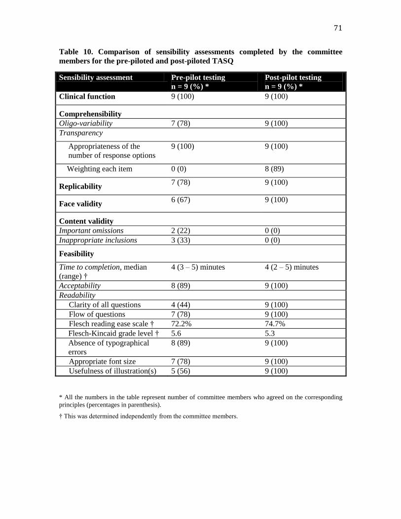
71
Table 10. Comparison of sensibility assessments completed by the committee
members for the pre-piloted and post-piloted TASQ
Sensibility assessment Pre-pilot testing
n = 9 (%) *
Post-pilot testing
n = 9 (%) *
Clinical function 9 (100) 9 (100)
Comprehensibility
Oligo-variability 7 (78) 9 (100)
Transparency
Appropriateness of the
number of response options
9 (100) 9 (100)
Weighting each item 0 (0) 8 (89)
Replicability 7 (78) 9 (100)
Face validity 6 (67) 9 (100)
Content validity
Important omissions 2 (22) 0 (0)
Inappropriate inclusions 3 (33) 0 (0)
Feasibility
Time to completion, median
(range) †
4 (3 – 5) minutes
4 (2 – 5) minutes
Acceptability 8 (89) 9 (100)
Readability
Clarity of all questions 4 (44) 9 (100)
Flow of questions 7 (78) 9 (100)
Flesch reading ease scale † 72.2% 74.7%
Flesch-Kincaid grade level † 5.6 5.3
Absence of typographical
errors
8 (89) 9 (100)
Appropriate font size 7 (78) 9 (100)
Usefulness of illustration(s) 5 (56) 9 (100)
* All the numbers in the table represent number of committee members who agreed on the corresponding
principles (percentages in parenthesis).
† This was determined independently from the committee members.
72
4 Version 4 of the Questionnaire
Version 4 (dated 7th
July 2011) was a one-page, double-sided questionnaire that was set
out in a clear and uncluttered format accompanied by a diagram of the back. It took 5
minutes or less to complete the questionnaire. At the beginning of the questionnaire,
demographic questions were asked, namely sex and date of birth. We did not include a
question on identification of the patient’s name to reassure patients of confidentiality.
Clear instructions on how to fill out the questionnaire were provided to ensure
standardization.
The questionnaire consisted of 16 items. The response options were presented as follows:
a binary response in 11 questions and 1 sub-question, 3-option responses in 2 questions
and 1 sub-question, and provision of continuous variables in 1 question and 1 sub-
question. We elected to retain additional 2 questions and 2 sub-questions in the final
questionnaire, although they will not be scored. These questions did not increase the
number of pages. Two questions serve as a confirmation that patients who complete the
questionnaire have ever had a history of chronic back pain or stiffness lasting 3 months or
more, and another question asks about the type of IBD. Two sub-questions help obtain
descriptive data including types of biologic drugs and whether the intake is current or
previous, and prior diagnosis of AS. The questionnaire is easy to read for a student at the
5th
grade or above. The Flesch Reading Ease is 74.7% which is considered fairly easy.
5 Description of the Items in the Final
Questionnaire
This section describes the item of the final version of the questionnaire. Figure 9
summarizes the domains which consists of IBD, inflammatory back symptoms, and
extra-axial features.

73
Figure 8. Description of the domains of the TASQ
Inflammatory bowel disease (IBD):
Treatment with biologic agents (infliximab or adalimumab, others).
Response of back symptoms after taking biologic agents.
Inflammatory back symptoms:
Age of onset of back pain or stiffness.
Morning stiffness.
Mode of onset of back pain.
Location of back pain: neck, upper / middle / lower back, buttocks.
Nocturnal pain with sleep disturbance.
Response to:
a. Physical activities.
b. Rest.
c. Non-steroidal anti-inflammatory drugs (NSAIDs).
Family history (first degree relatives) of AS.
Extra-axial features:
Prior diagnosis of iritis or uveitis.
Prior diagnosis of psoriasis.
Prior diagnosis of psoriatic arthritis.
Heel pain (enthesitis) ever.
Peripheral arthritis ever.
Dactylitis ever.

74
5.1 Inflammatory Bowel Disease (IBD)
Treatment with Biologic Agents
Choices of biologic agents here are infliximab, adalimumab, or others. As stated in the
Introduction, the first 2 TNF inhibitors are the main biologic drugs used in the
management of IBD patients. I added “other” to account for future biologic agents. This
question will not contribute to the total score.
Response of Back Pain or Stiffness to Biologic Agents
The questionnaire asks if patients ever had chronic back pain or stiffness lasting 3 months
or more that improved after taking biologic agents. A positive score is when there is a
response of the back pain or stiffness to biologic agents.
5.2 Inflammatory Back Symptoms
Chronic Back pain or Stiffness Lasting ≥ 3 Months
As discussed in the Introduction, since previous studies confirmed the strong association
between back pain and stiffness with axSpA, we used a double-barreled screening
question combining “chronic back pain” with “stiffness” lasting 3 months or more.
Age of Onset of Inflammatory Back Pain
This is an open-ended question. A positive score is when the age of onset of IBP is 45
years or less in accordance to the ASAS classification criteria for axSpA.
Mode of Onset of Back Pain
The item asks whether the initial development of the back pain was quickly “over hours
or days” or slowly “over weeks or months”. The latter response option constitutes a
positive score.

75
Morning Stiffness of the Back
This item starts with a closed-ended question and then branches if the answer is “Yes” to
presence of morning stiffness. The branching question then asks about duration. A patient
who has morning stiffness will be given a positive score. If morning stiffness lasts 30
minutes or more, a positive score will be given.
Nocturnal Pain with Sleep Disturbance
This item starts with a closed-ended question then branches if the patient answers “Yes”
to presence of nocturnal pain. The branching question then asks if the pain improves
upon getting out of bed, walking or stretching with 3 responses: “Yes”, “No. This would
not improve my back pain”, or “No. I do not get up”. A positive score is given to the
question and the sub-question i.e. presence of nocturnal pain with sleep disturbance, and
improvement upon getting out of bed, walking or stretching. Improvement of pain upon
getting up has been consistently found in previous studies to be a major finding in AS
patients (93, 95, 97, 119).
Locations of the Back Pain
Traditionally the low back has been the main complaint among axSpA patients (120).
Even though cervical spine was recently found to be significantly associated with primary
AS compared to IBD-associated AS (36), I did not want to miss those patients. In our
questionnaire, I provided a diagram of the back and asked the patients to check the areas
of back pain or stiffness including the neck, upper back, middle back, lower back and
buttock. A positive score is given to any of these locations with a total possible score of 5.
Response to Daily Physical Activity
I preferred to use this item over “response to exercise”, which is used in both Calin
criteria for IBP and the modified New York criteria, because it is more practical to ask
about response of back pain or stiffness to daily physical activities such as walking. This
item has also been used in the case ascertainment questionnaire (107). To increase the
sensitivity of this item, I asked, “Does your back pain or stiffness improve at all with

76
daily physical activities?” A positive score is given if the patient answers “Yes” to this
question.
Response to Rest
IBP typically lacks improvement with rest as was found in previous studies (3, 93, 119).
To increase the sensitivity of this item, I asked, “Does your back pain or stiffness
improve at all with rest?” A positive score is given if the patient answers “No” to this
question.
Responsiveness to NSAIDs
This question asks, “Does your back pain or stiffness improve after taking anti-
inflammatory drugs such as Motrin, Advil, Voltaren, Naprosyn, Aleve,
Indocin, Mobicox, or Celebrex?” The 3 responses are: “Yes”, (“No. These do not
improve my symptoms”, or “No. I do not take anti-inflammatory drugs”. A positive score
is given if the patient answers “Yes” to this question. Although this item may appear to
select NSAIDs respondents especially when there are patients with axSpA who are
NSAIDs non-respondents, this item addresses only one aspect of axSpA and that the non-
respondents are likely to score other features.
Family History of AS
Family history (first or second degree) of AS is a specific item that has been part of the
Amor and the ESSG criteria for SpA, and in the ASAS classification criteria for axSpA.
In my questionnaire, this item was asked as “Do you have a parent or sibling who has
been diagnosed with ankylosing spondylitis?” The 3 responses are: “Yes”, “No”, or
“Unknown”. The latter response was mainly added to include patients who are adopted.
A positive score is given if the patient answers “Yes” to this question.
77
5.3 Extra-axial manifestations
Prior Diagnosis of Uveitis or Iritis
In order to make this item more sensitive, I asked if a patient was diagnosed with iritis or
uveitis without asking about details of its symptoms. A positive score is given if the
patient answers “Yes” to this question.
Prior Diagnosis of Psoriasis
Psoriasis can co-exist in 10–25% of axSpA patients. This item is part of the ASAS
classification criteria for axSpA. A positive score is given if the patient answers “Yes” to
this question.
Prior Diagnosis of Psoriatic Arthritis
This item was initially added since some patients may have a prior diagnosis of psoriatic
arthritis without psoriasis. It may co-exist in IBD and result in symptoms of axSpA. A
positive score is given if the patient answers “Yes” to this question.
Peripheral Arthritis
This item is part of the ASAS classification criteria for axSpA. In order to make this item
more specific for peripheral arthritis, I asked whether the patient had pain and swelling in
any joint that was not related to injury. I then increased the sensitivity of this item by
adding “ever” to the description of this symptom. A picture of peripheral arthritis of the
hands was initially added to the questionnaire but was removed when the SpA team felt
that it might be confused with the picture of dactylitis. A positive score is given if the
patient answers “Yes” to this question.
Dactylitis
This item represents a highly specific feature of the SpA group of diseases, and is part of
the ASAS classification criteria for axSpA. I asked this specific question by describing
78
pain and swelling of the entire finger or toe that was not related to injury. I then increased
its sensitivity by adding “ever”. As in the case of “peripheral arthritis” item, a picture of
dactylitis was initially added to the questionnaire but was removed when some patients
felt the pictures of dactylitis looked like that of arthritis of a finger. A positive score is
given if the patient answers “Yes” to this question.
Heel Pain (Enthesitis)
This item is also part of the ASAS classification criteria for axSpA. A positive score is
given if the patient answers “Yes” to this question.
6 Reliability study
6.1 Response Rate of Reliability Questionnaires
Of the 77 mailed questionnaires, 34 were returned by the cut-off date. Mails for the
questionnaires of 6 patients were returned due to incorrect addresses. One patient
declined participation and the mail was returned. The response rate during the summer of
2011 was 44.2%. Since I was interested in a sample size of 24, the final number of
respondents (i.e. 34) was excellent. Therefore, I did not pursue follow-up mails for non-
respondents.
6.2 Missing Answers on the Questionnaire
Questionnaires from 5 of 34 responders had ≥ 15% missing answers in their respective
questionnaires. We were able to contact 4 of them by phone as soon as the mailed
questionnaires were received. We read the unanswered questions for them over the phone

79
as written without providing any further explanation to avoid interviewer bias.
Questionnaires from 33 out of 34 patients were usable for the descriptive analysis. We
could not identify one male patient due to missing date of birth on the questionnaire and
inability to crosscheck the code on his questionnaire with Spondylitis database (clerical
error). For the test-retest reliability, the final usable pairs of questionnaires were 33 as
one patient was excluded from the analysis because of incomplete data of the second
questionnaire.
6.3 Comparison between Responders and Non-
Responders
Table 11 summarizes the demographic and baseline characteristics of patients who did
and did not respond to the questionnaire. Respondents were slightly older than non-
respondents. The median of the highest level of education at the last clinic visit was
university in responders and college in non-responders. Ulcerative colitis was slightly
more common in non-responders than responders. The median ESR level was slightly
higher in non-responders than responders. Responders and non-responders were not
different in terms of average age, sex, highest level of education, types of IBD, activity of
axSpA, functional activities, ESR or CRP levels.

80
Table 11. Demographic and clinical characteristics of responders and non-
responders. Values are means (SD) unless otherwise indicated
Responders
(n = 33) *
Non-responders
(n = 43)
Age, yrs 45.7 ± 13.5 40.5 ± 11.7
Male (%) 27 (79.4%) 31 (72.1%)
Education, median 5 4
Crohn’s disease (%) 21 (63.6%) 26 (60.5%)
Ulcerative colitis (%) 11 (33.3%) 18 (41.9%)
Unclassified colitis (%) 1 (3%) 0
BASDAI 3.7 ± 2.6 4.1 ± 2.6
BASFI 3.7 ± 3.0 3.5 ± 3.1
ESR mm/hr, median 6.5 13.0
CRP mg/L, median 8 7
* The response of one patient was dropped because of incomplete information.
Note: All p values are > 0.05.
Abbreviations: SD, Standard Deviation; BASDAI: Bath Ankylosing Spondylitis Disease Activity Index;
BASFI: Bath Ankylosing Spondylitis Functional Index; ESR: Erythrocyte Sedimentation Rate; CRP: C-
Reactive Protein

81
6.4 Descriptive Statistics of the Items
I coded the items to the direction of the construct (axSpA), that is, the positive or
expected answers for a patient who has axSpA (for details, please refer to Description of
the Items in the Final Questionnaire). All respondents confirmed that they had back or
stiffness that lasted ≥ 3 months. Among the responses of the 34 patients, the most
prominent items are as follows: male gender, age of onset of back pain ≤ 45 years, slow
development of back pain initially (over weeks or months), current morning stiffness of
the back, current morning stiffness of the back lasting 30 minutes or more, pain or
stiffness in the neck, lower back and the buttock, improvement of back pain or stiffness at
all with daily physical activities, and history of peripheral arthritis. All patients were
aware that they had a diagnosis of AS. The least common associated features were
psoriatic arthritis and psoriasis successively. Table 12 summarized these findings.

82
Table 12. Summary of the responses for features of axSpA among patients with
axSpA and IBD
Items
Number of patients with
positive responses, n = 34 (%)
Male 27 (79.4)
Treatment of IBD with biologic drug that improved
the back pain or stiffness
16 (47.1)
Age of onset of back pain ≤ 45 years 33 (97.1)
Slow development of back pain initially 26 (76.5)
Current morning stiffness of the back 24 (70.6)
Current morning stiffness of the back lasting ≥ 30
minutes
22 (64.7)
Current back pain awakening at night 17 (50)
Current back pain awakening at night and improving
upon getting up, walking or stretching
11 (32.4)
Locations of back pain or stiffness
Neck 25 (73.5)
Upper back 14 (41.2)
Middle back 10 (29.4)
Lower back 26 (76.5)
Buttock 25 (73.5)
Improvement of back pain or stiffness at all with
daily physical activities
26 (76.5)
No improvement of back pain or stiffness at all with
rest
19 (55.9)
Improvement of back pain after taking NSAIDs 19 (55.9)
Family history (first degree) of AS 7 (20.6)
Prior diagnosis of AS 34 (100)
Prior diagnosis of iritis or uveitis 12 (35.3)
Prior diagnosis of psoriasis 3 (8.8)
Prior diagnosis of psoriatic arthritis 1 (2.9)
History of peripheral arthritis 26 (76.5)
History of dactylitis 12 (35.3)
History of heel enthesitis 21 (61.8)

83
At this stage, the committee members agreed on removing a response option “Prior
diagnosis of psoriatic arthritis” from item 13 (for more details, see Discussion - Test-
Retest Reliability). The final version 5 of TASQ (dated May 2012) is shown in Figure 10.

84

85
Figure 9. The Toronto Axial Spondyloarthritis Questionnaire (TASQ)

86
6.5 Test-Retest (Intra-rater) Reliability
Thirty-two pairs of questionnaires, indicating patients who completed 2 questionnaires at
both times, were used for analysis out of the 33 respondents. At this stage, I calculated
the kappa () coefficients for the questionnaire items including the ones which are not
going to be ultimately scored such as the IBD type and the different types of biologic
agents (Table 13). The coefficients of all items ranged between 0.81 to 1.00 which
indicate almost perfect agreements for test-retest reliability. The absolute percentage of
agreement across all items ranged from 91% to 100%.
The range of coefficients for each domain is as follows: 0.84 – 1.00 for the (IBD
domain), 0.85 – 1.00 for (Back pain or stiffness) domain, and 0.81 – 1.00 for (extra-axial
manifestations) domain. The ranges of absolute agreement between different domains are
as follows: 91 – 100% for the (IBD domain), 94–100% for (inflammatory back
symptoms) domain, and 91 – 100% for (extra-axial manifestations) domain.
The lower limit of 95% confidence interval of coefficients for some items ranged from
0.60 to 0.78 (moderate to substantial agreement) although was > 0.80 (almost perfect
correlation). These items include IBD type, pain and stiffness in the neck, middle back
and buttock, no improvement of back pain or stiffness with rest, history of peripheral
arthritis and history of dactylitits.
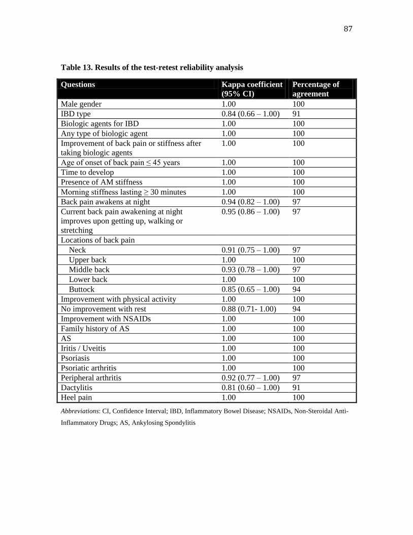
87
Table 13. Results of the test-retest reliability analysis
Questions Kappa coefficient
(95% CI)
Percentage of
agreement
Male gender 1.00 100
IBD type 0.84 (0.66 – 1.00) 91
Biologic agents for IBD 1.00 100
Any type of biologic agent 1.00 100
Improvement of back pain or stiffness after
taking biologic agents
1.00 100
Age of onset of back pain ≤ 45 years 1.00 100
Time to develop 1.00 100
Presence of AM stiffness 1.00 100
Morning stiffness lasting ≥ 30 minutes 1.00 100
Back pain awakens at night 0.94 (0.82 – 1.00) 97
Current back pain awakening at night
improves upon getting up, walking or
stretching
0.95 (0.86 – 1.00) 97
Locations of back pain
Neck 0.91 (0.75 – 1.00) 97
Upper back 1.00 100
Middle back 0.93 (0.78 – 1.00) 97
Lower back 1.00 100
Buttock 0.85 (0.65 – 1.00) 94
Improvement with physical activity 1.00 100
No improvement with rest 0.88 (0.71- 1.00) 94
Improvement with NSAIDs 1.00 100
Family history of AS 1.00 100
AS 1.00 100
Iritis / Uveitis 1.00 100
Psoriasis 1.00 100
Psoriatic arthritis 1.00 100
Peripheral arthritis 0.92 (0.77 – 1.00) 97
Dactylitis 0.81 (0.60 – 1.00) 91
Heel pain 1.00 100
Abbreviations: CI, Confidence Interval; IBD, Inflammatory Bowel Disease; NSAIDs, Non-Steroidal Anti-
Inflammatory Drugs; AS, Ankylosing Spondylitis

88
Chapter 5
Discussion

89
1 Questionnaire Development
The TASQ self-administered questionnaire was designed to serve as a case finding
instrument that could facilitate referrals of IBD patients with suspected axSpA to
rheumatologists for further evaluation. Early diagnosis allows for earlier intervention, an
important concept especially since TNF inhibitors have been found to be efficacious in
early disease. Because focusing only on characteristics of IBP cannot be sufficient to
make a diagnosis of axSpA, additional features are required, such as other relevant
clinical information (family history, articular symptoms, extra-axial manifestations),
laboratory tests (HLA-B27, ESR, CRP), and imaging (X-ray, MRI).
Previous questionnaires focused on refining the characteristics of IBP and did not
specifically target IBD patients. TASQ is to be administered at a single point in time to
patients with IBD who have chronic back pain or stiffness that has ever lasted 3 months
or more. This cut-off time which was chosen to minimize the referrals of mechanical
back pain was also used in previous studies (93, 106, 121). TASQ is cheap, easy to
administer and easy to understand by an average student in the 5th
grade. Completion of
the questionnaire takes 5 minutes or less which is in accordance to the recommended
completion time between 5 and 15 minutes (122). This also facilitates its feasibility
especially when this questionnaire is handed to IBD patients in a busy gasteroenterology
clinic or even when used online.
Designing a new questionnaire requires multiple steps to ensure a proper methodology in
order to yield a reliable instrument. During the development stage, I included sensitive
and specific items to help capture different aspects of the construct being sought i.e.
axSpA in IBD. I organized it into 3 domains (IBD, inflammatory back symptoms, and
extra-axial features). I strove to simplify the words in order to achieve our aims.
Hypothetically these questions represent a wide spectrum of discriminatory features that
increase the likelihood of axSpA and, therefore, can help gastroenterologists refer those
patients in a timely fashion.

90
The clinimetric method, as introduced by Feinstein, advocates for the use of clinical
judgment when developing a measuring instrument. The tools that I used for this
approach included sensibility and reliability. In 1993, Rowe and Oxman paved the road
shown by Feinstein and developed a questionnaire for sensibility assessment. Twelve
items were developed based on the principles of sensibility. The response options were
constructed on a 7-point Likert scale. Some of their questions appeared redundant, and
some contained double-barreled questions. This questionnaire was adapted during
development of recent measuring instruments such as the Pediatric Cardiopulmonary
Physiotherapy Discharge Tool (123), and HIV Disability Questionnaire (124). I also
adapted the sensibility questionnaire by modifying some of their questions and adding
new ones for feasibility. New items related to the concept of feasibility (ease of use)
consist of time to completion (in minutes), flow of questions, readability, typographical
errors, font size, and use of illustrations. I also changed the format of scaling responses
into dichotomous scale which goes along the theme of the scaling responses of TASQ.
The sensibility assessment proved to be a valuable and comprehensive approach during
selection of the items and pilot testing that enabled me to look at different facets of the
questionnaire and remedy its weaknesses. Using the sensibility assessment, the
questionnaire underwent 4 revisions incorporating feedback from the SpA team, general
rheumatologists, and patients before reaching the fourth and final version.
2 Clinimetric Measures in the Literature
Psychometric methods use classical test theory and item response theory. The former
involved the statistical analysis of the data to yield reliability (test retest reliability,
internal consistency, split half reliability) and validity. Item response theory uses other
statistical models with different assumptions and requires larger sample size. It is obvious
that these methods rely heavily on statistical analyses to select items of an index or
questionnaire. With regards to assessing the internal consistency of an instrument, it is
required to have high correlations between the items (inter-item correlation) and the

91
whole instrument (items-total correlation). While having a high internal consistency
coefficient is preferred, it may give a false impression that items are homogenous. For
example, the coefficient can increase by only increasing the number of questions (even if
they are redundant). In this case, the sensitivity of the items may actually be low which
consequently lowers the discriminatory property of the items (125). Having a high
coefficient of internal consistency requires an appropriate, and usually large, sample size.
Psychometric methods are not concerned with face validity and sensibility (usefulness of
the instrument). In addition, they do not necessarily result in constructing an accurate
measure (125).
Feinstein emphasized that judgment (by physicians and/or patients) is an essential part
during development of an instrument measuring a complex clinical phenomenon and that
psychometric techniques may be used to aid this process. Assessing internal consistency
is of less relevance here because the aim of developing a measuring tool is to capture
different aspects of the same (uni-dimensional) construct. Therefore, the internal
consistency is likely to be low although this is not always the case (126). There are some
indices which fell out of favor because they were not sensible and were difficult to apply
such as the Norris prognostic index for myocardial infarction (110). The clinimetric
approach has been used in the literature to construct indices which can comprise
classification or diagnostic criteria, guidelines, prognostic criteria, and questionnaires.
Examples of clinimetric measures are the New York Heart Association Functional
Classification (127), the Jones criteria for rheumatic fever (128), the Apgar criteria to
score the health of a newborn immediately after birth (129), and the Pittsburgh Sleep
Quality Index (130).
Two independent studies compared 2 different versions of questionnaires (Quality of Life
After Myocardial Infarction and Asthma Quality of Life Questionnaire) using methods of
clinical judgment and statistical analysis for item reduction. In each study, the clinical
judgment method performed slightly better than the statistical method when both versions
were tested for concurrent validity and responsiveness.

92
There are important issues that were raised for discussion during at the sensibility
assessment of TASQ. These will be discussed next.
3 Sensibility Assessment of TASQ
3.1 Comprehensibility (Transparency)
Assigning appropriate weighting to each item of the questionnaire is a challenge. There
are 4 methods that can shed some light on item weighting. The first method of scoring is
based on clinical judgment and intuition. For example, a total score can simply be the
sum of each item that is weighted equally with a score of 1. This gives the questionnaire
the advantages of being simple, easy to score and transparent. Pediatric Crohn’s Disease
Activity Index (PCDAI) was scored using this method (131). In addition, the contribution
of each item weight to the cumulative score will be clear as opposed to a questionnaire
that assigns different weights to each item. The disadvantage of this approach is the
implication that every item is of equal importance. The second method assigns a
weighted score for each domain e.g. double the total score of Domain 1 and half Domain
2. This indicates the greater significance of a domain and the lesser significance of
another. The third method uses a statistical test (factor analysis) where assigning weights
is based on factor loadings. However, this often requires large sample size i.e. 5 patients
for each item. In addition, the interpretation of factor analysis requires clinical judgment.
The fourth method relies on another statistical analysis i.e. multivariate logistic
regression analysis that quantifies each item depending on its importance in explaining
the outcome (dependent variable). This is the basis of scoring the Systemic Lupus
Erythromatosus Disease Activity Index (SLEDAI) and Crohn’s Disease Activity Index
(CDAI) (132, 133). While it may be scientifically appropriate to weight items based on
statistical analysis, the different statistical tests may give different final scores which also
depend on the characteristics of the population and the setting. Some scales were scored
using statistical models that resulted in assigning sophisticated scoring that requires a

93
special calculator. A clear example is the scoring system for the case ascertainment
questionnaire.
A recent Canadian questionnaire for inflammatory back pain (for details, refer to Section
4 of the Discussion) compared 2 methods of weighting items: summative (adding a score
of 1 for each item) and weighted items based on multivariate regression analysis. The
sensitivity, specificity, positive LR and the area under the curve were similar when any
method was used (134).
Another recent Canadian study compared weighting of the Pediatric Ulcerative Colitis
Activity Index (PUCAI) using mathematical and judgmental models. The PUCAI has 8
items including 6 historical and 2 laboratory parameters. Judgmental approach was found
to be as good as the mathematical approach only if the 2 laboratory items were removed
(135).
In my questionnaire, although there were 2 questions which are not going to be scored
(type of IBD and previous diagnosis of AS), I retained them in the final version to
provide important descriptive data especially that their removal would slightly reduce the
length of the questionnaire.
In conclusion, I will explore the scoring of the questionnaire in the next validation phase
and will compare the different scoring methods.
3.2 Content Validity
Many items had to be dropped because I wanted to keep the questionnaire simple and
concise (on 2 pages). A recent Cochrane systematic review found that the response rates
of patients in clinical trials increased when shorter self-reported questionnaires were
mailed (136). For example, the odds ratios (ORs) per page increase were 0.90 (95% CI
0.83–0.98) for one page compared with ≥ 4 pages, and 0.98 (95% CI 0.96–0.99) for one
page compared with ≥ 2 pages.

94
During the pilot testing, the item “fibromyalgia” was removed for 3 reasons related to
low discriminatory property: 1) patients with true axSpA may have enthesitis that could
have been misdiagnosed with fibromyalgia, 2) enthesitis may overlap with tender points
of fibromyalgia, and 3) fibromyalgia can co-exist independently in both axSpA and IBD
as part of its association with some chronic diseases (137, 138). In addition, patients did
not endorse this item in the pilot study.
3.3 Feasibility
3.3.1 Acceptability
An item on “race” was included in the Phase 2 (item generation) since axSpA is more
common in Caucasian people of European extraction. Therefore, I considered ethnicity in
this instance a risk factor for axSpA. During Stage 3 of the development of TASQ, one
member of our team pointed towards the inappropriate inclusion of an item about race. In
a recent Cochrane systematic review, the likelihood of not responding to mailed
questionnaires was shown to increase when they asked about sensitive questions (139). I
then removed this item in order to increase the response rate.
3.3.2 Readability
Clarity of Questions
Constructing a clear questionnaire proved to be a challenge. Patients may not necessarily
read and understand what health care workers perceive as a straightforward question
since each comes from different backgrounds. In this study, I had to rephrase certain
questions a few times and even drop certain items because they were unclear to patients,

95
even thought they were relevant to the complex construct of axSpA. I will elaborate more
on this in the following sections.
Insidious onset of back pain: The item has long been considered vague, as previous
studies did not agree on its exact definition. It is intended to measure the mode of onset of
back pain. However, does it mean slow onset or gradual onset? Can it be measured that in
weeks or months or even years? As a result of this unclear definition, studies showed an
overlap between mechanical low back pain and IBP when the poorly defined terms
“chronic” or “insidious onset” were used (93, 94). In the case ascertainment questionnaire
of Weisman and colleagues, the questionnaire developers defined “insidious onset” by
asking about the length of time (in months) since patients started to have back pain or
stiffness. At the end, I decided to keep the question as “When your back pain first began,
did it develop quickly (over hours or days) or slowly (over weeks or months). All patients
during the pilot and reliability studies were able to answer it easily.
Sleep disturbance at the second half of the night: Rudwaleit and colleagues found this
item an important characteristic of IBP when they administered their questionnaire (in
German) by an interviewer (93). I am unaware of any published cross-cultural adaptation
of their questionnaire. However, I used this item during the pilot testing phase but
decided to drop it because some patients found it difficult to define a cut-off for the
second half of the night.
Hip arthritis: Hip joint is localized in a deep-seated site, which may prevent patients
from expressing their pain clearly. The diagnosis of hip arthritis is often based on clinical
(history and physical examination) and radiographic evidence. There is a strong
association between hip arthritis and AS (37). A study showed that hip arthritis is
significantly associated with primary AS compared to IBD-associated AS (36). However,
I believe that this item is important and merits asking in a questionnaire. The problem
was how to ask about it properly using a single and relatively short question to fit in a
case-finding questionnaire. In clinical practice, rheumatologists ask patients different
questions about hip pain to increase the pre-test probability of hip arthritis. For example,
they may ask whether patients have pain or stiffness in the upper frontal side of the thigh

96
that radiates to the groin. Some ask whether patients feel or even hear a click during hip
joint movements. Some patients point towards the painful area by cupping their thumb
and index fingers around the hip joint, which can also be seen in other conditions such as
trochanteric bursitis or fibroacetabular impingement syndrome. In the pilot study, some
patients drew a line (without being asked) to point towards hip pain but they directed the
line at either the buttock or sacroiliac joint. At the end, I could not include this item in our
questionnaire due to lack of a standardized definition and its susceptibility to
misinterpretation. A recent study attempted to describe hip pain for patients by using of a
diagram with pre-shaded area for localizing the hip pain and compare it with a
standardized question asking, “In the past month have you had any pain in the hip lasting
one day or longer?” This study concluded that the use of combined methods was
associated with signs of hip disease (abnormal physical examination and radiographic
changes). However, if these combined methods were used to exclusively define hip pain,
the sensitivity of capturing those patients would be low which does not go in the same
direction of our questionnaire (140). Future studies are needed to best describe hip pain to
patients.
Alternating buttock pain: This item was found to be one of the specific characteristics of
IBP (although not sensitive) and was therefore incorporated in Amor criteria and in
Rudwaleit’s study (93, 102). However, we had to drop it for 3 reasons: 1) some patients
had difficulty understanding the question, 2) buttock pain can still be a complaint in some
patients with AS and mechanical back pain, and 3) this particular item was not included
in the description of IBP of the ASAS classification criteria for axSpA (105).
Illustrations
TASQ is unique compared to the previous questionnaires in that it shows for the first
time a diagram of the back to allow patients to identify the locations of their back pain or
stiffness. I initially wanted to include a location for the ribcage since this has been noted
in some studies. But I decided against this idea because it will make the diagram cluttered
with too many arrows.

97
The decision to remove the colored pictures of peripheral arthritis and dactylitis was
reached by consensus from the committee members and from the feedback of patients.
These pictures could be mutually misinterpreted. The picture of iritis was also removed
during selection of the items because it may be misinterpreted as conjunctivitis. However,
a future study is required to refine symptoms of iritis particularly if associated with
axSpA in IBD patients. A disadvantage of using colored pictures is the expense which
may limit the questionnaire feasibility. Questionnaires with colored pictures can be used
on the website but can only be used by patients who have access to the internet.
Recently, Gladman and colleagues developed a screening questionnaire for psoriatic
arthritis among patients with psoriasis and in general population; the Toronto Psoriatic
Arthritis Screening (ToPAS) (141). ToPAS has 3 domains, namely skin, joint and nail.
Among all the screening questionnaires for psoriatic arthritis, ToPAS was the first to
include colored pictures of the skin rash, and nail changes. A Dutch study found that
ToPAS performed slightly better compared to the Psoriatic Arthritis Screening and
Evaluation (PASE) questionnaire which could be partly related to the use of illustrations
(142). An update of ToPAS with more colored pictures is underway (143).
4 Reliability
4.1 Rating Responses of the Questionnaire Items
Upon looking at the responses of TASQ in the reliability study, all but one patient started
to have back pain or stiffness below the age of 45 years. The most prominent items
among patients with established IBD and axSpA were as follows: male gender, age of
onset of back pain ≤ 45 years, slow development of back pain initially (over weeks or
months), current morning stiffness of the back, current morning stiffness of the back
lasting 30 minutes or more, pain or stiffness in the neck, lower back and the buttock,

98
improvement of back pain or stiffness at all with daily physical activities, and history of
peripheral arthritis.
4.2 Test-Retest Reliability
I chose test-retest (intra-rater) reliability to assess the stability of the items over time in
order to quantify measurement error rather than true change in disease activity. I provided
detailed information on the reliability and agreement using coefficients of reliability
(along with statistical uncertainty) and percentage agreement as per the recent
recommendations of the 2011 Guidelines for Reporting Reliability and Agreement
Studies (GRRAS) (115).
There are various criteria for the interpretation of coefficients. These include the
criteria of Landis and Koch, Cicchetti and Sparrow, and Fleiss (118, 144, 145). It is
important to note that all these criteria are arbitrary. In our study, I chose the criteria of
Landis and Koch. My a priori hypothesis was that the reliability coefficient (kappa or )
would be estimated as 0.90. The coefficient for each item was more than 0.80
indicating almost perfect agreement. Similarly, the absolute agreement for each item was
more than 91%. Although it may seem that some items had reliability coefficients (range
0.81 – 0.88) of less than the hypothesized reliability of 0.90, the minimally accepted
reliability for this study was 0.80. Therefore, those items are still within acceptable range
of reliability i.e. > 0.80 indicating almost perfect agreement.
I observed that the 95% CIs of coefficients for some items were wide and that may be
due to our small sample size. If we used a larger sample size, we may improve the
precision around the k statistics.
The sub-question “Prior diagnosis of psoriatic arthritis” was removed after the reliability
study for 2 reasons. First, this item was rarity endorsed by patients (only 1 patient).
Second, during the validation phase of the case ascertainment questionnaire, this item did
not help differentiating AS from chronic back pain (107). We considered psoriasis

99
adequate for this questionnaire, as previous studies found it co-exist in 10-25% of AS
patients. Furthermore, psoriasis was recently shown to be more associated with non-
radiographic axSpA (OR 3.6) compared to classic AS (146). This additional item
reduction led to version 5 (dated May 1st, 2012).
5 Recent Questionnaire for Back Pain (April 2012)
A newly self-reported questionnaire has been published this year (April 2012). It is a
screening questionnaire for IBP developed by the Edmonton group of SPARCC. It
consists of 6 items with 3 branching questions. Items address morning stiffness of the
back and/or hip (branching question asked about the most noticeable time), nocturnal
pain (branching question asked about time of nocturnal awakening), diurnal variation of
pain in the back and/or hip, peripheral arthritis, response to exercise, and response to rest.
The questionnaire was administered to patients with established AS and mechanical back
pain (MBP).
In univariate analysis, 4 items distinguished IBP from MBP: morning stiffness, diurnal
variation, response to exercise and response to rest. In multivariate logistic regression
analysis, the last 3 items were independently associated with IBP, especially diurnal
variation (p = 0.0001). The “diurnal variation” item asked, “At what time of day your
back and/or hip symptoms the worse? Morning, Afternoon, Evening, Night, or Not
Applicable.” Using all the 6 items, the questionnaire outperformed Calin criteria and the
IBP criteria of ASAS in identifying patients with IBP. In my opinion, the way in which
the item “diurnal variation” was asked seems easier to understand than asking about
nocturnal pain at the second half of the night as suggested from Rudwaleit’s study (93).
In addition, this item showed the strongest association with IBP (OR 11.2, CI 3.7 – 34.2),
with a specificity of 92% and a sensitivity of 49%. As in the case of the previously
published questionnaires (Calin and case ascertainment questionnaire), the 6-item

100
questionnaire is not targeted to IBD patients, and focused on features of IBP with one
question on peripheral arthritis.
As discussed earlier, investigators, who are interested in early diagnosis of axSpA, have
long been searching for the optimal characteristics of IBP which can improve the current
increase in post-test probability from 5 to 14%. Further, the interpretation of IBP needs
some clinical experience. Therefore, IBP cannot be used solely as a referral criterion. The
recommendation is to combine IBP with other features of the complex disease (axSpA) in
order to achieve a high post-test probability of axSpA (more than 90%) (17, 104, 146).
Indeed, IBP is no longer considered by ASAS to be a mandatory element in the definition
of axSpA. My questionnaire is in agreement with this, since other elements in addition to
characteristics of the back pain, are given equal importance.

101
Chapter 6
Conclusions

102
TASQ is a newly developed self-reported instrument to be administered to patients with
IBD who have ever had chronic back pain or stiffness that has lasted 3 months or more. It
consists of 3 domains (inflammatory bowel disease, inflammatory back symptoms, and
extra-axial features) and 16 items. The items were chosen based on their high sensitivity
(i.e. found in most patients with axSpA) and reasonable specificity which is in
accordance with a purpose of case-finding questionnaire. I have demonstrated that TASQ
is sensible, reliable, and inexpensive. It is easy-to-apply and requires 5 minutes of less to
complete. It is suitable for a student in the 5th
grade.
TASQ is a very promising questionnaire that might facilitate early patients’ referral to
rheumatologists and avoid delay in diagnosis of axSpA.

103
Chapter 7
Limitations and Future
Directions

104
While the project achieved it primary goals, there are some limitations to point out. The
following sections will discuss the limitations of this research and future directions.
1 Development
The pilot study was conducted at the Spondylitis clinic using a convenience sample of
patients with axSpA. Ideally, a sample of the target patients should be used i.e. patients
with IBD and axSpA. However, we only had 2 questions specific to IBD patients (type of
IBD, response of back pain or stiffness to biologic drugs). In addition, patients in the pilot
study provided their feedback on the sensibility of the IBD items. Ideally, a random
sample of axSpA patients would be representative as it should include a mixture of
patients with different levels of disease severity and levels of education. The sampled
patients represented a wide spectrum of axSpA disease activity. The questionnaire is
limited to patients who have finished grade 5. It could potentially be too sophisticated for
patients who have lower educational levels. If this were truly shown in a future study, one
solution would be to use more illustrations.
This questionnaire is only applicable to English-speaking and literate patients. Therefore,
future research is needed for cross-cultural adaptation and validation to different
languages.
Some features of IBP need to be clarified in future studies. As described in the Results,
defining “alternating buttock pain” using simple words was challenging and led to
dropping this item from the final version. On the same token, finding an accurate
definition for “hip pain” needs to be studied in the future since hip arthritis is a feature of
axSpA. The classic item of Calin criteria “improvement of back pain or stiffness with
exercise” remains to be further clarified. It should be noted that this item is also observed
in patients with mechanical back pain. Does improvement of IBP imply any degree of
exercise? Is the improvement expected to occur after a certain period of time?

105
Future modifications for this questionnaire may emphasize shoulder involvement in
axSpA patients. Chronic rotator cuff and enthesitis at the insertion of the supraspinatus
muscle to the bone are common manifestations of axSpA. Inclusion of this item can be
achieved with the use of a diagram.
2 Future Validation Studies
The TASQ was demonstrated to be reliable in patients who had IBD and axSpA and
attended a hospital-based rheumatology clinic. An important next step is validation is this
questionnaire in gastroenterology clinics. We are currently in the process of validating
the TASQ through collaboration with the gastroenterologists at the University Health
Network including Mt. Sinai, Toronto General and Toronto Western Hospitals. Inclusion
criteria for the study include age ≥ 18 years, fluency in English, established IBD
diagnosis, chronic back pain or stiffness ever present ≥ 3 months in duration, and
competency to consent. Patients will be recruited and referred to the Spondylitis Clinic
for a thorough assessment according to a standardized protocol. They will complete the
TASQ in the waiting area. An advanced practice physiotherapist, who will be blinded to
the patients’ answers of the questionnaire, will then assess them. The diagnosis of back
symptoms will be determined. The ideal situation for validation of the questionnaire
requires 2 groups of IBD patients: patients with mechanical back pain and patients with
axSpA. Finally, the type of validity will be assessed such as concurrent construct validity.
Another validation of TASQ is underway in which we have collaborated with
gastroenterologists at a hospital-based IBD clinic in the US. This validation is based on a
case-control study. IBD patients have undergone CT scans of the abdomen and pelvis at
some point of time as part of investigations for their IBD. We determined that the clinic
has on file a bank over 900 CT scans of which approximately 130 patients were found to
have definite sacroiliitis on CT scan. These patients likely have IBD-associated AS. The
controls will consist of 200 IBD patients with no sacroiliitis on CT scan. After having just

106
recently obtained approval of the institutional research board at that hospital, my new
questionnaire will be administered to those patients either via email or mail. The scores
of the questionnaire will be determined using the receiver operator curve with the aim of
having high sensitivity.
A third future study is to validate the TASQ in the primary care setting in patients who
have IBD. This will involve approaching the Family Practice Units of the University of
Toronto affiliated teaching hospitals.
The reliability study of this questionnaire was conducted in patients with established IBD
and axSpA. This questionnaire applies to a subset of seronegative SpA patients who have
IBD and undetected axSpA. With the exception of IBD domain, there is no reason to
assume that this questionnaire cannot be applied to any patient at risk for axSpA such as
patients with reactive arthritis or uveitis. However, this would require future validation.

107
Bibliography
1. Sieper J, Rudwaleit M, Khan MA, Braun J. Concepts and epidemiology of spondyloarthritis. Best Pract Res Clin Rheumatol. 2006;20(3):401-17.
2. Reveille JD, Witter JP, Weisman MH. The prevalence of axial spondyloarthritis in the United States: Estimates from the U.S. National Health and Nutrition Examination Survey, 2009-10. Arthritis Care Res (Hoboken). 2012.
3. van der Linden S, Valkenburg HA, Cats A. Evaluation of diagnostic criteria for ankylosing spondylitis. A proposal for modification of the New York criteria. Arthritis Rheum. 1984;27(4):361-8.
4. Underwood MR, Dawes P. Inflammatory back pain in primary care. Br J Rheumatol. 1995;34(11):1074-7.
5. O'Shea FD, Boyle E, Salonen DC, Ammendolia C, Peterson C, Hsu W, et al. Inflammatory and degenerative sacroiliac joint disease in a primary back pain cohort. Arthritis Care Res (Hoboken). 2010;62(4):447-54.
6. De Angelis R, Salaffi F, Grassi W. Prevalence of spondyloarthropathies in an Italian population sample: a regional community-based study. Scand J Rheumatol. 2007;36(1):14-21.
7. Trontzas P, Andrianakos A, Miyakis S, Pantelidou K, Vafiadou E, Garantziotou V, et al. Seronegative spondyloarthropathies in Greece: a population-based study of prevalence, clinical pattern, and management. The ESORDIG study. Clin Rheumatol. 2005;24(6):583-9.
8. Bruges-Armas J, Lima C, Peixoto MJ, Santos P, Mendonca D, da Silva BM, et al. Prevalence of spondyloarthritis in Terceira, Azores: a population based study. Ann Rheum Dis. 2002;61(6):551-3.
9. Onen F, Akar S, Birlik M, Sari I, Khan MA, Gurler O, et al. Prevalence of ankylosing spondylitis and related spondyloarthritides in an urban area of Izmir, Turkey. J Rheumatol. 2008;35(2):305-9.
10. Bhatia K, Prasad ML, Barnish G, Koki G. Antigen and haplotype frequencies at three human leucocyte antigen loci (HLA-A, -B, -C) in the Pawaia of Papua New Guinea. Am J Phys Anthropol. 1988;75(3):329-40.
11. Gofton JP, Chalmers A, Price GE, Reeve CE. HL-A 27 and ankylosing spondylitis in B.C. Indians. J Rheumatol. 1984;11(5):572-3.
12. Erdesz S, Shubin SV, Shoch BP, Krylov M, Mylov NM, Chekalina NA, et al. Spondyloarthropathies in circumpolar populations of Chukotka (Eskimos and Chukchi): epidemiology and clinical characteristics. J Rheumatol. 1994;21(6):1101-4.
13. Alamanos Y, Papadopoulos NG, Voulgari PV, Karakatsanis A, Siozos C, Drosos AA. Epidemiology of ankylosing spondylitis in Northwest Greece, 1983-2002. Rheumatology (Oxford). 2004;43(5):615-8.
14. Bakland G, Nossent HC, Gran JT. Incidence and prevalence of ankylosing spondylitis in Northern Norway. Arthritis and rheumatism. 2005;53(6):850-5.

108
15. Feldtkeller E, Khan MA, van der Heijde D, van der Linden S, Braun J. Age at disease onset and diagnosis delay in HLA-B27 negative vs. positive patients with ankylosing spondylitis. Rheumatol Int. 2003;23(2):61-6.
16. Bennett AN, McGonagle D, O'Connor P, Hensor EM, Sivera F, Coates LC, et al. Severity of baseline magnetic resonance imaging-evident sacroiliitis and HLA-B27 status in early inflammatory back pain predict radiographically evident ankylosing spondylitis at eight years. Arthritis Rheum. 2008;58(11):3413-8.
17. Rudwaleit M, Sieper J. Referral strategies for early diagnosis of axial spondyloarthritis. Nat Rev Rheumatol. 2012.
18. Bakland G, Gran JT, Nossent JC. Increased mortality in ankylosing spondylitis is related to disease activity. Ann Rheum Dis. 2011;70(11):1921-5.
19. Montilla C, Del Pino-Montes J, Collantes-Estevez E, Font P, Zarco P, Mulero J, et al. Clinical Features of Late-onset Ankylosing Spondylitis: Comparison with Early-onset Disease. The Journal of rheumatology. 2012.
20. O'Shea FD, Haroon N, Salonen DC, Inman RD. Clinical and radiographic response to a local infliximab injection in a patient with chronic sacroiliitis. Nature clinical practice Rheumatology. 2009;5(3):171-3.
21. Lee W, Reveille JD, Davis JC, Jr., Learch TJ, Ward MM, Weisman MH. Are there gender differences in severity of ankylosing spondylitis? Results from the PSOAS cohort. Ann Rheum Dis. 2007;66(5):633-8.
22. McGonagle D, Gibbon W, Emery P. Classification of inflammatory arthritis by enthesitis. Lancet. 1998;352(9134):1137-40.
23. Banares A, Hernandez-Garcia C, Fernandez-Gutierrez B, Jover JA. Eye involvement in the spondyloarthropathies. Rheum Dis Clin North Am. 1998;24(4):771-84, ix.
24. Zeboulon N, Dougados M, Gossec L. Prevalence and characteristics of uveitis in the spondyloarthropathies: a systematic literature review. Ann Rheum Dis. 2008;67(7):955-9.
25. Van Praet L, Van den Bosch F, Mielants H, Elewaut D. Mucosal inflammation in spondylarthritides: past, present, and future. Curr Rheumatol Rep. 2011;13(5):409-15.
26. El Maghraoui A. Extra-articular manifestations of ankylosing spondylitis: prevalence, characteristics and therapeutic implications. Eur J Intern Med. 2011;22(6):554-60.
27. Hochberg MC, Silman AJ, Smolen JS, Weinblatt ME, Weisman MH. Rheumatology. In: Hochberg MC, editor. Rheumatology (Oxford). 5th ed. USA: Mosby; 2010.
28. Garrett S, Jenkinson T, Kennedy LG, Whitelock H, Gaisford P, Calin A. A new approach to defining disease status in ankylosing spondylitis: the Bath Ankylosing Spondylitis Disease Activity Index. J Rheumatol. 1994;21(12):2286-91.
29. Calin A, Garrett S, Whitelock H, Kennedy LG, O'Hea J, Mallorie P, et al. A new approach to defining functional ability in ankylosing spondylitis: the development of the Bath Ankylosing Spondylitis Functional Index. J Rheumatol. 1994;21(12):2281-5.

109
30. Moncur C. Ankylosing Spondylitis Measures: The Ankylosing Spondylitis Quality of Life (ASQOL) Scale, Bath Ankylosing Spondylitis Disease Activity Index (BASDAI), Bath Ankylosing Spondylitis Functional Index (BASFI), Bath Ankylosing Spondylitis Global Score (BAS-G), Bath Ankylosing Spondylitis Metrology Index (BASMI), Dougados Functional Index (DFI), Health Assessment Questionnaire for the Spondyloarthropathies (HAQ-S), and Revised Leeds Disability Questionnaire (RLDQ). Arthritis Care Res (Hoboken). 2003;49(5S):S197-S209.
31. van der Heijde D, Lie E, Kvien TK, Sieper J, Van den Bosch F, Listing J, et al. ASDAS, a highly discriminatory ASAS-endorsed disease activity score in patients with ankylosing spondylitis. Ann Rheum Dis. 2009;68(12):1811-8.
32. Machado P, Landewe R, Lie E, Kvien TK, Braun J, Baker D, et al. Ankylosing Spondylitis Disease Activity Score (ASDAS): defining cut-off values for disease activity states and improvement scores. Ann Rheum Dis. 2011;70(1):47-53.
33. Zochling J, Braun J. Quality indicators, guidelines and outcome measures in ankylosing spondylitis. Clin Exp Rheumatol. 2007;25(6 Suppl 47):147-52.
34. Chandran V, Tolusso DC, Cook RJ, Gladman DD. Risk factors for axial inflammatory arthritis in patients with psoriatic arthritis. J Rheumatol. 2010;37(4):809-15.
35. Gladman DD. Axial disease in psoriatic arthritis. Current rheumatology reports. 2007;9(6):455-60.
36. Perez Alamino R, Maldonado Cocco JA, Citera G, Arturi P, Vazquez-Mellado J, Sampaio-Barros PD, et al. Differential features between primary ankylosing spondylitis and spondylitis associated with psoriasis and inflammatory bowel disease. J Rheumatol. 2011;38(8):1656-60.
37. Jang JH, Ward MM, Rucker AN, Reveille JD, Davis JC, Jr., Weisman MH, et al. Ankylosing spondylitis: patterns of radiographic involvement--a re-examination of accepted principles in a cohort of 769 patients. Radiology. 2011;258(1):192-8.
38. van Hoeven L, Luime J, Han H, Weel A. Striking prevalence of axial spondyloarthritis in primary care patients with chronic back pain: a cross-sectional study (abstract). Arthritis Rheum. 2010;62(Suppl 10):2180.
39. Stebbings S, Jenks K, Treharne GJ, Garcia JA, Schultz M, Highton J, et al. Validation of the Dudley Inflammatory Bowel Symptom Questionnaire for the assessment of bowel symptoms in axial SpA: prevalence of clinically relevant bowel symptoms and association with disease activity. Rheumatology (Oxford). 2012;51(5):858-65.
40. Wanders A, Heijde D, Landewe R, Behier JM, Calin A, Olivieri I, et al. Nonsteroidal antiinflammatory drugs reduce radiographic progression in patients with ankylosing spondylitis: a randomized clinical trial. Arthritis Rheum. 2005;52(6):1756-65.
41. Braun J, van den Berg R, Baraliakos X, Boehm H, Burgos-Vargas R, Collantes-Estevez E, et al. 2010 update of the ASAS/EULAR recommendations for the management of ankylosing spondylitis. Ann Rheum Dis. 2011;70(6):896-904.
42. Paccou J, Solau-Gervais E, Houvenagel E, Salleron J, Luraschi H, Philippe P, et al. Efficacy in current practice of switching between anti-tumour necrosis factor-

110
alpha agents in spondyloarthropathies. Rheumatology (Oxford). 2011;50(4):714-20.
43. Lie E, van der Heijde D, Uhlig T, Mikkelsen K, Rodevand E, Koldingsnes W, et al. Effectiveness of switching between TNF inhibitors in ankylosing spondylitis: data from the NOR-DMARD register. Ann Rheum Dis. 2011;70(1):157-63.
44. Haibel H, Rudwaleit M, Listing J, Heldmann F, Wong RL, Kupper H, et al. Efficacy of adalimumab in the treatment of axial spondylarthritis without radiographically defined sacroiliitis: results of a twelve-week randomized, double-blind, placebo-controlled trial followed by an open-label extension up to week fifty-two. Arthritis Rheum. 2008;58(7):1981-91.
45. Barkham N, Keen HI, Coates LC, O'Connor P, Hensor E, Fraser AD, et al. Clinical and imaging efficacy of infliximab in HLA-B27-Positive patients with magnetic resonance imaging-determined early sacroiliitis. Arthritis Rheum. 2009;60(4):946-54.
46. Haibel H, Sieper J. Editorial review: how early should ankylosing spondylitis be treated with a tumor necrosis factor-blocker? Curr Opin Rheumatol. 2010;22(4):388-92.
47. Geboes K, Colombel JF, Greenstein A, Jewell DP, Sandborn WJ, Vatn MH, et al. Indeterminate colitis: a review of the concept--what's in a name? Inflamm Bowel Dis. 2008;14(6):850-7.
48. Tremaine WJ. Is indeterminate colitis determinable? Curr Gastroenterol Rep. 2012;14(2):162-5.
49. Bernstein CN, Wajda A, Svenson LW, MacKenzie A, Koehoorn M, Jackson M, et al. The epidemiology of inflammatory bowel disease in Canada: a population-based study. Am J Gastroenterol. 2006;101(7):1559-68.
50. Palm O, Moum B, Ongre A, Gran JT. Prevalence of ankylosing spondylitis and other spondyloarthropathies among patients with inflammatory bowel disease: a population study (the IBSEN study). J Rheumatol. 2002;29(3):511-5.
51. Peeters H, Vander Cruyssen B, Mielants H, de Vlam K, Vermeire S, Louis E, et al. Clinical and genetic factors associated with sacroiliitis in Crohn's disease. J Gastroenterol Hepatol. 2008;23(1):132-7.
52. Rudwaleit M, Baeten D. Ankylosing spondylitis and bowel disease. Best Pract Res Clin Rheumatol. 2006;20(3):451-71.
53. Weiner SR, Clarke J, Taggart NA, Utsinger PD. Rheumatic manifestations of inflammatory bowel disease. Semin Arthritis Rheum. 1991;20(6):353-66.
54. Acheson ED. An association between ulcerative colitis, regional enteritis, and ankylosing spondylitis. Q J Med. 1960;29:489-99.
55. Brakenhoff LK, van der Heijde DM, Hommes DW, Huizinga TW, Fidder HH. The joint-gut axis in inflammatory bowel diseases. J Crohns Colitis. 2010;4(3):257-68.
56. Orlando A, Renna S, Perricone G, Cottone M. Gastrointestinal lesions associated with spondyloarthropathies. World J Gastroenterol. 2009;15(20):2443-8.
57. Salvarani C, Fries W. Clinical features and epidemiology of spondyloarthritides associated with inflammatory bowel disease. World journal of gastroenterology : WJG. 2009;15(20):2449-55.

111
58. de Vlam K, Mielants H, Cuvelier C, De Keyser F, Veys EM, De Vos M. Spondyloarthropathy is underestimated in inflammatory bowel disease: prevalence and HLA association. J Rheumatol. 2000;27(12):2860-5.
59. Orchard TR, Wordsworth BP, Jewell DP. Peripheral arthropathies in inflammatory bowel disease: their articular distribution and natural history. Gut. 1998;42(3):387-91.
60. Salvarani C, Vlachonikolis IG, van der Heijde DM, Fornaciari G, Macchioni P, Beltrami M, et al. Musculoskeletal manifestations in a population-based cohort of inflammatory bowel disease patients. Scand J Gastroenterol. 2001;36(12):1307-13.
61. Lanna CC, Ferrari Mde L, Rocha SL, Nascimento E, de Carvalho MA, da Cunha AS. A cross-sectional study of 130 Brazilian patients with Crohn's disease and ulcerative colitis: analysis of articular and ophthalmologic manifestations. Clin Rheumatol. 2008;27(4):503-9.
62. Dekker-Saeys BJ, Meuwissen SG, Van Den Berg-Loonen EM, De Haas WH, Agenant D, Tytgat GN. Ankylosing spondylitis and inflammatory bowel disease. II. Prevalence of peripheral arthritis, sacroiliitis, and ankylosing spondylitis in patients suffering from inflammatory bowel disease. Ann Rheum Dis. 1978;37(1):33-5.
63. Turkcapar N, Toruner M, Soykan I, Aydintug OT, Cetinkaya H, Duzgun N, et al. The prevalence of extraintestinal manifestations and HLA association in patients with inflammatory bowel disease. Rheumatol Int. 2006;26(7):663-8.
64. McEniff N, Eustace S, McCarthy C, O'Malley M, O'Morain CA, Hamilton S. Asymptomatic sacroiliitis in inflammatory bowel disease. Assessment by computed tomography. Clin Imaging. 1995;19(4):258-62.
65. Scott WW, Jr., Fishman EK, Kuhlman JE, Caskey CI, O'Brien JJ, Walia GS, et al. Computed tomography evaluation of the sacroiliac joints in Crohn disease. Radiologic/clinical correlation. Skeletal Radiol. 1990;19(3):207-10.
66. Hwangbo Y, Kim HJ, Park JS, Ryu KN, Kim NH, Shim J, et al. Sacroiliitis is common in Crohn's disease patients with perianal or upper gastrointestinal involvement. Gut Liver. 2010;4(3):338-44.
67. Beslek A, Onen F, Birlik M, Akarsu M, Akar S, Sari I, et al. Prevalence of spondyloarthritis in Turkish patients with inflammatory bowel disease. Rheumatol Int. 2009;29(8):955-7.
68. Gladman DD, Rahman P, Cook RJ, Shen H, Zummer M, Thomson G, et al. The Spondyloarthritis Research Consortium of Canada registry for spondyloarthritis. J Rheumatol. 2011;38(7):1343-8.
69. Zochling J, Smith EU. Seronegative spondyloarthritis. Best Pract Res Clin Rheumatol. 2010;24(6):747-56.
70. Bernstein CN, Blanchard JF, Rawsthorne P, Yu N. The prevalence of extraintestinal diseases in inflammatory bowel disease: a population-based study. Am J Gastroenterol. 2001;96(4):1116-22.
71. Scarpa R, del Puente A, D'Arienzo A, di Girolamo C, della Valle G, Panarese A, et al. The arthritis of ulcerative colitis: clinical and genetic aspects. J Rheumatol. 1992;19(3):373-7.

112
72. Dekker-Saeys BJ, Meuwissen SG, Van Den Berg-Loonen EM, De Haas WH, Meijers KA, Tytgat GN. Ankylosing spondylitis and inflammatory bowel disease. III. Clinical characteristics and results of histocompatibility typing (HLA B27) in 50 patients with both ankylosing spondylitis and inflammatory bowel disease. Ann Rheum Dis. 1978;37(1):36-41.
73. O'Shea FD, Chandran V, Schentag CT, Salonen D, Gladman DD, Inman RD. Clinical and Radiographic Differences between Primary Ankylosing Spondylitis, Psoriatic Spondylitis and Spondylitis Associated with Inflammatory Bowel Disease. Arthritis Rheum. 2007;56:S259.
74. Orchard TR, Thiyagaraja S, Welsh KI, Wordsworth BP, Hill Gaston JS, Jewell DP. Clinical phenotype is related to HLA genotype in the peripheral arthropathies of inflammatory bowel disease. Gastroenterology. 2000;118(2):274-8.
75. Queiro R, Maiz O, Intxausti J, de Dios JR, Belzunegui J, Gonzalez C, et al. Subclinical sacroiliitis in inflammatory bowel disease: a clinical and follow-up study. Clin Rheumatol. 2000;19(6):445-9.
76. Takeuchi K, Smale S, Premchand P, Maiden L, Sherwood R, Thjodleifsson B, et al. Prevalence and mechanism of nonsteroidal anti-inflammatory drug-induced clinical relapse in patients with inflammatory bowel disease. Clin Gastroenterol Hepatol. 2006;4(2):196-202.
77. Meyer AM, Ramzan NN, Heigh RI, Leighton JA. Relapse of inflammatory bowel disease associated with use of nonsteroidal anti-inflammatory drugs. Dig Dis Sci. 2006;51(1):168-72.
78. Evans JM, McMahon AD, Murray FE, McDevitt DG, MacDonald TM. Non-steroidal anti-inflammatory drugs are associated with emergency admission to hospital for colitis due to inflammatory bowel disease. Gut. 1997;40(5):619-22.
79. Sandborn WJ, Stenson WF, Brynskov J, Lorenz RG, Steidle GM, Robbins JL, et al. Safety of celecoxib in patients with ulcerative colitis in remission: a randomized, placebo-controlled, pilot study. Clin Gastroenterol Hepatol. 2006;4(2):203-11.
80. El Miedany Y, Youssef S, Ahmed I, El Gaafary M. The gastrointestinal safety and effect on disease activity of etoricoxib, a selective cox-2 inhibitor in inflammatory bowel diseases. Am J Gastroenterol. 2006;101(2):311-7.
81. Bonner GF, Fakhri A, Vennamaneni SR. A long-term cohort study of nonsteroidal anti-inflammatory drug use and disease activity in outpatients with inflammatory bowel disease. Inflamm Bowel Dis. 2004;10(6):751-7.
82. Karmeli F, Cohen P, Rachmilewitz D. Cyclo-oxygenase-2 inhibitors ameliorate the severity of experimental colitis in rats. Eur J Gastroenterol Hepatol. 2000;12(2):223-31.
83. Oussalah A, Laclotte C, Chevaux JB, Bensenane M, Babouri A, Serre AA, et al. Long-term outcome of adalimumab therapy for ulcerative colitis with intolerance or lost response to infliximab: a single-centre experience. Aliment Pharmacol Ther. 2008;28(8):966-72.
84. Reinisch W, Sandborn WJ, Hommes DW, D'Haens G, Hanauer S, Schreiber S, et al. Adalimumab for induction of clinical remission in moderately to severely active ulcerative colitis: results of a randomised controlled trial. Gut. 2011;60(6):780-7.

113
85. Ford AC, Sandborn WJ, Khan KJ, Hanauer SB, Talley NJ, Moayyedi P. Efficacy of biological therapies in inflammatory bowel disease: systematic review and meta-analysis. Am J Gastroenterol. 2011;106(4):644-59, quiz 60.
86. Generini S, Giacomelli R, Fedi R, Fulminis A, Pignone A, Frieri G, et al. Infliximab in spondyloarthropathy associated with Crohn's disease: an open study on the efficacy of inducing and maintaining remission of musculoskeletal and gut manifestations. Ann Rheum Dis. 2004;63(12):1664-9.
87. van der Heijde D, Kivitz A, Schiff MH, Sieper J, Dijkmans BA, Braun J, et al. Efficacy and safety of adalimumab in patients with ankylosing spondylitis: results of a multicenter, randomized, double-blind, placebo-controlled trial. Arthritis Rheum. 2006;54(7):2136-46.
88. Baker SA, Weisman MH. Ankylosing Spondylitis and the Spondyloarthropathies. 1st ed. Weisman MH, Reveille JD, van der Heijde D, editors. Philadelphia: United States: Mosby; 2006.
89. Hart FD, Robinson KC, et al. Ankylosing spondylitis. Q J Med. 1949;18(71):217-34.
90. Hart FD. Ankylosing spondylitis. Lancet. 1948;1(6489):84. 91. Kellgren JH, Jeffrey MR, Ball J. The epidemiology of chronic rheumatism.
Oxford: Blackwell Scientific Publications; 1963. 92. Moll JM, Wright V. New York clinical criteria for ankylosing spondylitis. A
statistical evaluation. Ann Rheum Dis. 1973;32(4):354-63. 93. Rudwaleit M, Metter A, Listing J, Sieper J, Braun J. Inflammatory back pain in
ankylosing spondylitis: a reassessment of the clinical history for application as classification and diagnostic criteria. Arthritis Rheum. 2006;54(2):569-78.
94. Calin A, Porta J, Fries JF, Schurman DJ. Clinical history as a screening test for ankylosing spondylitis. JAMA. 1977;237(24):2613-4.
95. Moller P, Vinje O, Kass E. How does Bechterew's syndrome (ankylosing spondylitis) start? Interrelations between onset of symptoms. Scand J Rheumatol. 1983;12(3):289-98.
96. Sadowska-Wroblewska M, Filipowicz A, Garwolinska H, Michalski J, Rusiniak B, Wroblewska T. Clinical symptoms and signs useful in the early diagnosis of ankylosing spondylitis. Clin Rheumatol. 1983;2(1):37-43.
97. Gran JT. An epidemiological survey of the signs and symptoms of ankylosing spondylitis. Clin Rheumatol. 1985;4(2):161-9.
98. Zeidler H, Mau R, Mau W, Freyschmidt J, Majewski A, Deicher H. Evaluation of early diagnostic criteria including HLA-B27 for ankylosing spondylitis in a follow-up study. Z Rheumatol. 1985;44(6):249-53.
99. Mau W, Zeidler H, Mau R, Majewski A, Freyschmidt J, Stangel W, et al. Evaluation of early diagnostic criteria for ankylosing spondylitis in a 10 year follow-up. Z Rheumatol. 1990;49(2):82-7.
100. Cats A, Van der Linden SJ, Goei The HS, Khan MA. Proposals for diagnostic criteria of ankylosing spondylitis and allied disorders. Clin Exp Rheumatol. 1987;5(2):167-71.
101. Roelofs PD, Deyo RA, Koes BW, Scholten RJ, van Tulder MW. Nonsteroidal anti-inflammatory drugs for low back pain: an updated Cochrane review. Spine (Phila Pa 1976). 2008;33(16):1766-74.

114
102. Amor B, Dougados M, Mijiyawa M. [Criteria of the classification of spondylarthropathies]. Rev Rhum Mal Osteoartic. 1990;57(2):85-9.
103. Dougados M, van der Linden S, Juhlin R, Huitfeldt B, Amor B, Calin A, et al. The European Spondylarthropathy Study Group preliminary criteria for the classification of spondylarthropathy. Arthritis Rheum. 1991;34(10):1218-27.
104. Rudwaleit M, van der Heijde D, Khan MA, Braun J, Sieper J. How to diagnose axial spondyloarthritis early. Ann Rheum Dis. 2004;63(5):535-43.
105. Rudwaleit M, van der Heijde D, Landewe R, Listing J, Akkoc N, Brandt J, et al. The development of Assessment of SpondyloArthritis international Society classification criteria for axial spondyloarthritis (part II): validation and final selection. Ann Rheum Dis. 2009;68(6):777-83.
106. Rudwaleit M, Landewe R, van der Heijde D, Listing J, Brandt J, Braun J, et al. The development of Assessment of SpondyloArthritis international Society classification criteria for axial spondyloarthritis (part I): classification of paper patients by expert opinion including uncertainty appraisal. Ann Rheum Dis. 2009;68(6):770-6.
107. Weisman MH, Chen L, Clegg DO, Davis JC, Jr., Dubois RW, Prete PE, et al. Development and validation of a case ascertainment tool for ankylosing spondylitis. Arthritis Care Res (Hoboken). 2010;62(1):19-27.
108. Sackett DL, Holland WW. Controversy in the detection of disease. Lancet. 1975;2(7930):357-9.
109. Rudwaleit M, van der Heijde D, Landewe R, Akkoc N, Brandt J, Chou CT, et al. The Assessment of SpondyloArthritis International Society classification criteria for peripheral spondyloarthritis and for spondyloarthritis in general. Ann Rheum Dis. 2011;70(1):25-31.
110. Feinstein AR. Clinimetrics: New Haven: Yale University Press; 1987. p. 141-64. 111. Centers for Medicare and Medicaid Services. Toolkit for Making Written
Material Clear and Effective, Toolkit Part 7: Using readability formulas. [cited 2012 April 29th, 2012]; Available from: http://www.cms.gov/Outreach-and-Education/Outreach/WrittenMaterialsToolkit/ToolkitPart07.html.
112. Rowe BH, Oxman AD. An assessment of the sensibility of a quality-of-life instrument. Am J Emerg Med. 1993;11(4):374-80.
113. Streiner DL. A checklist for evaluating the usefulness of rating scales. Can J Psychiatry. 1993;38(2):140-8.
114. Dillman DA, Smyth JD, Christian LM. Internet, Mail, and Mixed-Mode Surveys: The Tailored Design Method. 3rd ed. New Jersey: John Wiley & Sons, Inc.; 2009.
115. Kottner J, Audige L, Brorson S, Donner A, Gajewski BJ, Hrobjartsson A, et al. Guidelines for Reporting Reliability and Agreement Studies (GRRAS) were proposed. J Clin Epidemiol. 2011;64(1):96-106.
116. Nunnally JC, Bernstein IH. Psychometric theory. New York: McGraw-Hill; 1994. 117. Streiner DL, Norman GR. Health measurement scales: a practical guide to their
development and use. 4th ed. New York, United States: Oxford University Press; 2008.
118. Landis JR, Koch GG. The measurement of observer agreement for categorical data. Biometrics. 1977;33(1):159-74.

115
119. Sieper J, van der Heijde D, Landewe R, Brandt J, Burgos-Vagas R, Collantes-Estevez E, et al. New criteria for inflammatory back pain in patients with chronic back pain: a real patient exercise by experts from the Assessment of SpondyloArthritis international Society (ASAS). Ann Rheum Dis. 2009;68(6):784-8.
120. Braun J, Inman R. Clinical significance of inflammatory back pain for diagnosis and screening of patients with axial spondyloarthritis. Ann Rheum Dis. 2010;69(7):1264-8.
121. Brandt HC, Spiller I, Song IH, Vahldiek JL, Rudwaleit M, Sieper J. Performance of referral recommendations in patients with chronic back pain and suspected axial spondyloarthritis. Ann Rheum Dis. 2007;66(11):1479-84.
122. McHorney CA, Tarlov AR. Individual-patient monitoring in clinical practice: are available health status surveys adequate? Qual Life Res. 1995;4(4):293-307.
123. Ellerton C, Davis A, Brooks D. Preliminary development and validation of a paediatric cardiopulmonary physiotherapy discharge tool. Physiother Can. 2011;63(1):34-44.
124. O’Brien K, Solomon P, Bayoumi AM, Alexander R, King K, Murray J, et al. Assessing the Sensibility of a New HIV Disability Questionnaire (abstract). Can J Infect Dis Med Microbiol. 2010;21(Suppl B):pp. 71B.
125. Fava GA, Tomba E, Sonino N. Clinimetrics: the science of clinical measurements. Int J Clin Pract. 2012;66(1):11-5.
126. Marx RG, Bombardier C, Hogg-Johnson S, Wright JG. Clinimetric and psychometric strategies for development of a health measurement scale. J Clin Epidemiol. 1999;52(2):105-11.
127. Association TCCOtNYH. Disease of the Heart and Blood Vessels. 6th ed. Boston: Little Brown; 1964.
128. Feinstein AR. T. Duckett Jones Memorial Lecture. The Jones criteria and the challenges of clinimetrics. Circulation. 1982;66(1):1-5.
129. Feinstein AR. Multi-item "instruments" vs Virginia Apgar's principles of clinimetrics. Arch Intern Med. 1999;159(2):125-8.
130. Buysse DJ, Reynolds CF, 3rd, Monk TH, Berman SR, Kupfer DJ. The Pittsburgh Sleep Quality Index: a new instrument for psychiatric practice and research. Psychiatry Res. 1989;28(2):193-213.
131. Hyams JS, Ferry GD, Mandel FS, Gryboski JD, Kibort PM, Kirschner BS, et al. Development and validation of a pediatric Crohn's disease activity index. J Pediatr Gastroenterol Nutr. 1991;12(4):439-47.
132. Bombardier C, Gladman DD, Urowitz MB, Caron D, Chang CH. Derivation of the SLEDAI. A disease activity index for lupus patients. The Committee on Prognosis Studies in SLE. Arthritis Rheum. 1992;35(6):630-40.
133. Best WR, Becktel JM, Singleton JW, Kern F, Jr. Development of a Crohn's disease activity index. National Cooperative Crohn's Disease Study. Gastroenterology. 1976;70(3):439-44.
134. Keeling SO, Majumdar SR, Conner-Spady B, Battie MC, Carroll LJ, Maksymowych WP. Preliminary Validation of a Self-reported Screening Questionnaire for Inflammatory Back Pain. J Rheumatol. 2012;39(4):822-9.

116
135. Turner D, Griffiths AM, Steinhart AH, Otley AR, Beaton DE. Mathematical weighting of a clinimetric index (Pediatric Ulcerative Colitis Activity Index) was superior to the judgmental approach. J Clin Epidemiol. 2009;62(7):738-44.
136. Edwards P, Roberts I, Sandercock P, Frost C. Follow-up by mail in clinical trials: does questionnaire length matter? Control Clin Trials. 2004;25(1):31-52.
137. Aloush V, Ablin JN, Reitblat T, Caspi D, Elkayam O. Fibromyalgia in women with ankylosing spondylitis. Rheumatol Int. 2007;27(9):865-8.
138. Palm O, Moum B, Jahnsen J, Gran JT. Fibromyalgia and chronic widespread pain in patients with inflammatory bowel disease: a cross sectional population survey. The Journal of rheumatology. 2001;28(3):590-4.
139. Edwards PJ, Roberts I, Clarke MJ, Diguiseppi C, Wentz R, Kwan I, et al. Methods to increase response to postal and electronic questionnaires. Cochrane Database Syst Rev. 2009(3):MR000008.
140. Birrell F, Lunt M, Macfarlane GJ, Silman AJ. Defining hip pain for population studies. Ann Rheum Dis. 2005;64(1):95-8.
141. Gladman DD, Schentag CT, Tom BD, Chandran V, Brockbank J, Rosen C, et al. Development and initial validation of a screening questionnaire for psoriatic arthritis: the Toronto Psoriatic Arthritis Screen (ToPAS). Ann Rheum Dis. 2009;68(4):497-501.
142. Diaconu-Popa DA, Driessen RJB, Kerkhof PCM, P.L.C.M. vR, de Jong EMGJ, Fransen J. Performance of two screening instruments, ToPAS and PASE, for detecting psoriatic arthritis in patients with psoriasis (abstract). Ann Rheum Dis. 2009;68(Suppl 3):656.
143. Chandran V, Gladman DD. Toronto Psoriatic Arthritis Screening (ToPAS) questionnaire: a report from the GRAPPA 2009 annual meeting. J Rheumatol. 2011;38(3):546-7.
144. Cicchetti DV, Sparrow SA. Developing criteria for establishing interrater reliability of specific items: applications to assessment of adaptive behavior. Am J Ment Defic. 1981;86(2):127-37.
145. Fleiss JL, Cohen J. The equivalence of weighted kappa and the intraclass correlation coefficient as measures of reliability. Educational and Psychological Measurement. 1973;33:613-9.
146. Braun A, Saracbasi E, Grifka J, Schnitker J, Braun J. Identifying patients with axial spondyloarthritis in primary care: how useful are items indicative of inflammatory back pain? Ann Rheum Dis. 2011;70(10):1782-7.

117
Appendix A: Consent to participate in a research study
Date: 13 July 2011
Dear Ms. / Mrs. / Mr.,
Many patients with inflammatory bowel disease such as Crohn’s disease, ulcerative colitis, or
indeterminate (unclassified) colitis are at high risk for developing inflammatory back disease which
is the main symptom of a group of chronic back diseases called spondyloarthritis. We have developed
a new questionnaire that could help in earlier detection of inflammatory back disease in patients
with inflammatory bowel disease in order to facilitate early treatment, with the aim to improve their
quality of life. Our goal now is to make sure this questionnaire is reliable.
We are asking you to complete a questionnaire (marked as 1), and then mail it back to us. After
about 1 week, fill out the same questionnaire (marked as 2) and send it back to us by 25th July 2011.
Two stamped, self-addressed envelopes have been provided.
We appreciate you time and cooperation with our request.
Sincerely,
Dr. Robert D. Inman, MD
Director of the Spondylitis Program
Director of the Arthritis Centre of Excellence
Professor of Medicine and Immunology University of Toronto