developmental and cell cycle regulation of the …indiana.edu/~calvilab/pdfs/4-2491.pdf ·...
TRANSCRIPT

Molecular Biology of the CellVol. 18, 2491–2502, July 2007
Developmental and Cell Cycle Regulation of theDrosophila Histone Locus Body□D
Anne E. White,* Michelle E. Leslie,† Brian R. Calvi,‡ William F. Marzluff,*†§�
and Robert J. Duronio*†�
*Department of Biology, †Curriculum in Genetics and Molecular Biology, §Department of Biochemistry andBiophysics, and �Program in Molecular Biology and Biotechnology, University of North Carolina, Chapel Hill,NC 27599; and ‡Department of Biology, Syracuse University, Syracuse, NY 13244
Submitted November 22, 2006; Revised April 4, 2007; Accepted April 6, 2007Monitoring Editor: Mark Solomon
Cyclin E/Cdk2 is necessary for replication-dependent histone mRNA biosynthesis, but how it controls this process inearly development is unknown. We show that in Drosophila embryos the MPM-2 monoclonal antibody, raised against aphosphoepitope from human mitotic cells, detects Cyclin E/Cdk2-dependent nuclear foci that colocalize with nascenthistone transcripts. These foci are coincident with the histone locus body (HLB), a Cajal body-like nuclear structureassociated with the histone locus and enriched in histone pre-mRNA processing factors such as Lsm11, a core componentof the U7 small nuclear ribonucleoprotein. Using MPM-2 and anti-Lsm11 antibodies, we demonstrate that the HLB isabsent in the early embryo and occurs when zygotic histone transcription begins during nuclear cycle 11. Whereas theHLB is found in all cells after its formation, MPM-2 labels the HLB only in cells with active Cyclin E/Cdk2. MPM-2 andLsm11 foci are present in embryos lacking the histone locus, and MPM-2 foci are present in U7 mutants, which cannotcorrectly process histone pre-mRNA. These data indicate that MPM-2 recognizes a Cdk2-regulated protein that assemblesinto the HLB independently of histone mRNA biosynthesis. HLB foci are present in histone deletion embryos, althoughthe MPM-2 foci are smaller, and some Lsm11 foci are not associated with MPM-2 foci, suggesting that the histone locusis important for HLB integrity.
INTRODUCTION
Cell cycle-regulated histone protein biosynthesis is con-trolled primarily through the regulation of histone mRNAabundance, which in cultured mammalian cells increases35-fold at the G1–S transition (Breindl and Gallwitz, 1973;Borun et al., 1975; Detke et al., 1979; Parker and Fitschen,1980; DeLisle et al., 1983; Heintz et al., 1983; Harris et al.,1991). This rapid rise in mRNA is achieved by increases inthe rate of transcription initiation and pre-mRNA processingas cells enter S phase, followed by rapid degradation ofhistone mRNA at the end of S phase (Marzluff and Duronio,2002). How these various aspects of histone mRNA metab-olism are linked to other events that drive progressionthrough the cell cycle by regulating the activity of the cyclin-dependent kinases (Cdks) remains incompletely understood.
In animal cells Cyclin E/Cdk2 promotes the G1-to-S tran-sition in part by phosphorylating proteins that mediatechanges in gene expression associated with the onset ofDNA replication (e.g., pRb; Du and Pogoriler, 2006). Theseinclude proteins that regulate histone expression. For
example, human NPAT and human HIRA are CyclinE/Cdk2 substrates that act to stimulate and repress, re-spectively, histone gene transcription in cell culture ex-periments (Ma et al., 2000; Zhao et al., 2000; Hall et al.,2001; Nelson et al., 2002; Miele et al., 2005). How theactivity of such factors is modulated by Cyclin E/Cdk2and integrated into cell cycle-regulated histone gene ex-pression in vivo is not known.
Cyclin E/Cdk2 may also regulate features of histonemRNA biosynthesis other than transcription, such as pre-mRNA processing. Rather than being polyadenylated, his-tone mRNAs terminate in a conserved stem loop structurethat regulates all aspects of replication-associated histonemRNA metabolism, including biosynthesis, translation, andstability (Marzluff, 2005). This unique mRNA 3� end isformed through a pre-mRNA processing reaction thatcleaves the histone pre-mRNA four to five nucleotides afterthe stem loop, producing mature histone mRNA (Dominskiand Marzluff, 1999; Marzluff, 2005). The cleavage endonu-clease complex is recruited to histone pre-mRNA by StemLoop Binding Protein (SLBP), which binds the stem loop inthe 3� untranslated region (UTR), and U7 small nuclearribonucleoprotein (snRNP), which binds a purine-rich se-quence located downstream of the cleavage site.
In mammalian cells and Xenopus oocytes U7 snRNP local-izes to Cajal bodies (CBs), which are subnuclear organellesinvolved in several aspects of RNA metabolism, includingsnRNP maturation (Kiss, 2004; Cioce and Lamond, 2005;Matera and Shpargel, 2006; Stanek and Neugebauer, 2006).Histone mRNA biosynthesis is thought to occur within ornear a subset of Cajal bodies. Unlike U7 snRNP, which isfound in all Cajal bodies (Frey and Matera, 1995), NPAT
This article was published online ahead of print in MBC in Press(http://www.molbiolcell.org/cgi/doi/10.1091/mbc.E06–11–1033)on April 18, 2007.□D The online version of this article contains supplemental materialat MBC Online (http://www.molbiolcell.org).
Address correspondence to: Robert J. Duronio ([email protected]).
Abbreviations used: CB, Cajal body; HLB, histone locus body; SLBP,stem-loop binding protein.
© 2007 by The American Society for Cell Biology 2491

localizes to the subset of Cajal bodies associated withhistone genes (Ma et al., 2000; Zhao et al., 2000). Thus,Cyclin E/Cdk2 may regulate Cajal body function or theactivity of proteins that act within Cajal bodies to regulatehistone mRNA biosynthesis.
Here, we examine the connection between Cyclin E/Cdk2activity and cell cycle-regulated histone mRNA biosynthesisin Drosophila embryos, which have provided fundamentalinsight into the regulation of the cell cycle and how thisregulation is coordinated with development (Lee and Orr-Weaver, 2003; Swanhart et al., 2005). Drosophila nuclei con-tain both Cajal bodies and a distinct nuclear body that isoften observed in proximity to the Cajal body called thehistone locus body (HLB) (Liu et al., 2006). The HLB isassociated with the histone genes, which are contained in a5-kb sequence present in �100 tandemly repeated copies,and it is enriched in U7 snRNP particles (Liu et al., 2006).Cyclin E/Cdk2 activity is necessary for histone gene expres-sion during embryogenesis (Lanzotti et al., 2004), but howthis occurs is not known. In this report, we demonstrate thatthe HLB contains a cell cycle-regulated, Cyclin E/Cdk2-dependent phospho-epitope recognized by the MPM-2monoclonal antibody.
The MPM-2 antibody was generated using a mitotic HeLacell extract and recognizes conserved cell cycle–dependentphospho-epitopes present in a variety of proteins across manyspecies (Davis et al., 1983). One epitope recognized by MPM-2is a consensus Cdk phosphorylation site (Westendorf et al.,1994). MPM-2 has been used extensively to study mitotic phos-pho-proteins in a variety of systems (Kuang et al., 1989; Hiranoand Mitchison, 1991; Yaffe et al., 1997; Logarinho and Sunkel,1998; do Carmo Avides et al., 2001; Albert et al., 2004; Lange etal., 2005). In Drosophila ovarian cells, MPM-2 labels a sphericalnuclear body whose cell cycle appearance is dependent onCyclin E/Cdk2 activity (Calvi et al., 1998). Here, we show thatMPM-2 nuclear foci are coincident with the HLB, and weexploit this finding to characterize the connection betweenCyclin E/Cdk2 activity, nuclear organization, and histonemRNA biosynthesis during early Drosophila development.
MATERIALS AND METHODS
Drosophila StocksSlbp15 (Sullivan et al., 2001), stg4 (Edgar and O’Farrell, 1989), cyclin EAR95
(Knoblich et al., 1994), U714 (Godfrey et al., 2006), Df(3R)stgAR2 (Lehman et al.,1999), the histone locus deletion Df(2L)Ds6 (Moore et al., 1983), cyclin Ehsp70
(Richardson et al., 1995), UAS:YFP-Lsm11 (Liu et al., 2006), and daGAL4(Wodarz et al., 1995) were all described previously. cyclin E mutant embryoswere unambiguously identified using a CyO P[wg-lacZ] balancer chromo-some. w1118 flies were used as wild type control, except in Figure 6A where asibling embryo of the stg mutant was used as control.
Immunostaining and In Situ HybridizationEmbryos were dechorionated, fixed in a 1:1 mixture of 5% formaldehyde/heptane for 25 min or 20% formaldehyde/heptane for 10 min, and incubatedwith primary and secondary antibodies each for 1 h at 25°C or overnight at 4°C.Yellow fluorescent protein (YFP)-Lsm11 embryos were fixed in a 1:1 mixture of4% formaldehyde/heptane for 20 min. Fat bodies were dissected in Schneider’smedia, fixed in 5% formaldehyde for 25 min, permeabilized with0.3% Triton X-100 (Acros Organics, Fairlawn, NJ) for 45 min, blocked with 1%bovine serum albumin, and incubated with primary antibodies overnight at 4°Cand with secondary antibodies for 1 h at 25°C. The following primary antibodieswere used: monoclonal mouse anti-Ser/Thr-ProMPM-2 (1:1000; Upstate Biotech-nology, Lake Placid, NY), monoclonal mouse anti-phospho-histone H3 (Ser10)(1:1000; Upstate Biotechnology), polyclonal rabbit anti-phospho-histone H3(Ser10) (1:1000; Upstate Biotechnology), polyclonal rabbit anti-phospho-tyrosine(1:100; Upstate Biotechnology), chicken anti-green fluorescent protein (GFP) (1:2000; Upstate Biotechnology), monoclonal rat anti-phospho-tyrosine (1:100; R&DSystems, Minneapolis, MN), and chicken anti-�-gal (1:1000; ProSci, Poway, CA);rabbit anti-GFP (1:2000; Abcam, Cambridge, MA); and affinity-purified poly-clonal rabbit anti-Lsm11 (1:1000; gift from Joe Gall, Department of Embryology,Carnegie Institution, Baltimore, MD; Liu et al., 2006). Embryos that were hybrid-
ized with H3/H4 DNA probes, cycEAR95, hsp70::cycE, and its control embryoswere incubated overnight with anti-Lsm11 antibodies at 37°C. The followingsecondary antibodies were used: goat anti-mouse IgG labeled with OregonGreen 488 (Invitrogen, Carlsbad, CA), Cy3 or Cy5 (Jackson ImmunoResearchLaboratories, West Grove, PA); goat anti-rabbit IgG labeled with rhodamine red(Invitrogen), Cy2 (Jackson ImmunoResearch Laboratories), or Cy5 (Abcam); goatanti-rat IgG labeled with Cy3 (Jackson ImmunoResearch Laboratories); donkeyanti-rat Cy5 (Jackson ImmunoResearch Laboratories); and donkey anti-chickenIgY labeled with Cy2, Cy3, or Cy5 (all from Jackson ImmunoResearch Labora-tories). DNA was detected by staining embryos with 4,6-diamidino-2-phe-nylindole (DAPI) (1:2000 of 1 mg/ml stock; Dako North America, Carpinteria,CA) for 30 s.
Histone H3 transcripts were detected by fluorescent in situ hybridizationby using digoxigenin-labeled H3 coding or H3-ds probes as described previ-ously (Lanzotti et al., 2002). Hybrids were detected using the fluoresceintyramide signal amplification fluorescence system (PerkinElmer Life andAnalytical Sciences, Boston, MA).
The histone locus was detected by fluorescent in situ hybridization with abiotin-labeled DNA probe (125 pg/�l) as described previously (Dernburg andSedat, 1998). The probe was derived from a clone containing both the H3 andH4 genes (Lanzotti et al., 2002) that was digested with MaeIII, RsaI, MseI, andHaeIII to generate fragments of an average length of 50 base pairs. DNAfragments were biotin labeled using the BrightStar psoralen-biotin labeling kit(Ambion, Austin, TX). Before hybridization, embryos were stained withMPM-2 and anti-Lsm11 antibodies by using methods described above. Bio-tinylated probe was detected using the cyanine 5 tyramide signal amplifica-tion fluorescence system (PerkinElmer Life and Analytical Sciences).
Cultured Cell Immunostaining and RNA Interference(RNAi)Dmel-2 cells were grown in Sf-900 II SFM serum-free media by usingstandard techniques. Double-stranded (ds)RNAs were made by in vitrotranscription by using a polymerase chain reaction (PCR) product astemplate and T7 polymerase. The following primer pairs were used toamplify Lsm11 and PTB (control), respectively: 5�-GGTAATACGACTCAC-TAT AGATGGAATCGAGGGACCGGAAAAC-3�, 5�-GGTAATACGACT-CACTATAGCAA CAGTTCACCCTCGACACTGCC-3�, and 5�-GGTAAT-ACGACTCACTATAGTGGAA TGAATTGTTCTTTGTGAA-3�, 5�-GGTA-ATACGACTCACTATAGGCCCATAGCG ACTACAGC-3�. Cells (2 � 106)were plated in six-well plates and treated with 10 �g of dsRNA daily for5 d, and they were split 1:1 on days 3 and 5. Knockdown was confirmed byWestern blot (data not shown). Cells were fixed directly to coverslips in10% formaldehyde for 10 min, extracted using 0.1% Triton X-100 for 15min, and blocked with 5% normal goat serum in phosphate-bufferedsaline/Tween 20 for 20 min. The same antibodies and incubation timesused to stain embryos were used to stain cells.
MicroscopyConfocal images were taken at a zoom of 1.0–2.0 with a 63� (numerical aperture1.40) Plan Apochromat objective on a Zeiss 510 laser scanning confocal micro-scope using the LSM data acquisition software (Carl Zeiss, Jena, Germany).YFP-Lsm11 embryo images were acquired on a Zeiss 410 laser scanning confocalmicroscope (Carl Zeiss). Image false coloring and contrast was adjusted usingPhotoshop (Adobe Systems, Mountain View, CA).
Measurement of MPM-2 Focus SizeMetaMorph software (Molecular Devices, Sunnyvale, CA) was used to char-acterize HLB structure from confocal images. Measurements were madeusing a 4-�m-deep compilation of confocal images from the tip of the ex-tended germ band in three embryos per genotype. Both the length and widthof MPM-2 foci were measured using the linescan function of MetaMorph.Two perpendicular, 1-pixel-thick lines from 10 to 30 pixels in length weredrawn across the nuclear MPM-2 focus in cells that contained only oneMPM-2 focus. The lines were long enough to include pixels that were outsideof the MPM-2 focus. The average background fluorescence was calculated bytaking the mean of six randomly chosen pixels from each linescan that wereoutside of the MPM-2 focus. The half-maximum value for each linescan wasdetermined by taking 50% of the difference of the peak of fluorescence in theMPM-2 focus and average background fluorescence. The number of pixelswhose fluorescence fell within or equaled the half-max was summed andmultiplied by a conversion factor of 0.12 �m/pixel, which was obtained fromthe LSM Image Browser software (Carl Zeiss). The sum of the length andwidth measurements in micrometers was calculated for 75 cells from wild-type and 75 cells from Df(2L)Ds6 homozygous mutant embryos. Data arereported as the average of these sums �SD. A p value was obtained byconducting a Student’s t test with two tails and unequal variance. The SE was�0.043 for wild-type and �0.031 for Df(2L)Ds6.
A. E. White et al.
Molecular Biology of the Cell2492
RESULTS
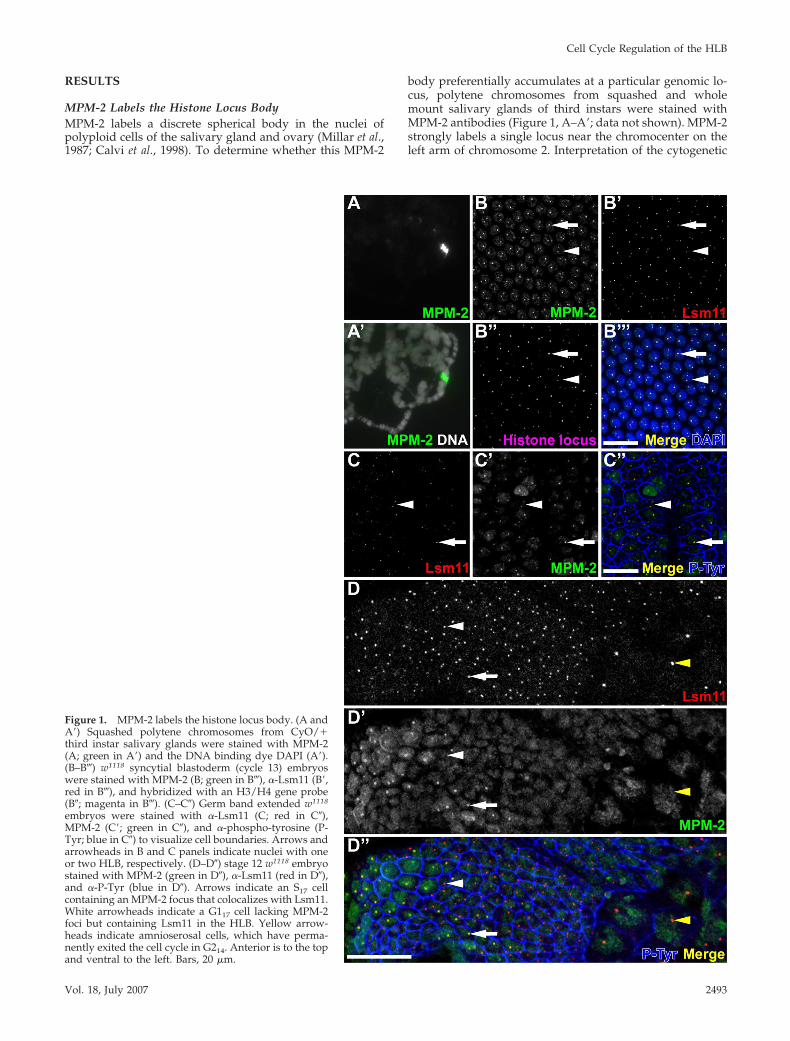
MPM-2 Labels the Histone Locus BodyMPM-2 labels a discrete spherical body in the nuclei ofpolyploid cells of the salivary gland and ovary (Millar et al.,1987; Calvi et al., 1998). To determine whether this MPM-2
body preferentially accumulates at a particular genomic lo-cus, polytene chromosomes from squashed and wholemount salivary glands of third instars were stained withMPM-2 antibodies (Figure 1, A–A�; data not shown). MPM-2strongly labels a single locus near the chromocenter on theleft arm of chromosome 2. Interpretation of the cytogenetic
Figure 1. MPM-2 labels the histone locus body. (A andA�) Squashed polytene chromosomes from CyO/�third instar salivary glands were stained with MPM-2(A; green in A�) and the DNA binding dye DAPI (A�).(B–B�) w1118 syncytial blastoderm (cycle 13) embryoswere stained with MPM-2 (B; green in B�), �-Lsm11 (B�,red in B�), and hybridized with an H3/H4 gene probe(B�; magenta in B�). (C–C�) Germ band extended w1118
embryos were stained with �-Lsm11 (C; red in C�),MPM-2 (C�; green in C�), and �-phospho-tyrosine (P-Tyr; blue in C�) to visualize cell boundaries. Arrows andarrowheads in B and C panels indicate nuclei with oneor two HLB, respectively. (D–D�) stage 12 w1118 embryostained with MPM-2 (green in D�), �-Lsm11 (red in D�),and �-P-Tyr (blue in D�). Arrows indicate an S17 cellcontaining an MPM-2 focus that colocalizes with Lsm11.White arrowheads indicate a G117 cell lacking MPM-2foci but containing Lsm11 in the HLB. Yellow arrow-heads indicate amnioserosal cells, which have perma-nently exited the cell cycle in G214. Anterior is to the topand ventral to the left. Bars, 20 �m.
Cell Cycle Regulation of the HLB
Vol. 18, July 2007 2493

banding pattern of these chromosomes indicated that thislabel is near or coincident with the chromosomal location ofthe histone gene cluster (data not shown). Cyclin E/Cdk2activity is required for MPM-2 labeling of this subnuclearbody, and for histone gene expression (Calvi et al., 1998;Lanzotti et al., 2004). We therefore hypothesized that MPM-2recognizes a phospho-protein(s) that localizes to the histonelocus body.
To test this hypothesis, we used confocal microscopy toanalyze embryos labeled with both MPM-2 and anti-Lsm11antibodies. Liu et al. (2006) originally visualized the HLBprimarily by detecting Lsm11, an Sm-like protein that is acore component of the U7 snRNP. Drosophila embryos un-dergo a stereotyped cell cycle program that is characterizedby distinct modes of cell cycle progression that occur atsuccessive stages of embryogenesis. During the first 2 h ofembryogenesis, maternally contributed factors drive 13rapid and synchronous nuclear cycles, consisting only ofS–M phases, in a syncytial cytoplasm. At the end of thisperiod (i.e., nuclear cycle 13), MPM-2 labels both the nucle-oplasm and distinct foci within the nucleus that colocalizewith Lsm11 foci (Figure 1, B–B�). MPM-2 foci also colocalizewith the histone locus, which was detected by fluorescencein situ hybridization by using a labeled DNA probe contain-ing the H3 and H4 genes (Figure 1, B�–B�). These dataindicate that an MPM-2 epitope is associated with the HLB.By Western blot analysis, MPM-2 is known to react with atleast a dozen targets in Drosophila (Logarinho and Sunkel,1998; Lange et al., 2005) (Leslie and Duronio, unpublisheddata), and thus the nucleoplasmic staining may not be thesame protein that concentrates in the HLB.
Cellularization takes place during cycle 14, when afterthe completion of S phase the nuclei pause for the firsttime in G2. Then, as gastrulation ensues, groups of cells indifferent regions of the embryo enter mitosis 14 together.These groups of cells, called “mitotic domains,” enter intomitosis 14 at different times, generating a reproducibleand well described pattern of mitosis (Foe, 1989). Entryinto mitosis 14 requires zygotic transcription of thestringcdc25 (stg) gene, which encodes a Cdc25-type phos-phatase that stimulates mitotic Cdk1 activity by removinginhibitory Y15 phosphorylation from Cdc2 (Edgar andO’Farrell, 1989). Cycles 15 and 16 are also regulated at theG2–M transition by developmentally controlled pulses ofstg transcription, and still lack G1 phase as in the earlysyncytial cycles (Edgar and O’Farrell, 1990; Edgar et al.,1994; Lehman et al., 1999). MPM-2 and Lsm11 nuclear focicolocalize and are continuously present during interphaseof these “postblastoderm” cell cycles (Figure 1, C–C�).MPM-2 foci are continuously present most likely becauseCyclin E/Cdk2 activity is also ubiquitous at this time,including during G2 phase (Sauer et al., 1995).
G1 phase first occurs during cycle 17, and subsequententry into S phase in all cell types requires zygotic expres-sion of cyclin E (Knoblich et al., 1994). Consistent with pre-vious observations that MPM-2 foci are Cyclin E dependent,MPM-2 only labels replicating cells where Cyclin E/Cdk2 isactive (Figure 1, D–D�, arrow). Cells that have exited the cellcycle, such as those in the amnioserosa (Figure 1, D–D�,yellow arrowhead) and epidermal cells arrested in G1 (Fig-ure 1, D–D�, white arrowhead), do not contain MPM-2 foci.In contrast, Lsm11 foci can be detected in all cells (Figure 1,arrows). Thus, the HLB is present ubiquitously, as describedpreviously for postembryonic stages of development (Liuet al., 2006), but the MPM-2 epitope occurs at the HLB onlyin cells that contain active Cyclin E/Cdk2.
One obvious feature of Lsm11 and MPM-2 staining ofblastoderm embryos is that each nucleus contains either oneor two foci (Figure 1, B–B�, C–C�, arrows and arrowheads,respectively). Homologous chromosomes are often paired inDrosophila cells, and we therefore hypothesized that one andtwo foci results from paired and unpaired homologous chro-mosomes, respectively. In the early embryo, the pairing ofhomologous chromosomes is dynamic, such that the fre-quency of pairing at any particular locus increases as theembryo ages (Fung et al., 1998). We counted the number ofcells with one or two MPM-2 foci in blastoderm embryosduring interphase of cell cycle 14. At this stage, 71% of cellscontained one MPM-2 focus and 29% contained two MPM-2foci (n � 293). Fung et al. (1998) reported a pairing frequencyfor the histone locus of 71 and 84% at 2.5 and 4 h ofdevelopment, respectively, which encompasses the cycle 14cells we analyzed (Fung et al., 1998). By the time of germband retraction during cycle 17, most cells contain a singlefocus (Figure 1D). These data are consistent with the HLBspecifically associating with the histone locus (Figure 1B���)(Liu et al., 2006). Also in support of this interpretation is theobservation that polyploid nurse cells in the ovary havepartially unpaired sister chromatids at the histone locus andcontain multiple MPM-2 foci (data not shown) (Hammondand Laird, 1985; Calvi et al., 1998). Together, these resultsindicate that MPM-2 recognizes an epitope at the histonelocus that is a component of the Lsm11-containing HLB.
Embryonic MPM-2 Foci Depend on Cyclin E/Cdk2ActivityIf the embryonic MPM-2 foci are related to the previouslydescribed MPM-2 foci in follicle cells, then their presenceshould depend on Cyclin E activity. To test this, we charac-terized MPM-2 staining in stage 13 (cycle 17 in the epider-mis) cyclin E mutant embryos relative to controls. Wild-typeembryos at this developmental stage contain proliferatingdiploid cells in the central and peripheral nervous systemsas well as endoreduplicating cells in various tissues (e.g.,midgut, salivary gland, and posterior spiracles). cyclin Emutant embryos develop normally until this stage becauseof maternal deposition of Cyclin E, and then they arrest inG1 of cycle 17. Consequently, DNA synthesis in both pro-liferating neuronal cells and endoreduplicating cells is se-verely compromised in cyclin E mutants (Knoblich et al.,1994). We focused on endoreduplicating cells in the poste-rior spiracle primordium, because they are near the surfaceand relatively easy to image. In control embryos these cellscontain robust MPM-2 foci (Figure 2, A–A�, arrow). In con-trast, the posterior spiracle cells of stage matched cyclin Emutants lack detectable MPM-2 foci (Figure 2, B–B�, arrow),whereas they still contain Lsm11 foci (Figure 2, C–C�, ar-row). These data indicate that Cyclin E is not required formaintenance of the HLB, but rather for the appearance of anMPM-2 epitope at the HLB. We also performed the recipro-cal experiment of Cyclin E overexpression. Epidermal cellsarrest in G1 of cycle 17, and they contain Lsm11 foci but notMPM-2 foci (Figure 2D). Ubiquitous expression of Cyclin Ewith a heat-inducible promoter (hsp70::cyclin E) drives theseG117 cells into S phase (Knoblich et al., 1994; Duronio andO’Farrell, 1995). This treatment also results in the appear-ance throughout the epidermis of MPM-2 foci that colocalizewith Lsm11 foci (Figure 2E, arrowheads). We conclude fromthese data that Cyclin E/Cdk2 activity in the embryo is bothnecessary and sufficient to produce nuclear MPM-2 foci thatcolocalize with the HLB. Because histone gene expression islost in cyclin E mutant embryos when they undergo cell cyclearrest in G1 of cycle 17, we hypothesize that MPM-2 recog-
A. E. White et al.
Molecular Biology of the Cell2494

nizes a Cyclin E/Cdk2 substrate that contributes to histonemRNA biosynthesis. We therefore characterized embryonicMPM-2 foci in more detail.
MPM-2 Foci Colocalize with Nascent Histone TranscriptsIf MPM-2 recognizes an HLB phospho-protein involved inhistone gene expression, then it should label sites of histonemRNA biosynthesis. We previously developed a cytologicalassay that detects nascent, chromatin-associated histonemRNA transcripts in intact Slbp mutant embryos (Lanzottiet al., 2002). Slbp mutants are defective in normal histonepre-mRNA processing, and accumulate inappropriatelylong, polyadenylated histone mRNAs (Sullivan et al., 2001).These aberrant mRNAs result from the use of cryptic poly-adenylation signals located downstream of the normal pre-mRNA processing site in each of the five replication-associ-ated histone genes (Lanzotti et al., 2002). Because the 3� UTRof these polyadenylated histone mRNAs contains sequencesnot found in the wild-type mRNA, we were able to developa probe (H3-ds) that specifically recognizes the polyadenyl-ated form of histone H3 mRNA. Cryptic polyadenylation isless efficient than the normal 3�-end processing, causingnascent pre-mRNA to accumulate on the chromatin tem-plate in Slbp mutants. These nascent transcripts can be visu-alized as a nuclear focus by fluorescent whole mount in situhybridization with the H3-ds probe (Figure 3, A and B)
(Lanzotti et al., 2002, 2004). In addition, nascent histonetranscripts occur soon after zygotic transcription of the his-tone genes begins, because there is no maternal SLBP in theearly embryo. We previously showed that nascent H3 tran-scripts arise in very late G214 immediately preceding eachmitosis and persist into S phase of cycle 15 (Lanzotti et al.,2004). Fluorescent in situ hybridization of Slbp mutant em-bryos with the H3-ds probe in conjunction with MPM-2staining showed that MPM-2 foci colocalize with these nas-cent H3 transcripts (Figure 3, A and B, arrows). Thus,MPM-2 foci colocalize with sites of active histone mRNAbiosynthesis in embryonic cells. MPM-2 also stains foci incells in early G214 that lack nascent H3 transcripts but thatare known to contain active Cyclin E/Cdk2 (Figure 3, Aand C, arrowhead). This suggests that MPM-2 foci can bepresent without ongoing active histone gene transcription(see below; Figure 6).
The HLB Disassembles during MitosisComponents of the mammalian Cajal body such as NPATdisassemble at the metaphase–anaphase transition and reas-semble in the following interphase (Ma et al., 2000). Todetermine whether HLBs behave similarly, embryos werestained with MPM-2 or anti-Lsm11 antibodies and anti-phospho-histone H3 antibody (PH3) to mark mitotic cells.MPM-2 staining was examined in the early mitotic domains
Figure 2. Embryonic MPM-2 foci depend on Cyclin Eactivity. (A–C) Images of the posterior spiracles of germband retracted (stage 13) embryos. (A) w1118 embryosstained with MPM-2 (A; green in A�) and �-P-Tyr (blue inA�). (B and C) cycEAR95 homozygous mutant embryosstained with MPM-2 (B; green in B�) or �-Lsm11 (C; red inC�) and �-P-Tyr (blue in B� and C�). Arrows indicate aposterior spiracle cell. w1118 control (D) or hsp70::cyclin Etransgenic (E) embryos were given a 37°C heat shock for30 min before fixation and then stained with MPM-2(green), �-Lsm11 (red), and �-P-Tyr (blue). Epidermal cellsof the first thoracic segment are shown. Arrowheads indi-cate the HLB. Anterior is to the top. Bar, 20 �m.
Cell Cycle Regulation of the HLB
Vol. 18, July 2007 2495

of cell cycle 14 in gastrulating embryos (Figure 4). Becausethe groups of cells making up a mitotic domain do not entermitosis synchronously, we could examine all stages of mi-tosis within a single domain. The nuclear MPM-2 focipresent in G214 persist into early mitosis, and all M14prophase cells examined contained MPM-2 foci associatedwith condensing chromosomes (n � 125; Figure 4A, arrow).In contrast, only 77% of metaphase cells (n � 106; Figure 4A,arrowhead) and none of the anaphase cells (n � 102; Figure4A, yellow arrowhead) contain detectable chromosome-as-sociated MPM-2 foci. At the following interphase, the nu-clear MPM-2 foci reappear. In syncytial blastoderm embryoswhere progression through mitosis occurs synchronously,we also failed to detect MPM-2 foci during anaphase andsome metaphase embryos (Supplemental Figure 1). Thesedata indicate that the MPM-2 phospho-epitope begins todecline during metaphase and becomes undetectable by an-aphase, either because the HLB disassembles or because theprotein containing the MPM-2 epitope is destroyed or de-phosphorylated. A similar analysis was performed in cycle14 embryos with Lsm11 antibodies. As with MPM-2 foci,Lsm11 foci are present in prophase, become undetectable byanaphase, and return in interphase (Figure 4B). These datasuggest that components of the HLB disassemble duringmitosis and are similar to what has been reported previouslyfor NPAT in fibroblast cell lines (Ma et al., 2000; Zhao et al.,2000). Drosophila centrosomes have been reported to containproteins with phospho-epitopes that are recognized byMPM-2 during mitosis (Logarinho and Sunkel, 1998; doCarmo Avides et al., 2001; Lange et al., 2005). With the
fixation conditions we used, MPM-2 staining increasesthroughout the cell during mitosis, but we did not observespecific staining of centrosomes.
MPM-2 Foci Assemble in the Absence of the HistonePre-mRNA Processing Factors SLBP and U7 snRNPOur analysis thus far suggests that MPM-2 recognizes aCyclin E/Cdk2 substrate found in the HLB that could beinvolved in histone mRNA biosynthesis. One possibility isthat one or more components of the histone pre-mRNAprocessing machinery are required for the formation ofMPM-2 foci. To test this, we stained Slbp mutants withMPM2 antibodies. As shown above (Figure 3), MPM-2 fociare observed in Slbp mutant embryos, which are deficient inSLBP because there is no maternal store of SLBP protein(Lanzotti et al., 2002). This indicates that SLBP does notcontain the primary MPM-2 epitope in the HLB and sug-gests that components of the HLB assemble independentlyof SLBP. Consistent with this, Lsm11 foci are readily de-tected in Slbp mutant embryos, and they colocalize with sitesof nascent histone H3 transcription (data not shown).
To test whether the U7snRNP is required for eitherMPM-2 foci or the HLB to assemble, we stained tissues fromU7 snRNA mutant third instars. Because of maternal load-ing of U7 snRNA, the third instar is the earliest time duringdevelopment when U7 snRNA is depleted and the U7 mu-tant phenotype occurs (Godfrey et al., 2006). We confirmedthe disruption of the U7 snRNP by assaying for Lsm11, a U7snRNP-specific protein that no longer accumulates in theHLB in U7 mutant cells (Figure 5, B and D). MPM-2 fociform in U7 mutant salivary gland-associated fat bodies andeye discs (Figure 5, A and C), as they do in SLBP mutantembryos. Similarly, the MPM-2 antigen, but not Lsm11,localizes to the HLB in U7 mutant follicle cells in the adultovary (Gall, personal communication). Therefore, the HLBwith an associated protein recognized by MPM-2 assemblesindependently of the U7 snRNP. To confirm this result, weperformed RNAi knockdown of Lsm11 in Dmel-2 cells, andwe observed that MPM-2 staining was unperturbed afterLsm11 dsRNA treatment, although the Lsm11 foci were nolonger detectable (Supplemental Figure 2). In sum, our re-sults indicate that the MPM-2 phospho-epitope and Lsm11are detected in the HLB whether or not SLBP is present inthe cell. Unlike Lsm11, however, the detection of the MPM-2phospho-epitope in the HLB does not depend on U7 snRNA,suggesting that at least a partial HLB can form indepen-dently of an intact U7 snRNP complex.
Embryonic MPM-2 Foci Do Not Require Stringcdc25
or Histone TranscriptionCyclin E/Cdk2 phosphorylation of the protein containingthe MPM-2 epitope may regulate, or even result from, thetranscription of histone genes. To test this, we examinedMPM-2 staining in string mutant embryos. In late G214,histone gene transcription occurs in a pattern that preciselycorrelates with the mitotic domain pattern, and this tran-scription is lost in string mutants, which arrest in G214 afterdestruction of maternal string mRNA and protein (Figure 6,A and B) (Lanzotti et al., 2004). MPM-2 foci can be readilydetected in string Slbp null mutant embryos that lack nascenthistone transcripts (Figure 6, A and C). This result indicatesthat formation of MPM-2 foci does not require histone genetranscription or the function of mitotic Cdks activated bystringcdc25. Consistent with this interpretation, MPM-2 fociare present in all cells throughout interphase 14 (i.e., both Sphase and G2 when Cyclin E/Cdk2 is active) at times whenneither stringcdc25 is active nor histone transcription occurs
Figure 3. MPM-2 foci colocalize with the histone locus body inreplicating cells. (A) An Slbp15 homozygous mutant postblastodermembryo was stained with �-P-Tyr (left in blue), MPM-2 (green inmerge), and hybridized with a fluorescent probe that recognizesmisprocessed, polyadenylated H3 mRNA as nascent transcripts inthe nucleus (H3-ds; left in red). Arrows indicate a cell in S15, whichcontains an MPM-2 focus that colocalizes with nascent H3 tran-scripts. Arrowheads indicate an MPM-2 focus in a G214 cell thatlacks nascent H3 transcripts. Anterior is to the top and ventral to theright. Bar, 20 �m. (B) S15 cell marked by arrows in A. Bar, 2 �m. (C)G214 cell marked by arrowheads in A. Bar, 2 �m.
A. E. White et al.
Molecular Biology of the Cell2496

(Figure 3C). These data indicate that the formation ofMPM-2 foci occurs independently of stringcdc25 (and thus byinference Cdk1 activity) and histone transcription.
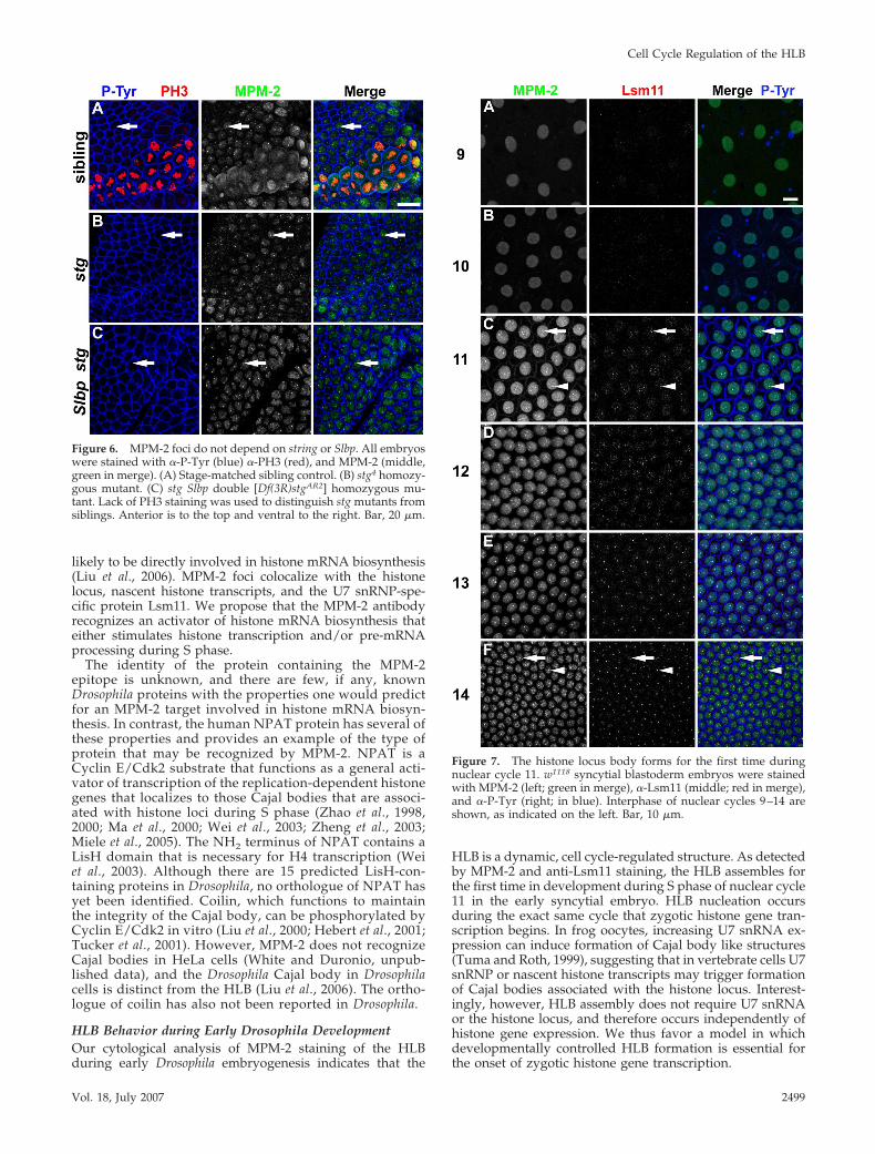
MPM-2 Foci Occur Coincidentally with Activationof Zygotic Histone Gene ExpressionMost zygotic gene expression begins at the blastodermstage during cycle 14 (Edgar and Schubiger, 1986). His-tone genes are an exception, and become transcriptionallyactive precisely in nuclear cycle 11 (Edgar and Schubiger,1986). If the protein containing the MPM-2 epitope regu-lates some aspect(s) of histone mRNA biosynthesis, thenwe expect MPM-2 foci to be present once zygotic expres-sion of the histone genes has begun. To determinewhether there is a relationship between the onset of zy-gotic histone transcription, the assembly of the HLB, andthe appearance of the MPM-2 epitope at the HLB, wecarefully analyzed syncytial blastoderm embryos stainedwith MPM-2 and anti-Lsm11 antibodies. Nuclear densitywas used to accurately stage the embryos with respect toeach nuclear cycle. Uniform MPM-2 staining throughoutthe nucleus was detectable in all syncytial blastodermcycles, perhaps because maternal Cyclin E/Cdk2 ispresent throughout the early embryo. MPM-2 foci occurfor the first time precisely during nuclear cycle 11, con-comitantly with the onset of zygotic histone gene tran-
scription (Figure 7). Similarly, nuclear Lsm11 staining isundetectable before cycle 11, and Lsm11 foci first occurduring nuclear cycle 11 and colocalize with MPM-2 foci(Figure 7). The appearance of Lsm11 foci in cycle 11 is notthe result of new, zygotic synthesis, because Lsm11 pro-tein is present in 0- to 30-min embryos (data not shown),before the onset of zygotic gene expression. In addition,maternal expression of a YFP–Lsm11 fusion protein inda-GAL4/UAS-YFP-Lsm11 females results in appearanceof YFP–Lsm11 foci in embryonic cycle 11 (SupplementalFigure 3). These data indicate that the onset of zygotichistone gene transcription and formation of the MPM-2–positive HLB during early development are temporallyand spatially coincident.
MPM-2 Foci Form Independently of the Histone LocusThe coincidence in developmental timing of the onset ofzygotic histone transcription and formation of the HLB sug-gests two possibilities. Histone gene transcription may di-rectly result in the nucleation of the HLB at the histone locus,or de novo assembly of the HLB may trigger the initiation ofhistone transcription. To attempt to distinguish betweenthese possibilities, we stained embryos containing a ho-mozygous deletion of the entire histone locus [Df(2L)Ds6]with MPM-2 and anti-Lsm11 antibodies. We hypothesizedthat chromatin at or near the histone locus is necessary for
Figure 4. MPM-2 and Lsm11 foci are cell cycle depen-dent. Postblastoderm w1118 embryos in cell cycle 14 werestained with �-P-Tyr (blue), �-PH3 (red), and MPM-2 (A)or �-Lsm11 (B) (middle; green in merge). Arrows indicateprophase cells, arrowheads indicate metaphase cells, andyellow arrowheads indicate anaphase cells. Bar, 20 �m.Single prophase, metaphase, and anaphase cells areshown in A� and B�, A� and B�, and A� and B�, respec-tively. Bars, 2 �m. Mitotic domain 5 is shown in A, andmitotic domain 1 is shown in B. Note that the rat �-P-Tyrused here recognizes an antigen at the HLB and the mouse�-PH3 used in B stains interphase cells weakly.
Cell Cycle Regulation of the HLB
Vol. 18, July 2007 2497

the assembly of the HLB, as it might function as a scaffold onwhich the HLB is assembled to carry out histone mRNAbiosynthesis. Surprisingly, both MPM-2 and Lsm11 foci aredetected in Df(2L)Ds6 homozygous embryos lacking the hi-stone genes (Figure 8, arrows), which were distinguishedfrom siblings containing histone genes by in situ hybridiza-tion of histone H3 mRNA (Figure 8, left). Moreover, thedistribution of nuclei with one versus two MPM-2 foci issimilar between wild-type and Df(2L)Ds6 embryos, suchthat the percentage of cells containing one MPM-2 focus inwild type and Df(2L)Ds6 embryos is 89 and 84%, respec-tively, and the percentage of cells containing two MPM-2foci in wild-type and Df(2L)Ds6 embryos is 11 and 7.8%,respectively. This result indicates that the histone locus, andtherefore histone transcription, is not required for the initialnucleation of the HLB. This is consistent with our analysis ofstring mutant embryos, which also indicated that histonetranscription is not required to maintain MPM-2 foci.
Although HLBs form in the absence of the histone locus,we observed differences in MPM-2 and Lsm11 staining be-tween wild-type and Df(2L)Ds6 embryos. First, the lack ofthe histone gene cluster altered the high incidence of MPM-2and Lsm11 colocalization (Table 1). Using fluorescent im-ages, nuclei were assigned to a category based on thetype of MPM-2 and Lsm11 foci present in the nucleus of
stage 11 wild-type or Df(2L)Ds6 embryos. Whereas in wild-type embryos only 5% of interphase nuclei contained Lsm11foci that did not colocalize with MPM-2, in Df(2L)Ds6 mu-tants the proportion of this class of nuclei increased to �35%(Table 1). In addition, 8% of the nuclei contained more thanone Lsm11 focus that did not colocalize with MPM-2 foci(Table 1). These data indicate that full association of the U7snRNP and the protein containing the MPM-2 epitope re-quires the histone locus. In addition, the HLBs in Df(2L)Ds6embryos are smaller than in controls as revealed by measur-ing the size (see Materials and Methods) of the MPM-2 nuclearfocus in 75 cells from two stage-matched, stage 11 embryos.The average size (defined as length plus width) of theMPM-2 focus in control and Df(2L)Ds6 embryos is 1.48 �0.37 and 1.06 � 0.27 �m, respectively (p 0.001). Thus,although the histone locus is not absolutely essential for theformation of the HLB, as assessed by colocalization ofMPM-2 and Lsm11 foci, full, stable assembly of the HLBrequires the histone locus.
DISCUSSION
Using the MPM-2 monoclonal antibody as a tool, we havepresented evidence that a Cyclin E/Cdk2 substrate localizesto the HLB, a recently described Drosophila nuclear organelle
Figure 5. MPM-2 foci form independently of the U7snRNP. Fat bodies (A and B) and eye discs (C and D)dissected from w1118 and U714 homozygous mutantthird instars were stained with MPM-2 (left; green inmerge) and �-Lsm11 (middle; red in merge). Fat bodieswere also stained with DAPI (blue in merge). Arrowsand arrowheads indicate nuclei with and withoutMPM-2 foci, respectively. Note that in wild type Lsm11foci are present in cells without MPM-2 (arrowhead inC) and that Lsm11 foci are absent in the U7 mutant cells.Bars, 20 �m.
A. E. White et al.
Molecular Biology of the Cell2498
likely to be directly involved in histone mRNA biosynthesis(Liu et al., 2006). MPM-2 foci colocalize with the histonelocus, nascent histone transcripts, and the U7 snRNP-spe-cific protein Lsm11. We propose that the MPM-2 antibodyrecognizes an activator of histone mRNA biosynthesis thateither stimulates histone transcription and/or pre-mRNAprocessing during S phase.
The identity of the protein containing the MPM-2epitope is unknown, and there are few, if any, knownDrosophila proteins with the properties one would predictfor an MPM-2 target involved in histone mRNA biosyn-thesis. In contrast, the human NPAT protein has several ofthese properties and provides an example of the type ofprotein that may be recognized by MPM-2. NPAT is aCyclin E/Cdk2 substrate that functions as a general acti-vator of transcription of the replication-dependent histonegenes that localizes to those Cajal bodies that are associ-ated with histone loci during S phase (Zhao et al., 1998,2000; Ma et al., 2000; Wei et al., 2003; Zheng et al., 2003;Miele et al., 2005). The NH2 terminus of NPAT contains aLisH domain that is necessary for H4 transcription (Weiet al., 2003). Although there are 15 predicted LisH-con-taining proteins in Drosophila, no orthologue of NPAT hasyet been identified. Coilin, which functions to maintainthe integrity of the Cajal body, can be phosphorylated byCyclin E/Cdk2 in vitro (Liu et al., 2000; Hebert et al., 2001;Tucker et al., 2001). However, MPM-2 does not recognizeCajal bodies in HeLa cells (White and Duronio, unpub-lished data), and the Drosophila Cajal body in Drosophilacells is distinct from the HLB (Liu et al., 2006). The ortho-logue of coilin has also not been reported in Drosophila.
HLB Behavior during Early Drosophila DevelopmentOur cytological analysis of MPM-2 staining of the HLBduring early Drosophila embryogenesis indicates that the
HLB is a dynamic, cell cycle-regulated structure. As detectedby MPM-2 and anti-Lsm11 staining, the HLB assembles forthe first time in development during S phase of nuclear cycle11 in the early syncytial embryo. HLB nucleation occursduring the exact same cycle that zygotic histone gene tran-scription begins. In frog oocytes, increasing U7 snRNA ex-pression can induce formation of Cajal body like structures(Tuma and Roth, 1999), suggesting that in vertebrate cells U7snRNP or nascent histone transcripts may trigger formationof Cajal bodies associated with the histone locus. Interest-ingly, however, HLB assembly does not require U7 snRNAor the histone locus, and therefore occurs independently ofhistone gene expression. We thus favor a model in whichdevelopmentally controlled HLB formation is essential forthe onset of zygotic histone gene transcription.
Figure 6. MPM-2 foci do not depend on string or Slbp. All embryoswere stained with �-P-Tyr (blue) �-PH3 (red), and MPM-2 (middle,green in merge). (A) Stage-matched sibling control. (B) stg4 homozy-gous mutant. (C) stg Slbp double [Df(3R)stgAR2] homozygous mu-tant. Lack of PH3 staining was used to distinguish stg mutants fromsiblings. Anterior is to the top and ventral to the right. Bar, 20 �m.
Figure 7. The histone locus body forms for the first time duringnuclear cycle 11. w1118 syncytial blastoderm embryos were stainedwith MPM-2 (left; green in merge), �-Lsm11 (middle; red in merge),and �-P-Tyr (right; in blue). Interphase of nuclear cycles 9–14 areshown, as indicated on the left. Bar, 10 �m.
Cell Cycle Regulation of the HLB
Vol. 18, July 2007 2499

Although the HLB is capable of forming independentlyof the histone locus, the histone locus contributes to thestructural integrity of the HLB. In histone deletion em-bryos, HLBs are smaller and some interphase nuclei con-tain Lsm11 foci that do not colocalize with MPM-2. Thesize of the nucleolus is determined by the amount ofribosomal gene transcription (Hernandez-Verdun, 2006).Thus, the size and overall composition of the HLB maybe similarly dependent on transcription of the histonegenes. Consistent with this, MPM-2 and Lsm11 foci arepresent at maximal size during prophase of cycle 14, justafter the initiation of histone transcription in late G214.Nascent histone transcripts are likely aborted during mi-tosis (Shermoen and O’Farrell, 1991), which correlateswith the loss of MPM-2 and Lsm11 staining we observe inanaphase. Alternatively, reduced HLB size may be a sec-ondary consequence of cell cycle arrest, resulting fromlack of histone biosynthesis (Smith et al., 1993).
In wild-type embryos, we observe either one or twoMPM-2 or Lsm11 foci per cell at frequencies similar to theknown pairing frequencies of the histone loci on homolo-gous second chromosomes (Fung et al., 1998). This is alsoconsistent with the association of the HLB with histonegenes (Liu et al., 2006). Surprisingly, we often detect one ortwo MPM-2 foci in histone deletion embryos, suggestingthat HLB number is not dictated solely by homologouspairing at the histone locus. HLB components may associatewith another chromosomal locus in the absence of the his-tone genes. Alternatively, the de novo formation of one ortwo HLBs may actually drive homologous pairing at thehistone locus.
Cell Cycle Regulation of the HLBHistone mRNA is greatly depleted in cyclin E mutantembryos (Lanzotti et al., 2004). Because cyclin E mutant
embryos arrest in G1 phase, it is difficult to know thisobservation is indicative of a direct involvement of CyclinE/Cdk2 in histone gene expression, or arises as a secondaryconsequence of cell cycle arrest. Because interphase MPM-2foci are coincident with the HLB, are present in cells whereCyclin E/Cdk2 is active, and they are absent in cells thatlack Cyclin E/Cdk2 activity, we favor the interpretation thatCyclin E/Cdk2 phosphorylates a protein directly involvedin histone mRNA biosynthesis. How Cyclin E/Cdk2 partic-ipates in this process is not known, but it is not required forrecruitment of U7 snRNP to the HLB, because Lsm11 re-mains a stable component of the HLB in wild-type cellsarrested in G1 and in cells of cyclin E mutant embryos.
During the early embryonic cell cycles that have con-stitutive Cyclin E/Cdk2 activity, MPM-2 foci disappear ascells progress through mitosis and are undetectable byanaphase. Focal Lsm11 staining is also lost during mitosis,suggesting the disassembly of HLB components into thecytoplasm subsequent to nuclear envelope breakdownrather than simply dephosphorylation or destruction ofthe MPM-2 target(s). The HLB rapidly reforms during thesubsequent interphase. These behaviors are similar to thebehavior of NPAT foci during mitosis in cultured mam-malian fibroblasts (Ma et al., 2000; Zhao et al., 2000). Thenucleolus also disassembles during mitosis (Hernandez-Verdun, 2006), suggesting that dynamic disassembly/re-assembly of specific nuclear compartments involved ingene expression is a general feature of nuclear behaviorduring metazoan mitosis.
SummaryThe HLB is a dynamic structure capable of receiving inputfrom both the histone locus and the cell cycle. Our analysisraises many questions, including how HLB assembly occursat a specific time in a syncytial cytoplasm from abundant
Figure 8. Aberrant histone locus bodies formin the absence of the histone locus. Postblas-toderm embryos hybridized with an H3 RNAprobe (magenta) were stained with MPM-2(green), �-Lsm11 (red), and DAPI (blue).Df(2L)Ds6 homozygous mutant embryos thatlack the histone gene cluster (B) were distin-guished from control siblings (A) by the lackof zygotic histone H3 mRNA. Arrows indicatefoci of MPM-2 and �-Lsm11 that colocalize.Arrowheads indicate cells containing a focusof colocalizing MPM-2 and �-Lsm11 as well asa focus of just �-Lsm11. Anterior is to the top.Bar, 20 �m.
Table 1. Characterization of the HLB in histone deletion embryos
1 MLa 2 ML 1 ML � 1 L 2 L 1 L 1 ML � 2 Ls 2 M Other
w1118 (n � 191) 81.2 11.0 5.2 0 0.5 0 0 2.1Ds6 (n � 192) 54.7 4.2 20.3 3.1 3.6 7.8 0.5 5.7
a ML indicates a focus of colocalized MPM-2 and Lsm11 staining, L indicates a focus containing only Lsm11, and M indicates a focuscontaining only MPM-2. “Other” refers to individual patterns of staining that are not represented in the indicated categories and that eachcomprise 0.5% of cells.
A. E. White et al.
Molecular Biology of the Cell2500

maternal components, even in the absence of a histone locustemplate, and how Cyclin E/Cdk2 regulates HLB functionand histone mRNA biosynthesis. Determining the identityof the HLB protein(s) recognized by MPM-2 antigen willhelp answer such questions, and it will provide a useful toolto examine how the regulation of such a fundamental pro-cess as histone mRNA biosynthesis is modulated by devel-opmental programs.
ACKNOWLEDGMENTS
We are grateful to Joe Gall for generous sharing of reagents, especially Lsm11antibodies, and of unpublished information. We thank Julie Norseen forcontributing to MPM-2 polytene staining. We thank Mark Peifer for criticalreading of the manuscript. This work was supported by National Institutes ofHealth grant GM-057859 (to R.J.D.).
REFERENCES
Albert, A. L., Lavoie, S. B., and Vincent, M. (2004). Multisite phosphorylationof Pin1-associated mitotic phosphoproteins revealed by monoclonal antibod-ies MPM-2 and CC-3. BMC Cell Biol. 5, 22.
Borun, T. W., Gabrielli, F., Ajiro, K., Zweidler, A., and Baglioni, C. (1975).Further evidence of transcriptional and translational control of histone mes-senger RNA during the HeLa S3 cycle. Cell 4, 59–67.
Breindl, M., and Gallwitz, D. (1973). Identification of histone messenger RNAfrom HeLa cells. Appearance of histone mRNA in the cytoplasm and itstranslation in a rabbit-reticulocyte cell-free system. Eur. J. Biochem. 32,381–391.
Calvi, B. R., Lilly, M. A., and Spradling, A. C. (1998). Cell cycle control ofchorion gene amplification. Genes Dev. 12, 734–744.
Cioce, M., and Lamond, A. I. (2005). Cajal bodies: a long history of discovery.Annu. Rev. Cell Dev. Biol. 21, 105–131.
Davis, F. M., Tsao, T. Y., Fowler, S. K., and Rao, P. N. (1983). Monoclonalantibodies to mitotic cells. Proc. Natl. Acad. Sci. USA 80, 2926–2930.
DeLisle, A. J., Graves, R. A., Marzluff, W. F., and Johnson, L. F. (1983).Regulation of histone mRNA production and stability in serum-stimulatedmouse 3T6 fibroblasts. Mol. Cell. Biol. 3, 1920–1929.
Dernburg, A. F., and Sedat, J. W. (1998). Mapping three-dimensional chro-mosome architecture in situ. Methods Cell Biol. 53, 187–233.
Detke, S., Lichtler, A., Phillips, I., Stein, J., and Stein, G. (1979). Reassessmentof histone gene expression during cell cycle in human cells by using homol-ogous H4 histone cDNA. Proc. Natl. Acad. Sci. USA 76, 4995–4999.
do Carmo Avides, M., Tavares, A., and Glover, D. M. (2001). Polo kinase andAsp are needed to promote the mitotic organizing activity of centrosomes.Nat. Cell Biol. 3, 421–424.
Dominski, Z., and Marzluff, W. F. (1999). Formation of the 3� end of histonemRNA. Gene 239, 1–14.
Du, W., and Pogoriler, J. (2006). Retinoblastoma family genes. Oncogene 25,5190–5200.
Duronio, R. J., and O’Farrell, P. H. (1995). Developmental control of the G1 toS transition in Drosophila: cyclin Eis a limiting downstream target of E2F.Genes Dev. 9, 1456–1468.
Edgar, B. A., and O’Farrell, P. H. (1989). Genetic control of cell divisionpatterns in the Drosophila embryo. Cell 57, 177–187.
Edgar, B. A., and O’Farrell, P. H. (1990). The three postblastoderm cell cyclesof Drosophila embryogenesis are regulated in G2 by string. Cell 62, 469–480.
Edgar, B. A., Lehman, D. A., and O’Farrell, P. H. (1994). Transcriptionalregulation of string (cdc25): a link between developmental programming andthe cell cycle. Development 120, 3131–3143.
Edgar, B. A., and Schubiger, G. (1986). Parameters controlling transcriptionalactivation during early Drosophila development. Cell 44, 871–877.
Foe, V. E. (1989). Mitotic domains reveal early commitment of cells in Dro-sophila embryos. Development 107, 1–22.
Frey, M. R., and Matera, A. G. (1995). Coiled bodies contain U7 small nuclearRNA and associate with specific DNA sequences in interphase human cells.Proc. Natl. Acad. Sci. USA 92, 5915–5919.
Fung, J. C., Marshall, W. F., Dernburg, A., Agard, D. A., and Sedat, J. W.(1998). Homologous chromosome pairing in Drosophila melanogaster proceedsthrough multiple independent initiations. J. Cell Biol. 141, 5–20.
Godfrey, A. C., Kupsco, J. M., Burch, B. D., Zimmerman, R. M., Dominski, Z.,Marzluff, W. F., and Duronio, R. J. (2006). U7 snRNA mutations in Drosophilablock histone pre-mRNA processing and disrupt oogenesis. RNA 12, 396–409.
Hall, C., Nelson, D. M., Ye, X., Baker, K., DeCaprio, J. A., Seeholzer, S.,Lipinski, M., and Adams, P. D. (2001). HIRA, the human homologue of yeastHir1p and Hir2p, is a novel cyclin-cdk2 substrate whose expression blocksS-phase progression. Mol. Cell. Biol. 21, 1854–1865.
Hammond, M. P., and Laird, C. D. (1985). Chromosome structure and DNAreplication in nurse and follicle cells of Drosophila melanogaster. Chromosoma91, 267–278.
Harris, M. E., Bohni, R., Schneiderman, M. H., Ramamurthy, L., Schumperli,D., and Marzluff, W. F. (1991). Regulation of histone mRNA in the unper-turbed cell cycle: evidence suggesting control at two posttranscriptional steps.Mol. Cell. Biol. 11, 2416–2424.
Hebert, M. D., Szymczyk, P. W., Shpargel, K. B., and Matera, A. G. (2001).Coilin forms the bridge between Cajal bodies and SMN, the spinal muscularatrophy protein. Genes Dev. 15, 2720–2729.
Heintz, N., Sive, H. L., and Roeder, R. G. (1983). Regulation of human histonegene expression: kinetics of accumulation and changes in the rate of synthesisand in the half-lives of individual histone mRNAs during the HeLa cell cycle.Mol. Cell. Biol. 3, 539–550.
Hernandez-Verdun, D. (2006). The nucleolus: a model for the organization ofnuclear functions. Histochem. Cell Biol. 126, 135–148.
Hirano, T., and Mitchison, T. J. (1991). Cell cycle control of higher-orderchromatin assembly around naked DNA in vitro. J. Cell Biol. 115, 1479–1489.
Kiss, T. (2004). Biogenesis of small nuclear RNPs. J. Cell Sci. 117, 5949–5951.
Knoblich, J. A., Sauer, K., Jones, L., Richardson, H., Saint, R., and Lehner, C. F.(1994). Cyclin E controls S phase progression and its down-regulation duringDrosophila embryogenesis is required for the arrest of cell proliferation. Cell77, 107–120.
Kuang, J., Zhao, J., Wright, D. A., Saunders, G. F., and Rao, P. N. (1989).Mitosis-specific monoclonal antibody MPM-2 inhibits Xenopus oocyte matu-ration and depletes maturation-promoting activity. Proc. Natl. Acad. Sci. USA86, 4982–4986.
Lange, B. M., Kirfel, G., Gestmann, I., Herzog, V., and Gonzalez, C. (2005).Structure and microtubule-nucleation activity of isolated Drosophila embryocentrosomes characterized by whole mount scanning and transmission elec-tron microscopy. Histochem. Cell Biol. 124, 325–334.
Lanzotti, D. J., Kaygun, H., Yang, X., Duronio, R. J., and Marzluff, W. F. (2002).Developmental control of histone mRNA and dSLBP synthesis during Dro-sophila embryogenesis and the role of dSLBP in histone mRNA 3� end pro-cessing in vivo. Mol. Cell. Biol. 22, 2267–2282.
Lanzotti, D. J., Kupsco, J. M., Marzluff, W. F., and Duronio, R. J. (2004).string(cdc25) and cyclin E are required for patterned histone expression atdifferent stages of Drosophila embryonic development. Dev. Biol. 274, 82–93.
Lee, L. A., and Orr-Weaver, T. L. (2003). Regulation of cell cycles in Drosophiladevelopment: intrinsic and extrinsic cues. Annu. Rev. Genet. 37, 545–578.
Lehman, D. A., Patterson, B., Johnston, L. A., Balzer, T., Britton, J. S., Saint, R.,and Edgar, B. A. (1999). Cis-regulatory elements of the mitotic regulator,string/Cdc25. Development 126, 1793–1803.
Liu, J., Hebert, M. D., Ye, Y., Templeton, D. J., Kung, H., and Matera, A. G.(2000). Cell cycle-dependent localization of the CDK2-cyclin E complex inCajal (coiled) bodies. J. Cell Sci. 113, 1543–1552.
Liu, J. L., Murphy, C., Buszczak, M., Clatterbuck, S., Goodman, R., and Gall,J. G. (2006). The Drosophila melanogaster Cajal body. J. Cell Biol. 172, 875–884.
Logarinho, E., and Sunkel, C. E. (1998). The Drosophila POLO kinase localisesto multiple compartments of the mitotic apparatus and is required for thephosphorylation of MPM2 reactive epitopes. J. Cell Sci. 111, 2897–2909.
Ma, T., Van Tine, B. A., Wei, Y., Garrett, M. D., Nelson, D., Adams, P. D.,Wang, J., Qin, J., Chow, L. T., and Harper, J. W. (2000). Cell cycle-regulatedphosphorylation of p220(NPAT) by cyclin E/Cdk2 in Cajal bodies promoteshistone gene transcription. Genes Dev. 14, 2298–2313.
Marzluff, W. F. (2005). Metazoan replication-dependent histone mRNAs: adistinct set of RNA polymerase II transcripts. Curr. Opin. Cell Biol. 17,274–280.
Marzluff, W. F., and Duronio, R. J. (2002). Histone mRNA expression: multiplelevels of cell cycle regulation and important developmental consequences.Curr. Opin. Cell Biol. 14, 692–699.
Matera, A. G., and Shpargel, K. B. (2006). Pumping RNA: nuclear bodybuild-ing along the RNP pipeline. Curr. Opin. Cell Biol. 18, 317–324.
Cell Cycle Regulation of the HLB
Vol. 18, July 2007 2501

Miele, A. et al. (2005). HiNF-P directly links the cyclin E/CDK2/p220NPATpathway to histone H4 gene regulation at the G1/S phase cell cycle transition.Mol. Cell. Biol. 25, 6140–6153.
Millar, S. E., Freeman, M., and Glover, D. M. (1987). The distribution of a‘mitosis-specific’ antigen during Drosophila development. J. Cell Sci. 87, 95–104.
Moore, G., Sinclair, D., and T. Grigliatti. (1983). Histone gene multiplicity andposition effect variegation in Drosophila melanogaster. Genetics 105, 327–344.
Nelson, D. M., Ye, X., Hall, C., Santos, H., Ma, T., Kao, G. D., Yen, T. J., Harper,J. W., and Adams, P. D. (2002). Coupling of DNA synthesis and histonesynthesis in S phase independent of cyclin/cdk2 activity. Mol. Cell. Biol. 22,7459–7472.
Parker, I., and Fitschen, W. (1980). Histone mRNA metabolism during themouse fibroblast cell cycle. Cell Differ. 9, 23–30.
Richardson, H., O’Keefe, L. V., Marty, T., and Saint, R. (1995). Ectopic cyclinE expression induces premature entry into S phase and disrupts patternformation in the Drosophila eye imaginal disc. Development 121, 3371–3379.
Sauer, K., Knoblich, J. A., Richardson, H., and Lehner, C. F. (1995). Distinctmodes of cyclin E/cdc2c kinase regulation and S-phase control in mitotic andendoreduplication cycles of Drosophila embryogenesis. Genes Dev. 9, 1327–1339.
Shermoen, A. W., and O’Farrell, P. H. (1991). Progression of the cell cyclethrough mitosis leads to abortion of nascent transcripts. Cell 67, 303–310.
Smith, A. V., King, J. A., and Orr-Weaver, T. L. (1993). Identification ofgenomic regions required for DNA replication during Drosophila embryogen-esis. Genetics 135, 817–829.
Stanek, D., and Neugebauer, K. M. (2006). The Cajal body: a meeting place forspliceosomal snRNPs in the nuclear maze. Chromosoma 115, 343–354.
Sullivan, E., Santiago, C., Parker, E. D., Dominski, Z., Yang, X., Lanzotti, D. J.,Ingledue, T. C., Marzluff, W. F., and Duronio, R. J. (2001). Drosophila stem loopbinding protein coordinates accumulation of mature histone mRNA with cellcycle progression. Genes Dev. 15, 173–187.
Swanhart, L., Kupsco, J., and Duronio, R. J. (2005). Developmental control ofgrowth and cell cycle progression in Drosophila. Methods Mol. Biol. 296,69–94.
Tucker, K. E., Berciano, M. T., Jacobs, E. Y., LePage, D. F., Shpargel, K. B.,Rossire, J. J., Chan, E. K., Lafarga, M., Conlon, R. A., and Matera, A. G. (2001).Residual Cajal bodies in coilin knockout mice fail to recruit Sm snRNPs andSMN, the spinal muscular atrophy gene product. J. Cell Biol. 154, 293–307.
Tuma, R. S., and Roth, M. B. (1999). Induction of coiled body-like structuresin Xenopus oocytes by U7 snRNA. Chromosoma 108, 337–344.
Wei, Y., Jin, J., and Harper, J. W. (2003). The cyclin E/Cdk2 substrate and Cajalbody component p220(NPAT) activates histone transcription through a novelLisH-like domain. Mol. Cell. Biol. 23, 3669–3680.
Westendorf, J. M., Rao, P. N., and Gerace, L. (1994). Cloning of cDNAs forM-phase phosphoproteins recognized by the MPM2 monoclonal antibodyand determination of the phosphorylated epitope. Proc. Natl. Acad. Sci. USA91, 714–718.
Wodarz, A., Hinz, U., Engelbert, M., and Knust, E. (1995). Expression ofcrumbs confers apical character on plasma membrane domains of ectodermalepithelia of Drosophila. Cell 82, 67–76.
Yaffe, M. B. et al. (1997). Sequence-specific and phosphorylation-dependentproline isomerization: a potential mitotic regulatory mechanism. Science 278,1957–1960.
Zhao, J., Dynlacht, B., Imai, T., Hori, T., and Harlow, E. (1998). Expression ofNPAT, a novel substrate of cyclin E-CDK2, promotes S-phase entry. GenesDev. 12, 456–461.
Zhao, J., Kennedy, B. K., Lawrence, B. D., Barbie, D. A., Matera, A. G.,Fletcher, J. A., and Harlow, E. (2000). NPAT links cyclin E-Cdk2 to theregulation of replication-dependent histone gene transcription. Genes Dev.14, 2283–2297.
Zheng, L., Roeder, R. G., and Luo, Y. (2003). S phase activation of the histoneH2B promoter by OCA-S, a coactivator complex that contains GAPDH as akey component. Cell 114, 255–266.
A. E. White et al.
Molecular Biology of the Cell2502