developmental biology - gross lab · a section of molecular cell and developmental biology and...
TRANSCRIPT

Developmental Biology 319 (2008) 10–22
Contents lists available at ScienceDirect
Developmental Biology
j ourna l homepage: www.e lsev ie r.com/deve lopmenta lb io logy
Zebrafish blowout provides genetic evidence for Patched1-mediated negativeregulation of Hedgehog signaling within the proximal optic vesicle of thevertebrate eye
Jiwoon Lee a, Jason R. Willer c, Gregory B. Willer c, Kierann Smith d, Ronald G. Gregg c, Jeffrey M. Gross a,b,⁎a Section of Molecular Cell and Developmental Biology and Institute for Cell and Molecular Biology, USAb Institute of Neuroscience, The University of Texas at Austin, Box C1000, 1 University Station, Austin, TX 78712, USAc Department of Biochemistry and Molecular Biology, University of Louisville, Louisville KY, 40202, USAd Department of Molecular and Cellular Biology, Harvard University, Cambridge, MA 02138, USA
⁎ Corresponding author. Section of Molecular Cell anUniversity of Texas at Austin, Box C1000, 1 University SFax: +1 512 471 3878.
E-mail address: [email protected] (J.M. Gross
0012-1606/$ – see front matter © 2008 Elsevier Inc. Aldoi:10.1016/j.ydbio.2008.03.035
A B S T R A C T
A R T I C L E I N F OArticle history:
In this study, we have cha Received for publication 24 August 2007Revised 21 March 2008Accepted 25 March 2008Available online 4 April 2008Keywords:ZebrafishOptic vesicleColobomaHedgehogPatched1
racterized the ocular defects in the recessive zebrafish mutant blowout thatpresents with a variably penetrant coloboma phenotype. blowout mutants develop unilateral or bilateralcolobomas and as a result, the retina and retinal pigmented epithelium are not contained within the opticcup. Colobomas result from defects in optic stalk morphogenesis whereby the optic stalk extends into theretina and impedes the lateral edges of the choroid fissure from meeting and fusing. The expression domainof the proximal optic vesicle marker pax2a is expanded in blowout at the expense of the distal optic vesiclemarker pax6, suggesting that the initial patterning of the optic vesicle into proximal and distal territories isdisrupted in blowout. Later aspects of distal optic cup formation (i.e. retina development) are normal inblowout mutants, however. Positional cloning of blowout identified a nonsense mutation in patched1, anegative regulator of the Hedgehog pathway, as the underlying cause of the blowout phenotype. Expandeddomains of expression of the Hedgehog target genes patched1 and patched2 were observed in blowout,consistent with a loss of Patched1 function and upregulation of Hedgehog pathway activity. Moreover,colobomas in blowout could be suppressed by pharmacologically inhibiting the Hedgehog pathway withcyclopamine, and maximal rescue occurred when embryos were exposed to cyclopamine between 5.5 and 13hours post-fertilization. These observations highlight the critical role that Hedgehog pathway activity playsin mediating patterning of the proximal/distal axis of the optic vesicle during the early phases of eyedevelopment and they provide genetic confirmation for the integral role that patched1-mediated negativeregulation of Hedgehog signaling plays during vertebrate eye development.
© 2008 Elsevier Inc. All rights reserved.
Introduction
Vertebrate eye formation commenceswith the symmetric, bilateralevagination of optic vesicles (OV) from the diencephalon (Adler andCanto-Soler, 2007; Chow and Lang, 2001). Each OV then undergoes acomplex series ofmorphogeneticmovements that ultimately results inthe formation of a bilayered optic cup, containing the prospectiveretina and retinal pigment epithelium (RPE). The optic cup remainsattached to the diencephalon by the optic stalk, a transient structurethatwill eventually befilled by retinal ganglion cell axons and form theoptic nerve. During optic cup morphogenesis, the neuroectodermallayers of the OV invaginate ventrally and fuse along the proximo-distalaxis such that the retina and RPE are confined within the cup. Fusion
d Developmental Biology, Thetation, Austin, TX 78712, USA.
).
l rights reserved.
occurs along a distinct ventral region of the optic cup called the choroidfissure. The choroid fissure is also a transient structure that allows forthe exit of retinal axons into the optic nerve, and for the entrance of thehyaloid artery into the eye. Defects in choroid fissure closure result incolobomas; ocularmalformations characterized by the persistence of acleft or hole at the back of the eye (Chang et al., 2006; Fitzpatrick andvan Heyningen, 2005; Gregory-Evans et al., 2004). While choroidfissure closure is a critical aspect of ocular development that requires aprecise interplay between growth, morphogenesis and regulated geneexpression, the molecular and cellular mechanisms underlying thisprocess have not yet been fully elucidated in any vertebrate organism.
Many developmental events in the OV are regulated by secretedsignaling molecules of the Bone Morphogenetic Protein (BMP),Transforming Growth Factor Beta (TGF-β), Wnt, Fibroblast GrowthFactor (FGF) and Hedgehog (Hh) families (Adler and Canto-Soler, 2007;Chow and Lang, 2001; Esteve and Bovolenta, 2006). Perhaps the beststudied of these in early eye development is Hh, whose membersregulate several aspects of OV formation, patterning andmorphogenesis

11J. Lee et al. / Developmental Biology 319 (2008) 10–22
(Amato et al., 2004). Sonic hedgehog (Shh), secreted from the ventralmidline, first functions in directing the separation of the eye field intotwo bilateral OVs in both mice and human embryos (Chiang et al., 1996;Roessler et al., 1996). In the zebrafish embryo, mutations in Shh do notlead to cyclopia, however, likely due to redundancy with other Hhrelated proteins (Barresi et al., 2000; Karlstrom et al., 1999; Schauerte etal., 1998). A second key role for Shh signaling in the OV is to promoteproximal cell fates (i.e. optic stalk and choroid fissure), and to repressdistal cell fates (i.e. retina, RPE and lens). The pax2 transcription factor isa critical Shh target in this process; pax2 expression in the optic stalk andchoroid fissure is dependent on Shh (Chiang et al., 1996; Perron et al.,2003; Varga et al., 2001; Zhang and Yang, 2001) and Shh overexpressionis sufficient to induce the expression ofpax2 inmore distal OV territorieswhere it is normally absent (Ekker et al., 1995; Macdonald et al., 1995;Perron et al., 2003; Zhang and Yang, 2001). pax2 and pax6 repress eachother’s transcription and thereby form a precise boundary between theoptic stalk and retina (Schwarz et al., 2000). Loss of pax2 function leadsto optic nerve hypoplasia and colobomas in humanpatients, aswell as ina number of animal model systems (Macdonald et al., 1997; Otteson etal.,1998; Sanyanusin et al.,1995; Schwarz et al., 2000; Torres et al.,1996).
Zebrafish provide an excellent model system in which eyedevelopment can be studied and through which the molecularmechanisms underlying human ocular diseases can be elucidated(Goldsmith and Harris, 2003; Gross and Perkins, 2008). Indeed, loss offunction phenotypes for aussicht, pax2a, vax1/vax2, fgf19, n-cadherin,apc, laminin β1 and laminin γ1 each lead to colobomas in zebrafishand have been informative in furthering our understanding of choroidfissure closure in vivo (Heisenberg et al., 1999; Nakayama et al., 2008;Gross and Perkins, 2008). Of particular relevance in this group are thevax1 and vax2 genes. The vax genes are co-expressed in the optic stalkand ventral optic cup, with vax1 enriched in the optic stalk and vax2enriched in the ventral optic cup (Barbieri et al., 1999; Hallonet et al.,1998; Ohsaki et al., 1999; Take-uchi et al., 2003). Similar to pax2, atthese early developmental stages, vax1/vax2 gene expression is alsodependent on Shh (Perron et al., 2003; Take-uchi et al., 2003), and Shhoverexpression is sufficient to increase both vax1 and vax2 levels(Sasagawa et al., 2002; Zhang and Yang, 2001). Targeted knockout ofeither vax1 or vax2 leads to ventral optic cup defects and colobomas inmouse (Barbieri et al., 2002; Bertuzzi et al., 1999; Hallonet et al., 1999;Mui et al., 2002), and similarly, morpholino disruption of vax1 andvax2 results in ventral optic cup defects and colobomas in zebrafish(Take-uchi et al., 2003). The vax proteins function in the optic stalkand ventral optic cup by repressing pax6 expression to promoteproximal OV fates over more distal ones (Mui et al., 2005; Take-uchi etal., 2003). Combined, what emerges from these studies is a model inwhich Shh signaling activates pax2 and vax1/vax2 pathways, wherethe pax2 pathway directly regulates optic stalk and choroid fissureformation while the vax1/vax2 pathway represses retina and RPEdifferentiation in the optic stalk and ventral optic cup.
With an interest in better understanding the molecular mechan-isms underlying OV morphogenesis and choroid fissure closure, wehave positionally cloned and characterized a recessive zebrafishmutant named blowout (blw), that was originally identified in thelarge-scale mutagenesis screen in Tübingen (Karlstrom et al., 1996).blw mutants possess colobomas and defects in retinotectal projec-tions, although visual function is relatively normal when tested inoptokinetic response assays and by electroretinogram (Karlstrom etal., 1996; Neuhauss et al., 1999). Positional cloning of blw revealed anonsense mutation in patched1 (ptc1) as the cause of these oculardefects. Loss of ptc1 function leads to an expansion of Hh target geneexpression in blw mutants. Within the OV, pax2a expression isexpanded distally at the expense of pax6 and as a result, the opticstalk expands into the ventral optic cup, appearing to physicallyprevent the lateral edges of the choroid fissure from meeting andfusing. Blocking Hh pathway activity with cyclopamine suppressedcolobomas in blw mutants indicating that constitutive Hh pathway
activity is likely the molecular mechanism underlying the colobomaphenotype in blw. These observations highlight the critical role thatHedgehog pathway activity plays in mediating patterning of theproximal/distal axis of the optic vesicle during the early phases of eyedevelopment and they provide genetic confirmation for the integralrole that patched1-mediated negative regulation of Hedgehog signal-ing plays during vertebrate eye development.
Materials and methods
Zebrafish maintenance and strains
Zebrafish (Danio rerio) were maintained at 28.5 °C on a 14 h light/10 h dark cycle.Embryos were obtained from the natural spawning of heterozygous carriers orhomozygous mutants setup in pairwise crosses. Embryos were collected and raised at28.5 °C after Westerfield (1995) and were staged according to Kimmel et al. (1995).blwtc294z outcrosses were provided by Dr. Hans Georg Frohnhöfer at the Max PlanckInstitute for Developmental Biology and were propagated by repeated outcrosses to TLfish. All animals were treated in accordance with provisions established at theUniversity of Texas at Austin governing animal use and care.
Histology
Histology was performed as described in Nuckels and Gross (2007). Briefly, mutantandwild-type sibling embryoswere collected and fixed overnight at 4 °C in a solution of1% (w/v) paraformaldehyde (PFA), 2.5% glutaraldehyde and 3% sucrose in phosphatebuffered saline (PBS). They were washed 3×5 min in PBS and re-fixed for 90 min at 4 °Cin a 2% OsO4 solution, washed 3×5 min in PBS at room temperature (RT) anddehydrated through a graded ethanol series (50, 70, 80, 90, 2×100%). Embryos werefurther dehydrated 2×10 min in propylene oxide and infiltrated 1–2 h in a 50%propylene oxide/50% Epon/Araldite mixture (Polysciences, Inc.). Embryos were thenincubated overnight at RT in 100% Epon/Araldite resin with caps open to allow forpropylene oxide evaporation and resin infiltration, embedded and baked at 60 °C for 2–3 days. Sections 1–1.25 μm were cut, mounted on glass slides and stained in a 1%methylene blue/1% borax solution. Sections weremounted in DPX (ElectronMicroscopySciences) and photographed on a Leica DMRB microscope mounted with a DFC320digital camera.
Immunohistochemistry
Laminin immunohistochemistry was performed as described in Lee and Gross(2007). Briefly, embryos were collected and fixed overnight at 4 °C in a solution of 4%PFA and 3% sucrose in PBS. Embryos were washed at RT 3×5 min in PBS and processedimmediately for whole mount immunohistochemistry. Whole-mount embryos werewashed in PBS/0.1% Tween-20 (PBST) and permeabilized 12′ with 100% acetoneprechilled to −20 °C. Embryos were washed three times at RT with PBST and digestedwith proteinase K (10 μg/mL diluted in PBST) for 12–30 min depending on age. Embryoswere then washed with PBST, refixed in 4% PFA for 10′ at RT, washed 3× in PBST andblocked for 1 h at RT in block [2% normal goat serum (NGS), 1% DMSO in PBST]. Embryoswere then incubated overnight at 4 °C in anti-laminin-111 antibody (Sigma) diluted1:400 in block. Embryos were washed 5×30′ in PBST and incubated overnight at 4 °C inbiotin-SP conjugated affinity purified F(ab’)2 goat anti-rabbit IgG diluted 1:500 in block.Embryos were thenwashed 5×30′ in PBSTand incubated 3 h at RT in Avidin-PeroxidaseComplex Reagent (ABC Reagent; Vector Labs). Embryos werewashed 3×30′ in PBSTandthen developed for 10–30′ with DAB reagent (Sigma). After development, embryoswere fixed briefly in 4%PFA, cryosectioned and imaged as above. Immunohistochem-istry with zpr1, zpr3 and zn8 monoclonal antibodies (Zebrafish International ResourceCenter) was performed as described by Uribe and Gross (2007). Primary antibodieswere diluted at 1:200, Cy3 secondary antibody at 1:300 and Sytox-Green (MolecularProbes) at 1:10,000. Imaging was performed on a Zeiss LSM5 Pascal laser scanningconfocal microscope. Three to five optical sections (1 μm in thickness) were collectedand projected using Zeiss confocal software. Images were overlaid using AdobePhotoshop CS2.
5-Bromo-2-deoxyuridine (BrdU) staining
Embryos were dechorionated and incubated in fish water with 10 mM BrdU(Sigma) for defined time periods and either immediately sacrificed or washed threetimes into fish water and grown for additional periods before sacrifice. Embryos wereprocessed for immunohistochemistry after Uribe and Gross (2007) with the additionof a 10 min incubation in 4 N HCl at 37° prior to blocking to relax chromatin andfacilitate BrdU detection. Mouse anti-BrdU was used at a 1:50 dilution and Cy3 anti-mouse secondaries were used at a 1:200 dilution. Nuclei were counterstained withSytox-Green (1:10,000; Molecular Probes). Imaging was performed on a Zeiss LSM5Pascal laser scanning confocal microscope. Three to five optical sections (1 μm inthickness) were collected and projected using Zeiss confocal software. BrdU positivecells and nuclei were counted in three to five eyes from different embryos andaverages were compared by Fisher’s exact test for statistical significance (GraphpadPrism).

12 J. Lee et al. / Developmental Biology 319 (2008) 10–22
Positional cloning
Mapping was essentially performed as in (Willer et al., 2005). A mapping panel wasgenerated by outcrossing a TL+/blw with a WIK +/+ fish and then backcrossing aresulting WIK+/blw male with a TL+/blw female. Genomic DNA was isolated fromhomozygous embryos and wild-type siblings and used for bulk segregant analysis.Simple sequence length polymorphisms (SSLPs) roughly 20 cM apart across the genomewere amplified by PCR and analyzed on E-Gel 4% agarose gels (Invitrogen). Once linkagewas detected, 96 individual mutants were genotyped to confirm linkage and refine theinterval. For high-resolution mapping, new mapping panels were created by back-crossing two individual WIK+/blw females to TL+/blw males to create homozygousmutants. Flanking markers Z11410 and Z13521 were used to genotype a total of 526mutant embryos. The Ensembl Zv6 Zebrafish assembly was used to identify completedBAC sequences in the interval and newmarkers were designed within these BACs usingthe Zebrafish SSR search Web site (http://danio.mgh.harvard.edu/markers/ssr.html). A917 kb critical interval was identified and the open reading frames (ORFs) from sixcandidate loci in the interval were cloned and sequenced from blw and wild-typeembryos using Big-Dye chemistry and an ABI 3130XL DNA sequencer (AppliedBiosystems). Mutations in atp6v0b (NM_199561) and ptc1 (NM_130988) wereidentified. A ptc1 mutation in blw was first reported by Koudijs et al. (2008). Con-firmation of the identified T→A mutation at position 520 in atp6v0b was obtained bygenotyping genomic DNA frommutant, WIK+/blw, and TL+/blw fish using an SNP assayrun on a PSQ HS 96 Pyrosequencer (Biotage AB). Confirmation of the identified G→Amutation at position 3119 in ptc1 was obtained by PCR amplifying the correspondingregion of genomic DNA from mutant, WIK+/blw and TL+/blw fish and then directsequencing. G3119A also disrupts a conserved AvaII restriction site enabling blwmutants to be genotyped by restriction fragment length polymorphism (RFLP) assayswhere genomic DNA was isolated from embryos and a region of the ptc1 gene fromExon 16 (5′-CCA TGATAAGTACGACAC CAC TGGAGAG-3′) to Intron 17 (5′-CAC TAC ACCAAA TCC CTG ATG GAT GG-3′) spanning the mutation was PCR amplified, gel purified,digested overnight with AvaII and analyzed on a 2% agarose gel.
Morpholino, mRNA and BAC injections
ptc1 and atp6v0b morpholinos (MOs) were purchased from Open Biosystems(Huntsville, AL). MOs were resuspended in water and injections performed at the 1-cellstage into wild-type Oregon AB embryos. A standard control morpholino (5′-CCT-CTTACCTCAGTTACAATTTATA-3′) was used for injection control embryos and 13 ng wasinjected. Ptc1MO (5′-CATAGTCCAAACGGGAGGCAGAAGA-3′) targeted the translationinitiation site of the ptc1 transcript (Wolff et al., 2003) and 1.3 ng was injected into1-cell stage wild-type Oregon AB embryos. atp6v0b MO (5′-AAGGTTTTATTAGCACT-TACCGACG-3′) targeted the exon1/intron1 junction of the atp6v0b transcript and 13 ngwas injected into 1-cell stage wild-type Oregon AB embryos. Splicing was verified byRT-PCR as described by Gross and Dowling (2005). For mRNA overexpression, full-length ptc1 was PCR amplified and subcloned into pCR4-TOPO, and atp6v0b and
Fig. 1. blowout mutants present with colobomas. Wild-type (A, C) and blw (B, D) mutantColobomas can be unilateral or bilateral, and these blwmutants show bilateral incidence. Arrhas been outlined to illustrate the extent of the coloboma in panels C and D. Transverse histolin blw (F); however, at later time points their severity varies ranging frommild (H) to severeA–D. Dorsal is up in panels E–I. Scale bars are 100 μm.
atp6v0bN113K were PCR amplified and subcloned into pCS2. Plasmid containing cDNAswere linearized and used for in vitro translation (mMessage Machine, Ambion). mRNAwas resuspended in water and 50 and 100 pg was injected into embryos derived fromblw+ /− × blw+ /−, blw− /− × blw+ /− crosses and/or 1-cell stage wild-type Oregon ABembryos. BAC DKEY-31M5 was purchased from RZPD (Berlin, Germany), isolated frombacteria (Qiagen) and injected into 1-cell stage embryos derived from heterozygous blwincrosses.
Riboprobes and in situ hybridization
Hybridizations were performed essentially as described by Jowett and Lettice(1994) using digoxigenin labeled antisense RNA probes. ptc1 was cloned from 24 hpfcDNA, ligated into pGEM-T and used for probe synthesis (cloning details available uponrequest). In situ hybridizations on embryos younger than 48 hpf were performed onembryos derived from homozygous incrosses such that all embryos were mutant andgenotyping was unnecessary. Probe synthesis constructs for the listed genes weregenerously provided by the following researchers: pax2a (Bruce Riley, Texas A&MUniversity), ptc2 (Brian Perkins, Texas A&M University) and pax6 (Brian Link, MedicalCollege of Wisconsin).
Cyclopamine treatments
Cyclopamine (Sigma) was resuspended at 10 mg/mL in 100% ethanol and dilutedinto fish water for exposures. Ethanol (100%) was used for vehicle controls. Embryosderived from homozygous blw incrosses were used for all exposures. Embryos wereremoved from cyclopamine at defined times and washed into fish water for furtherculturing. Cyclopamine rescue data were analyzed by Fisher’s exact test for statisticalsignificance (Graphpad Prism).
Results
Blowout mutants possess defects in choroid fissure closure
blw mutants present with obvious colobomas (Figs. 1A–D). In mu-tants, the choroid fissure has not closed and as a result, retinal and RPEtissue are not contained within the eyecup (Figs. 1E–I). Severity of thephenotype varies between homozygous embryos with phenotypesranging from subtle, unilateral colobomas in some embryos to severe,bilateral colobomas in others. Retinal and RPE tissue extend from theeyecup into the forebrain in the most severely affected mutants (Fig.1I), while in those less severely affected, a cleft is observed sur-rounding the choroid fissure but little to no retinal tissue has expelled
embryos at 50 hpf (A, B) and 5 dpf (C, D) imaged laterally (A, B) and ventrally (C, D).ows in panels A and B point to the choroid fissure. Retinal tissue expelled from each eyeogical sectioning of wild-type (E, G) and blw (F, H, I) eyes. Colobomas are obvious at 3 dpf(I). Lens and retinal development appear normal in blwmutants. Anterior is up in panels
13J. Lee et al. / Developmental Biology 319 (2008) 10–22
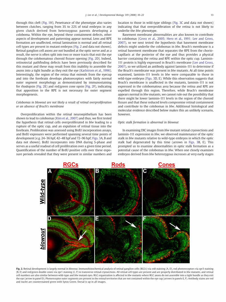
through this cleft (Fig. 1H). Penetrance of the phenotype also variesbetween clutches, ranging from 3% to 22% of total embryos in anygiven clutch derived from heterozygous parents developing acoloboma. Within the eye, beyond these containment defects, otheraspects of development and patterning appear normal. Lens and RPEformation are unaffected, retinal lamination is normal and all retinalcell types are present in mutant embryos (Fig. 2 and data not shown).Retinal ganglion cell axons are not bundled at the optic nerve and as aresult, the nerve is often split into two or more tracts that exit the eyethrough the colobomatous choroid fissure opening (Fig. 2D). Indeed,retinotectal pathfinding defects have been previously described forthis mutant and these may result from this inability to assemble theaxons into a tight bundle as they exit the eye (Karlstrom et al., 1996).Interestingly, the region of the retina that extends from the eyecupand into the forebrain develops photoreceptors with fairly normalouter segment morphology as demonstrated by immunostainingfor rhodopsin (Fig. 2E) and red/green cone opsin (Fig. 2F), indicatingthat apposition to the RPE is not necessary for outer segmentmorphogenesis.
Colobomas in blowout are not likely a result of retinal overproliferationor an absence of Bruch’s membrane
Overproliferation within the retinal neuroepithelium has beenshown to lead to colobomas (Kim et al., 2007) and thus, we first testedthe hypothesis that retinal cells overproliferated in blw leading to arupture of the optic cup, and an expulsion of retinal tissue into theforebrain. Proliferation was assessed using BrdU incorporation assays,and BrdU exposures were performed spanning several time points ofdevelopment (e.g. 24–36 hpf, 42–48 hpf and 72–96 hpf; Figs. 3A, B anddata not shown). BrdU incorporates into DNA during S-phase andserves as a useful readout of cell proliferation over a given time period.Quantification of the number of BrdU positive cells over these expo-sure periods revealed that they were present in similar numbers and
Fig. 2. Retinal development is largely normal in blowout. Immunohistochemical analysis of r(B, E) and red/green double cones via zpr1 staining (C, F) in transverse retinal cryosections. Acell numbers are also similar betweenwild-type and blwmutant eyes. RGC organization is afthe eye (arrow in panel D). Photoreceptor outer segments are present in the retinal territoriesand nuclei are counterstained green with Sytox Green. Dorsal is up in all images.
location to those in wild-type siblings (Fig. 3C and data not shown)indicating that that overproliferation of the retina is not likely tounderlie the blw phenotype.
Basement membrane abnormalities are also known to contributeto colobomas (Gross et al., 2005; Hero et al., 1991; Lee and Gross,2007), so we next tested the hypothesis that basement membranedefects might underlie the colobomas in blw. Bruch’s membrane is aretinal basement membrane that separates the RPE from the chorio-capillaris at the posterior of the eye and thus provides a physicalbarrier containing the retina and RPE within the optic cup. Laminin-111 protein is highly expressed in Bruch’s membrane (Lee and Gross,2007), so we utilized an antibody against laminin-111 to determine ifthe Bruch’s membrane was present in blw mutants. At all time pointsexamined, laminin-111 levels in blw were comparable to those inwild-type embryos (Figs. 3D, E). While this observation suggests thatBruch’s membrane is unaffected in the mutants, laminin-111 is notexpressed in the colobomatous area because the retina and RPE areexpelled through this region. Therefore, while Bruch’s membraneappears normal in blwmutants, we cannot rule out the possibility thatthere might be lower laminin-111 levels in the region of the choroidfissure and that these reduced levels compromise retinal containmentand contribute to the colobomas in blw. Additional histological andmolecular evidence described below makes this an unlikely scenario,however.
Optic stalk formation is abnormal in blowout
In examining DIC images from blwmutant retinal cryosections andlaminin-111 expression in blw, we observed maintenance of the opticstalk in blw mutants relative to wild-type embryos in which the opticstalk had degenerated by this time (arrows in Figs. 3B, E). Thisprompted us to examine abnormalities in optic stalk formation as apotential cause of the colobomas in blw. When one closely examinesembryos derived from blw heterozygous incrosses at very early stages
etinal ganglion cells (RGCs) via zn8 staining (A, D), rod photoreceptors via zpr3 stainingll retinal cell types are present and are properly distributed in blw mutants, and retinalfected in blwmutants where RGC axons do not assemble into a tight bundle as they exitthat are not contained within the eye cup (arrows in panels E, F). Antibody stains are red
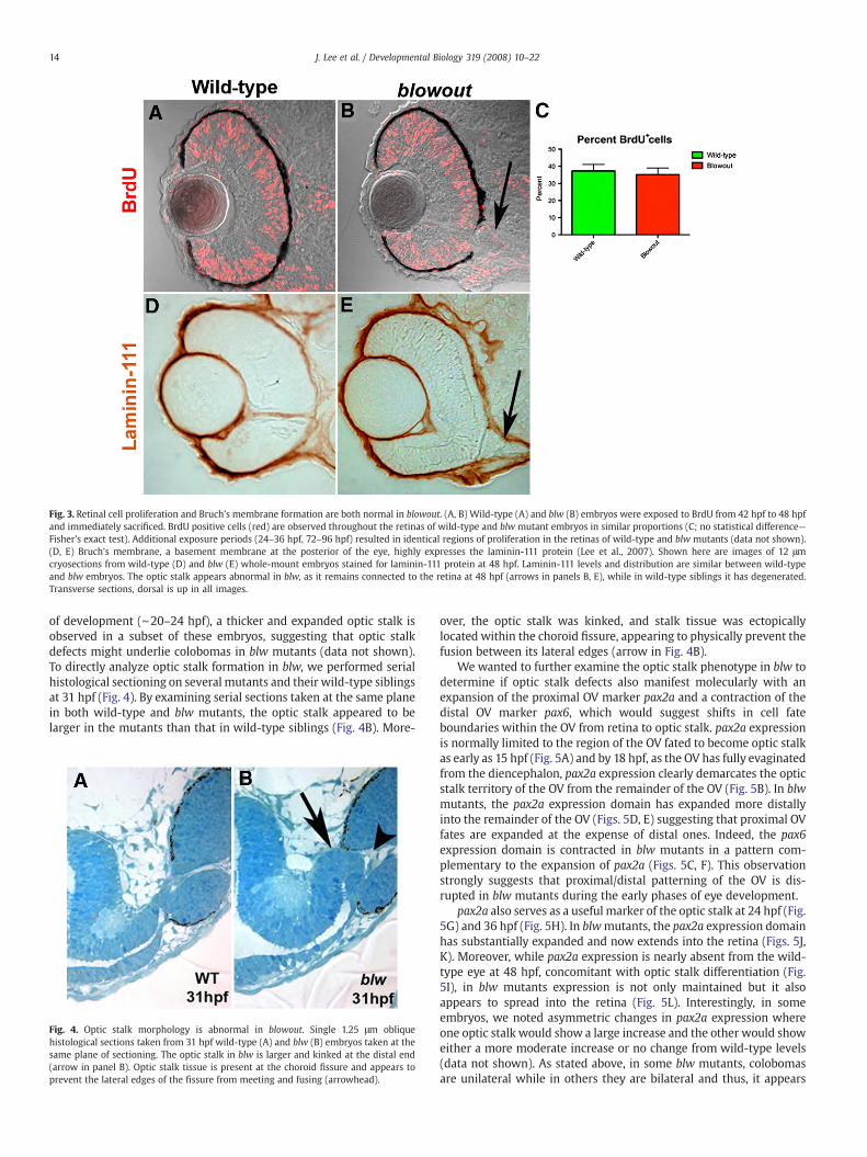
Fig. 3. Retinal cell proliferation and Bruch’s membrane formation are both normal in blowout. (A, B) Wild-type (A) and blw (B) embryos were exposed to BrdU from 42 hpf to 48 hpfand immediately sacrificed. BrdU positive cells (red) are observed throughout the retinas of wild-type and blw mutant embryos in similar proportions (C; no statistical difference—Fisher’s exact test). Additional exposure periods (24–36 hpf, 72–96 hpf) resulted in identical regions of proliferation in the retinas of wild-type and blw mutants (data not shown).(D, E) Bruch’s membrane, a basement membrane at the posterior of the eye, highly expresses the laminin-111 protein (Lee et al., 2007). Shown here are images of 12 μmcryosections from wild-type (D) and blw (E) whole-mount embryos stained for laminin-111 protein at 48 hpf. Laminin-111 levels and distribution are similar between wild-typeand blw embryos. The optic stalk appears abnormal in blw, as it remains connected to the retina at 48 hpf (arrows in panels B, E), while in wild-type siblings it has degenerated.Transverse sections, dorsal is up in all images.
14 J. Lee et al. / Developmental Biology 319 (2008) 10–22
of development (∼20–24 hpf), a thicker and expanded optic stalk isobserved in a subset of these embryos, suggesting that optic stalkdefects might underlie colobomas in blw mutants (data not shown).To directly analyze optic stalk formation in blw, we performed serialhistological sectioning on several mutants and their wild-type siblingsat 31 hpf (Fig. 4). By examining serial sections taken at the same planein both wild-type and blw mutants, the optic stalk appeared to belarger in the mutants than that in wild-type siblings (Fig. 4B). More-
Fig. 4. Optic stalk morphology is abnormal in blowout. Single 1.25 μm obliquehistological sections taken from 31 hpf wild-type (A) and blw (B) embryos taken at thesame plane of sectioning. The optic stalk in blw is larger and kinked at the distal end(arrow in panel B). Optic stalk tissue is present at the choroid fissure and appears toprevent the lateral edges of the fissure from meeting and fusing (arrowhead).
over, the optic stalk was kinked, and stalk tissue was ectopicallylocated within the choroid fissure, appearing to physically prevent thefusion between its lateral edges (arrow in Fig. 4B).
We wanted to further examine the optic stalk phenotype in blw todetermine if optic stalk defects also manifest molecularly with anexpansion of the proximal OV marker pax2a and a contraction of thedistal OV marker pax6, which would suggest shifts in cell fateboundaries within the OV from retina to optic stalk. pax2a expressionis normally limited to the region of the OV fated to become optic stalkas early as 15 hpf (Fig. 5A) and by 18 hpf, as the OV has fully evaginatedfrom the diencephalon, pax2a expression clearly demarcates the opticstalk territory of the OV from the remainder of the OV (Fig. 5B). In blwmutants, the pax2a expression domain has expanded more distallyinto the remainder of the OV (Figs. 5D, E) suggesting that proximal OVfates are expanded at the expense of distal ones. Indeed, the pax6expression domain is contracted in blw mutants in a pattern com-plementary to the expansion of pax2a (Figs. 5C, F). This observationstrongly suggests that proximal/distal patterning of the OV is dis-rupted in blw mutants during the early phases of eye development.
pax2a also serves as a useful marker of the optic stalk at 24 hpf (Fig.5G) and 36 hpf (Fig. 5H). In blwmutants, the pax2a expression domainhas substantially expanded and now extends into the retina (Figs. 5J,K). Moreover, while pax2a expression is nearly absent from the wild-type eye at 48 hpf, concomitant with optic stalk differentiation (Fig.5I), in blw mutants expression is not only maintained but it alsoappears to spread into the retina (Fig. 5L). Interestingly, in someembryos, we noted asymmetric changes in pax2a expression whereone optic stalk would show a large increase and the other would showeither a more moderate increase or no change from wild-type levels(data not shown). As stated above, in some blw mutants, colobomasare unilateral while in others they are bilateral and thus, it appears

Fig. 5. The proximal/distal axis of the optic vesicle is disrupted in blowout. pax2a marks the proximal OV (optic stalk) in wild-type embryos at 15 hpf (A), 18 hpf (B), 24 hpf (G) and36 hpf (H), while it is absent at 48 hpf (I) owing to the degeneration of the stalk by this time. Expression in blwmutants is observed in a broader domainwithin the optic stalk regionas early as 15 hpf (B) and this expansion is maintained at 18 hpf (E), 24 hpf (J) and 36 hpf (K, arrow). Expression is not extinguished at 48 hpf and can be observed to extend into theretina (L, arrow). Conversely, pax6marks the distal optic vesicle (retina and RPE) at 18 hpf (C), and expression is substantially contracted within distal optic vesicle of blwmutants (F)in a pattern complementary to the expanded domain of pax2a.
15J. Lee et al. / Developmental Biology 319 (2008) 10–22
that this asymmetry manifests at the molecular level as well as themorphological.
Positional cloning of blowout
To enable a molecular characterization of blw and its optic stalkphenotypes, we next set out to positionally clone the gene and identifythe responsible mutation. Linkage mapping with microsatellitemarkers placed the mutation on chromosome 2, between Z11410 andZ13521. High-resolution mapping was then performed using addi-
tional microsatellite markers to refine the blw locus. Using markersz8451 and CH211-261P7-SSR2, and 526 blw mutant embryos, wedefined a critical interval that must contain the blw locus (Fig. 6A).Assembly of BACs spanning this region identified 10 genes and 1pseudogene within it (Fig. 6A and data not shown). cDNAs represent-ing six of these genes were cloned and sequenced and, interestingly,separate mutations in two different genes were identified in all 526blw mutant embryos.
The first mutation identified was a missense mutation in atp6v0b(NM_199561) that changed bp 520 from T-to-A, resulting in a non-

Fig. 6. Positional cloning of the blowoutmutation. (A) Genetic map of Chromosome 2 containing the blwmutation. Microsatellite markers and BACs are indicated with the number ofrecombinants and embryos genotyped. (B) Sequence chromatogram showing the atp6v0bmutation in blw. DNA sequence fromwild-type (left) and homozygous (right) blwmutantsare shown and the affected base is marked by an arrow. Amino acid sequence is listed below the DNA sequence. (C). Asn113, changed to Lys in blw (red), is conserved in vertebrateatp6v0b proteins. (D) Sequence chromatogram showing the ptc1 mutation in blw. DNA sequence from heterozygous (left) and homozygous (right) blw mutants are shown and theaffected base is marked by an arrow. (E) Schematic of the patched1 protein and the approximate position of the stop codon in blw situated after the 8th transmembrane domain.
16 J. Lee et al. / Developmental Biology 319 (2008) 10–22
conservative Asn to Lys change at position 113 of the protein (Fig. 6B;www.ensembl.org/Danio_rerio). atp6v0b encodes a 22 kDa proteoli-pid subunit (v0c″) of the vacuolar ATPase complex (v-ATPase). The v-ATPase is a multi-protein complex consisting of 13 different subunittypes that are assembled stoichiometrically into a multimeric proteincomplex (Nishi and Forgac, 2002). v-ATPases are best known for theirroles in H+ transport through which they are important for intra-cellular and extracellular acidification events, protein transport andmembrane fusion (Nelson and Harvey, 1999; Nishi and Forgac, 2002).Additionally, several recent studies have identified v-ATPase complex-independent functions for individual v-ATPase subunits (Hiesinger etal., 2005; Kontani et al., 2005), and perhaps the most interesting ofthese is in C. elegans where v0 subunits have been shown to be in-volved in mediating the release of Hedgehog-like ligands (Liegeois etal., 2006). The v0c″ protein is highly conserved in eukaryotes andprokaryotes and Asn113 is also conserved in vertebrate v0c″ proteins,suggesting that it may be important for protein function (Fig. 6C).
The secondmutationwe identified in blwwas a nonsensemutationin patched1 (ptc1; NM_130988) that changes bp3119 from G-to-A,resulting in a premature stop codon at position 1040 of the Patched1protein (Fig. 6D; http://www.ncbi.nlm.nih.gov/entrez). This truncatesPatched1 just after the 8th transmembrane helix (Fig. 6E). Patched1 is a
receptor for Hh ligands and in the unoccupied state, it serves as anegative regulator of Hh signaling by inhibiting the Smoothened pro-tein. Upon bindingof aHh ligand byPatched, inhibition of Smoothenedis relieved and aHh-dependent intracellular signaling pathway begins.Two patched genes are found in zebrafish, ptc1 and ptc2 (Concordet etal., 1996; Lewis et al., 1999), and ptc1 is strongly expressed in the opticstalk during the stages of OVmorphogenesis (Lewis et al.,1999). Oculardefects have not been described in ptc1morphants (Beales et al., 2007;Wolff et al., 2003), and OV morphogenesis does not appear to bedisrupted in the zebrafish ptc2 mutant, leprechaun (Koudijs et al.,2005).
Loss of patched1 function is responsible for the blowout phenotype
ptc1 is expressed in the zebrafish optic stalk (Figs. 9A, C) (Lewis etal.,1999), and gene expression therein is known to be dependent onHhsignaling (Macdonald et al., 1995; Take-uchi et al., 2003)making ptc1 alikely candidate for underlying the blowout phenotype. Indeed, thatweobserve Hh overexpression-like phenotypes in blw (e.g. expansion ofpax2a expression at the expense of pax6 expression) supports thehypothesis that loss of ptc1 function may underlie these phenotypes.To determine if loss of ptc1 function leads to colobomas, we injected a

17J. Lee et al. / Developmental Biology 319 (2008) 10–22
translation blocking MO targeting the ptc1 transcript into 1-cellembryos (after Wolff et al., 2003). Knock-down of ptc1 translationresulted in severe colobomas in all injected embryos, as well asadditional overt phenotypes in the eye, brain and muscles (Figs. 7A, Banddata not shown). Histological analysis of ptc1morphants confirmedthe degree to which the eye is affected in these embryos (Figs. 7C–E).Colobomas in ptc1 morphants were often more severe than thoseobserved in blw and the eyes of ptc1 morphants are rotated laterallyrelative to wild-type embryos, and their overall position is displacedmedially, likely owing to the substantial coloboma and ventral retinadefects. Many ptc1 mutants also showed small, displaced lenses, aphenotype that does not appear in blwmutants. The lens phenotype inptc1 morphants is identical, however, to that reported to occur as aresult of Shh overexpressionwhere lens tissue is substantially reducedor absent (Barth and Wilson, 1995; Dutta et al., 2005; Yamamoto et al.,
Fig. 7. Loss of ptc1 leads to colobomas and ventral optic cup defects. 1.3 ng injection of a traLateral views of a (A) Control morpholino (ControlMO) injected and (B) ptc1 morphant at 2severely affected than blw mutants, showing medial displacement and lateral flexure of theTransverse histological sections from a 5 dpf wild-type (C) and ptc1morphants (D, E). Severe cthe lens is also displaced and lies out of the plane of the section. pax2a expression is expandgenes ptc1 (J, K) and ptc2 (L, M) also show expanded domains of expression in ptc1 morpha
2004). Unfortunately, rescue experiments via injection of full-lengthptc1 mRNA into blw mutants and/or ptc1 morphants were notsuccessful, possibly owing to the large size of the ptc1 transcript and/or degradation of the mRNA once injected (data not shown). Thus, thebiological relevance of the increased severity of the ptc1 morphantphenotype remains uncertain. However, and of particular note,molecular changes in ptc1 morphants are identical to those observedin blw mutants where pax2a expression is expanded and extends intothe retina (Figs. 7F, G) and pax6 expression is contracted (Figs. 7H, I).These results support the hypothesis that loss of ptc1 function is verylikely to be the underlying cause of the colobomas in blw.
We also wanted to determine if atp6v0bN113K was causative or if itcontributed to the colobomas observed in blw. As mentioned above,v0 subunits have been shown to be involved in mediating the releaseof Hedgehog-like ligands in C. elegans (Liegeois et al., 2006). Changes
nslation blocking morpholino targeting ptc1 transcripts (ptc1MO) results in colobomas..5 dpf. Morphants possess bilateral colobomas (arrow in panel B). Morphants are moreeyes, and lens displacement. Overt brain and muscle defects are also observed. (C–E)olobomas are obvious in themorphants (arrows in panels D, E), and in themany of themed into the retina at 24 hpf (F, G) and pax6 expression is contracted (H, I). The Hh targetnts.
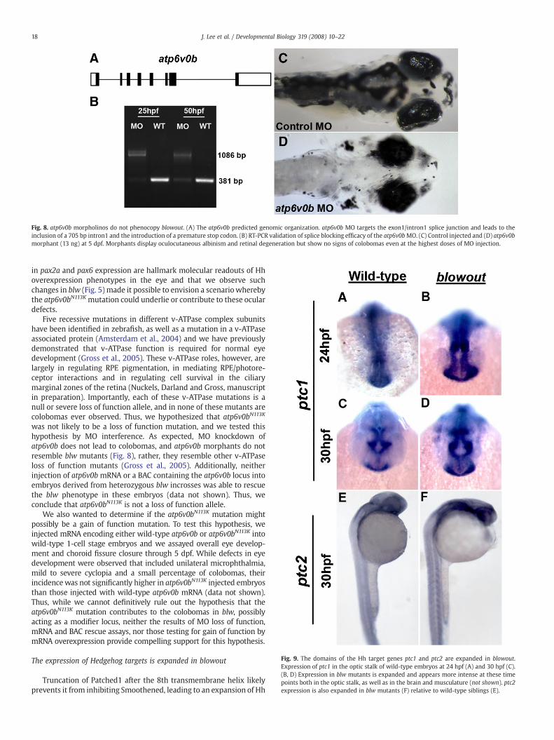
Fig. 8. atp6v0b morpholinos do not phenocopy blowout. (A) The atp6v0b predicted genomic organization. atp6v0b MO targets the exon1/intron1 splice junction and leads to theinclusion of a 705 bp intron1 and the introduction of a premature stop codon. (B) RT-PCR validation of splice blocking efficacy of the atp6v0bMO. (C) Control injected and (D) atp6v0bmorphant (13 ng) at 5 dpf. Morphants display oculocutaneous albinism and retinal degeneration but show no signs of colobomas even at the highest doses of MO injection.
Fig. 9. The domains of the Hh target genes ptc1 and ptc2 are expanded in blowout.Expression of ptc1 in the optic stalk of wild-type embryos at 24 hpf (A) and 30 hpf (C).(B, D) Expression in blw mutants is expanded and appears more intense at these timepoints both in the optic stalk, as well as in the brain and musculature (not shown). ptc2expression is also expanded in blw mutants (F) relative to wild-type siblings (E).
18 J. Lee et al. / Developmental Biology 319 (2008) 10–22
in pax2a and pax6 expression are hallmark molecular readouts of Hhoverexpression phenotypes in the eye and that we observe suchchanges in blw (Fig. 5) made it possible to envision a scenariowherebythe atp6v0bN113K mutation could underlie or contribute to these oculardefects.
Five recessive mutations in different v-ATPase complex subunitshave been identified in zebrafish, as well as a mutation in a v-ATPaseassociated protein (Amsterdam et al., 2004) and we have previouslydemonstrated that v-ATPase function is required for normal eyedevelopment (Gross et al., 2005). These v-ATPase roles, however, arelargely in regulating RPE pigmentation, in mediating RPE/photore-ceptor interactions and in regulating cell survival in the ciliarymarginal zones of the retina (Nuckels, Darland and Gross, manuscriptin preparation). Importantly, each of these v-ATPase mutations is anull or severe loss of function allele, and in none of these mutants arecolobomas ever observed. Thus, we hypothesized that atp6v0bN113K
was not likely to be a loss of function mutation, and we tested thishypothesis by MO interference. As expected, MO knockdown ofatp6v0b does not lead to colobomas, and atp6v0b morphants do notresemble blw mutants (Fig. 8), rather, they resemble other v-ATPaseloss of function mutants (Gross et al., 2005). Additionally, neitherinjection of atp6v0b mRNA or a BAC containing the atp6v0b locus intoembryos derived from heterozygous blw incrosses was able to rescuethe blw phenotype in these embryos (data not shown). Thus, weconclude that atp6v0bN113K is not a loss of function allele.
We also wanted to determine if the atp6v0bN113K mutation mightpossibly be a gain of function mutation. To test this hypothesis, weinjected mRNA encoding either wild-type atp6v0b or atp6v0bN113K intowild-type 1-cell stage embryos and we assayed overall eye develop-ment and choroid fissure closure through 5 dpf. While defects in eyedevelopment were observed that included unilateral microphthalmia,mild to severe cyclopia and a small percentage of colobomas, theirincidencewas not significantly higher in atp6v0bN113K injected embryosthan those injected with wild-type atp6v0b mRNA (data not shown).Thus, while we cannot definitively rule out the hypothesis that theatp6v0bN113K mutation contributes to the colobomas in blw, possiblyacting as a modifier locus, neither the results of MO loss of function,mRNA and BAC rescue assays, nor those testing for gain of function bymRNA overexpression provide compelling support for this hypothesis.
The expression of Hedgehog targets is expanded in blowout
Truncation of Patched1 after the 8th transmembrane helix likelyprevents it from inhibiting Smoothened, leading to an expansion of Hh

Fig. 10. Cyclopamine suppression of colobomas in blowout. (A) On average, 89.8% ofvehicle (EtOH) treated embryos derived from homozygous incrosses displayedcolobomas, while exposure of siblings to 2 μM or 3 μM cyclopamine from 5.5 hpf to24 hpf suppressed colobomas to 26.7% and 16.7%, respectively (⁎⁎⁎pb0.0001, Fisher’sexact test). (B) Treatment of embryos derived from homozygous incrosses to vehicle(EtOH), or 3 μM cyclopamine from 5.5 hpf to 13 hpf, or 13 hpf to 24 hpf was also able tosuppress colobomas from an average of 82% in vehicle controls to 4% in 5.5–13 hpftreated embryos (⁎⁎⁎pb0.0001, Fisher’s exact test), and to 65.3% in 13 hpf–24 hpftreated embryos (⁎p=0.0278, Fisher's exact test).
19J. Lee et al. / Developmental Biology 319 (2008) 10–22
target gene expression in blw mutants. This model is consistent withpublished reports demonstrating that Hh overexpression alters geneexpression in the optic stalk and optic cup in zebrafish (Ekker et al.,1995), and consistent with our observations that the expressiondomain of the Shh target pax2a is expanded in blw mutants and ptc1morphants, while the pax6 domain is contracted (Figs. 5, 7). ptc1 andptc2 are also targets of Hh pathway activation (Lewis et al., 1999;Concordet et al., 1996), likely acting in a negative feedbackmechanismto limit the diffusion of the Hh morphogen and thus limit the range ofHh signaling (Chen and Struhl, 1996). To directly assess whether ptc1and/or ptc2 expression is altered in blw, we assayed their distributionsby in situ hybridization (Fig. 9). In wild-type embryos, ptc1 is stronglyexpressed in the optic stalk at 24 and 30 hpf (Figs. 9A, C), as well asthroughout the ventral brain and the somites (Concordet et al., 1996,and data not shown). In blw mutants, ptc1 expression appears to beexpanded and the message is distributed in a substantially broaderdomain of expression (Figs. 9B, D). Expanded domains of expressionwere also observed in the ventral brain and the somites (data notshown). Similarly, expansion of ptc1 expression was also observed inptc1 morphants (Figs. 7J, K). Examination of ptc2 expression in blwmutants yielded similar results (Figs. 9E, F), although changes in ptc2distribution were variable in blw mutants with only a subset (∼40%)showing grossly obvious changes. ptc2 expression was substantiallyaltered in all ptc1 morphant embryos, however (Figs. 7L, M). Wehighlight that while in situ hybridizations are not quantitative assays,that the ptc1 and ptc2 in situ signals appear to be more intense andexpanded in the brain and somites in both blw mutant and ptc1morphant embryos strongly suggest that Hh pathway activity isupregulated as a result of loss of Patched1 function.
Homozygous blowout mutants are viable and reveal post-metamorphicroles for Patched1 function in the adult zebrafish
We were able to rear ∼2–3% of homozygous blw mutant embryosto adulthood and the resulting fish displayed a number of obviousmorphological defects (Supplemental Fig. 1). blw homozygotes weregenerally smaller in size than their heterozygous and wild-typesiblings. Males and females were recovered in approximately equalnumbers and fertility was normal in the homozygous mutant fish.Embryos derived from homozygous mothers continued to showvariable rates of phenotypic penetrance, never reaching predictedMendelian ratios. On average, the incidence of colobomas wasapproximately 35% when a homozygous female was mated with aheterozygous male, and incidences rose to over 80% when ahomozygous female was mated to a homozygous male. In nearly allmutants derived from homozygous mothers that displayed colobo-mas, these were almost always observed bilaterally. Importantly,beyond this difference, blw mutants derived from homozygousmothers did not show markedly more severe or more widespreaddevelopmental defects than those derived from heterozygousmothers, indicating that there is not likely to be a significant maternalmRNA or protein rescue of the blw phenotype that masks defects inembryos derived from heterozygous carriers.
Is upregulation of Hh pathway activity the mechanism that leads tocolobomas in blowout?
Given the similarities between Hh overexpression and the blwmutation (e.g. changes to pax2a, pax6, ptc1 and ptc2 expression), wenext sought to directly test the hypothesis that upregulation of Hhpathway activity is the molecular mechanism underlying colobomasin blw. To test this hypothesis, we utilized cyclopamine, a pharmaco-logical inhibitor of the Hh pathway that acts downstream of Patched(Cooper et al., 1998; Taipale et al., 2002), and asked whether low dosesof cyclopamine were capable of suppressing colobomas in blwmutants. Indeed, a similar rescue paradigm has been successfully
utilized for zebrafish mutations in other negative regulators of the Hhpathway (e.g. lep/ptc2 and uki/Hip; (Koudijs et al., 2005). For theseassays, we took advantage of homozygous viable blw adults toincrease the incidence of coloboma phenotypes and to abrogate theneed to genotype each rescued embryo as we knew a priori that allwere homozygousmutants. Low levels of cyclopamine (≤3 μM) did notlead to noticeable defects in eye development (data not shown) andthus, we assayed whether these subthreshold levels were able tosuppress colobomas in blw. Exposure of blw mutants to 2 μM or 3 μMof cyclopamine between 5.5 hpf and 24 hpf was highly effective insuppressing the incidence of colobomas (Fig. 10A). In vehicle treatedsibling controls, 89.8% displayed colobomas while 2 μM cyclopaminesuppressed this to 26.7% and 3 μM cyclopamine to 16.7% (pb0.0001;Fisher’s exact test).
We next sought to determine if we could identify awindow of timebetween 5.5 hpf and 24 hpf during which maximal rescue ofcolobomas in blw mutants could be achieved, thereby indicating thewindow of time when Hh signaling is likely to be required forproximal/distal patterning of the OV during normal embryogenesis.Given that 3 μM cyclopamine was able to significantly rescuecolobomas in blw mutants (Fig. 10A), we utilized this concentrationand exposed embryos over two time windows: 5.5 hpf–13 hpf and13 hpf–24 hpf (Fig.10B). Maximal rescuewas achieved by cyclopamineexposure during the early 5.5 hpf–13 hpf window with incidences ofcolobomas dropping from 82% to 4% (pb0.0001; Fisher’s exact test).Cyclopamine exposure from 13 hpf–24 hpf resulted in 65.3% ofembryos displaying colobomas (p=0.0278; Fisher’s exact test). Whilethe rescue from 13 hpf to 24 hpf cyclopamine exposure is statisticallysignificant when compared to vehicle treated controls, it is substan-tially lower than that achieved during the early 5.5 hpf–13 hpfwindow of treatment suggesting that it is during this early timewindow that the Hh signal is normally conveyed to the OV tosegregate it into the appropriate proximal and distal territories.

20 J. Lee et al. / Developmental Biology 319 (2008) 10–22
Discussion
Blowout is a loss of function mutation in Patched1
In this study, we have characterized zebrafish blw, a recessivemutation that presents with ocular coloboma and defects in retino-tectal axon pathfinding (Karlstrom et al., 1996). Positional cloning ofblw identified two mutations; the first of these affects ptc1, resultingin a stop codon that truncates the Patched1 protein after the 8thtransmembrane domain, and the second affects atp6v0b, changing aconserved asparagine residue to a lysine. MO targeting of ptc1 trans-cripts results in colobomas while MO targeting of atp6v0b transcriptsdid not lead to colobomas; rather, loss of atp6v0b function resulted inoculocutaneous albinism and retinal degeneration. These phenotypeswere identical to those observed in six other loss of function muta-tions in v-ATPase components or v-ATPase associated proteins,suggesting that the atp6v0bN113K mutation was not a loss of functionallele (Gross et al., 2005; Nuckels et al., in preparation). Indeed, neitheratp6v0b mRNA nor BAC injections were able to rescue or alter theocular defects in blw mutants. Additionally, overexpression ofatp6v0bN113K mRNA did not lead to ocular defects at a higher levelthan that resulting from overexpression of wild-type atp6v0b mRNA,also suggesting that the atp6v0bN113K mutation was not a neomorphicor gain of function allele. Thus, whilewe cannot exclude the possibilitythat atp6v0bN113K acts as a modifier of ptc1W1040X, our results support amodel in which the coloboma phenotypes in blw stem solely from aloss of Patched1 function.
Interestingly, ptc1 morphants showed more severe ocular pheno-types than those observed in blw mutants. We were able to rearhomozygous mutants to adulthood and embryos derived fromincrosses between homozygous parents did not display more severeocular phenotypes than those derived from heterozygous parents.This indicates that there is not a maternal supply of mRNA or proteinthat partially rescues the ocular phenotypes in blw mutants derivedfrom heterozygous mothers. Phenotypic penetrance varies in clutchesof embryos derived from heterozygous parents and while thephenotypic penetrance was higher in clutches derived from homo-zygous parents, they still never reached predicted ratios. Theseobservations in conjunction with the more severe ocular phenotypespresent in ptc1 morphants suggest that the ptcW1040X mutation maybe a hypomorphic partial loss of function ptc1 allele.
While direct biochemical analysis of the truncated Ptc1W1040X
proteinwill be required to test this hypothesis, should it retain some ofits ability to repress Smoothened, it would be interesting with respectto other mutations that have been identified in the C-terminus of thePatched1 protein. For example, two missense mutations in the humanPATCHED1 (PTCH) gene have been identified that alter the C-terminaldomain of PTCH (Ming et al., 2002). The first of these mutationsresides in the extracellular loop between the 7th and 8th transmem-brane domains of PTCH (PTCHT728M) and the second resides in theintracellular loop between the 8th and 9th transmembrane domainsof PTCH (PTCHT1052M). Patients with these mutations display holo-prosencephaly and other developmental defects, but they do notdisplay colobomas. Interestingly, their phenotypes more closelyresemble loss of function mutations in SONIC HEDGEHOG (SHH) thanloss of function mutations in PTCH. The biochemical nature of thesehuman mutations has not yet been characterized but given thesimilarity in phenotypes between them and SHHmutations, it may bethat they disrupt the portion of the protein that is required for theinactivation of PTCH function in the presence of SHH. Thus, PTCHT728M
and PTCHT1052M may act as a dominant negative such that even in thepresence of SHH, thesemutated proteins still represses SMOOTHENEDactivity and keep the Hh pathway inactive.
By comparison, truncation of the zebrafish Ptc1 protein at aminoacid 1040, between the 8th and 9th intracellular loops, behaves quitedifferently than these point mutations. In blw/ptc1 mutants, the Shh
pathway appears to be constitutively active although with variablepenetrance and relativelymilder phenotypeswhen compared to thosethat arise from Shh overexpression (Ekker et al., 1995). It is possiblethat the remaining extracellular loop between the 7th and 8thtransmembrane domains still retains some of its repressive ability toblock Smoothened functionwhen Hh signals are present and thus it isable to ameliorate some of the phenotypes that would be expectedto arise if the Hh pathway was fully activated (i.e. to resemble Shhoverexpression phenotypes). It is also possible that there are otherredundant negative regulators of the Hh pathway that prevent fullactivation of the pathway in blw/ptc1 mutants, which thereby in-fluence the penetrance of the phenotype. Ptc2 is an ideal candidate topossess a redundant function and indeed, a recent report by Koudijset al. (2008) demonstrates that the ocular phenotypes in ptc1/ptc2double mutants are much more similar to Shh overexpressionphenotypes than those present in the single mutants.
Ptc1-dependent regulation of Hh signaling is required for formation ofthe optic stalk-retina interface
Shh signaling is a key regulator of proximal OV cell fates (Amato etal., 2004). pax2a, a Shh target, has been shown to directly regulateoptic stalk and choroid fissure formation in the proximal OV, whilepax6 encodes a key regulatory factor involved in the specification ofretina and RPE cell fates within the distal OV (Chow and Lang, 2001;Adler and Canto-Soler, 2007). In the blw mutant OV, the early domainof pax2a expression is expanded distally and pax6 expression iscontracted (Fig. 5). These results support a model in which proximalOV cell fates are expanded in blw mutants at the expense of distalones, and this leads to an enlargement of the optic stalk. Indeed, thesechanges in gene expression in blw are identical to those observed fromShh overexpression in zebrafish, Xenopus and chick (Ekker et al., 1995;Macdonald et al., 1995; Perron et al., 2003; Sasagawa et al., 2002;Zhang and Yang, 2001). One problemwith this model, however, is thatdespite the optic stalk expansion and coloboma phenotypes, lateraspects of distal OV (i.e. retinal) formation are normal in blwmutants.Onewould expect that if early distal OV patterning was compromised,later retinal and RPE fates would necessarily be affected. This is not thecase in blwmutants; retinal patterning is unaffected and overall retinasize is similar to that in wild-type siblings. While we do not yet knowhow distal fates recover in blw, one possible mechanism is that theremaining pax6 expressing distal OV is able to utilize pax6 in a non-cell autonomous fashion (e.g. Lesaffre et al., 2007), and therebycounteract the effects of the expanded proximal pax2a expressingregions. In this scenario, the optic stalk would be refractory to this“rescue” and thus still expand into the choroid fissure, but theremainder of the distal OV is able to develop relatively normally.Additional analyses are required to test this hypothesis and todetermine why blw mutants do not display distal OV defects despitethe early changes in gene expression domains observed within the OVat 18 hpf.
Colobomas in blw were suppressed by pharmacological inhibitionof Hh pathway activity indicating that it is the dysregulation of thispathway that underlies the coloboma phenotype. We suspect thatcolobomas result from the expansion of the optic stalk into the choroidfissure which prevents its lateral edges from meeting and fusing;however, it is also possible that overproliferation of cells within theoptic stalk itself contributes to these colobomas by further expandingthe optic stalk into the ventral optic cup, and thus impeding its closure.Hh signaling is known to regulate cell proliferation in a number ofcellular and developmental contexts, including the eye, through theactivation of key cell cycle regulators like cyclin D1, cyclin B1 and cyclinA2, as well as the phosphatase, Cdc25, that collectively function tomaintain proliferative cells in the cell cycle (Adolphe et al., 2006;Agathocleous et al., 2007; Locker et al., 2006). In blw, an Hh-dependentdysregulation of cell proliferation may also be occurring within the
21J. Lee et al. / Developmental Biology 319 (2008) 10–22
proximal OV which increases the amount of stalk tissue and therebyphysically impedes closure of the choroid fissure. It will be interestingin future studies to determine if proliferation and the expression ofthese cell cycle components are upregulated in the optic stalk of blwmutants.
Hh signaling is required for later aspects of optic stalk develop-ment in mouse whereby retinal ganglion cell derived Shh is requiredfor optic stalk neuroepithelial cells to develop into astroglia, as well asto suppress pigment cell formation around the optic nerve (Dakubo etal., 2003; Torres et al., 1996). We observed continued expression ofpax2a mRNA in the optic stalk and ectopically in the retina of blwmutants when compared to that in wild-type siblings, where ex-pression had ceased and differentiation commenced by 48 hpf. Be-yond the coloboma inducedmorphological abnormalities in the retinaof blw mutants, histologically and immunohistochemically theirretinas appear relatively normal. That said, we do not know the effectof the mutation on later aspects of optic stalk development, as wehave not yet assayed the differentiated state of the optic stalk in blwmutants. Based on the results in mouse, one might hypothesize thatneuroepithelial cells of the blw optic stalk would remain undiffer-entiated due to the prolonged expression of these specification genes.Conversely, one might also hypothesize that the optic stalk in blwmight differentiate but that glial cells would be overrepresented as aresult of dysregulated Hh target gene expression. blw was originallyidentified based on defects in retinotectal pathfinding (Karlstrom etal., 1996), and in either of the above scenarios, retinal ganglion cellaxons would likely not properly find their way to the optic nerve and/or the optic tectum. Future studies will be required to examine thesepossibilities and determine the molecular basis for the pathfindingdefects in blw.
Finally, underscoring the importance of Patched-dependentregulation of Hh signaling in human disease, loss of function muta-tions in human PTCH lead to Basal Cell Naevus or Gorlin syndrome(BCNS; OMIM #109400), a developmental disorder characterized bydental, skeletal and ocular defects (Hahn et al., 1996; Johnson et al.,1996). Ocular phenotypes associated with BCNS include abnormalmyelination of the optic nerve, retinal dysplasia and colobomas;however, themechanism throughwhich PTCHmutations lead to theseocular defects has not been established (Black et al., 2003; De Jong etal., 1985; Manners et al., 1996). Our results demonstrate a critical rolefor zebrafish Patched1 in negatively regulating Hedgehog signalingwithin the proximal OV and possibly in functioning to restrict Hh-dependent cell proliferation therein. BCNS-related pathologies mayresult from unrestricted proliferation within the retina, a modelrecently proposed by Black et al. (2003). That a subset of homozygousadult blwmutants are viable will enable us to test this hypothesis andto identify later roles for ptc1-dependent Hh regulation in oculardevelopment and homeostasis, as well as to model other non-ocularBCNS associated pathologies in zebrafish.
Acknowledgments
This work was supported by grants from the Knights TemplarEye Foundation and the E. Matilda Ziegler Foundation for the Blindto J.M.G., NIH RO1 RR020357 to R.G.G. and an undergraduateresearch grant from Harvard University to K.S. We thank Sam Cookefor technical support, and Brian Perkins, Brian Link, John Wall-ingford and members of the Gross Lab for comments on themanuscript. The Zebrafish International Resource Center providedseveral monoclonal antibodies utilized in this study and it issupported by grant P40 RR012546 from the NIH-NCRR.
Appendix A. Supplementary data
Supplementary data associated with this article can be found, inthe online version, at doi:10.1016/j.ydbio.2008.03.035.
References
Adler, R., Canto-Soler, M.V., 2007. Molecular mechanisms of optic vesicle development:complexities, ambiguities and controversies. Dev. Biol. 305, 1–13.
Adolphe, C., Hetherington, R., Ellis, T., Wainwright, B., 2006. Patched1 functions as agatekeeper by promoting cell cycle progression. Cancer Res. 66, 2081–2088.
Agathocleous, M., Locker, M., Harris, W.A., Perron, M., 2007. A general role of hedgehogin the regulation of proliferation. Cell Cycle 6, 156–159.
Amato, M.A., Boy, S., Perron, M., 2004. Hedgehog signaling in vertebrate eye dev-elopment: a growing puzzle. Cell Mol. Life Sci. 61, 899–910.
Amsterdam, A., Nissen, R.M., Sun, Z., Swindell, E.C., Farrington, S., Hopkins, N., 2004.Identification of 315 genes essential for early zebrafish development. Proc. Natl.Acad. Sci. USA 101, 12792–12797.
Barbieri, A.M., Lupo, G., Bulfone, A., Andreazzoli, M., Mariani, M., Fougerousse, F.,Consalez, G.G., Borsani, G., Beckmann, J.S., Barsacchi, G., Ballabio, A., Banfi, S., 1999. Ahomeobox gene, vax2, controls the patterning of the eye dorsoventral axis. Proc.Natl. Acad. Sci. USA 96, 10729–10734.
Barbieri, A.M., Broccoli, V., Bovolenta, P., Alfano, G., Marchitiello, A., Mocchetti, C.,Crippa, L., Bulfone, A., Marigo, V., Ballabio, A., Banfi, S., 2002. Vax2 inactivation inmouse determines alteration of the eye dorsal–ventral axis, misrouting of the opticfibres and eye coloboma. Development 129, 805–813.
Barresi, M.J., Stickney, H.L., Devoto, S.H., 2000. The zebrafish slow-muscle-omitted geneproduct is required for Hedgehog signal transduction and the development of slowmuscle identity. Development 127, 2189–2199.
Barth, K.A., Wilson, S.W., 1995. Expression of zebrafish nk2.2 is influenced by sonichedgehog/vertebrate hedgehog-1 and demarcates a zone of neuronal differentia-tion in the embryonic forebrain. Development 121, 1755–1768.
Beales, P.L., Bland, E., Tobin, J.L., Bacchelli, C., Tuysuz, B., Hill, J., Rix, S., Pearson, C.G., Kai,M., Hartley, J., Johnson, C., Irving, M., Elcioglu, N., Winey, M., Tada, M., Scambler, P.J.,2007. IFT80, which encodes a conserved intraflagellar transport protein, is mutatedin Jeune asphyxiating thoracic dystrophy. Nat. Genet. 39, 727–729.
Bertuzzi, S., Hindges, R., Mui, S.H., O’Leary, D.D., Lemke, G., 1999. The homeodomainprotein vax1 is required for axon guidance and major tract formation in thedeveloping forebrain. Genes Dev. 13, 3092–3105.
Black, G.C., Mazerolle, C.J., Wang, Y., Campsall, K.D., Petrin, D., Leonard, B.C., Damji, K.F.,Evans, D.G., McLeod, D., Wallace, V.A., 2003. Abnormalities of the vitreoretinalinterface caused by dysregulated Hedgehog signaling during retinal development.Hum. Mol. Genet. 12, 3269–3276.
Chang, L., Blain, D., Bertuzzi, S., Brooks, B.P., 2006. Uveal coloboma: clinical and basicscience update. Curr. Opin. Ophthalmol. 17, 447–470.
Chen, Y., Struhl, G., 1996. Dual roles for patched in sequestering and transducingHedgehog. Cell 87, 553–563.
Chiang, C., Litingtung, Y., Lee, E., Young, K.E., Corden, J.L., Westphal, H., Beachy, P.A.,1996.Cyclopia and defective axial patterning in mice lacking Sonic hedgehog genefunction. Nature 383, 407–413.
Chow, R.L., Lang, R.A., 2001. Early eye development in vertebrates. Annu. Rev. Cell Dev.Biol. 17, 255–296.
Concordet, J.P., Lewis, K.E., Moore, J.W., Goodrich, L.V., Johnson, R.L., Scott, M.P., Ingham,P.W., 1996. Spatial regulation of a zebrafish patched homologue reflects the roles ofsonic hedgehog and protein kinase A in neural tube and somite patterning.Development 122, 2835–2846.
Cooper, M.K., Porter, J.A., Young, K.E., Beachy, P.A., 1998. Teratogen-mediated inhibitionof target tissue response to Shh signaling. Science 280, 1603–1607.
Dakubo, G.D., Wang, Y.P., Mazerolle, C., Campsall, K., McMahon, A.P., Wallace, V.A., 2003.Retinal ganglion cell-derived sonic hedgehog signaling is required for optic disc andstalk neuroepithelial cell development. Development 130, 2967–2980.
De Jong, P.T., Bistervels, B., Cosgrove, J., de Grip, G., Leys, A., Goffin, M., 1985.Medullated nerve fibers. A sign of multiple basal cell nevi (Gorlin’s) syndrome.Arch. Ophthalmol. 103, 1833–1836.
Dutta, S., Dietrich, J.E., Aspock, G., Burdine, R.D., Schier, A., Westerfield, M., Varga, Z.M.,2005. pitx3 defines an equivalence domain for lens and anterior pituitary placode.Development 132, 1579–1590.
Ekker, S.C., Ungar, A.R., Greenstein, P., von Kessler, D.P., Porter, J.A., Moon, R.T., Beachy,P.A., 1995. Patterning activities of vertebrate hedgehog proteins in the developingeye and brain. Curr. Biol. 5, 944–955.
Esteve, P., Bovolenta, P., 2006. Secreted inducers in vertebrate eye development: morefunctions for old morphogens. Curr. Opin. Neurobiol. 16, 13–19.
Fitzpatrick, D.R., van Heyningen, V., 2005. Developmental eye disorders. Curr. Opin.Genet. Dev. 15, 348–353.
Goldsmith, P., Harris, W.A., 2003. The zebrafish as a tool for understanding the biologyof visual disorders. Semin. Cell Dev. Biol. 14, 11–18.
Gregory-Evans, C.Y., Williams, M.J., Halford, S., Gregory-Evans, K., 2004. Ocularcoloboma: a reassessment in the age of molecular neuroscience. J. Med. Genet.41, 881–891.
Gross, J.M., Dowling, J.E., 2005. Tbx2b is essential for neuronal differentiation along thedorsal/ventral axis of the zebrafish retina. Proc. Natl Acad. Sci. USA 102, 4371–4376.
Gross, J.M., Perkins, B.D., 2008. Zebrafish mutants as models for congenital oculardisorders in humans. Mol. Reprod. Dev. 75, 547–555.
Gross, J.M., Perkins, B.D., Amsterdam, A., Egana, A., Darland, T., Matsui, J.I., Sciascia, S.,Hopkins, N., Dowling, J.E., 2005. Identification of zebrafish insertional mutants withdefects in visual system development and function. Genetics 170, 245–261.
Hahn, H., Wicking, C., Zaphiropoulous, P.G., Gailani, M.R., Shanley, S., Chidambaram, A.,Vorechovsky, I., Holmberg, E., Unden, A.B., Gillies, S., Negus, K., Smyth, I., Pressman,C., Leffell, D.J., Gerrard, B., Goldstein, A.M., Dean, M., Toftgard, R., Chenevix-Trench,G., Wainwright, B., Bale, A.E., 1996. Mutations of the human homolog of Drosophilapatched in the nevoid basal cell carcinoma syndrome. Cell 85, 841–851.

22 J. Lee et al. / Developmental Biology 319 (2008) 10–22
Hallonet, M., Hollemann, T., Wehr, R., Jenkins, N.A., Copeland, N.G., Pieler, T., Gruss, P.,1998. Vax1 is a novel homeobox-containing gene expressed in the developinganterior ventral forebrain. Development 125, 2599–2610.
Hallonet, M., Hollemann, T., Pieler, T., Gruss, P., 1999. Vax1, a novel homeobox-containing gene, directs development of the basal forebrain and visual system.Genes Dev. 13, 3106–3114.
Heisenberg, C.P., Brennan, C., Wilson, S.W., 1999. Zebrafish aussicht mutant embryosexhibit widespread overexpression of ace (fgf8) and coincident defects in CNSdevelopment. Development 126, 2129–2140.
Hiesinger, P.R., Fayyazuddin, A., Mehta, S.Q., Rosenmund, T., Schulze, K.L., Zhai, R.G.,Verstreken, P., Cao, Y., Zhou, Y., Kunz, J., Bellen, H.J., 2005. The v-ATPase V0 subunita1 is required for a late step in synaptic vesicle exocytosis in Drosophila. Cell 121,607–620.
Hero, I., Farjah, M., Scholtz, C.L., 1991. The prenatal development of the optic fissure incolobomatous microphthalmia. Invest. Ophthalmol. Vis. Sci. 32, 2622–2635.
Johnson, R.L., Rothman, A.L., Xie, J., Goodrich, L.V., Bare, J.W., Bonifas, J.M., Quinn, A.G.,Myers, R.M., Cox, D.R., Epstein Jr., E.H., Scott, M.P.,1996. Human homolog of patched,a candidate gene for the basal cell nevus syndrome. Science 272, 1668–1671.
Jowett, T., Lettice, L., 1994. Whole-mount in situ hybridizations on zebrafish embryosusing a mixture of digoxigenin- and fluorescein-labelled probes. Trends Genet. 10,73–74.
Karlstrom, R.O., Trowe, T., Klostermann, S., Baier, H., Brand, M., Crawford, A.D.,Grunewald, B., Haffter, P., Hoffmann, H., Meyer, S.U., Muller, B.K., Richter, S., vanEeden, F.J., Nusslein-Volhard, C., Bonhoeffer, F., 1996. Zebrafish mutations affectingretinotectal axon pathfinding. Development 123, 427–438.
Karlstrom, R.O., Talbot, W.S., Schier, A.F., 1999. Comparative synteny cloning of zebrafishyou-too: mutations in the Hedgehog target gli2 affect ventral forebrain patterning.Genes Dev. 13, 388–393.
Kim, T.H., Goodman, J., Anderson, K.V., Niswander, L., 2007. Phactr4 regulates neuraltube and optic fissure closure by controlling PP1-, Rb-, and E2F1-regulated cell-cycle progression. Dev. Cell 13, 87–102.
Kontani, K., Moskowitz, I.P., Rothman, J.H., 2005. Repression of cell–cell fusion bycomponents of the C. elegans vacuolar ATPase complex. Dev. Cell 8, 787–794.
Koudijs, M.J., den Broeder, M.J., Keijser, A., Wienholds, E., Houwing, S., van Rooijen, E.M.,Geisler, R., van Eeden, F.J., 2005. The zebrafish mutants dre, uki, and lep encodenegative regulators of the hedgehog signaling pathway. PLoS Genet. 1, e19.
Koudijs, M.J., den Broeder, M.J., Groot, E., van Eeden, F.J., 2008. Genetic analysis of twozebrafish patched homologues identifies novel roles for the hedgehog signalingpathway. BMC Dev. Biol. 8, 15.
Lee, J., Gross, J.M., 2007. Laminin {beta}1 and {gamma}1 containing laminins areessential for basement membrane integrity in the zebrafish eye. Invest. Ophthal-mol. Vis. Sci. 48, 2483–2490.
Lesaffre, B., Joliot, A., Prochiantz, A., Volovitch, M., 2007. Direct non-cell autonomousPax6 activity regulates eye development in the zebrafish. Neural. Develop. 2, 2.
Lewis, K.E., Concordet, J.P., Ingham, P.W., 1999. Characterisation of a second patchedgene in the zebrafish Danio rerio and the differential response of patched genes toHedgehog signalling. Dev. Biol. 208, 14–29.
Liegeois, S., Benedetto, A., Garnier, J.M., Schwab, Y., Labouesse, M., 2006. The V0-ATPasemediates apical secretion of exosomes containing Hedgehog-related proteins inCaenorhabditis elegans. J. Cell Biol. 173, 949–961.
Locker, M., Agathocleous, M., Amato, M.A., Parain, K., Harris, W.A., Perron, M., 2006.Hedgehog signaling and the retina: insights into the mechanisms controlling theproliferative properties of neural precursors. Genes Dev. 20, 3036–3048.
Macdonald, R., Barth, K.A., Xu, Q., Holder, N., Mikkola, I., Wilson, S.W., 1995. Midlinesignalling is required for Pax gene regulation and patterning of the eyes.Development 121, 3267–3278.
Macdonald, R., Scholes, J., Strahle, U., Brennan, C., Holder, N., Brand, M., Wilson, S.W.,1997. The Pax protein Noi is required for commissural axon pathway formation inthe rostral forebrain. Development 124, 2397–2408.
Manners, R.M., Morris, R.J., Francis, P.J., Hatchwell, E., 1996. Microphthalmos inassociation with Gorlin’s syndrome. Br. J. Ophthalmol. 80, 378.
Ming, J.E., Kaupas, M.E., Roessler, E., Brunner, H.G., Golabi, M., Tekin, M., Stratton, R.F.,Sujansky, E., Bale, S.J., Muenke, M., 2002. Mutations in PATCHED-1, the receptor forSONIC HEDGEHOG, are associated with holoprosencephaly. Hum. Genet. 110,297–301.
Mui, S.H., Hindges, R., O’Leary, D.D., Lemke, G., Bertuzzi, S., 2002. The homeodomainprotein Vax2 patterns the dorsoventral and nasotemporal axes of the eye.Development 129, 797–804.
Mui, S.H., Kim, J.W., Lemke, G., Bertuzzi, S., 2005. Vax genes ventralize the embryoniceye. Genes Dev. 19, 1249–1259.
Nakayama, Y., Miyake, A., Nakagawa, Y., Mido, T., Yoshikawa, M., Konishi, M., Itoh, N.,2008. Fgf19 is required for zebrafish lens and retina development. Dev. Biol. 313,752–766.
Nelson, N., Harvey, W.R., 1999. Vacuolar and plasma membrane proton-adenosine-triphosphatases. Physiol. Rev. 79, 361–385.
Neuhauss, S.C., Biehlmaier, O., Seeliger, M.W., Das, T., Kohler, K., Harris, W.A., Baier, H.,1999. Genetic disorders of vision revealed by a behavioral screen of 400 essentialloci in zebrafish. J. Neurosci. 19, 8603–8615.
Nishi, T., Forgac, M., 2002. The vacuolar (H+)-ATPases—nature’s most versatile protonpumps. Nat. Rev. Mol. Cell Biol. 3, 94–103.
Ohsaki, K., Morimitsu, T., Ishida, Y., Kominami, R., Takahashi, N., 1999. Expression of theVax family homeobox genes suggests multiple roles in eye development. GenesCells 4, 267–276.
Otteson, D.C., Shelden, E., Jones, J.M., Kameoka, J., Hitchcock, P.F., 1998. Pax2 expressionand retinal morphogenesis in the normal and Krd mouse. Dev. Biol. 193, 209–224.
Perron, M., Boy, S., Amato, M.A., Viczian, A., Koebernick, K., Pieler, T., Harris, W.A., 2003.A novel function for Hedgehog signalling in retinal pigment epithelium differentia-tion. Development 130, 1565–1577.
Roessler, E., Belloni, E., Gaudenz, K., Jay, P., Berta, P., Scherer, S.W., Tsui, L.C., Muenke, M.,1996. Mutations in the human Sonic Hedgehog gene cause holoprosencephaly. Nat.Genet. 14, 357–360.
Sanyanusin, P., Schimmenti, L.A., McNoe, L.A., Ward, T.A., Pierpont, M.E., Sullivan, M.J.,Dobyns, W.B., Eccles, M.R., 1995. Mutation of the PAX2 gene in a family with opticnerve colobomas, renal anomalies and vesicoureteral reflux. Nat. Genet. 9, 358–364.
Sasagawa, S., Takabatake, T., Takabatake, Y., Muramatsu, T., Takeshima, K., 2002. Axesestablishment during eye morphogenesis in Xenopus by coordinate and antag-onistic actions of BMP4, Shh, and RA. Genesis 33, 86–96.
Schauerte, H.E., van Eeden, F.J., Fricke, C., Odenthal, J., Strahle, U., Haffter, P., 1998. Sonichedgehog is not required for the induction of medial floor plate cells in thezebrafish. Development 125, 2983–2993.
Schwarz, M., Cecconi, F., Bernier, G., Andrejewski, N., Kammandel, B., Wagner, M., Gruss,P., 2000. Spatial specification of mammalian eye territories by reciprocaltranscriptional repression of Pax2 and Pax6. Development 127, 4325–4334.
Taipale, J., Cooper, M.K., Maiti, T., Beachy, P.A., 2002. Patched acts catalytically tosuppress the activity of Smoothened. Nature 418, 892–897.
Take-uchi, M., Clarke, J.D., Wilson, S.W., 2003. Hedgehog signalling maintains the opticstalk-retinal interface through the regulation of Vax gene activity. Development130, 955–968.
Torres, M., Gomez-Pardo, E., Gruss, P., 1996. Pax2 contributes to inner ear patterning andoptic nerve trajectory. Development 122, 3381–3391.
Varga, Z.M., Amores, A., Lewis, K.E., Yan, Y.L., Postlethwait, J.H., Eisen, J.S., Westerfield,M., 2001. Zebrafish smoothened functions in ventral neural tube specification andaxon tract formation. Development 128, 3497–3509.
Willer, G.B., Lee, V.M., Gregg, R.G., Link, B.A., 2005. Analysis of the Zebrafish perplexedmutation reveals tissue-specific roles for de novo pyrimidine synthesis duringdevelopment. Genetics 170, 1827–1837.
Wolff, C., Roy, S., Ingham, P.W., 2003. Multiple muscle cell identities induced by distinctlevels and timing of hedgehog activity in the zebrafish embryo. Curr. Biol.13,1169–1181.
Yamamoto, Y., Stock, D.W., Jeffery, W.R., 2004. Hedgehog signalling controls eyedegeneration in blind cavefish. Nature 431, 844–847.
Zhang, X.M., Yang, X.J., 2001. Temporal and spatial effects of Sonic hedgehog signaling inchick eye morphogenesis. Dev. Biol. 233, 271–290.