developmental biology · haploid male germ cells differentiate and undergo remarkable structural...
TRANSCRIPT

Developmental Biology 320 (2008) 446–455
Contents lists available at ScienceDirect
Developmental Biology
j ourna l homepage: www.e lsev ie r.com/deve lopmenta lb io logy
Nuclear regulator Pygo2 controls spermiogenesis and histone H3 acetylation
Mahalakshmi Nair a,1, Ippei Nagamori b,1, Peng Sun a,1, Durga Prasad Mishra b,1, Catherine Rhéaume a,Boan Li c, Paolo Sassone-Corsi b,⁎, Xing Dai a,d,⁎a Department of Biological Chemistry, University of California, Irvine, CA 92697, USAb Department of Pharmacology, University of California, Irvine, CA 92697, USAc School of Life Sciences, Xiamen University, Xiamen, Fujian 361005, PR Chinad Developmental Biology Center, University of California, Irvine, CA 92697, USA
⁎ Corresponding authors. Department of Biological CUniversity of California, Irvine, CA 92697-1700, USA. Fax
E-mail address: [email protected] (X. Dai).1 These authors have contributed equally to this work
0012-1606/$ – see front matter © 2008 Elsevier Inc. Aldoi:10.1016/j.ydbio.2008.05.553
a b s t r a c t
a r t i c l e i n f oArticle history:
Mammalian spermiogenesi Received for publication 5 October 2007Revised 28 May 2008Accepted 28 May 2008Available online 6 June 2008Keywords:Pygo2PygopusSpermatogenesisSpermiogenesisWnt/β-catenin signalingChromatinHistone acetylationTestisGerm cells
s, a process where haploid male germ cells differentiate to become maturespermatozoa, entails dramatic morphological and biochemical changes including remodeling of the germ cellchromatin. Proteins that contain one or more plant homeodomain (PHD) fingers have been implicated in theregulation of chromatin structure and function. Pygopus 2 (Pygo2) belongs to a family of evolutionarilyconserved PHD finger proteins thought to act as co-activators of Wnt signaling effector complexes composedof β-catenin and LEF/TCF transcription factor. Here we analyze mice containing hypomorphic alleles ofpygopus 2 (Pygo2 or mpygo2) and uncover a β-catenin-independent involvement of the Pygo2 protein inspermiogenesis. Pygo2 is expressed in elongating spermatids at stages when chromatin remodeling occurs,and block of Pygo2 function leads to spermiogenesis arrest and consequent infertility. Analysis ofspermiogenesis in Pygo2 mutants reveals reduced expression of select post-meiotic genes includingprotamines, transition protein 2, and H1fnt, all of which are required for germ cell chromatin condensation,and drastically altered pattern of histone H3 hyperacetylation. These findings suggest that Pygo2 is involvedin the chromatin remodeling events that lead to nuclear compaction of male germ cells.
© 2008 Elsevier Inc. All rights reserved.
Introduction
The canonicalWnt signalingcascade is required fordevelopment andhomeostasis of a large array of tissues, and its mis-regulation causes anumber of diseases including cancer (Clevers, 2006; Logan and Nusse,2004). A key event in this signaling pathway is the stabilization andaccumulation of β-catenin (Logan and Nusse, 2004). β-catenin translo-cates into the nucleus, where it forms a complex with the LEF/TCFtranscription factor as well as other co-activators such as the Pygopusfamily of proteins to regulate gene expression (Jessen et al., in press).Drosophila Pygopus was originally identified as a highly specific andobligatory component of canonicalWg signaling (Belenkaya et al., 2002;Kramps et al., 2002; Parker et al., 2002; Thompson et al., 2002). Recentgene knockout studies of mammalian pygopus homologs support aninvolvement of Pygo2 in Wnt signaling in select mammalian tissues,although its function in eye development is Wnt-independent (Li et al.,2007; Schwab et al., 2007; Song et al., 2007). Pygopus proteins contain aPHDfinger at their C-termini, a domain throughwhich theprotein is ableto interact with β-catenin via the adaptor protein Legless/BCL9. Several
hemistry, College of Medicine,: +1 949 824 2688.
.
l rights reserved.
studies suggest that by virtue of this ability to bind β-catenin, Pygopusproteins act as devoted co-activators and/or facilitate nuclear retentionof the β-catenin/LEF/TCF complex (Kramps et al., 2002; Krieghoff et al.,2006; Stadeli and Basler, 2005; Thompson, 2004; Townsley et al., 2004).Importantly, β-catenin-independent association ofDrosophila PygowithLEF/TCF target genes has also been reported (de la Roche and Bienz,2007).
Mammalian spermiogenesis is a post-meiotic process duringwhichhaploid male germ cells differentiate and undergo remarkablestructural and biochemical transformations to become maturespermatozoa. During mid-late spermiogenesis, spermatids elongatetheir nuclei, cease transcription, and dramatically remodel theirchromatin (Kimmins and Sassone-Corsi, 2005). Somatic histones aredisplaced from the chromatin by germ cell-specific DNA packagingproteins protamine 1 (Prm1) and 2 (Prm2), resulting in a highlycondensed chromatin configuration. The importance of protamines areunderscored by the findings that disturbances in histone–protaminedisplacement associate with infertility in a large number of malepatients (Balhorn et al., 1988; Chevaillier et al., 1987; de Yebra et al.,1993), and that genetic manipulations to reduce overall protaminelevels cause defective spermatid nuclear shaping and condensation(Cho et al., 2001). Transition proteins (Tnp) are intermediates in thehistone–protamine transition, and recent gene knockout studiessuggest overlapping functions for the two Tnp proteins, Tnp1 and

447M. Nair et al. / Developmental Biology 320 (2008) 446–455
Tnp2, in chromatin condensation and sperm development (Meistrichet al., 2003; Shirley et al., 2004; Zhao et al., 2004).
The chromatin incorporation of histone variants and the hyper-acetylation of histones have been proposed to underlie the process ofhistone replacement during spermiogenesis (Govin et al., 2004). Thehistone variant H1FNT is expressed in developing spermatids and isimportant for proper DNA condensation during spermatid elongation(Martianov et al., 2005; Tanaka et al., 2005). H1fnt mutant male miceare characterized by delayed germ cell nuclear condensation, aberrantelongation of spermatids, and greatly reduced fertility. It is generallybelieved that histone hyperacetylation weakens the histone–DNAinteraction, thereby creating a more open chromatin structure. Theobservation that histone H4 becomes hyperacetylated immediatelybefore the histone-to-protamine transition has led to the proposal thatH4 hyperacetylation facilitates histone displacement (Grimes andHenderson, 1984b; Meistrich et al., 1992). Although hyperacetylationof additional histones such as H3 has been found to co-exist with H4hyperacetylation (Grimes and Henderson, 1984a; Hazzouri et al.,2000), less attention has been given to the potential involvement ofthese histone modifications in germ cell chromatin remodeling.
Herewe showthat Pygo2 is expressed in elongating spermatids andthat mice containing hypomorphic alleles of Pygo2 are infertile withdrastic spermiogenic defects and reduced expression of Prm1, Prm2,Tnp2 and H1fnt. Furthermore, we provide evidence that Pygo2 in thetestis associates with a histone acetyltransferase (HAT) activity, and areduction in Pygo2 expression levels leads to a specific decrease inlysine (K) 9/14 acetylation of histone H3. Finally, we show thatalthough Wnt/β-catenin signaling is active in differentiating germcells, the function of Pygo2 in spermiogenesis is not linked to nuclearβ-catenin.
Materials and methods
Mouse breeding
Mice used for analysis were maintained in a 129Sv (129)×C57BL/6 (B6) mixed(50:50) genetic background. Genotyping was performed as previously described, withSouthern blot analysis of DNAs isolated from f/f Pygo2 mice showing no evidence ofgene duplication or deletion (Li et al., 2007).
Histology, immunofluorescence and immunohistochemistry
Whole testes, epididymis, and vas deference from male mice were fixed overnightin Bouin's fixative or 4% paraformaldehyde, processed and embedded in paraffinwax orOCT. Sections (6–8 μm)were stained with periodic acid/Schiff sulfite leucofuchsin (PAS)or hematoxylin/esosin, DAPI, or the appropriate antibodies. Immunofluorescence wasperformed as described (Nair et al., 2006). The following antibodies were used: rabbitpolyclonal α-Pygo2 antibody (Li et al., 2007), rabbit α-LDHC4 antibody (Hintz andGoldberg, 1977), mouse monoclonal α-β-catenin antibody (Sigma, Cat # C7207), α-acetyl-K9/14-histone H3 antibody (Upstate, Cat # 06-599), α-acetyl-K9-histone H3antibody (Genetex, Cat # GTX12179), α-acetyl-K9/K14 histone H3 antibody (Upstate,Cat # 06-599), α-acetyl-K8-histone H4 and α-acetyl-K12-histone H4 antibodies(generous gifts from Michael Grunstein, UCLA), monoclonal α-trimethyl-K4-histoneH3 antibody (Upstate, Cat # 05-745), and α-β-galactosidase antibody (MP Biomedicals,Cat # 55976). Immunohistochemistry was performed using a rat monoclonal IgM α-GCNA1 antibody (Enders and May, 1994) and the VECTASTAIN elite ABC kit (Vector)according to the manufacturer's suggestions.
Northern and RT-PCR
Total RNA was extracted from testis of 40-days old mice using Trizol (Invitrogen),and northern blot analysis performed using 35 μg of total RNA as described previously(Dai et al.,1998). A 374 bp fragment, generated bydigesting the 3′ untranslated sequenceof Pygo2 (position 1927–2820 of the Pygo2 cDNA; (Li et al., 2004)) with Pst I and Ear I,was used as a probe. For RT-PCR experiments, 5 μg of total RNAwas reverse transcribedinto cDNA using the Superscript III reverse transcriptase (Invitrogen). Sequences ofprimers used for PCR reactions are available upon request.
Seminiferous tubule squash preparation, germ cell and chromatin fractionation, andWestern blot analysis
Whole testes from 40-day old mice was decapsulated in PBS. Stage-specificsegments of seminiferous tubules were isolated using the transillumination-assisted
microdissection technique (Kotaja et al., 2004). Squash preparations were performed aspreviously described (Kotaja et al., 2004). For germ cell fractionation, freshly dissectedepididymis (including caput, corpus and cauda epididymis) from 3- to 4 testes of 40-dayold mice were minced in 10ml of ice-cold PBS, and kept at 4 °C for 15minwith constantagitation. The supernatant was filtered through a 74-mm mesh and centrifuged at5000 ×g for 20 min. Pellets were washed once in 1 ml of ice-cold PBS and twice in10 mM Tris–HCl, pH 8.0, 1 mM EDTA, which led to the lysis of contaminating blood andepithelial cells. Subsequently, centrifugal elutriation (Meistrich, 1977) was carried outto obtain enriched fractions of pachytene spermatocytes, round spermatids, andelongating/elongated spermatids. Purity was monitored and the number of resultingspermatozoa evaluated using a Nikon Diaphot TMD inverted microscope. Proteinextraction and Western blotting were performed using standard procedures, and blotswere probed with α-β-actin (Abcam, Cat # ab6276), α-Pygo2 (Li et al., 2007), and α-H1FNT (Martianov et al., 2005) antibodies. Chromatin fractionation was performed aspreviously described (Martianov et al., 2005). Briefly, whole testes were decapsulated inPBS and agitated on ice for 15 min after mincing. The cell mixture was transfer to 15 mltubes and kept for 15 min, after which the supernatant was collected and centrifuged at1000 ×g to remove tubular and Sertoli cells. The pellet, composed of mainly germ cells,was resuspended in N250 buffer (15 mM Tris, pH 7.5, 10 mMMgCl2, 60 mM KCl, 15 mMNaCl, 1 mM CaCl2, and 250mM Sucrose) containing 0.3% NP40. After rocking for 15 min,the mixture was centrifuged at 2000 ×g, and the supernatant collected as thecytoplasmic fraction. The pellet was washed by N250 buffer three times andresuspended in PIPES (10 mM PIPES and 10 mM EDTA, pH 8.0) buffer. The resultingmixture was centrifuged at 6000 ×g, and the supernatant collected as thenuculeoplasmic fraction and the pellet as the chromatin fraction. Throughout theprocedure, the f/f samples were resuspended in smaller volumes than the wild-type toensure comparable cell/volume ratios.
Co-immunoprecipitation (Co-IP) and histone acetyltransferase (HAT) activity assay
Co-IP of whole testis extracts prepared from 40-day old mice was carried out in20 mM HEPES pH 7.9, 1 mM EDTA, 1 mM EGTA, 150 mM NaCl, 10% glycerol, 1 mM DTT,1 mM Na3VO4, plus protease inhibitors, using rabbit polyclonal α-Pygo2 antibody (Li etal., 2007) or control normal rabbit IgG (Santa Cruz Biotechnology, Santa Cruz, CA).Following overnight incubation at 4 °C, immuno-complexes were collected with ProteinA/G beads, washed 4 times, eluted by boiling in 2× sample buffer, and subjected to SDS-PAGE and Western blotting with biotinylated α-Pygo2 (R&D systems, Minneapolis,MN), or α-β-catenin (Sigma, St. Louis, MO). HAT assay was performed according tomanufacturer's instructions (Upstate, Cat # 17-289). Briefly, streptavidin-coated stripplates were incubated with biotinylated histone H3 or H4 peptides. Immunoprecipi-tates were then added in appropriate buffers, and strip plates washed and probed foracetylation of the histone peptides using an α-acetyl-lysine antibody. Acetylatedhistone H3 or H4 peptides were used as positive controls for antibody reaction andcolorimetric analysis.
Results
Pygo2 is expressed in elongating/condensing spermatids and associateswith the chromatin
In order to study the expression profile of Pygo2 inmale germ cells,an α-Pygo2 antibody (Li et al., 2007) that specifically recognizes anexpected 50 kDa protein in testicular extracts (Supplemental Figs. 1and 2) was used to immunostain frozen testis sections and squashpreparations of stage-specific segments of seminiferous tubules. Asystematic analysis of seminiferous stage-specific germcells revealed astrong presence of Pygo2 protein in spermatids from step 8 to step 12,encompassing those that undergo elongation and subsequentlynuclear condensation (Fig. 1). The Pygo2 protein was found to benuclear in step 8–9 spermatids (Figs. 1B, E, H), but as elongationproceeds it became localized to what appears to be acrosome at theanterior dorsal aspect of step 10 spermatids (Fig. 1K). In step 12spermatids, the protein is predominantly present at the nuclearperiphery of the sperm head (Fig. 1N). Biochemical fractionationexperiments revealed the presence of Pygo2 protein in both nuclearand cytoplasmic fractions of purified elongating/elongated sperma-tids, and demonstrated that in the nucleus, the protein is predomi-nantly chromatin-associated (Fig. 1O).
Nuclear Pygo2 was also detected in A-type spermatogonia, Sertolicells, Leydig cells, and peritubular myoid cells (Fig. 1A, SupplementalFig. 2). Consistently, the Pygo2 transcripts were detected in testis of 1week-old males, where only spermatogonia and somatic cell typeswere present (Supplemental Fig. 2G). Weak to no signal was

Fig. 2. Reduced Pygo2 expression in f/f testis. (A) Results of northern blot analysis ofRNAs isolated from wild-type and f/f testes. A single Pygo2 transcript of 3.2 kb wasdetected in both wild-type and mutant samples. (B) Results of Western blot analysis oftesticular extracts prepared fromwild-type and f/f testes. Note reduced levels of the 50-kDa Pygo2 protein in mutant samples. GAPDH transcripts and β-actin proteins wereused as loading controls. The Pygo2/Pygo2 band intensities were quantified bydensitometer tracing and fold reduction was determined from multiple experimentsafter normalization against band intensities of loading controls. (C) The f/f mutationpreferentially impacts nuclear Pygo2 levels.
Fig. 1. Pygo2 protein is detected in elongating spermatids between steps 8–12. (A) Results of immunostaining of a testis section containing seminiferous tubules of different stages.Note Pygo2 expression in spermatids of step 9 (stage IX), step 10/11 (stage X/XI), but not in round spermatids or step 14 maturing spermatozoa (stage II/III). Panel B is a mergedimage of Pygo2 (green) and DAPI (artificially colored red) staining. (C–N) Squash preparations containing step 8 (C–E), 9 (F–H), 10 (I–K), and 12 (L–N) spermatids immunostainedwith anti-Pygo2 antibody. Arrowheads point to spermatids of step 8 (ES8), 9 (ES9), 10 (ES10), and 12 (ES12). Panels D, G, J, M are corresponding DAPI images of panels C, F, I, L,respectively. Panels E, H, K, and N are merged images with DAPI artificially colored red. (O) Nuclear Pygo2 associates with the chromatin. Nt, total nuclear fraction; Cy, cytoplasmicfraction; Nu, nucleoplasmic fraction; Ch, chromatin fraction. Histone H3 and H1FNT proteins serve as positive controls for chromatin fractionation. Scale bar: 60 μm in panels A, B;6 μm in panels C–N.
448 M. Nair et al. / Developmental Biology 320 (2008) 446–455
detected in spermatocytes and round spermatids (Fig. 1 and data notshown). These results raised the possibility that Pygo2 plays a role inmale germ cell differentiation, especially during spermiogenesis.
Reduced levels of Pygo2 results in male infertility
We previously generated both null and conditional floxed (f)alleles of Pygo2 and showed that germline deletion of Pygo2 resultsin death shortly after birth (Li et al., 2007). Analysis of testes fromE18.5 wild-type and null embryos revealed no apparent difference(data not shown), suggesting that embryonic germ cell developmentis normal in the absence of Pygo2. Interestingly, mice homozygousfor the f allele of Pygo2 (Li et al., 2007) survived to adulthood butshowed male-specific sterility, offering a potential model to explorePygo2 function in postnatal testis. f/f males displayed normal matingbehavior but multiple breedings yielded no offspring. Results ofnorthern and Western blot analyses showed reduced levels of the3.2-kb Pygo2 transcript (2.2±0.3-fold) and 50-kD Pygo2 protein (2.4±0.5-fold) in f/f testis (Figs. 2A, B), suggesting that the presence ofloxP sites in introns flanking exon 3 of Pygo2 (Li et al., 2007) resultsin a hypomorphic allele. The reduction of Pygo2 levels in spermatidswas confirmed by immunofluorescence of frozen testis sections(data not shown). Interestingly, the nuclear/cytoplasmic distributionof Pygo2 was abnormal in f/f testis, as there was a disproportionaldecrease in Pygo2 levels in the nucleus (Fig. 2C). Although f/f micedisplayed no visible morphological defects in somatic tissues (datanot shown), a reduction in Pygo2 transcript levels was also observed
for several somatic tissues in f/f mice when compared with wild-type controls (Supplemental Fig. 3), further supporting the notionthat the floxed allele of Pygo2 is hypomorphic. In contrast to f/fmales, +/f and +/null heterozygous males are fertile. Together, thesedata suggest that reduced Pygo2 levels in f/f mice associate withmale infertility.
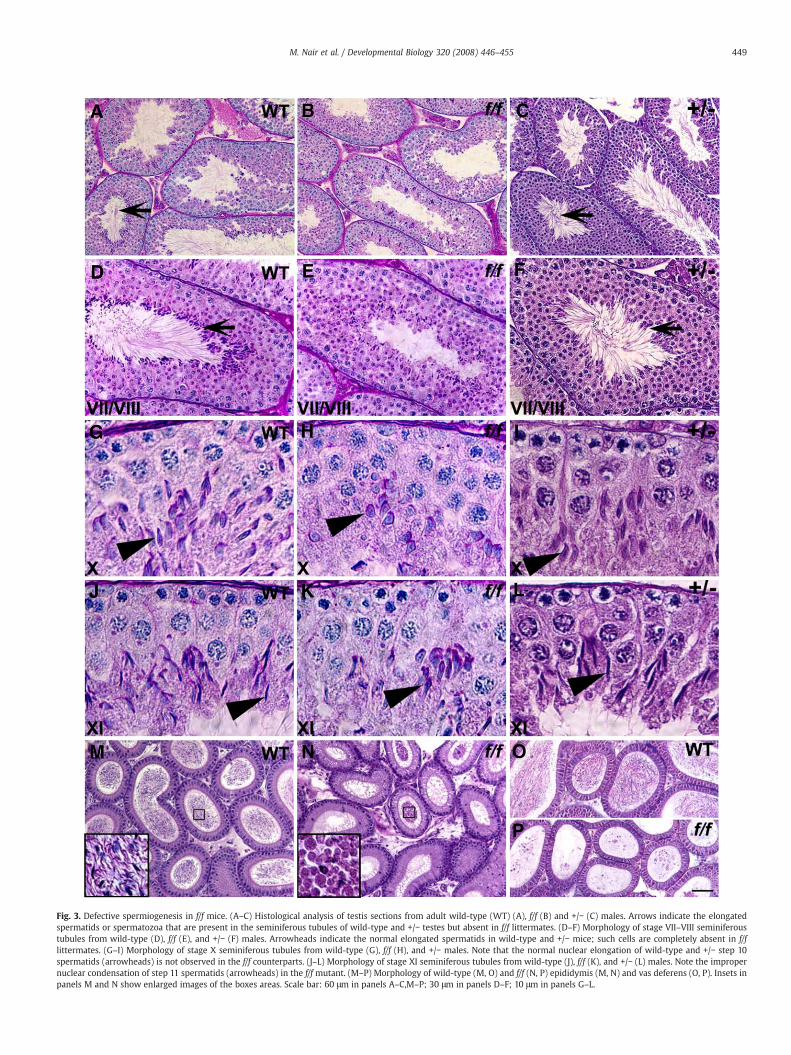
Fig. 3. Defective spermiogenesis in f/f mice. (A–C) Histological analysis of testis sections from adult wild-type (WT) (A), f/f (B) and +/− (C) males. Arrows indicate the elongatedspermatids or spermatozoa that are present in the seminiferous tubules of wild-type and +/− testes but absent in f/f littermates. (D–F) Morphology of stage VII–VIII seminiferoustubules from wild-type (D), f/f (E), and +/− (F) males. Arrowheads indicate the normal elongated spermatids in wild-type and +/− mice; such cells are completely absent in f/flittermates. (G–I) Morphology of stage X seminiferous tubules from wild-type (G), f/f (H), and +/− males. Note that the normal nuclear elongation of wild-type and +/− step 10spermatids (arrowheads) is not observed in the f/f counterparts. (J–L) Morphology of stage XI seminiferous tubules from wild-type (J), f/f (K), and +/− (L) males. Note the impropernuclear condensation of step 11 spermatids (arrowheads) in the f/f mutant. (M–P) Morphology of wild-type (M, O) and f/f (N, P) epididymis (M, N) and vas deferens (O, P). Insets inpanels M and N show enlarged images of the boxes areas. Scale bar: 60 μm in panels A–C,M–P; 30 μm in panels D–F; 10 μm in panels G–L.
449M. Nair et al. / Developmental Biology 320 (2008) 446–455

Fig. 5. Select reduction of haploid gene expression in f/f spermatids. Shown are resultsof semi-quantitative (A) and real-time (B) PCR analysis revealing decreasedmRNA levelsof Prm1, Prm2, Tnp2, and H1fnt but not other genes. Real-time PCR data representaverage of four different mice, each assayed in triplicates.
450 M. Nair et al. / Developmental Biology 320 (2008) 446–455
Defective spermiogenesis in f/f Pygo2 males
At a histological level, themost striking defect in adultmutant testiswas the lack of elongated spermatids (Fig. 3B). Analysis of stage-matched seminiferous tubules from juvenile wild-type and f/f micedemonstrated that in the mutant the cellular associations normallyobserved in wild-type testis were disrupted, that round, elongating,and elongated spermatids sometimes co-exist in a single tubuleindicative of asynchronous spermatid development, and that step 16spermatids were completely absent (Fig. 3E). The earliest morpholo-gical defects were detected in step 9–10 spermatids, coinciding withthe stages when Pygo2 expression in haploid germ cells becameprominent and is primarily nuclear. However, the total number ofelongating spermatids was maintained in the f/f tubules (53% of totalgerm cells in stage X/XI f/f tubules are step 10 and 11 spermatids, ascompared to 54% in the wild-type). Comparing to their wild-typecounterparts, step 10 spermatids in the mutant did not properlyelongate their nuclei (Fig. 3H), and the nuclei of step 11 mutantspermatids were not properly condensed (Fig. 3K). These resultshighlight defective nuclear elongation and condensation as theprimary defects in f/f testis. In contrast to f/f mice, heterozygous (+/−)males showed no detectable spermiogenic defects (Figs. 3C, F, I, L).Examination of the histology of epidydimis and vas deferens revealedthat while wild-type and +/− epidydimis and vas deferens containedabundant mature spermatozoa (Figs. 3M, O and data not shown), fewspermatozoa were observed in the f/f mutant littermates (Figs. 3N, P).The occasionally seen spermatozoa displayed round heads with notails (Fig. 3N inset), confirming a failure in nuclear shaping.
At a biochemical level, expression of LDHC4, a marker for germcells from mid/late-pachytene stage onward (Hintz and Goldberg,1977), was not affected (Figs. 4A, B). Furthermore, expression ofGCNA1 (germ cell nuclear antigen), a marker for spermatogonia andspermatocytes prior to mid-pachytene (Enders and May, 1994), waslargely normal, and there is no reduction in the number of GCNA1-positive cells in the mutant (Figs. 4C, D). These results suggest that the
Fig. 4. Biochemistry of wild-type and f/f testes. Shown are results of immunofluorescence stGCNA1 antibody (C, D) on wild-type (A, C) and f/f (B, D) testes. Scale bar: 80 μm.
f/f mutation does not significantly impact the early steps ofspermatogenesis.
Decreased expression of select post-meiotic genes in f/f testis
To explore the molecular defects of Pygo2-reduced germ cells, weperformed RT-PCR analysis to examine the expression of regulatory
aining using anti-LDHC4 antibody (A, B) and immunohistochemical analysis using anti-

Fig. 6. H1FNT protein levels are reduced in elongating/elongated f/f spermatids. (A)Western blot analysis showing reduced H1FNT protein levels in total testis lysates (TL)and elongating/elongated spermatids (ES), but not in spermatocytes (SC) and roundspermatids (RS). Actinwas used as a loading control. (B, C) The polarized H1FNT proteinlocalization (red) in wild-type spermatids (B) is greatly diminished in the f/f (C)counterparts. Blue, DAPI staining to visualize nuclei. Scale bar: 6 μm.
451M. Nair et al. / Developmental Biology 320 (2008) 446–455
and structural genes that have been implicated in spermatogenesis.Genes that are normally expressed in Sertoli cells, including GATA1,RARα, and FSHR (Heckert and Griswold, 1991; Kliesch et al., 1992;Vernet et al., 2006; Yomogida et al., 1994), showed unaltered ex-pression in f/f testis (Fig. 5A). Similarly, genes the expression of whichis Leydig cell-specific or -enriched, including LHR and RXRβ (Gaemerset al., 1998; Zhang et al., 1994), were not affected. AR has been
Fig. 7.Hyperacetylation at K9/K14 of histone H3 is specifically reduced in elongating f/f spermusing anti-acetyl-K9 H3 (A, B) and anti-acetyl-K12 H4 (E, F) antibodies. Shown are stage IX seG−J) Results of immunostaining of wild-type (C, G, I) and f/f (D, H, J) testes using anti-acetyl-Western blot analysis of lysates prepared from fractionated elongating/elongated spermatidsignals of chromatin or total nuclear fractions were quantified from two independent exper
reported to be expressed in both somatic (Sertoli, Leydig, peritubularmyoid cells) and germ cells (Vornberger et al., 1994), and no changewas seen in f/f testis. These results suggest that the f/f mutation ofPygo2 may not impact the somatic Sertoli and Leydig cells.
Germ cell-expressing genes were differentially affected. Thosewith a reported expression in round spermatids and earlier, such asCREM (Delmas et al., 1993), ACT (Fimia et al., 1999), Pgk2 (Gold et al.,1983), Tpap (Kashiwabara et al., 2000), produced normal levels ofmRNAs in f/f testis. In contrast, there was a dramatic reduction ofmRNA levels of several genes with expression normally activated inlate round spermatids and high in elongating spermatids (Mali et al.,1989; Saunders et al., 1992), including Prm1 (decreased by 2.6-fold),Prm2 (decreased by 3.4-fold), Tnp2 (decreased by 2.1-fold), and H1fnt(decreased by 4.3-fold), in f/f testis (Figs. 5A, B). However, thisreduction is not universal for all late-spermatid-expressing genes, asMCS (Nam et al., 1997), TRF2/TLF (Martianov et al., 2001), CaMKIV (Wuand Means, 2000), HSC70t (Tsunekawa et al., 1999), and RT7/ODF(Morales et al., 1994; van der Hoorn et al., 1990) were all expressed atapparently normal levels. Importantly, Tnp1 is normally expressed in apattern reminiscent of those of Prm1, Prm2, and Tnp2 (Mali et al.,1989), yet its expression level was only minimally affected in f/f testis.
We next performed proof-of-principle experiments to see if thealtered mRNA levels observed above indeed translate into alteredprotein levels. A significant reduction in H1FNT protein levels was
atids. (A, B, E, F) Results of double-immunostaining of wild-type (A, E) and f/f (B, F) testesminiferous tubules. Note reduced histone H3 K9 acetylation in stage 9 spermatids. (C, D,K9/K14 H3 (C, D), anti-acetyl-K8 H4 (G, H) or anti-trimethyl-K4 H3 (I, J) antibodies. (K)s. (L) Plot of chromatin/nuclear (Ch/Nt) ratios for histone H3 and Pygo2. Western blotiments and average values are shown. Scale bar: 30 μm.
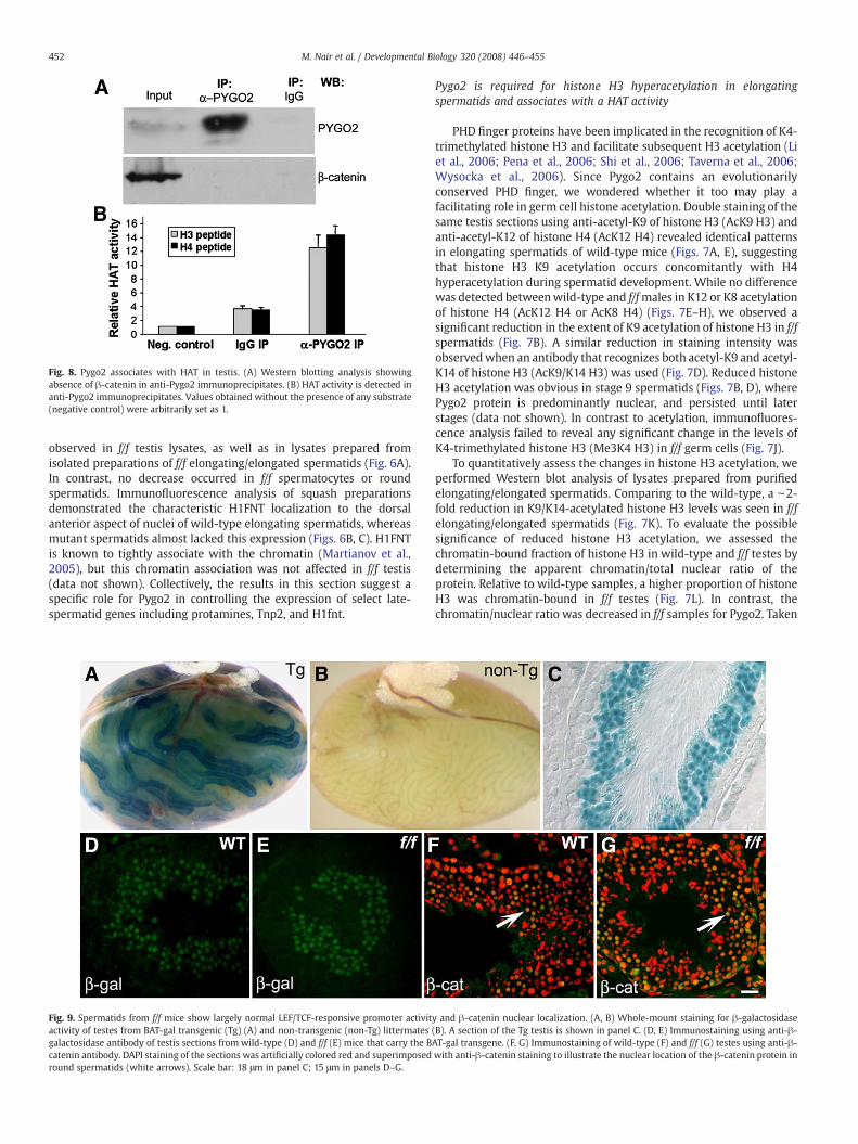
Fig. 8. Pygo2 associates with HAT in testis. (A) Western blotting analysis showingabsence of β-catenin in anti-Pygo2 immunoprecipitates. (B) HAT activity is detected inanti-Pygo2 immunoprecipitates. Values obtained without the presence of any substrate(negative control) were arbitrarily set as 1.
452 M. Nair et al. / Developmental Biology 320 (2008) 446–455
observed in f/f testis lysates, as well as in lysates prepared fromisolated preparations of f/f elongating/elongated spermatids (Fig. 6A).In contrast, no decrease occurred in f/f spermatocytes or roundspermatids. Immunofluorescence analysis of squash preparationsdemonstrated the characteristic H1FNT localization to the dorsalanterior aspect of nuclei of wild-type elongating spermatids, whereasmutant spermatids almost lacked this expression (Figs. 6B, C). H1FNTis known to tightly associate with the chromatin (Martianov et al.,2005), but this chromatin association was not affected in f/f testis(data not shown). Collectively, the results in this section suggest aspecific role for Pygo2 in controlling the expression of select late-spermatid genes including protamines, Tnp2, and H1fnt.
Fig. 9. Spermatids from f/f mice show largely normal LEF/TCF-responsive promoter activitactivity of testes from BAT-gal transgenic (Tg) (A) and non-transgenic (non-Tg) littermatesgalactosidase antibody of testis sections from wild-type (D) and f/f (E) mice that carry the Bcatenin antibody. DAPI staining of the sections was artificially colored red and superimposedround spermatids (white arrows). Scale bar: 18 μm in panel C; 15 μm in panels D–G.
Pygo2 is required for histone H3 hyperacetylation in elongatingspermatids and associates with a HAT activity
PHD finger proteins have been implicated in the recognition of K4-trimethylated histone H3 and facilitate subsequent H3 acetylation (Liet al., 2006; Pena et al., 2006; Shi et al., 2006; Taverna et al., 2006;Wysocka et al., 2006). Since Pygo2 contains an evolutionarilyconserved PHD finger, we wondered whether it too may play afacilitating role in germ cell histone acetylation. Double staining of thesame testis sections using anti-acetyl-K9 of histone H3 (AcK9 H3) andanti-acetyl-K12 of histone H4 (AcK12 H4) revealed identical patternsin elongating spermatids of wild-type mice (Figs. 7A, E), suggestingthat histone H3 K9 acetylation occurs concomitantly with H4hyperacetylation during spermatid development. While no differencewas detected betweenwild-type and f/fmales in K12 or K8 acetylationof histone H4 (AcK12 H4 or AcK8 H4) (Figs. 7E–H), we observed asignificant reduction in the extent of K9 acetylation of histone H3 in f/fspermatids (Fig. 7B). A similar reduction in staining intensity wasobservedwhen an antibody that recognizes both acetyl-K9 and acetyl-K14 of histone H3 (AcK9/K14 H3) was used (Fig. 7D). Reduced histoneH3 acetylation was obvious in stage 9 spermatids (Figs. 7B, D), wherePygo2 protein is predominantly nuclear, and persisted until laterstages (data not shown). In contrast to acetylation, immunofluores-cence analysis failed to reveal any significant change in the levels ofK4-trimethylated histone H3 (Me3K4 H3) in f/f germ cells (Fig. 7J).
To quantitatively assess the changes in histone H3 acetylation, weperformed Western blot analysis of lysates prepared from purifiedelongating/elongated spermatids. Comparing to the wild-type, a ∼2-fold reduction in K9/K14-acetylated histone H3 levels was seen in f/felongating/elongated spermatids (Fig. 7K). To evaluate the possiblesignificance of reduced histone H3 acetylation, we assessed thechromatin-bound fraction of histone H3 in wild-type and f/f testes bydetermining the apparent chromatin/total nuclear ratio of theprotein. Relative to wild-type samples, a higher proportion of histoneH3 was chromatin-bound in f/f testes (Fig. 7L). In contrast, thechromatin/nuclear ratio was decreased in f/f samples for Pygo2. Taken
y and β-catenin nuclear localization. (A, B) Whole-mount staining for β-galactosidase(B). A section of the Tg testis is shown in panel C. (D, E) Immunostaining using anti-β-AT-gal transgene. (F, G) Immunostaining of wild-type (F) and f/f (G) testes using anti-β-with anti-β-catenin staining to illustrate the nuclear location of the β-catenin protein in

453M. Nair et al. / Developmental Biology 320 (2008) 446–455
together, these results identify Pygo2 as a regulator of global histoneH3 hyperacetylation in elongating spermatids, and correlate mis-regulated acetylation with aberrant chromatin retention of histoneH3 in Pygo2-reduced testis.
We next asked whether Pygo2, like PHD finger protein Yng1(Taverna et al., 2006), associates with a HAT activity. Extracts frommouse testis were used for immunoprecipitation using anti-Pygo2antibody, and it was evident that the antibody precipitated theendogenous Pygo2 protein (Fig. 8A). When HAT activity wasmeasured, a significant enrichment of an activity that was able toacetylate both histone H3 and H4 peptide substrates was observed inα-Pygo2 immunoprecipitates over IgG controls (Fig. 8B). Interestingly,β-catenin was not detected in the immunoprecipitates. As a control,we note that Pygo2-β-catenin interactionwas detected in 293Tcells inour hands (data not shown). Collectively, our results demonstrate thatPygo2 in testis associates with a HAT activity likely in a β-catenin-independent manner, providing a possible biochemical mechanismfor Pygo2's regulation of histone acetylation in germ cells.
In vivo evidence that Pygo2 and β-catenin have independent regulatorypathways in male germ cells
Next we performed experiments to directly address whetherPygo2 functions in spermiogenesis by regulating the transcriptionaloutput of Wnt/β-catenin signaling. We first examined the expressionof BAT-gal, a widely used Wnt reporter gene in which LacZ expressionis under the control of LEF/TCF-responsive elements (Maretto et al.,2003), in testis. Testes from BAT-gal transgenic mice stained blue withβ-galactosidase substrates (Fig. 9A), while non-transgenic littermatesappeared white (Fig. 9B). The predominant sites of LacZ expressionwere round spermatids (Figs. 9C, D). The β-galactosidase proteinpersisted in early elongating (step 9–10) spermatids but diminishedupon further maturation (data not shown). These results imply thepresence of factors in round spermatids that can activate LEF/TCFtarget gene expression. Indeed, nuclear β-catenin, presumably pro-duced as a result of active Wnt signaling, was detected in roundspermatids (Fig. 9F) and weakly in pachytene spermatocytes, but notin elongating spermatids that normally express Pygo2 (data notshown). No significant reduction in β-galactosidase activity orexpression was observed in round and early elongating spermatidsof f/fmice (Fig. 9E and data not shown), indicating that reduced Pygo2levels have little effect on the activity of the LEF/TCF-responsivepromoter that is present in the BAT-gal transgene. Furthermore,nuclear localization of β-catenin was still detected in f/f roundspermatids (Fig. 9G). These results, together with the biochemicaldata above showing that β-catenin and Pygo2 in testis do not co-immunoprecipitate, suggest that the actions of β-catenin and Pygo2are spatially separated, and that Pygo2 function in spermiogenesis isβ-catenin-independent.
Discussion
Spermiogenesis entails a major biochemical and morphologicalrestructuring of the germ cell involving replacement of somatichistones by protamines packing the DNA into the condensedspermatid nucleus during the elongation phase. Our results uncoveran important role for Pygo2 in mammalian spermiogenesis, as re-duced levels of Pygo2 originated from hypomorphic floxed allelesresult in a complete spermiogenic arrest and male infertility. Pygo2-reduced spermatids fail to properly elongate and shape their nuclei,and these phenotypes partially overlap those observed in mousemutants of protamines, transition proteins, and H1fnt. Indeed, weobserved a decreased expression of Prm1, Prm2, Tnp2, and H1fnt in f/f testis. This effect is rather specific, as other late-spermatid-expressing genes such as Tnp1 are not affected. Therefore, it isunlikely that the reduced expression of H1fnt, Prm1, Prm2, and Tnp2
is simply due to a reduced presence of late spermatids in f/f testis. Inmice and humans, genes encoding Prm1, Prm2, and Tnp2 areclustered together on one chromosome and their expression coor-dinately regulated in a haploid-specific manner during spermatogen-esis (Nelson and Krawetz, 1993), whereas the Tnp1 gene is located onanother chromosome. The uniform down-regulation of the clusteredgenes in f/f testis raises an intriguing possibility that Pygo2 directly orindirectly regulates the activation of this gene cluster in a concertedmanner. This notion is worth testing experimentally. Prm1, Prm2,Tnp1, and RT7 are all downstream targets of CREM, a key haploidgerm cell transcription factor (Delmas et al., 1993). The largelyunaltered expression of Tnp1 and RT7 also suggests that Pygo2reduction does not result in a general decrease in CREM-dependenttranscription.
Besides a potential role in regulating specific haploid genes, afunctional Pygo2 protein is essential in vivo for K9/K14 hyperacetyla-tion of histone H3. We show that H3 hyperacetylation occurs duringspermatid elongation and nuclear condensation, and that it is se-lectively reduced in f/f mutant spermatids. While it is formally pos-sible that this defect is a secondary consequence of other cellular/molecular changes such as those that may occur in the somaticsupport cells or in premeiotic germ cells, two pieces of evidence areconsistent with a more direct role for Pygo2 in the acetylation ofhistone H3: 1) histone H3 hyperacetylation and its decrease in f/fmutant spatiotemporally correlate with abundant nuclear expressionof Pygo2; 2) Pygo2 associates with a HATactivity in testis extracts. Oneof the proposed consequences of histone hyperacetylation is thefacilitated displacement of histones from the chromatin. Consistentlywith this hypothesis, we observed an increased chromatin association(retention) of histone H3 in f/f mutant testis. Our results offer noinsights as to the relative importance of Pygo2's effect on haploid geneexpression and histone H3 acetylation. Our current thinking is thatPygo2 regulates both events in elongating spermatids, therebycoordinating histone displacement with the production of proteinsthat can take histones' place to package the germ cell DNA in a morecompact fashion.
Which HAT does Pygo2 associate with? Our results of HAT assayson purified histone peptide substrates indicate that this HAT does notdistinguish between H3 and H4 in vitro. Therefore, it is unlikely thatthe Pygo2-associated HAT is CDYL, which has been previouslyidentified as a histone H4-specific HAT expressed in maturingspermatids (Lahn et al., 2002). Despite the in vitro promiscuity ofPygo2-associated HAT, reduced Pygo2 levels affect histone H3 but notH4 hyperacetylation. This in vivo specificity implicates the existence ofadditional mechanisms that target Pygo2-associated activity tohistone H3. PHD fingers have recently been found to be a recognitionmotif for histone H3 trimethylated at K4 (Li et al., 2006; Pena et al.,2006; Shi et al., 2006; Wysocka et al., 2006). Furthermore, K4trimethylation of histone H3 facilitates its subsequent acetylation byYng1-associated HAT (Taverna et al., 2006). Global K4 trimethylationof H3 during germ cell development has recently been shown tospatiotemporally overlap global histone hyperacetylation (Godmannet al., 2007), but the underlying molecular link between these twomodification events is unclear. We propose that the PHD domain ofPygo2 binds to K4-trimethyl histone H3 in germ cell chromatin,thereby targeting Pygo2-associated HATactivity to histone H3. Clearly,future studies beyond the scope of this work are needed to test ourmodel and to identify the Pygo2-associated HAT.
Testes of f/fmice express Pygo2 at a level that is approximately 40%of the wild type. Since +/null heterozygous – presumably expressing50% of wild-type level of Pygo2 – and +/f males are fertile with noapparent defects in spermatid elongation, we surmise that there is acritical threshold of Pygo2 dosage below which spermiogenesiscannot be sustained. We note the interesting finding that the nuclearconcentration of Pygo2 appears to be disproportionally impacted bythe mutation comparing to the cytoplasmic concentration. Perhaps a

454 M. Nair et al. / Developmental Biology 320 (2008) 446–455
threshold level of Pygo2 is necessary for efficient nuclear localization;when Pygo2 levels drop below this threshold, the cytoplasmic/nucleardistribution is altered, leading to a specific reduction in protein levelsinside the nucleus. Dissecting the underling mechanism of thisobservation is outside the scope of the current work. Alternatively,the f/f mutation may have an allele-specific effect. f/f mice do notdisplay apparent defects in somatic epithelial tissues that are knownto be affected by a Pygo2 null mutation (Li et al., 2007). This differencereveals a tissue- and cell type-specific sensitivity of developmentalprocesses to Pygo2 protein levels. Remodeling of the germ cellchromatin during spermatid elongation is global in nature, whereaschromatin remodeling in somatic cells is more gene- and locus-specific. Therefore, elongating spermatids may entail a higher level ofnuclear Pygo2 proteins for proper chromatin modifications, and aretherefore particularly sensitive to the f/f-induced reduction in Pygo2levels.
Our data demonstrate that canonical Wnt signaling is active (byvirtue of expressing nuclear β-catenin and activating a LEF/TCF targetpromoter) in differentiating germ cells, particularly in spermatocytesand round spermatids. However, this activation does not seem torequire Pygo2, as Pygo2 protein is not detected in these cells. Instead,Pygo2 is present in elongating spermatids and is required for theirfurther differentiation. Although we cannot fully exclude the pos-sibility that there are subtle differences inWnt signaling betweenwildtype and mutant that are beyond our method of detection, ouranalysis supports the notion that the function of Pygo2 in male germcells is independent of β-catenin, and by inference,Wnt signaling. Thiswork is consistent with previous reports to show that mammalianPygopus genes have evolved to perform Wnt-independent functionsin addition to their roles in mediating canonical Wnt signaling (Li etal., 2007; Schwab et al., 2007; Song et al., 2007).
Acknowledgments
We thank Wen-Hwa Lee for microscope use, Grant MacGregor fordiscussions, and anonymous reviewers for helpful suggestions. We aregrateful to Erwin Goldberg and George Enders for kindly providing theα-LDHC4 and α-GCNA1 antibodies, respectively. This work wassupported by a DOD grant W81XWH-04-1-0516, and NIH GrantsR01-AR47320 and K02-AR51482 awarded to X. D.
Appendix A. Supplementary data
Supplementary data associated with this article can be found, inthe online version, at doi:10.1016/j.ydbio.2008.05.553.
References
Balhorn, R., Reed, S., Tanphaichitr, N., 1988. Aberrant protamine 1/protamine 2 ratios insperm of infertile human males. Experientia 44, 52–55.
Belenkaya, T.Y., Han, C., Standley, H.J., Lin, X., Houston, D.W., Heasman, J., 2002. pygopusEncodes a nuclear protein essential for wingless/Wnt signaling. Development 129,4089–4101.
Chevaillier, P., Mauro, N., Feneux, D., Jouannet, P., David, G., 1987. Anomalous proteincomplement of sperm nuclei in some infertile men. Lancet 2, 806–807.
Cho, C., Willis, W.D., Goulding, E.H., Jung-Ha, H., Choi, Y.C., Hecht, N.B., Eddy, E.M., 2001.Haploinsufficiency of protamine-1 or -2 causes infertility in mice. Nat. Genet. 28,82–86.
Clevers, H., 2006. Wnt/beta-catenin signaling in development and disease. Cell 127,469–480.
Dai, X., Schonbaum, C., Degenstein, L., Bai, W., Mahowald, A., Fuchs, E., 1998. The ovogene required for cuticle formation and oogenesis in flies is involved in hairformation and spermatogenesis in mice. Genes Dev. 12, 3452–3463.
de la Roche, M., Bienz, M., 2007. Wingless-independent association of Pygopus withdTCF target genes. Curr. Biol.
de Yebra, L., Ballesca, J.L., Vanrell, J.A., Bassas, L., Oliva, R., 1993. Complete selectiveabsence of protamine P2 in humans. J. Biol. Chem. 268, 10553–10557.
Delmas, V., van der Hoorn, F., Mellstrom, B., Jegou, B., Sassone-Corsi, P., 1993. Inductionof CREM activator proteins in spermatids: down-stream targets and implicationsfor haploid germ cell differentiation. Mol. Endocrinol. 7, 1502–1514.
Enders, G.C., May II, J.J., 1994. Developmentally regulated expression of a mouse germcell nuclear antigen examined from embryonic day 11 to adult in male and femalemice. Dev. Biol. 163, 331–340.
Fimia, G.M., De Cesare, D., Sassone-Corsi, P., 1999. CBP-independent activation of CREMand CREB by the LIM-only protein ACT. Nature 398, 165–169.
Gaemers, I.C., van Pelt, A.M., van der Saag, P.T., Hoogerbrugge, J.W., Themmen, A.P., deRooij, D.G., 1998. Differential expression pattern of retinoid X receptors in adultmurine testicular cells implies varying roles for these receptors in spermatogenesis.Biol. Reprod. 58, 1351–1356.
Godmann, M., Auger, V., Ferraroni-Aguiar, V., Di Sauro, A., Sette, C., Behr, R., Kimmins, S.,2007. Dynamic regulation of histone H3 methylation at lysine 4 in mammalianspermatogenesis. Biol. Reprod.
Gold, B., Fujimoto, H., Kramer, J.M., Erickson, R.P., Hecht, N.B., 1983. Haploidaccumulation and translational control of phosphoglycerate kinase-2 messengerRNA during mouse spermatogenesis. Dev. Biol. 98, 392–399.
Govin, J., Caron, C., Lestrat, C., Rousseaux, S., Khochbin, S., 2004. The role of histones inchromatin remodelling during mammalian spermiogenesis. Eur. J. Biochem. 271,3459–3469.
Grimes Jr., S.R., Henderson, N., 1984a. Acetylation of rat testis histones H2B and TH2B.Dev. Biol. 101, 516–521.
Grimes Jr., S.R., Henderson, N., 1984b. Hyperacetylation of histone H4 in rat testisspermatids. Exp. Cell Res. 152, 91–97.
Hazzouri, M., Pivot-Pajot, C., Faure, A.K., Usson, Y., Pelletier, R., Sele, B., Khochbin, S.,Rousseaux, S., 2000. Regulated hyperacetylation of core histones during mousespermatogenesis: involvement of histone deacetylases. Eur. J. Cell Biol. 79, 950–960.
Heckert, L.L., Griswold, M.D., 1991. Expression of follicle-stimulating hormone receptormRNA in rat testes and Sertoli cells. Mol. Endocrinol. 5, 670–677.
Hintz, M., Goldberg, E., 1977. Immunohistochemical localization of LDH-x duringspermatogenesis in mouse testes. Dev. Biol. 57, 375–384.
Jessen, S., Gu, B., Dai, X. Pygopus and the Wnt signaling pathway a diverse set ofconnections. Bioessays, in press.
Kashiwabara, S., Zhuang, T., Yamagata, K., Noguchi, J., Fukamizu, A., Baba, T., 2000.Identification of a novel isoform of poly(A) polymerase, TPAP, specifically present inthe cytoplasm of spermatogenic cells. Dev. Biol. 228, 106–115.
Kimmins, S., Sassone-Corsi, P., 2005. Chromatin remodelling and epigenetic features ofgerm cells. Nature 434, 583–589.
Kliesch, S., Penttila, T.L., Gromoll, J., Saunders, P.T., Nieschlag, E., Parvinen, M., 1992. FSHreceptor mRNA is expressed stage-dependently during rat spermatogenesis. Mol.Cell Endocrinol. 84, R45–R49.
Kotaja, N., Kimmins, S., Brancorsini, S., Hentsch, D., Vonesch, J.L., Davidson, I., Parvinen, M.,Sassone-Corsi, P., 2004. Preparation, isolation and characterization of stage-specificspermatogenic cells for cellular and molecular analysis. Nat. Methods 1, 249–254.
Kramps, T., Peter, O., Brunner, E., Nellen, D., Froesch, B., Chatterjee, S., Murone, M., Zullig, S.,Basler, K., 2002. Wnt/wingless signaling requires BCL9/legless-mediated recruitmentof pygopus to the nuclear beta-catenin–TCF complex. Cell 109, 47–60.
Krieghoff, E., Behrens, J., Mayr, B., 2006. Nucleo-cytoplasmic distribution of beta-cateninis regulated by retention. J. Cell Sci. 119, 1453–1463.
Lahn, B.T., Tang, Z.L., Zhou, J., Barndt, R.J., Parvinen, M., Allis, C.D., Page, D.C., 2002.Previously uncharacterized histone acetyltransferases implicated in mammalianspermatogenesis. Proc. Natl. Acad. Sci. U. S. A. 99, 8707–8712.
Li, B., Mackay, D.R., Ma, J., Dai, X., 2004. Cloning and developmental expression of mousepygopus 2, a putative Wnt signaling component. Genomics 84, 398–405.
Li, H., Ilin, S., Wang, W., Duncan, E.M., Wysocka, J., Allis, C.D., Patel, D.J., 2006. Molecularbasis for site-specific read-out of histone H3K4me3 by the BPTF PHD finger of NURF.Nature 442, 91–95.
Li, B., Rheaume, C., Teng, A., Bilanchone, V., Munguia, J.E., Hu, M., Jessen, S., Piccolo, S.,Waterman, M.L., Dai, X., 2007. Developmental phenotypes and reduced Wntsignaling in mice deficient for pygopus 2. Genesis 45, 318–325.
Logan, C.Y., Nusse, R., 2004. The Wnt signaling pathway in development and disease.Annu. Rev. Cell. Dev. Biol. 20, 781–810.
Mali, P., Kaipia, A., Kangasniemi, M., Toppari, J., Sandberg, M., Hecht, N.B., Parvinen, M.,1989. Stage-specific expression of nucleoprotein mRNAs during rat and mousespermiogenesis. Reprod. Fertil. Dev. 1, 369–382.
Maretto, S., Cordenonsi, M., Dupont, S., Braghetta, P., Broccoli, V., Hassan, A.B., Volpin, D.,Bressan, G.M., Piccolo, S., 2003. Mapping Wnt/beta-catenin signaling during mousedevelopment and in colorectal tumors. Proc. Natl. Acad. Sci. U. S. A. 100, 3299–3304.
Martianov, I., Brancorsini, S., Catena, R., Gansmuller, A., Kotaja, N., Parvinen, M., Sassone-Corsi, P., Davidson, I., 2005. Polar nuclear localization of H1T2, a histone H1 variant,required for spermatid elongation and DNA condensation during spermiogenesis.Proc. Natl. Acad. Sci. U. S. A. 102, 2808–2813.
Martianov, I., Fimia, G.M., Dierich, A., Parvinen, M., Sassone-Corsi, P., Davidson, I., 2001.Late arrest of spermiogenesis and germ cell apoptosis in mice lacking the TBP-likeTLF/TRF2 gene. Mol. Cell 7, 509–515.
Meistrich, M.L., 1977. Separation of spermatogenic cells and nuclei from rodent testes.Methods Cell Biol. 15, 15–54.
Meistrich, M.L., Trostle-Weige, P.K., Lin, R., Bhatnagar, Y.M., Allis, C.D., 1992. Highlyacetylated H4 is associated with histone displacement in rat spermatids. Mol.Reprod. Dev. 31, 170–181.
Meistrich, M.L., Mohapatra, B., Shirley, C.R., Zhao, M., 2003. Roles of transition nuclearproteins in spermiogenesis. Chromosoma 111, 483–488.
Morales, C.R., Oko, R., Clermont, Y., 1994. Molecular cloning and developmentalexpression of an mRNA encoding the 27 kDa outer dense fiber protein of ratspermatozoa. Mol. Reprod. Dev. 37, 229–240.
Nair, M., Teng, A., Bilanchone, V., Agrawal, A., Li, B., Dai, X., 2006. Ovol1 regulates thegrowth arrest of embryonic epidermal progenitor cells and represses c-myctranscription. J. Cell Biol. 173, 253–264.

455M. Nair et al. / Developmental Biology 320 (2008) 446–455
Nam, S.Y., Maeda, S., Ogawa, K., Kurohmaru, M., Hayashi, Y., 1997. Expression pattern ofthemitochondrial capsule selenoproteinmRNA in themouse testis after puberty; insitu hybridization study. J. Vet. Med. Sci. 59, 983–988.
Nelson, J.E., Krawetz, S.A., 1993. Linkage of human spermatid-specific basic nuclearprotein genes. Definition and evolution of the P1–NP2–NTP2 locus. J. Biol. Chem.268, 2932–2936.
Parker, D.S., Jemison, J., Cadigan, K.M., 2002. Pygopus, a nuclear PHD-finger proteinrequired for Wingless signaling in Drosophila. Development 129, 2565–2576.
Pena, P.V., Davrazou, F., Shi, X., Walter, K.L., Verkhusha, V.V., Gozani, O., Zhao, R.,Kutateladze, T.G., 2006. Molecular mechanism of histone H3K4me3 recognition byplant homeodomain of ING2. Nature 442, 100–103.
Saunders, P.T., Millar, M.R., Maguire, S.M., Sharpe, R.M., 1992. Stage-specific expressionof rat transition protein 2mRNA and possible localization to the chromatoid body ofstep 7 spermatids by in situ hybridization using a nonradioactive riboprobe. Mol.Reprod. Dev. 33, 385–391.
Schwab, K.R., Patterson, L.T., Hartman, H.A., Song, N., Lang, R.A., Lin, X., Potter, S.S., 2007.Pygo1 and Pygo2 roles in Wnt signaling in mammalian kidney development. BMCBiol. 5, 15.
Shi, X., Hong, T., Walter, K.L., Ewalt, M., Michishita, E., Hung, T., Carney, D., Pena, P.,Lan, F., Kaadige, M.R., Lacoste, N., Cayrou, C., Davrazou, F., Saha, A., Cairns, B.R.,Ayer, D.E., Kutateladze, T.G., Shi, Y., Cote, J., Chua, K.F., Gozani, O., 2006. ING2 PHDdomain links histone H3 lysine 4 methylation to active gene repression. Nature442, 96–99.
Shirley, C.R., Hayashi, S., Mounsey, S., Yanagimachi, R., Meistrich, M.L., 2004. Abnormalitiesand reduced reproductive potential of sperm from Tnp1- and Tnp2-null doublemutant mice. Biol. Reprod. 71, 1220–1229.
Song, N., Schwab, K.R., Patterson, L.T., Yamaguchi, T., Lin, X., Potter, S.S., Lang, R.A., 2007.pygopus 2 has a crucial, Wnt pathway-independent function in lens induction.Development 134, 1873–1885.
Stadeli, R., Basler, K., 2005. Dissecting nuclear Wingless signalling: recruitment of thetranscriptional co-activator Pygopus by a chain of adaptor proteins. Mech. Dev. 122,1171–1182.
Tanaka, H., Iguchi, N., Isotani, A., Kitamura, K., Toyama, Y., Matsuoka, Y., Onishi, M.,Masai, K., Maekawa, M., Toshimori, K., Okabe, M., Nishimune, Y., 2005. HANP1/H1T2, a novel histone H1-like protein involved in nuclear formation and spermfertility. Mol. Cell Biol. 25, 7107–7119.
Taverna, S.D., Ilin, S., Rogers, R.S., Tanny, J.C., Lavender, H., Li, H., Baker, L., Boyle, J., Blair, L.P.,Chait, B.T., Patel, D.J., Aitchison, J.D., Tackett, A.J., Allis, C.D., 2006. Yng1 PHD finger
binding to H3 trimethylated at K4 promotes NuA3 HAT activity at K14 of H3 andtranscription at a subset of targeted ORFs. Mol. Cell 24, 785–796.
Thompson, B.J., 2004. A complex of Armadillo, Legless, Pygopus coactivates dTCF toactivate wingless target genes. Curr. Biol. 14, 458–466.
Thompson, B., Townsley, F., Rosin-Arbesfeld, R., Musisi, H., Bienz, M., 2002. A newnuclear component of the Wnt signalling pathway. Nat. Cell Biol. 4, 367–373.
Townsley, F.M., Cliffe, A., Bienz, M., 2004. Pygopus and Legless target Armadillo/beta-catenin to the nucleus to enable its transcriptional co-activator function. Nat. CellBiol. 6, 626–633.
Tsunekawa, N., Matsumoto, M., Tone, S., Nishida, T., Fujimoto, H., 1999. The Hsp70homolog gene, Hsc70t, is expressed under translational control during mousespermiogenesis. Mol. Reprod. Dev. 52, 383–391.
van der Hoorn, F.A., Tarnasky, H.A., Nordeen, S.K., 1990. A new rat gene RT7 isspecifically expressed during spermatogenesis. Dev. Biol. 142, 147–154.
Veeman, M.T., Axelrod, J.D., Moon, R.T., 2003. A second canon. Functions andmechanisms of beta-catenin-independent Wnt signaling. Dev. Cell 5, 367–377.
Vernet, N., Dennefeld, C., Rochette-Egly, C., Oulad-Abdelghani, M., Chambon, P.,Ghyselinck, N.B., Mark, M., 2006. Retinoic acid metabolism and signaling pathwaysin the adult and developing mouse testis. Endocrinology 147, 96–110.
Vornberger, W., Prins, G., Musto, N.A., Suarez-Quian, C.A., 1994. Androgen receptordistribution in rat testis: new implications for androgen regulation of spermato-genesis. Endocrinology 134, 2307–2316.
Wu, J.Y., Means, A.R., 2000. Ca(2+)/calmodulin-dependent protein kinase IV isexpressed in spermatids and targeted to chromatin and the nuclear matrix. J.Biol. Chem. 275, 7994–7999.
Wysocka, J., Swigut, T., Xiao, H., Milne, T.A., Kwon, S.Y., Landry, J., Kauer, M., Tackett, A.J.,Chait, B.T., Badenhorst, P., Wu, C., Allis, C.D., 2006. A PHD finger of NURF coupleshistone H3 lysine 4 trimethylationwith chromatin remodelling. Nature 442, 86–90.
Yomogida, K., Ohtani, H., Harigae, H., Ito, E., Nishimune, Y., Engel, J.D., Yamamoto, M.,1994. Developmental stage- and spermatogenic cycle-specific expression oftranscription factor GATA-1 in mouse Sertoli cells. Development 120, 1759–1766.
Zhang, F.P., Hamalainen, T., Kaipia, A., Pakarinen, P., Huhtaniemi, I., 1994. Ontogeny ofluteinizing hormone receptor gene expression in the rat testis. Endocrinology 134,2206–2213.
Zhao, M., Shirley, C.R., Hayashi, S., Marcon, L., Mohapatra, B., Suganuma, R., Behringer, R.R.,Boissonneault, G., Yanagimachi, R., Meistrich, M.L., 2004. Transition nuclear proteinsare required for normal chromatin condensation and functional sperm development.Genesis 38, 200–213.