developmental cell article - case western reserve universityko.cwru.edu/pr/lefebvre.pdf ·...
TRANSCRIPT

Developmental Cell
Article
Sox9 Directs Hypertrophic Maturationand Blocks Osteoblast Differentiationof Growth Plate ChondrocytesPeter Dy,1,3 Weihuan Wang,1,3 Pallavi Bhattaram,1 Qiuqing Wang,1 Lai Wang,2 R. Tracy Ballock,2
and Veronique Lefebvre1,*1Department of Cell Biology2Departments of Biomedical Engineering and Orthopaedic SurgeryOrthopaedic and Rheumatologic Research Center, Cleveland Clinic Lerner Research Institute, Cleveland, OH 44195, USA3These authors contributed equally to this work
*Correspondence: [email protected]
DOI 10.1016/j.devcel.2011.12.024
SUMMARY
The transcription factor Sox9 is necessary for earlychondrogenesis, but its subsequent roles in the carti-lage growth plate, a highly specialized structure thatdrives skeletal growth and endochondral ossifica-tion, remain unclear. Using a doxycycline-inducibleCre transgene and Sox9 conditional null alleles inthemouse, we show that Sox9 is required tomaintainchondrocyte columnar proliferation and generatecell hypertrophy, two key features of functionalgrowth plates. Sox9 keeps Runx2 expression andb-catenin signaling in check and thereby inhibitsnot only progression from proliferation to prehyper-trophy, but also subsequent acquisition of an osteo-blastic phenotype. Sox9 protein outlives Sox9RNA inupper hypertrophic chondrocytes, where it contrib-utes with Mef2c to directly activate the major markerof these cells, Col10a1. These findings thus revealthat Sox9 remains a central determinant of thelineage fate and multistep differentiation programof growth plate chondrocytes and thereby illuminateour understanding of key molecular mechanismsunderlying skeletogenesis.
INTRODUCTION
The skeleton is critically important in vertebrates. It forms the
body framework, assists in locomotion, and fulfills key physio-
logical functions. It is subject to prevalent inherited and acquired
diseases in humans, many of which remain poorly treatable
(Woolf and Pfleger, 2003; Rimoin et al., 2007). To help find
efficient cures, it is thus essential that we reach a fuller under-
standing of the basis of the skeleton complex structure and
regulation, and in particular the mechanisms underlying its
development.
Building the vertebrate skeleton requires the generation of two
main tissues, cartilage and bone, at the right time in over 200
locations in the embryo, and subsequently ensuring their proper
Develo
growth and maturation (Provot and Schipani, 2005). These hard
connective tissues greatly differ in composition, function, and
regulation, but develop through closely related, mutually inter-
acting processes. Chondrocytes (cartilage-forming cells) and
osteoblasts (bone-making cells) derive from osteochondropro-
genitors, bipotent cells that arise frommultipotent mesenchyme.
Skull vault bones and other flat bones form intramembranously
through direct differentiation of osteochondroprogenitors into
osteoblasts. Other bones form endochondrally, that is, through
a cartilage intermediate. Osteochondroprogenitors first con-
dense into precartilaginous masses. Inner cells differentiate
into early chondrocytes and perichondrium cells remain uncom-
mitted. Early chondrocytes then undergo further steps of differ-
entiation in a staggered manner, establishing structures referred
to as growth plates in reference to their prime contribution to
skeletal elongation. Chondrocytes flatten, stack in longitudinal
columns, and proliferate actively until reaching the prehypertro-
phic stage. This stage is a major step in skeletogenesis because
the cells switch to a mature phenotypic program and induce
osteogenesis in the perichondrium. Following subsequent
hypertrophy, another key step in skeleton elongation, chondro-
cytes terminally mature, and most if not all undergo apoptosis.
Osteoblast precursors, endothelial cells, osteoclasts, and hema-
topoietic cells migrate from the perichondrium into the cartilage
remnant to remodel the tissue and lay down bone and marrow
(Maes et al., 2010). Each chondrocyte and osteoblast develop-
mental stage is characterized by expression of specific genes
(Lefebvre and Smits, 2005). Typical markers include Col2a1
(collagen 2) and Acan (aggrecan) for early chondrocytes; Fgfr3
(fibroblast growth factor receptor 3) for columnar cells; Ppr
(parathyroid hormone-related protein receptor), Ihh (Indian
hedgehog), and Col10a1 (collagen 10) for prehypertrophic cells;
and Col10a1 only for hypertrophic cells. Terminal chondrocytes
express Mmp13 (matrix metalloproteinase 13) and Bsp (bone
sialoprotein), and mineralize the extracellular matrix, as do
mature osteoblasts, whereas early osteoblasts express Osx
(Osterix) and Col1a1 (collagen 1).
Like other developmental processes, skeletogenesis is
spatially and temporally governed by intricate networks of regu-
latory molecules, among which lineage-specific transcription
factors have key fate-determining roles (Karsenty et al., 2009).
The Sry-related transcription factor Sox9 is one of them
pmental Cell 22, 597–609, March 13, 2012 ª2012 Elsevier Inc. 597

Developmental Cell
Sox9 in the Cartilage Growth Plate
(Akiyama, 2008). Research on its functions started when SOX9
heterozygous mutations were found to cause campomelic
dysplasia, a severe form of dwarfism affecting all cartilage and
endochondral structures (Foster et al., 1994; Wagner et al.,
1994). Sox9 expression is turned on inmesenchymal precursors,
maintained in developing chondrocytes until prehypertrophy,
but turned off in other lineages. Sox9 is absolutely necessary
for chondrocyte specification and early differentiation (Bi et al.,
1999; Akiyama et al., 2002). It directly activates all major carti-
lage-specific extracellular matrix genes expressed by early
chondrocytes, and is helped in this function by two distant rela-
tives, Sox5 and Sox6 (Lefebvre and Smits, 2005). The three Sox
proteins are needed and sufficient for early chondrogenesis, and
thus referred to as the chondrogenic trio (Ikeda et al., 2004).
Subsequent differentiation of chondrocytes is directed from
the prehypertrophic stage by the Runt domain transcription
factors Runx2 and Runx3, and by MADS box transcription
factors, mainly Mef2c (Takeda et al., 2001; Yoshida and Komori,
2005; Arnold et al., 2007). Runx2 is also necessary for osteoblast
specification and differentiation (Ducy et al., 1997; Komori et al.,
1997; Otto et al., 1997), along with the zinc finger transcription
factor Osx (Nakashima et al., 2002).
Strong expression of Sox9 in growth plate chondrocytes until
prehypertrophy and marked shortening of campomelic dys-
plasia growth plates strongly suggest that Sox9 has important
roles in growth plates. These roles, however, remain unclear.
Sox9 was first proposed to inhibit chondrocyte proliferation
and hypertrophy (Akiyama et al., 2002, 2004), but was more
recently proposed to be necessary for chondrocyte survival
and hypertrophy, and to delay terminal maturation (Hattori
et al., 2010; Ikegami et al., 2011). Some of the data in these
previous studies were difficult to interpret because the mouse
transgenes that were used to inactivate or overexpress Sox9
were active from the precursor or early chondrocyte stage,
causing defects in cartilage primordia that precluded definitive
identification of growth plate-specific roles for Sox9. To solve
this problem and clarify the roles of Sox9 in the growth plate,
we used in this study mice harboring Sox9 conditional null alleles
and a Cre transgene inducible in differentiated growth plate
chondrocytes. We show that Sox9 continues to fulfill essential
roles at several stages of differentiation of these cells to ensure
cartilage-mediated skeletal growth and coordinate this process
with endochondral ossification.
RESULTS
Generation of a Cre Transgene Inducible inDifferentiated ChondrocytesWe previously showed that an Acan (aggrecan) upstream
enhancer was sufficient to activate theCol2a1 promoter in differ-
entiated chondrocytes in transgenic mice (Han and Lefebvre,
2008). Here, we cloned these regulatory elements into a bigenic
template (Utomo et al., 1999) to generate a mouse line express-
ing an Acan enhancer-driven, tetracycline-inducible Cre (ATC)
transgene (see Figure S1A available online). We characterized
transgene activity using a dual R26TG Cre reporter (Muzumdar
et al., 2007). This reporter expresses Tomato ubiquitously before
Cre recombination andGFP following recombination.R26TGATC
fetuses at embryonic day 17.5 (E17.5) showed Cre activity in few
598 Developmental Cell 22, 597–609, March 13, 2012 ª2012 Elsevier
cells in the end of growth plates, nuclei pulposi and bone in
absence of tetracycline (Figures S1B–S1D). When their mothers
drank water containing the tetracycline compound doxycycline
(Dox) from E15.5, they showed Cre-mediated recombination
within 2 days in all differentiated chondrocytes (except in epiph-
yseal lateral sides) and nucleus pulposus cells, and in some
myoblasts and bone cells, but none in perichondrium cells and
other cell types (Figures S1B–S1E). We concluded that ATC
should be an excellent model to study gene functions in growth
plate chondrocytes independently of functions in precursors and
most other cell types.
Sox9 Is Necessary to Maintain Functional Growth PlatesTo determine whether Sox9 has specific roles in the growth
plate, we bred females carrying Sox9 conditional null alleles
(Sox9fl/fl; Kist et al., 2002) with Sox9fl/+ATC males, treated them
with Dox from E15.5, and analyzed fetuses daily afterward.
Sox9 RNA remained abundant in nonhypertrophic chondrocytes
of Sox9fl/fl and Sox9fl/+ control fetuses through E18.5, but it was
lost in most growth plate cells of Sox9fl/flATC mutants by E17.5
(Figure 1A). Control epiphyses and growth plates kept the
same length over time while primary ossification centers elon-
gated, reflecting balanced turnover of cartilage, and Sox9fl/+ATC
growth plates remained virtually normal (Figures 1A and 1B). In
contrast, columnar zones shortened in Sox9fl/flATC growth
plates, chondrocytes lost the ability to enlarge, and endochon-
dral bones stopped elongating by E17.5, resulting in severe
dwarfism (Figures 1A and 1C). These data thus revealed that
Sox9 is essential to maintain functional growth plates.
Sox9 Is Needed to Maintain Growth Plate ChondrocyteProliferation and Viability, Delay Prehypertrophy,and Allow HypertrophyShortening of Sox9 mutant columnar zones could result from
chondrocyte slow proliferation, untimely death, or precocious
maturation. In bromodeoxyuridine (BrdU) incorporation assays
performed after 2 days of fetus treatment with Dox, growth
plate chondrocytes from Sox9fl/flATC fetuses showed normal
proliferation rates in epiphyses, but columnar cells growth
arrested about three times as fast as control cells (Figure 2A).
This result was confirmed by immunostaining for Ki-67 (Fig-
ure S2A). In TUNEL assay, Sox9fl/fl growth plates never showed
a significant rate of chondrocyte death until the cells reached
the ossification front (Figure 2B). In contrast, Sox9fl/flATC
growth plates showed 0.1% of dying cells in the columnar
zone and 4.5% in the prehypertrophic zone after 2 days on
Dox, and 2.8% and 24.8% in the same respective zones by
the next day. Immunoreactivity for cleaved caspase 3 indicated
that mutant cell death occurred through apoptosis (Figure S2B).
Thus, Sox9 deletion led to premature growth arrest of columnar
cells, followed by apoptosis. Consistent with precocious prehy-
pertrophy, Sox9fl/flATC chondrocytes turned on Ppr and Ihh
concomitantly with growth arrest (Figure 2C). Surprisingly,
however, they failed to activate the hypertrophic markers
Col10a1, Bmp6, and Has2, but ultimately expressed the
terminal chondrocyte and osteoblast marker Mmp13 (Figure 2D
and Figure S2C). They expressed the chondrocyte proliferation
inhibitor Fgfr3 at a normal level, and chondrocyte maturation
inhibitors Pthrp (parathyroid hormone related peptide) and Ptc
Inc.

Figure 1. Sox9 Is Required to Maintain Functional Growth Plates
(A) Histological aspect and Sox9 expression of proximal tibia growth plates from Sox9fl/fl and Sox9fl/flATC fetuses treated with Dox from E15.5 and collected at
E16.5, E17.5, and E18.5. Sections were stained with Alcian blue (which binds aggrecan) and nuclear fast red, or hybridized with a Sox9RNA probe (red) and DNA-
binding DAPI dye (blue). CZ, columnar zone; EP, epiphyses; HZ, hypertrophic zone.
(B) Histological aspect of the entire tibia of Sox9fl/fl, Sox9fl/+ATC, and Sox9fl/flATC littermates treated as described in (A). The dotted line designates the middle of
the bone. EB, endochondral bone.
(C) High-magnification images of hypertrophic zones at E17.5.
See also Figure S1.
Developmental Cell
Sox9 in the Cartilage Growth Plate
(Patched) at a high level (Figure 2E). They strongly expressed
Runx2 and Mef2c, which encode transcription factors required
for chondrocyte maturation, and Hdac4, encoding a histone
deacetylase inhibitor of these factors (Vega et al., 2004), indi-
cating that Sox9 may delay prehypertrophy by downregulating
Runx2 and Mef2c, but must control hypertrophy differently.
Overall, these data thus showed that Sox9 is required to main-
tain growth plate chondrocyte proliferation, delay prehypertro-
phy, and allow hypertrophy before terminal maturation and
apoptosis.
Sox9 Contributes with Mef2c to DirectlyActivate Col10a1
Based on the facts that Col10a1 is expressed exclusively in pre-
hypertrophic and hypertrophic chondrocytes and that Sox9 is
required for hypertrophy, we investigated whether Sox9 directly
controls Col10a1. As a prerequisite to this function, Sox9 protein
must be present in Col10a1-expressing chondrocytes. Using
RNA in situ hybridization, we confirmed (Lefebvre et al., 1998)
that Sox9 and Col10a1 RNAs overlap each other in prehypertro-
phic cells, and immunostaining revealed that the Sox9 protein
Develo
outlives its RNA and remains nuclear in upper hypertrophic
chondrocytes (Figures 3A and 3B). Concomitant loss of Sox9
RNA and protein in Dox-treated Sox9fl/flATC growth plates as-
certained antibody specificity. Thus, Sox9 protein is present in
cells activating Col10a1.
To test whether Sox9 could transactivate Col10a1, we
searched for putative Sox9 binding sites in theCol10a1 promoter
and hypertrophic enhancer (Zheng et al., 2009). We found only
one sequence that was evolutionarily conserved and matched
a Sox9 binding site: a pair of sites resembling the Sox consensus
CA/TTTGA/T
A/T, oriented head to head, and separated by 3–5
nucleotides (Sock et al., 2003). This sequence was located
at �186/�169 (Figures 3C and 3D), near a Mef2c binding site
(Arnold et al., 2007). Electrophoretic mobility shift assays
showed the ability of each protein to bind its site (Figure 3E),
but no evidence of cooperative binding (Figure 3F). Chromatin
immunoprecipitation assays revealed efficient binding of both
proteins to the Col10a1 promoter in primary growth plate chon-
drocytes forced to upregulate Col10a1 by treatment with
okadaic acid, a potent inhibitor of Pp2a phosphatase (Kozhe-
myakina et al., 2009) (Figures 3G and 3H).
pmental Cell 22, 597–609, March 13, 2012 ª2012 Elsevier Inc. 599

Figure 2. Sox9 Is Required for Chondrocyte Proliferation, Survival, and Hypertrophy but Delays Prehypertrophy
(A) BrdU incorporation assay in tibia proximal growth plates of Sox9fl/fl and Sox9fl/flATC littermates treated with Dox from E15.5 and harvested at E17.5. Left,
representative images of BrdU antibody staining. The box shows 35 segments in the epiphysis (EP) and columnar zone (CZ), in which BrdU-positive cells (dark
brown nuclei) were counted. Right, graph showing the percentage of positive cells in control andmutant growth plates in each segment. Data are presented as the
average with SD of measurements made in three nonadjacent sections from each of three embryos per genotype.
(B) TUNEL assay in embryos similar to those shown in (A), but collected at E17.5 and E18.5. Left, representative images of data. Dying cells are seen as green
dots; cell nuclei are seen as blue dots. Right, quantification of the percentage of TUNEL-positive cells in the columnar (CZ) and prehypertrophic/hypertrophic (PH)
zones. Data are shown as the averages with SD obtained for three nonadjacent sections in each of three embryos per genotype.
Developmental Cell
Sox9 in the Cartilage Growth Plate
600 Developmental Cell 22, 597–609, March 13, 2012 ª2012 Elsevier Inc.

Developmental Cell
Sox9 in the Cartilage Growth Plate
To test whether Sox9 binding to the Col10a1 promoter re-
sulted in transactivation, we constructed Col10a1/bgeo/EGFP
reporters (Figure S3A). As expected, the activity of the Col10a1
promoter was upregulated in primary chondrocytes when the
reporters contained the Col10a1 hypertrophic enhancer and
the cells were treated with okadaic acid (Figure 4A). Forced
expression either humanSOX9 ormouseMef2c enhanced trans-
activation, and forced expression of both proteins resulted in
additive or slightly stronger effects. The proteins acted similarly
in nonchondrocytic 293T cells, even though the enhancer was
virtually inactive in these cells (Figure S3B). In agreement with
the protein binding locations, promoter mutagenesis experi-
ments revealed that SOX9 and Mef2c largely mediated their
activity through the Col10a1 �210/�94 sequence (Figure S3C).
The mutation of either protein site reduced reporter activity in
chondrocytes in absence and presence of exogenous protein,
and the mutation of both sites abolished reporter activity (Fig-
ure 4B). Taken together, these results support the concept that
Sox9 and Mef2c transactivate Col10a1, and act additively.
To test this concept in vivo, we generated Sox9/Mef2c
compound mutants. Single and double heterozygotes obtained
using Prx1Cre, a transgene active in limb bud mesenchyme,
including chondrocyte precursors (Logan et al., 2002), displayed
no major histological defects at E18.5 (Figure 4C). Col10a1
RNA level was slightly but not significantly decreased in single
heterozygotes, but was significantly reduced by 2.4-fold in
double heterozygotes. Further, and consistent with the reported
roles of Mef2c (Arnold et al., 2007), Mef2cfl/flATC fetuses on
Dox since E15.5 showed greatly expanded growth plates by
E17.5 and severely delayed Col10a1 activation (Figure 4D).
Sox9fl/+Mef2cfl/flATC littermates had similar histological defects,
but virtually failed to express Col10a1. Thus, Sox9 and Mef2c
also act together to activate Col10a1 in vivo. Interestingly,
Mef2cfl/flATC mutants initiated Ppr and Runx2 expression in a
timely manner and delayed Ppr deactivation and Ihh activation,
whereas Sox9fl/+Mef2cfl/flATC littermates exhibited stronger
and earlier activation of Ppr, and a partial rescue of the Ihh
activation delay. Thus, Sox9 inhibits chondrocyte prehypertro-
phy from the Ppr expression stage, Mef2c activates it from the
Ihh expression stage, and both proteins are then required for
chondrocyte hypertrophy and Col10a1 expression.
Sox9 Prevents Osteoblastic Differentiation ofPrehypertrophic ChondrocytesBesides Col10a1 transactivation, we considered the possibility
that Sox9 could use additional, indirect mechanisms to account
for its absolute requirement for chondrocyte hypertrophy. Since
Runx2 is necessary for osteoblast differentiation and chondro-
cyte maturation, its inability to cause hypertrophy of Sox9-defi-
cient chondrocytes, despite being strongly expressed, made
us wonder whether Sox9fl/flATC prehypertrophic chondrocytes
were switching lineage. After 2 days on Dox, control and mutant
prehypertrophic cells, as identified by expression of their exclu-
sive marker, Ihh, still contained RNA for Col2a1, but none for
(C) Ppr and Ihh RNA in situ hybridization of sections similar to those in (A). The d
(D) Col10a1 RNA in situ hybridization of sections from Sox9fl/fl and Sox9fl/flATC litt
(E) RNA in situ hybridization of sections similar to those in (A). Probes are indicat
See also Figure S2.
Develo
Sox9, Sox5, and Sox6 (Figure 5A and data not shown). Since
the Sox trio is required for Col2a1 transcription, the presence
of Col2a1 RNA in these cells was unlikely due to recent produc-
tion, but more likely due to earlier production of this slowly
decaying RNA (Murakami et al., 2000). Importantly, this result
attested to the chondrocyte origin of the cells. Strikingly, mutant
prehypertrophic cells upregulated not only Runx2, but also Osx,
which is required for osteoblast differentiation. Osx RNA is
normally present at an extremely low level in prehypertrophic
chondrocytes compared to osteoblasts, but Sox9-deficient
chondrocytes contained as much Osx RNA as periosteum and
endochondral bone osteoblasts. Further, they contained low
levels of Col1a1 RNA, which is abundant in osteoblasts but
absent in chondrocytes, and Bsp RNA (bone sialoprotein),
a marker of osteoblasts and terminal chondrocytes. Sox9fl/flATC
prehypertrophic chondrocytes were thus transitioning into early
osteoblasts within 2 days on Dox. One day later, these mutant
cells still contained Col2a1 RNA, but no longer contained Ihh
RNA, and they were expressingCol1a1 andBspRNA as strongly
as wild-type osteoblasts (Figure 5B). Furthermore, they were
strongly expressing genes involved in matrix mineralization,
such as Alpl (alkaline phosphatase) and Ank (ankylosis), and
were surrounded by mineralized matrix (Figure 5C). They had
thus completed differentiation into osteoblasts and reached
the mature stage in this lineage. Confirming conversion into a
single lineage, Sox9fl/flATC growth plate cells treated for 3 days
with Dox were negative for markers of other mesenchyme-
derived cell types, including Gdf5 (joint precursors), MyoD
(myoblasts), Pparg (adipocytes), and Scx (tenocytes) (Figure S4).
We concluded thatSox9 is necessary tomaintain the lineage fate
of prehypertrophic cells and prevent acquisition of an osteo-
blastic phenotype.
Sox9 Secures the Fate of Growth Plate Chondrocytesby Reducing b-Catenin ActivitySox9 and b-catenin oppose each other in osteochondroprogeni-
tors to specify the chondrocyte and osteoblast lineages, respec-
tively (Day et al., 2005; Hill et al., 2005). We thus considered the
possibility that Sox9 blocks expression of an osteoblastic
phenotype by growth plate chondrocytes by opposing b-catenin
signaling. We observed that Sox9fl/flATC prehypertrophic chon-
drocytes on Dox for 2 days were avidly expressing Tcf1 (T cell-
specific transcription factor 1) andCcnd1 (cyclin D1), two targets
of b-catenin signaling very active in osteoblasts (Figure 6). Inac-
tivation of b-catenin along with Sox9 in Sox9fl/flCtnnbfl/flATC
fetuses resulted in repression of these targets and partial
downregulation of Osx. Importantly, it prevented Col1a1 and
Bsp activation, thus osteoblastic differentiation, but did not
prevent precocious activation of Ihh and upregulation of
Runx2, and did not rescue cell hypertrophy and Col10a1 expres-
sion (Figure 6 and Figure S5A). Thus, Sox9 blocks expression of
an osteoblast phenotype by chondrocytes at least in part by
suppressing b-catenin signaling, but does not use this mecha-
nism to keep Runx2 expression low and permit hypertrophy. In
ouble arrow shows the length of the columnar zone.
ermates treated with Dox from E15.5 and collected at E16.5, E17.5, and E18.5.
ed.
pmental Cell 22, 597–609, March 13, 2012 ª2012 Elsevier Inc. 601
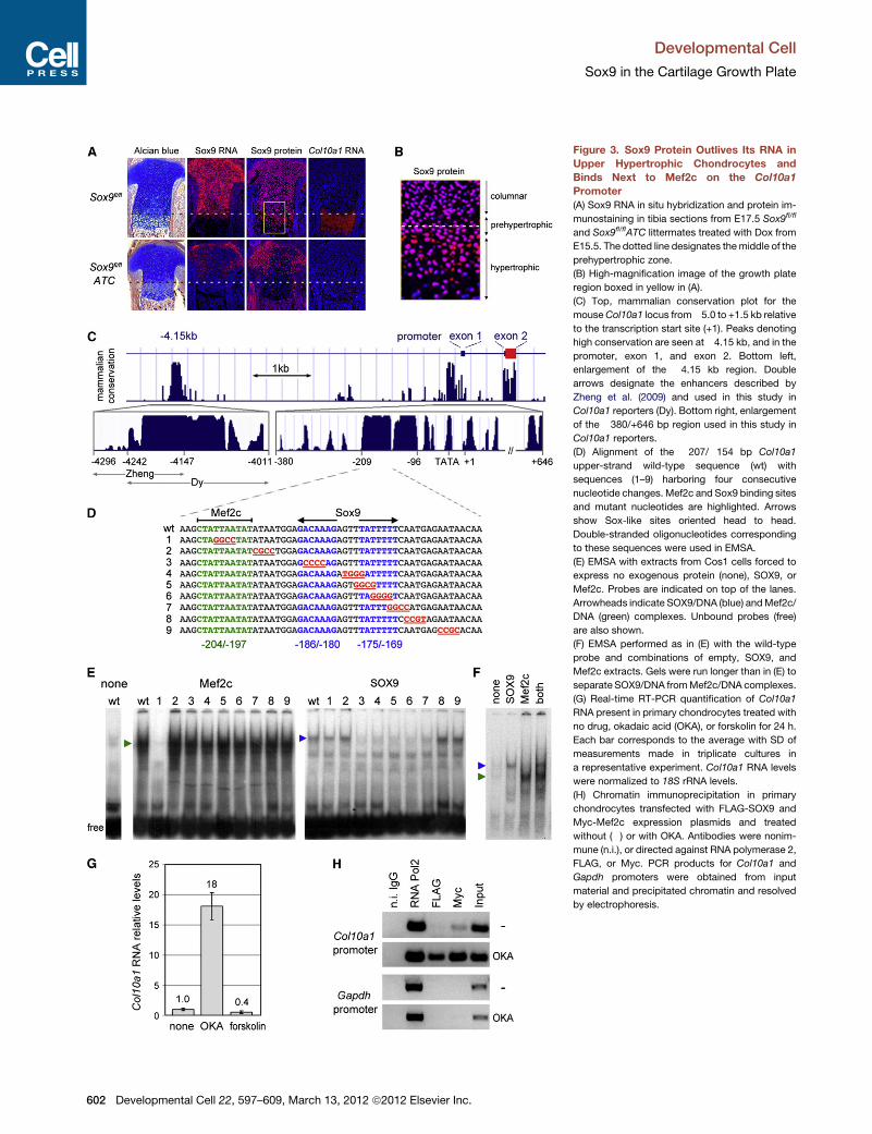
Figure 3. Sox9 Protein Outlives Its RNA in
Upper Hypertrophic Chondrocytes and
Binds Next to Mef2c on the Col10a1
Promoter
(A) Sox9 RNA in situ hybridization and protein im-
munostaining in tibia sections from E17.5 Sox9fl/fl
and Sox9fl/flATC littermates treated with Dox from
E15.5. The dotted line designates themiddle of the
prehypertrophic zone.
(B) High-magnification image of the growth plate
region boxed in yellow in (A).
(C) Top, mammalian conservation plot for the
mouseCol10a1 locus from�5.0 to +1.5 kb relative
to the transcription start site (+1). Peaks denoting
high conservation are seen at�4.15 kb, and in the
promoter, exon 1, and exon 2. Bottom left,
enlargement of the �4.15 kb region. Double
arrows designate the enhancers described by
Zheng et al. (2009) and used in this study in
Col10a1 reporters (Dy). Bottom right, enlargement
of the �380/+646 bp region used in this study in
Col10a1 reporters.
(D) Alignment of the �207/�154 bp Col10a1
upper-strand wild-type sequence (wt) with
sequences (1–9) harboring four consecutive
nucleotide changes. Mef2c and Sox9 binding sites
and mutant nucleotides are highlighted. Arrows
show Sox-like sites oriented head to head.
Double-stranded oligonucleotides corresponding
to these sequences were used in EMSA.
(E) EMSA with extracts from Cos1 cells forced to
express no exogenous protein (none), SOX9, or
Mef2c. Probes are indicated on top of the lanes.
Arrowheads indicate SOX9/DNA (blue) andMef2c/
DNA (green) complexes. Unbound probes (free)
are also shown.
(F) EMSA performed as in (E) with the wild-type
probe and combinations of empty, SOX9, and
Mef2c extracts. Gels were run longer than in (E) to
separate SOX9/DNA fromMef2c/DNA complexes.
(G) Real-time RT-PCR quantification of Col10a1
RNA present in primary chondrocytes treated with
no drug, okadaic acid (OKA), or forskolin for 24 h.
Each bar corresponds to the average with SD of
measurements made in triplicate cultures in
a representative experiment. Col10a1 RNA levels
were normalized to 18S rRNA levels.
(H) Chromatin immunoprecipitation in primary
chondrocytes transfected with FLAG-SOX9 and
Myc-Mef2c expression plasmids and treated
without (�) or with OKA. Antibodies were nonim-
mune (n.i.), or directed against RNA polymerase 2,
FLAG, or Myc. PCR products for Col10a1 and
Gapdh promoters were obtained from input
material and precipitated chromatin and resolved
by electrophoresis.
Developmental Cell
Sox9 in the Cartilage Growth Plate
602 Developmental Cell 22, 597–609, March 13, 2012 ª2012 Elsevier Inc.

Figure 4. Sox9 and Mef2c Act Cooperatively to Activate Col10a1
(A) Relative activity of Col10a1 reporters transfected in primary chondrocytes along with SOX9 and Mef2c expression plasmids. Cells were treated with forskolin
or okadaic acid. Reporter activities were normalized for transfection efficiency and are presented as averagewith SD of biological triplicates in one representative
experiment. Dotted lines designate expected values from additive effects of SOX9 and Mef2c.
(B) Effect of mutations (D) in the Sox9 and Mef2c binding sites on the relative activity of a Col10a1 reporter transfected in primary chondrocytes treated with
okadaic acid. Reporter activities were normalized for transfection efficiency and are presented as average with SD of biological triplicates in one representative
experiment. Dotted lines designate expected values from additive effects of SOX9 and Mef2c.
(C) Histology andCol10a1 expression in tibia proximal growth plates of E18.5Sox9/Mef2cmutants generated usingPrx1Cre. Left, images of sections stainedwith
Alcian blue and hybridized with a Col10a1 RNA probe. Right, quantification of the density of Col10a1 RNA level in hypertrophic zones. Data are presented as the
averages with SD obtained for three sections per embryo. *p < 0.05 (Student’s t test).
(D) Histology and gene expression analysis of tibia proximal growth plates of E18.5 Sox9/Mef2cmutants generated using ATC and treated with Dox from E15.5.
Double arrows designate the distance from the articular surface to the onset of marker gene expression.
See also Figure S3.
Developmental Cell
Sox9 in the Cartilage Growth Plate
a complementary experiment, we asked whether forced upregu-
lation of b-catenin signaling in growth plate chondrocytes would
suffice to change the fate of the cells. For this purpose, we gener-
ated ATC fetuses harboring CtnnbflEx3, an allele that allows
constitutive activation of b-catenin (Harada et al., 1999). The
growth plates of these fetuses exhibited expanded hypertrophic
zones, but looked otherwise normal after 2 days on Dox (Fig-
ure S5B). Strong expression of Tcf1 and Ccnd1 in columnar
and prehypertrophic chondrocytes proved upregulation of
Develo
b-catenin signaling (Figure S5C). Ihh and Runx2 were upregu-
lated and their expression domains were broadened, as was
that of Col10a1. Osx was slightly expressed, but Col1a1 re-
mained silent. Not surprisingly, therefore, mutant chondrocytes
still contained high levels of Sox9 RNA and protein (Figure S5D).
Thus, upregulation of b-catenin signaling in a Sox9 wild-type
context promotes chondrocyte maturation, as previously shown
(Tamamura et al., 2005; Guo et al., 2009), but is insufficient to
induce osteoblastic differentiation.
pmental Cell 22, 597–609, March 13, 2012 ª2012 Elsevier Inc. 603

Figure 5. Sox9 Prevents Osteoblastic Differentiation of Chondrocytes
(A) RNA in situ hybridization of tibia proximal growth plates from E17.5 Sox9fl/fl and Sox9fl/flATC littermates treated with Dox from E15.5. Double arrows designate
the distance from the articular surface to the prehypertrophic zone. Dotted lines delineate the prehypertrophic/osteoblastic zone.
(B) RNA in situ hybridization of tibia proximal growth plates from E18.5 Sox9fl/fl and Sox9fl/flATC littermates treated with Dox from E15.5.
(C) Staining with the von Kossa reagent of the same growth plates as in (B). The region boxed in green on the left is shown at highmagnification on the right. Black
arrows indicate the upper edge of the prehypertrophic/osteoblastic zone. Double green arrows designate mineralized cartilage matrix (dark brown signal).
See also Figure S4.
Developmental Cell
Sox9 in the Cartilage Growth Plate
DISCUSSION
This study clarified and provided novel insights on the roles of
Sox9 in the growth plate (Figure 7). It showed that Sox9 main-
tains columnar proliferation, delays prehypertrophy, and then
prevents osteoblastic differentiation of chondrocytes by
lowering b-catenin signaling and Runx2 expression. Further,
Sox9 is required for chondrocyte hypertrophy, both indirectly
by keeping the lineage fate of chondrocytes and directly by re-
maining present in upper hypertrophic cells and transactivating
Col10a1 along with Mef2c. It is thus essential to form and main-
tain functional growth plates.
Most of our findings were made using an ATC conditional
deletion system, which allows for precise temporal and spatial
deletion in differentiated chondrocytes upon induction by doxy-
cycline. The drug was delivered for several days without adverse
effects, whereas tamoxifen, which is used for many inducible
transgenes, is often abortive in pregnancy. ATC was instru-
mental to uncover growth plate-specific roles of Sox9 indepen-
dently of roles in precursor, perichondrium, and joint cells and
should be equally useful to study the roles of many other genes
in differentiated chondrocytes.
604 Developmental Cell 22, 597–609, March 13, 2012 ª2012 Elsevier
In contrast to inhibition of chondrocyte proliferation, which
was inferred from the analysis of fetuses overexpressing SOX9
from the Col2a1 locus (Akiyama et al., 2004), we found that
Sox9 sustains columnar cell proliferation. Akiyama and
colleagues compared cell proliferation in control and mutant
fetuses using an average value for entire cartilage elements,
without considering differences in cell developmental stages
that existed between controls and mutants. We measured cell
proliferation in discrete areas succeeding each other from the
top to the end of the growth plate to account for cell differentia-
tion changes, and we did this in established growth plates
promptly after Sox9 inactivation. This precise assay pinpointed
to premature growth arrest of columnar cells, coincident with
prehypertrophy, as reported in Sox5/6 mutants (Smits et al.,
2004). Since the Sox trio transactivates cartilage matrix genes,
it may influence chondrocyte destiny via regulatory events taking
place in this matrix. In addition, it may interact with Ihh or Pthrp
signaling, which control the transition from proliferation to prehy-
pertrophy (Karsenty et al., 2009). Supporting this idea, Sox9 is a
target of Pthrp signaling (Huang et al., 2001) and pathway
components are normal or upregulated in Sox9 and Sox5/6
mutants.
Inc.

Figure 6. Sox9 Inhibits b-Catenin Signaling in the Growth Plate
Histology analysis and RNA in situ hybridization of tibia proximal growth plates from E17.5 Sox9fl/fl, Sox9fl/flATC, and Sox9fl/flCtnnbfl/flATC littermates treated with
Dox from E15.5. Dotted lines delineate the prehypertrophic/osteoblastic zones.
See also Figure S5.
Developmental Cell
Sox9 in the Cartilage Growth Plate
We observed extensive apoptosis in Sox9-deficient growth
plates, but only 3 days after Sox9 inactivation when the cells
had converted into mature osteoblasts. This cell fate change
and time lapse argue against a direct role for Sox9 in chondro-
cyte survival. This conclusion contrasts with a recent report by
Ikegami et al. (2011) that Sox9 sustains chondrocyte survival
through Pik3ca-Akt pathways. These authors showed that
control chondrocytes were positive for pAkt and Sox9 until pre-
hypertrophy. The cells then underwent hypertrophy and terminal
maturation before apoptosis. Similarly, mutant chondrocytes
lost pAkt upon losing Sox9, and as in our study, went on to
undergo prehypertrophy and mineralize their matrix before
dying. It is thus unclear that Sox9 and pAkt directly control chon-
drocyte viability.
Concurring with Ikegami et al. (2011), we found that Sox9 is
required for chondrocyte hypertrophy, including Col10a1
expression. The presence of Sox9 protein in prehypertrophic
and upper hypertrophic chondrocyte nuclei, Sox9 binding to
the Col10a1 promoter in hypertrophic cells, and upregulation
of Col10a1 reporters by Sox9 through this site point to a direct
role for Sox9 in this process. The late onset of Col10a1 expres-
sion in chondrocytes implies that early and hypertrophic chon-
drocytes use distinct mechanisms to specify Sox9 activity.
Candidate partners of Sox9 in prehypertrophy and hypertrophy
include Runx2/3 and Mef2c. Several Runx2 and Mef2c binding
sites in the Col10a1 enhancer, promoter, and intervening region
are needed for reporter activity, and whereas Mef2c is sufficient
to activate a full-length Col10a1 reporter, Runx2 is not (Zheng
et al., 2003; Arnold et al., 2007; Li et al., 2011). Runx2 was insuf-
ficient in our experiments as well, even in combination with Sox9
andMef2c (data not shown). In contrast, we found that Sox9 and
Mef2c act cooperatively to activate Col10a1 and Col10a1
reporters. They bind to adjacent sites in the promoter, and the
Develo
simultaneous mutation of their sites inactivated reporters. The
mutation of either site, however, only partially repressed reporter
activation by either or both proteins. This suggests that a func-
tional transactivation complex can be assembled on the
Col10a1 promoter as long as one of the two proteins is present
and allowed to bind DNA. This possibility would explain that
Col10a1 might still be transcribed in lower hypertrophic chon-
drocytes, where Sox9 protein is no longer detected. However,
since Sox9 inactivation abolishes Col10a1 expression, despite
upregulation of Mef2c, we proposed and provided evidence
that Sox9 contributes to chondrocyte hypertrophy through
both direct and indirect mechanisms.
A major function of Sox9 revealed in this study is to maintain
the lineage decision of chondrocytes during prehypertrophy to
prevent osteoblastic differentiation and allow hypertrophy. Oste-
oblastic differentiation of chondrocytes during wild-type endo-
chondral ossification has been debated for decades. Using
inducible CreER transgenes and an R26lacZ reporter, Maes and
colleagues (2010) recently showed that chondrocytes accumu-
late at chondro-osseous junctions while precursor cells emerge
from the perichondrium to contribute to the trabecular osteo-
blast population. The time frame of the experiments was unfortu-
nately too short to definitively conclude on the ultimate fate
of chondrocytes, i.e., death or contribution to the trabecular
osteoblast population. Because it remains uncertain that
chondrocytes can turn into osteoblasts in vivo, we used gold
standard markers for cell lineages and differentiation stages to
unmistakably prove that Sox9 mutant chondrocytes acquired a
fully differentiated osteoblastic phenotype. This included
demonstration that cells containing residual amounts of Col2a1
RNA, thus of chondrocyte origin, also contained a specific signa-
ture of osteoblasts (i.e., both Col1a1 RNA and high levels of Osx
RNA). No markers for other mesenchyme-derived lineages were
pmental Cell 22, 597–609, March 13, 2012 ª2012 Elsevier Inc. 605

Figure 7. Proposed Roles of Sox9 in the
Growth Plate
In awild-type growth plate (left),Sox9 is expressed
in the columnar, prehypertrophic, and upper
hypertrophic zones. It promotes columnar chon-
drocyte proliferation (green) and prevents pre-
hypertrophy (red) by keeping expression of the
major chondrocyte maturation regulators Runx2
and Mef2c in check. Sox9 then contributes, along
with Mef2c, to activate hypertrophy (green). Sox9
deletion in growth plate chondrocytes (right)
swiftly leads to columnar cell growth arrest, pre-
hypertrophy, and osteoblastic differentiation
through upregulation of Runx2, Osx, and Mef2c
expression and increased b-catenin signaling.
Developmental Cell
Sox9 in the Cartilage Growth Plate
expressed in these cells. Although osteoblastic differentiation
was thus irrefutable, it occurred only in prehypertrophic cells,
indicating that permissive conditions weremet only at that stage.
These conditions undoubtedly included Ihh expression, as Ihh
induces osteoblastogenesis of adjacent perichondrial cells
(St-Jacques et al., 1999). A key difference that normally allows
prehypertrophic chondrocytes to maintain a distinct lineage
fate from perichondrium neighbors is their expression of Sox9.
Wild-type terminal chondrocytes resemble osteoblasts in that
they express Mmp13 and induce matrix mineralization, but
they do not express RNA for Col1a1 or the osteoblast dif-
ferentiation factor Osx. They may thus contribute to bone for-
mation as mature osteoblast-like cells rather than genuine
osteoblasts, while perichondrium-derived cells undergo full
osteoblastogenesis.
Searching for mechanisms whereby Sox9 prevents osteo-
blastic conversion of chondrocytes, we focused on b-catenin,
because the two factors specify the fate of osteochondroproge-
nitors in opposite ways. Whereas Sox9 inactivation in neural
crest led to bone formation in prospective cartilage sites
(Mori-Akiyama et al., 2003), b-catenin inactivation in skull vault
precursors and endochondral bone perichondrium led to ectopic
chondrogenesis (Day et al., 2005; Hill et al., 2005). Moreover,
endogenous and constitutively activated b-catenin were shown
to silence Sox9 in undifferentiated synovial joint and limb bud
mesenchyme, respectively (Hill et al., 2006; Spater et al.,
2006). In addition to b-catenin blocking Sox9 expression, the
two proteinswere shown to oppose each other’s action bymutu-
ally inducing their proteasomal degradation (Akiyama et al.,
2004; Topol et al., 2009). Our study shows that endogenous
Sox9 inhibits b-catenin signaling in the growth plate and thereby
blocks osteoblastic conversion of prehypertrophic cells. This
result is consistent with Sox9 inducing b-catenin degradation.
606 Developmental Cell 22, 597–609, March 13, 2012 ª2012 Elsevier Inc.
Interestingly, we showed that increasing
b-catenin signaling through constitutive
activation of the protein was insufficient
to change the fate of Sox9 wild-type
growth plate chondrocytes, and in
contrast to findings in undifferentiated
mesenchyme, these differentiated chon-
drocytes were protected from b-catenin
signaling-mediated Sox9 gene repres-
sion and protein degradation. One possible explanation can be
provided based on the facts that Sox9 induces degradation of
endogenous b-catenin by recruiting glycogen synthase kinase
3 (Topol et al., 2009) and that the constitutively active form of
b-catenin expressed in our mutants is insensitive to this enzyme.
Thus, Sox9 protein may have escaped degradation because it
would normally be codegraded with b-catenin. This explanation,
however, does not explain how b-catenin signaling leads to Sox9
repression unless, as previously proposed (Kumar and Lassar,
2009), Sox9 maintains its own expression in chondrocytes
through positive feedback. Upregulation of Runx2 is certainly
another factor that led Sox9-deficient chondrocytes toward os-
teoblastogenesis, since Runx2 is required for the latter process.
However, Runx2 upregulation occurred to the same extent in
Sox9/Ctnnb-deficient and Sox9-deficient chondrocytes, proving
that it is not sufficient on its own to change the fate of Sox9-defi-
cient chondrocytes. Supporting this conclusion, forced expres-
sion of a Runx2 transgene in chondrocytes was shown to lead
to chondrocyte ectopicmaturation, but not to osteoblastic differ-
entiation (Takeda et al., 2001). Our constitutively activated b-cat-
enin mutants showed slight upregulation of Runx2 expression,
but increased chondrocyte maturation rather than osteoblastic
differentiation, proving that the combination of both events
was insufficient to change the fate Sox9wild-type chondrocytes.
It is possible, however, that it was sufficient in the absence of
Sox9, as Sox9 may be critical to shift the activities of Runx2
and b-catenin from osteoblast differentiation to chondrocyte
maturation.
In conclusion, this study has demonstrated that Sox9 has
more roles in promoting chondrogenesis than previously real-
ized. Like other fate-determining transcription factors in their
respective lineages (Tapscott, 2005; Karsenty et al., 2009),
Sox9 is involved at many steps of the chondrocyte differentiation

Developmental Cell
Sox9 in the Cartilage Growth Plate
pathway. It is necessary both to specify and maintain the lineage
choice of the cells and to activate stage-specific markers. Sox9
inactivation in the growth plate resulted in dwarfism due to short-
ening of columnar and hypertrophic zones and in advanced
ossification due to premature prehypertrophy and matrix miner-
alization. These typical features of campomelic dysplasia imply
that the disease is likely due to growth plate defects in addition to
cartilage primordia defects, as previously proposed (Bi et al.,
2001). They also suggest that changes in Sox9 gene expression
or protein activity in growth plates may contribute to many other
types of defective growth diseases. Further, while conversion of
chondrocytes into osteoblastic cells may not to be a normal
process, this study has revealed that differentiated chondro-
cytes maintain a high degree of lineage plasticity, and thus
suggests that chondrocyte ectopic hypertrophy and osteophyte
formation, typical features of osteoarthritis, could result from
changes in Sox9 activity. This study thus illuminates our under-
standing of the essential roles of Sox9 in chondrogenesis and
provides new insights on mechanisms that may underlie both
inherited and acquired skeletal diseases.
EXPERIMENTAL PROCEDURES
Mice
Mice were used according to federal guidelines and as approved by the Cleve-
land Clinic Institutional Animal Care and Use Committee. ATC mice were
generated as described (Figure S1). Other alleles and transgenes are
described in Results. Data were reproduced with two or more pairs of control
and mutant littermates. Dox (D9891, Sigma) was administered at 2 mg/ml in
drinking water with 5% sucrose.
Assays on Tissue Sections
Sections of paraformaldehyde-fixed paraffin-embedded tissues were
analyzed histologically following staining with nuclear fast red and Alcian
blue or the von Kossa reagent using standard protocols. RNA in situ hybridiza-
tion was performed using 35S-labeled RNA probes (Smits et al., 2004;
Nakashima et al., 2002; Arnold et al., 2007; Dy et al., 2010; Table S1). Sox9
immunostaining was performed after antigen retrieval in sodium citrate using
rabbit polyclonal anti-Sox9 antibody (1/200; AB5535, Chemicon) and Alexa
Fluor 594-conjugated goat anti-rabbit IgG antibody (1/500; Invitrogen).
Mounting was performed with DAPI-containing Vectashield medium (Vector
Laboratories). TUNEL staining was performed using an alkaline phosphatase
In Situ Cell Death Detection Kit (Roche). BrdU incorporation was measured
as described (Smits et al., 2004). Data were visualized with a Leica DM2500
microscope, captured with a Qimaging Micropublisher 5.0 RTV digital
camera, and processed with Adobe Photoshop CS4 software. RNA in situ
signal density was quantified using Image-Pro Plus 6.1 software (Media
Cybernetics).
Col10a1 Sequence Analysis and Reporters
Col10a1 sequences and conservation plots were downloaded from the Univer-
sity of California in Santa Cruz genome browser (http://genome.ucsc.edu/).
Col10a1 reporters were constructed as described (Figure S3A).
Transfection and Real-Time RT-PCR
Primary chondrocytes were prepared from newborn mouse rib cages as
described (Han and Lefebvre, 2008) and cultured in Dulbecco’s modified
Eagle’s medium/F12 medium with 5% fetal calf serum and 0.53 ITS (GIBCO).
For reporter assay, 105 cells were plated per 24-well dish in the presence of
4 U/ml hyaluronidase (Sigma). Transfection mixtures contained 1.2 ml FuGENE
6 (Roche), 250 ng Col10a1 reporter, 40 ng pGL3 control plasmid (Promega),
100 ng expression plasmid encoding no protein, FLAG-SOX9 (30 ng), or
Myc-Mef2c (70 ng). Cells were treated with 50 nM okadaic acid or 25 mM for-
skolin (Kozhemyakina et al., 2009) after 24 hr and assayed for luciferase and
Develo
b-galactosidase activities (Applied Biosystems) 24 hr later. Reporter activities
were normalized for transfection efficiency and are presented as average with
SD of biological triplicates in one representative experiment. Total RNA was
prepared using TRIzol (Invitrogen) and assayed by real-time RT-PCR as
described (Wang et al., 2007).
Electrophoretic Mobility Shift Assay and Chromatin
Immunoprecipitation
EMSA, Cos1 protein extracts, and oligonucleotide probes were prepared as
described (Dy et al., 2010). Chromatin immunoprecipitation was performed
as described (Bhattaram et al., 2010) using newborn rat growth plate primary
chondrocytes transiently transfected with FLAG-SOX9 and Myc-Mef2c
expression plasmids. Antibodies were mouse anti-FLAG M2 (Sigma), Myc-
Tag (Cell Signaling), nonimmune IgG (Sigma), and anti-RNA polymerase II
(Upstate). PCR primers were as described (Table S2; Han and Lefebvre, 2008).
SUPPLEMENTAL INFORMATION
Supplemental Information includes two tables and five figures and can be
found with this article online at doi:10.1016/j.devcel.2011.12.024.
ACKNOWLEDGMENTS
We thank G. Scherer for providing Sox9fl/fl mice; E.N. Olson for Mef2cfl/fl mice
and plasmids; M. Taketo forCnntbflEx3mice; B. de Crombrugghe, G. Karsenty,
H. Kronenberg, W. Lee, and A. McMahon for plasmids; C. Grundy, A. Silvester,
and P.Mehta for technical help; the Case Transgenic Facility forATC founders;
and T. Mead for critical advice on the manuscript. This work was funded by
National Institute of Arthritis and Musculoskeletal and Skin Diseases grants
AR46249, AR54153, and AR60016 (to V.L.) and an Arthritis Foundation Post-
doctoral Fellowship (to P.B.).
Received: March 28, 2011
Revised: November 3, 2011
Accepted: December 29, 2011
Published online: March 12, 2012
REFERENCES
Akiyama, H. (2008). Control of chondrogenesis by the transcription factor
Sox9. Mod. Rheumatol. 18, 213–219.
Akiyama, H., Chaboissier, M.C., Martin, J.F., Schedl, A., and de Crombrugghe,
B. (2002). The transcription factor Sox9 has essential roles in successive steps
of the chondrocyte differentiation pathway and is required for expression of
Sox5 and Sox6. Genes Dev. 16, 2813–2828.
Akiyama, H., Lyons, J.P., Mori-Akiyama, Y., Yang, X., Zhang, R., Zhang, Z.,
Deng, J.M., Taketo, M.M., Nakamura, T., Behringer, R.R., et al. (2004).
Interactions between Sox9 and beta-catenin control chondrocyte differentia-
tion. Genes Dev. 18, 1072–1087.
Arnold, M.A., Kim, Y., Czubryt, M.P., Phan, D., McAnally, J., Qi, X., Shelton,
J.M., Richardson, J.A., Bassel-Duby, R., and Olson, E.N. (2007). MEF2C tran-
scription factor controls chondrocyte hypertrophy and bone development.
Dev. Cell 12, 377–389.
Bhattaram, P., Penzo-Mendez, A., Sock, E., Colmenares, C., Kaneko, K.J.,
Vassilev, A., Depamphilis, M.L., Wegner, M., and Lefebvre, V. (2010).
Organogenesis relies on SoxC transcription factors for the survival of neural
and mesenchymal progenitors. Nat Commun 1, 9.
Bi, W., Deng, J.M., Zhang, Z., Behringer, R.R., and de Crombrugghe, B. (1999).
Sox9 is required for cartilage formation. Nat. Genet. 22, 85–89.
Bi,W., Huang,W.,Whitworth, D.J., Deng, J.M., Zhang, Z., Behringer, R.R., and
de Crombrugghe, B. (2001). Haploinsufficiency of Sox9 results in defective
cartilage primordia and premature skeletal mineralization. Proc. Natl. Acad.
Sci. USA 98, 6698–6703.
Day, T.F., Guo, X., Garrett-Beal, L., and Yang, Y. (2005). Wnt/beta-catenin
signaling in mesenchymal progenitors controls osteoblast and chondrocyte
differentiation during vertebrate skeletogenesis. Dev. Cell 8, 739–750.
pmental Cell 22, 597–609, March 13, 2012 ª2012 Elsevier Inc. 607
Developmental Cell
Sox9 in the Cartilage Growth Plate
Ducy, P., Zhang, R., Geoffroy, V., Ridall, A.L., and Karsenty, G. (1997). Osf2/
Cbfa1: a transcriptional activator of osteoblast differentiation. Cell 89,
747–754.
Dy, P., Smits, P., Silvester, A., Penzo-Mendez, A., Dumitriu, B., Han, Y., de la
Motte, C.A., Kingsley, D.M., and Lefebvre, V. (2010). Synovial joint morpho-
genesis requires the chondrogenic action of Sox5 and Sox6 in growth plate
and articular cartilage. Dev. Biol. 341, 346–359.
Foster, J.W., Dominguez-Steglich, M.A., Guioli, S., Kwok, C., Weller, P.A.,
Stevanovi�c, M., Weissenbach, J., Mansour, S., Young, I.D., Goodfellow,
P.N., et al. (1994). Campomelic dysplasia and autosomal sex reversal caused
by mutations in an SRY-related gene. Nature 372, 525–530.
Guo, X., Mak, K.K., Taketo, M.M., and Yang, Y. (2009). The Wnt/beta-catenin
pathway interacts differentially with PTHrP signaling to control chondrocyte
hypertrophy and final maturation. PLoS ONE 4, e6067.
Han, Y., and Lefebvre, V. (2008). L-Sox5 and Sox6 drive expression of the ag-
grecan gene in cartilage by securing binding of Sox9 to a far-upstream
enhancer. Mol. Cell. Biol. 28, 4999–5013.
Harada, N., Tamai, Y., Ishikawa, T., Sauer, B., Takaku, K., Oshima, M., and
Taketo, M.M. (1999). Intestinal polyposis in mice with a dominant stable muta-
tion of the beta-catenin gene. EMBO J. 18, 5931–5942.
Hattori, T., Muller, C., Gebhard, S., Bauer, E., Pausch, F., Schlund, B., Bosl,
M.R., Hess, A., Surmann-Schmitt, C., von der Mark, H., et al. (2010). SOX9
is a major negative regulator of cartilage vascularization, bone marrow forma-
tion and endochondral ossification. Development 137, 901–911.
Hill, T.P., Spater, D., Taketo, M.M., Birchmeier, W., and Hartmann, C. (2005).
Canonical Wnt/beta-catenin signaling prevents osteoblasts from differenti-
ating into chondrocytes. Dev. Cell 8, 727–738.
Hill, T.P., Taketo, M.M., Birchmeier, W., and Hartmann, C. (2006). Multiple
roles of mesenchymal beta-catenin during murine limb patterning.
Development 133, 1219–1229.
Huang, W., Chung, U.I., Kronenberg, H.M., and de Crombrugghe, B. (2001).
The chondrogenic transcription factor Sox9 is a target of signaling by the para-
thyroid hormone-related peptide in the growth plate of endochondral bones.
Proc. Natl. Acad. Sci. USA 98, 160–165.
Ikeda, T., Kamekura, S., Mabuchi, A., Kou, I., Seki, S., Takato, T., Nakamura,
K., Kawaguchi, H., Ikegawa, S., and Chung, U.I. (2004). The combination of
SOX5, SOX6, and SOX9 (the SOX trio) provides signals sufficient for induction
of permanent cartilage. Arthritis Rheum. 50, 3561–3573.
Ikegami, D., Akiyama, H., Suzuki, A., Nakamura, T., Nakano, T., Yoshikawa, H.,
and Tsumaki, N. (2011). Sox9 sustains chondrocyte survival and hypertrophy
in part through Pik3ca-Akt pathways. Development 138, 1507–1519.
Karsenty, G., Kronenberg, H.M., and Settembre, C. (2009). Genetic control of
bone formation. Annu. Rev. Cell Dev. Biol. 25, 629–648.
Kist, R., Schrewe, H., Balling, R., and Scherer, G. (2002). Conditional inactiva-
tion of Sox9: a mouse model for campomelic dysplasia. Genesis 32, 121–123.
Komori, T., Yagi, H., Nomura, S., Yamaguchi, A., Sasaki, K., Deguchi, K.,
Shimizu, Y., Bronson, R.T., Gao, Y.H., Inada, M., et al. (1997). Targeted disrup-
tion of Cbfa1 results in a complete lack of bone formation owing to matura-
tional arrest of osteoblasts. Cell 89, 755–764.
Kozhemyakina, E., Cohen, T., Yao, T.P., and Lassar, A.B. (2009). Parathyroid
hormone-related peptide represses chondrocyte hypertrophy through
a protein phosphatase 2A/histone deacetylase 4/MEF2 pathway. Mol. Cell.
Biol. 29, 5751–5762.
Kumar, D., and Lassar, A.B. (2009). The transcriptional activity of Sox9 in chon-
drocytes is regulated by RhoA signaling and actin polymerization. Mol. Cell.
Biol. 29, 4262–4273.
Lefebvre, V., and Smits, P. (2005). Transcriptional control of chondrocyte fate
and differentiation. Birth Defects Res. C Embryo Today 75, 200–212.
Lefebvre, V., Li, P., and de Crombrugghe, B. (1998). A new long form of Sox5
(L-Sox5), Sox6 and Sox9 are coexpressed in chondrogenesis and coopera-
tively activate the type II collagen gene. EMBO J. 17, 5718–5733.
Li, F., Lu, Y., Ding, M., Napierala, D., Abbassi, S., Chen, Y., Duan, X., Wang, S.,
Lee, B., and Zheng, Q. (2011). Runx2 contributes to murine Col10a1 gene
608 Developmental Cell 22, 597–609, March 13, 2012 ª2012 Elsevier
regulation through direct interaction with its cis-enhancer. J. Bone Miner.
Res. 26, 2899–2910. 10.1002/jbmr.504.
Logan, M., Martin, J.F., Nagy, A., Lobe, C., Olson, E.N., and Tabin, C.J. (2002).
Expression of Cre Recombinase in the developing mouse limb bud driven by
a Prxl enhancer. Genesis 33, 77–80.
Maes, C., Kobayashi, T., Selig, M.K., Torrekens, S., Roth, S.I., Mackem, S.,
Carmeliet, G., and Kronenberg, H.M. (2010). Osteoblast precursors, but not
mature osteoblasts, move into developing and fractured bones along with
invading blood vessels. Dev. Cell 19, 329–344.
Mori-Akiyama, Y., Akiyama, H., Rowitch, D.H., and de Crombrugghe, B.
(2003). Sox9 is required for determination of the chondrogenic cell lineage in
the cranial neural crest. Proc. Natl. Acad. Sci. USA 100, 9360–9365.
Murakami, S., Lefebvre, V., and de Crombrugghe, B. (2000). Potent inhibition
of the master chondrogenic factor Sox9 gene by interleukin-1 and tumor
necrosis factor-alpha. J. Biol. Chem. 275, 3687–3692.
Muzumdar, M.D., Tasic, B., Miyamichi, K., Li, L., and Luo, L. (2007). A global
double-fluorescent Cre reporter mouse. Genesis 45, 593–605.
Nakashima, K., Zhou, X., Kunkel, G., Zhang, Z., Deng, J.M., Behringer, R.R.,
and de Crombrugghe, B. (2002). The novel zinc finger-containing transcription
factor osterix is required for osteoblast differentiation and bone formation. Cell
108, 17–29.
Otto, F., Thornell, A.P., Crompton, T., Denzel, A., Gilmour, K.C., Rosewell, I.R.,
Stamp, G.W., Beddington, R.S., Mundlos, S., Olsen, B.R., et al. (1997). Cbfa1,
a candidate gene for cleidocranial dysplasia syndrome, is essential for osteo-
blast differentiation and bone development. Cell 89, 765–771.
Provot, S., and Schipani, E. (2005). Molecular mechanisms of endochondral
bone development. Biochem. Biophys. Res. Commun. 328, 658–665.
Rimoin, D.L., Cohn, D., Krakow, D., Wilcox, W., Lachman, R.S., and Alanay, Y.
(2007). The skeletal dysplasias: clinical-molecular correlations. Ann. N Y Acad.
Sci. 1117, 302–309.
Smits, P., Dy, P., Mitra, S., and Lefebvre, V. (2004). Sox5 and Sox6 are needed
to develop and maintain source, columnar, and hypertrophic chondrocytes in
the cartilage growth plate. J. Cell Biol. 164, 747–758.
Sock, E., Pagon, R.A., Keymolen, K., Lissens, W., Wegner, M., and Scherer, G.
(2003). Loss of DNA-dependent dimerization of the transcription factor SOX9
as a cause for campomelic dysplasia. Hum. Mol. Genet. 12, 1439–1447.
Spater, D., Hill, T.P., O’sullivan, R.J., Gruber, M., Conner, D.A., and Hartmann,
C. (2006). Wnt9a signaling is required for joint integrity and regulation of Ihh
during chondrogenesis. Development 133, 3039–3049.
St-Jacques, B., Hammerschmidt, M., and McMahon, A.P. (1999). Indian
hedgehog signaling regulates proliferation and differentiation of chondrocytes
and is essential for bone formation. Genes Dev. 13, 2072–2086.
Takeda, S., Bonnamy, J.P., Owen, M.J., Ducy, P., and Karsenty, G. (2001).
Continuous expression of Cbfa1 in nonhypertrophic chondrocytes uncovers
its ability to induce hypertrophic chondrocyte differentiation and partially
rescues Cbfa1-deficient mice. Genes Dev. 15, 467–481.
Tamamura, Y., Otani, T., Kanatani, N., Koyama, E., Kitagaki, J., Komori, T.,
Yamada, Y., Costantini, F., Wakisaka, S., Pacifici, M., et al. (2005).
Developmental regulation of Wnt/beta-catenin signals is required for growth
plate assembly, cartilage integrity, and endochondral ossification. J. Biol.
Chem. 280, 19185–19195.
Tapscott, S.J. (2005). The circuitry of a master switch: Myod and the regulation
of skeletal muscle gene transcription. Development 132, 2685–2695.
Topol, L., Chen,W., Song, H., Day, T.F., and Yang, Y. (2009). Sox9 inhibits Wnt
signaling by promoting beta-catenin phosphorylation in the nucleus. J. Biol.
Chem. 284, 3323–3333.
Utomo, A.R.H., Nikitin, A.Y., and Lee, W.-H. (1999). Temporal, spatial, and cell
type-specific control of Cre-mediated DNA recombination in transgenic mice.
Nat. Biotechnol. 17, 1091–1096.
Vega, R.B., Matsuda, K., Oh, J., Barbosa, A.C., Yang, X., Meadows, E.,
McAnally, J., Pomajzl, C., Shelton, J.M., Richardson, J.A., et al. (2004).
Histone deacetylase 4 controls chondrocyte hypertrophy during skeletogene-
sis. Cell 119, 555–566.
Inc.

Developmental Cell
Sox9 in the Cartilage Growth Plate
Wagner, T., Wirth, J., Meyer, J., Zabel, B., Held, M., Zimmer, J., Pasantes, J.,
Bricarelli, F.D., Keutel, J., Hustert, E., et al. (1994). Autosomal sex reversal and
campomelic dysplasia are caused bymutations in and around the SRY-related
gene SOX9. Cell 79, 1111–1120.
Wang, L., Shao, Y.Y., and Ballock, R.T. (2007). Thyroid hormone interacts with
the Wnt/beta-catenin signaling pathway in the terminal differentiation of
growth plate chondrocytes. J. Bone Miner. Res. 22, 1988–1995.
Woolf, A.D., and Pfleger, B. (2003). Burden of major musculoskeletal condi-
tions. Bull. World Health Organ. 81, 646–656.
Develo
Yoshida, C.A., and Komori, T. (2005). Role of Runx proteins in chondrogenesis.
Crit. Rev. Eukaryot. Gene Expr. 15, 243–254.
Zheng, Q., Zhou, G., Morello, R., Chen, Y., Garcia-Rojas, X., and Lee, B.
(2003). Type X collagen gene regulation by Runx2 contributes directly to its
hypertrophic chondrocyte-specific expression in vivo. J. Cell Biol. 162,
833–842.
Zheng, Q., Keller, B., Zhou, G., Napierala, D., Chen, Y., Zabel, B., Parker, A.E.,
and Lee, B. (2009). Localization of the cis-enhancer element for mouse type X
collagen expression in hypertrophic chondrocytes in vivo. J. Bone Miner. Res.
24, 1022–1032.
pmental Cell 22, 597–609, March 13, 2012 ª2012 Elsevier Inc. 609

1
Developmental Cell, Volume 22
Supplemental Information
Sox9 Directs Hypertrophic Maturation
and Blocks Osteoblast Differentiation
of Growth Plate Chondrocytes
Peter Dy, Weihuan Wang, Pallavi Bhattaram, Qiuqing Wang, Lai Wang, R. Tracy Ballock,
and Véronique Lefebvre
SUPPLEMENTAL INVENTORY
Table S1. Generation of Alpl and Ank RNA probes
Table S2. Primers used to amplify the rat Col10a1 promoter sequence encompassing the Sox9
and Mef2c binding sites in chromatin immunoprecipitation
Figure S1. Generation and characterization of ATC transgenic mice. This supplemental figure
shows the composition of the ATC transgene and its specific activity in mouse embryos before
and upon treatment with doxycycline.
Figure S2. Sox9 promotes growth plate chondrocyte proliferation, survival, and hypertrophy.
This supplemental figure complements the main figure 2 by showing additional assays for cell
proliferation and death and additional markers for chondrocyte hypertrophy.
Figure S3. Transactivation of Col10a1 reporters by Sox9 and Mef2c. This supplemental figure
complements the main figure 4 by showing the structure of Col10a1 reporters, the activity of
these reporters in non-chondrocytic cells, and the delineation of the promoter region responsive
to SOX9 and Mef2c.
Figure S4. Expression of various lineage markers in Sox9 mutant growth plates. This
supplemental figure complements the main figure 5 by showing that deletion of Sox9 in the

2
growth plate does not result in activation of markers for presumptive joint cells, myoblasts,
adipocytes, and tenocytes.
Figure S5. Histology analysis of growth plates in -catenin mutants. This supplemental figure
complements the main figure 6 by comparing growth plates lacking -catenin and expressing a
constitutively active form of -catenin.

3
SUPPLEMENTAL INFORMATION
Table S1. Generation of Alpl and Ank RNA probes
Gene name cDNA region Accession number
Alpl 1030 - 1995 NM_007431.2
Ank 1303 - 2169 NM_020332
Primers, cDNA regions, and accession numbers are indicated. PCR products were obtained
from mouse embryo total cDNA, cloned in pCR-TOPO (Invitrogen), and sequence-verified.
Table S2. Primers used to amplify the rat Col10a1 promoter sequence encompassing the
Sox9 and Mef2c binding sites in chromatin immunoprecipitation
Forward primer CCATCATGAACCAACATTGGAGTCAGAAC
Reverse primer AGTCCTATTGGGCAGGCGTACAAA

4

5
Figure S1. Generation and characterization of ATC transgenic mice
A. Schematic of ATC. ATC was constructed using a previously described bigenic template
(Utomo et al., 1999). One gene was comprised of the -608/+309bp mouse Col2a1 region (Col2
prom), four tandem copies (4x) of a cartilage-specific Acan enhancer (Han and Lefebvre, 2008)
cloned in an intron, a splice acceptor site (not shown), the reverse tetracycline-controlled
transactivator coding sequence (rtTA), and a polyadenylation site (pA). The other gene
contained 7 tandem copies of tetracycline-responsive element (tetO), a widely expressed
human cytomegalovirus promoter (CMV prom), the Cre coding sequence, and a polyadenylation
site. A 4kb p53 intron separated the two genes to avoid mutual interference. Upon treatment
with doxycycline (Dox), rtTA is activated and binds tetO to induce Cre expression.
B. Analysis of ATC activity in mid-sagittal sections of E17.5 mouse fetuses carrying the R26TG
allele. Red and green fluorescence emitted by the products of Tomato and GFP genes,
respectively, was visualized on frozen sections of tissues fixed with 4% paraformaldehyde. +
Dox, treatment with doxycycline from E15.5.
C. Proximal tibia growth plate and knee region of the same fetuses as in panel B. EP,
epiphysis; CZ, columnar zone; HP, hypertrophic zone; EB, endochondral bone.
D. Sagittal sections through the temporal cartilage and lumbar vertebral column of the same
fetuses as in panel B. AP, annulus fibrosus cartilage; NP, nucleus pulposus.
E. High-magnification of the boxed region in panel C. Dashed lines delineate the perichondrium.

6

7
Figure S2. Sox9 promotes growth plate chondrocyte proliferation, survival, and
hypertrophy
A. Immunostaining of Ki-67 in paraffin sections of tibia proximal growth plates from E17.5 and
E18.5 Sox9fl/fl and Sox9fl/flATC littermates treated with Dox from E15.5. After antigen retrieval,
Ki-67 was detected using rabbit polyclonal anti-Ki-67 IgG (1/1000; AB9260, Millipore),
biotinylated goat anti-rabbit (H+L) IgG (1/1000; Vector Laboratories), and Vectastain Elite ABC
kit (PK-6100, Vector Laboratories). Both control and mutant sections show strong signals
(brown/red) in many cells of the epiphyses and columnar zones, but weak or no signal in
prehypertrophic and hypertrophic zones. The boxed regions on the left are shown at higher
magnification on the right. Examples of positive cells are shown with black arrows. CZ and
purple arrow, columnar zone. HZ and brown arrow, hypertrophic zones.
B. Immunostaining of cleaved caspase 3 in frozen sections of tibia proximal growth plates from
E18.5 Sox9fl/fl and Sox9fl/flATC littermates treated with Dox from E15.5. The protein was
revealed using rabbit polyclonal anti-cleaved caspase 3 IgG (1/200; 9661S, Cell Signaling) and
Alexa Fluor 594-conjugated goat anti-rabbit IgG. Positive signal (red) within the growth plates is
seen at the level of the mutant prehypertrophic zone only. Cell nuclei are stained with DAPI.
Boxed regions in the left pictures are shown at high magnification on the right.
C. Expression of hypertrophic markers in proximal tibia growth plates from E18.5 Sox9fl/fl and
Sox9fl/flATC littermates treated with Dox from E15.5. The left pictures were stained with Alcian
blue. The other pictures show adjacent sections hybridized with RNA probes.

8
Figure S3. Transactivation of Col10a1 reporters by Sox9 and Mef2c
A. Schematic of Col10a1 reporters. Reporters were constructed in the pBluescript KS+ cloning
vector (Stratagene). They featured the geo gene, encoding a fusion protein with a nuclear
translocation signal and E. coli -galactosidase and neomycin resistance activities, and the
enhanced green fluorescent protein (EGFP) gene. The two genes were separated by an internal
ribosome entry site (IRES) and were followed by a bovine growth hormone polyadenylation site
(pA). They were driven by the -380/+646bp mouse Col10a1 sequence, corresponding to the

9
conserved promoter region (magenta box), first exon (red), first intron (pink), and 5’ part of exon
2 (red) up to the first codon (ATG). Reporters were assembled with or without the -4kb
hypertrophic chondrocyte enhancer (-4242/-4011) placed directly upstream of the promoter
(blue box). The position of Mef2c and Sox9 binding sites in the promoter is shown (yellow
band). Reporters contained either intact or mutant binding sites for these proteins, as described
in Fig. 3D. Mutations were introduced using Stratagene Quick Change Mutagenesis kit. Angled
arrow, transcription start site; ATG, first codon of the Col10a1 gene cloned in frame with the
bgeo coding sequence; V-shaped line, intron.
B. Relative activity of Col10a1 reporters transfected in 293T cells along with expression
plasmids for SOX9 and Mef2c. Reporter activities were normalized for transfection efficiency
and are presented as average with standard deviation of biological triplicates in one
representative experiment. Dotted lines, expected values from additive effects of SOX9 and
Mef2c.
C. Delineation of the Col10a1 promoter region responsive to SOX9 and Mef2c in transiently
transfected 293T cells. Reporters were made as shown in panel A, but with the indicated
segments of the promoter region. Reporter activities were normalized for transfection efficiency
and are presented as average with standard deviation of biological triplicates in one
representative experiment.

10
Figure S4. Expression of various lineage markers in Sox9 mutant growth plates
Sections were made through proximal tibia growth plates from E17.5 and E18.5 Sox9fl/fl and
Sox9fl/flATC littermates treated with Dox from E15.5. Sections were stained with Alcian blue or
hybridized with RNA probes for the presumptive synovial joint cell marker Gdf5, myogenic
marker Myf5, adipogenic marker Pparg, and tenogenic marker Scx. Arrows show typical sites of
expression of these genes. None of these genes was expressed in control and mutant growth
plates in the prehypertrophic/osteoblastic zone.

11
Figure S5. Histology analysis of growth plates in -catenin mutants
A. Histological analysis of tibias from Sox9fl/fl, Sox9fl/flATC, and Sox9fl/flCtnnbfl/flATC littermates
treated with Dox from E15.5 and collected at E17.5. Top, the two types of mutants show similar
reduction of growth plates. Bottom, high-magnification pictures of the proximal growth plates
show a less severe loss of Alcian blue-stained cartilage extracellular matrix in the
prehypertrophic zone (PZ) of the Sox9fl/flCtnnbfl/flATC mutant than in the Sox9fl/flATC mutant.
This reduced loss is likely due to lack of osteoblastic differentiation of the cells.

12
B. Histological analysis of tibias from CtnnbflEx3/+ and CtnnbflEx3/+ATC littermates treated with Dox
from E15.5 and collected at E17.5. Top, the hypertrophic zone (HZ) of the mutant is elongated
at the expense of endochondral bone (EB) formation. Bottom, high-magnification pictures
showing the normal appearance of the mutant proximal growth plate, but for an elongated
hypertrophic zone.
C. Histology analysis and RNA in situ hybridization of tibia proximal growth plates from E17.5
CtnnbflEx3/+ and CtnnbflEx3/+ATC littermates treated with Dox from E15.5. Dotted lines delineate
the prehypertrophic/osteoblastic zones.
D. Sox9 RNA in situ hybridization and protein immunostaining in similar sections as in panel C.