development/plasticity/repair ... · the microscope from all spinal-cord-injured animals. two...
TRANSCRIPT

Development/Plasticity/Repair
Nerve Regeneration Restores Supraspinal Control of BladderFunction after Complete Spinal Cord Injury
Yu-Shang Lee,1,3 Ching-Yi Lin,1,3 Hai-Hong Jiang,2,4 Marc DePaul,5 Vernon W. Lin,3 and Jerry Silver5
Departments of 1Neurosciences, 2Biomedical Engineering, and 3Physical Medicine and Rehabilitation and 4Glickman Urological and Kidney Institute,Cleveland Clinic, Cleveland, Ohio 44195, and 5Department of Neurosciences, Case Western Reserve University, Cleveland, Ohio 44106
A life-threatening disability after complete spinal cord injury is urinary dysfunction, which is attributable to lack of regeneration ofsupraspinal pathways that control the bladder. Although numerous strategies have been proposed that can promote the regrowth ofsevered axons in the adult CNS, at present, the approaches by which this can be accomplished after complete cord transection are quitelimited. In the present study, we modified a classic peripheral nerve grafting technique with the use of chondroitinase to facilitate theregeneration of axons across and beyond an extensive thoracic spinal cord transection lesion in adult rats. The novel combinationtreatment allows for remarkably lengthy regeneration of certain subtypes of brainstem and propriospinal axons across the injury site andis followed by markedly improved urinary function. Our studies provide evidence that an enhanced nerve grafting strategy represents apotential regenerative treatment after severe spinal cord injury.
IntroductionClassic studies have repeatedly demonstrated (Ramon y Cajal,1928; Aguayo et al., 1981) that a favorable environment forCNS axon regrowth can be created by engraftment of a seg-ment of peripheral nerve, which can be further augmented viathe addition of growth factors (Cheng et al., 1996; Lee et al.,2002b; Tsai et al., 2005). However, the lack of significant exo-dus of axons from the graft had derailed the use of the periph-eral nerve bridging strategy since it was first implemented byTello in Ramon y Cajal’s laboratory nearly a century ago (Ra-mon y Cajal, 1928). Importantly, recent papers have shownthat a buildup of inhibitory reactive glial-derived chondroitinsulfate proteoglycans (CSPGs) at the peripheral nervous sys-tem (PNS)/CNS interfaces is a major impediment that curtailsregenerating axons from entering but especially exiting thebridge (Houle et al., 2006; Yang et al., 2006; Busch and Silver,2007; Alilain et al., 2011). In high cervical hemisection modelsof spinal cord injury (SCI), treatment at the doorways of thegraft with chondroitinase ABC (ChABC), which cleaves theinhibition mediating sugar chains of CSPGs (Shen et al.,2009), can lead to regeneration of a portion of the descending
innervation to the cervical cord, lying just beyond the lesion,followed by restoration of a good measure of forepaw mobility(Houle et al., 2006) or diaphragm function (Alilain et al.,2011). Having bypassed the lesion, but being directed intogray matter, regenerating axons tended to remain within theirsegment of exit from the graft, so it was unknown whethercritical functions associated with the far caudal spinal cord,such as bladder control, could also be impacted by this approach.Also, it was unknown whether a similar strategy could be used in amore clinically challenging complete transection injury.
Proper urine storage and micturition in normal animals andhumans is complex, requiring the spinal cord to integrate infor-mation arising from the brain, bladder, and urethra (de Groat etal., 1998; Fowler et al., 2008; Cruz and Cruz, 2011). SCI results inthe loss of supraspinal control of the lower urinary tract and aninability to coordinate activity between urinary bladder smoothmuscle and external urethral sphincter (EUS) striated muscle,resulting in a severe condition known as detrusor-sphincter dys-synergia (DSD) (Kruse et al., 1993; Fraser, 2011). Although muchwork in bladder recovery after SCI has focused on pharmacolog-ical intervention and/or functional electrical stimulation of thebladder or peripheral nerves (Kontani and Hayashi, 1997; Cam-eron, 2010), relatively few studies have focused on central neuro-nal regeneration for the recovery of efficient micturition (Cruzand Cruz, 2011). Here we used adult rats with complete thoraciccord transections that have been bridged by a combination ofmultiple peripheral nerve autografts (PNGs) covered by an acidicfibroblast growth factor (aFGF)-laden fibrin matrix plus ChABCdelivered to the graft and at the graft/host interfaces. We providephysiological, anatomical, and pharmacological evidence of asurprising degree of regeneration from particular brainstem cen-ters past the distal graft/cord interface, with some axons reachingall the way to lumbo-sacral levels and with return of bladdercontrol.
Received March 6, 2012; revised March 28, 2013; accepted April 18, 2013.Author contributions: Y.-S.L., V.W.L., and J.S. designed research; Y.-S.L., C.-Y.L., H.-H.J., M.D., and J.S. performed
research; Y.-S.L. contributed unpublished reagents/analytic tools; Y.-S.L., C.-Y.L., and J.S. analyzed data; Y.-S.L. andJ.S. wrote the paper.
This project was funded by the Cleveland Clinic Foundation and National Institute of Neurological Disorders andStroke Grants NS069765 (Y.-S.L.) and NS25713 (J.S.). We thank Brian Balog for taking care of the animals aftersurgery and Kevin Li assisting with animal care and data analysis. We thank Dr. Margot Damaser for providingknowledge of the evaluation of bladder pathology and sharing equipment for the bladder functional assessments.
Correspondence should be addressed to either of the following: Dr. Yu-Shang Lee, Departments of Neurosciencesand Physical Medicine and Rehabilitation, Cleveland Clinic, 9500 Euclid Avenue, NE63, Cleveland, OH 44195, E-mail:[email protected]; or Dr. Jerry Silver, Department of Neurosciences, School of Medicine, E653, Case Western ReserveUniversity, 10900 Euclid Avenue, Cleveland, OH 44106, E-mail: [email protected].
DOI:10.1523/JNEUROSCI.1116-12.2013Copyright © 2013 the authors 0270-6474/13/3310591-16$15.00/0
The Journal of Neuroscience, June 26, 2013 • 33(26):10591–10606 • 10591

Materials and MethodsAnimal groups. One hundred twelve adult fe-male Sprague Dawley rats (225–250 g; Harlan)were assigned randomly and equally into sevengroups (16 rats per group): (1) sham group(laminectomy only); (2) Tx-only group (T8spinal cord transection only); (3) PNG group(T8 transection with PNG treatment); (4) aFGF �ChABC group (T8 transection with aFGF �ChABC treatment); (5) PNG � ChABC group(T8 transection with PNG � ChABC treatment);(6) PNG � aFGF group (T8 transection withPNG � aFGF treatment); and (7) PNG � aFGF �ChABC group (T8 transection with PNG �aFGF � ChABC treatment). One rat in each ofthe sham, aFGF � ChABC, PNG � ChABC, andPNG � aFGF � ChABC groups died prema-turely. Two rats from each of the Tx-only, PNG,and PNG � aFGF groups also died prematurely.Therefore, a total of 102 rats completed the entiresequence of tests. All animal procedures followedNational Institutes of Health guidelines and wereapproved by the Institutional Animal Care andUse Committee of the Cleveland Clinic. Animalswere housed in ventilated, humidity-controlled(50%), and temperature-controlled (23–25°C)rooms on a 12 h light/dark cycle.
Spinal cord surgery, multiple peripheral nervesegment transplantation, and ChABC injection.All surgical procedures were conducted underaseptic conditions. Before surgery, all animalswere anesthetized by 2% isoflurane mixed withoxygen. The animals were maintained on aheating pad, and the rectal temperature wasmonitored and maintained within �1.5°C ofnormal temperature during surgery. Bipolarelectrocauterization was used to minimizebleeding. Rats in the sham group underwent apartial laminectomy only at the T8 level. Ratsin the Tx-only group underwent a similar par-tial laminectomy, followed by two completetransverse cuts of the spinal cord at the T8 level,creating a gap of �5 mm. A surgical micro-scope was used to ensure the complete removalof neural tissue from the 5 mm gap (nothingwas inserted in this gap). Rats in the PNGgroup underwent a spinal cord transection asdescribed above, and then the 5 mm gap wasbridged using PNG auto-transplantation (�18intercostal nerve segments) as described previ-ously (Lee et al., 2002b). The nerve segmentswere slightly inserted into both rostral and cau-dal stumps (up to 0.5 mm) to bridge the gap. Acritical next step in this procedure is the fixa-tion of the vertebral column in a dorsiflexedposition using a compressive S-shaped mono-filament surgical steel wire (B&S 20 gauge, DS-20; Ethicon) loop. The loop is composed of twohooks and a central handle for twisting. After hooking at the bases of theT7 and T10 spinal processes, the loop is secured to the vertebral columnwith two sets of non-absorbable threads (Cheng and Olson, 1995). Thethread is passed underneath the rib and then tightened with the loop. Thecentral handle is twisted to provide stabilization. The continuing pres-ence of the monofilament was confirmed by postmortem analysis. Rats inthe ChABC � aFGF group also underwent T8 spinal cord transection,but then 3 �l (1.5 �l at each cord stump adjacent to the lesion) of ChABC(1 U/ml; Seikagaku America) was injected into the spinal cord throughglass pipettes via a Hamilton syringe. After the injections, a mixture of
aFGF (1 �g; R&D Systems) and fibrin glue was applied to fill the 5 mmgap. Rats in the PNG � ChABC group received spinal cord transections,which were followed by PNG auto-transplantation with 3 �l (1.5 �l ateach cord stump adjacent to the lesion) of ChABC injected into the spinalcord. Rats in the PNG � aFGF group received spinal cord transections,which were followed by PNG auto-transplantation with only the aFGF/fibrin mixture application on top of the grafts and fixation of the verte-bral column that was described above. Rats in the PNG � aFGF �ChABC group received spinal cord transection and all the treatments infull combination as described above (for a schematic diagram of thisentire intervention, see Fig. 1). In all experiments in which ChABC was
Figure 1. A schematic diagram illustrates the placement of the PNG (collected from intercostal nerves and soaked in ChABC before thetransplantation), aFGF in fibrin glue, microinjection of ChABC into the spinal cord, and spinal column stabilization in PNG � aFGF �ChABC-treated animals. Vertebrae T8 and T9 have been juxtaposed to save space.
Figure 2. PNG � aFGF � ChABC improves micturition patterns after complete SCI. Representative metabolic cage voidingpatterns were from select groups (A) and quantification of the results (B, C) from each group at 3 and 6 months after SCI. In eachstep-like curve of the graph, the horizontal line represents individual micturition events, and the vertical lines are the volume ofurine expelled (change from baseline). PNG � aFGF � ChABC animals showed higher frequencies and lower volumes per mictu-rition than the other SCI groups. There results were significant when compared with all SCI groups (*p � 0.05), when comparedwith Tx-only, PNG, and aFGF�ChABC groups ( †p �0.05), when compared with the Tx-only, PNG, aFGF�ChABC, PNG�ChABC,and PNG � aFGF group ( §p � 0.05), and when compared with 3 months with its own group ( �p � 0.05).
10592 • J. Neurosci., June 26, 2013 • 33(26):10591–10606 Lee et al. • Bladder Recovery after Spinal Cord Injury
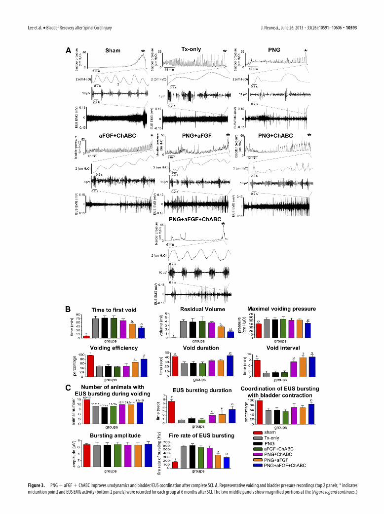
Figure 3. PNG � aFGF � ChABC improves urodynamics and bladder/EUS coordination after complete SCI. A, Representative voiding and bladder pressure recordings (top 2 panels; * indicatesmicturition point) and EUS EMG activity (bottom 2 panels) were recorded for each group at 6 months after SCI. The two middle panels show magnified portions at the (Figure legend continues.)
Lee et al. • Bladder Recovery after Spinal Cord Injury J. Neurosci., June 26, 2013 • 33(26):10591–10606 • 10593

used, the nerve segments were collected andsoaked in ChABC solution (10 �l of ChABC in1 ml of saline) for �1 h before implantation(Neubauer et al., 2007). The bladders of allspinal-cord-transected rats were expressedmanually at least twice per day throughout theexperimental period.
The measurement of micturition patterns us-ing metabolic cages. At 3 and 6 months afterspinal cord surgery, animals were placed in ametabolic cage (Braintree Scientific) for themeasurement of voiding patterns. The voidedurine was collected by a computerized systemto record micturition volume and frequency.Animals were kept in this cage for 16 h withample water and food during the period ofurine collection and measurement. Each ani-mal was recorded at least three times (with 3 dseparating each recording session), and thenthis data were averaged to represent the volun-tary micturition pattern of an individual ani-mal. The criteria for the micturition patternanalysis included the frequency of voiding per16 h and the volume per voids. The total vol-ume of expelled urine was not included be-cause of variation of water intake between theindividual animals.
Urodynamics/cystometrogram and electro-myography recordings. After the metabolic cagestudies, rats at 6 months after surgery were anes-thetized with 2% isoflurane, and a catheter wasinserted through the urethra into the bladder forthe subsequent delivery of saline. Fine-wire elec-trodes also were inserted percutaneously on bothsides of urethra for subsequent measurements ofEUS electromyography (EMG) activity. We used50-�m-diameter Teflon-insulated platinum wire(2 mm exposed tip; A-M Systems) that was in-serted into the urethra via the vagina bilaterallyalong the mid-urethra using a 30 gauge needle.The urethra can be easily identified where itbulges in the anterior vaginal wall. Next, the ratswere placed in a restraining apparatus and al-lowed to recover from isoflurane anesthesia for 1 h before the followingevaluations were performed. Continuous cystometrograms (CMGs) werecollected using constant infusion (0.08 ml/min) of saline (SP 100i single-syringe infusion pump; World Precision Instruments) through the catheterinto the bladder to elicit repetitive voids, which allowed collection of data fora large number of voiding cycles. The electrodes were connected to an alter-nating current amplifier (model P511; Astro-Med) with high- and low-passfrequency filters at 10 Hz and 1 kHz and a recording system (Dash 8X;Astro-Med) at a sample frequency of 10 kHz.
Spinal cord re-transection and pharmacological studies. At 6 monthsafter spinal cord surgery and/or treatment, the T5 level of the spinal cord
was exposed and then completely transected by two cuts. A pledget ofGelfoam soaked with 4 �l of 10% fluorescent conjugated biotinylateddextran amine (BDA) (Sigma) also was placed in the T5 spinal cordlesion site for the anterograde tracing study (see below). At 2 weeks afterspinal cord re-transection, the animals were used to retest bladder func-tion again using metabolic cages as well as analyses of urodynamics andEUS EMG recordings. In addition, animals in each SCI group were giveneither a serotonin (5-HT) receptor antagonist (5 mg/kg methysergide,i.p., once per day for 3 d) or �-methyl-DL-tyrosine [which depletes ty-rosine hydroxylase (TH) at nerve terminals; 250 mg/kg, i.p., once per dayfor 3 d], and then bladder function was tested again at the end of the thirdday of drug treatment.
Bladder morphology studies. Animals were perfused transcardially with4% neutral paraformaldehyde (PFA) in 0.01 M PBS, pH 7.4. The bladdersof all animals were collected, and weight wet was recorded. The bladderswere further fixed with 4% PFA for 1 d. The bladders were then trans-ferred to 30% sucrose, cryostat sectioned transversely (8 �m) at the levelof mid-bladder, and stained with Masson’s trichrome for additionalmorphological analysis under the light microscope.
Immunostaining of nerve fibers in the spinal cord. After transcardialperfusion with 4% PFA in 0.01 M PBS, the spinal cords were subsequentlypostfixed in the perfusing solution at 4°C. Then the tissues were cryopro-tected in 30% sucrose in PBS for 24 – 48 h at 4°C. Forty micrometersagittal cryostat sections, including the graft and spinal cord rostrally andcaudally, were examined immunohistologically for the distribution of5-HT or TH fibers at the interfaces of the PNG and beyond into the distal
4
(Figure legend continued.) point at which micturition occurred. Animals that received PNG�aFGF � ChABC treatment had much reduced amplitudes and frequencies of non-voiding blad-der contractions and much better coordination between EUS EMG bursting activity and bladdercontractions during the micturition point. Quantification of the CMG results (B) and EUS EMGbursting patterns (C) during voiding shows that the triple-combination animals had the longestdurations of EUS EMG bursting activity, better coordination between bladder contractions andEUS bursting activity during voiding, and the lowest firing rates of bursting when comparedwith animals that received alternative treatments. There results were significant when com-pared with all SCI groups ( †p � 0.05), when compared with Tx-only, PNG, and aFGF � ChABCgroups ( �p � 0.05), when compared with Tx-only, PNG, aFGF � ChABC, and PNG � ChABCgroups ( §p � 0.05), and when compared with the Tx-only, PNG, aFGF � ChABC, PNG �ChABC, and PNG � aFGF group ( Øp � 0.05).
Figure 4. PNG � aFGF � ChABC treatment improves bladder morphology. A–N, The Tx-only, PNG, aFGF � ChABC, and PNG �ChABC groups show edematous connective tissue and the proliferation of urothelial layers. Both the PNG � aFGF and PNG �aFGF � ChABC groups show less tissue edema and less proliferation of the urothelial layers (arrow). Scale bars: A, C, E, G, I, K, M,500 �m; B, D, F, H, J, L, N, 250 �m. O, PNG � aFGF � ChABC animals had the most significantly reduced bladder weight amongall the SCI groups. The red color indicates muscle fibers, and the blue color indicates collagen. There results were significant whencompared with all SCI groups ( †p � 0.05), when compared with Tx-only, PNG, aFGF � ChABC, and PNG � ChABC groups ( §p �0.05), and when compared with the Tx-only, PNG, aFGF � ChABC, PNG � ChABC, and PNG � aFGF group ( Øp � 0.05).
10594 • J. Neurosci., June 26, 2013 • 33(26):10591–10606 Lee et al. • Bladder Recovery after Spinal Cord Injury
end of the spinal cord tissue. Sections were blocked in 3% normal horseserum with 0.25% Triton X-100 in PBS for 1 h. After blocking, sectionswere exposed to an anti-5-HT polyclonal antibody (1:1500 dilution; Dia-Sorin) with anti-glial fibrillary acidic protein (GFAP) for astrocytes (1:500; Dako) or anti-TH polyclonal antibody (1:1000 dilution; ProtosBiotechnology) with anti-GFAP antibody or anti-TH antibody withanti-synaptophysin antibody (1:200 dilution; Sigma) and then incu-bated overnight at 4°C. Sections were washed, incubated with eitherfluorescein-conjugated or rhodamine-conjugated secondary antibodyfor 90 min, washed, and coverslipped with Vectashield (Vector Labora-tories). The images were collected using a Carl Zeiss LSM 510METAconfocal microscope.
To process cord tissue for 5-HT or TH immunohistochemistry in thelumbar area, transverse sections were blocked in 3% normal horse serumwith 0.25% Triton X-100 in PBS for 1 h. After blocking, the sections wereexposed to an anti-5-HT polyclonal antibody (1:1500 dilution; DiaSorin)or an anti-TH polyclonal antibody (1:1000 dilution; Protos Biotechnol-
ogy) and incubated overnight at 4°C. Afterthree rinses in PBS, the sections were exposedto a biotinylated secondary antibody (1:200;Vector Laboratories), followed by the ABCElite kit (Vector Laboratories) for 1 h each. Thereaction was visualized by treatment with0.02% 3,3�-diaminobenzidine with 0.001%H2O2 in Tris–saline for 2– 6 min, and thesections then were examined using a lightmicroscope.
Anatomical tracing procedures. For antero-grade tracing, 2 weeks after BDA placement atthe T5 level, animals were perfused with 4%PFA in PBS, and the spinal cords were collectedto perform cryosectioning at 40 �m. Immuno-staining with an anti-GFAP antibody wasadded to the tracer-labeled sections for double-labeling purposes. Z-stack images were col-lected and compiled using a Carl Zeiss LSM510META confocal microscope and softwarewith a 25� objective and excitation wave-lengths of 594 and 488 nm. For retrograde trac-ing, a solution of tracer in normal saline [4%Fluoro-Gold (FG), 2 � 2 �l] was injected care-fully into both sides of the L4 spinal cord. Twoweeks later, the rats were perfused, and thenthe brains and spinal cords were removed andsliced in the transverse plane at 40 �m thick-ness. The slices were examined using a fluores-cence microscope with ultraviolet filter.
CMG and EUS EMG data analyses. The anal-yses of CMG pressure parameters derived fromfive consecutive voiding cycles collected foreach animal included the time to first voiding,maximal voiding pressure, voiding interval,voiding duration, residual volume, and void-ing efficiency. After the last voiding cycle andafter stopping the pump infusion, the remain-ing saline in the bladder was collected and mea-sured to determine the residual volume. Thetotal volume of the bladder was measured asthe bladder capacity between the beginning ofsaline infusion and the first leaking/micturi-tion point, and then this value was multipliedby the saline infusion rate of 0.08 ml/min.Thus, the voiding efficiency was determined asthe percentage of [(total volume � residualvolume)/total volume � 100]. The EUS EMGactivity during the middle phase of bladderstorage, which was considered baseline activity,and bladder micturition represented by burst-ing activity were identified and archived in 2 ssamples (AstroVIEW X; Astro-Med). The 2 s
samples were filtered and smoothed as described previously with modi-fication (Jiang et al., 2009). Briefly, the signals were rectified, and meanamplitude and firing rates were calculated after the above correction(MyosoticSignaPoint; Myosotic). In addition, the total times of EUSEMG bursting activity were measured during the voiding period. We alsodetermined the level (by percentage) of coordination between bladdercontractions and EUS bursting activity during voiding periods for the 2 ssamples. Such coordination was defined by the alignment of the bottompart of the intraluminal pressure trace and the period of high-frequencyoscillation (IPHFO) of the EUS bursting signal. Thus, the coordinationlevel was determined as the percentage of [(coordinated number of EUSEMG signal/total number of bottom IPHFO) � 100].
Quantitative assessment of regenerating 5-HT, TH, and anterogradelylabeled nerve fibers. The numbers of 5-HT- and TH-positive fibers invarious regions below the lesion/PNG (i.e., at the graft/host interface andat 250 �m, 1 mm, 2 mm, 5 mm, 8 mm, and 12 mm) were counted under
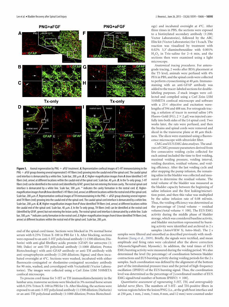
Figure 5. Axonal regeneration by PNG � aFGF treatment. A, Representative confocal images of 5-HT immunostaining in thePNG � aFGF group showing several regenerated 5-HT fibers (red) growing into the caudal end of the spinal cord. The caudal spinalcord interface is demarcated by a white line. Scale bar, 200 �m. B, C, Higher-magnification images from A show identified 5-HTfibers (red, arrow) at different locations within the caudal end of the spinal cord. Scale bar, 40 �m. D, In the Tx-only group, 5-HTfibers (red) can be identified in the rostral cord (identified by GFAP, green) but not entering the lesion cavity. The rostral spinal cordinterface is demarcated by a white line. Scale bar, 500 �m. * indicates the cavity formation in the rostral cord. E, Higher-magnification images from D show identified 5-HT fibers (red, arrow) at different locations within the rostral end of the spinal cord.Scale bar, 200 �m. F, Representative confocal images of TH immunostaining in the PNG � aFGF group showing several regener-ated TH fibers (red) growing into the caudal end of the spinal cord. The caudal spinal cord interface is demarcated by a white line.Scale bar, 200 �m. G, H, Higher-magnification images from F show identified TH fibers (red, arrow) at different locations withinthe caudal end of the spinal cord. Scale bar, 40 �m. I, In the Tx-only group, TH fibers (red) can be identified at the rostral cord(identified by GFAP, green) but not entering the lesion cavity. The rostral spinal cord interface is demarcated by a white line. Scalebar, 500 �m. * indicates cavity formation in the rostral cord. J, Higher-magnification images from I show identified TH fibers (red,arrow) at different locations within the rostral end of the spinal cord. Scale bar, 200 �m.
Lee et al. • Bladder Recovery after Spinal Cord Injury J. Neurosci., June 26, 2013 • 33(26):10591–10606 • 10595

the microscope from all spinal-cord-injuredanimals. Two observers blinded to all treat-ment groups counted the fibers. Every 1-in-6sections from each animal were used to countnerve fiber segments and then multiplied by 6to represent a quantitative estimate of totalnumbers of fiber segments beyond the lesionper animal. The numbers of BDA-positive fi-bers in our anterograde tracing study werecounted in several areas, including 1 mm ros-tral to the lesion/PNG, within the middle of thelesion/PNG, and at different sites caudal to thelesion/PNG (i.e., at the graft/host interface andat 250 �m, 1 mm, 2 mm, 5 mm, and 8 mm).The section selection and the blinded methodof counting fibers were the same as describedabove.
Statistical analyses. All data are reported asmean � SEM. A two-way (group � time)ANOVA followed by Tukey’s post hoc ANOVAtest were used to determine significant differ-ences between groups. Linear regressions wereperformed using SigmaStat statistical software.Significant differences were determined at p �0.05. All behavioral tests and data analyses weredone in a blinded manner during this entirestudy.
ResultsMultiple PNGs with aFGF/fibrin andChABC lead to markedly improvedmicturition patterns in rats that hadsustained complete T8 spinal cordtransectionVoiding patterns were recorded with theuse of metabolic cages at 3 and 6 monthsafter spinal cord surgery. All the animalsin each group were used in this study. Twomain criteria, including the total numberof voids over a set time and the volume permicturition, were used to make compari-sons between the groups. Representativemicturition patterns of sham, Tx-only,and PNG � aFGF � ChABC groups areshown in Figure 2A. In each “step-like”segment of the graph, the horizontal linerepresents the time period between indi-vidual micturition events, and the verticallines depict the volume of urine expelled(change from baseline). At 3 months, thesham group (29 voids/16 h) showed sig-nificantly higher total numbers of voidsthan the spinal-cord-injured as well as theinjury � treatment groups (Fig. 2B; p �0.05). However, a comparison of the sixSCI groups revealed that the frequency ofmicturition in the PNG � aFGF �ChABC group (13 voids/16 h) was signif-icantly higher than the Tx-only (6voids/16 h), PNG (7 voids/16 h), aFGF �ChABC (6 voids/16 h), PNG � ChABC (8voids/16 h), and PNG � aFGF (8.5voids/16 h) groups (Fig. 2B; p � 0.05).The sham group (0.3 ml/void) also dem-onstrated a significantly lower volume per
Figure 6. Significant axonal regeneration with PNG�aFGF�ChABC treatment. The triple combination of ChABC with PNG and aFGFpromotes regeneration of both 5-HT and TH fibers beyond the PNG/caudal spinal cord interface. A, Confocal images of a sagittal section ofthe spinal cord showing that regenerated 5-HT fibers (red, white arrow head) have grown beyond the PNG (GFAP-negative area) andpenetrated deep into the CNS (identified by GFAP, green). The caudal spinal cord interface is demarcated by a white line. Scale bar, 300�m.B–D, Higher-magnification images from A show identified 5-HT fibers (white arrows) at different locations within the caudal end of thespinal cord. Scale bar, 40 �m. Note in A the alignment of the astrocytes at a point at which axons reenter the cord. E, Confocal photomi-crographs of a sagittal section of the spinal cord showing that TH fibers (red, white arrow head) have regenerated beyond the graft (GFAPnegative area) and well into the CNS (identified by GFAP, green). The arrowheads indicate TH fibers crossing the PNG/caudal spinal cordinterface (demarcated by a white line). Scale bar, 300 �m. F–H, Higher-magnification images from E show identified TH fibers (whitearrows) at different distances beyond the PNG/caudal spinal cord interface. Scale bar, 40 �m. I, Confocal images from immunostainingreveal a close proximity between the regenerated fibers and synapsin (SYN) puncta in PNG � aFGF � ChABC-treated animals. Double-staining image indicates the colocalization of TH fibers and SYN (arrow). Scale bar, 70 mm. J, Regenerated 5-HT fibers with expansiveterminal arborizations were identified within the lumbar cord as depicted in this representative transverse section. Scale bar, 200�m. K, L,Higher-magnification images from J. Note the bouton structures on 5-HT fibers (white arrow). The interface between gray matter andwhite matter is demarcated by a white dashed line. GM, Gray matter; WM, white matter. Scale bar, 50 �m. M, N, TH fibers were alsoidentified well into the lumbar cord as shown in this representative transverse section. Note the bouton structures on TH fibers (whitearrow). The interface between gray matter and white matter is demarcated by a white dashed line. GM, Gray matter; WM, white matter.Scale bars: M, 200 �m; N, 50 �m. O, Quantitative analysis showed greater numbers and longer-distance regeneration of 5-HT fibersbeyond the lesion in the PNG � aFGF � ChABC treatment group compared with the other SCI groups. P, Quantitative analysis showedmore and longer-distance regeneration of TH fibers beyond the lesion in the PNG� aFGF � ChABC treatment group versus the other SCIgroups. †p�0.05whencomparedwithTx-only,PNG,andaFGF�ChABCgroups,*p�0.05whencomparedwithTx-only,PNG,aFGF�ChABC,andPNG�ChABCgroups,and**p�0.05whencomparedwithTx-only,PNG,aFGF�ChABC,PNG�ChABC,andPNG�aFGFgroups.
10596 • J. Neurosci., June 26, 2013 • 33(26):10591–10606 Lee et al. • Bladder Recovery after Spinal Cord Injury

micturition than all the other groups (Fig. 2C; p � 0.05). Again, acomparison of the six SCI groups showed that the volume permicturition in the PNG � aFGF � ChABC group (1.3 ml/void)was significantly lower than the Tx-only (2.6 ml/void), PNG (2.4ml/void), aFGF � ChABC (2.5 ml/void), PNG � ChABC (2.2ml/void), and the PNG � aFGF (2.1 ml/void) groups (Fig. 2C;p � 0.05). In addition, the PNG � ChABC and PNG � aFGFgroups also showed significantly lower volumes per micturitionwhen compared with the Tx-only, PNG, and aFGF � ChABCgroups (Fig. 2B,C; p � 0.05).
At 6 months, the sham group still had the highest frequency(28 voids/16 h) and lower volume per micturition (0.25 ml) thanall six SCI groups (Fig. 2B; p � 0.05). Similar to but now muchmore obvious than the patterns at 3 months, the PNG � aFGF �ChABC group demonstrated a much higher frequency (18voids/16 h) than the Tx-only (5 voids/16 h), PNG (6 voids/16 h),aFGF � ChABC (7 voids/16 h), PNG � ChABC (10 voids/16 h),and PNG � aFGF (11 voids/16 h) groups (Fig. 2B; p � 0.05). Thecomplete combination group also had markedly lower volumesper micturition (1.2 ml) than the Tx-only (3.2 ml), PNG (2.9 ml),aFGF � ChABC (2.7 ml), PNG � ChABC (1.9 ml), and PNG �aFGF (1.8 ml) groups (Fig. 2C; p � 0.05). In addition, the PNG �ChABC and PNG � aFGF groups also showed significantlyhigher frequency and lower volume per micturition when com-pared with the Tx-only, PNG, and aFGF � ChABC groups (Fig.2B,C; p � 0.05). When one compares the 6 month versus 3month data within groups, the Tx-only, PNG, and aFGF �ChABC animals showed significantly increasing volumes permicturition over time, which is indicative of ongoing pathologywithin the bladder. In contrast, the PNG � aFGF � ChABCgroup especially, rather than declining, showed improvements inbladder function at 6 months, with more voids and lower vol-umes per micturition than at the 3 month time period (Fig. 2B,C;p � 0.05). In general, the metabolic cage study demonstrated thatthe PNG � aFGF � ChABC group had developed a more normalmicturition pattern with higher-frequency micturition episodesand less voiding volume per micturition than the other five SCIgroups. This indicates that the PNG � aFGF � ChABC animalsdid not need to store excessively large volumes of urine in the
bladder to void, which is moving furthertoward a normal pattern.
PNG with aFGF/fibrin and ChABCmarkedly improves CMG and EUSEMG activity in previously spinal-cord-transected ratsMeasurements of bladder contractionsand EUS activity were used to further in-vestigate the quality of bladder function at6 months after SCI. Six animals in thesham control group, eight animals in theTx-only group, and eight animals in eachof the repaired groups were used inthis investigation. During continuous-infusion CMG in sham control rats beforethe onset of voiding (a period when thebladder is not expelling urine), the EUSdisplayed low-amplitude tonic activity.There are no overt bladder contractionsbefore the voiding event (see a typicalbladder pressure trace of sham animals inFig. 3A). The tonic EUS activity increasesin amplitude at the onset of micturition
and then (peculiar to rats) rises precipitously to a large-amplitudebursting pattern during the voiding period (see a typical EUSEMG trace of sham animals in Fig. 3A). The “ramp-like” pairs ofdashed lines on both bladder pressure and EUS EMG recordingsindicate the point during the voiding event from which a greatlyexpanded portion of the trace is depicted. At the micturitionpoint, the sham group demonstrates the coordination that nor-mally occurs during the micturition cycle between bladder con-tractions and EUS EMG bursting patterns (Fig. 3A). At 6 monthsafter T8 spinal cord complete transection, there was significantDSD that was displayed as large-amplitude non-voiding contrac-tions and atypical EUS EMG bursting activity during the voidingattempt (both bladder pressure trace and EUS EMG trace of Tx-only in Fig. 3A). The PNG, aFGF � ChABC, and PNG � ChABCgroups (Fig. 3A) had similar maladaptive patterns of bladderpressure and EUS EMG activity. Although the PNG � aFGFgroup (Fig. 3A) showed somewhat diminished amplitudes ofnon-voiding bladder contractions, with partial EUS EMG burst-ing activity during voiding, there was no coordination betweenthe EUS and bladder contractions during micturition. In contrastto all the other treated SCI groups, the PNG � aFGF � ChABCgroup (Fig. 3A) showed the greatest reduction in frequency ofDSD as well as much reduced amplitudes of non-voiding bladdercontractions. In addition, and rather remarkably, the PNG �aFGF � ChABC group also demonstrated the characteristic pha-sic EUS EMG bursting activity that was well coordinated withbladder contractions during voiding.
The CMG analyses also showed that the PNG � aFGF �ChABC group (at 39.8 min) had the shortest time to the first void(p � 0.05; Fig. 3B) among all the SCI groups (the others were54.6 –75.4 min). The sham group waited 9.4 min to first void. Theresidual volume (Fig. 3B) in the PNG � aFGF � ChABC group(1.5 ml) was the lowest amount (p � 0.05; Fig. 3B) comparedwith other SCI groups (the others were 2.7– 4.1 ml), whereas thesham group was 0.03 ml. The maximal voiding pressure (Fig. 3B)in the PNG � aFGF � ChABC group (43.2 cm H2O) was signif-icantly lowest (p � 0.05) among all SCI groups (the others were52.5–54.3 cm H2O), whereas the sham group was 41.2 cm H2O.The voiding efficiency (Fig. 3B) in the PNG � aFGF � ChABC
Figure 7. A few BDA-positive fibers grow beyond the graft in the PNG � aFGF animals. A, A low-magnification sagittal sectionshows, with the combination of BDA labeling (red) and GFAP staining (green), the overall anatomy of the PNG and spinal cord (leftside is rostral and right side is caudal). Scale bar, 500 �m. B, High-magnification image shows a few BDA-labeled fibers (red)entering the caudal end of the spinal cord (GFAP-positive area) from PNG (GFAP-negative area). The interface of the PNG and spinalcord is demarcated by a white line. Scale bar, 100 �m.
Lee et al. • Bladder Recovery after Spinal Cord Injury J. Neurosci., June 26, 2013 • 33(26):10591–10606 • 10597

group (82%) was the highest amount (p � 0.05) compared withthe other SCI groups (48 – 67%), whereas the sham group was98.2%. The voiding duration (Fig. 3B) in the PNG � aFGF �ChABC group (43.9 s) was clearly the longest (p � 0.05) com-pared with other SCI groups (which were 27.5–33.6 s), whereasthe sham group was 42.1 s. The voiding interval (Fig. 3B) in thePNG � ChABC, PNG � aFGF, and PNG � aFGF � ChABC
groups (8.8 –10.1 min) was again the longer (p � 0.05) comparedwith the Tx-only, PNG, and aFGF � ChABC groups (1.9 –2.3min), whereas the sham group was 8.9 min. Together, the CMGdata indicate that the cord-injured animals treated with a combi-nation of PNG � aFGF � ChABC demonstrated far less bladderincontinence and distension as well as fewer non-voiding bladdercontractions, indicating a reduction of bladder hyperreflexia.
Figure 8. Anterograde tracing to label regenerating axons in the PNG � aFGF � ChABC treatment group. A, These three low-magnification sagittal sections of confocal images show with BDAlabeling (red) and GFAP staining (green) and the two combined, the overall anatomy of the PNG and spinal cord (left side is rostral and right side is caudal). Scale bar, 1000 �m. B, High-magnificationconfocal image shows the large number of BDA-labeled fibers (red) entering the PNG (GFAP negative area) from the rostral end of the spinal cord (GFAP-positive area). The interface of the PNG andspinal cord is demarcated by a white line. Scale bar, 100 �m. Significant numbers of BDA-labeled fibers were identified within the PNG (C) and penetrating deeply into the interface of the PNG withthe caudal end of the spinal cord (D) (demarcated by a white line). Note the alignment of the GFAP-positive astrocyte processes. D�, An adjacent section to D showing more evidence of theBDA-labeled fibers (red and arrowheads) crossing the PNG/caudal spinal cord interface. E–G, The BDA-labeled fibers (red, white arrows) were identified at various locations continuing far caudallyinto the spinal cord. Scale bar, 50 �m. H, Quantitative analysis showed greater numbers and longer-distance penetration of BDA-labeled fibers beyond the lesion in the PNG � aFGF � ChABCtreatment group than the other SCI groups. �p � 0.05 when compared with Tx-only and aFGF � ChABC groups, �p � 0.05 when compared with Tx-only group, †p � 0.05 when compared withTx-only, PNG, and aFGF � ChABC groups, *p � 0.05 when compared with Tx-only, PNG, aFGF � ChABC, and PNG � ChABC groups, and **p � 0.05 when compared with Tx-only, PNG, aFGF �ChABC, PNG � ChABC, and PNG � aFGF groups.
10598 • J. Neurosci., June 26, 2013 • 33(26):10591–10606 Lee et al. • Bladder Recovery after Spinal Cord Injury

The EUS EMG analyses revealed that 100% of the animals inthe sham group showed the presence of EUS EMG bursting ac-tivity during voiding. Seventy percent of the animals among allthe SCI groups had some sort of EUS EMG activity as well. How-ever, the duration of EUS EMG bursting in the PNG � aFGF �ChABC group (3.8 s) was significantly longer (p � 0.05) than theother SCI groups (which were 0.8 –2.2 s), whereas the shamgroup was 5.6 s (Fig. 3C). In addition, overall coordination be-tween bladder contractions and EUS bursting during voiding inthe PNG � aFGF � ChABC group (85%) was significantly higherthan the other SCI groups (which were 55–70%). There were nosignificant differences in the EUS EMG bursting amplitudes
among the groups (Fig. 3C). The firing rateof EMG bursting in the PNG � aFGF �ChABC group (297 Hz) was significantlylower (p � 0.05) than the other SCI groups(380–596 Hz), whereas the sham was 177Hz (Fig. 3C). In general, the PNG�aFGF�ChABC treatment provides significant re-covery of EUS EMG activity, allowing forbetter coordination with bladder contrac-tions. All the aforementioned measure-ments are indicative of a reduction of DSDand higher efficiency of micturition controlin the full-combination treatment animals,with some indicators achieving normal ornear-normal levels. Additional improve-ments in urinary function beyond that at 6months were not seen in three animals at 9months after lesion and treatment with thefull-combination therapy.
PNG � aFGF � ChABC treatmentimproves bladder morphologyBecause of the need to store abnormallylarge amounts of urine, SCI animals dis-play considerably increased bladder sizesand weights than in sham animals at 6months after complete spinal cord tran-section. The bladder wall of the Tx-only,PNG, and aFGF � ChABC groups had de-veloped edematous connective tissue withextensive proliferation of the urotheliallayers facing the lumen (Fig. 4A–N). ThePNG � aFGF and the PNG � ChABCgroups, but especially the PNG � aFGF �ChABC group, showed much less tissueedema and less proliferation of theurothelial layers, resulting in significantlyless total bladder weight (Fig. 4O).
Evidence of regeneration and synapseformation by 5-HT and TH fiberscrossing the PNG/caudal spinal cordinterfaceGiven the improvements in voidingbehavior in rats that received the ChABC �PNG � aFGF treatment, we next exam-ined whether axonal regrowth couldextend beyond the caudal interface be-tween the PNG and spinal cord. Immuno-staining demonstrated in PNG � aFGF(Fig. 5A–C,F–H), PNG � ChABC (data
not shown), and especially PNG � aFGF � ChABC (Fig. 6A–J)groups that both TH- and 5-HT-positive fibers (these systems areimportant in controlling micturition) had extended into thebridge, across the caudal PNG/spinal cord interface and well intothe caudal cord (the CNS compartment was demarcated by thepresence of GFAP). 5-HT- and TH-positive fibers were identifiedonly in the rostral penumbra of the lesion in the Tx-only group(Fig. 5D,E, I, J). There was absolutely no evidence that TH or5-HT fibers had crossed into the caudal cord beyond the lesion inthe PNG and aFGF � ChABC groups. Interestingly, precisely atthe interface between the PNG and CNS tissue (especially in theadditionally aFGF- or ChABC-treated or both treated animals),
Figure 9. Retrograde anatomical tracing indicated the neuronal populations that have regenerated axons below the lesion sitein the PNG � aFGF � ChABC treatment group. A, FG-labeled cells were identified in the pontine micturition center, includingBarrington’s nucleus (arrow) and D region (arrowhead). Scale bar, 800 �m. B, FG-labeled cells were identified in both raphe nuclei(arrow) and the reticular formation (arrowhead). Scale bar, 400 �m. C, The retrogradely FG-labeled neurons (arrow) could beidentified in locus ceruleus. Scale bar, 250 �m. D, Quantification of the FG-labeled cells in the retrograde tracing study. Me,Mesencephalic nucleus of trigeminal nerve; DTN, dorsal tegmental nucleus; 4V, fourth ventricle; VCA, ventral cochlear nucleus,anterior; Py and PT, pyramidal tract. *p � 0.05 when compared with Tx-only, PNG, and aFGF � ChABC groups, and **p � 0.05when compared with Tx-only, PNG, aFGF � ChABC, PNG � ChABC, and PNG � aFGF groups.
Lee et al. • Bladder Recovery after Spinal Cord Injury J. Neurosci., June 26, 2013 • 33(26):10591–10606 • 10599

there often occurred a rectilinear align-ment of the astrocytes and at these pointsaxons appear to have gained access to thedistal cord. For example, in Figure 5A, ax-ons reenter the cord at a dorsal locationthrough a zone of aligned astrocytes in-stead of more ventrally in which there wasa more “wall-like” arrangement (Fig. 6A).Importantly, there were far greater num-bers of regenerated 5-HT or TH axons andregenerated axons that traveled longerdistances beyond the graft site to lumbarlevels in the PNG � aFGF � ChABCgroup (Fig. 6J–N) than in the PNG �ChABC and PNG � aFGF groups.
With the use of double labeling, it wasapparent that regenerated fibers that en-tered the distal end of the spinal cordcould express synapse-associated pro-teins, suggesting the possibility of func-tional reconnection (Fig. 6I). Overall,detection of considerable numbers of5-HT and TH fibers growing far beyondthe interface of the PNG/caudal spinalcord in the PNG � aFGF � ChABC group(for a quantitative analysis, see Fig. 6O,P)is consistent with their possible involve-ment in the improvement of bladderfunction.
Anterograde and retrograde labeling ofregenerating neuronsBDA for anterograde tracing was placed ata fresh transection site at T5, which wasabove the original transection site (T8).No BDA-labeled fibers were found beyond the caudal end of thebridge in any of the Tx-only, PNG, and aFGF � ChABC animals.The PNG animals showed BDA-labeled fibers growing into butnot exiting the graft (data not shown). There were scattered fibersjust beyond the graft in the PNG � aFGF group (Fig. 7), as well asthe PNG � ChABC group (data not shown). Remarkably, thePNG � aFGF � ChABC animals (Fig. 8A–G) had substantialregeneration into and beyond the graft. Confocal reconstructionsand quantitative analysis (Fig. 8H) confirmed that regeneratedfibers had penetrated deeply into the gray matter near the PNG/caudal spinal interface, and many were identified in the whitematter traveling several millimeters farther down the cord, with afew making it to the very end of our sections.
Retrograde tracing by injecting FG into the spinal cord (atapproximately L4; nearly 2 cm below the rostral graft site) wasused to further determine the neural populations that may con-tribute to nerve regeneration well beyond the bridge/cord inter-face. In the Tx-only, PNG, and aFGF � ChABC groups, therewere no FG-labeled neurons anywhere in the rostral cord orbrain. In contrast, FG-labeled neurons could be clearly identifiedwithin different regions and in several nuclei in the PNG �ChABC, PNG � aFGF, and PNG � aFGF � ChABC groups, withfar more in the triple-combination animals. The PNG � aFGF �ChABC group showed FG-labeled cells in the all important pon-tine micturition center (Barrington’s nuclei and the D-region), aswell as the locus ceruleus, nuclei raphe magnus, and the reticularformation (gigantocellular reticular nuclei) (Fig. 9A–C). BesidesFG-labeled cells in the brain, the cervical and thoracic spinal cord
also contained labeled cells likely to be part of the propriospinalsystem. The PNG � aFGF and PNG � ChABC groups alsoshowed a few labeled cells in the D-region, raphe magnus nuclei,reticular formation, and the cervical spinal cord. Importantly, thePNG � aFGF � ChABC group contained far more FG-labeledcells and in more neural populations than did the PNG � ChABCor PNG � aFGF groups (Fig. 9D).
Effects of re-transecting the cord in animals with and withoutrepair bridgesAnimals in all SCI groups (five animals in each group) were usedto perform spinal cord re-transection at the T5 level. They werereevaluated for bladder function 2 weeks later. In the Tx-only,PNG, aFGF � ChABC, and PNG � ChABC groups, there wereno or only minor differences in their already abnormal overnightmicturition patterns after spinal cord re-transection (Fig. 10A).There also were no significant differences in both CMG/EUSEMG parameters after spinal cord re-transection, including EUSbursting activity during voiding in the non-regenerating groups,but there were some changes in the relatively poorly regeneratinggroups (Fig. 11A,B). CMG analysis in the PNG � ChABC groupdid show significant increases in the time to first void and de-creases in voiding intervals (p � 0.05), but there was no differ-ence in EUS EMG analysis after spinal cord re-transection.Metabolic cage analyses of the PNG � aFGF group showed sig-nificant differences (p � 0.05) in increased volume per micturi-tion but not in voiding frequency after spinal cord re-transection(Fig. 10A). CMG and EUS EMG analysis showed continuing in-
Figure 10. Spinal cord re-transection eliminates or reduces the improvements in bladder function after regeneration. A, Rawdata analysis of a representative PNG � aFGF � ChABC-treated animal at 2 weeks after T5 spinal cord re-transection. Themicturition patterns revert to lower frequencies of voiding and higher volumes per micturition. The PNG � aFGF group alsoincreased volume per micturition after re-transection. Tx-only, PNG, and aFGF � ChABC animals did not change. †p � 0.05 whencompared with the before treatment. B, After re-transection, the PNG � aFGF � ChABC group developed increased amplitudesand frequencies of non-voiding bladder contractions. Coordination between bladder contractions and EUS EMG bursting activityduring voiding period was eliminated (* indicates micturition point).
10600 • J. Neurosci., June 26, 2013 • 33(26):10591–10606 Lee et al. • Bladder Recovery after Spinal Cord Injury
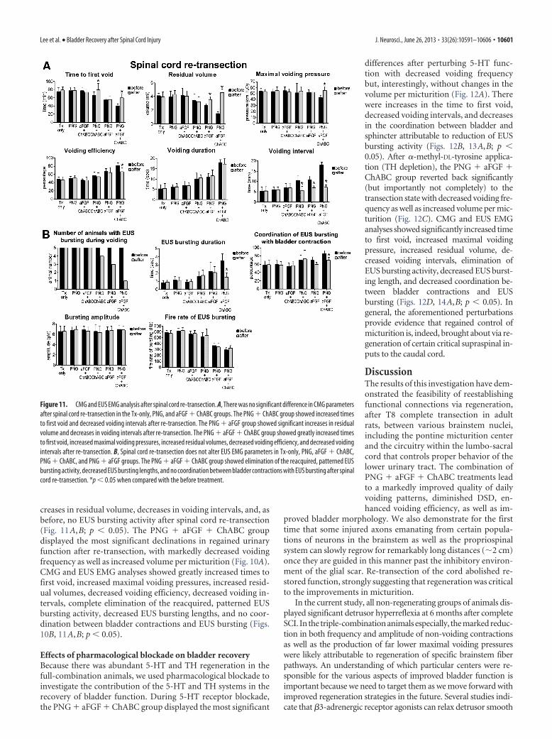
creases in residual volume, decreases in voiding intervals, and, asbefore, no EUS bursting activity after spinal cord re-transection(Fig. 11A,B; p � 0.05). The PNG � aFGF � ChABC groupdisplayed the most significant declinations in regained urinaryfunction after re-transection, with markedly decreased voidingfrequency as well as increased volume per micturition (Fig. 10A).CMG and EUS EMG analyses showed greatly increased times tofirst void, increased maximal voiding pressures, increased resid-ual volumes, decreased voiding efficiency, decreased voiding in-tervals, complete elimination of the reacquired, patterned EUSbursting activity, decreased EUS bursting lengths, and no coor-dination between bladder contractions and EUS bursting (Figs.10B, 11A,B; p � 0.05).
Effects of pharmacological blockade on bladder recoveryBecause there was abundant 5-HT and TH regeneration in thefull-combination animals, we used pharmacological blockade toinvestigate the contribution of the 5-HT and TH systems in therecovery of bladder function. During 5-HT receptor blockade,the PNG � aFGF � ChABC group displayed the most significant
differences after perturbing 5-HT func-tion with decreased voiding frequencybut, interestingly, without changes in thevolume per micturition (Fig. 12A). Therewere increases in the time to first void,decreased voiding intervals, and decreasesin the coordination between bladder andsphincter attributable to reduction of EUSbursting activity (Figs. 12B, 13A,B; p �0.05). After �-methyl-DL-tyrosine applica-tion (TH depletion), the PNG � aFGF �ChABC group reverted back significantly(but importantly not completely) to thetransection state with decreased voiding fre-quency as well as increased volume per mic-turition (Fig. 12C). CMG and EUS EMGanalyses showed significantly increased timeto first void, increased maximal voidingpressure, increased residual volume, de-creased voiding intervals, elimination ofEUS bursting activity, decreased EUS burst-ing length, and decreased coordination be-tween bladder contractions and EUSbursting (Figs. 12D, 14A,B; p � 0.05). Ingeneral, the aforementioned perturbationsprovide evidence that regained control ofmicturition is, indeed, brought about via re-generation of certain critical supraspinal in-puts to the caudal cord.
DiscussionThe results of this investigation have dem-onstrated the feasibility of reestablishingfunctional connections via regeneration,after T8 complete transection in adultrats, between various brainstem nuclei,including the pontine micturition centerand the circuitry within the lumbo-sacralcord that controls proper behavior of thelower urinary tract. The combination ofPNG � aFGF � ChABC treatments leadto a markedly improved quality of dailyvoiding patterns, diminished DSD, en-hanced voiding efficiency, as well as im-
proved bladder morphology. We also demonstrate for the firsttime that some injured axons emanating from certain popula-tions of neurons in the brainstem as well as the propriospinalsystem can slowly regrow for remarkably long distances (�2 cm)once they are guided in this manner past the inhibitory environ-ment of the glial scar. Re-transection of the cord abolished re-stored function, strongly suggesting that regeneration was criticalto the improvements in micturition.
In the current study, all non-regenerating groups of animals dis-played significant detrusor hyperreflexia at 6 months after completeSCI. In the triple-combination animals especially, the marked reduc-tion in both frequency and amplitude of non-voiding contractionsas well as the production of far lower maximal voiding pressureswere likely attributable to regeneration of specific brainstem fiberpathways. An understanding of which particular centers were re-sponsible for the various aspects of improved bladder function isimportant because we need to target them as we move forward withimproved regeneration strategies in the future. Several studies indi-cate that �3-adrenergic receptor agonists can relax detrusor smooth
Figure 11. CMG and EUS EMG analysis after spinal cord re-transection. A, There was no significant difference in CMG parametersafter spinal cord re-transection in the Tx-only, PNG, and aFGF � ChABC groups. The PNG � ChABC group showed increased timesto first void and decreased voiding intervals after re-transection. The PNG � aFGF group showed significant increases in residualvolume and decreases in voiding intervals after re-transection. The PNG � aFGF � ChABC group showed greatly increased timesto first void, increased maximal voiding pressures, increased residual volumes, decreased voiding efficiency, and decreased voidingintervals after re-transection. B, Spinal cord re-transection does not alter EUS EMG parameters in Tx-only, PNG, aFGF � ChABC,PNG � ChABC, and PNG � aFGF groups. The PNG � aFGF � ChABC group showed elimination of the reacquired, patterned EUSbursting activity, decreased EUS bursting lengths, and no coordination between bladder contractions with EUS bursting after spinalcord re-transection. *p � 0.05 when compared with the before treatment.
Lee et al. • Bladder Recovery after Spinal Cord Injury J. Neurosci., June 26, 2013 • 33(26):10591–10606 • 10601

muscle in rats (Woods et al., 2001; Frazier etal., 2006). Because pharmacological block-ade of TH synthesis mostly eliminated theimprovements in detrusor hyperreflexia,this points to the important contribution ofregenerated TH fibers (some of which werelikely adrenergic) in the reduction of abnor-mal bladder wall activity.
Poor-quality EUS bursting activity,which can lead to some ability to urinate,albeit abnormally, can slowly return inrats after mid-thoracic complete spinalcord transection as was demonstratedwith the present data and which confirmsother studies (Cheng and de Groat, 2004;Leung et al., 2007). This is likely attribut-able to synaptic plasticity in the vicinity ofthe central pattern generator for reflexEUS bursting activity that has been dis-covered near the L4 level (Chang et al.,2007). Indeed, in the present study, spinalcord re-transection at 6 months did notaffect the irregular patterns of EUS burst-ing that had developed in the Tx-only,PNG, and aFGF � ChABC groups, withno evidence of nerve regenerationthrough the lesion site. The application of5-HT1A receptor agonists as well as �2/3-adrenoceptor agonists has been shown toimprove EUS activity and voiding effi-ciency (Morita et al., 2000; Dolber et al.,2007), and, indeed, the quality of the re-turned, more regularly patterned EUSbursting particularly in the PNG � aFGF �ChABC group was degraded but, impor-tantly, not completely eliminated via phar-macological applications that block both5-HT and TH. This indicates that these par-ticular reformed supraspinal inputs, butalso other regenerating pathways likely fromthe pontine micturition center or reticularformation, may directly foster recovery orindirectly help regulate neuroplasticity andallow the animal to regain control of thiscritical sphincter.
Why did this particular combinationtherapy allow for such robust axonal re-generation well beyond that of our previ-ous studies (Houle et al., 2006; Alilain etal., 2011) in which we used ChABC andsingle, longer bypass PNS bridges thatwere directed just above ventral horn graymatter? We suggest that maximizing surface area with the use ofmultiple grafts coupled with the enzyme effectively increases ac-cess to the distal white matter, which, in turn, can permit long-distance regeneration (Davies et al., 1997, 1999; Silver and Miller,2004; Gensel et al., 2009; Lu et al., 2012b). In addition, the use ofFGF may have a variety of beneficial synergistic effects. The FGFfamily members have multiple functions, including their capacityto modulate cell proliferation, migration, differentiation, andsurvival (Teng et al., 1998; Rabchevsky et al., 2000; Lee et al.,2006). Previous work using organotypic slice cultures (Lee et al.,2002a) and an in vivo SCI model combining aFGF with a PNG
(Cheng et al., 1996; Lee et al., 2002b; Tsai et al., 2005) has shownthe important role of aFGF in enhancing axonal regeneration. Inother studies, aFGF was demonstrated to promote axonal regen-eration and reduce death of neurons after SCI or brain ischemia(Russell et al., 2006). In addition, FGF has been shown to be asupportive molecular enhancer of long-distance, unbranchedaxon regrowth from adult DRG transplants across the corpuscallosum (Jin et al., 2008; Ziemba et al., 2008). Therefore, theapplication of aFGF in our repair strategy is important not only tofacilitate axonal growth in general but also to help allow axons togrow straight, especially at the entrances and exits of the PNG.
Figure 12. Pharmacological manipulations eliminate or reduce the improvements in bladder function after regeneration. A,Raw data of a representative PNG� aFGF� ChABC-treated animal at 3 d after methysergide application. The micturition patternsreturned to lower frequencies of voiding, but there was no difference in volume per micturition. B, Coordination between bladdercontractions and EUS EMG bursting activity during voiding was diminished, but no significant differences occurred in the ampli-tudes of non-voiding contractions. C, Raw data of a representative PNG � aFGF � ChABC-treated animal at 3 d after �-methyl-DL-tyrosine application, the micturition patterns returned to lower frequencies of voiding and higher volumes per micturition. D,Amplitudes of non-voiding bladder contractions increased, and coordination between bladder contractions and EUS EMG burstingactivity during the voiding period was eliminated. †p � 0.05 when compared with the before treatment.
10602 • J. Neurosci., June 26, 2013 • 33(26):10591–10606 Lee et al. • Bladder Recovery after Spinal Cord Injury

FGF as a trophic factor is unusual in this respect because otherneurotrophins, such as NGF, BDNF, or neurotrophin-3, pro-mote a more branched pattern of axonal growth that tends to beconstraining to elongation (Lu and Tuszynski, 2008; Lu et al.,2012a). Thus, FGF seems to allow axons to grow more readilythrough the territory in which it is expressed. FGF may also alterthe maturation state (Hoke and Silver, 1996; Fawcett and Asher,1999) or change the morphology of astrocytes at the graft/cordinterface into a more primitive bipolar shape (Imura et al., 2006;Goldshmit et al., 2012) that improves the ability of axons to passthrough. ChABC may also aid in aligning astrocytes because theenzyme has been shown to enhance integration of Schwann cellsand astrocytes by altering the production of inhibitory matricesthat form between them when they are compelled to interact withone another after transplantation (Grimpe et al., 2005).
It is now well established that the application of ChABC aloneor in combination with other interventions, including PNGs orcellular transplantation, can promote sprouting as well as nerveregeneration and functional recovery after SCI (Bradbury et al.,2002; Caggiano et al., 2005; Fouad et al., 2005; Houle et al., 2006;Massey et al., 2006; Karimi-Abdolrezaee et al., 2010; Alilain et al.,2011; Bukhari et al., 2011; Imagama et al., 2011; Jefferson et al.,
2011; Wang et al., 2011). Typically, how-ever, the extent of axonal regrowth/sprouting within the CNS compartment isfairly restricted to the region of CSPG deg-radation (Barritt et al., 2006; Starkey et al.,2012). We have identified a subset of neu-rons situated primarily within the brains-tem and reticular formation (and which areknown to play important roles in bladderfunction) (McMahon and Spillane, 1982) aswell as the propriospinal system that can re-grow lengthy axons once a permissive envi-ronment that allows them past the glial scaris provided. We saw no evidence of regener-ation by cortico-spinal or rubro-spinal ax-ons (unlike the results by Cheng et al., 1996),which is one possible reason for the limitedrecovery of locomotor function (Basso–Beattie–Bresnahan score improved from 2to �7 in the triple-combination animals;data not shown). Although propriospinal(Bareyre et al., 2004; Fenrich and Rose,2011; van den Brand et al., 2012), 5-HT(Azmitia et al., 1978; Molliver et al., 1990;Hawthorne et al., 2011), and certain otherbrainstem neurons (Hill et al., 2001) are wellknown to possess an enhanced capacity toregenerate or sprout axons relatively shortdistances after injury, the lengthy regenera-tion of these as well as those from severalother nuclei so far down the cord was quitesurprising. Propriospinal neurons have thecapacity to regenerate an axon right thoughvery small, carefully crafted lesions in theventral white commissure (Fenrich andRose, 2011). Serotonergic neurons expresshigher levels of GAP-43 and �1 integrinsthan do cortical neurons, which helps themmaintain an active growth cone and sproutrobustly rather than becoming dystrophicand dying back within the lesion environ-
ment (Hawthorne et al., 2011). Indeed, these neuronal popula-tions appear to behave as if they were intrinsically conditioned(Erturk et al., 2012). Recently, an exciting series of papers hasrevealed that robust axonal regeneration can be elicited viamanipulation of the mTOR/PTEN and SOCS3/STAT3 path-ways (Park et al., 2010; Sun et al., 2011; de Lima et al., 2012). Itstill needs to be determined whether the long-distance regen-erating neurons intrinsically modulate these growth regula-tory genes. An understanding of the special strengths of theseneurons may aid in devising strategies to instill their uniquenavigational attributes into other types of neurons that fareless well after injury or when they are presented with aregeneration-promoting environment.
The current study provides an experimental framework forstimulating functional regeneration after acute severe SCI. Mic-turition control is complex and recovery takes a long time, but,rather remarkably, it would appear that certain primitivebrainstem-mediated functions lost to SCI such as respiration(Alilain et al., 2011) and urination do possess the capacity torewire themselves even when a relatively small number of axonscan be induced to regenerate beyond the glial scar. However, andimportantly, we do not yet know precisely how many and where
Figure 13. CMG and EUS EMG analysis after methysergide application. A, There is no significant difference in CMG parametersafter methysergide application in the Tx-only, PNG, and aFGF � ChABC groups. The PNG � ChABC and PNG � aFGF groupsshowed significantly decreased voiding intervals after methysergide application. The PNG � aFGF � ChABC group showedsignificantly increased time to first void and decreased voiding intervals. B, Methysergide application did not alter EUS EMGparameters in Tx-only, PNG, aFGF � ChABC, and PNG � ChABC groups. EUS bursting length was reduced in PNG � aFGF groupafter methysergide application. The PNG � aFGF � ChABC group showed elimination of EUS bursting activity and no coordinationbetween bladder contractions with EUS bursting after methysergide application. *p � 0.05 when compared with the beforetreatment.
Lee et al. • Bladder Recovery after Spinal Cord Injury J. Neurosci., June 26, 2013 • 33(26):10591–10606 • 10603

beyond the lesion the functionally rele-vant synapses need to be re-formed. Thecritical synapses may occur within the in-terneuronal pool just beyond the graft inwhich most fibers end. The future chal-lenge will be to provide even more favor-able environments (Deng et al., 2011,2013) and increase the intrinsic growthpotential of these particular supraspinalneurons to facilitate more substantial andrapid axonal regeneration to further en-hance recovery of these but also other sys-tems not only after acute but also afterchronic SCI.
NotesSupplemental material for this article is available athttp://filer.case.edu/jxs10/Regeneration_Movies/.Movie 1, Limited axon regeneration in a PNGonly animal. A movie made from confocal recon-structions showing that the BDA-labeled fibersgrew into the PNG but did not exit. Movie 2,Long-distance axon regeneration in a triple-combination animal. A movie made from confo-cal reconstructions clearly showing nerveregeneration (BDA-labeled fibers) beyond thePNG/caudal cord interface in a PNG � aFGF �ChABC animal. This material has not been peerreviewed.
ReferencesAguayo AJ, David S, Bray GM (1981) Influences
of the glial environment on the elongation ofaxons after injury: transplantation studies inadult rodents. J Exp Biol 95:231–240. Medline
Alilain WJ, Horn KP, Hu H, Dick TE, Silver J(2011) Functional regeneration of respira-tory pathways after spinal cord injury. Nature475:196 –200. CrossRef Medline
Azmitia EC, Buchan AM, Williams JH (1978)Structural and functional restoration by col-lateral sprouting of hippocampal 5-HT axons.Nature 274:374 –376. CrossRef Medline
Bareyre FM, Kerschensteiner M, Raineteau O,Mettenleiter TC, Weinmann O, Schwab ME (2004) The injured spinalcord spontaneously forms a new intraspinal circuit in adult rats. NatNeurosci 7:269 –277. CrossRef Medline
Barritt AW, Davies M, Marchand F, Hartley R, Grist J, Yip P, McMahon SB,Bradbury EJ (2006) Chondroitinase ABC promotes sprouting of intactand injured spinal systems after spinal cord injury. J Neurosci 26:10856 –10867. CrossRef Medline
Bradbury EJ, Moon LD, Popat RJ, King VR, Bennett GS, Patel PN, FawcettJW, McMahon SB (2002) Chondroitinase ABC promotes functional re-covery after spinal cord injury. Nature 416:636 – 640. CrossRef Medline
Bukhari N, Torres L, Robinson JK, Tsirka SE (2011) Axonal regrowth afterspinal cord injury via chondroitinase and the tissue plasminogen activator(tPA)/plasmin system. J Neurosci 31:14931–14943. CrossRef Medline
Busch SA, Silver J (2007) The role of extracellular matrix in CNS regenera-tion. Curr Opin Neurobiol 17:120 –127. CrossRef Medline
Caggiano AO, Zimber MP, Ganguly A, Blight AR, Gruskin EA (2005) Chon-droitinase ABCI improves locomotion and bladder function followingcontusion injury of the rat spinal cord. J Neurotrauma 22:226 –239.CrossRef Medline
Cameron AP (2010) Pharmacologic therapy for the neurogenic bladder.Urol Clin North Am 37:495–506. CrossRef Medline
Chang HY, Cheng CL, Chen JJ, de Groat WC (2007) Serotonergic drugs andspinal cord transections indicate that different spinal circuits are involvedin external urethral sphincter activity in rats. Am J Physiol Renal Physiol292:F1044 –F1053. CrossRef Medline
Cheng CL, de Groat WC (2004) The role of capsaicin-sensitive afferentfibers in the lower urinary tract dysfunction induced by chronic spinalcord injury in rats. Exp Neurol 187:445– 454. CrossRef Medline
Cheng H, Olson L (1995) A new surgical technique that allows proximodis-tal regeneration of 5-HT fibers after complete transection of the rat spinalcord. Exp Neurol 136:149 –161. CrossRef Medline
Cheng H, Cao Y, Olson L (1996) Spinal cord repair in adult paraplegic rats:partial restoration of hind limb function. Science 273:510 –513. CrossRefMedline
Cruz CD, Cruz F (2011) Spinal cord injury and bladder dysfunction: newideas about an old problem. ScientificWorldJournal 11:214 –234.CrossRef Medline
Davies SJ, Fitch MT, Memberg SP, Hall AK, Raisman G, Silver J (1997)Regeneration of adult axons in white matter tracts of the central nervoussystem. Nature 390:680 – 683. CrossRef Medline
Davies SJ, Goucher DR, Doller C, Silver J (1999) Robust regeneration ofadult sensory axons in degenerating white matter of the adult rat spinalcord. J Neurosci 19:5810 –5822. Medline
de Groat WC, Araki I, Vizzard MA, Yoshiyama M, Yoshimura N, Sugaya K,Tai C, Roppolo JR (1998) Developmental and injury induced plasticityin the micturition reflex pathway. Behav Brain Res 92:127–140. CrossRefMedline
de Lima S, Koriyama Y, Kurimoto T, Oliveira JT, Yin Y, Li Y, Gilbert HY,Fagiolini M, Martinez AM, Benowitz L (2012) Full-length axon regen-eration in the adult mouse optic nerve and partial recovery of simple
Figure 14. CMG and EUS EMG analysis after �-methyl-DL-tyrosine application. A, There was no significant difference in CMGparameters after �-methyl-DL-tyrosine application in the Tx-only, PNG, aFGF � ChABC, and PNG � ChABC groups. The PNG �aFGF group showed significantly decreased voiding intervals after �-methyl-DL-tyrosine application. The PNG � aFGF � ChABCgroup showed significantly increased time to first void, increased maximal voiding pressure, increased residual volume, decreasedvoiding efficiency, and decreased voiding intervals. B, �-Methyl-DL-tyrosine application did not alter EUS EMG parameters in theTx-only, PNG, aFGF � ChABC, and PNG � ChABC groups. EUS bursting length was reduced in PNG � aFGF group after �-methyl-DL-tyrosine application. The PNG � aFGF � ChABC group showed elimination of EUS bursting activity and significantly decreasingEUS bursting length after �-methyl-DL-tyrosine application. *p � 0.05 when compared with the before treatment.
10604 • J. Neurosci., June 26, 2013 • 33(26):10591–10606 Lee et al. • Bladder Recovery after Spinal Cord Injury

visual behaviors. Proc Natl Acad Sci U S A 109:9149 –9154. CrossRefMedline
Deng LX, Hu J, Liu N, Wang X, Smith GM, Wen X, Xu XM (2011) GDNFmodifies reactive astrogliosis allowing robust axonal regenerationthrough Schwann cell-seeded guidance channels after spinal cord injury.Exp Neurol 229:238 –250. CrossRef Medline
Deng LX, Deng P, Ruan Y, Xu ZC, Liu NK, Wen X, Smith GM, Xu XM (2013)A novel growth-promoting pathway formed by GDNF-overexpressionSchwann cells promotes propriospinal axonal regeneration, synapse for-mation, and partial recovery of function after spinal cord injury J Neuro-sci 33:5655–5667. CrossRef
Dolber PC, Gu B, Zhang X, Fraser MO, Thor KB, Reiter JP (2007) Activa-tion of the external urethral sphincter central pattern generator by a5-HT1A receptor agonist in rats with chronic spinal cord injury. Am JPhysiol Regul Integr Comp Physiol 292:R1699 –R1706. CrossRef Medline
Erturk A, Mauch CP, Hellal F, Forstner F, Keck T, Becker K, Jahrling N,Steffens H, Richter M, Hubener M, Kramer E, Kirchhoff F, Dodt HU,Bradke F (2012) Three-dimensional imaging of the unsectioned adultspinal cord to assess axon regeneration and glial responses after injury.Nat Med 18:166 –171. CrossRef Medline
Fawcett JW, Asher RA (1999) The glial scar and central nervous systemrepair. Brain Res Bull 49:377–391. CrossRef Medline
Fenrich KK, Rose PK (2011) Axons with highly branched terminal regionssuccessfully regenerate across spinal midline transections in the adult cat.J Comp Neurol 519:3240 –3258. CrossRef Medline
Fouad K, Schnell L, Bunge MB, Schwab ME, Liebscher T, Pearse DD (2005)Combining Schwann cell bridges and olfactory-ensheathing glia graftswith chondroitinase promotes locomotor recovery after complete tran-section of the spinal cord. J Neurosci 25:1169 –1178. CrossRef Medline
Fowler CJ, Griffiths D, de Groat WC (2008) The neural control of micturi-tion. Nat Rev Neurosci 9:453– 466. CrossRef Medline
Fraser MO (2011) New insights into the pathophysiology of detrusor-sphincter dyssynergia. Curr Bladder Dysfunct Rep 6:93–99. CrossRef
Frazier EP, Schneider T, Michel MC (2006) Effects of gender, age and hy-pertension on beta-adrenergic receptor function in rat urinary bladder.Naunyn Schmiedebergs Arch Pharmacol 373:300 –309. CrossRef Medline
Gensel JC, Nakamura S, Guan Z, van Rooijen N, Ankeny DP, Popovich PG(2009) Macrophages promote axon regeneration with concurrent neu-rotoxicity. J Neurosci 29:3956 –3968. CrossRef Medline
Goldshmit Y, Sztal TE, Jusuf PR, Hall TE, Nguyen-Chi M, Currie PD (2012)Fgf-dependent glial cell bridges facilitate spinal cord regeneration in ze-brafish. J Neurosci 32:7477–7492. CrossRef Medline
Grimpe B, Pressman Y, Lupa MD, Horn KP, Bunge MB, Silver J (2005) Therole of proteoglycans in Schwann cell/astrocyte interactions and in regen-eration failure at PNS/CNS interfaces. Mol Cell Neurosci 1:18 –29.CrossRef Medline
Hawthorne AL, Hu H, Kundu B, Steinmetz MP, Wylie CJ, Deneris ES, SilverJ (2011) The unusual response of serotonergic neurons after CNS In-jury: lack of axonal dieback and enhanced sprouting within the inhibitoryenvironment of the glial scar. J Neurosci 31:5605–5616. CrossRef Medline
Hill CE, Beattie MS, Bresnahan JC (2001) Degeneration and sprouting ofidentified descending supraspinal axons after contusive spinal cord injuryin the rat. Exp Neurol 171:153–169. CrossRef Medline
Hoke A, Silver J (1996) Proteoglycans and other repulsive molecules in glialboundaries during development and regeneration of the nervous system.Prog Brain Res 108:149 –163. CrossRef Medline
Houle JD, Tom VJ, Mayes D, Wagoner G, Phillips N, Silver J (2006) Com-bining an autologous peripheral nervous system “bridge” and matrixmodification by chondroitinase allows robust, functional regenerationbeyond a hemisection lesion of the adult rat spinal cord. J Neurosci 26:7405–7415. CrossRef Medline
Imagama S, Sakamoto K, Tauchi R, Shinjo R, Ohgomori T, Ito Z, Zhang H,Nishida Y, Asami N, Takeshita S, Sugiura N, Watanabe H, Yamashita T,Ishiguro N, Matsuyama Y, Kadomatsu K (2011) Keratan sulfate restrictsneural plasticity after spinal cord injury. J Neurosci 31:17091–17102.CrossRef Medline
Imura T, Nakano I, Kornblum HI, Sofroniew MV (2006) Phenotypic andfunctional heterogeneity of GFAP-expressing cells in vitro: differentialexpression of LeX/CD15 by GFAP-expressing multipotent neural stemcells and non-neurogenic astrocytes. Glia 53:277–293. CrossRef Medline
Jefferson SC, Tester NJ, Howland DR (2011) Chondroitinase ABC pro-motes recovery of adaptive limb movements and enhances axonal growth
caudal to a spinal hemisection. J Neurosci 31:5710 –5720. CrossRefMedline
Jiang HH, Gustilo-Ashby AM, Salcedo LB, Pan HQ, Sypert DF, Butler RS,Damaser MS (2009) Electrophysiological function during voiding aftersimulated childbirth injuries. Exp Neurol 215:342–348. CrossRef Medline
Jin Y, Ziemba KS, Smith GM (2008) Axon growth across a lesion site alonga preformed guidance pathway in the brain. Exp Neurol 210:521–530.CrossRef Medline
Karimi-Abdolrezaee S, Eftekharpour E, Wang J, Schut D, Fehlings MG(2010) Synergistic effects of transplanted adult neural stem/progenitorcells, chondroitinase, and growth factors promote functional repair andplasticity of the chronically injured spinal cord. J Neurosci 30:1657–1676.CrossRef Medline
Kontani H, Hayashi K (1997) Urinary bladder response to hypogastricnerve stimulation after bilateral resection of the pelvic nerve or spinalcord injury in rats. Int J Urol 4:394 – 400. CrossRef Medline
Kruse MN, Belton AL, de Groat WC (1993) Changes in bladder and externalurethral sphincter function after spinal cord injury in the rat. Am J Physiol264:R1157–1163. Medline
Lee YS, Baratta J, Yu J, Lin VW, Robertson RT (2002a) AFGF promotesaxonal growth in rat spinal cord organotypic slice co-cultures. J Neu-rotrauma 19:357–367. CrossRef Medline
Lee YS, Hsiao I, Lin VW (2002b) Peripheral nerve grafts and aFGF restorepartial hindlimb functions in adult paraplegic rats. J Neurotrauma 19:1203–1216. CrossRef Medline
Lee YS, Lin CY, Robertson RT, Yu J, Deng X, Hsiao I, Lin VW (2006) Re-growth of catecholaminergic fibers and protection of cholinergic spinalcord neurons in spinal repaired rats. Eur J Neurosci 23:693–702. CrossRefMedline
Leung PY, Johnson CS, Wrathall JR (2007) Comparison of the effects ofcomplete and incomplete spinal cord injury on lower urinary tract func-tion as evaluated in unanesthetized rats. Exp Neurol 208:80 –91. CrossRefMedline
Lu P, Tuszynski MH (2008) Growth factors and combinatorial therapies forCNS regeneration. Exp Neurol 209:313–320. CrossRef Medline
Lu P, Blesch A, Graham L, Wang Y, Samara R, Banos K, Haringer V, HavtonL, Weishaupt N, Bennett D, Fouad K, Tuszynski MH (2012a) Motoraxonal regeneration after partial and complete spinal cord transection.J Neurosci 32:8208 – 8218. CrossRef Medline
Lu P, Wang Y, Graham L, McHale K, Gao M, Wu D, Brock J, Blesch A,Rosenzweig ES, Havton LA, Zheng B, Conner JM, Marsala M, TuszynskiMH (2012b) Long-distance growth and connectivity of neural stem cellsafter severe spinal cord injury. Cell 150:1264 –1273. CrossRef Medline
Massey JM, Hubscher CH, Wagoner MR, Decker JA, Amps J, Silver J, OniferSM (2006) Chondroitinase ABC digestion of the perineuronal net pro-motes functional collateral sprouting in the cuneate nucleus after cervicalspinal cord injury. J Neurosci 26:4406 – 4414. CrossRef Medline
McMahon SB, Spillane K (1982) Brain stem influences on the parasympa-thetic supply to the urinary bladder of the cat. Brain Res 234:237–249.CrossRef Medline
Molliver ME, Berger UV, Mamounas LA, Molliver DC, O’Hearn E, WilsonMA (1990) Neurotoxicity of MDMA and related compounds: anatomicstudies. Ann N Y Acad Sci 600:649 – 661; discussion 661– 664. Medline
Morita T, Iizuka H, Iwata T, Kondo S (2000) Function and distribution ofbeta3-adrenoceptors in rat, rabbit and human urinary bladder and exter-nal urethral sphincter. J Smooth Muscle Res 36:21–32. CrossRef Medline
Neubauer D, Graham JB, Muir D (2007) Chondroitinase treatment in-creases the effective length of acellular nerve grafts. Exp Neurol 207:163–170. CrossRef Medline
Park KK, Liu K, Hu Y, Kanter JL, He Z (2010) PTEN/mTOR and axonregeneration. Exp Neurol 223:45–50. CrossRef Medline
Rabchevsky AG, Fugaccia I, Turner AF, Blades DA, Mattson MP, Scheff SW(2000) Basic fibroblast growth factor (bFGF) enhances functional recov-ery following severe spinal cord injury to the rat. Exp Neurol 164:280 –291. CrossRef Medline
Ramon y Cajal S (1928) Degeneration and regeneration of the nervous sys-tem. New York: Oxford UP.
Russell JC, Szuflita N, Khatri R, Laterra J, Hossain MA (2006) Transgenicexpression of human FGF-1 protects against hypoxic-ischemic injury inperinatal brain by intervening at caspase-XIAP signaling cascades. Neu-robiol Dis 22:677– 690. CrossRef Medline
Shen Y, Tenney AP, Busch SA, Horn KP, Cuascut FX, Liu K, He Z, Silver J,
Lee et al. • Bladder Recovery after Spinal Cord Injury J. Neurosci., June 26, 2013 • 33(26):10591–10606 • 10605

Flanagan JG (2009) PTPsigma is a receptor for chondroitin sulfate pro-teoglycan, an Inhibitor of neural regeneration. Science 326:592–596.CrossRef Medline
Silver J, Miller JH (2004) Regeneration beyond the glial scar. Nat Rev Neu-rosci 5:146 –156. CrossRef Medline
Starkey ML, Bartus K, Barritt AW, Bradbury EJ (2012) Chondroitinase ABCpromotes compensatory sprouting of the intact corticospinal tract andrecovery of forelimb function following unilateral pyramidotomy in adultmice. Eur J Neurosci 36:3665–3678. CrossRef Medline
Sun F, Park KK, Belin S, Wang D, Lu T, Chen G, Zhang K, Yeung C, Feng G,Yankner BA, He Z (2011) Sustained axon regeneration induced by co-deletion of PTEN and SOCS3. Nature 480:372–375. CrossRef Medline
Teng YD, Mocchetti I, Wrathall JR (1998) Basic and acidic fibroblast growthfactors protect spinal motor neurons in vivo after experimental spinalcord injury. Eur J Neurosci 10:798 – 802. CrossRef Medline
Tsai EC, Krassioukov AV, Tator CH (2005) Corticospinal regeneration intolumbar grey matter correlates with locomotor recovery after completespinal cord transection and repair with peripheral nerve grafts, fibroblastgrowth factor 1, fibrin glue, and spinal fusion. J Neuropathol Exp Neurol64:230 –244. Medline
van den Brand R, Heutschi J, Barraud Q, DiGiovanna J, Bartholdi K,Huerlimann M, Friedli L, Vollenweider I, Moraud EM, Duis S, Domi-nici N, Micera S, Musienko P, Courtine G (2012) Restoring volun-tary control of locomotion after paralyzing spinal cord injury. Science336:1182–1185. CrossRef Medline
Wang D, Ichiyama RM, Zhao R, Andrews MR, Fawcett JW (2011) Chon-droitinase combined with rehabilitation promotes recovery of fore-limb function in rats with chronic spinal cord injury. J Neurosci 31:9332–9344. CrossRef Medline
Woods M, Carson N, Norton NW, Sheldon JH, Argentieri TM (2001) Effi-cacy of the beta3-adrenergic receptor agonist CL-316243 on experimentalbladder hyperreflexia and detrusor instability in the rat. J Urol 166:1142–1147. CrossRef Medline
Yang LJ, Lorenzini I, Vajn K, Mountney A, Schramm LP, Schnaar RL(2006) Sialidase enhances spinal axon outgrowth in vivo. Proc NatlAcad Sci U S A 103:11057–11062. CrossRef Medline
Ziemba KS, Chaudhry N, Jin Y, Rabchevsky AG, Smith GM (2008) Target-ing axon growth from neuronal transplantation in the adult central ner-vous system along preformed guidance pathways. J Neurosci 28:340 –348.CrossRef Medline
10606 • J. Neurosci., June 26, 2013 • 33(26):10591–10606 Lee et al. • Bladder Recovery after Spinal Cord Injury