development/plasticity/repair ... the organ of corti, the auditory organ of the inner ear that...
TRANSCRIPT

Development/Plasticity/Repair
BMP Signaling Is Necessary for Patterning the Sensory andNonsensory Regions of the Developing Mammalian Cochlea
Takahiro Ohyama,1,6 Martin L. Basch,2 Yuji Mishina,4 Karen M. Lyons,5 Neil Segil,1,7 and Andrew K. Groves2,3
1Division of Cell Biology and Genetics, House Ear Institute, Los Angeles, California 90057, 2Department of Neuroscience, Department of Molecular andHuman Genetics, and 3Program in Developmental Biology, Baylor College of Medicine, Houston, Texas 77030, 4Department of Biologic and MaterialsSciences, University of Michigan, Ann Arbor, Michigan 48109, 5Department of Orthopedic Surgery, Department of Molecular, Cellular, and DevelopmentalBiology, and Department of Biological Chemistry, University of California, Los Angeles, California 90095, and 6Department of Otolaryngology and7Department of Cell and Neurobiology, Keck School of Medicine, University of Southern California, Los Angeles, California 90033
The mammalian inner ear detects sound with the organ of Corti, an intricately patterned region of the cochlea in which one row of innerhair cells and three rows of outer hair cells are surrounded by specialized supporting cells. The organ of Corti derives from a prosensorydomain that runs the length of the cochlear duct and is bounded by two nonsensory domains, Kolliker’s organ on the neural side and theouter sulcus on the abneural side. Although much progress has been made in identifying the signals regulating organ of Corti inductionand differentiation, less is known about the mechanisms that establish sensory and nonsensory territories in the cochlear duct. Here, weshow that a gradient of bone morphogenetic protein (BMP) signaling is established in the abneural–neural axis of the cochlea. Analysisof compound mutants of Alk3/6 type I BMP receptors shows that BMP signaling is necessary for specification of the prosensory domaindestined to form the organ of Corti. Reduction of BMP signaling in Alk3/6 compound mutants eliminates both the future outer sulcus andthe prosensory domain, with all cells expressing markers of Kolliker’s organ. BMP4 upregulates markers of the future outer sulcus anddownregulates marker genes of Kolliker’s organ in cochlear organ cultures in a dose-dependent manner. Our results suggest BMPsignaling is required for patterning sensory and nonsensory tissue in the mammalian cochlea.
IntroductionThe mammalian cochlea has recently emerged as an excellentsystem to study pattern formation during development. The co-chlea develops as a ventral outgrowth of the inner ear primor-dium and is initially patterned into several molecularly distinctdomains. The central, prosensory domain is destined to give riseto the organ of Corti, the auditory organ of the inner ear thatcontains sensory hair cells and different types of supporting cells(Kelley, 2006, 2007). The prosensory domain is bounded by twononsensory domains. The domain closest to the auditory gan-glion, the neural side, is termed Kolliker’s organ and will developinto the inner sulcus, whereas the domain on the opposite, ab-neural side of the prosensory domain will develop into the outersulcus (see Fig. 1H).
Although the signals that specify particular hair cell and sup-porting cell types within the organ of Corti and define their pre-cise planar orientation are beginning to be defined (Kelley, 2007;Kelly and Chen, 2007), the earlier signals that establish the early
developmental domains of the cochlea are poorly understood.The Notch pathway has been proposed to specify the prosensorydomain of the cochlea as a result of signaling by the Notch ligandJagged1 (Brooker et al., 2006; Kiernan et al., 2006). However, wehave shown recently that the prosensory domain is still inducedand differentiates into hair cells and supporting cells in RBPJ�(recombination signal-binding protein 1 for J�) conditionalknock-out (CKO) mice that lack all canonical Notch signaling inthe inner ear (Doetzlhofer et al., 2009), suggesting that othersignals must be responsible for induction of the prosensorydomain.
Bone morphogenetic proteins (BMPs) are good candidatesfor regulating development of sensory tissue in the cochlea. Bmp4is expressed in all developing sensory organs in the chick ear (Wuand Oh, 1996) and in the developing cristae of the mouse (Morsliet al., 1998). Moreover, Bmp4 is expressed adjacent to the devel-oping prosensory domain of the cochlea in cells destined to be-come Hensen’s and Claudius’ cells of the outer sulcus (Morsli etal., 1998). Blockade of BMP signaling with the secreted BMPantagonist Noggin disrupts the formation of ear sensory tissue inchick embryos (Chang et al., 1999; Gerlach et al., 2000). BMPsignaling also modulates the production of hair cells in culturedchick otocysts (Li et al., 2005; Pujades et al., 2006) and in organ ofCorti explants (Puligilla et al., 2007).
Because Bmp4 is expressed in the cochlear duct from its earli-est stages, we speculated that BMP signaling plays an early role inspecifying the sensory and nonsensory regions of the cochlea. Wetested the role of BMP signaling in cochlear development by an-
Received July 8, 2010; revised Aug. 27, 2010; accepted Aug. 28, 2010.This work was supported by National Institutes of Health Grant DC006185 (A.K.G., N.S.) and P30 Core Grant
DC006276 and by the House Ear Institute. We are very grateful to Juan Llamas, Welly Makmura, and Francesca DellaRipa for excellent technical assistance, to Suzi Mansour, Brigid Hogan, David Kingsley, Thomas Vogt, Guy Richardson,Matthew Kelley, and Robin Lovell-Badge for probes, and to Doris Wu for offering advice on paint-filling.
Correspondence should be addressed to either of the following: Takahiro Ohyama, House Ear Institute, 2100 West3rd Street, Los Angeles, CA 90057, E-mail: [email protected]; or Andrew Groves, Baylor College of Medicine,BCM295, 1 Baylor Plaza, Houston, TX 77030, E-mail: [email protected].
DOI:10.1523/JNEUROSCI.3547-10.2010Copyright © 2010 the authors 0270-6474/10/3015044-08$15.00/0
15044 • The Journal of Neuroscience, November 10, 2010 • 30(45):15044 –15051
alyzing compound mouse mutants for the Alk3 and Alk6 type IBMP receptors. We show that BMP signaling is necessary for thedevelopment of the outer sulcus and the prosensory domain. Wealso show that BMP4 suppresses markers of Kolliker’s organ butpromotes markers of the outer sulcus. We propose that establish-ing a gradient of BMP signaling is an important step in patterningthe cochlea across its abneural–neural axis.
Materials and MethodsMice. The following lines of mice were used in this study: Pax2–Cre(Ohyama and Groves, 2004), Alk3-null and Alk3-floxed (Mishina et al.,2002), and Alk6-null (Yi et al., 2000). To generate Alk3-CKO; Alk6�/�,Pax2–Cre mice are crossed with Alk3 �/� and Alk6 �/� mice to generatePax2–Cre�/�; Alk3�/�; Alk6�/� animals. Alk3floxed/floxed mice were crossedwith Alk6�/� mice and then intercrossed to generate Alk3floxed/floxed; Alk6�/�
animals. These mice were crossed to generateAlk3-CKO; Alk6�/�. The genetic background ofthese mice is a mixture of CD1 and C57BL/6. Allmouse experiments were approved by the HouseEar Institute Institutional Animal Care and UseCommittee.
Bromodeoxyuridine incorporation assays. Toanalyze cell proliferation in the embryonic co-chlea, pregnant mice were injected with bro-modeoxyuridine (BrdU) on day 13 postcoitumfollowing the procedure described by Lee et al.(2006). BrdU (10 mg/ml) was prepared in PBSand injected intraperitoneally three times with2 h intervals at 50 �g/g body weight. Pregnantmice were killed 2 h after the last injection, andthe embryos were isolated and fixed by immer-sion in 4% paraformaldehyde overnight.
In situ hybridization and immunohistochemis-try. Embryonic day 12.5 (E12.5) to E15.5 headswere fixed in 4% paraformaldehyde in PBS over-night at 4°C, sunk in 30% sucrose in PBS at 4°C,incubated in Tissue-Tek O.C.T. compound(Sakura Finetek) at room temperature for 10min, and frozen on dry ice. Sections, 14 �m thick,were cut using a Leica 3050 S cryostat.Digoxigenin-labeled antisense riboprobes weresynthesized using standard protocols (Stern,1998). The following probes were used: Bmp4(clone identification number 4192158; OpenBiosystems), Fgf10 (Suzanne Mansour, Univer-sity of Utah, Salt Lake City, UT), Lfng (ThomasVogt, Merck Research Laboratories, Boston,MA), Id2 (Matthew Kelley, National Institute onDeafness and Other Communications Disorders,Bethesda, MD), Sox2 (Robin Lovell-Badge, Na-tional Institute for Medical Research, London,UK), Bmp7 (Brigid Hogan, Duke University,Durham, UK), Bmp5 (David Kingsley, StanfordUniversity, Palo Alto, CA), and Alk3 and Alk6(Carl Heldin, Ludwig Institute for Cancer Re-search, Uppsala, Sweden). The in situ hybridiza-tion procedure was modified from a protocol byDomingos Henrique (Henrique et al., 1995). De-tailed protocols are available on request. Anti-bodies used in this study were anti-BrdU(Fitzgerald), anti-JAG1 (Santa Cruz Biotechnol-ogies), anti-MYO6 (Proteus), anti-P27KIP1
(NeoMarker), anti-active CASPASE3 (R & DSystems), anti-phospho-SMAD1/5/8 (Cell Sig-naling Technology), and anti-SOX2 (MilliporeBioscience Research Reagents). Fluorescently la-beled secondary antibodies were from Invitro-gen. For anti- P27KIP1, JAG1, and SOX2 staining,sections were boiled for 10 min in 10 mM citric
acid, pH 6.0. For anti-BrdU staining, cultures were hydrolyzed in 2N HCl for10 min. Cell nuclei were fluorescently labeled using 4�,6�-diamidino-2-phenylindole (DAPI) (Invitrogen).
Paint-filling of mouse embryo ears. E13.5 embryos were fixed in Bodi-an’s solution. Embryos were cut sagitally, and the brains were removed toexpose the inner ears. The mouse heads were dehydrated by 100% etha-nol and cleared in methyl salicylate. A 1% solution of high-gloss whiteenamel paint in methyl salicylate was injected into the inner ear ductthrough a pulled glass capillary needle (Sutter Instruments) with a Pico-spritzer II pressure injector (General Valve Corporation).
Organotypic cochlear culture and reverse transcription-PCR. Inner earcapsules of stage E11.5 embryos were collected in PBS (Invitrogen). Tofree the cochlear duct from surrounding condensed mesenchyme, tissuewas incubated in calcium–magnesium-free PBS (Invitrogen) containingdispase (1 mg/ml; Invitrogen) and collagenase (1 mg/ml; Worthington
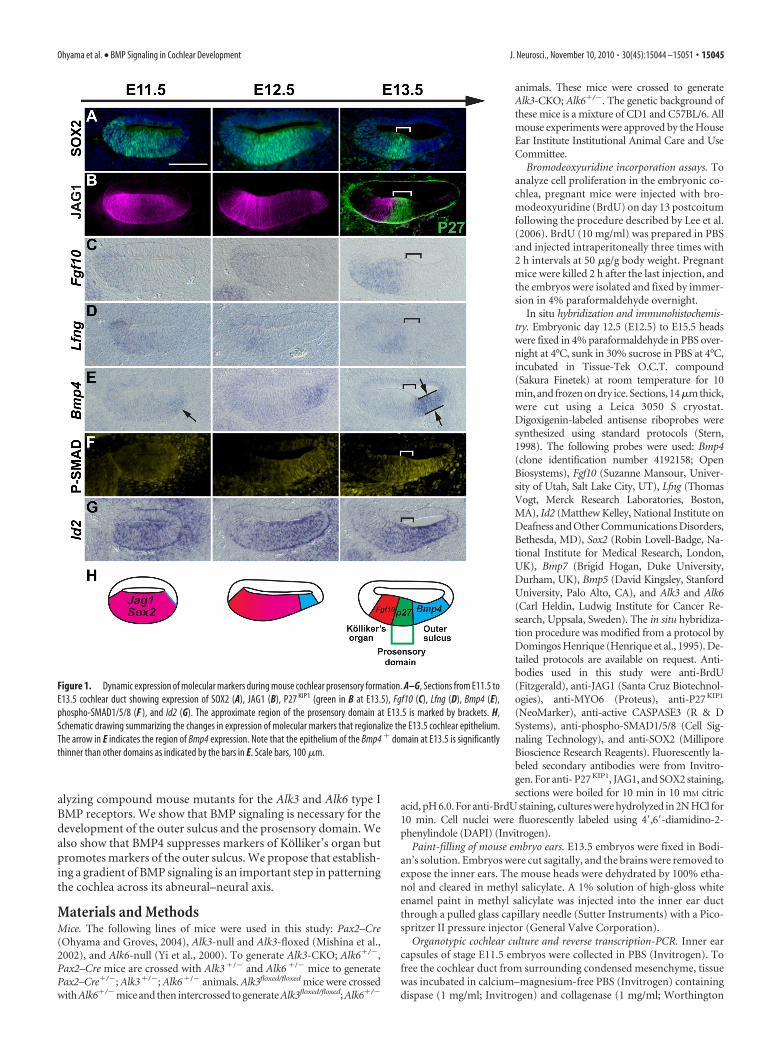
Figure 1. Dynamic expression of molecular markers during mouse cochlear prosensory formation. A–G, Sections from E11.5 toE13.5 cochlear duct showing expression of SOX2 (A), JAG1 (B), P27 KIP1 (green in B at E13.5), Fgf10 (C), Lfng (D), Bmp4 (E),phospho-SMAD1/5/8 (F ), and Id2 (G). The approximate region of the prosensory domain at E13.5 is marked by brackets. H,Schematic drawing summarizing the changes in expression of molecular markers that regionalize the E13.5 cochlear epithelium.The arrow in E indicates the region of Bmp4 expression. Note that the epithelium of the Bmp4 � domain at E13.5 is significantlythinner than other domains as indicated by the bars in E. Scale bars, 100 �m.
Ohyama et al. • BMP Signaling in Cochlear Development J. Neurosci., November 10, 2010 • 30(45):15044 –15051 • 15045
Biochemicals) as described previously (Doetzlhofer et al., 2004). Toavoid contamination of the vestibular sensory organs, only the apical tomid-turn region of the cochlea was isolated with 30 gauge 1⁄2 needles.Isolated cochlear explants (four per well) were cultured on SPI blackmembranes (SPI Supplies) in DMEM–F-12 (Invitrogen) with N2 sup-plement (Invitrogen) and 5 ng/ml epidermal growth factor (R & D Sys-tems). Cultures were maintained in a 5% CO2/20% O2 humidifiedincubator (Forma Scientific). BMP4 (R & D Systems) was stored as a 10mM stock in 4 mM HCl, 0.1% BSA at �80°C. For RNA extraction, totalRNA from four explants was isolated using a Qiagen RNeasy Micro kitwith QIAshredder. cDNA was synthesized using iScript (Bio-Rad).Quantitative PCR was performed with a PerfeCTA SYBR Green FastMix(Quanta) and gene-specific primer sets on a 7500 Real-time PCR Detec-tion System (Applied Biosystems). Relative gene expression was analyzedusing the ��CT method (Livak and Schmittgen, 2001). Primer sets arelisted in supplemental Table S1 (available at www.jneurosci.org as sup-plemental material).
ResultsThe developing cochlear duct is progressively patterned intothree distinct domainsThe mouse cochlea starts to develop as a ventral extension of theotocyst between 10 and 11 d of development (Morsli et al., 1998).The thickened region of cochlear duct initially expresses the tran-scription factor Sox2 and the Notch ligand Jagged 1 (Jag1) atE11.5 (Fig. 1A,B). Bmp4 starts to be expressed asymmetrically inthe abneural side of the cochlea from E11.5 (Fig. 1E, arrow)(Morsli et al., 1998). The cochlear duct shows additional evidenceof asymmetry from the earliest point of its outgrowth; for exam-ple, Fgf10 and Lfng are expressed in the neural side of the cochlearduct from E11.5 onward, marking the future Kolliker’s organ(Fig. 1C,D). After the onset of Bmp4 expression, the SOX2�,JAG1� cochlear duct becomes regionalized into three molecu-
larly distinct compartments. From E12.5 onward, cells in thecochlea destined to form the organ of Corti express the cell-cycleinhibitor p27kip1 and exit the cell cycle, forming a region termedthe prosensory domain (Chen and Segil, 1999; Lee et al., 2006).By E13.5, Sox2, which is necessary for the differentiation of sen-sory cells in the inner ear (Kiernan et al., 2005; Dabdoub et al.,2008) becomes restricted to the prosensory domain and part ofKolliker’s organ destined to give rise to the inner sulcus of thecochlea (Fig. 1A), whereas JAG1 is progressively restricted toKolliker’s organ (Fig. 1B). The expression of Bmp4 in the abneu-ral side of the sensory precursor domain progressively expandsfrom E11.5 onward, marking the future outer sulcus character-ized by a significantly thinner epithelium (Fig. 1E) that will in-clude Hensen’s and Claudius’ cells (Morsli et al., 1998). Of theother BMP family members we tested (Bmp2, Bmp5, Bmp6, andBmp7), Bmp7 is expressed ubiquitously at low levels in the co-chlear epithelium at this stage, whereas expression of Bmp2,Bmp5, and Bmp6 could not be detected (supplemental Fig. S1,available at www.jneurosci.org as supplemental material) (Kier-nan et al., 2005; Dabdoub et al., 2008).
We used antibodies to phosphorylated SMAD1/5/8 proteinsto visualize BMP signaling in the cochlea. These SMAD proteins arephosphorylated by the serine–threonine kinase activities of Bmpr-Iaand Bmpr-Ib receptors and mediate transcriptional regulation of theBMP target genes by interacting with Smad4 (Kishigami andMishina, 2005; Sieber et al., 2009). At E11.5, phospho-SMAD1/5/8 isdetected at low levels throughout the cochlear epithelium (Fig. 1F,left). As the cochlea elongates, we observed the highest level of phos-pho-SMAD1/5/8 in the Bmp4� future outer sulcus, a reduced levelof SMAD phosphorylation in the prosensory domain, and the lowestlevels in Kolliker’s organ (Fig. 1F, right) (supplemental Fig. S2, avail-able at www.jneurosci.org as supplemental material). Expression ofa known BMP target gene, Id2 (Hollnagel et al., 1999), appears tocorrelate well with phospho-SMAD levels in the cochlea at E13.5(Fig. 1F,G, right). These results suggest that a gradient of BMP sig-naling across the abneural–neural axis of the cochlear duct is pro-gressively established during cochlear outgrowth.
BMP signaling is necessary for prosensory specification in themouse cochleaTo test the consequences of perturbing the BMP signaling gradi-ent in the cochlea, we disrupted two BMP type I receptors, Alk3(Bmpr-Ia) and Alk6 (Bmpr-Ib), in mice. Alk3 and Alk6 are ex-pressed ubiquitously at low levels in the cochlear epithelium dur-ing prosensory specification (supplemental Fig. S1, available atwww.jneurosci.org as supplemental material). Because Alk3 playsa crucial early role in the induction of mesoderm (Mishina et al.,1995), we conditionally inactivated Alk3 in the inner ear withPax2–Cre mice (Mishina et al., 2002; Ohyama and Groves, 2004)to avoid premature lethality. Alk6 is not required for embryonicdevelopment, and we therefore used mice carrying an Alk6 nullallele for our experiments (Yi et al., 2000).
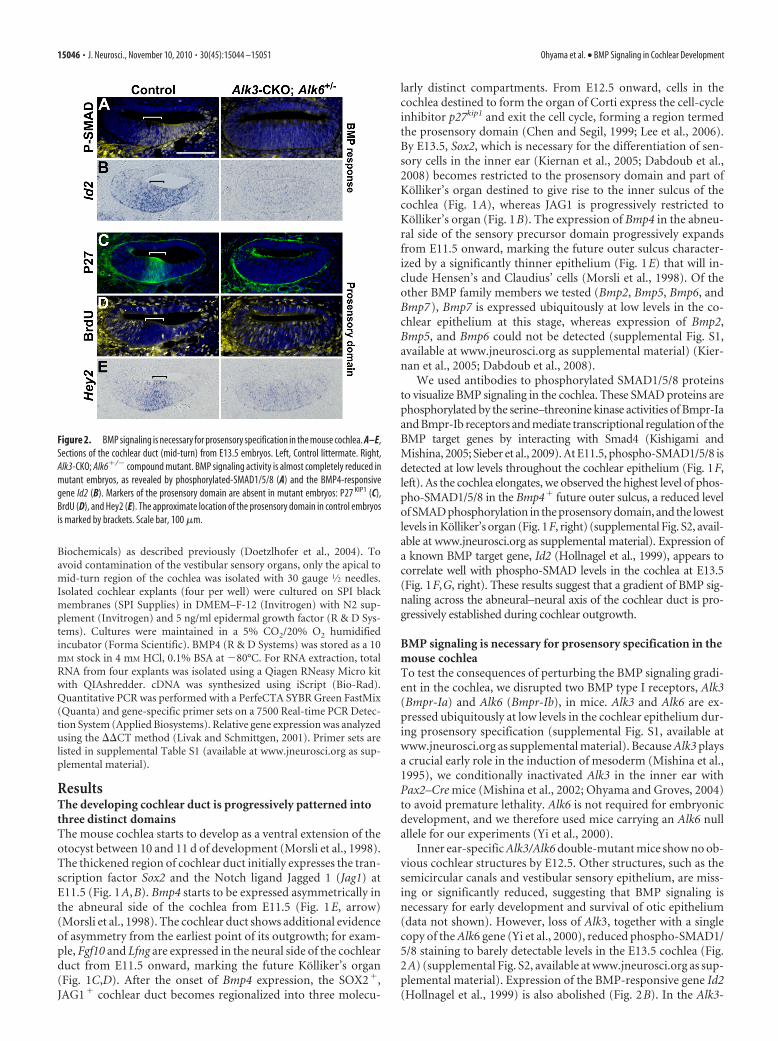
Inner ear-specific Alk3/Alk6 double-mutant mice show no ob-vious cochlear structures by E12.5. Other structures, such as thesemicircular canals and vestibular sensory epithelium, are miss-ing or significantly reduced, suggesting that BMP signaling isnecessary for early development and survival of otic epithelium(data not shown). However, loss of Alk3, together with a singlecopy of the Alk6 gene (Yi et al., 2000), reduced phospho-SMAD1/5/8 staining to barely detectable levels in the E13.5 cochlea (Fig.2A) (supplemental Fig. S2, available at www.jneurosci.org as sup-plemental material). Expression of the BMP-responsive gene Id2(Hollnagel et al., 1999) is also abolished (Fig. 2B). In the Alk3-
Figure 2. BMP signaling is necessary for prosensory specification in the mouse cochlea. A–E,Sections of the cochlear duct (mid-turn) from E13.5 embryos. Left, Control littermate. Right,Alk3-CKO; Alk6�/� compound mutant. BMP signaling activity is almost completely reduced inmutant embryos, as revealed by phosphorylated-SMAD1/5/8 (A) and the BMP4-responsivegene Id2 (B). Markers of the prosensory domain are absent in mutant embryos: P27 KIP1 (C),BrdU (D), and Hey2 (E). The approximate location of the prosensory domain in control embryosis marked by brackets. Scale bar, 100 �m.
15046 • J. Neurosci., November 10, 2010 • 30(45):15044 –15051 Ohyama et al. • BMP Signaling in Cochlear Development

CKO; Alk6�/� compound mutant cochlea, we saw no evidencefor differentiation of the prosensory domain, which is normallymarked by expression of p27 KIP1 (Fig. 2C). BrdU labeling of theE13 compound mutants showed that cells continued to prolifer-ate throughout the cochlear duct, consistent with a lack ofp27 KIP1 expression in the prosensory region (Fig. 2D). The tran-scription factor Hey2, which is expressed strongly in the prosen-sory domain (Doetzlhofer et al., 2009), is significantly reduced inthe compound mutant (Fig. 2E). In compound mutant embryos,the majority of cochlear epithelium expressed markers of Kollik-er’s organ (Fgf10, Jag1, and Lfng) (Fig. 3A–C), whereas markers ofthe future outer sulcus (Bmp4) were entirely absent (Fig. 3D,E).As a result of greatly reduced BMP signaling in Alk3; Alk6�/�
compound mutants, most cochlear duct cells undergo apoptosisat approximately E15.5 (Fig. 3F). We observed no evidence ofhair cell differentiation in compound mutants on the basis ofMyosin VI staining (data not shown), although SOX2 is ex-pressed in most cochlear duct cells in compound mutants at lowlevels similar to the levels in wild-type Kolliker’s organ (Fig. 3G).These results suggest that BMP signaling is necessary for the in-duction and survival of the prosensory domain that gives rise tothe organ of Corti.
BMP4 suppresses markers of Kolliker’s organ and promotesprosensory and outer sulcus fates in a dose-dependentmannerBecause Alk3-CKO; Alk6�/� compound mutants show barely de-tectable levels of BMP signaling in the cochlea and express mark-ers of Kolliker’s organ throughout the cochlear duct, it is not clearwhether Kolliker’s organ is induced by low levels of BMP signal-ing or actively repressed by high or moderate BMP levels. To testthis, we cultured fragments of the cochlear duct from E11.5mouse embryos in a defined medium containing different dosesof BMP4 and measured RNA levels of different cochlear genes by
reverse transcription-PCR after 2 d.BMP4 downregulated expression of Kol-liker’s organ markers (Jag1, Fgf10, andLfng) in cochlear cultures in a dose-dependent manner (Fig. 4A). Addition ofBMP4 to cochlear organ cultures also up-regulated levels of known BMP targetgenes such as Id2 (Hollnagel et al., 1999),Lmx1a (Chizhikov and Millen, 2004), andLmo4 (de la Calle-Mustienes et al., 2003;Chang et al., 2008) in a dose-dependentmanner (Fig. 4C) (supplemental Fig. S3,available at www.jneurosci.org as supple-mental material). Ectopic expression of Idgenes has been shown to inhibit hair celldifferentiation (Jones et al., 2006). Be-cause Id genes have been shown to nega-tively regulate transcription factors,including basic helix–loop– helix and Etsfamily members (Ruzinova and Benezra,2003), BMP signals may act to inhibit Kol-liker’s organ and prosensory domain dif-ferentiation in the future outer sulcusthrough regulation of the Id2 transcrip-tional repressor. Markers of the futureouter sulcus such as Cv2 and Frzb (Qian etal., 2007), which are expressed betweenE14.5 and E15.5 (supplemental Fig. S3,available at www.jneurosci.org as supple-
mental material), are upregulated in 4 d cultures in the presenceof BMP4 in a dose-dependent manner (Fig. 4D), suggesting thatBMP4 promotes outer sulcus differentiation. We also testedwhether BMP4 affects production of sensory hair cells by usingMath1– green fluorescent protein (GFP) transgenic mice inwhich differentiating Math1� sensory hair cells are marked byGFP (Lumpkin et al., 2003). Culture of Math1–GFP cochlearexplants with BMP4 for 4 d revealed that the number of hair cellsincreased compared with controls at intermediate levels of BMP4(10 ng/ml) but decreased with high levels of BMP4 (50 ng/ml)(Fig. 4E).
Reduction in BMP signaling causes mild defects ofvestibular morphologyBMP signaling has been suggested to play a role in the formationof the cristae and semicircular canals (Chang et al., 2002, 2008;Blauwkamp et al., 2007; Omata et al., 2007; Hammond et al.,2009; Vervoort et al., 2010). To determine the effects of Alk3 andAlk6 deletion on inner ear morphogenesis, we used paint-fillingto analyze the morphology of Alk3-CKO and Alk3-CKO; Alk6�/�
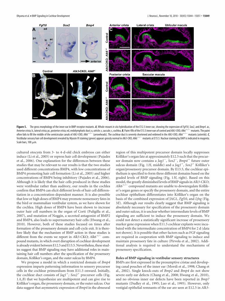
mutants at E13.5. Alk3-CKO mutants and Alk3�/�; Alk6�/� mu-tants showed no obvious morphological defects in the vestibularsystem but displayed a partially shortened cochlear duct (data notshown). Alk3-CKO; Alk6�/� mutants had partially truncated andnarrowed semicircular canals (Fig. 5B, arrowheads) and an evenmore severely truncated cochlear duct (Fig. 5B, asterisk). AlthoughBmp4 is expressed in vestibular sensory patches (Fig. 5A) (Morsli etal., 1998), vestibular sensory structures seem to develop normally inAlk3-CKO; Alk6�/� mutants at E15.5 (Fig. 5C).
DiscussionThe mammalian cochlea is one of the most intricately patternedtissues in any vertebrate. However, it is still essentially unknownwhich signals confer positional identity and polarity on the dif-
Figure 3. Markers of Kolliker’s organ expand at the expense of the prosensory domain and the outer sulcus in Alk3-CKO;Alk6�/� compound mutants. A–D, Sections of the cochlear duct (mid-turn) at E13.5. Left, Control littermate. Right, Alk3-CKO;Alk6�/� compound mutant. Markers of Kolliker’s organ such as Fgf10 (A), JAG1 (B), and Lfng (C) are expressed throughout thedorsal cochlear duct of compound mutants, whereas Bmp4 (D), a marker of outer sulcus, is absent. The approximate location of theprosensory domain is marked by brackets. E, A schematic drawing representing the phenotype of the Alk3-CKO; Alk6�/� com-pound mutant. In the Alk3-CKO; Alk6�/� compound mutant, Kolliker’s organ domain (red) expands at the expense of theprosensory domain (green) and the outer sulcus domain (blue). F, G, Sections of the cochlear duct (mid-turn) at E15.5. F, The arrowindicates apoptotic cells marked by activated CASPASE3 (green). Nuclei are labeled with DAPI (blue). G, P27 KIP1 (green) and SOX2(magenta) staining, showing an absence of the p27 kip1� domain in the compound mutants. Scale bars, 100 �m.
Ohyama et al. • BMP Signaling in Cochlear Development J. Neurosci., November 10, 2010 • 30(45):15044 –15051 • 15047

ferent regions of the cochlea. Our currentdata suggests that BMP signaling is neces-sary for the induction of the prosensorydomain that gives rise to the organ ofCorti. However, this induction of theprosensory domain is only one manifesta-tion of BMP signaling that also appearsto specify the nonsensory regions on ei-ther side of the organ of Corti in aconcentration-dependent manner.
A number of genes have been impli-cated in the development of the cochlearprosensory domain. The transcriptionfactor Sox2 is expressed in all developingprosensory regions of the ear and is neces-sary for their formation (Kiernan et al.,2005). Recent work suggests that Notchsignaling is sufficient to induce ectopicvestibular sensory tissue but that Wnt sig-nals, rather than Notch signals, may initi-ate this process in the early otocyst(Daudet and Lewis, 2005; Daudet et al.,2007; Jayasena et al., 2008). Notch signal-ing can also induce Sox2 expression innonsensory regions of the cochlea (Dab-doub et al., 2008). Based on results fromJagged1 conditional mutant mice, Notchsignaling has been suggested to induce theprosensory domain in the mammalian co-chlea (Brooker et al., 2006; Kiernan et al.,2006). However, we have shown recentlythat the prosensory domain and later haircells and supporting cells are induced nor-mally in conditional mutants of RBPJ�,the transcriptional coeffector of canonicalNotch signaling (Doetzlhofer et al., 2009).Together with our present results, we nowpropose that canonical Notch signalingserves to regulate hair cell and supportingdifferentiation through lateral inhibition (Kelley, 2006) but thatBMP signaling is a more likely candidate to induce the prosensorydomain of the cochlea.
BMP signaling is necessary for sensory patterning of themammalian cochleaBMPs are indispensable for a variety of cell fate decisions, such asthose between neural and non-neural ectoderm, and for devel-opment of germ cells, lens, limb, and kidney (Dudley et al., 1995;Godin et al., 1999; Lawson et al., 1999; Nakayama et al., 2000; Liuand Niswander, 2005). A Drosophila homolog of Bmp2 and Bmp4,decapentaplegic (dpp), can act as a morphogen that patterns thewing imaginal disc and the dorsoventral axis of the early embryo, inwhich different levels of BMP signaling can specify neurogenic, ec-todermal, or amnioserosa fates (O’Connor et al., 2006; Affolter andBasler, 2007). A variety of mechanisms, including positive feedback,degradation, and modulation of ligand diffusion and modulation byligand binding proteins, have been proposed to contribute to theformation of distinct cell fates by signaling gradients (Mizutani et al.,2005; Wang and Ferguson, 2005; Lander, 2007; Serpe et al., 2008).Moreover, both qualitative and quantitative differences in BMPfamily signaling contribute to the patterning of the dorsal neuraltube (Liem et al., 1995, 1997; Lee et al., 1998; Lee and Jessell, 1999;Liu and Niswander, 2005).
Our results suggest that a BMP signaling gradient is requiredto pattern the mammalian cochlear sensory epithelium. First,Bmp4 is expressed on the abneural side of the cochlear duct,accompanied by a gradient of phospho-SMAD1/5/8 staining anda graded expression pattern of the BMP-responsive gene Id2 (Fig.1F,G). Bmp7 is also expressed at lower levels throughout thecochlear duct, although it is not localized to any particular celltype (supplemental Fig. S1, available at www.jneurosci.org assupplemental material). We did not observe expression of otherBMPs such as Bmp2 and Bmp5 (supplemental Fig. S1, available atwww.jneurosci.org as supplemental material) in the cochlear ep-ithelium at this stage. Second, greatly reduced BMP signaling inAlk3-CKO; Alk6�/� mutant cochlea results in an expansion ofthe domain expressing Kolliker’s organ markers (JAG1, Fgf10,and Lfng) at the expense of the prosensory and the outer sulcusdomains (Figs. 2, 3). Interestingly, Bmp4 is itself downregulatedin Alk3-CKO; Alk6�/� mutants, suggesting that it may be regu-lated by positive feedback through BMP receptor signaling. Fi-nally, activation of BMP signaling in cultured cochlear explantssuggests that BMP4 promotes outer sulcus fates at the expense ofKolliker’s organ (Fig. 4A–D), and intermediate levels of BMPsignaling increase the number of sensory hair cells (Fig. 4E).Several recent studies have reported differing effects of BMP4 onthe differentiation of hair cells. For example, addition of BMP4 to
Figure 4. BMP4 represses Kolliker’s organ gene expression and activates outer sulcus genes in a dose-dependent manner.Quantitative PCR analysis of 2 d cultures of E11.5 cochlear explants with genes downregulated (A) and upregulated (C) by increas-ing concentrations of BMP4 protein (n � 4, �SEM). B, Whole-mount in situ hybridization of explants with Fgf10 and Id2 probesshows that Fgf10 is downregulated by BMP4, whereas Id2 is upregulated. D, Quantitative PCR analysis of 4 d cultures of E11.5cochlear explants with outer sulcus marker genes (n � 4, �SEM). E, Four day culture of cochlear explants from Math1–GFP miceat the presence or in the absence of BMP4 protein in culture medium. The number of GFP � cells per explant is quantified andshown in the graph (n � 20, �SEM).
15048 • J. Neurosci., November 10, 2010 • 30(45):15044 –15051 Ohyama et al. • BMP Signaling in Cochlear Development
cultured otocysts from 3- to 4-d-old chick embryos can eitherinduce (Li et al., 2005) or repress hair cell development (Pujadeset al., 2006). One explanation for the differences between thesestudies that may be relevant to our results is that the two studiesused different concentrations BMP4, with low concentrations ofBMP4 promoting hair cell formation (Li et al., 2005) and higherconcentrations of BMP4 being inhibitory (Pujades et al., 2006).Although it is likely that the hair cells produced in these studieswere vestibular rather than auditory, our results in the cochleaconfirm that BMP4 can elicit different levels of hair cell differen-tiation in a concentration-dependent manner. It is also possiblethat low or high doses of BMP4 may promote nonsensory fates inthe bird or mammalian vestibular system, as we have shown forthe cochlea. High doses of BMP4 have been shown to increaseouter hair cell numbers in the organ of Corti (Puligilla et al.,2007), and mutation of Noggin, a secreted antagonist of BMP2and BMP4, also leads to supernumerary hair cells (Hwang et al.,2010). However, both of these studies focused on times afterformation of the prosensory domain and cell-cycle exit. It is there-fore likely that the mechanism of BMP action in these studies isdifferent from the events we report in Alk3-CKO; Alk6�/� com-pound mutants, in which overt disruption of cochlear developmentis already evident between E12.5 and E13.5. Nevertheless, these stud-ies suggest that BMP signaling may have additional roles in main-taining hair cell numbers after the specification of the prosensorydomain, Kolliker’s organ, and the outer sulcus by BMP4.
We propose a model in which a restricted domain of Bmp4expression imparts patterning information to sensory precursorcells in the cochlear primordium from E11.5 onward. Initially,the cochlear duct consists of Jag1�; Sox2� precursor cells (Fig.1A,B) that we hypothesize are multipotent and can give rise toKolliker’s organ, the prosensory domain, or the outer sulcus. Ourdata suggest that asymmetric expression of Bmp4 in the abneural
region of this multipotent precursor domain locally suppressesKolliker’s organ fate at approximately E12.5 such that the precur-sor domain now contains a Jag1�, Sox2�, Bmp4� future outersulcus domain (Fig. 1H, middle) and a Jag1�, Sox2� Kolliker’sorgan/prosensory precursor domain. By E13.5, the cochlear epi-thelium is specified to form three different domains based on thegraded levels of BMP signaling (Fig. 1H, right). Based on thismodel, the greatly diminished levels of BMP signals in Alk3-CKO;Alk6�/� compound mutants are unable to downregulate Kollik-er’s organ genes or specify the prosensory domain, and the entirecochlear epithelium differentiates into Kolliker’s organ on thebasis of the combined expression of JAG1, Fgf10, and Lfng (Fig.3E). Although our results clearly suggest that BMP signaling isabsolutely necessary for specification of the prosensory domainand outer sulcus, it is unclear whether intermediate levels of BMPsignaling are sufficient to induce the prosensory domain. Wecould not detect a statistically significant increase of prosensorymarker gene expression when E11.5 cochlear explants were incu-bated with the intermediate concentration of BMP4 for 2 d (datanot shown). It is possible that other factors such as FGF signalingare required in cooperation with BMP signaling to induce andmaintain prosensory fate in culture (Pirvola et al., 2002). Addi-tional analysis is required to understand the mechanisms ofprosensory specification.
Roles of BMP signaling in vestibular sensory structuresBMPs are first expressed in the presumptive cristae and develop-ing canal pouches of the inner ear (Morsli et al., 1998; Chang etal., 2002). Single knock-outs of Bmp2 and Bmp4 do not showsevere early ear defects (Chang et al., 2008; Hwang et al., 2010),and no obvious inner ear defects have been reported in Bmp7mutants (Dudley et al., 1995; Luo et al., 1995). However, onlyvestigial epithelial remnants of the ear are seen at E12.5 in Alk3-
Figure 5. The gross morphology of the inner ear in BMP receptor mutants. A, Whole-mount in situ hybridization of the E12.5 inner ear, showing the expression of Fgf10, Sox2, and Bmp4. ac,Anterior crista; lc, lateral crista; pc, posterior crista; ed, endolymphatic duct; u, utricle; s, saccule; c, cochlea. B, Paint-fills of the E13.5 inner ears of control and Alk3-CKO; Alk6�/� mutants. The paintoften fails to fill the middle of the semicircular canals of Alk3-CKO; Alk6�/� (arrowheads). The cochlear duct is severely shortened and widened in the Alk3-CKO; Alk6�/� mutants (asterisk). C,Vestibular sensory hair cell development revealed by Myosin VI staining (green) appears grossly normal in Alk3-CKO; Alk6�/� mutants at E15.5. Nuclear staining by DAPI is indicated in magenta.Scale bars, 100 �m.
Ohyama et al. • BMP Signaling in Cochlear Development J. Neurosci., November 10, 2010 • 30(45):15044 –15051 • 15049

CKO; Alk6�/� mutants (T.O., unpublished observations), sug-gesting that minimum levels of BMP signaling through both Alk3and Alk6 receptors are required for survival and proliferation ofotocyst epithelium. Alk3-CKO; Alk6�/� mutants develop innerears with partial vestibular defects (Fig. 5), although much of thecochlear epithelium is lost by E15.5 (Fig. 3F,G). All Alk3-CKO,Alk3-CKO; Alk6�/�, and Alk3-CKO; Alk6�/� mutants die shortlyafter birth for reasons that are currently unknown. Conditionaldeletion of Bmp4 with either Foxg1–Cre or Pax2–Cre mice leadsto more severe vestibular defects than those seen in our Alk3-CKO; Alk6�/� mutants, accompanied by frequent loss of cristaeand semicircular canals (Chang et al., 2008). This suggests thatBmp4 signaling through both Alk3 and Alk6 receptors is necessaryfor correct formation of the cristae and semicircular canals.
In conclusion, our data suggest that an asymmetric BMP sig-naling gradient is key to inducing and patterning the sensorydomains of the mammalian cochlea. Additional understandingof the role of BMP signaling in cochlear development and a pre-cise control of BMP signaling levels may enable more efficientgeneration of hair cells from pluripotent stem cells (Li et al.,2003). We suggest the mammalian cochlea as an attractive systemto understand the mechanisms of BMP signaling gradient in cellfate specification and asymmetric patterning of precursor cellsduring development.
ReferencesAffolter M, Basler K (2007) The Decapentaplegic morphogen gradient:
from pattern formation to growth regulation. Nat Rev Genet 8:663– 674.Blauwkamp MN, Beyer LA, Kabara L, Takemura K, Buck T, King WM, Dolan
DF, Barald KF, Raphael Y, Koenig RJ (2007) The role of bone morpho-genetic protein 4 in inner ear development and function. Hear Res225:71–79.
Brooker R, Hozumi K, Lewis J (2006) Notch ligands with contrasting func-tions: Jagged1 and Delta1 in the mouse inner ear. Development133:1277–1286.
Chang W, Nunes FD, De Jesus-Escobar JM, Harland R, Wu DK (1999) Ec-topic noggin blocks sensory and nonsensory organ morphogenesis in thechicken inner ear. Dev Biol 216:369 –381.
Chang W, ten Dijke P, Wu DK (2002) BMP pathways are involved in oticcapsule formation and epithelial-mesenchymal signaling in the develop-ing chicken inner ear. Dev Biol 251:380 –394.
Chang W, Lin Z, Kulessa H, Hebert J, Hogan BL, Wu DK (2008) Bmp4 isessential for the formation of the vestibular apparatus that detects angularhead movements. PLoS Genet 4:e1000050.
Chen P, Segil N (1999) p27(Kip1) links cell proliferation to morphogenesisin the developing organ of Corti. Development 126:1581–1590.
Chizhikov VV, Millen KJ (2004) Control of roof plate formation by Lmx1ain the developing spinal cord. Development 131:2693–2705.
Dabdoub A, Puligilla C, Jones JM, Fritzsch B, Cheah KS, Pevny LH, KelleyMW (2008) Sox2 signaling in prosensory domain specification and sub-sequent hair cell differentiation in the developing cochlea. Proc Natl AcadSci U S A 105:18396 –18401.
Daudet N, Lewis J (2005) Two contrasting roles for Notch activity in chickinner ear development: specification of prosensory patches and lateralinhibition of hair-cell differentiation. Development 132:541–551.
Daudet N, Ariza-McNaughton L, Lewis J (2007) Notch signalling is neededto maintain, but not to initiate, the formation of prosensory patches in thechick inner ear. Development 134:2369 –2378.
de la Calle-Mustienes E, Lu Z, Cortes M, Andersen B, Modolell J, Gomez-Skarmeta JL (2003) Xenopus Xlmo4 is a GATA cofactor during ventralmesoderm formation and regulates Ldb1 availability at the dorsal meso-derm and the neural plate. Dev Biol 264:564 –581.
Doetzlhofer A, White PM, Johnson JE, Segil N, Groves AK (2004) In vitrogrowth and differentiation of mammalian sensory hair cell progenitors: arequirement for EGF and periotic mesenchyme. Dev Biol 272:432– 447.
Doetzlhofer A, Basch ML, Ohyama T, Gessler M, Groves AK, Segil N (2009)Hey2 regulation by FGF provides a Notch-independent mechanism formaintaining pillar cell fate in the organ of Corti. Dev Cell 16:58 – 69.
Dudley AT, Lyons KM, Robertson EJ (1995) A requirement for bone mor-
phogenetic protein-7 during development of the mammalian kidney andeye. Genes Dev 9:2795–2807.
Gerlach LM, Hutson MR, Germiller JA, Nguyen-Luu D, Victor JC, Barald KF(2000) Addition of the BMP4 antagonist, noggin, disrupts avian innerear development. Development 127:45–54.
Godin RE, Robertson EJ, Dudley AT (1999) Role of BMP family membersduring kidney development. Int J Dev Biol 43:405– 411.
Hammond KL, Loynes HE, Mowbray C, Runke G, Hammerschmidt M, Mul-lins MC, Hildreth V, Chaudhry B, Whitfield TT (2009) A late role forbmp2b in the morphogenesis of semicircular canal ducts in the zebrafishinner ear. PLoS One 4:e4368.
Henrique D, Adam J, Myat A, Chitnis A, Lewis J, Ish-Horowicz D (1995)Expression of a Delta homologue in prospective neurons in the chick.Nature 375:787–790.
Hollnagel A, Oehlmann V, Heymer J, Ruther U, Nordheim A (1999) Idgenes are direct targets of bone morphogenetic protein induction in em-bryonic stem cells. J Biol Chem 274:19838 –19845.
Hwang CH, Guo D, Harris MA, Howard O, Mishina Y, Gan L, Harris SE, WuDK (2010) Role of bone morphogenetic proteins on cochlear hair cellformation: analyses of Noggin and Bmp2 mutant mice. Dev Dyn239:505–513.
Jayasena CS, Ohyama T, Segil N, Groves AK (2008) Notch signaling aug-ments the canonical Wnt pathway to specify the size of the otic placode.Development 135:2251–2261.
Jones JM, Montcouquiol M, Dabdoub A, Woods C, Kelley MW (2006) In-hibitors of differentiation and DNA binding (Ids) regulate Math1 andhair cell formation during the development of the organ of Corti. J Neu-rosci 26:550 –558.
Kelley MW (2006) Regulation of cell fate in the sensory epithelia of the innerear. Nat Rev Neurosci 7:837– 849.
Kelley MW (2007) Cellular commitment and differentiation in the organ ofCorti. Int J Dev Biol 51:571–583.
Kelly M, Chen P (2007) Shaping the mammalian auditory sensory organ bythe planar cell polarity pathway. Int J Dev Biol 51:535–547.
Kiernan AE, Pelling AL, Leung KK, Tang AS, Bell DM, Tease C, Lovell-BadgeR, Steel KP, Cheah KS (2005) Sox2 is required for sensory organ devel-opment in the mammalian inner ear. Nature 434:1031–1035.
Kiernan AE, Xu J, Gridley T (2006) The Notch ligand JAG1 is required forsensory progenitor development in the mammalian inner ear. PLoSGenet 2:e4.
Kishigami S, Mishina Y (2005) BMP signaling and early embryonic pattern-ing. Cytokine Growth Factor Rev 16:265–278.
Lander AD (2007) Morpheus unbound: reimagining the morphogen gradi-ent. Cell 128:245–256.
Lawson KA, Dunn NR, Roelen BA, Zeinstra LM, Davis AM, Wright CV,Korving JP, Hogan BL (1999) Bmp4 is required for the generation ofprimordial germ cells in the mouse embryo. Genes Dev 13:424 – 436.
Lee KJ, Jessell TM (1999) The specification of dorsal cell fates in the verte-brate central nervous system. Annu Rev Neurosci 22:261–294.
Lee KJ, Mendelsohn M, Jessell TM (1998) Neuronal patterning by BMPs: arequirement for GDF7 in the generation of a discrete class of commissuralinterneurons in the mouse spinal cord. Genes Dev 12:3394 –3407.
Lee YS, Liu F, Segil N (2006) A morphogenetic wave of p27Kip1 transcrip-tion directs cell cycle exit during organ of Corti development. Develop-ment 133:2817–2826.
Li H, Liu H, Heller S (2003) Pluripotent stem cells from the adult mouseinner ear. Nat Med 9:1293–1299.
Li H, Corrales CE, Wang Z, Zhao Y, Wang Y, Liu H, Heller S (2005) BMP4signaling is involved in the generation of inner ear sensory epithelia. BMCDev Biol 5:16.
Liem KF Jr, Tremml G, Roelink H, Jessell TM (1995) Dorsal differentiationof neural plate cells induced by BMP-mediated signals from epidermalectoderm. Cell 82:969 –979.
Liem KF Jr, Tremml G, Jessell TM (1997) A role for the roof plate and itsresident TGFbeta-related proteins in neuronal patterning in the dorsalspinal cord. Cell 91:127–138.
Liu A, Niswander LA (2005) Bone morphogenetic protein signalling andvertebrate nervous system development. Nat Rev Neurosci 6:945–954.
Livak KJ, Schmittgen TD (2001) Analysis of relative gene expression datausing real-time quantitative PCR and the 2(-Delta Delta C(T)) method.Methods 25:402– 408.
15050 • J. Neurosci., November 10, 2010 • 30(45):15044 –15051 Ohyama et al. • BMP Signaling in Cochlear Development

Lumpkin EA, Collisson T, Parab P, Omer-Abdalla A, Haeberle H, Chen P,Doetzlhofer A, White P, Groves A, Segil N, Johnson JE (2003) Math1-driven GFP expression in the developing nervous system of transgenicmice. Gene Expr Patterns 3:389 –395.
Luo G, Hofmann C, Bronckers AL, Sohocki M, Bradley A, Karsenty G (1995)BMP-7 is an inducer of nephrogenesis, and is also required for eye devel-opment and skeletal patterning. Genes Dev 9:2808 –2820.
Mishina Y, Suzuki A, Ueno N, Behringer RR (1995) Bmpr encodes a type Ibone morphogenetic protein receptor that is essential for gastrulationduring mouse embryogenesis. Genes Dev 9:3027–3037.
Mishina Y, Hanks MC, Miura S, Tallquist MD, Behringer RR (2002) Gen-eration of Bmpr/Alk3 conditional knockout mice. Genesis 32:69 –72.
Mizutani CM, Nie Q, Wan FY, Zhang YT, Vilmos P, Sousa-Neves R, Bier E,Marsh JL, Lander AD (2005) Formation of the BMP activity gradient inthe Drosophila embryo. Dev Cell 8:915–924.
Morsli H, Choo D, Ryan A, Johnson R, Wu DK (1998) Development of themouse inner ear and origin of its sensory organs. J Neurosci 18:3327–3335.
Nakayama T, Cui Y, Christian JL (2000) Regulation of BMP/Dpp signalingduring embryonic development. Cell Mol Life Sci 57:943–956.
O’Connor MB, Umulis D, Othmer HG, Blair SS (2006) Shaping BMP mor-phogen gradients in the Drosophila embryo and pupal wing. Development133:183–193.
Ohyama T, Groves AK (2004) Generation of Pax2-Cre mice by modifica-tion of a Pax2 bacterial artificial chromosome. Genesis 38:195–199.
Omata Y, Nojima Y, Nakayama S, Okamoto H, Nakamura H, Funahashi J(2007) Role of bone morphogenetic protein 4 in zebrafish semicircularcanal development. Dev Growth Differ 49:711–719.
Pirvola U, Ylikoski J, Trokovic R, Hebert JM, McConnell SK, Partanen J(2002) FGFR1 is required for the development of the auditory sensoryepithelium. Neuron 35:671– 680.
Pujades C, Kamaid A, Alsina B, Giraldez F (2006) BMP-signaling regulatesthe generation of hair-cells. Dev Biol 292:55– 67.
Puligilla C, Feng F, Ishikawa K, Bertuzzi S, Dabdoub A, Griffith AJ, Fritzsch B,Kelley MW (2007) Disruption of fibroblast growth factor receptor 3 sig-naling results in defects in cellular differentiation, neuronal patterning,and hearing impairment. Dev Dyn 236:1905–1917.
Qian D, Jones C, Rzadzinska A, Mark S, Zhang X, Steel KP, Dai X, Chen P(2007) Wnt5a functions in planar cell polarity regulation in mice. DevBiol 306:121–133.
Ruzinova MB, Benezra R (2003) Id proteins in development, cell cycle andcancer. Trends Cell Biol 13:410 – 418.
Serpe MJ, Rivera M, Kersey FR, Clark RL, Craig SL (2008) Time and dis-tance dependence of reversible polymer bridging followed by single-molecule force spectroscopy. Langmuir 24:4738 – 4742.
Sieber C, Kopf J, Hiepen C, Knaus P (2009) Recent advances in BMP recep-tor signaling. Cytokine Growth Factor Rev 20:343–355.
Stern CD (1998) Detection of multiple gene products simultaneously by insitu hybridization and immunohistochemistry in whole mounts of avianembryos. Curr Top Dev Biol 36:223–243.
Vervoort R, Ceulemans H, Van Aerschot L, D’Hooge R, David G (2010)Genetic modification of the inner ear lateral semicircular canal phenotypeof the Bmp4 haplo-insufficient mouse. Biochem Biophys Res Commun394:780 –785.
Wang YC, Ferguson EL (2005) Spatial bistability of Dpp-receptor inter-actions during Drosophila dorsal-ventral patterning. Nature434:229 –234.
Wu DK, Oh SH (1996) Sensory organ generation in the chick inner ear.J Neurosci 16:6454 – 6462.
Yi SE, Daluiski A, Pederson R, Rosen V, Lyons KM (2000) The type I BMPreceptor BMPRIB is required for chondrogenesis in the mouse limb. De-velopment 127:621– 630.
Ohyama et al. • BMP Signaling in Cochlear Development J. Neurosci., November 10, 2010 • 30(45):15044 –15051 • 15051