dharmacon decode pooleddharmacon.horizondiscovery.com/uploadedfiles/resources/decode... · a quote...
TRANSCRIPT

Technical ManualDharmacon™ Decode™ Pooled
Lentiviral shRNA Screening Libraries
GE Healthcare
Contents
1 Lentiviral Particle Product Safety Level Information ...............................................................................................3
2 Laboratory Protocols and Calculation Tracking Sheets .........................................................................................4
3 Introduction ......................................................................................................................................................................7
4 Decode Pooled Lentiviral shRNA Screening Required Materials .........................................................................10
5 Assay Development and Optimization: Transduction Parameters .....................................................................11A. Optimization of Lentiviral Transduction .................................................................................................................................................................. 11
B. Determination of Functional Titer .............................................................................................................................................................................. 11
C. Optimization of Puromycin Selection ....................................................................................................................................................................... 13
6 Assay Development and Optimization: Screening Parameters ...........................................................................13A. Assay-specific Screening Conditions ....................................................................................................................................................................... 13
B. Average shRNA Fold Representation During Transduction and Number of Biological Replicates ......................................... 13
C. Number of Cells Needed for Transduction ............................................................................................................................................................ 14
D. Volume of Lentiviral Particles Needed for Transduction ................................................................................................................................ 15
7 Primary Screen ...............................................................................................................................................................16A. Cell Transduction and Selection Screening ............................................................................................................................................................17
B. Genomic DNA Isolation .....................................................................................................................................................................................................17
C. PCR Amplification of shRNA from Genomic DNA ............................................................................................................................................... 18
i. Number of PCR Reactions ......................................................................................................................................................................................... 18
ii. Multiplexing of High-throughput Sequencing Samples ........................................................................................................................... 19
iii. PCR from Genomic DNA ........................................................................................................................................................................................... 20
8 Illumina Platform Sequencing ....................................................................................................................................21
9 Hit Identification and Follow Up ................................................................................................................................21
10 Frequently Asked Questions (FAQs) ..........................................................................................................................22
11 Troubleshooting ............................................................................................................................................................22
12 References ......................................................................................................................................................................23
13 Label Licenses ..............................................................................................................................................................23
3
Decode Pooled Lentiviral shRNA Screening Libraries
Technical Manual
1 Lentiviral Particle Product Safety Level InformationThis Lentiviral Particle Product Safety Level Information constitutes Product Documentation according to clause 1 of the Product Terms and Conditions.
Dharmacon™ Decode™ Pooled Lentiviral shRNA Screening Libraries are solely for internal research use (as set forth in the Product Terms and Conditions) in laboratories where the containment measures stated below and in applicable laws and regulations are met. Products may not be used for diagnostic, therapeutic or other commercial purposes and may not to be administered to humans for any purpose or to animals for therapeutic purposes. The Products are replication-incompetent, self-inactivating (SIN) and non-pathogenic (do not cause infectious human disease).
Any investigator who purchases Dharmacon Lentiviral Particle Products is responsible for consulting with their institution’s health and biosafety personnel for specific guidelines on the handling of lentiviral vector particles. Furthermore, each investigator is fully responsible for obtaining the required permissions for research usage and the acceptance of replication-incompetent SIN lentiviral vectors and replication-defective lentiviral particles into their local jurisdiction and institution.
For questions concerning the design or production of the Products, please contact: Technical Support ([email protected], 800 235 9880).
Biosafety Level 2 Containment Measures in the USFor US guidance on containment for lentiviral vectors, please refer to the Recombinant DNA Advisory Committee (RAC) guidelines for research with lentiviral vectors here.
The U.S. Department of Health and Human Services Centers for Disease Control and Prevention and National Institutes of Health, Biosafety in Microbiological and Biomedical Laboratories (BMBL), Fifth Edition, Dec 2009 downloadable here.
See also the NIH Guidelines for Research Involving Recombinant or Synthetic Nucleic Acid Molecules (NIH Guidelines), downloadable here.
Biosafety Level 2 Containment Measures in the EUFor the EU directives, please consult the following:
1. Council Directive 2009/41/EC of the European Parliament and of the Council of 6 May 2009 on the contained use of genetically modified micro-organisms. (revised version of Directive 90/219/EEC of the European Parliament and of the Council of 23 April 1990 on the contained use of genetically modified micro-organisms, amended by Council Directive 98/81/EC of 26 October 1998); and
2. Council Directive 2001/18/EC of the European Parliament and of the Council of 12 March 2001 on the deliberate release into the environment of genetically modified organisms and repealing Council Directive 90/220/EEC.
Required Containment Measures in GermanyThe containment requirements as stated in the German Genetic Safety Ordinance (Gentechnik-Sicherheitsverordnung) of Safety Level 2*or higher have been assigned to the handling of the above-mentioned lentiviral vector particles. Please note that a higher Security Level might be required if the lentiviral vector particles are used for genetic engineering operations with other products which require a higher Security Level. *Safety Level 2: activities of low risk for human health and the environment by the state of scientific knowledge (Stand der Wissenschaft). For the German regulations, please consult the following:
1. German Genetic Engineering Act (Gentechnikgesetz - GenTG); and
2. Genetic Engineering Safety Ordinance (Gentechnik-Sicherheitsverordnung - GenTSV).
Safety is a focus of all lentiviral vector technology. For this reason, lentiviral vector packaging systems have divided the essential functions amongst multiple plasmids to reduce the risk of generating replication-competent lentiviral particles (RCL). The split-genome packaging system is designed so that multiple recombination events between the components are required for autonomous replication. Clinical trials using a split-genome packaging system have shown that this strategy effectively eliminates the creation of RCLs (Levine et al. 2006). Commercially available 3rd generation lentiviral vector systems separate the lentiviral envelope, env (such as VSV-G), from the gag-pro-pol, which encodes structural and enzymatic functions. The Pooled Lentiviral shRNA Screening Libraries are produced using the Dharmacon Trans-Lentiviral Packaging System. The Trans-Lentiviral Packaging System provides an even higher level of safety over 3rd generation packaging systems by further splitting the viral pol [reverse transcriptase (RT) and integrase (IN) functions] from gag-pro. Because the RT and IN enzymes are provided in trans to gag-pro, additional recombination events are necessary to produce RCLs.

4
Decode Pooled Lentiviral shRNA Screening Libraries
Technical Manual
2 Laboratory Protocols and Calculation Tracking Sheets The Laboratory Protocols provide a brief description of the Decode Pooled shRNA Screening workflow. Before using these Laboratory Protocols, we strongly recommend that new users familiarize themselves with the detailed protocols provided in this manual. An electronic version of the Laboratory Protocol Worksheet can also be downloaded from the Decode Pooled Lentiviral shRNA Screening Libraries under Resources. The downloadable version of these protocols allow users to incorporate specific input related to their screen and performs key calculations to simplify protocol planning.
5
Decode Pooled Lentiviral shRNA Screening Libraries
Technical Manual
(0.3 recommended)

6
Decode Pooled Lentiviral shRNA Screening Libraries
Technical Manual
C. PCR Amplification of shRNA from gDNA
Table 10. PCR cycling parameters.
Temperature Time
98 °C 3 minutes
98 °C 10 seconds
23 C
ycle
s
57 °C 15 seconds
72 °C 15 seconds
72 °C 5 minutes

7
Decode Pooled Lentiviral shRNA Screening Libraries
Technical Manual
3 IntroductionDecode Lentiviral RNAi screening libraries are pools of Dharmacon™ GIPZ™ short hairpin RNAs (shRNAs) that are packaged into high-titer lentiviral particles. Through RNAi-mediated silencing of hundreds or thousands of genes in parallel, a pooled lentiviral shRNA screen can be performed to identify genes that regulate cellular responses and signaling pathways, or to discover novel gene functions. In contrast to the costly automated techniques that are required to screen using individually arrayed RNAi reagents, Decode pooled shRNA screening libraries allow the researcher to transduce and screen a population of cells within a few tissue culture dishes. Table 1 lists the pre-defined Decode pooled lentiviral shRNA libraries targeting genes within the human genome that are available. These screening libraries contain an average of five shRNAs per target gene and are supplied in sufficient volume of lentiviral particles to allow high biological reproducibility in relevant cells that are refractory to transfection. In addition, Custom Decode pooled lentiviral shRNA libraries are available for order. Researchers may choose any available GIPZ clones (human or mouse) for pooling (from 50 to 10,000 clones per pool), any number of pools, and lentiviral volumes from 100 µL to 5 mL. To request a quote go to dharmacon.gelifesciences.com or contact us at: [email protected]. Also available from Dharmacon for use with Decode screening libraries are optimized primers and experimentally tested protocols for reliable identification of shRNA hits by high-throughput sequencing on an Illumina platform.
Table 1. Available Decode Pooled Lentiviral shRNA Screening Libraries.
Library Cat #Number of genes
targetedNumber of pools × number of
shRNA constructs per poolLentiviral particle volume per pool
Ubiquitin Conjugation RHS6076 571 1 pool of 3,830 shRNA 2 tubes × 25 µL (50 µL total)
Phosphatase RHS6077 254 1 pool of 1,561 shRNA 2 tubes × 25 µL (50 µL total)
Protein Kinase RHS6078 709 1 pool of 4,675 shRNA 2 tubes × 25 µL (50 µL total)
Ion Channel RHS6079 347 1 pool of 1,884 shRNA 2 tubes × 25 µL (50 µL total)
GPCR RHS6080 382 1 pool of 2,591 shRNA 2 tubes × 25 µL (50 µL total)
Protease RHS6081 478 1 pool of 2,559 shRNA 2 tubes × 25 µL (50 µL total)
Druggable Genome RHS6082 7494 5 pools of 8,490 shRNA 4 tubes × 25 µL (100 µL total)
Human Genome RHS6083 18205 10 pools of 9,570 shRNA 4 tubes × 25 µL (100 µL total)
The Decode Pooled shRNA screening protocols begin by transducing cells at a low multiplicity of infection (MOI) with a lentiviral pool containing between 50 and 10,000 unique shRNAs (see Figure 1 for a screening workflow diagram). Individual cells in the resulting transduced population will contain unique shRNAs integrated into their genomes. Following transduction, a selective pressure is applied to identify shRNAs that target genes involved in a specific biological response. As a result of the selective pressure, cells expressing shRNA are either enriched or depleted in the cellular population. To identify hits, genomic DNA (gDNA) is isolated from the initial transduced cell population (reference cells) and from the transduced cell population that remains following the application of selective pressure and phenotypic selection (experimental cells). shRNA sequences within the isolated gDNA are amplified using Illumina-adapted Decode Forward and Reverse Indexed PCR primers that have been designed and optimized to minimize amplification bias. Following amplification, indexed PCR products can be directly loaded onto Illumina flow cells and sequenced using Decode sequencing primers. The differences in shRNA abundance between reference and experimental cell populations can then be determined.

8
Decode Pooled Lentiviral shRNA Screening Libraries
Technical ManualAs
say
deve
lopm
ent a
nd o
ptim
izatio
nPr
imar
y sc
reen
Hit
iden
tific
atio
n
Transduction parameters
Select phenotype
Select transduced cells
Reference sample
Isolate gDNA and analyze sequence of integrated shRNA
Log mean countsLog
Expe
rimen
tal/R
efer
ence
Experimental sample
Screening parameters
Selective pressure
Transduce with lentiviral shRNA pool
Selective pressure
Enriched shRNAs
Depleted shRNAs
Figure 1. Pooled shRNA screening workflow.
Assay Development and Optimization: Establish optimal experimental conditions, including those for lentiviral transduction and screening parameters, such as selective pressure and time between collection of reference and experimental samples.
Primary Screen: A stable population of cells expressing single integrants of shRNAs is created by transducing Decode lentiviral pools at low MOI. Transduced cells are then split into reference and experimental populations for application of a selective pressure and phenotypic selection. gDNA is then isolated from reference and experimental populations of transduced cells. Illumina-adapted primers and Phusion™ Hot Start II High-Fidelity DNA Polymerase are used to PCR-amplify integrated shRNA sequences and add Illumina flow-cell binding sequences. The resulting amplicons are sequenced on Illumina platform sequencers, using the sequencing primers provided.
Hit Identification and Follow Up: shRNA sequences are identified in reference and experimental libraries. shRNAs that are enriched or depleted during the screen are identified as hits, and the genes that they target are identified. Hits can be confirmed and studied further using individual shRNA constructs that can be ordered from the GIPZ lentiviral shRNA collection.

9
Decode Pooled Lentiviral shRNA Screening Libraries
Technical Manual
Decode Pooled Lentiviral shRNA Screening Libraries are comprised of the GIPZ lentiviral shRNA, in which the gene-specific silencing sequence is embedded in a primary microRNA transcript, thus creating an RNAi trigger that silences genes with increased specificity and minimal cellular toxicity (Figures 2 and 3). The Decode Pooled Lentiviral shRNA Screening Libraries combine the advantages of the microRNA-adapted GIPZ shRNA design with the convenience of high-titer lentiviral delivery to create a powerful multiplexed RNAi screening resource capable of producing loss-of-function phenotypes in many dividing and non-dividing cells.
Decode shRNA pools are created using experimentally tested methods that ensure uniform representation of shRNA constructs in every lentiviral pool. Specifically, individual shRNA clones are transferred from 96-well plate collections onto agar plates and allowed to grow overnight. Colonies are then combined into a slurry and grown for a defined period of time. High quality DNA is prepared from cultures of E. coli and analyzed by high-throughput sequencing to examine shRNA representation and identity. This quality control allows us to verify that the abundance of 70% of the shRNAs is less than 5-fold different from each other and the abundance of 90% of the shRNAs is less than 25-fold different from each other. During this quality control process, the relative representation and identity of every shRNA in the plasmid pool is determined and provided with the product to ensure high quality screening materials and confidence in Decode lentiviral pool starting materials.
Figure 2. GIPZ shRNAs effectively knockdown genes of interest. HEK293T cells were transduced with lentiviral particles containing shRNA to the indicated genes and a non-silencing (NS) control, at an average MOI of ten. Forty-eight hours after transduction, cells were selected with puromycin for four days prior to RNA isolation. Expression of indicated genes was determined by RT-qPCR and normalized to non-silencing control.
Vector Element
Utility
hCMV Human cytomegalovirus promoter drives strong transgene expression
tGFP TurboGFP reporter for visual tracking of transduction and expression
PuroR Puromycin resistance permits antibiotic-selective pressure and propagation of stable integrants
IRESInternal ribosomal entry site allows expression of TurboGFP and puromycin resistance genes in a single transcript
shRNA microRNA-adapted shRNA (based on miR-30) for gene knockdown
5' LTR 5' long terminal repeat
3' SIN LTR 3' self-inactivating long terminal repeat for increased safety
Ψ Psi packaging sequence allows lentiviral genome packaging using lentiviral packaging systems
RRE Rev response element enhances titer by increasing packaging efficiency of full-length lentiviral genomes
WPREWoodchuck hepatitis posttranscriptional regulatory element enhances transgene expression in the target cells
PuroRIRES
Amp R SV40 oripUC ori
tGFP
hCMV
RRE
shRNA
WPRE
5' LT
R
3' SIN LTR
Ψ
pGIPZ
pTRIPZ
rtTA3tRFP
TRE UBC
From PowerPoint (R&D)Revised versions
pLOC
pLKO.1
U6 hPGK
pSMART 2.0
SV40 ori
hCMV
pSMART 2.0
pSMART 2.0
AmpR pUCori SV40 ori
5ʹ LT
R
3ʹ SIN LTR
Ψ
RRE tGFP
hCMV
IRES PuroR shRNA WPREcPPT/CTS
hPGKU6
AmpRpUCori
pLKO.1
RSV/
5ʹ LT
R
3ʹ SIN LTR
Ψ
RRE PuroRshRNA
pSMART 2.0
AmpR pUCori SV40 ori
5ʹ LT
R
3ʹ SIN LTR
Ψ
RRE tGFP
hCMV
IRES PuroR microRNA WPRE
pLOC
AmpR pUCori SV40 ori
5ʹ LT
R
3ʹ SIN LTR
Ψ
RRE tGFPnuc
hCMV
IRES BlastR-2a- WPREORF
Multi TagCloning Site
AmpR pUCori SV40 ori
pGIPZ5ʹ
LTR
3ʹ SIN LTR
Ψ
RRE tGFP
hCMV
IRES PuroR WPRE
shRNA
AmpR pUCori SV40 ori
UBC
pTRIPZ
5ʹ LT
R
3ʹ SIN LTR
Ψ
RRE tRFP
TRE
IRES PuroR WPRErtTA3
shRNA
hCMVmCMVhEF1αmEF1αCAGPGKUBC
SMARTchoice shRNA
5ʹ LTR 3ʹ SIN LTRΨ RRE tGFPor
tRFP
IRES PuroR WPRE
SMAR
Tcho
ice
prom
oter
s
SMARTvector2.0 universal scaffold
5' LT
RΨ
PuroRIRES WPRERRE
shRNA
3' SIN LTR
Amp R SV40 oripUC ori
hCMV
5' LT
RΨ
5' LT
RΨ
RSV/
5' LT
RΨ
PuroR
PuroR
IRES WPRERRE
RRE
RRE
shRNA
shRNA
3' SIN LTR
3' SIN LTR
3' SIN LTR
5' LT
RΨ
3' SIN LTR
Amp R
Amp R
Amp R
SV40 oripUC ori
pUC ori
pUC ori
SV40 oriAmp R pUC ori
tGFP
PuroRIRES WPREmicroRNAtGFP
hCMV
Blast RIRES WPRE-2a-tGFPnuc
Multi TagCloning Site
ORF
hCMVmCMVhEF1
or
αmEF1 αCAGPGKUBC
SMAR
Tcho
ice
prom
oter
s
5' LTR Ψ tGFP PuroRIRES
tRFP
WPRE 3' SIN LTR
SMARTvector universal scaffold
SMARTchoice shRNA
RRE
Figure 3. pGIPZ lentiviral vector.
10
Decode Pooled Lentiviral shRNA Screening Libraries
Technical Manual
4 Decode Pooled Lentiviral shRNA Screening Required MaterialsThe Decode shRNA Screening Workflow Requires:• Pools of concentrated lentiviral particles (Table 1).
• 2 × 25 μL tubes of GIPZ non-silencing shRNA control lentiviral particles (Dharmacon Cat #RHS4348).
• Decode Indexing PCR and Sequencing Primer Kit (Dharmacon Cat #PRM6178). Note: We recommend purchasing two Decode Indexing PCR and Sequencing Kits for use with the Decode Human Genome Library.
• Phusion™ Hot Start II High-Fidelity DNA Polymerase and 5x Phusion™ HF Buffer (Thermo Scientific Cat #F-549S, F-549L). Note: Please see section 7 to determine the amount of DNA polymerase required for a screening workflow.
Additional Recommended Materials But Not Supplied Include:• Betaine Solution 5M (Sigma-Aldrich Cat #B0300)
• Qiagen™ QIAquick™ PCR Purification Kit (Cat #28104)
• Thermo Scientific™ GeneRuler™ Low Range DNA Ladder, ready-to-use, 25-700 bp (Cat #SM1193)
• Qiagen™ Blood and Cell Culture DNA Maxi Kit (Cat #13362)
• Thermo Scientific™ dNTP™ Mix, 10 mM each (Cat #R0191)
• HyClone™ Puromycin 2 HCl (Cat #SV30075.01)
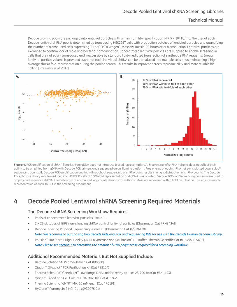
Figure 4. PCR amplification of shRNA libraries from gDNA does not introduce biased representation. A. Free energy of shRNA hairpins does not affect their ability to be amplified from gDNA with Decode PCR primers and sequenced on an Illumina platform. Free energy of each shRNA hairpin is plotted against log10 sequencing counts. B. Decode PCR amplification and high-throughput sequencing of shRNA pools results in a tight distribution of shRNA counts. The Decode Phosphatase library was transduced into HEK293T cells at 1000-fold representation and gDNA was isolated. Decode PCR and Sequencing primers were used to amplify and sequence shRNA. The histogram of normalized log2 counts demonstrates that shRNAs are recovered with a tight distribution. This ensures ample representation of each shRNA in the screening experiment.
A. B.
Decode plasmid pools are packaged into lentiviral particles with a minimum titer specification of ≥ 5 × 10⁸ TU/mL. The titer of each Decode lentiviral shRNA pool is determined by transducing HEK293T cells with production batches of lentiviral particles and quantifying the number of transduced cells expressing TurboGFP™ (Evrogen™, Moscow, Russia) 72 hours after transduction. Lentiviral particles are examined to confirm lack of mold and bacterial contamination. Concentrated lentiviral particles are supplied to enable screening in cells that are not easily transduced and inaccessible by standard lipid-mediated transfection of synthetic siRNA reagents. Enough lentiviral particle volume is provided such that each individual shRNA can be transduced into multiple cells, thus maintaining a high average shRNA fold-representation during the pooled screen. This results in improved screen reproducibility and more reliable hit calling (Strezoska et al. 2012).

11
Decode Pooled Lentiviral shRNA Screening Libraries
Technical Manual
5 Assay Development and Optimization: Transduction ParametersA. Optimization of Lentiviral Transduction
While GIPZ shRNA lentiviral particles exhibit broad cell tropism, the conditions for successful and efficient delivery can vary significantly. It is essential to determine the optimal lentiviral transduction conditions in each cell line or type of interest. Please keep in mind that the conditions you select during these optimization steps must be compatible with your primary screening protocols and conditions. GIPZ non-silencing control lentiviral particles can be used for optimization of transduction conditions. Parameters that may influence the efficiency of lentiviral transduction include, but are not limited to:
Transduction medium: When possible, the transduction of cells with lentiviral particles should be performed in a small volume of low-serum (0.5-2%) or serum-free medium. For cells sensitive to low serum conditions, transduction optimization can be performed in complete medium.
Transduction duration: Incubation time can vary between 4 and 24 hours and will depend on your cell line.
Transduction medium additives: Cationic polymers such as hexadimethrine bromide (Polybrene) may be added to enhance lentiviral particle binding to the cell surface. We recommend testing a range of concentrations, from 0-10 µg/mL, for identification of optimal transduction efficiency with minimal or no cell toxicity.
Cell density at transduction: The density at which cells are seeded may also influence transduction efficiency. We recommend seeding cells at a range of densities for optimization of transduction efficiency. Plate sizes for screening should be chosen accordingly.
B. Determination of Functional TiterPooled shRNA libraries and GIPZ non-silencing shRNA controls are supplied as concentrated lentiviral particles. Specifications can be found on the Certificate of Analysis (C of A) provided with lentiviral particles. Functional titer can be determined either by counting GFP-positive colonies using fluorescence microscopy or by FACS analysis of GFP-positive cells. The following protocol describes how to estimate functional titer of each Decode lentiviral pool by using GIPZ non-silencing control lentiviral particles and determining titer by fluorescence microscopy.
1. The day before transduction, seed a 96-well cell culture plate (Destination Plate) with your cells at the density determined during transduction optimization. Grow cells overnight.
2. Make dilution medium using serum and Polybrene conditions determined during transduction optimization. Make dilutions of GIPZ non-silencing control lentiviral particles in a round-bottom 96-well plate (Dilution Plate). As shown in Figure 5 and Table 2, use one row of the plate for each replicate of the dilution series of the lentiviral stock. We recommend performing two replicates. The procedure for dilution of the lentiviral stock is described below and results in a series of five-fold dilutions to reach a final dilution of 390,625-fold. a. Add 40 µL of dilution medium to wells A1 and B1. Add 80 µL of dilution medium to each well A2-A8 and B2-B8.b. Thaw GIPZ non-silencing control lentiviral particles on ice and then add 10 µL each to wells A1 and B1. Mix contents of each
well by pipetting up and down 10-15 times. Discard pipette tip.c. Transfer 20 µL from wells A1 and B1 to the corresponding wells in column 2. Mix contents of each well by pipetting up and
down 10-15 times. Discard pipette tip.d. Repeat transfer of 20 µL for columns 2 through 8, mixing 10-15 times for each dilution.e. Allow lentiviral-Polybrene complexes to form for 3-5 minutes at room temperature.
3. Remove culture medium from the cells in the 96-well plate Destination Plate.4. Transfer 25 µL of each dilution of lentiviral particles from the Dilution Plate to the corresponding wells in the Destination Plate,
being careful to not create bubbles.5. Incubate the cells for 4-24 hours (as determined during transduction optimization).6. Add 75 μL of normal growth medium to cells.
A detailed guide to optimization
of lentiviral transduction conditions can be found
under Resources for shRNA reagents
GIPZ shRNA constructs express the Evrogen TurboGFP reporter gene, facilitating determination of trans-duction efficiency using fluores-cence microscopy.
Please note that conditions can vary between batches and passages of cells. We recommend banking enough cells for optimization, primary screen and follow up work.

12
Decode Pooled Lentiviral shRNA Screening Libraries
Technical Manual
7. Culture cells for 48-72 hours (as determined during transduction optimization).8. Choose one well in the Destination Plate for counting TurboGFP-expressing colonies of cells. This should be a well in which
individual colonies of cells can be visualized and counted. Count each multi-cell colony as one transduction event, as the cells have been dividing over the culture period (Figure 6). Calculate the average number of TurboGFP-positive colonies from the same destination well of each replicate.
Remove medium from Destination Plate.
Add 25 µL diluted lentiviral particles from each well in the
Dilution Plate to cells in the corresponding well of the
Destination Plate
Dilution Plate Destination Plate
Make five-fold dilutions into Dilution Plate according to protocol and Table 2.
A1 2 3 4 5 6 7 8
BCDEFGH
A
1 2 3 4 5 6 7 8
BCDEFGH
Figure 5. Diagram for dilution series of lentiviral particles (Dilution Plate) and addition to cells (Destination Plate).
Table 2. Example setup for lentiviral particle dilution series.
Dilution Plate
Dilution factorVolume of diluted lentiviral
particles used in transduction (Destination Plate)
Well Lentiviral particle serial dilution volume
Volume of dilution medium
A1 10 µL (*control) 40 µL 5 25 µL
A2 20 µL (from A1) 80 µL 25 25 µL
A3 20 µL (from A2) 80 µL 125 25 µL
A4 20 µL (from A3) 80 µL 625 25 µL
A5 20 µL (from A4) 80 µL 3125 25 µL
A6 20 µL (from A5) 80 µL 15625 25 µL
A7 20 µL (from A6) 80 µL 78125 25 µL
A8 20 µL (from A7) 80 µL 390625 25 µL*Control (GIPZ non-silencing shRNA control lentiviral particles). Repeat identical dilution series in wells B1 to B8.
Figure 6. Example of individual colonies in HEK293T cells 72 hour post-transduction. Four colonies are circled. Imaged at 40x magnification.
Functional titer in transducing units per mL (TU/mL) can be determined using the following formula: Number of TurboGFP-positive colonies × Dilution factor (Table 2) ÷ 0.025 mL (Volume of diluted lentiviral particles used) = Functional titer of GIPZ non-silencing control lentiviral particles stock in your cell line (TU/mL)
Relative transduction efficiency of your cell type can be determined by using the following formula: Functional titer of non-silencing control in your cell line (TU/mL) ÷ Titer of non-silencing control lentiviral particles stock as calculated by Dharmacon in HEK293T (TU/mL) (reported on the C of A) = Relative transduction efficiency of your cell line
Use the calculated relative transduction efficiency of your cell line to calculate the anticipated functional titer for each Decode lentiviral pool using the following formula: Relative transduction efficiency of your cell line × Titer of the lentiviral pool as calculated by in HEK293T cells (TU/mL) = Anticipated functional titer of the pool in your cell line (TU/mL)

13
Decode Pooled Lentiviral shRNA Screening Libraries
Technical Manual
Calculation Examples
If you counted 58 Evrogen TurboGFP-positive colonies in well A7 of the destination plate, the titer of the GIPZ non-silencing control lentiviral particles in your cell line would be calculated as follows: 58 (TurboGFP positive colonies) × 78,125 (dilution factor) ÷ 0.025 mL (volume of diluted lentiviral particles used) = 1.8 × 10⁸ TU/mL functional titer of GIPZ non-silencing control in your cell line
If the titer for the GIPZ non-silencing control lentiviral particles on the product insert was listed as 9.0 × 10⁸ TU/mL, the relative transduction efficiency of your cell type would be determined as follows: 1.8 × 10⁸ TU/mL (functional titer in your cell line) ÷ 9.0 × 10⁸ TU/mL (titer as indicated on product insert) = 0.2 relative transduction efficiency
If the relative transduction efficiency of your cell line is 0.2 and the titer of a Decode Lentiviral Pool, as indicated on the C of A, is 5.0 × 10⁸ TU/mL, the anticipated functional titer of the pool in your cell line would be determined as follows: 0.2 (relative transduction efficiency) × 5.0 × 10⁸ TU/mL (titer as indicated on product insert) = 1.0 × 10⁸ TU/mL anticipated functional titer in your cell line
C. Optimization of Puromycin SelectionThe GIPZ shRNA vector contains the puromycin selectable marker and TurboGFP reporter, which allow for selection of cells that have integrated the GIPZ shRNA construct. Selection can be performed using an appropriate concentration of puromycin or by fluorescence-activated cell sorting (FACS) to isolate cells expressing TurboGFP. If using puromycin selection to generate a purely transduced population of cells, it is important to determine the optimal concentration of puromycin required to kill non-transduced cells. This concentration can be identified by generating a puromycin kill curve.
1. On day 0, plate cells at a density appropriate for your cell type. Incubate overnight.
2. On day 1 change to fresh medium supplemented with puromycin at a range of concentrations (0-15 μg/mL). Incubate for 3-6 days. We recommend using HyClone™ Puromycin 2 HCl (Cat #SV30075.01).
3. Approximately every 2-3 days replace with freshly prepared puromycin medium.
4. Monitor the cells daily and visually observe the percentage of surviving cells. Optimum effectiveness should be reached in 3-6 days under puromycin selection.
5. The minimum antibiotic concentration to use is the lowest concentration that kills 100% of the cells in 3-6 days from the start of antibiotic selection.
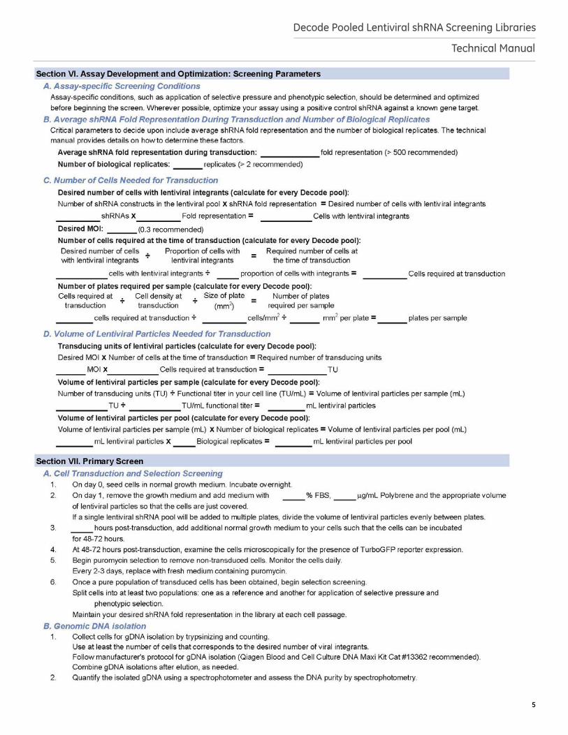
6 Assay Development and Optimization: Screening ParametersA. Assay-specific Screening ConditionsThe pooled shRNA screening workflow described here facilitates identification of genetic regulators of a range of biological processes. Hundreds or thousands of different shRNAs are introduced into a population of cells such that each cell expresses a single shRNA. These cells are subsequently subjected to selective pressure and phenotypic selection. Cells expressing shRNAs targeting genes involved in the phenotype of interest are enriched or depleted relative to the reference population of transduced cells. These shRNAs are identified by comparing the relative shRNA abundance between the experimental and reference population of cells. Screening approaches can be designed to identify genes involved in a phenotype of interest. shRNA-mediated knockdown screens can be used to identify genes that affect cell proliferation and/or survival, cause changes in cellular behavior (such as migration or adhesion), modulate response to different treatments (such as drugs or radiation) or change reporter or surface marker expression. Variables to consider when planning screening conditions include, but are not limited to, assay duration, conditions of selective pressure (such as concentration or duration), and method of phenotypic selection (such as viability, surface marker expression or migration). We recommend that you optimize all assay conditions prior to beginning a pooled shRNA screen, if possible, using an shRNA for a target known to be involved in the phenotype(s) of interest.
B. Average shRNA Fold Representation During Transduction and Number of Biological ReplicatesA critical and necessary consideration of pooled lentiviral shRNA screening is the extent to which any given shRNA construct in a pooled library will be represented in the screen; in other words, the number of cells that contain an independent genomic integration of any given shRNA or the number of biological replicates of each shRNA integration event and subsequent phenotypic selection. High shRNA representation results in high reproducibility between biological replicates and ensures that there is a sufficient window for detection of changes in shRNAs representation after phenotypic selection (Strezoska et al. 2012). A high shRNA fold representation is desirable, if technically feasible, for your assay. We recommend between 500 and 1,000 independent integrations per shRNA, particularly if you are interested in observing shRNA depletion hits. Increasing the number of biological replicates in a screen can also improve the ability to identify hits; however, we have observed that shRNA fold representation has a greater impact on the ability to identify hits than the number of biological replicates. Therefore, we recommend at least two biological replicates, while maintaining an average shRNA fold representation as high as is practical for your screen.
14
Decode Pooled Lentiviral shRNA Screening Libraries
Technical Manual
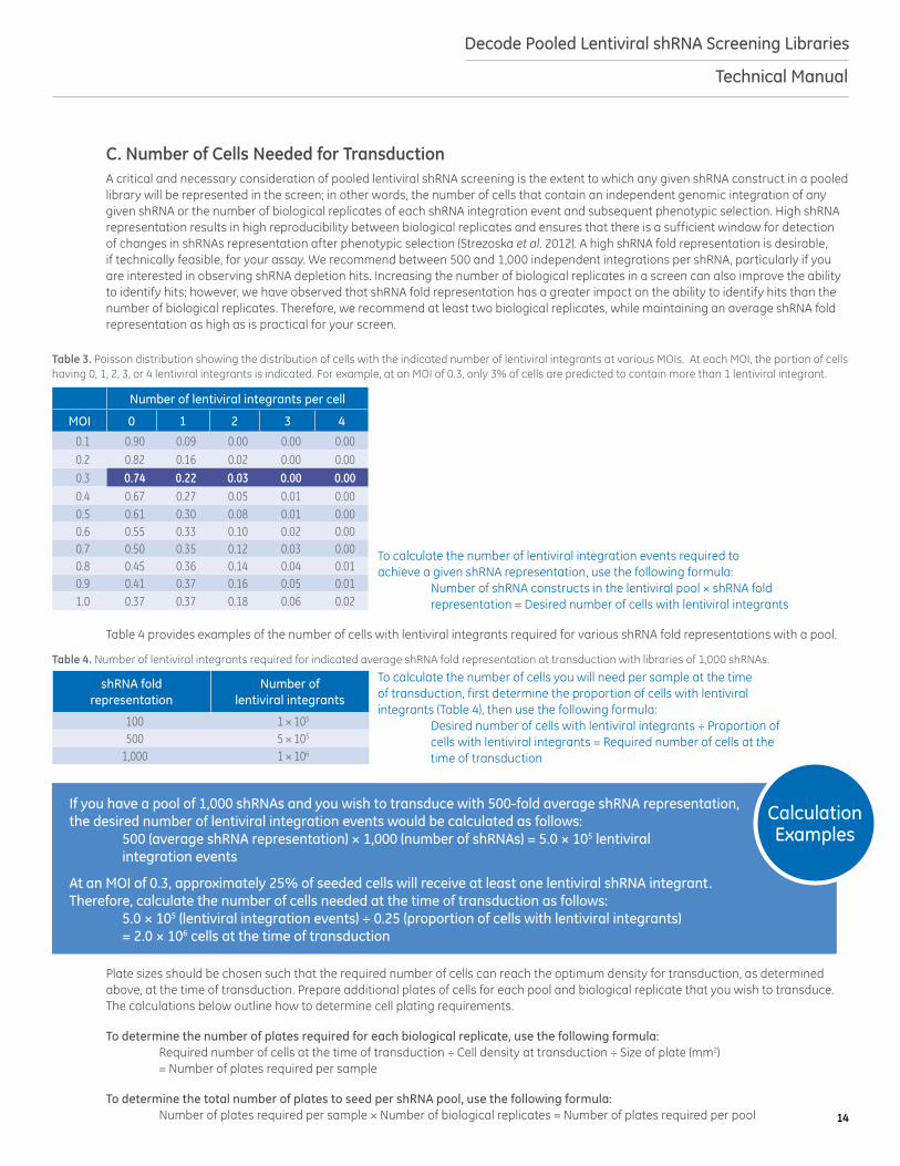
C. Number of Cells Needed for TransductionA critical and necessary consideration of pooled lentiviral shRNA screening is the extent to which any given shRNA construct in a pooled library will be represented in the screen; in other words, the number of cells that contain an independent genomic integration of any given shRNA or the number of biological replicates of each shRNA integration event and subsequent phenotypic selection. High shRNA representation results in high reproducibility between biological replicates and ensures that there is a sufficient window for detection of changes in shRNAs representation after phenotypic selection (Strezoska et al. 2012). A high shRNA fold representation is desirable, if technically feasible, for your assay. We recommend between 500 and 1,000 independent integrations per shRNA, particularly if you are interested in observing shRNA depletion hits. Increasing the number of biological replicates in a screen can also improve the ability to identify hits; however, we have observed that shRNA fold representation has a greater impact on the ability to identify hits than the number of biological replicates. Therefore, we recommend at least two biological replicates, while maintaining an average shRNA fold representation as high as is practical for your screen.
Table 4. Number of lentiviral integrants required for indicated average shRNA fold representation at transduction with libraries of 1,000 shRNAs.
shRNA fold representation
Number of lentiviral integrants
100 1 × 10⁵
500 5 × 10⁵
1,000 1 × 10⁶
Table 3. Poisson distribution showing the distribution of cells with the indicated number of lentiviral integrants at various MOIs. At each MOI, the portion of cells having 0, 1, 2, 3, or 4 lentiviral integrants is indicated. For example, at an MOI of 0.3, only 3% of cells are predicted to contain more than 1 lentiviral integrant.
Number of lentiviral integrants per cell
MOI 0 1 2 3 4
0.1 0.90 0.09 0.00 0.00 0.000.2 0.82 0.16 0.02 0.00 0.000.3 0.74 0.22 0.03 0.00 0.000.4 0.67 0.27 0.05 0.01 0.000.5 0.61 0.30 0.08 0.01 0.000.6 0.55 0.33 0.10 0.02 0.000.7 0.50 0.35 0.12 0.03 0.000.8 0.45 0.36 0.14 0.04 0.010.9 0.41 0.37 0.16 0.05 0.011.0 0.37 0.37 0.18 0.06 0.02
Calculation Examples
If you have a pool of 1,000 shRNAs and you wish to transduce with 500-fold average shRNA representation, the desired number of lentiviral integration events would be calculated as follows: 500 (average shRNA representation) × 1,000 (number of shRNAs) = 5.0 × 10⁵ lentiviral integration events
At an MOI of 0.3, approximately 25% of seeded cells will receive at least one lentiviral shRNA integrant. Therefore, calculate the number of cells needed at the time of transduction as follows: 5.0 × 10⁵ (lentiviral integration events) ÷ 0.25 (proportion of cells with lentiviral integrants) = 2.0 × 10⁶ cells at the time of transduction
To calculate the number of lentiviral integration events required to achieve a given shRNA representation, use the following formula: Number of shRNA constructs in the lentiviral pool × shRNA fold representation = Desired number of cells with lentiviral integrants
To calculate the number of cells you will need per sample at the time of transduction, first determine the proportion of cells with lentiviral integrants (Table 4), then use the following formula: Desired number of cells with lentiviral integrants ÷ Proportion of cells with lentiviral integrants = Required number of cells at the time of transduction
Plate sizes should be chosen such that the required number of cells can reach the optimum density for transduction, as determined above, at the time of transduction. Prepare additional plates of cells for each pool and biological replicate that you wish to transduce. The calculations below outline how to determine cell plating requirements.
To determine the number of plates required for each biological replicate, use the following formula: Required number of cells at the time of transduction ÷ Cell density at transduction ÷ Size of plate (mm2) = Number of plates required per sample
To determine the total number of plates to seed per shRNA pool, use the following formula: Number of plates required per sample × Number of biological replicates = Number of plates required per pool
Table 4 provides examples of the number of cells with lentiviral integrants required for various shRNA fold representations with a pool.
15
Decode Pooled Lentiviral shRNA Screening Libraries
Technical Manual
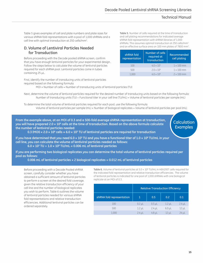
Table 5 gives examples of cell and plate numbers and plate sizes for various shRNA fold representations with a pool of 1,000 shRNAs and a cell line with optimal transduction at 250 cells/mm2.
D. Volume of Lentiviral Particles Needed for TransductionBefore proceeding with the Decode pooled shRNA screen, confirm that you have enough lentiviral particles for your experimental design. Follow the steps below to calculate the volume of lentiviral particles required for each shRNA pool. Lentiviral particles come in tubes containing 25 μL.
First, identify the number of transducing units of lentiviral particles required based on the following formula: MOI × Number of cells = Number of transducing units of lentiviral particles (TU)
Next, determine the volume of lentiviral particles required for the desired number of transducing units based on the following formula: Number of transducing units (TU) ÷ Functional titer in your cell line (TU/mL) = Volume of lentiviral particles per sample (mL)
To determine the total volume of lentiviral particles required for each pool, use the following formula: Volume of lentiviral particles per sample (mL) × Number of biological replicates = Volume of lentiviral particles per pool (mL)
Before proceeding with a Decode Pooled shRNA screen, carefully consider whether you have obtained a sufficient amount of lentiviral particles to perform a screen at the desired fold coverage, given the relative transduction efficiency of your cell line and the number of biological replicates you wish to perform. Table 6 outlines the volume of lentiviral particles needed for various shRNA fold representations and relative transduction efficiencies. Additional lentiviral particles can be ordered separately.
Table 5. Number of cells required at the time of transduction and cell plating recommendations for indicated average shRNA fold representation with shRNA libraries of 1,000 shRNAs. This assumes optimal transduction at 250 cells/mm2 and an effective surface area on 100 mm plates of 7800 mm2.
shRNA fold representation
Number of cells required at
transduction
Recommended cell plating
100 4.0 × 10⁵ 1 × 100 mm
500 2.0 × 10⁶ 1 × 100 mm
1,000 4.0 × 10⁶ 2 × 100 mm
Calculation Examples
From the example above, at an MOI of 0.3 and a 500-fold average shRNA representation at transduction, you will have prepared 2.0 × 10⁶ cells at the time of transduction. Based on the above formula calculate the number of lentiviral particles needed: 0.3 (MOI) × 2.0 × 10⁶ cells = 6.0 × 10⁵ TU of lentiviral particles are required for transduction
If you have determined that you need 6.0 × 10⁵ TU and you have a functional titer of 1.0 × 10⁸ TU/mL in your cell line, you can calculate the volume of lentiviral particles needed as follows: 6.0 × 10⁵ TU ÷ 1.0 × 10⁸ TU/mL = 0.006 mL of lentiviral particles
If you are performing two biological replicates you can determine the total volume of lentiviral particles required per pool as follows: 0.006 mL of lentiviral particles × 2 biological replicates = 0.012 mL of lentiviral particles
Table 6. Volume of lentiviral particles at 5.0 × 10⁸ TU/mL in HEK293T cells required for the indicated fold representation and relative transduction efficiencies. The volume of lentiviral particles is indicated for one pool of 1,000 shRNAs with one biological replicate at an MOI of 0.3.
Relative Transduction Efficiency
shRNA fold representation 1 0.5 0.2 0.1
100 0.2 µL 0.5 µL 1.2 µL 2.4 µL
500 1.2 µL 2.4 µL 6.0 µL 12 µL
1,000 2.4 µL 4.8 µL 12 µL 24 µL
16
Decode Pooled Lentiviral shRNA Screening Libraries
Technical Manual
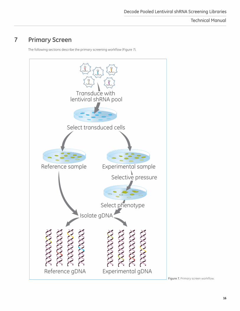
Primary screen
Reference sample
Isolate gDNA
Reference gDNA Experimental gDNA
Experimental sample
Transduce with lentiviral shRNA pool
Selective pressure
Select phenotype
Select transduced cells
Figure 7. Primary screen workflow.
7 Primary ScreenThe following sections describe the primary screening workflow (Figure 7).

17
Decode Pooled Lentiviral shRNA Screening Libraries
Technical Manual
A. Cell Transduction and Selection ScreeningThe experimental conditions described here, and in Figure 8, serve as a guide for performing lentiviral transductions. However, the precise cell number and volume of lentiviral particles necessary to achieve the desired MOI and average shRNA fold representation in the library should be determined specifically for each cell line of interest and each intended screening experiment, as outlined above. Similarly, conditions should be clearly defined prior to starting the screen for application of selective pressure and selection of phenotype of interest. For libraries comprised of multiple pools, we recommend that cells be transduced with individual shRNA pools in separate plates.
1. On day 0, seed cells in normal growth medium in the number of plates determined in section 6. The number of cells seeded should be determined by extrapolating from the number of cells needed at the time of transduction and the doubling time of your cell type. Incubate overnight.
2. The next day (day 1), remove the medium and add optimized transduction medium (section 5) with the appropriate amount of lentiviral particles (section 6) so that the cells are just covered. If a single lentiviral pool will be added to multiple plates, as determined in section 6, divide volume of lentiviral particles evenly between plates.
3. After the appropriate transduction time (section 5), add additional normal growth medium to your cells such that the cells can be incubated for 48-72 hours.
4. At 48-72 hours post-transduction examine the cells microscopically for the presence of TurboGFP reporter expression; this will be the first indication as to the efficiency of transduction. Note: When using a MOI = 0.3 you should expect approximately 25% of the cells to express TurboGFP.
5. Begin puromycin selection to remove non-transduced cells. Use the appropriate concentration of puromycin, as determined in section 5. Monitor the cells daily and observe the percentage of TurboGFP-positive cells. Every 48-72 hours, replace with fresh medium containing puromycin and passage cells as needed. Selection should be performed for the number of days determined in section 5.
6. Once a pure population of transduced cells has been obtained (3-6 days), begin selection screening. Split cells into at least two populations: one as a reference and another for application of selective pressure and phenotypic selection. To maintain your desired shRNA fold representation in the library at each cell passage, always retain at least the number of cells that corresponds to the desired number of lentiviral integrants.
B. Genomic DNA Isolation
Following selection, gDNA should be isolated from control and experimental cell populations. Note: Isolation of gDNA from cells transduced with Decode pools has been optimized in the protocol below using Qiagen Blood and Cell Culture DNA Maxi Kit (Cat #13362); however, kits from other manufacturers may also be suitable.
7. Collect cells for gDNA isolation by trypsinizing and counting. To maintain your desired fold representation during gDNA isolation, use at least the number of cells that corresponds to the desired number of lentiviral integrants. The most accurate results can be obtained by counting cell number prior to gDNA isolation. Follow the manufacturer’s protocol for purification of gDNA from cell cultures.
Note: It is important that you elute gDNA samples in EDTA-free buffer to prevent inhibition of subsequent PCR reactions. To ensure good DNA quality and yield, do not use more than the manufacturer’s recommended cell number and be sure that your gDNA is fully solubilized. If multiple purification columns are required to maintain representation of your sample, combine gDNA isolations after elution.
8. Quantify the isolated gDNA using a spectrophotometer and assess the DNA purity by measuring the ratio of the absorbance at 260 and 280 nm (A260/280) and at A230 (A260/230). High-quality gDNA samples should have an A260/A280 ratio of 1.8 to 2.0, indicating the absence of contaminating proteins, and an A260/A230 ratio of > 2.0, indicating the absence of other organic contaminants.
Seed cells Transducecells
Confirmtransduction
Puromycinselection
Selectivepressure
Phenotypicselection
12-18 hours
48-72 hours
3-6 days
Screenspecific
Figure 8. Timeline of primary screen.
18
Decode Pooled Lentiviral shRNA Screening Libraries
Technical Manual
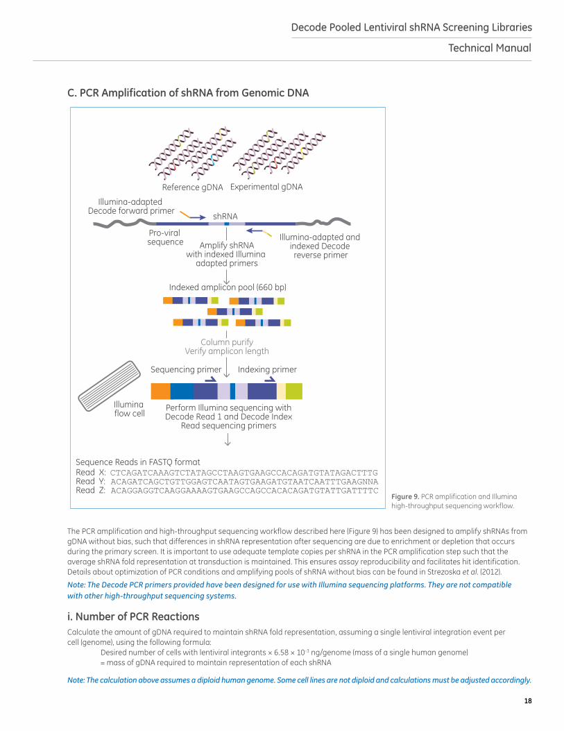
C. PCR Amplification of shRNA from Genomic DNA
The PCR amplification and high-throughput sequencing workflow described here (Figure 9) has been designed to amplify shRNAs from gDNA without bias, such that differences in shRNA representation after sequencing are due to enrichment or depletion that occurs during the primary screen. It is important to use adequate template copies per shRNA in the PCR amplification step such that the average shRNA fold representation at transduction is maintained. This ensures assay reproducibility and facilitates hit identification. Details about optimization of PCR conditions and amplifying pools of shRNA without bias can be found in Strezoska et al. (2012).
Note: The Decode PCR primers provided have been designed for use with Illumina sequencing platforms. They are not compatible with other high-throughput sequencing systems.
i. Number of PCR ReactionsCalculate the amount of gDNA required to maintain shRNA fold representation, assuming a single lentiviral integration event per cell (genome), using the following formula: Desired number of cells with lentiviral integrants × 6.58 × 10⁻³ ng/genome (mass of a single human genome) = mass of gDNA required to maintain representation of each shRNA
Note: The calculation above assumes a diploid human genome. Some cell lines are not diploid and calculations must be adjusted accordingly.
Section I: PCR amplification and Illumina sequencing
Experimental gDNAReference gDNA
Indexed amplicon pool (660 bp)
Column purifyVerify amplicon length
Pro-viral sequence
Illumina-adapted Decode forward primer
Illumina-adapted and indexed Decode reverse primer
Amplify shRNAwith indexed Illumina
adapted primers
shRNA
Sequencing primer
Illumina flow cell
Indexing primer
Sequence Reads in FASTQ formatRead X: CTCAGATCAAAGTCTATAGCCTAAGTGAAGCCACAGATGTATAGACTTTGRead Y: ACAGATCAGCTGTTGGAGTCAATAGTGAAGATGTAATCAATTTGAAGNNARead Z: ACAGGAGGTCAAGGAAAAGTGAAGCCAGCCACACAGATGTATTGATTTTC
Perform Illumina sequencing with Decode Read 1 and Decode Index
Read sequencing primers
Figure 9. PCR amplification and Illumina high-throughput sequencing workflow.
19
Decode Pooled Lentiviral shRNA Screening Libraries
Technical Manual
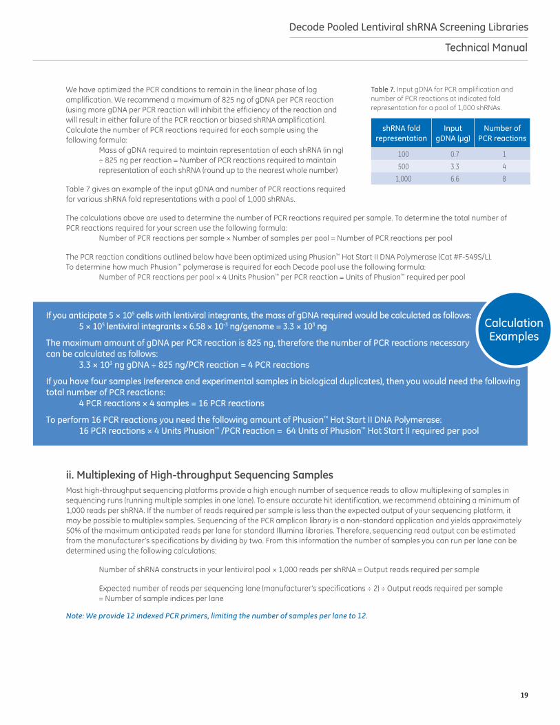
We have optimized the PCR conditions to remain in the linear phase of log amplification. We recommend a maximum of 825 ng of gDNA per PCR reaction (using more gDNA per PCR reaction will inhibit the efficiency of the reaction and will result in either failure of the PCR reaction or biased shRNA amplification). Calculate the number of PCR reactions required for each sample using the following formula: Mass of gDNA required to maintain representation of each shRNA (in ng) ÷ 825 ng per reaction = Number of PCR reactions required to maintain representation of each shRNA (round up to the nearest whole number)
Table 7 gives an example of the input gDNA and number of PCR reactions required for various shRNA fold representations with a pool of 1,000 shRNAs.
The calculations above are used to determine the number of PCR reactions required per sample. To determine the total number of PCR reactions required for your screen use the following formula: Number of PCR reactions per sample × Number of samples per pool = Number of PCR reactions per pool
The PCR reaction conditions outlined below have been optimized using Phusion™ Hot Start II DNA Polymerase (Cat #F-549S/L). To determine how much Phusion™ polymerase is required for each Decode pool use the following formula: Number of PCR reactions per pool × 4 Units Phusion™ per PCR reaction = Units of Phusion™ required per pool
ii. Multiplexing of High-throughput Sequencing SamplesMost high-throughput sequencing platforms provide a high enough number of sequence reads to allow multiplexing of samples in sequencing runs (running multiple samples in one lane). To ensure accurate hit identification, we recommend obtaining a minimum of 1,000 reads per shRNA. If the number of reads required per sample is less than the expected output of your sequencing platform, it may be possible to multiplex samples. Sequencing of the PCR amplicon library is a non-standard application and yields approximately 50% of the maximum anticipated reads per lane for standard Illumina libraries. Therefore, sequencing read output can be estimated from the manufacturer’s specifications by dividing by two. From this information the number of samples you can run per lane can be determined using the following calculations:
Number of shRNA constructs in your lentiviral pool × 1,000 reads per shRNA = Output reads required per sample
Expected number of reads per sequencing lane (manufacturer’s specifications ÷ 2) ÷ Output reads required per sample = Number of sample indices per lane
Note: We provide 12 indexed PCR primers, limiting the number of samples per lane to 12.
Table 7. Input gDNA for PCR amplification and number of PCR reactions at indicated fold representation for a pool of 1,000 shRNAs.
shRNA fold representation
Input gDNA (µg)
Number of PCR reactions
100 0.7 1
500 3.3 4
1,000 6.6 8
Calculation Examples
If you anticipate 5 × 10⁵ cells with lentiviral integrants, the mass of gDNA required would be calculated as follows: 5 × 10⁵ lentiviral integrants × 6.58 × 10⁻³ ng/genome = 3.3 × 10³ ng
The maximum amount of gDNA per PCR reaction is 825 ng, therefore the number of PCR reactions necessary can be calculated as follows: 3.3 × 10³ ng gDNA ÷ 825 ng/PCR reaction = 4 PCR reactions
If you have four samples (reference and experimental samples in biological duplicates), then you would need the following total number of PCR reactions: 4 PCR reactions × 4 samples = 16 PCR reactions
To perform 16 PCR reactions you need the following amount of Phusion™ Hot Start II DNA Polymerase: 16 PCR reactions × 4 Units Phusion™ /PCR reaction = 64 Units of Phusion™ Hot Start II required per pool
20
Decode Pooled Lentiviral shRNA Screening Libraries
Technical Manual
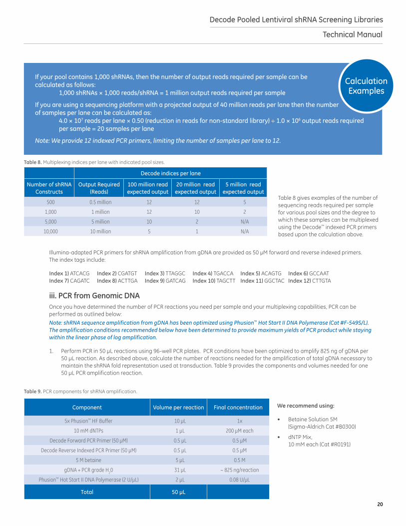
Table 8 gives examples of the number of sequencing reads required per sample for various pool sizes and the degree to which these samples can be multiplexed using the Decode™ indexed PCR primers based upon the calculation above.
Illumina-adapted PCR primers for shRNA amplification from gDNA are provided as 50 μM forward and reverse indexed primers. The index tags include:
Index 1) ATCACG Index 2) CGATGT Index 3) TTAGGC Index 4) TGACCA Index 5) ACAGTG Index 6) GCCAAT Index 7) CAGATC Index 8) ACTTGA Index 9) GATCAG Index 10) TAGCTT Index 11) GGCTAC Index 12) CTTGTA
iii. PCR from Genomic DNAOnce you have determined the number of PCR reactions you need per sample and your multiplexing capabilities, PCR can be performed as outlined below: Note: shRNA sequence amplification from gDNA has been optimized using Phusion™ Hot Start II DNA Polymerase (Cat #F-549S/L). The amplification conditions recommended below have been determined to provide maximum yields of PCR product while staying within the linear phase of log amplification.
1. Perform PCR in 50 µL reactions using 96-well PCR plates. PCR conditions have been optimized to amplify 825 ng of gDNA per 50 µL reaction. As described above, calculate the number of reactions needed for the amplification of total gDNA necessary to maintain the shRNA fold representation used at transduction. Table 9 provides the components and volumes needed for one 50 µL PCR amplification reaction.
We recommend using:
• Betaine Solution 5M (Sigma-Aldrich Cat #B0300)
• dNTP Mix, 10 mM each (Cat #R0191)
Calculation Examples
If your pool contains 1,000 shRNAs, then the number of output reads required per sample can be calculated as follows: 1,000 shRNAs × 1,000 reads/shRNA = 1 million output reads required per sample
If you are using a sequencing platform with a projected output of 40 million reads per lane then the number of samples per lane can be calculated as: 4.0 × 10⁷ reads per lane × 0.50 (reduction in reads for non-standard library) ÷ 1.0 × 10⁶ output reads required per sample = 20 samples per lane
Note: We provide 12 indexed PCR primers, limiting the number of samples per lane to 12.
Table 8. Multiplexing indices per lane with indicated pool sizes.
Decode indices per lane
Number of shRNA Constructs
Output Required (Reads)
100 million read expected output
20 million read expected output
5 million read expected output
500 0.5 million 12 12 5
1,000 1 million 12 10 2
5,000 5 million 10 2 N/A
10,000 10 million 5 1 N/A
Component Volume per reaction Final concentration
5x Phusion™ HF Buffer 10 µL 1x
10 mM dNTPs 1 µL 200 µM each
Decode Forward PCR Primer (50 µM) 0.5 µL 0.5 µM
Decode Reverse Indexed PCR Primer (50 µM) 0.5 µL 0.5 µM
5 M betaine 5 µL 0.5 M
gDNA + PCR grade H20 31 µL ~ 825 ng/reaction
Phusion™ Hot Start II DNA Polymerase (2 U/µL) 2 µL 0.08 U/µL
Total 50 µL
Table 9. PCR components for shRNA amplification.
21
Decode Pooled Lentiviral shRNA Screening Libraries
Technical Manual
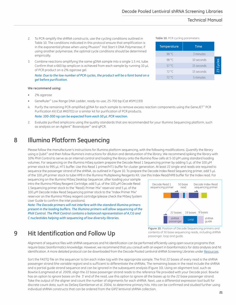
2. To PCR-amplify the shRNA constructs, use the cycling conditions outlined in Table 10. The conditions indicated in this protocol ensure that amplification is in the exponential phase when using Phusion™ Hot Start II DNA Polymerase; if using another polymerase, the optimal cycle conditions should be determined empirically.
3. Combine reactions amplifying the same gDNA sample into a single 1.5 mL tube. Confirm that a 660 bp amplicon is achieved from each sample by running 10 µL of PCR product on a 2% agarose gel.
Note: Due to the low number of PCR cycles, the product will be a faint band on a gel before purification.
We recommend using:
• 2% agarose
• GeneRuler™ Low Range DNA Ladder, ready-to-use, 25-700 bp (Cat #SM1193)
4. Purify the remaining PCR-amplified gDNA for each sample to remove excess reaction components using the GeneJET™ PCR Purification Kit (Cat #K0701) or a similar kit for purification of PCR products. Note: 100-300 ng can be expected from each 50 μL PCR reaction.
2. Evaluate purified amplicons using the quality standards that are recommended for your Illumina Sequencing platform, such as analysis on an Agilent™ Bioanalyzer™ and qPCR.
8 Illumina Platform SequencingPlease follow the manufacturer’s instructions for Illumina platform sequencing, with the following modifications. Quantify the library using a Qubit™ and then follow Illumina’s instructions for dilution and denaturation of the library. We recommend spiking the library with 10% PhiX Control to serve as an internal control and loading the library onto the Illumina flow cells at 5-10 pM using standard loading volumes. For sequencing on the Illumina HiSeq system prepare the Decode Read 1 Sequencing primer by adding 5 µL of the 100 µM primer stock to 995 µL HT1 buffer. Use this Read 1 primer/HT1 buffer for cluster generation. At least 22 single-end reads are required to sequence the passenger strand of the shRNA, as outlined in Figure 10. To prepare the Decode Index Read Sequencing primer, add 5 µL of the 100 µM primer stock to tube HP8 in the Illumina Multiplexing Reagents Kit. Use this Index Read/HP8 buffer for the Index read. For sequencing on the Illumina MiSeq Desktop Sequencer, after loading your sample intro the Illumina MiSeq Reagent Cartridge, add 5 μL of the 100 μM Decode Read 1 Sequencing primer stock to the "Read1 Primer Mix" reservoir and 5 μL of the 100 μM Decode Index Read Sequencing primer stock to the "Index Primer Mix" reservoir on the Illumina MiSeq reagent cartridge (please check the MiSeq System User Guide to confirm the inlet positions). Note: The Decode primers will not interfere with the standard Illumina primers present in the loading buffers. The Illumina primers will allow sequencing of the PhiX Control. The PhiX Control contains a balanced representation of A,T,G and C nucleotides helping with sequencing of low diversity libraries.
9 Hit Identification and Follow UpAlignment of sequence files with shRNA sequences and hit identification can be performed efficiently using open source programs that require basic bioinformatics knowledge. However, we recommend that you consult with an expert in bioinformatics for data analysis and hit identification. A more detailed protocol can be downloaded from the Decode Pooled Lentiviral shRNA Screening Libraries under Resources.
Sort the FASTQ file on the sequencer to bin each index tag with the appropriate sample. The first 22 bases of every read is the shRNA passenger strand (the variable region) and is sufficient to differentiate the shRNAs. The remaining bases in the read include the shRNA and a partial guide strand sequence and can be ignored in the subsequent analysis (Figure 10). Using an alignment tool, such as Bowtie (Langmead et al. 2009), align the 22 base passenger strand reads to the reference file provided with your Decode pool. Bowtie has an option to ignore bases on the 3' end of the read, use this option to ignore all the bases up to the 22 base passenger strand. Take the output of the aligner and count the number of alignments for each shRNA. Next, use a differential expression tool built for discrete count data, such as DeSeq (Gentleman et al. 2004), to determine primary hits. Hits can be confirmed and studied further using individual shRNA constructs that can be ordered from the GIPZ lentiviral shRNA collection.
Temperature Time
98 °C 3 minutes
98 °C 10 seconds
23 C
ycle
s
57 °C 15 seconds
72 °C 15 seconds
72 °C 5 minutes
Table 10. PCR cycling parameters.
Position of shRNA information in 50 bp sequencing output
Decode Read 1 sequencing primer
Decode Index Read sequencing primer
22 bases 19 bases
50 base read
9 bases
shRNAguide
LoopshRNA passenger
Figure 10. Position of Decode Sequencing primers and contents of 50 base sequencing reads, including shRNA passenger, loop and guide.

22
Decode Pooled Lentiviral shRNA Screening Libraries
Technical Manual
10 Frequently Asked Questions (FAQs)
11 TroubleshootingFor answers to questions that are not addressed here, please email Technical Support at [email protected] with the answers to the questions below, your sales order or purchase order number and the Cat # or Clone ID of the construct or pooled library with which you are having trouble.
1. What was the relative transduction efficiency between your cell line and that reported by Dharmacon?
2. What was your lentiviral titer for your cell line?
3. At what MOI did you use the pools?
4. What is your cell line?
5. Did you maintain the cells on puromycin after transduction?
6. How much time elapsed from transduction to puromycin selection?
7. How much puromycin did you use?
8. How long did your cells stay on puromycin selection?
9. In brief, what is your assay?
Questions Answers
Where can I find titer information for my pools?The titer of your Lentiviral shRNA pools will be indicated on the Certificate of Analysis (C of A). C of As for purchased Decode Lentiviral shRNA pools can also be requested from Technical Support.
Where can I find the sequence of an individual shRNA included in my pool(s)?
The full insert sequences are in the Decode Data Files CD sent with your shipment. Please contact Technical Support with your Purchase Order number if you did not receive this CD in your shipment.
How many screens can I perform with my pools?
The number of screens which can be performed per lentiviral pool will depend on:1. The transducibility of your specific cell line/cell type.2. Your target MOI.3. The shRNA fold representation you choose to maintain throughout the screen. 4. The number of biological replicates you intend to include.
Can I run my PCR samples on a Life Technologies™ Ion Torrent Sequencing Platform?
No. The adaptors that are integrated into our Decode PCR primers are specific to the Illumina sequencing platforms and are not compatible with other sequencing platforms.
Can I use Sanger sequencing instead of high-throughput sequencing to deconvolute my screen with the Decode primers?
No. Decode PCR Primers, Read 1 Sequencing and Index Read Primers are designed for high-throughput sequencing on an Illumina instrument only. If you wish to perform Sanger sequencing, you can design your own primers.
How is gDNA input calculated to maintain desired shRNA fold representation?
The quantity of gDNA required for maintaining a desired shRNA fold representation can be calculated using the formulas and values (Table 11) below.
Note: this calculation is for a diploid genome. Some cell lines are not diploid and calculations should be adjusted accordingly.
Table 11. Constants needed for calculating gDNA input.
Constants Value
Mass of a base pair 660 g/mol/bp
Base pairs per diploid human genome 6 × 10⁹ bp
Avogadro’s number 6.02 × 10²³ mol
Calculate the mass of human genome: (6 × 10⁹ bp/genome) × (660 g/mol/bp) = 3.96 × 10¹² g/mol/genome (3.96 × 10¹² g/mol/genome) ÷ (6.02 x 10²³ mol) = 6.58 × 10⁻¹² g/genome
23
Decode Pooled Lentiviral shRNA Screening Libraries
Technical Manual
12 ReferencesCited and Recommended References:1. C. Fellmann, J. Zuber, Functional identification of optimized RNAi triggers using a massively parallel sensor assay. Mol. Cell 41,
733 (2011).
2. C. Gazin, N. Wajapeyee, An elaborate pathway required for Ras-mediated epigenetic silencing. Nature 449, 1073 (2007).
3. R. C. Gentleman, V. J. Carey, Bioconductor: open software development for computational biology and bioinformatics. Genome biol. 5, R80 (2004).
4. S. Gobeil, X. Zhu, A genome-wide shRNA screen identifies GAS1 as a novel melanoma metastasis suppressor gene. Genes & Dev. 22, 2932 (2008).
5. K. Gumireddy, A. Li, KLF17 is a negative regulator of epithelial-mesenchymal transition and metastasis in breast cancer. Nat. Cell. Biol. 11, 1297 (2009).
6. K. E. Hurov, C. Cotta-Ramusino, A genetic screen identifies the Triple T complex required for DNA damage signaling and ATM and ATR stability. Genes & Dev. 24, 1939 (2010).
7. J. C. Kappes, X. Wu, Safety considerations in vector development. Somat. Cell Mol. Genet. 26, 147 (2001).
8. B. L. Levine, L. M. Humeau, Gene transfer in humans using a conditionally replicating lentiviral vector. PNAS 103, 17372 (2006).
9. B. C. O'Connell, B. Adamson, A genome-wide camptothecin sensitivity screen identifies a mammalian MMS22L-NFKBIL2 Complex required for genomic stability. Mol. Cell 40, 645 (2010).
10. R. K. Palakurthy, N. Wajapeyee, Epigenetic silencing of the RASSF1A tumor suppressor gene through HOXB3-mediated induction of DNMT3B expression. Mol. Cell 36, 219 (2009).
11. M. R. Schlabach, J.Luo, Cancer proliferation gene discovery through functional genomics. Science 319, 620 (2008).
12. J. M. Silva, K. Marran, Profiling essential genes in human mammary cells by multiplex RNAi screening. Science 319, 617 (2008).
13. D. Sims, A. Mendes-Pereira, High-throughput RNA interference screening using pooled shRNA libraries and next generation sequencing. Genome Biol. 12, R104 (2011).
14. A. Smogorzewska, R. Desetty, A genetic screen identifies FAN1, a Fanconi anemia-associated nuclease necessary for DNA interstrand crosslink repair. Mol. Cell 39, 36 (2010).
15. G. A. Smolen, J. Zhang, A genome-wide RNAi screen identifies multiple RSK-dependent regulators of cell migration. Genes & Dev. 24, 2654 (2010).
16. Ž. Strezoska, A. Licon, Optimized PCR conditions and increased shRNA fold representation improve reproducibility of pooled shRNA screens. PLoS One 7, e42341 (2012).
17. T. Westbrook, E. Martin, A genetic screen forcandidate tumor suppressors identifies REST. Cell 121, 837 (2005).
18. L. Zender, W. Xue, An oncogenomics-based in vivo RNAi screen identifies tumor suppressors in liver cancer. Cell 135, 852 (2008).
13 Label Licenses The shRNA and gene expression Products, use and applications, are covered by pending and issued patents. Certain Label licenses govern the use of the products, these can be found at http://www.gelifesciences.com/webapp/wcs/stores/servlet/catalog/en/GELifeSciences-us/about-us/licensing-statements. It is each Buyer’s responsibility to determine which intellectual property rights held by third parties may restrict the use of Products for a particular application. Please review the Label Licenses governing all use of the shRNA and gene expression Products.
dharmacon.gelifesciences.com/uploadedFiles/product-terms-and-conditions.pdf
TurboGFP is a registered trademark of Evrogen. Bioanalyzer is a registered trademark of Agilent. QIAquick is a trademark of Qiagen. dNTP. GeneRule, HyClone, Phusion, Qubit, and Life Technologies are trademarks of Thermo Fisher Scientific, Inc. GE, imagination at work and GE monogram are trademarks of General Electric Company. Dharmacon, Inc., a General Electric company doing business as GE Healthcare. All other trademarks are the property of General Electric Company or one of its subsidiaries. ©2014 General Electric Company—All rights reserved. First published September 2014. GE Healthcare UK Limited, Amersham Place, Little Chalfont, Buckinghamshire, HP7 9NA, UK
V5-0
316
Orders can be placed at:gelifesciences.com/dharmacon
Customer Support: [email protected] Support: [email protected] or1.800.235.9880; 303.604.9499 if you have any questions.