diabetes mellitus by dr. zahoor 1. diabetes mellitus (dm) diabetes mellitus is a syndrome of chronic...
TRANSCRIPT
1
DIABETES MELLITUS
By Dr. Zahoor
2
DIABETES MELLITUS (DM)
• Diabetes Mellitus is a syndrome of chronic hyperglycaemia due to insulin deficiency or insulin resistance or both
• It affects 220 million people world wide• DM is usually irreversible, and patient can lead
a reasonably normal life style • Its late complications result in reduced life
expectancy and major health costs
3
DIABETES MELLITUS
Complications include • Macro vascular e.g. coronary artery disease,
stroke, peripheral vascular disease• Micro vascular damage e.g. diabetic-
retinopathy, nephropathy, neuropathy
4
DIABETES MELLITUS
• Insulin is synthesized by beta cells in pancreas • Body blood glucose is maintained in range of
3.5 – 8 mmol/L (63 – 144 mg%)• Brain uses glucose and brain glucose uptake is
not dependent on insulin
5
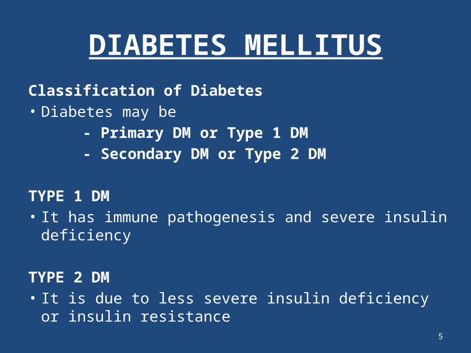
DIABETES MELLITUSClassification of Diabetes• Diabetes may be - Primary DM or Type 1 DM - Secondary DM or Type 2 DM
TYPE 1 DM• It has immune pathogenesis and severe insulin deficiency
TYPE 2 DM• It is due to less severe insulin deficiency or insulin
resistance
6
7
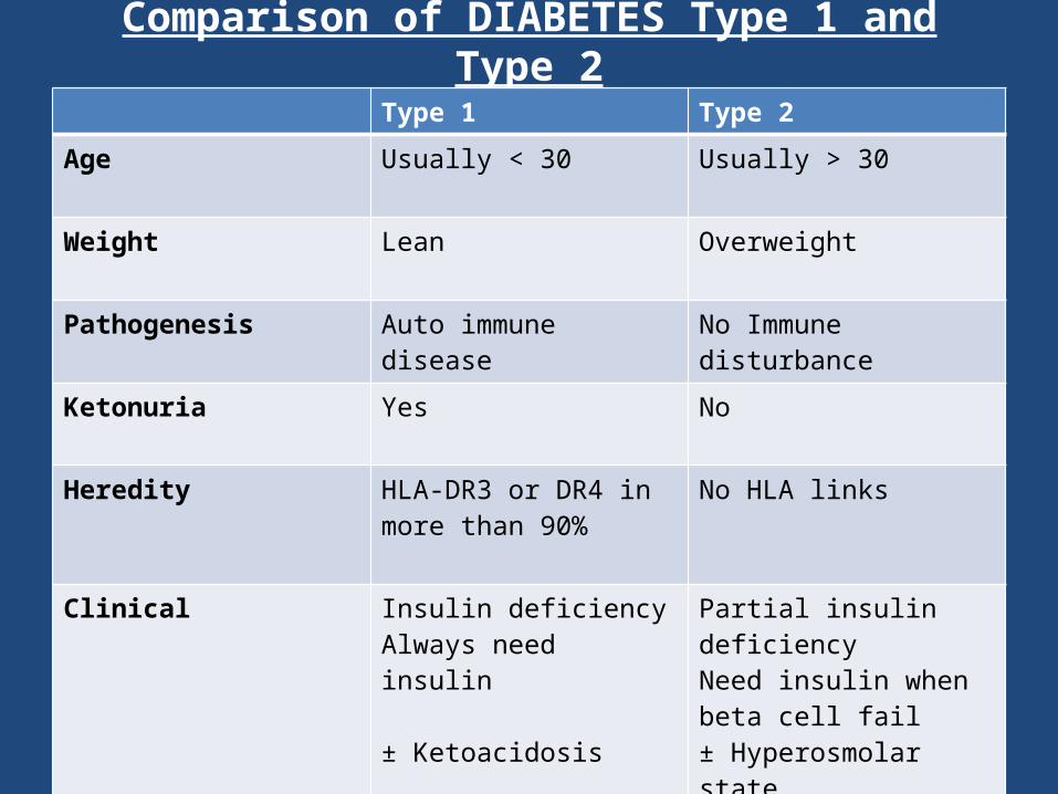
Comparison of DIABETES Type 1 and Type 2Type 1 Type 2
Age Usually < 30 Usually > 30
Weight
Lean Overweight
Pathogenesis Auto immune disease No Immune disturbance
Ketonuria Yes No
Heredity HLA-DR3 or DR4 in more than 90%
No HLA links
Clinical Insulin deficiencyAlways need insulin
± Ketoacidosis
Partial insulin deficiency Need insulin when beta cell fail± Hyperosmolar state
Biochemical C-peptide disappears C-peptide persists
8
TYPE 1 DM
Aetiology • Immune mediated – anti bodies against beta
cell of pancreas• Disease starts in childhood, reaching peak at
the time of puberty, but can present at any age• Type 1 DM is associated with auto immune
thyroid disease, Coeliac disease, Addison’s disease and pernicious anemia
9
TYPE 2 DM
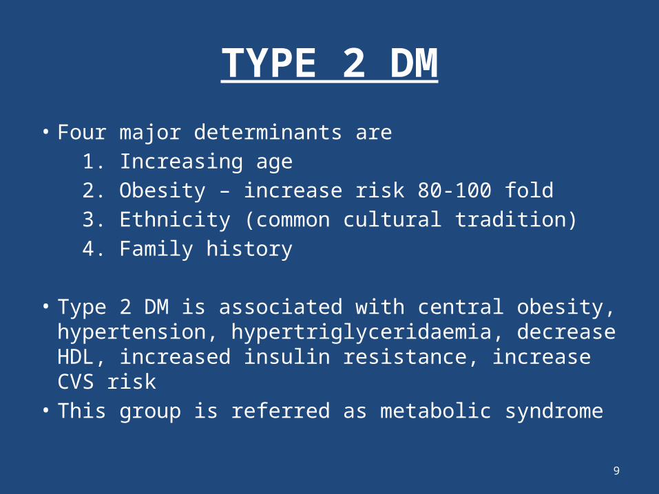
• Four major determinants are 1. Increasing age 2. Obesity – increase risk 80-100 fold 3. Ethnicity (common cultural tradition) 4. Family history
• Type 2 DM is associated with central obesity, hypertension, hypertriglyceridaemia, decrease HDL, increased insulin resistance, increase CVS risk
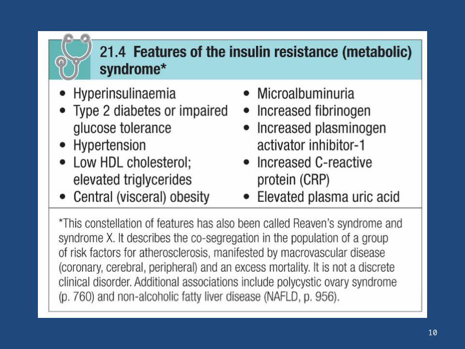
• This group is referred as metabolic syndrome
10
11
DIABETES TYPE 2
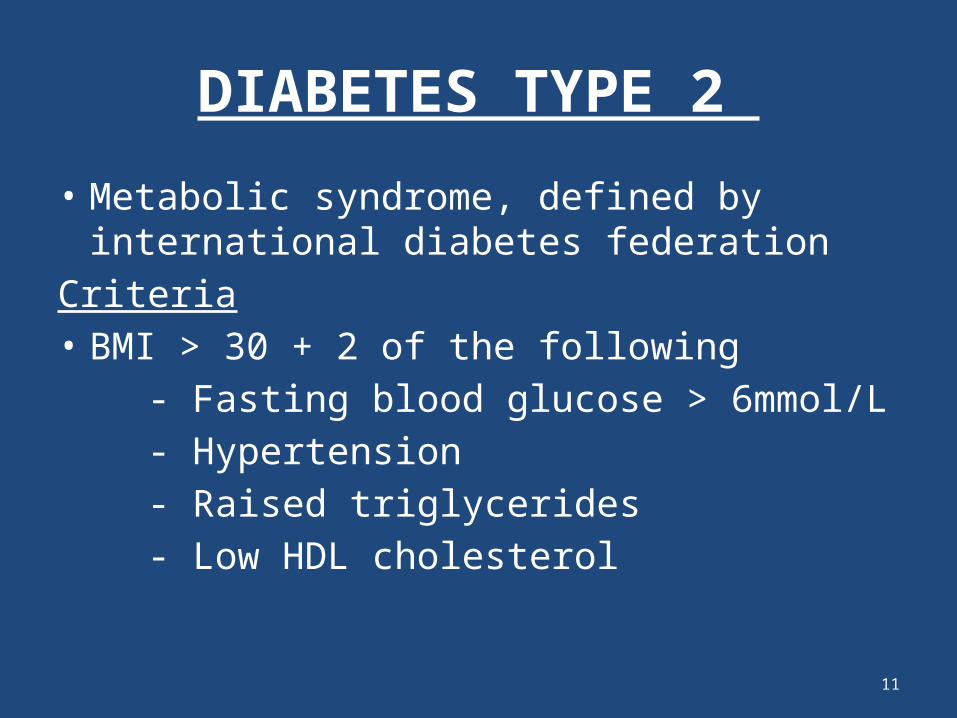
• Metabolic syndrome, defined by international diabetes federation
Criteria • BMI > 30 + 2 of the following - Fasting blood glucose > 6mmol/L - Hypertension - Raised triglycerides - Low HDL cholesterol
12
DIABETES TYPE 2
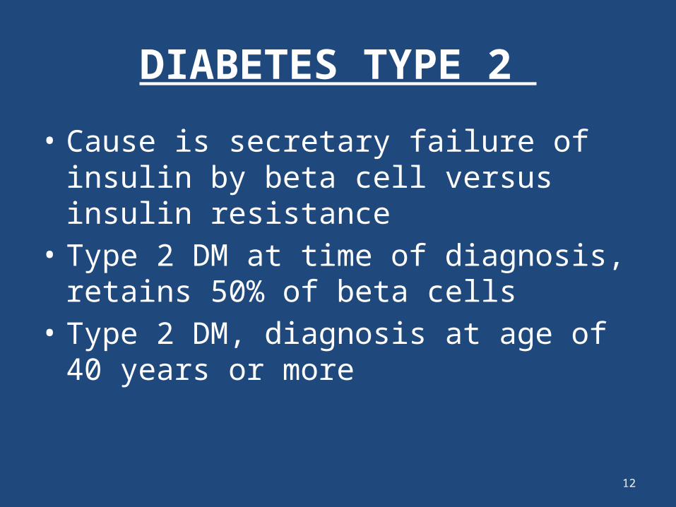
• Cause is secretary failure of insulin by beta cell versus insulin resistance
• Type 2 DM at time of diagnosis, retains 50% of beta cells
• Type 2 DM, diagnosis at age of 40 years or more
13
DIABETES TYPE 2
Clinical Presentation • Presentation may be acute, sub acute or asymptomatic Acute presentation • Usually young people, present with 2-6 week history of - Polyurea – due to osmotic diuresis as blood glucose level
is high - Thirst – due to loss of fluid and electrolyte - Weight loss – due to breakdown of fat and muscle
secondary to insulin deficiency
14
DIABETES TYPE 2
Sub acute presentation• Patient present with history of thirst, polyurea and
weight loss• Patient may complain of loss of energy, visual blurring
(due to glucose induced changes in refraction) • Following complication as presenting feature. These
include: - Staphylococcal skin infection - Polyneuropathy causing tingling and numbness in feet - Retinopathy noted by optician on visit
15
DIABETES TYPE 2
Asymptomatic presentation• Glycosuria or raised blood glucose may be
detected on routine examination e.g. insurance purpose
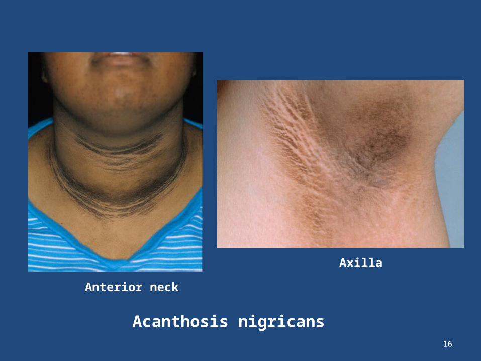
• If patient has insulin resistance, they may have Acanthosis nigricans (characterized by blackish pigmentation at the neck and in axillae)
16
Acanthosis nigricans
Anterior neck
Axilla
17
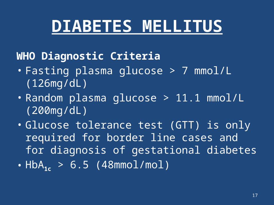
DIABETES MELLITUS
WHO Diagnostic Criteria• Fasting plasma glucose > 7 mmol/L (126mg/dL)• Random plasma glucose > 11.1 mmol/L
(200mg/dL)• Glucose tolerance test (GTT) is only required for
border line cases and for diagnosis of gestational diabetes
• HbA1c > 6.5 (48mmol/mol)
18
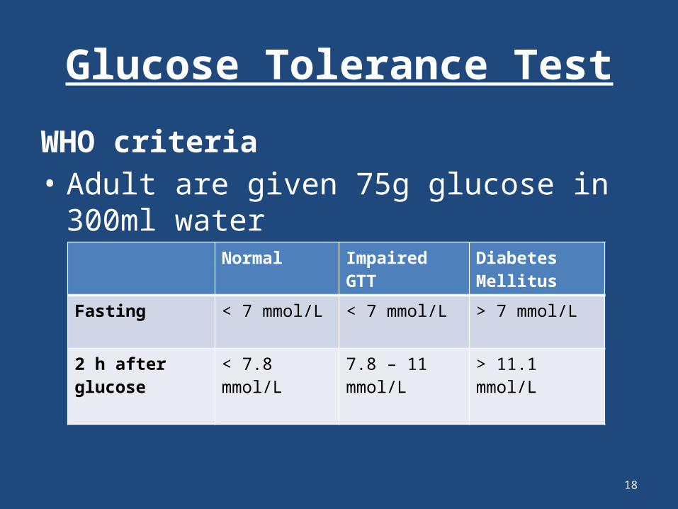
Glucose Tolerance Test
WHO criteria• Adult are given 75g glucose in 300ml water
Normal Impaired GTT Diabetes Mellitus
Fasting < 7 mmol/L < 7 mmol/L > 7 mmol/L
2 h after glucose < 7.8 mmol/L 7.8 – 11 mmol/L > 11.1 mmol/L
19
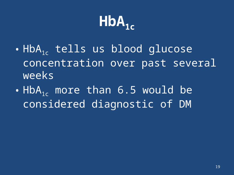
HbA1c
• HbA1c tells us blood glucose concentration over past several weeks
• HbA1c more than 6.5 would be considered diagnostic of DM
20
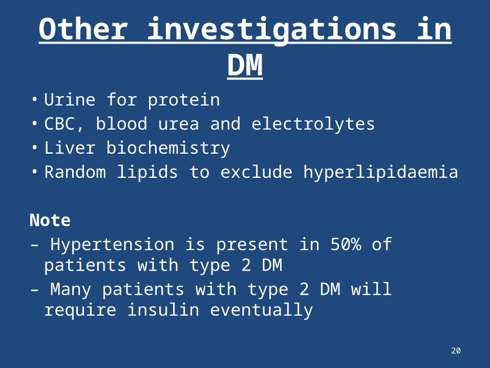
Other investigations in DM
• Urine for protein• CBC, blood urea and electrolytes • Liver biochemistry• Random lipids to exclude hyperlipidaemia
Note – Hypertension is present in 50% of patients with type 2
DM– Many patients with type 2 DM will require insulin
eventually
21
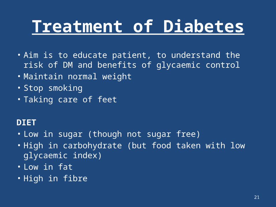
Treatment of Diabetes
• Aim is to educate patient, to understand the risk of DM and benefits of glycaemic control
• Maintain normal weight • Stop smoking • Taking care of feet
DIET• Low in sugar (though not sugar free)• High in carbohydrate (but food taken with low glycaemic index)• Low in fat • High in fibre
22
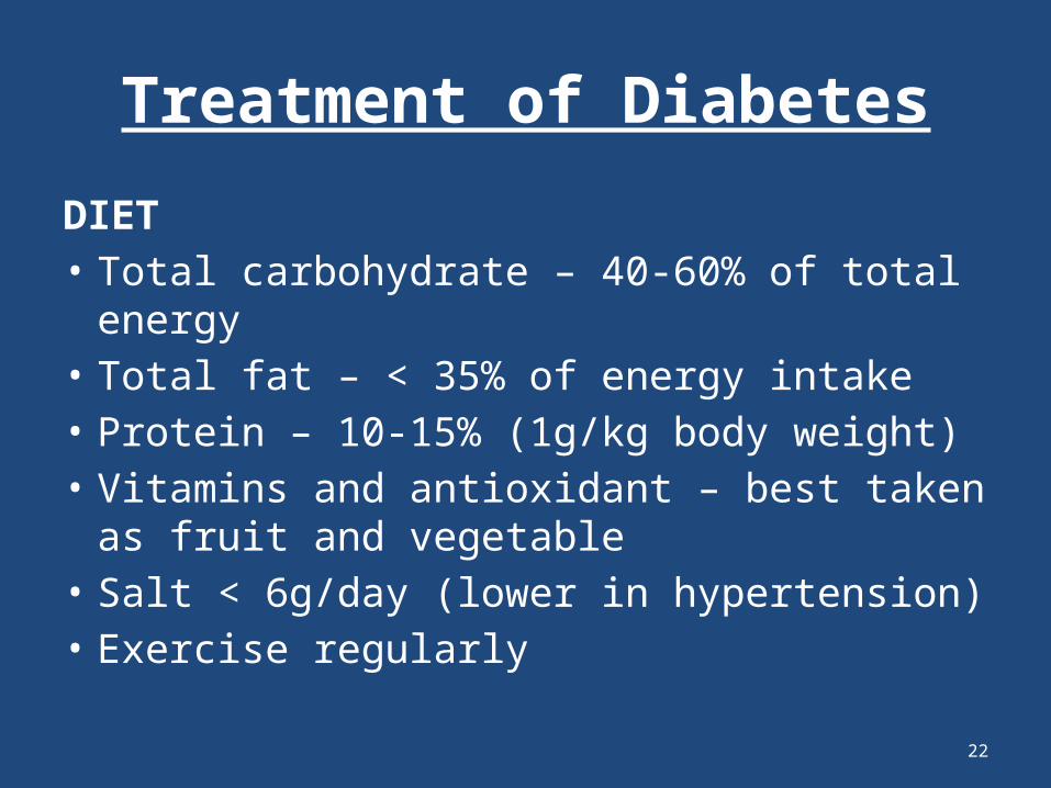
Treatment of Diabetes
DIET• Total carbohydrate – 40-60% of total energy• Total fat – < 35% of energy intake • Protein – 10-15% (1g/kg body weight)• Vitamins and antioxidant – best taken as fruit
and vegetable• Salt < 6g/day (lower in hypertension)• Exercise regularly
23
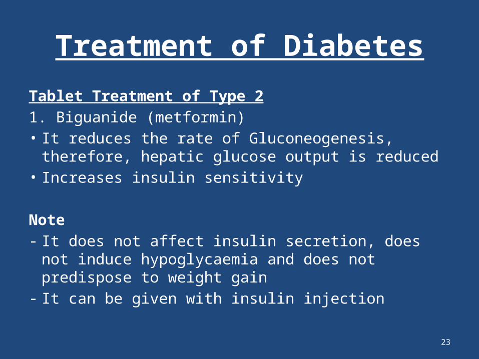
Treatment of Diabetes
Tablet Treatment of Type 21. Biguanide (metformin)• It reduces the rate of Gluconeogenesis, therefore,
hepatic glucose output is reduced • Increases insulin sensitivity
Note- It does not affect insulin secretion, does not induce
hypoglycaemia and does not predispose to weight gain - It can be given with insulin injection
24
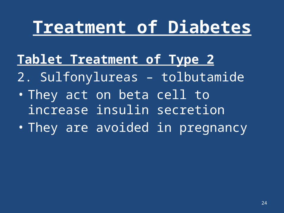
Treatment of Diabetes
Tablet Treatment of Type 22. Sulfonylureas – tolbutamide• They act on beta cell to increase insulin
secretion• They are avoided in pregnancy
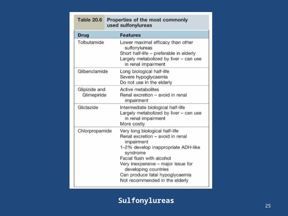
25Sulfonylureas
26
Treatment of Diabetes(cont)
3. Meglitinids e.g. repaglinide • They increase insulin secretion
4. Thiazolidinediones also known as glitazones e.g. Piogliatazone
• They reduce insulin resistance, they also reduce hepatic glucose production and increase glucose utilization at periphery
• They potentiate the effect of injected insulin• Side effect – weight gain, heart failure, osteoporosis,
bladder cancer
27
Treatment of Diabetes
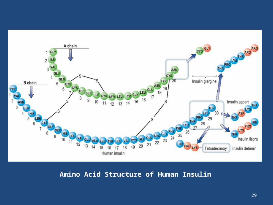
New drugs• Dipeptidyl peptidase -4 (DPP 4) inhibitors• DPP4 inhibitors increase insulin Injection therapy for Type 2 DM • Insulin is formed in every vertebrate, but there are
species differences• Insulin derived from beef or pig pancreas have been
replaced in most countries by biosynthetic human insulin• They are produced by adding a DNA sequence coding
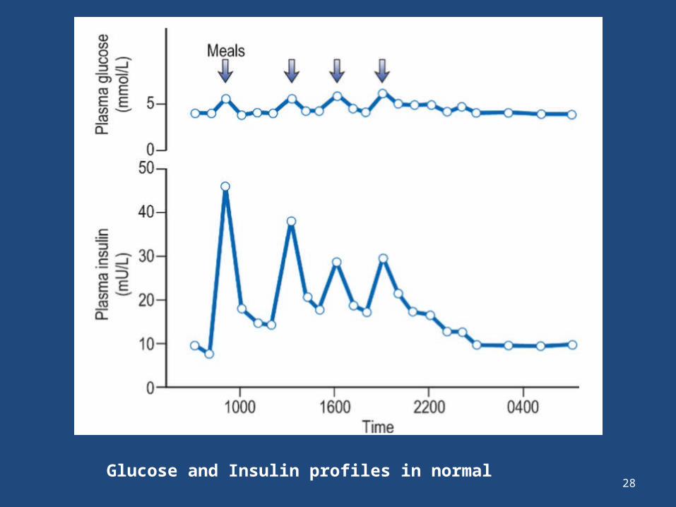
28Glucose and Insulin profiles in normal
29
Amino Acid Structure of Human Insulin
30
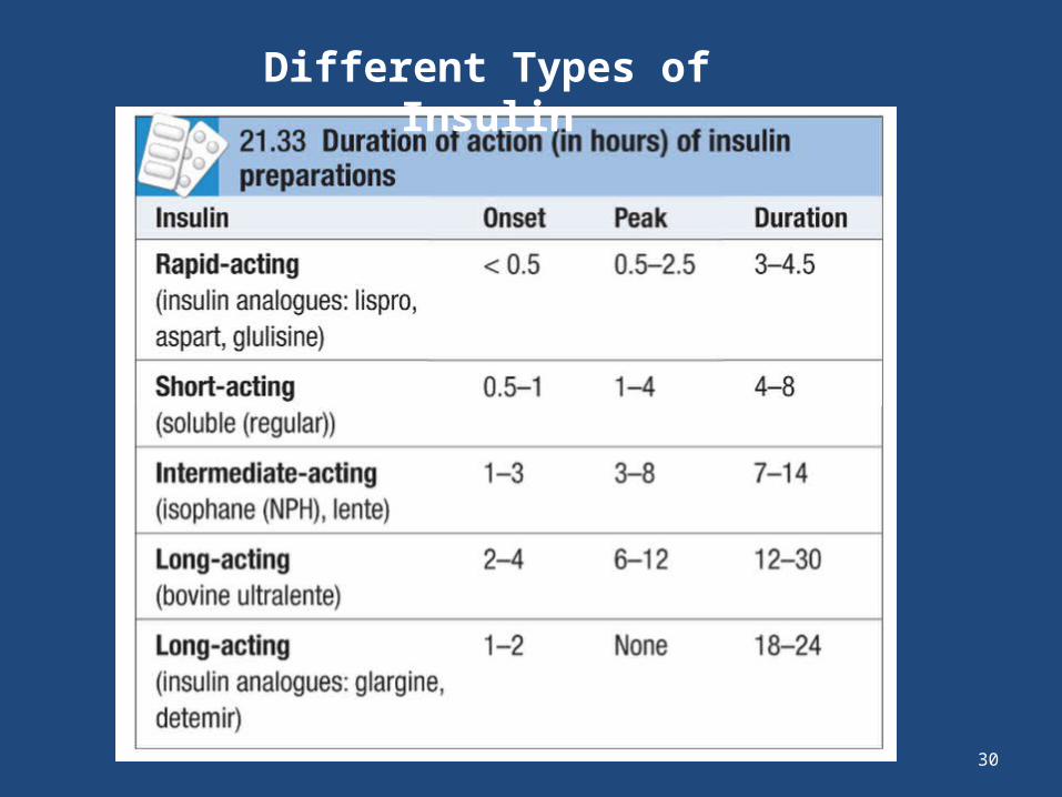
Different Types of Insulin
31
Practical Management of Diabetes
Type 2 DM • Oral therapy Oral + Insulin
Type 1 DM• Insulin is required
32
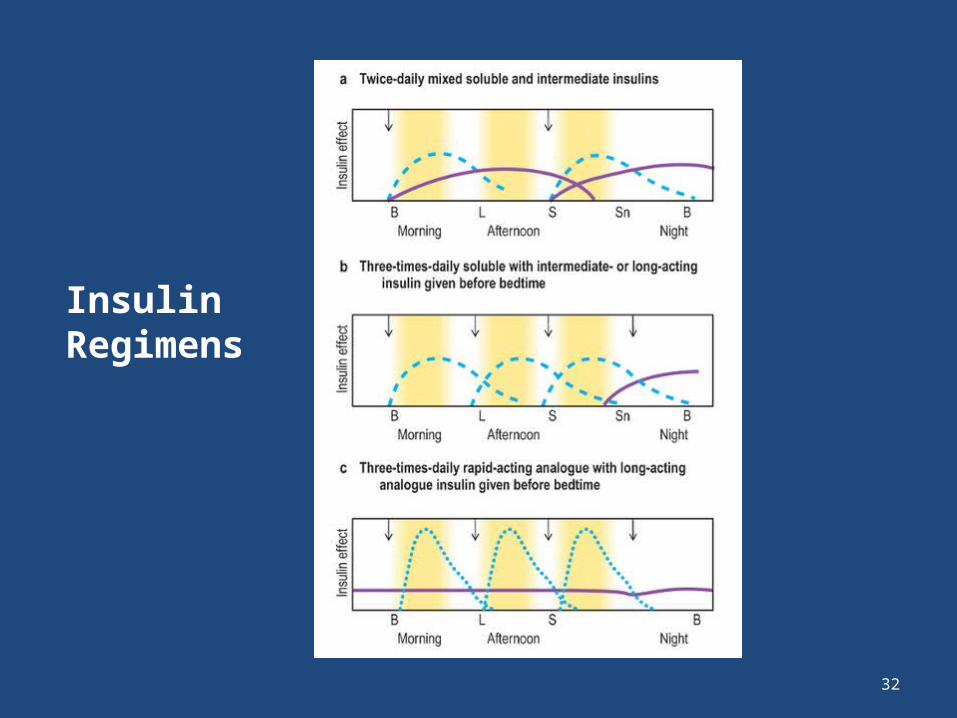
Insulin Regimens
33
Practical Management of Diabetes
Infusion Devices• CS II (Continuous Subcutaneous Insulin
Infusion) • Insulin is delivered by a small pump strapped
around the waist that infuses a constant insulin via a needle in the subcutaneous tissue
• At meal time, doses are given as bolus of insulin at start of meal by the pump
34
Complication of Insulin Therapy
• Pain at time of delivery of injection • Fatty lumps – lipohypertrophy (usually occur due to single
injection site)• Weight gain• Hypoglycemia (occurs when blood glucose falls below
3mmol/L) and most patient experience adrenergic response that is sweating, tremor and pounding heart beat
- Hypoglycemia can cause convulsions, coma NOTE - Patient should always be advised to carry glucose or
sweets to use when he feels hypoglycemic symptoms
35
DM Treatment
Whole Pancreas and Pancreatic Islet Transplantation• Whole pancreas transplantation has been performed
for 30 years • Immunosuppressive therapy is needed• Islet transplantation is done by harvesting pancreatic
islets from 2-3 cadavers pancreas and then injected into the portal vein and they seed themselves in liver
• Immunosuppressive therapy is needed
36
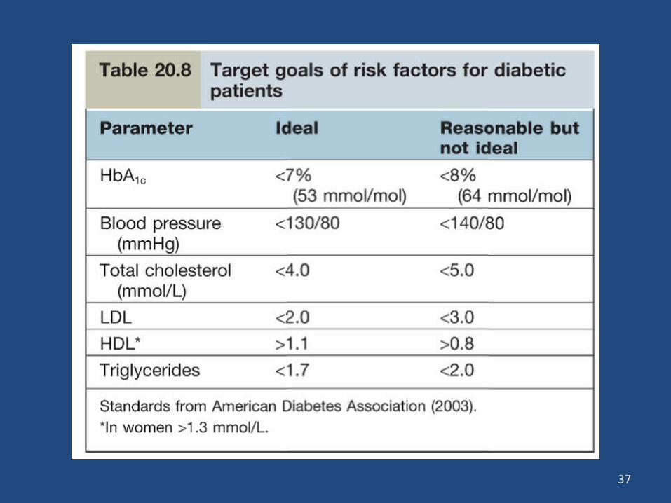
Measuring the metabolic control of Diabetes
• Urine Test – Dipstic test• Home blood glucose testing – Prick at the side
of the finger and not at the tip of the finger as it is densely innervated
• HbA1c – target for glucose control HbA1c less than 7%
37
38
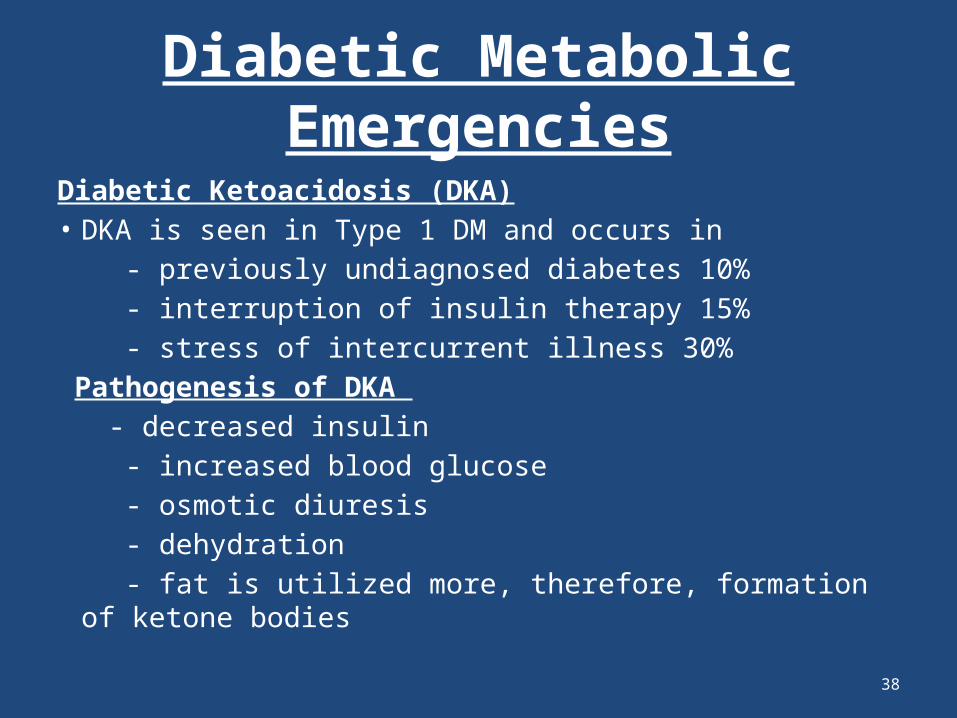
Diabetic Metabolic Emergencies
Diabetic Ketoacidosis (DKA)• DKA is seen in Type 1 DM and occurs in - previously undiagnosed diabetes 10% - interruption of insulin therapy 15% - stress of intercurrent illness 30% Pathogenesis of DKA - decreased insulin - increased blood glucose - osmotic diuresis - dehydration - fat is utilized more, therefore, formation of ketone bodies
39
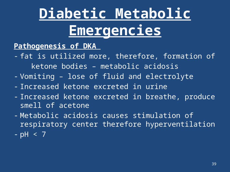
Diabetic Metabolic Emergencies
Pathogenesis of DKA - fat is utilized more, therefore, formation of ketone bodies – metabolic acidosis - Vomiting – lose of fluid and electrolyte- Increased ketone excreted in urine- Increased ketone excreted in breathe, produce smell of
acetone - Metabolic acidosis causes stimulation of respiratory
center therefore hyperventilation - pH < 7
40
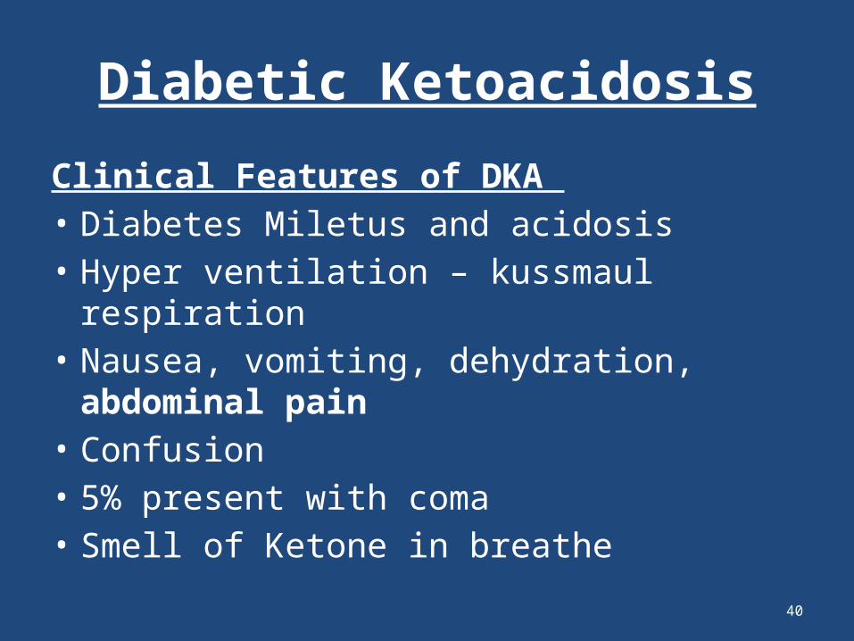
Diabetic Ketoacidosis
Clinical Features of DKA • Diabetes Miletus and acidosis • Hyper ventilation – kussmaul respiration • Nausea, vomiting, dehydration, abdominal
pain• Confusion • 5% present with coma• Smell of Ketone in breathe
41
Diabetic Ketoacidosis
Diagnosis• Increased blood glucose • Ketonaemia, heavy ketone urea• Arterial blood gases – acidosisClinical • Pulse > 100 b.p.m or < 60 b.p.m • Systolic BP < 90 mmHg• Oxygen saturation < 92% on room air
42
Diabetic Ketoacidosis
Diagnosis (cont)• Blood Ketone > 6mmol/L• Bicarbonate < 12mmol/L• Arterial pH < 7.1 • Hypokalemia – K < 3.5 mmol/L
43
Diabetic Ketoacidosis
Management• Replace fluid with 0.9% saline, average 5-7L is
required• Replace electrolyte loses – K may be normal
initially but insulin therapy leads to uptake of K by cells, therefore K level falls, therefore K is given as soon as insulin is started
• Restore acid base balance – HCO3 is seldom necessary, only used when pH is < 7
44
Diabetic Ketoacidosis
Management (cont)• Short acting insulin as IV intravenous infusion or
hourly IM injection (avoid SC injection as blood flow is decreased in shock)
• Monitor blood glucose • Look for underlying cause • Do X-ray chest, blood CBC, cultures• ECG • Serum amylase

45
Hyperosmolar Hyperglycaemic State
46
Hyperosmolar Hyperglycaemic State
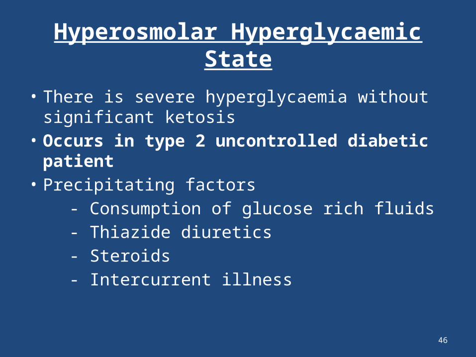
• There is severe hyperglycaemia without significant ketosis
• Occurs in type 2 uncontrolled diabetic patient• Precipitating factors - Consumption of glucose rich fluids - Thiazide diuretics - Steroids - Intercurrent illness
47
Hyperosmolar Hyperglycaemic State
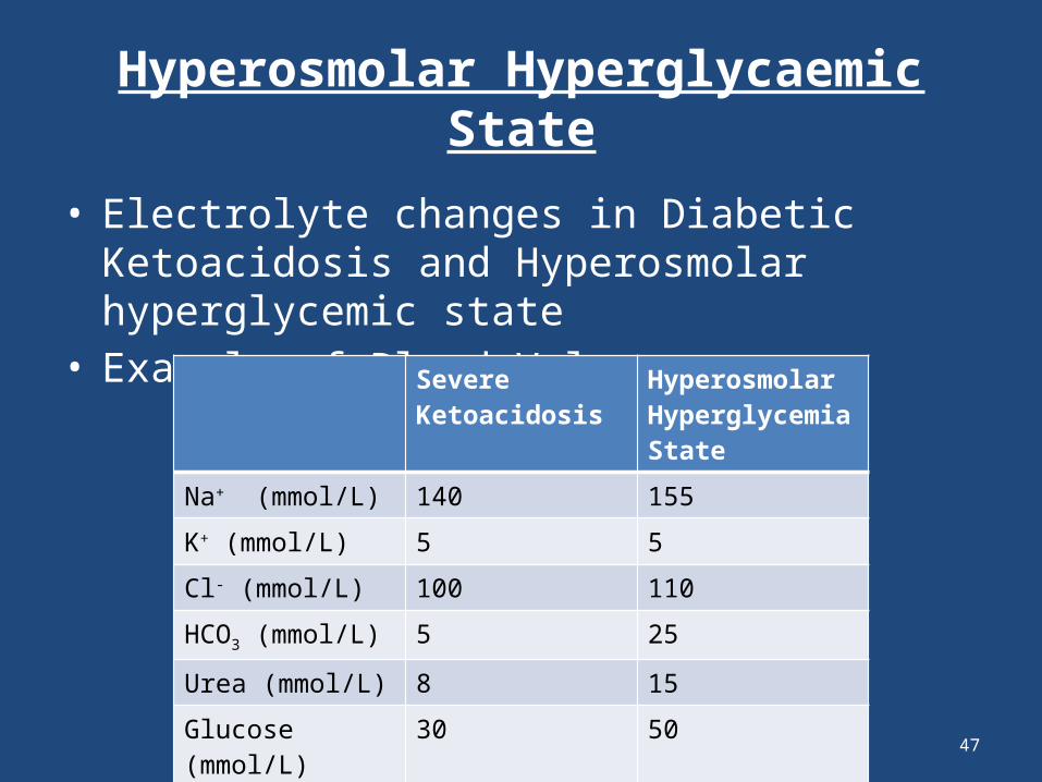
• Electrolyte changes in Diabetic Ketoacidosis and Hyperosmolar hyperglycemic state
• Example of Blood ValuesSevere Ketoacidosis
Hyperosmolar Hyperglycemia State
Na+ (mmol/L) 140 155
K+ (mmol/L) 5 5
Cl- (mmol/L) 100 110
HCO3 (mmol/L) 5 25
Urea (mmol/L) 8 15
Glucose (mmol/L) 30 50
Arterial pH 7.0 7.35
48
Hyperosmolar Hyperglycaemic State
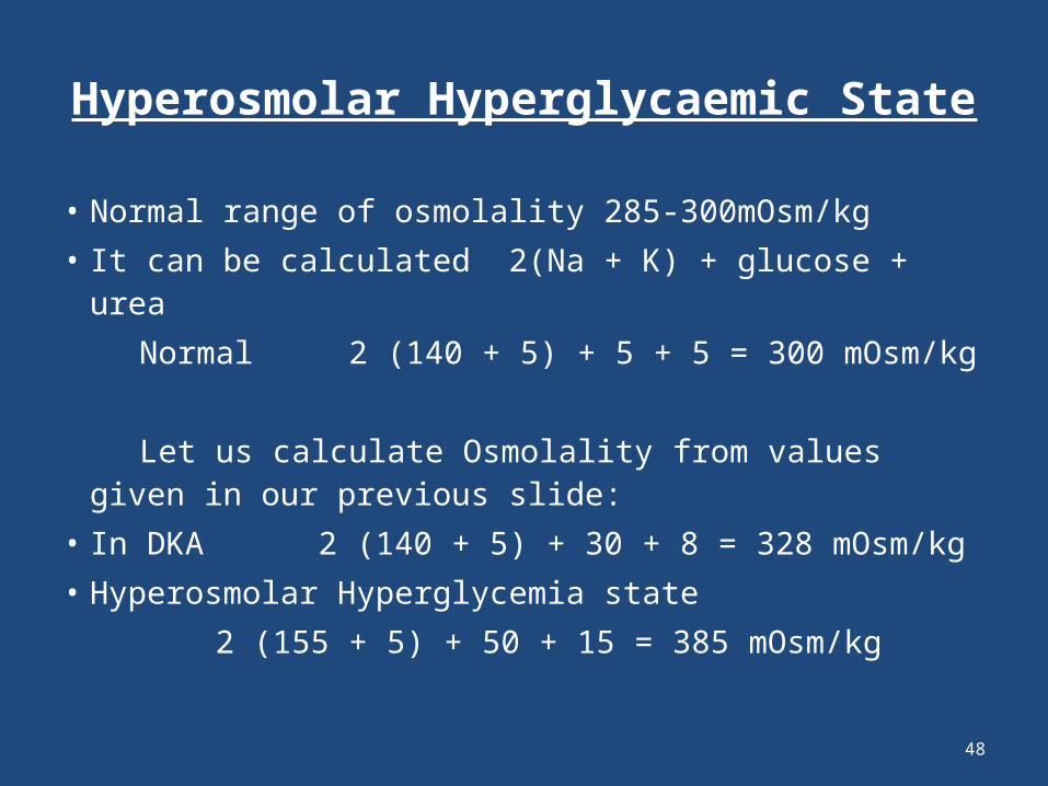
• Normal range of osmolality 285-300mOsm/kg• It can be calculated 2(Na + K) + glucose + urea Normal 2 (140 + 5) + 5 + 5 = 300 mOsm/kg Let us calculate Osmolality from values given in
our previous slide: • In DKA 2 (140 + 5) + 30 + 8 = 328 mOsm/kg• Hyperosmolar Hyperglycemia state 2 (155 + 5) + 50 + 15 = 385 mOsm/kg
49
Normal ANION GAP
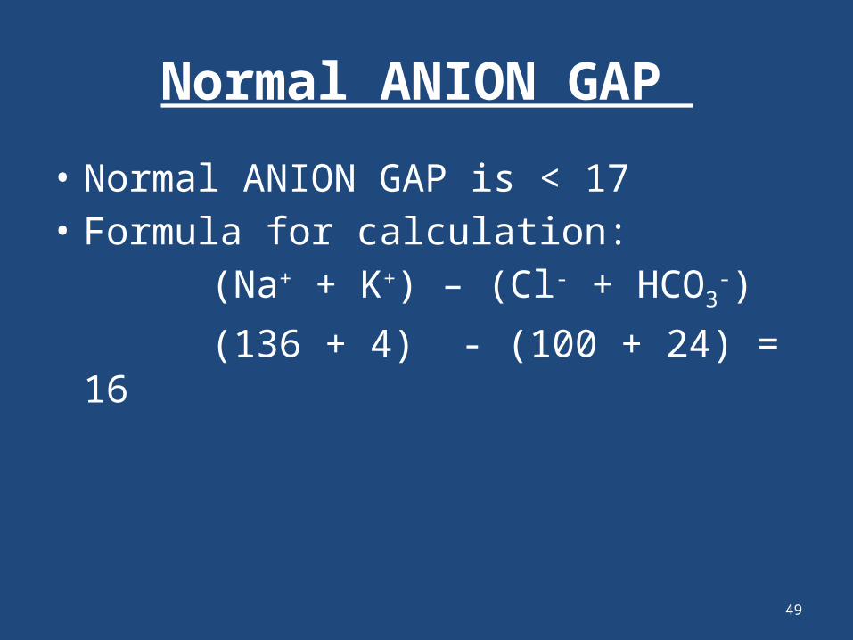
• Normal ANION GAP is < 17• Formula for calculation: (Na+ + K+) – (Cl- + HCO3
-)
(136 + 4) - (100 + 24) = 16
50
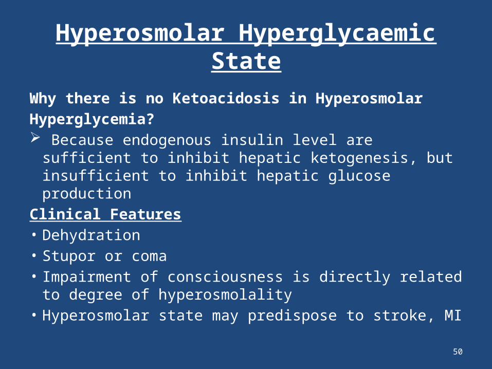
Hyperosmolar Hyperglycaemic State
Why there is no Ketoacidosis in Hyperosmolar Hyperglycemia? Because endogenous insulin level are sufficient to inhibit
hepatic ketogenesis, but insufficient to inhibit hepatic glucose production
Clinical Features• Dehydration • Stupor or coma • Impairment of consciousness is directly related to degree of
hyperosmolality • Hyperosmolar state may predispose to stroke, MI
51
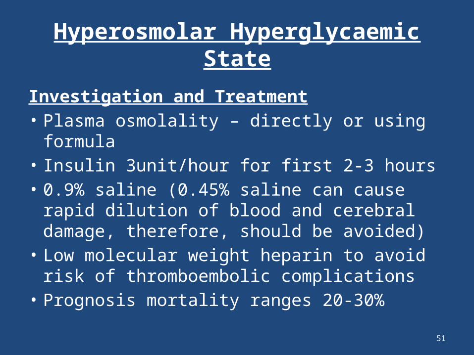
Hyperosmolar Hyperglycaemic State
Investigation and Treatment• Plasma osmolality – directly or using formula• Insulin 3unit/hour for first 2-3 hours • 0.9% saline (0.45% saline can cause rapid
dilution of blood and cerebral damage, therefore, should be avoided)
• Low molecular weight heparin to avoid risk of thromboembolic complications
• Prognosis mortality ranges 20-30%

52
Lactic Acidosis
• It can occur in diabetic patient on Biguanide therapy,for patient taking metformin risk is low
• Patient has severe metabolic acidosis with large ANION GAP (normal < 17 mmol/L), usually without hyperglycemia or ketosis
• Treatment – rehydration – infusion of isotonic 1.26% bicarbonate – mortality is high – 50%
53
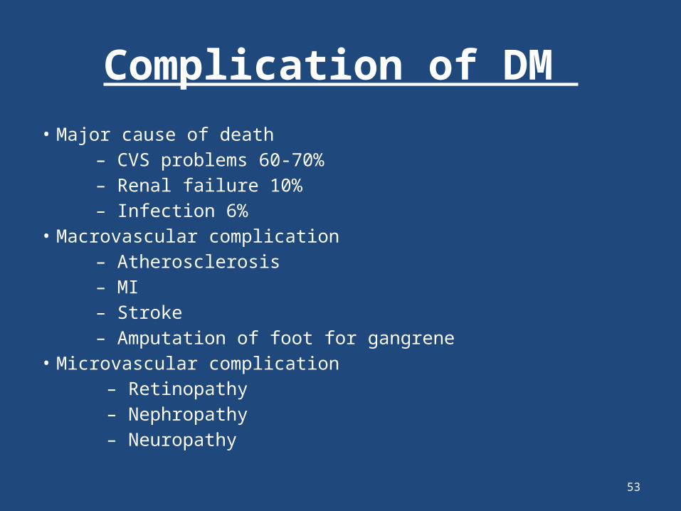
Complication of DM • Major cause of death – CVS problems 60-70% – Renal failure 10% – Infection 6%• Macrovascular complication – Atherosclerosis – MI – Stroke – Amputation of foot for gangrene• Microvascular complication – Retinopathy – Nephropathy – Neuropathy
54
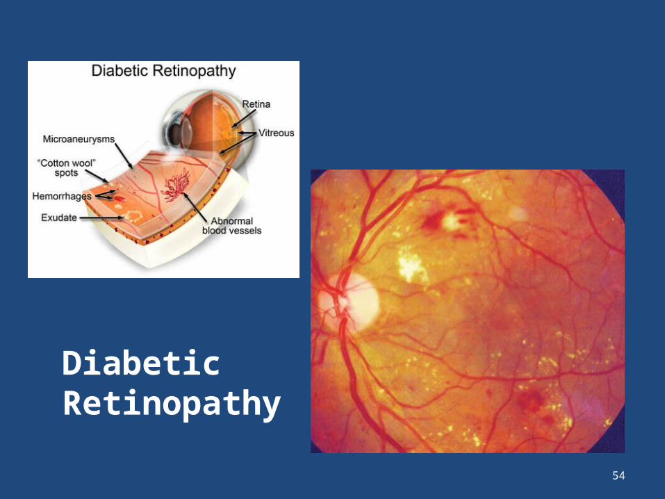
Diabetic Retinopathy

55
Thank you