diagnosis and thyroid cancer incidence is … - pdf of slides.pdf · • 2008 estimate for united...
TRANSCRIPT
1
Diagnosis and Management of Thyroid Cancer: A Case-based
ApproachMatthew D. Ringel. M.D.
Professor, Department of MedicineDivision of Endocrinology, Diabetes, and
MetabolismThe Ohio State University College of Medicine
Goals and Objectives• Review current recommendations for the
following in a case-based manner:• Diagnostic testing for thyroid cancer
diagnosis.• Initial treatments for patients diagnosed
with thyroid cancer.• Key concepts in monitoring thyroid cancer.• Controversies in the management of
recurrent thyroid cancer
• 2008 Estimate for United States from NCI and American Cancer Society 1, 2
37,340 New Cases1,590 Cause-related Mortalities388,386 patients with thyroid cancer
• Incidence rate is increasing in both men and women at fastest rate of all cancers
• Mortality has been stable since the early 1970s and remains very low, but dependent on stage at diagnosis.
Thyroid Cancer Incidence is Rising in the United States
1 http://SEER.cancer.gov2 www.cancer.org
• Increased use of thyroid ultrasound• Increased availability of ultrasound-guided
FNA of non-palpable nodulesSupported by data from SEER database demonstrating increase is due nearly completely to small, early stage papillary thyroid cancers 1
• True increased in incidence due to environmental or genetic factors
Less likely due to no change in mortality rate
Potential Causes for the Increase in Thyroid Cancer Incidence
1Davies, et al. JAMA 2006;295:2164
2
Examples of Recent Society Guidelines for Thyroid Cancer
Diagnosis and Management• NCCN: Sherman SI: J Natl Compr Cancer Netw.
5:568—621; 2007.• American Thyroid Association: Cooper DS, et al.
Thyroid. 16: 109-42, 2006.• European Thyroid Association: Pacini F, et al. Eur
J Endocrinol. 154: 787-803, 2006.• British Thyroid Association: London, Royal
College of Physicians. 2002.• AACE/AAES: Endocr Pract. 7: 202-220, 2001.
Guidelines Levels of Evidence• Modified USPSTF categories• Level A: Strongly Recommends: Based on strong
evidence that a service or intervention improves health outcomes.
• Level B: Recommends: Based on fair evidence limited by the number, quality, or consistence of studies.
• Level C: Recommends based on expert opinion• Level D: Recommends against based on expert opinion.• Level E: Recommends against based in fair evidence.• Level F: Recommends against based on strong evidence.• Level I: Recommends neither for nor against based on
insufficient, poor quality, or conflicting evidence.1. Cooper DS, et al Thyroid 2006 16; 20062. US Preventive Services Task Force Ratings: Strengths of Recommendations and Quality of evidence. Guide to Clinical Preventive Services, 3rd ed. Periodic Updates 2000-2003. Agency for Healthcare Research and Quality. Rockville, MD.
Illustrative Case• A 42 year old woman is in the office
for and annual physical.• She has no specific complaints other
than fatigue.• She has no dry skin, constipation, or
other signs/symptoms of hypothyroidism.
• She has no heart palpitations, heat intolerance or tremor.
Case• On physical examination, BP is 112/72;
HR: 72
• Neck exam reveals an ~2 cm rubbery thyroid nodule on the left that moves easily with swallowing. There are no palpable nodes. It is non-tender. No nodularity is noted in the right side.
• Remainder of the examination is normal.
3
Case• What are the key historical questions
you might ask to determine a risk of thyroid cancer?
Case• Have you noticed a persistent hoarse voice
or difficulty swallowing?• Do you have a family history of thyroid
cancer? (particularly important for medullary thyroid cancer risk)
• Do you have a history of radiation exposure, particularly in childhood?
• In this case, the answer to all is “no”.
Case• What are the appropriate diagnostic tests
for this presumed solitary thyroid nodule?
CaseR1: ATA guidelines
“Measure serum TSH in the initial evaluation of a thyroid nodule.” Level: C
R2: “Thyroid sonography should be performed in all patients with one or more selected nodules.”Level: B
Note: Nuclear Imaging of thyroid nodules at this stage should be performed in individuals with low TSH levels to determine if a nodule is “hot.”
Cooper DS, et al. Thyroid 2006. 16:109-142
4
Case• TSH is 1.2 m U/L (normal range 0.4-4.2
mU/L).
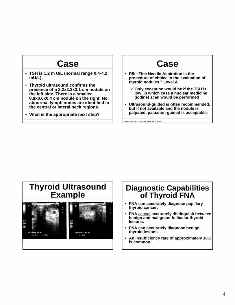
• Thyroid ultrasound confirms the presence of a 2.2x2.3x2.1 cm nodule on the left side. There is a smaller 0.8x0.6x0.4 cm nodule on the right. No abnormal lymph nodes are identified in the central or lateral neck regions.
• What is the appropriate next step?
Thyroid Ultrasound Example
Case• R5: “Fine Needle Aspiration is the
procedure of choice in the evaluation of thyroid nodules.” Level A
Only exception would be if the TSH is low, in which case a nuclear medicine (iodine) scan would be performed
• Ultrasound-guided is often recommended, but if not available and the nodule is palpated, palpation-guided is acceptable.
Cooper DS, et al. Thyroid 2006. 16:109-142
• FNA can accurately diagnose papillary thyroid cancer.
• FNA cannot accurately distinguish between benign and malignant follicular thyroid lesions.
• FNA can accurately diagnose benign thyroid lesions
• An insufficiency rate of approximately 10% is common
Diagnostic Capabilities of Thyroid FNA
5
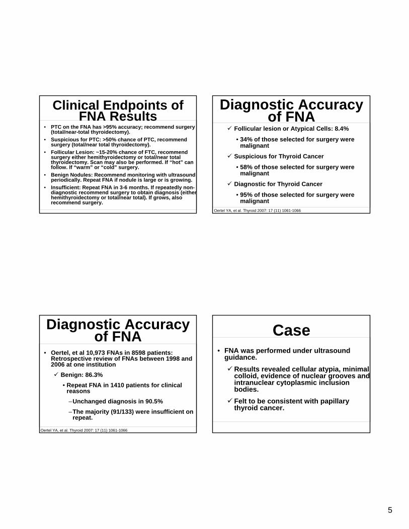
• PTC on the FNA has >95% accuracy; recommend surgery (total/near-total thyroidectomy).
• Suspicious for PTC: >50% chance of PTC, recommend surgery (total/near total thyroidectomy).
• Follicular Lesion: ~15-20% chance of FTC, recommend surgery either hemithyroidectomy or total/near total thyroidectomy. Scan may also be performed. If “hot” can follow. If “warm” or “cold” surgery.
• Benign Nodules: Recommend monitoring with ultrasound periodically. Repeat FNA if nodule is large or is growing.
• Insufficient: Repeat FNA in 3-6 months. If repeatedly non-diagnostic recommend surgery to obtain diagnosis (either hemithyroidectomy or total/near total). If grows, also recommend surgery.
Clinical Endpoints of FNA Results
Diagnostic Accuracy of FNA
• Oertel, et al 10,973 FNAs in 8598 patients: Retrospective review of FNAs between 1998 and 2006 at one institution
Benign: 86.3%• Repeat FNA in 1410 patients for clinical
reasons–Unchanged diagnosis in 90.5%–The majority (91/133) were insufficient on
repeat.
Oertel YA, et al. Thyroid 2007: 17 (11) 1061-1066
Diagnostic Accuracy of FNA
Follicular lesion or Atypical Cells: 8.4%
• 34% of those selected for surgery were malignant
Suspicious for Thyroid Cancer
• 58% of those selected for surgery were malignant
Diagnostic for Thyroid Cancer
• 95% of those selected for surgery were malignant
Oertel YA, et al. Thyroid 2007: 17 (11) 1061-1066
Case• FNA was performed under ultrasound
guidance.Results revealed cellular atypia, minimal colloid, evidence of nuclear grooves and intranuclear cytoplasmic inclusion bodies.Felt to be consistent with papillary thyroid cancer.
6
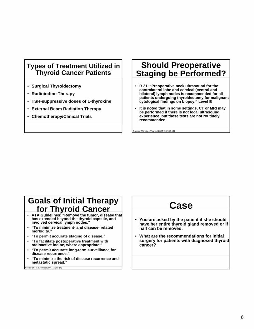
Types of Treatment Utilized in Thyroid Cancer Patients
• Surgical Thyroidectomy
• Radioiodine Therapy
• TSH-suppressive doses of L-thyroxine
• External Beam Radiation Therapy
• Chemotherapy/Clinical Trials
• ATA Guidelines: “Remove the tumor, disease that has extended beyond the thyroid capsule, and involved cervical lymph nodes.”
• “To minimize treatment- and disease- related morbidity.”
• “To permit accurate staging of disease.”• “To facilitate postoperative treatment with
radioactive iodine, where appropriate.”• “To permit accurate long-term surveillance for
disease recurrence.”• “To minimize the risk of disease recurrence and
metastatic spread.”
Goals of Initial Therapy for Thyroid Cancer
Cooper DS, et al. Thyroid 2006. 16:109-142
• R 21. “Preoperative neck ultrasound for the contralateral lobe and cervical (central and bilateral) lymph nodes is recommended for all patients undergoing thyroidectomy for malignant cytological findings on biopsy.” Level B
• It is noted that in some settings, CT or MRI may be performed if there is not local ultrasound experience, but these tests are not routinely recommended.
Should Preoperative Staging be Performed?
Cooper DS, et al. Thyroid 2006. 16:109-142
Case• You are asked by the patient if she should
have her entire thyroid gland removed or if half can be removed.
• What are the recommendations for initial surgery for patients with diagnosed thyroid cancer?
7
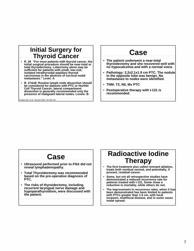
• R. 26 “For most patients with thyroid cancer, the initial surgical procedure should be near-total or total thyroidectomy. Lobectomy alone may be sufficient for patients with small, low-risk, isolated intrathyroidal papillary thyroid carcinomas in the absence of cervical nodal metastases.” Level: A
• R. 27&28. Routine lymph node dissection should be considered for patients with PTC or HurthleCell Thyroid Cancer; lateral compartment dissection is generally recommended only the presence of malignant lateral nodes. Levels: B
Initial Surgery for Thyroid Cancer
Cooper DS, et al. Thyroid 2006. 16:109-142
Case• Ultrasound performed prior to FNA did not
reveal lymphadenopathy.• Total Thyroidectomy was recommended
based on the pre-operative diagnosis of PTC.
• The risks of thyroidectomy, including recurrent laryngeal nerve damage and hypoparathyroidism, were discussed with the patient.
Case• The patient underwent a near-total
thyroidectomy and she recovered well with no hypocalcemia and with a normal voice.
• Pathology: 2.2x2.1x1.9 cm PTC. The nodule in the opposite lobe was benign. No metastases to nodes were identified.
• TNM: T2, N0, Mx PTC• Postoperative therapy with I-131 is
recommended.
• The first treatment also called remnant ablation, treats both residual normal, and potentially, if present, residual cancer.
• Some, but not all retrospective studies have demonstrated a reduced recurrence rate for patients treated with I-131. Some show a reduction in mortality, while others do not.
• The improvement in recurrence rates, when it has been demonstrated has been limited to patients with PTCs greater than 1.5 cm, with local invasion, multifocal disease, and in some cases nodal spread.
Radioactive Iodine Therapy
8
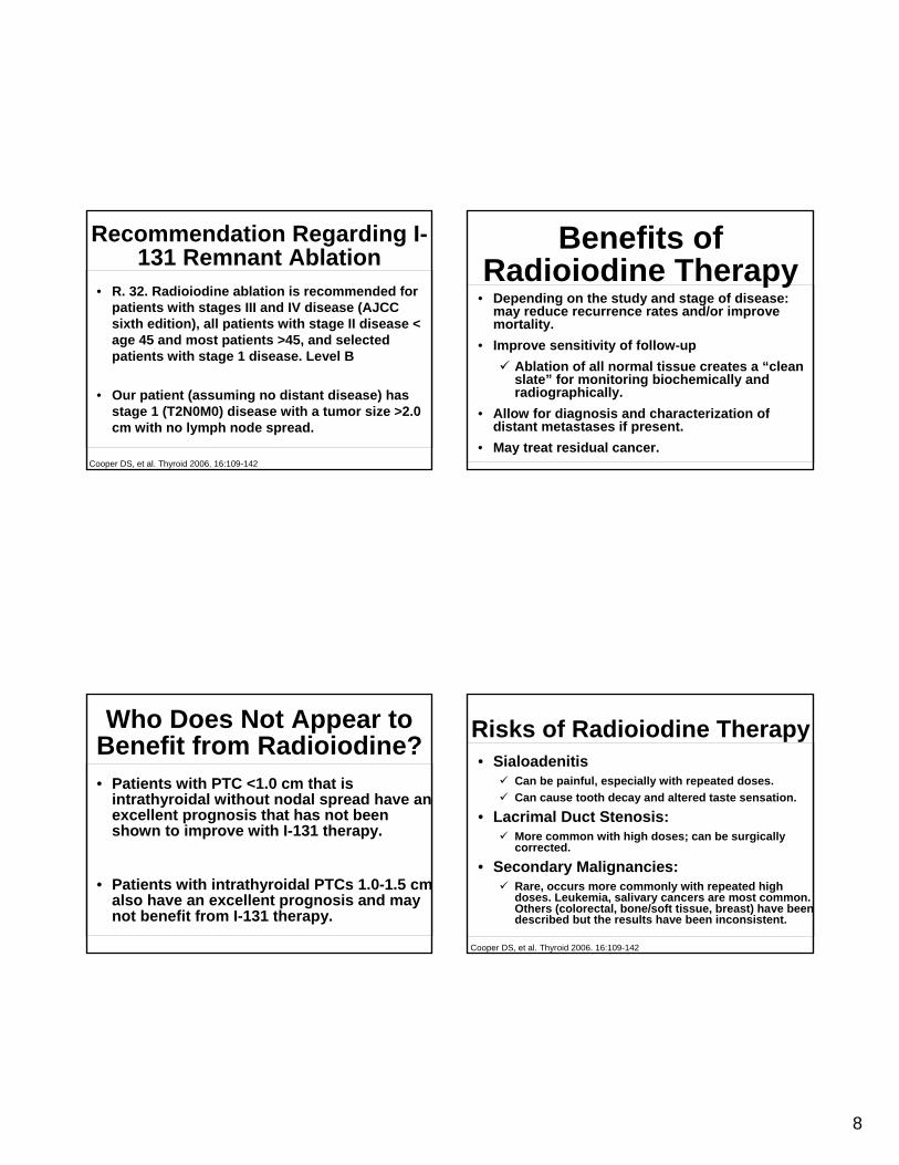
Recommendation Regarding I-131 Remnant Ablation
• R. 32. Radioiodine ablation is recommended for patients with stages III and IV disease (AJCC sixth edition), all patients with stage II disease < age 45 and most patients >45, and selected patients with stage 1 disease. Level B
• Our patient (assuming no distant disease) has stage 1 (T2N0M0) disease with a tumor size >2.0 cm with no lymph node spread.
Cooper DS, et al. Thyroid 2006. 16:109-142
Who Does Not Appear to Benefit from Radioiodine?• Patients with PTC <1.0 cm that is
intrathyroidal without nodal spread have an excellent prognosis that has not been shown to improve with I-131 therapy.
• Patients with intrathyroidal PTCs 1.0-1.5 cm also have an excellent prognosis and may not benefit from I-131 therapy.
• Depending on the study and stage of disease: may reduce recurrence rates and/or improve mortality.
• Improve sensitivity of follow-upAblation of all normal tissue creates a “clean slate” for monitoring biochemically and radiographically.
• Allow for diagnosis and characterization of distant metastases if present.
• May treat residual cancer.
Benefits of Radioiodine Therapy
Risks of Radioiodine Therapy• Sialoadenitis
Can be painful, especially with repeated doses.Can cause tooth decay and altered taste sensation.
• Lacrimal Duct Stenosis:More common with high doses; can be surgically corrected.
• Secondary Malignancies:Rare, occurs more commonly with repeated high doses. Leukemia, salivary cancers are most common. Others (colorectal, bone/soft tissue, breast) have been described but the results have been inconsistent.
Cooper DS, et al. Thyroid 2006. 16:109-142
9
Risks of Radioiodine Therapy• Early Menopause: ~12 months in one
study.• Delay in pregnancy: Recommended to wait
12 months to become pregnant.• Marrow and Lung Toxicity: Occasionally
seen in patients who have received multiplehigh doses of I-131.
• Reduce Sperm Count: Rarely seen with high doses.
Cooper DS, et al. Thyroid 2006. 16:109-142
Case• After discussion of the Risks and Benefits
of Radioiodine, the patient is scheduled to receive therapy.
• What special preparations needed for patients scheduled to receive radioiodine therapy?
Preparation for Radioiodine Therapy
• High Levels of TSH are needed to facilitate radioiodine therapy.
Withdrawal from Thyroid hormone• Effective• Causes often-time severe, transient
symptoms• Inexpensive
Preparation for Radioiodine Therapy
Recombinant human TSH administration• IM injections• Well-tolerated• Studied for a shorter period of time• Expensive• Approved for Remnant Ablation only
10
• rhTSH was approved in 2007 for remnant ablation.
Pacini et al. Randomized, controlled multicenter study63 patients randomized to thryoxinewithdrawal vs rhTSH preparation for remnant ablative therapy with 100 mCi I-131.Rate of remnant ablation defined as uptake <0.1% on scan 8 months later. Tg levels were a secondary endpoint.
RhTSH versus ThyroxineWithdrawal for I-131 Therapy
Pacini F, et al. J Clin Endocrinol Metab. 2006: 91;926-932.
• 100% of patients in both groups had successful ablation of iodine uptake.
• The rates of patients with stimulated Tg levels <1 ng/ml or <2 ng/ml were the same between the two groups.
• Quality of life scores (Billewicz and SF36 scales) was better in patients treated with rhTSH
• The authors concluded that rhTSH preparation for remnant ablative I-131 therapy was as effective and hypothyroid preparation at achieving ablation with fewer hypothyroid symptoms.
RhTSH versus ThyroxineWithdrawal for I-131 Therapy
Pacini F, et al. J Clin Endocrinol Metab. 2006: 91;926-932.
Low-iodine Diet• In addition to a high TSH, patients should not
have high levels of iodine at the time of I-131 therapy.
A careful history must be taken to determine if the patient has received any IV contrast in the recent past (<6 months).If needed, a urine iodine level can be measured to be sure the contrast has cleared.
Low-iodine DietA two week low iodine diet is generally prescribed.• Available in several sources on the internet
or from NCI.A urine iodine is sometimes performed to confirm the efficacy of the diet prior to treatment.
11
• R33. “Patients undergoing radioiodine therapy or diagnostic testing can be prepared by LT4 withdrawal…with measurement of serum TSH to determine timing of testing (TSH>30 mU/L).” Level B.
• R37. “Remnant ablation can be performed following thyroxine withdrawal or rhTSH stimulation.” Level B.
• R38. “A low iodine diet for 1-2 weeks is recommended for patients undergoing radioiodine remnant ablation, particularly for those patients with high iodine intake.” Level B.
Recommendations for Radioiodine Preparation
Cooper DS, et al. Thyroid 2006. 16:109-142
Dosing of Remnant Ablative Radioiodine
• Successful remnant ablation can be defined as the loss of all uptake on a subsequent scan or theabsence of detectable thyroglobulin (or both). The latter is now most frequently used.
• In general, the goal is to use the smallest activity required to achieve ablation.
• Higher doses are employed in patients with a high risk of residual thyroid cancer tissue or tumors with poorly differentiated features that may not concentrate iodine as well.
Dosing Recommendations
• R35. “The minimum activity (30-100 mCi) necessary to achieve successful remnant ablation, particularly for low-risk patients.”Level B
• R36. “If residual microscopic disease is suspected or documented, or if there is a more aggressive tumor histology, then higher activities (100-200 mCi) may be appropriate.” Level C
Cooper DS, et al. Thyroid 2006. 16:109-142
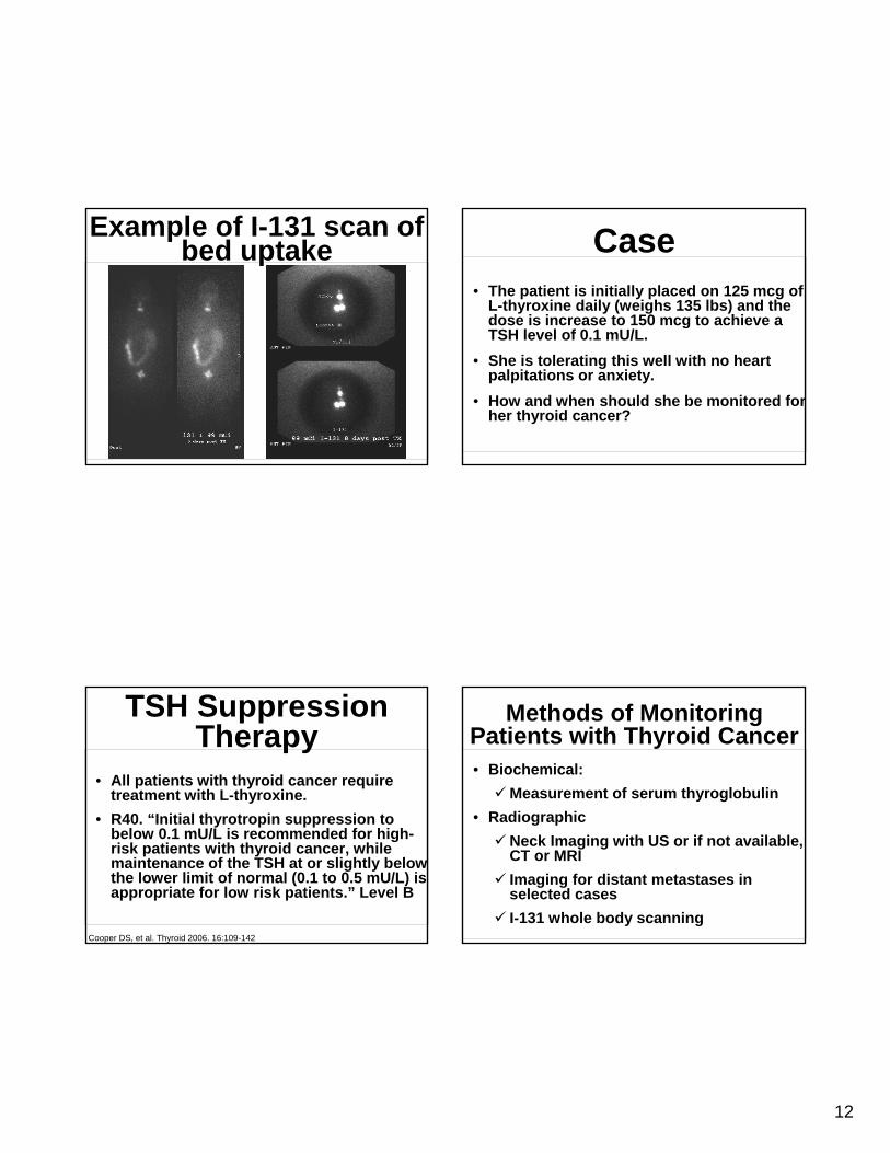
Case• Because the tumor was staged as a T2 I-
131 remnant ablation was recommended. • She received with 50 mCi of I-131 following
thyroxine withdrawal.• Pre- and post-therapy diagnostic scans
revealed uptake in the thyroid bed with no lateral uptake or iodine-avid distant metastases.
12
Example of I-131 scan of bed uptake
• All patients with thyroid cancer require treatment with L-thyroxine.
• R40. “Initial thyrotropin suppression to below 0.1 mU/L is recommended for high-risk patients with thyroid cancer, while maintenance of the TSH at or slightly below the lower limit of normal (0.1 to 0.5 mU/L) is appropriate for low risk patients.” Level B
TSH Suppression Therapy
Cooper DS, et al. Thyroid 2006. 16:109-142
Case• The patient is initially placed on 125 mcg of
L-thyroxine daily (weighs 135 lbs) and the dose is increase to 150 mcg to achieve a TSH level of 0.1 mU/L.
• She is tolerating this well with no heart palpitations or anxiety.
• How and when should she be monitored for her thyroid cancer?
Methods of Monitoring Patients with Thyroid Cancer• Biochemical:
Measurement of serum thyroglobulin • Radiographic
Neck Imaging with US or if not available, CT or MRIImaging for distant metastases in selected casesI-131 whole body scanning
13
• Highly specific and sensitive for thyroid tissue.• Becomes specific for thyroid cancer after
thyroidectomy and I-131 ablation therapy.• Must be measured with simultaneous anti-Tg
antibodies as these can interfere and cause false abnormal (often undetectable) results.
• Must be measured with TSH as this can stimulate Tg levels.
• Tg levels are the cornerstone of monitoring, especially in patients who do not have anti-Tg antibodies (~85% of patients).
Serum Thyroglobulin (Tg) Measurement
Neck Ultrasound• A neck ultrasound to examine nodes bilaterally
by a skilled operator is highly sensitive for detecting recurrent or residual PTC. FNA can also be performed if indicated.
• FTC rarely spreads to neck nodes and may be less sensitive in these patients.
• Other neck imaging such as CT or MRI may be ordered in selected cases or based on preferences at individual practice sites.
• Chest CT scans are generally performed in the following situations:
High risk of distant metastasesVery high Tg levelsElevated Tg levels with negative neck imaging or FTCPositive iodine scans in the lungSome patients with persistently elevated anti-Tg antibody levels with negative neck imaging.
• Chest radiographs are insensitive for thyroid cancer as many metastases are small.
Chest Imaging
• Whole body radioiodine scanning
Less sensitive than Tg levels, particularly for detecting neck disease.
May still be performed in selected cases, particularly those patients who have anti-Tg antibodies.
Nuclear Imaging
1 Robbins RJ, et al. J Clin Endocrinol Metab. 2006:91; 498-5052 Chin BB, et al. J Clin Endocrinol Metab. 2004:89;91-95.
14
Nuclear Imaging• FDG PET or PET/CT imaging
PET imaging is most helpful in patients with poorly differentiated tumors or with high Tg levels and negative neck and chest imaging.May Provide prognostic information in patients with distant metastases1. TSH stimulation may improve sensitivity2.
1 Robbins RJ, et al. J Clin Endocrinol Metab. 2006:91; 498-5052 Chin BB, et al. J Clin Endocrinol Metab. 2004:89;91-95.
Case• Three months after I-131 therapy; the
patient had a TSH level of 0.15 mU/L and a serum Tg level <0.2 ng/ml with undetectable anti-Tg antibodies.
• Nine months after I-131 therapy; TSH was 0.08 mU/L; Tg was <0.2 ng/ml with undetectable anti-Tg antibodies.
• What is recommended at this point?
• R45. “In low risk patients who have had remnant ablation and negative cervical ultrasound and TSH-suppressed Tg 6 months after treatment, serum Tg should be measured after thyroxine withdrawal or rhTSH stimulation approximately 12 months after the ablation to very absence of disease. The timing or necessity of subsequent stimulation is uncertain for those found to be free of disease.” Level A
Surveillance for Thyroid Cancer
Cooper DS, et al. Thyroid 2006. 16:109-142
• R46. After the first whole body scan performed at the time of remnant ablation, low-risk patients with negative TSH-stimulated thyroglobulin and cervical ultrasound do not require routine diagnostic whole body scan during follow-up. Level A
Surveillance for Thyroid Cancer
Cooper DS, et al. Thyroid 2006. 16:109-142
15
Case• 12 months after treatment a TSH-stimulated Tg
level is obtained after rhTSH administration. The stimulated Tg level 72 hours after the second dose is <0.2 ng/ml with undetectable anti-Tg antibodies. Following this, a neck ultrasound is performed that does not reveal obvious evidence of thyroid cancer.
• This is consistent with a complete remission!
• Long-term thyrotoxicosis is associated with the development of atrial fibrillation. It is also associated with osteoporosis and fractures in post-menopausal women.
• This is discussed with the patient in detail.
• What is the benefit of long-term TSH suppression for this patient?
Long-Term ThyroxineSuppression
• R51. “In patients free of disease, especially those at low risk for recurrence, the TSH may be kept within the low normal range (0.3 to 2.0 mU/l).” Level C
• Based on this recommendation and the data leading to it, the patient’s L-thyroxinedose is adjusted to achieve a TSH in the 0.4-1.0 mU/L range.
Long-term ThyroxineSuppression
Cooper DS, et al. Thyroid 2006. 16:109-142
Does a negative TSH-stimulated Tg predict a
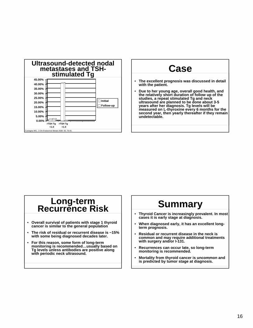
lack of tumor recurrence?• Castagna, et al reported on a cohort of
patients with undetectable stimulated Tg one year after I-131 therapy (n=68)
• 67 patients underwent repeat stimulated Tg and neck ultrasound 2.5 yrs later
• 1 patient (1.5%) developed a recurrence at follow up detected both by stimulated Tg and the neck ultrasound, but not basal Tg did not.
Castagna MG. J Clin Endocrinol Metab 2008: 93; 76-81.
16
Ultrasound-detected nodal metastases and TSH-
stimulated Tg
Castagna MG. J Clin Endocrinol Metab 2008: 93; 76-81.
0.00%5.00%
10.00%15.00%20.00%25.00%30.00%35.00%40.00%45.00%
rTSH Tg<1.0
rTSH Tg>1.0
InitialFollow-up
Long-term Recurrence Risk
• Overall survival of patients with stage 1 thyroid cancer is similar to the general population
• The risk of residual or recurrent disease is ~15% with some being diagnosed decades later.
• For this reason, some form of long-term monitoring is recommended…usually based on Tg levels unless antibodies are positive along with periodic neck ultrasound.
Case• The excellent prognosis was discussed in detail
with the patient.• Due to her young age, overall good health, and
the relatively short duration of follow up of the studies, a repeat stimulated Tg and neck ultrasound are planned to be done about 3-5 years after her diagnosis. Tg levels will be measured on L-thyroxine every 6 months for the second year, then yearly thereafter if they remain undetectable.
• Thyroid Cancer is increasingly prevalent. In most cases it is early stage at diagnosis.
• When diagnosed early, it has an excellent long-term prognosis.
• Residual or recurrent disease in the neck is common and may require additional treatments with surgery and/or I-131.
• Recurrences can occur late, so long-term monitoring is recommended.
• Mortality from thyroid cancer is uncommon and is predicted by tumor stage at diagnosis.
Summary