diagnosis and treatment of cervical radiculopathy
TRANSCRIPT
NASS Clinical Guidelines – Diagnosis and Treatment of Cervical Radiculopathy from Degenerative Disorders 1
This clinical guideline should not be construed as including all proper methods of care or excluding other acceptable methods of care reasonably directed to obtaining the same results. The ultimate judgment regarding any specific procedure or treatment is to be made by the physician and patient in light of all circumstances presented by the patient and the needs and resources particular to the locality or institution.
North American Spine SocietyEvidence-Based Clinical Guidelines
for Multidisciplinary Spine Care
NASS Evidence-Based Guideline Development CommitteeChristopher M. Bono, MD, Committee ChairGary Ghiselli, MD, Outcome Measures ChairThomas J. Gilbert, MD, Diagnosis/Imaging ChairD. Scott Kreiner, MD, Medical/Interventional ChairCharles Reitman, MD, Surgical Treatment ChairJeffrey Summers, MD, Natural History ChairJamie Baisden, MDJohn Easa, MD
Robert Fernand, MDTim Lamer, MDPaul Matz, MDDan Mazanec, MDDaniel K. Resnick, MDWilliam O. Shaffer, MDAnil Sharma, MDReuben Timmons, MDJohn Toton, MD
Diagnosis and Treatment of Cervical Radiculopathy from Degenerative Disorders
NASS Clinical Guidelines – Diagnosis and Treatment of Cervical Radiculopathy from Degenerative Disorders 2
This clinical guideline should not be construed as including all proper methods of care or excluding other acceptable methods of care reasonably directed to obtaining the same results. The ultimate judgment regarding any specific procedure or treatment is to be made by the physician and patient in light of all circumstances presented by the patient and the needs and resources particular to the locality or institution.
Financial StatementThis clinical guideline was developed and funded in its entirety by the North American Spine Society (NASS). All participating authors have submitted a disclosure form relative to potential conflicts of interest which is kept on file at NASS.
CommentsComments regarding the guideline may be submitted to the North American Spine Society and will be consid-ered in development of future revisions of the work.
North American Spine SocietyEvidence-Based Clinical Guidelines for Multidisciplinary Spine CareDiagnosis and Treatment of Cervical Radiculopathy from Degenerative Disorders
Copyright © 2010 North American Spine Society
7075 Veterans Boulevard Burr Ridge, IL 60527630.230.3600www.spine.org
ISBN: 1-929988-25-7
I. Introduction . . . . . . . . . . . . . . . . . . . . . . . . . . . . . . . . . . . . . . . . .4
II. Guideline Development Methodology . . . . . . . . . . . . . . . . . . . .5
III. Natural History of Cervical Radiculopathy from
Degenerative Disorders . . . . . . . . . . . . . . . . . . . . . . . . . . . . . . . .9
IV. Recommendations for Diagnosis and Treatment of Cervical Radiculopathy from Degenerative Disorders
A. Diagnosis/Imaging . . . . . . . . . . . . . . . . . . . . . . . . . . . . . . . . . . . . . . . . . . . . . . . . . . . . . . . . . 12
B. Outcome Measures for Treatment . . . . . . . . . . . . . . . . . . . . . . . . . . . . . . . . . . . . . . . . . . . . 31
C. Medical and Interventional Treatment . . . . . . . . . . . . . . . . . . . . . . . . . . . . . . . . . . . . . . . . . 41
D. Surgical Treatment . . . . . . . . . . . . . . . . . . . . . . . . . . . . . . . . . . . . . . . . . . . . . . . . . . . . . . . . . 50
V. AppendicesA. Acronyms . . . . . . . . . . . . . . . . . . . . . . . . . . . . . . . . . . . . . . . . . . . . . . . . . . . . . . . . . . . . . . . 72
B. Levels of Evidence for Primary Research Questions . . . . . . . . . . . . . . . . . . . . . . . . . . . . . . 73
C. Grades of Recommendations for Summaries or Reviews of Studies . . . . . . . . . . . . . . . . . 74
D. Protocol for NASS Literature Searches . . . . . . . . . . . . . . . . . . . . . . . . . . . . . . . . . . . . . . . . 75
E. Literature Search Parameters . . . . . . . . . . . . . . . . . . . . . . . . . . . . . . . . . . . . . . . . . . . . . . . . 77
F. Evidentiary Tables . . . . . . . . . . . . . . . . . . . . . . . . . . . . . . . . . . . . . . . . . . . . . . . . . . . . . . . . . 86
VI. References . . . . . . . . . . . . . . . . . . . . . . . . . . . . . . . . . . . . . . . . . . .168
Table of Contents
3
This clinical guideline should not be construed as including all proper methods of care or excluding other acceptable methods of care reasonably directed to obtaining the same results. The ultimate judgment regarding any specific procedure or treatment is to be made by the physician and patient in light of all circumstances presented by the patient and the needs and resources particular to the locality or institution.
NASS Clinical Guidelines – Diagnosis and Treatment of Cervical Radiculopathy from Degenerative Disorders 4
This clinical guideline should not be construed as including all proper methods of care or excluding other acceptable methods of care reasonably directed to obtaining the same results. The ultimate judgment regarding any specific procedure or treatment is to be made by the physician and patient in light of all circumstances presented by the patient and the needs and resources particular to the locality or institution.
I. IntroductionObjectiveThe objective of the North American Spine Society (NASS) Clinical Guideline for the Diagnosis and Treatment of Cervical Radiculopathy from Degen-erative Disorders is to provide evidence-based rec-ommendations to address key clinical questions surrounding the diagnosis and treatment of cervi-cal radiculopathy from degenerative disorders. The guideline is intended to reflect contemporary treat-ment concepts for cervical radiculopathy from de-generative disorders as reflected in the highest qual-ity clinical literature available on this subject as of May 2009. The goals of the guideline recommenda-tions are to assist in delivering optimum, efficacious treatment and functional recovery from this spinal disorder.
Scope, Purpose and Intended UserThis document was developed by the North Ameri-can Spine Society Evidence-Based Guideline Devel-opment Committee as an educational tool to assist practitioners who treat patients with cervical radic-ulopathy from degenerative disorders. The goal is to provide a tool that assists practitioners in improving the quality and efficiency of care delivered to pa-tients with cervical radiculopathy from degenera-tive disorders. The NASS Clinical Guideline for the Diagnosis and Treatment of Cervical Radiculopathy from Degenerative Disorders provides a definition and explanation of the natural history of cervical ra-diculopathy from degenerative disorders, outlines a
reasonable evaluation of patients suspected to have cervical radiculopathy from degenerative disorders and outlines treatment options for adult patients with a diagnosis of cervical radiculopathy from de-generative disorders.
THIS GUIDELINE DOES NOT REPRESENT A “STANDARD OF CARE,” nor is it intended as a fixed treatment protocol. It is anticipated that there will be patients who will require less or more treatment than the average. It is also acknowledged that in atypical cases, treatment falling outside this guide-line will sometimes be necessary. This guideline should not be seen as prescribing the type, frequen-cy or duration of intervention. Treatment should be based on the individual patient’s need and physi-cian’s professional judgment. This document is de-signed to function as a guideline and should not be used as the sole reason for denial of treatment and services. This guideline is not intended to expand or restrict a health care provider’s scope of practice or to supersede applicable ethical standards or provi-sions of law.
Patient PopulationThe patient population for this guideline encom-passes adults (18 years or older) with a chief com-plaint of pain in a radicular pattern in one or both upper extremities related to compression and/or ir-ritation of one or more cervical nerve roots.
NASS Clinical Guidelines – Diagnosis and Treatment of Cervical Radiculopathy from Degenerative Disorders 5
This clinical guideline should not be construed as including all proper methods of care or excluding other acceptable methods of care reasonably directed to obtaining the same results. The ultimate judgment regarding any specific procedure or treatment is to be made by the physician and patient in light of all circumstances presented by the patient and the needs and resources particular to the locality or institution.
II. Guideline Development Methodology Through objective evaluation of the evidence and transparency in the process of making recommen-dations, it is NASS’ goal to develop evidence-based clinical practice guidelines for the diagnosis and treatment of adult patients with various spinal con-ditions. These guidelines are developed for educa-tional purposes to assist practitioners in their clini-cal decision-making processes. It is anticipated that where evidence is very strong in support of recom-mendations, these recommendations will be opera-tionalized into performance measures.
Multidisciplinary CollaborationWith the goal of ensuring the best possible care for adult patients suffering with spinal disorders, NASS is committed to multidisciplinary involvement in the process of guideline and performance measure development. To this end, NASS has ensured that representatives from medical, interventional and surgical spine specialties have participated in the development and review of all NASS guidelines. It is also important that primary care providers and musculoskeletal specialists who care for patients with spinal complaints are represented in the de-velopment and review of guidelines that address treatment by first contact physicians, and NASS has involved these providers in the development pro-cess as well. To ensure broad-based representation, NASS has invited and welcomes input from other societies and specialties.
Evidence Analysis Training of All NASS Guideline DevelopersNASS has initiated, in conjunction with the Universi-ty of Alberta’s Centre for Health Evidence, an online training program geared toward educating guideline developers about evidence analysis and guideline development. All participants in guideline develop-ment for NASS have completed the training prior to participating in the guideline development program at NASS. This training includes a series of readings
and exercises, or interactivities, to prepare guideline developers for systematically evaluating literature and developing evidence-based guidelines. The on-line course takes approximately 15-30 hours to com-plete, and participants have been awarded CME credit upon completion of the course.
Disclosure of Potential Conflicts of InterestAll participants involved in guideline development have disclosed their relationships with other entities and potential conflicts of interest to their colleagues and their potential conflicts have been documented for future reference. They will not be published in any guideline, but kept on file for reference, if need-ed. Participants have been asked to update their dis-closures regularly throughout the guideline devel-opment process.
Levels of Evidence and Grades of RecommendationNASS has adopted standardized levels of evidence (Appendix B) and grades of recommendation (Ap-pendix C) to assist practitioners in easily under-standing the strength of the evidence and recom-mendations within the guidelines. The levels of evidence range from Level I (high quality random-ized controlled trial) to Level V (expert consensus). Grades of recommendation indicate the strength of the recommendations made in the guideline based on the quality of the literature.
Grades of Recommendation: A: Good evidence (Level I studies with consistent
findings) for or against recommending interven-tion.
B: Fair evidence (Level II or III studies with consis-tent findings) for or against recommending in-tervention.
NASS Clinical Guidelines – Diagnosis and Treatment of Cervical Radiculopathy from Degenerative Disorders 6
This clinical guideline should not be construed as including all proper methods of care or excluding other acceptable methods of care reasonably directed to obtaining the same results. The ultimate judgment regarding any specific procedure or treatment is to be made by the physician and patient in light of all circumstances presented by the patient and the needs and resources particular to the locality or institution.
C: Poor quality evidence (Level IV or V studies) for or against recommending intervention.
I: Insufficient or conflicting evidence not allowing a recommendation for or against intervention.
Guideline recommendations are written utilizing a standard language that indicates the strength of the recommendation. “A” recommendations indi-cate a test or intervention is “recommended”; “B” recommendations “suggest” a test or intervention and “C” recommendations indicate a test or inter-vention “may be considered” or “is an option.” “I” or “Insufficient Evidence” statements clearly indicate that “there is insufficient evidence to make a rec-ommendation for or against” a test or intervention. Work group consensus statements clearly state that “in the absence of reliable evidence, it is the work group’s opinion that” a test or intervention may be appropriate.
The levels of evidence and grades of recommenda-tion implemented in this guideline have also been adopted by the Journal of Bone and Joint Surgery, the American Academy of Orthopaedic Surgeons, Clinical Orthopaedics and Related Research, the journal Spine and the Pediatric Orthopaedic Society of North America.
In evaluating studies as to levels of evidence for this guideline, the study design was interpreted as es-tablishing only a potential level of evidence. As an example, a therapeutic study designed as a random-ized controlled trial would be considered a poten-tial Level I study. The study would then be further analyzed as to how well the study design was imple-mented and significant short comings in the execu-tion of the study would be used to downgrade the levels of evidence for the study’s conclusions. In the example cited previously, reasons to downgrade the results of a potential Level I randomized controlled trial to a Level II study would include, among other possibilities: an underpowered study (patient sam-ple too small, variance too high), inadequate ran-domization or masking of the group assignments and lack of validated outcome measures.
In addition, a number of studies were reviewed sev-eral times in answering different questions within this guideline. How a given question was asked might influence how a study was evaluated and interpreted as to its level of evidence in answering that particular question. For example, a random-ized control trial reviewed to evaluate the differenc-es between the outcomes of surgically treated ver-sus untreated patients with lumbar spinal stenosis might be a well designed and implemented Level I therapeutic study. This same study, however, might be classified as giving Level II prognostic evidence if the data for the untreated controls were extracted and evaluated prognostically.
Guideline Development Process
Step 1: Identification of Clinical QuestionsTrained guideline participants were asked to submit a list of clinical questions that the guideline should address. The lists were compiled into a master list, which was then circulated to each member with a request that they independently rank the ques-tions in order of importance for consideration in the guideline. The most highly ranked questions, as determined by the participants, served to focus the guideline.
Step 2: Identification of Work GroupsMultidisciplinary teams were assigned to work groups and assigned specific clinical questions to ad-dress. Because NASS is comprised of surgical, medi-cal and interventional specialists, it is imperative to the guideline development process that a cross-section of NASS membership is represented on each group. This also helps to ensure that the potential for inadvertent biases in evaluating the literature and formulating recommendations is minimized.
Step 3: Identification of Search Terms and ParametersOne of the most crucial elements of evidence analy-sis to support development of recommendations for appropriate clinical care is the comprehensive litera-

NASS Clinical Guidelines – Diagnosis and Treatment of Cervical Radiculopathy from Degenerative Disorders 7
This clinical guideline should not be construed as including all proper methods of care or excluding other acceptable methods of care reasonably directed to obtaining the same results. The ultimate judgment regarding any specific procedure or treatment is to be made by the physician and patient in light of all circumstances presented by the patient and the needs and resources particular to the locality or institution.
ture search. Thorough assessment of the literature is the basis for the review of existing evidence and the formulation of evidence-based recommendations. In order to ensure a thorough literature search, NASS has instituted a Literature Search Protocol (Appen-dix D) which has been followed to identify literature for evaluation in guideline development. In keep-ing with the Literature Search Protocol, work group members have identified appropriate search terms and parameters to direct the literature search.
Specific search strategies, including search terms, parameters and databases searched, are document-ed in the appendices (Appendix E).
Step 4: Completion of the Literature SearchOnce each work group identified search terms/pa-rameters, the literature search was implemented by a medical/research librarian, consistent with the Literature Search Protocol.
Following these protocols ensures that NASS recom-mendations (1) are based on a thorough review of relevant literature; (2) are truly based on a uniform, comprehensive search strategy; and (3) represent the current best research evidence available. NASS maintains a search history in Endnote, for future use or reference.
Step 5: Review of Search Results/Identification of Literature to ReviewWork group members reviewed all abstracts yielded from the literature search and identified the litera-ture they will review in order to address the clini-cal questions, in accordance with the Literature Search Protocol. Members have identified the best research evidence available to answer the targeted clinical questions. That is, if Level I, II and or III lit-erature is available to answer specific questions, the work group was not required to review Level IV or V studies. Work group members reviewed the evi-dence on the topic of cervical radiculopathy, and studies eligible for review were required to address
radiculopathy alone or include a subgroup analysis of patients with radiculopathy. Many of the studies considered for potential inclusion in this guideline included groups of patients with myelopathy, with-out appropriate subgroup analyses of those patients with cervical radiculopathy alone. For this reason, in the absence of subgroup analyses, a large number of studies were excluded from consideration in ad-dressing the questions and formulating recommen-dations. These studies, having been reviewed, are included in the reference sections.
Step 6: Evidence AnalysisMembers have independently developed evidentia-ry tables summarizing study conclusions, identify-ing strengths and weaknesses and assigning levels of evidence. In order to systematically control for potential biases, at least two work group members have reviewed each article selected and indepen-dently assigned levels of evidence to the literature using the NASS levels of evidence. Any discrepan-cies in scoring have been addressed by two or more reviewers. The consensus level (the level upon which two-thirds of reviewers were in agreement) was then assigned to the article.
As a final step in the evidence analysis process, members have identified and documented gaps in the evidence to educate guideline readers about where evidence is lacking and help guide further needed research by NASS and other societies.
Step 7: Formulation of Evidence-Based Recommendations and Incorporation of Expert ConsensusWork groups held webcasts to discuss the evidence-based answers to the clinical questions, the grades of recommendations and the incorporation of expert consensus. Expert consensus has been incorporat-ed only where Level I-IV evidence is insufficient and the work group has deemed that a recommendation is warranted. Transparency in the incorporation of consensus is crucial, and all consensus-based rec-
NASS Clinical Guidelines – Diagnosis and Treatment of Cervical Radiculopathy from Degenerative Disorders 8
This clinical guideline should not be construed as including all proper methods of care or excluding other acceptable methods of care reasonably directed to obtaining the same results. The ultimate judgment regarding any specific procedure or treatment is to be made by the physician and patient in light of all circumstances presented by the patient and the needs and resources particular to the locality or institution.
ommendations made in this guideline very clearly indicate that Level I-IV evidence is insufficient to support a recommendation and that the recom-mendation is based only on expert consensus.
Consensus Development ProcessVoting on guideline recommendations was conduct-ed using a modification of the nominal group tech-nique in which each work group member indepen-dently and anonymously ranked a recommendation on a scale ranging from 1 (“extremely inappropri-ate”) to 9 (“extremely appropriate”). Consensus was obtained when at least 80% of work group members ranked the recommendation as 7, 8 or 9. When the 80% threshold was not attained, up to three rounds of discussion and voting were held to resolve dis-agreements. If disagreements were not resolved af-ter these rounds, no recommendation was adopted.
After the recommendations were established, work group members developed the guideline content, addressing the literature which supports the recom-mendations.
Step 8: Submission of the Draft Guidelines for Review/CommentGuidelines were submitted to the full Evidence-Based Guideline Development Committee and the Research Council Director for review and comment. Revisions to recommendations were considered for incorporation only when substantiated by a prepon-derance of appropriate level evidence.
Step 9: Submission for Board ApprovalOnce any evidence-based revisions were incor-porated, the drafts were prepared for NASS Board review and approval. Edits and revisions to recom-mendations and any other content were considered for incorporation only when substantiated by a pre-ponderance of appropriate level evidence.
Step 10: Submission for Endorsement, Publication and National Guideline Clearinghouse (NGC) Inclusion
Following NASS Board approval, the guidelines have been slated for publication, submitted for endorse-ment to all appropriate societies and submitted for inclusion in the National Guidelines Clearinghouse (NGC). No revisions were made at this point in the process, but comments have been and will be saved for the next iteration.
Step 11: Identification and Development of Performance Measures The recommendations will be reviewed by a group experienced in performance measure development (eg, the AMA Physician’s Consortium for Perfor-mance Improvement) to identify those recommen-dations rigorous enough for measure develop-ment. All relevant medical specialties involved in the guideline development and at the Consortium will be invited to collaborate in the development of evidence-based performance measures related to spine care.
Step 12: Review and Revision Process The guideline recommendations will be reviewed every three years by an EBM-trained multidisci-plinary team and revised as appropriate based on a thorough review and assessment of relevant litera-ture published since the development of this version of the guideline.
Use of AcronymsThroughout the guideline, readers will see many ac-ronyms with which they may not be familiar. A glos-sary of acronyms is available in Appendix A.
Nomenclature for Medical/Interventional TreatmentThroughout the guideline, readers will see that what has traditionally been referred to as “nonoperative,” “nonsurgical” or “conservative” care is now referred to as “medical/interventional care.” The term medi-cal/interventional is meant to encompass pharma-cological treatment, physical therapy, exercise ther-apy, manipulative therapy, modalities, various types of external stimulators and injections.
NASS Clinical Guidelines – Diagnosis and Treatment of Cervical Radiculopathy from Degenerative Disorders 9
This clinical guideline should not be construed as including all proper methods of care or excluding other acceptable methods of care reasonably directed to obtaining the same results. The ultimate judgment regarding any specific procedure or treatment is to be made by the physician and patient in light of all circumstances presented by the patient and the needs and resources particular to the locality or institution.
III. Definition and Natural History of Cervical Radiculopathy from Degenerative Disorders
What is the best working definition of cervical radiculopathy from degenerative disorders?
Cervical radiculopathy from degenerative disorders can be defined as pain in a radicular pattern in one or both upper extremities related to compression and/or irritation of one or more cervical nerve roots. Frequent signs and symptoms include varying degrees of sensory, motor and reflex changes as well as dysesthesias and paresthesias related to nerve root(s) without evidence of spinal cord dysfunction (myelopathy).
Work Group Consensus Statement
What is the natural history of cer-vical radiculopathy from degener-ative disorders?To address the natural history of cervical radicul-opathy from degenerative disorders, the work group performed a comprehensive literature search and analysis. The group reviewed 31 articles that were selected from a search of MEDLINE (PubMed), Co-chrane Register of Controlled Trials, Web of Science and EMBASE Drugs & Pharmacology. However, all identified studies failed to meet the guideline’s in-clusion criteria because they did not ade-quately present data about the natural history of cervical radiculopathy. The plurality of studies did not re-port results of untreated patients, thus limiting con-clusions about natural history. This includes works that have been frequently cited as so-called natural history studies but are in fact reports of the results of one or more medical/interventional treatment
measures.5,12,18,22,28 In other investigations, data were reported for untreated and conservatively-treated patients together without an analysis specific to the untreated group. Other commonly cited studies did not report subgroup analyses of patients with cervi-cal radiculopathy alone and thereby presented gen-eralized natural history data regarding a heteroge-neous cohort of patients with isolated neck pain, cervical radiculopathy or cervical myelopathy.
Because of the limitations of available literature, the work group was unable to definitively answer the question posed related to the natural history of cer-vical radiculopathy from degenerative disorders. In lieu of an evidence-based answer, the work group did reach consensus on the following statement ad-dressing natural history.
It is likely that for most patients with cervical radiculopathy from degenerative disorders signs and symptoms will be self-limited and will resolve spontaneously over a variable length of time without specific treatment.
Work Group Consensus Statement
Future Directions for ResearchThe work group identified the following potential studies, which could generate meaningful evidence to assist in further defining the natural history of cervical radiculopathy from degenerative disorders.
Recommendation #1: A prospective study of patients with cervical radicu-lopathy from degenerative disorders without treat-ment, notwithstanding nonprescription analgesics, would provide Level I evidence regarding the natu-ral history of this disorder.
Recommendation #2: A systematic study of patients with untreated cer-
NASS Clinical Guidelines – Diagnosis and Treatment of Cervical Radiculopathy from Degenerative Disorders 10
This clinical guideline should not be construed as including all proper methods of care or excluding other acceptable methods of care reasonably directed to obtaining the same results. The ultimate judgment regarding any specific procedure or treatment is to be made by the physician and patient in light of all circumstances presented by the patient and the needs and resources particular to the locality or institution.
vical radiculopathy from degenerative disorders would provide evidence regarding the natural his-tory of the disease in this patient population.
Natural History References1. Anderberg L, Annertz M, Persson L, Brandt L, Saveland H.
Transforaminal steroid injections for the treatment of cer-vical radiculopathy: a prospective and randomised study. Eur Spine J. Mar 2007;16(3):321-328.
2. Carette S, Fehlings MG. Clinical practice. Cervical radicu-lopathy. N Engl J Med. Jul 28 2005;353(4):392-399.
3. Garvey TA, Eismont FJ. Diagnosis and treatment of cer-vical radiculopathy and myelopathy. Orthop Rev. Jul 1991;20(7):595-603.
4. Gore DR, Carrera GF, Glaeser ST. Smoking and degen-erative changes of the cervical spine: a roentgenographic study. Spine J. Sep-Oct 2006;6(5):557-560.
5. Gore DR, Sepic SB, Gardner GM, Murray MP. Neck pain: a long-term follow-up of 205 patients. Spine. Jan-Feb 1987;12(1):1-5.
6. Hamalainen O, Toivakka-Hamalainen SK, Kuronen P. +Gz associated stenosis of the cervical spinal canal in fighter pilots. Aviat Space Environ Med. Apr 1999;70(4):330-334.
7. Harrop JS, Hanna A, Silva MT, Sharan A. Neurological manifestations of cervical spondylosis: an overview of signs, symptoms, and pathophysiology. Neurosurgery. Jan 2007;60(1 Supp1 1):S14-20.
8. Healy JF, Healy BB, Wong WH, Olson EM. Cervical and lumbar MRI in asymptomatic older male lifelong athletes: frequency of degenerative findings. J Comput Assist To-mogr. Jan-Feb 1996;20(1):107-112.
9. Hendriksen IJ, Holewijn M. Degenerative changes of the spine of fighter pilots of the Royal Netherlands Air Force (RNLAF). Aviat Space Environ Med. Nov 1999;70(11):1057-1063.
10. Humphreys SC, Hodges SD, Patwardhan A, Eck JC, Coving-ton LA, Sartori M. The natural history of the cervical fora-men in symptomatic and asymptomatic individuals aged 20-60 years as measured by magnetic resonance imaging. A descriptive approach. Spine. Oct 15 1998;23(20):2180-2184.
11. Kang JD, Stefanovic-Racic M, McIntyre LA, Georgescu HI, Evans CH. Toward a biochemical understanding of human intervertebral disc degeneration and herniation. Contributions of nitric oxide, interleukins, prostaglan-din E2, and matrix metalloproteinases. Spine. May 15 1997;22(10):1065-1073.
12. Lees F, Turner JW. Natural history and prognosis of cervi-cal spondylosis. Br Med J. Dec 28 1963;2(5373):1607-1610.
13. Murphey F, Simmons JC, Brunson B. Chapter 2. Ruptured cervical discs, 1939 to 1972. Clin Neurosurg. 1973;20:9-17.
14. Murphy DR, Hurwitz EL, Gregory A, Clary R. A nonsurgi-
cal approach to the management of patients with cervical radiculopathy: A prospective observational cohort study. J Manipulative Physiol Ther. May 2006;29(4):279-287.
15. Peng B, Hao J, Hou S, et al. Possible pathogenesis of painful intervertebral disc degeneration. Spine. Mar 1 2006;31(5):560-566.
16. Petren-Mallmin M, Linder J. Cervical spine degeneration in fighter pilots and controls: a 5-yr follow-up study. Aviat Space Environ Med. May 2001;72(5):443-446.
17. Petren-Mallmin M, Linder J. MRI cervical spine findings in asymptomatic fighter pilots. Aviat Space Environ Med. Dec 1999;70(12):1183-1188.
18. Radhakrishnan K, Litchy WJ, O’Fallon WM, Kurland LT. Epidemiology of cervical radiculopathy. A population-based study from Rochester, Minnesota, 1976 through 1990. Brain. Apr 1994;117 ( Pt 2):325-335.
19. Rao R. Neck pain, cervical radiculopathy, and cervical my-elopathy: pathophysiology, natural history, and clinical evaluation. J Bone Joint Surg Am. Oct 2002;84-A(10):1872-1881.
20. Ross JS, Modic MT, Masaryk TJ, Carter J, Marcus RE, Bohl-man H. Assessment of extradural degenerative disease with Gd-DTPA-enhanced MR imaging: correlation with surgical and pathologic findings. AJR Am J Roentgenol. Jan 1990;154(1):151-157.
21. Rubin D. Cervical radiculitis: diagnosis and treatment. Arch Phys Med Rehabil. Dec 1960;41:580-586.
22. Saal JS, Saal JA, Yurth EF. Nonoperative management of herniated cervical intervertebral disc with radiculopathy. Spine. Aug 1996;21(16):1877-1883.
23. Sambrook PN, MacGregor AJ, Spector TD. Genetic influ-ences on cervical and lumbar disc degeneration: a mag-netic resonance imaging study in twins. Arthritis Rheum. Feb 1999;42(2):366-372.
24. Sampath P, Bendebba M, Davis JD, Ducker T. Outcome in patients with cervical radiculopathy. Prospective, multi-center study with independent clinical review. Spine. Mar 15 1999;24(6):591-597.
25. Swezey RL. Conservative treatment of cervical radiculop-athy. J Clin Rheumatol. Apr 1999;5(2):65-73.
26. Teresi LM, Lufkin RB, Reicher MA, et al. Asymptomatic degenerative disk disease and spondylosis of the cervical spine: MR imaging. Radiology. Jul 1987;164(1):83-88.
27. Van Zundert J, Harney D, Joosten EA, et al. The role of the dorsal root ganglion in cervical radicular pain: diagnosis, pathophysiology, and rationale for treatment. Reg Anesth Pain Med. Mar-Apr 2006;31(2):152-167.
28. Wainner RS, Gill H. Diagnosis and nonoperative manage-ment of cervical radiculopathy. J Orthop Sports Phys Ther. Dec 2000;30(12):728-744.
29. Yoo K, Origitano TC. Familial cervical spondylosis. Case report. J Neurosurg. Jul 1998;89(1):139-141.
30. Yoshida M, Tamaki T, Kawakami M, Hayashi N, Ando M.
NASS Clinical Guidelines – Diagnosis and Treatment of Cervical Radiculopathy from Degenerative Disorders 11
This clinical guideline should not be construed as including all proper methods of care or excluding other acceptable methods of care reasonably directed to obtaining the same results. The ultimate judgment regarding any specific procedure or treatment is to be made by the physician and patient in light of all circumstances presented by the patient and the needs and resources particular to the locality or institution.
Indication and clinical results of laminoplasty for cervical myelopathy caused by disc herniation with developmen-tal canal stenosis. Spine. Nov 1998;23(22):2391-2397.
31. Zejda JE, Stasiow B. Cervical spine degenerative changes (nar-rowed intervertebral disc spaces and osteophytes) in coal miners. Int J Occup Med Environ Health. 2003;16(1):49-53.
NASS Clinical Guidelines – Diagnosis and Treatment of Cervical Radiculopathy from Degenerative Disorders 12
This clinical guideline should not be construed as including all proper methods of care or excluding other acceptable methods of care reasonably directed to obtaining the same results. The ultimate judgment regarding any specific procedure or treatment is to be made by the physician and patient in light of all circumstances presented by the patient and the needs and resources particular to the locality or institution.
IV. Recommendations for Diagnosis and Treatment of Cervical Radiculopathy from Degenerative Disorders
A. Diagnosis and Imaging
What history and physical exami-nation findings best support a di-agnosis of cervical radiculopathy from degenerative disorders?
RECOMMENDATION: It is suggested that the diagnosis of cervical radiculopathy be considered in patients with arm pain, neck pain, scapular or periscapular pain, and paresthesias, numbness and sensory changes, weakness, or abnormal deep tendon reflexes in the arm. These are the most common clinical findings seen in patients with cervical radiculopathy.
Grade of Recommendation: B
Henderson et al30 presented findings of a retrospec-tive observational study reporting results of PLF in the treatment of 736 patients with cervical radicul-opathy. Patients included in the study reported the following symptoms: arm pain (99.4%), neck pain (79.7%), scapular pain (52.5%), anterior chest pain (17.8%) and headache (9.7%). Eleven patients pre-sented with only left chest and arm pain (“cervical angina”). Pain or paresthesia in a dermatomal pat-tern was reported by 53.9% of patients, while 45.5% experienced pain or paresthesia in a diffuse or non-dermatomal pattern. No pain or paresthesia was re-ported by 0.6% of patients. Of patients included in the study, 85.2% reported a sensory change to pin-prick, 68% had a specific motor deficit and 71.2% had a specific decrease in a DTR. One nerve root level was thought to be primarily responsible for symptoms in 87.3% of patients and two levels were felt to be equally involved for the remaining 12.7%. The correlation between pain/paresthesia, motor deficit, DTR change and the primary operative level
was 73.8%, 84.8% and 83.5%, respectively. There was a 71.5% incidence of correlation between preopera-tive clinical findings and operative findings. Good or excellent results were reported by 91.5% of patients. Good or excellent relief of arm pain was found in 95.5% of patients, neck pain in 88.8%, scapular pain in 95.9%, chest pain in 95.4% and headache in 89.8%. Resolution of DTR abnormalities was re-ported in 96.9%. Residual sensory deficit was found in 20.9% of patients and motor deficit in 2.3%. In a large group of patients with cervical radiculopathy, this study elucidates the common clinical findings of pain, paresthesia, motor deficit and decreased DTRs, along with their respective frequencies. These data present evidence that the surgical site can be accurately predicted on the basis of clinical findings 71.5% of the time.
In critique, no validated outcome measures were used in the study. Thus, it provides Level II evidence that 71.5% of the time, the surgical site can be accu-rately predicted on the basis of clinical findings.
Jenis et al31 described a retrospective case series re-porting the results of surgical intervention in 11 cer-vical radiculopathy patients with neck pain from C4 radiculopathy. Pain was localized to the posterior aspect of the neck and lateralized to the side with C4 root involvement. Pain was also reported in trape-zial areas and upper extremities depending on the presence of more caudal radiculopathies. Neck pain was exacerbated by flexion and extension in all pa-tients. Decreased sensation in the C4 dermatome was present in all patients. MRI was obtained in all patients and CT scan in three patients prior to sur-gery. Excluding a single myelopathic patient, four patients were treated with anterior cervical discec-tomy and fusion (ACDF) and seven with posterior

NASS Clinical Guidelines – Diagnosis and Treatment of Cervical Radiculopathy from Degenerative Disorders 13
This clinical guideline should not be construed as including all proper methods of care or excluding other acceptable methods of care reasonably directed to obtaining the same results. The ultimate judgment regarding any specific procedure or treatment is to be made by the physician and patient in light of all circumstances presented by the patient and the needs and resources particular to the locality or institution.
foraminotomy (PLF). Evaluating fusion status, pain relief and level of activity based on Odom’s criteria, good or excellent results were obtained in 10 of the 11 patients. The authors concluded that patients with neck pain should be evaluated for C4 radiculopathy, the examination should include C4 sensory testing, and neck pain from C4 radiculopathy can respond to surgical decompression unlike neck pain arising from degenerative disc disease.
In critique, no validated outcome measures were used and the sample size was small. This study pro-vides Level IV evidence that neck pain with or with-out upper extremity clinical findings should prompt evaluation for a C4 radiculopathy and that this eval-uation should include C4 sensory testing.
Post et al38 reported a retrospective case series re-viewing experience with the surgical management of a series of 10 patients with C7-T1 herniations. Symptoms included shoulder pain radiating into the lateral aspect of the hand, hand weakness and weakness in finger flexion, finger extension and in-trinsic hand muscles. Sensation and DTRs were un-remarkable. MRI on each patient revealed a soft disc compressing the C8 nerve root. Recovery of hand strength was noted in each patient; however, recov-ery was incomplete in two patients with symptoms greater than four months. In critique, no validated outcome measures were used and the sample size was small. This study provides Level IV evidence that C8 radiculopathy usually presents as weakness of the hand and pain radiating to shoulder, scapu-lar area, and to the fourth and fifth fingers. Physi-cal exam may reveal normal sensation and DTRs. Motor examination may show weakness of finger flexion and extension and weakness of the intrinsic muscles of the hand.
Tanaka et al48 described a prospective observational study examining whether or not pain in the neck or scapular regions in 50 consecutive patients with cer-vical radiculopathy originated from a compressed nerve root, and whether the site of pain is useful for
identifying the level involved. Patients underwent single level nerve root decompression using a pos-terior open foraminotomy. The surgical level was determined by correlation of symptoms and imag-ing, with selective nerve root block (SNRB) in five patients. Cervical disc herniation (CDH) was found in 20 patients and stenosis in 30. Neck or scapu-lar pain preceeded the arm/finger symptoms in 35 patients (70%) and was relieved early in 46 (92%). When the pain was suprascapular, C5 or C6 radicu-lopathy was frequent; when interscapular, C7 or C8 radiculopathy was frequent; and when scapular, C8 was frequent. Arm and finger symptoms improved significantly in all groups after decompression. Six-ty-one painful sites were noted before surgery: one in 39 patients and two in 11 patients. One month af-ter surgery, 27 patients reported complete pain re-lief, 23 complained of pain in 24 subregions, seven of which were the same as before surgery. Seven-teen pain sites were new since surgery. All but one new site were nuchal and suprascapular. At one year follow-up, 45 patients reported no pain, five patients had pain in six sites, three of which were the same as before surgery. The authors concluded that pain in the suprascapular, interscapular or scapular regions can orginate from a compressed cervical nerve root and is valuable for determing the nerve root in-volved.
This study provides Level I evidence that cervical ra-diculopathy at C5, C6, C7 and C8 frequently causes pain in suprascapular, interscapular and scapular areas and is useful in determining the level of nerve root involvement. Pain in the suprascapular region suggests C5 or C6 radiculopathy, pain in the inter-scapular region suggests C7 or C8 radiculopathy, and pain in the scapular region suggests C8 radicu-lopathy.
Yoss et al55 conducted a retrospective observational study of 100 patients to correlate clinical findings with surgical findings when a single cervical nerve root (C5, C6, C7, C8) is compressed by a disc hernia-tion. Symptoms included pain in the neck, shoulder,
NASS Clinical Guidelines – Diagnosis and Treatment of Cervical Radiculopathy from Degenerative Disorders 14
This clinical guideline should not be construed as including all proper methods of care or excluding other acceptable methods of care reasonably directed to obtaining the same results. The ultimate judgment regarding any specific procedure or treatment is to be made by the physician and patient in light of all circumstances presented by the patient and the needs and resources particular to the locality or institution.
scapular or interscapular regions, arm, forearm or hand; paresthesias in forearm, and hand; and weak-ness of upper extremity. Signs included diminution of triceps, biceps and brachioradialis reflexes, mus-cle weakness and sensory loss. Pain or paresthe-sia in the neck, shoulder, scapular or interscapular region were present in cases of C5, C6, C7 or C8 compression. The presence of pain in the arm cor-responded to the site compression in 23% of cases. The presence of pain or paresthesia in the forearm corresponded to a single root or one of two roots in 32% and 66%, respectively. Hand pain and paresthe-sia corresponded to a single root or one of two roots in 70% and 27%, respectively. Subjective weakness corresponded to a single level in 22/34 (79%) cases.
When a diminution of DTR was present, the lesion could be correctly localized to a single level or one of two levels in 11% and 82%, respectively. Objective muscle weakness corresponded to a single root or one of two roots in 77% and 12%, respectively. In all cases in which the C5 and C8 nerve root was involved and objective weakness was present, the level was correctly localized. Sensory loss corresponded to a single root or one of two roots in 65% and 35%, re-spectively. The authors concluded that clinical find-ings related to the fingers are the most accurate for localizing a CDH to a single level. A single level CDH may produce signs and symptoms that correspond to overlapping dermatomal levels.
This study provides Level II evidence that clinical findings related to the fingers are the most accurate for localizing a CDH to a single level. Single level CDH may produce signs and symptoms that corre-spond to overlapping dermatomal levels. RECOMMENDATION: It is suggested that the diagnosis of cervical radiculopathy be considered in patients with atypical findings such as deltoid weakness, scapular winging, weakness of the intrinsic muscles of the hand, chest or deep breast pain, and headaches. Atypical symptoms
and signs are often present in patients with cervical radiculopathy, and can improve with treatment.
Grade of Recommendation: B Henderson et al30 presented findings of a retrospec-tive observational study reporting results of PLF in the treatment of 736 patients with cervical radicul-opathy. Patients included in the study reported the following symptoms: arm pain (99.4%), neck pain (79.7%), scapular pain (52.5%), anterior chest pain (17.8%) and headache (9.7%). Eleven patients pre-sented with only left chest and arm pain (“cervical angina”). Pain or paresthesia in a dermatomal pat-tern was reported by 53.9% of patients, while 45.5% experienced pain or paresthesia in a diffuse or non-dermatomal pattern. No pain or paresthesia was re-ported by 0.6% of patients. Of patients included in the study, 85.2% reported a sensory change to pin-prick, 68% had a specific motor deficit and 71.2% had a specific decrease in a DTR. One nerve root level was thought to be primarily responsible for symptoms in 87.3% of patients and two levels were felt to be equally involved for the remaining 12.7%. The correlation between pain/paresthesia, motor deficit, DTR change and the primary surgical level was 73.8%, 84.8% and 83.5%, respectively. There was a 71.5% incidence of correlation between presurgi-cal clinical findings and surgical findings. Good or excellent results were reported by 91.5% of patients. Good or excellent relief of arm pain was found in 95.5% of patients, neck pain in 88.8%, scapular pain in 95.9%, chest pain in 95.4% and headache in 89.8%. Resolution of DTR abnormalities was re-ported in 96.9%. Residual sensory deficit was found in 20.9% of patients and motor deficit in 2.3%. In a large group of patients with cervical radiculopathy, this study elucidates the common clinical findings of pain, paresthesia, motor deficit, and decreased DTRs, along with their respective frequencies. These data present evidence that the operative site can be accurately predicted on the basis of clinical findings 71.5% of the time.
NASS Clinical Guidelines – Diagnosis and Treatment of Cervical Radiculopathy from Degenerative Disorders 15
This clinical guideline should not be construed as including all proper methods of care or excluding other acceptable methods of care reasonably directed to obtaining the same results. The ultimate judgment regarding any specific procedure or treatment is to be made by the physician and patient in light of all circumstances presented by the patient and the needs and resources particular to the locality or institution.
In critique, no validated outcome measures were used in the study. Thus, it provides Level II evidence that 71.5% of the time, the operative site can be ac-curately predicted on the basis of clinical findings.
Chang et al13 described a retrospective case series identifying the characteristics of cervical radicul-opathy causing deltoid paralysis, and reporting on the surgical outcomes of ACDF for the treatment of deltoid paralysis. All 14 patients had pain radiat-ing to the scapula, shoulder or arm, with weakness of shoulder abduction due to paralysis of deltoid (graded 0-5). Severity of radicular pain was graded on a visual analog scale (VAS) from zero to 10. Plain radiographs and MRI were correlated with clinical findings. Surgery was performed on patients with single level CDH or cervical spondylotic radicul-opathy (CSR). Patients with multilevel disease were excluded. The following lists the single levels im-plicated in deltoid paralysis and their respective frequencies: 1-C3-4 CDH (central), 4-C4-5 CDH, 1-C5-6 CDH, 3-C4-5 CSR, 5-C5-6 CSR. Both radicu-lopathy and deltoid paralysis improved significantly with surgery. The authors found that a painful cervi-cal radiculopathy with deltoid paralysis arose from the C4-5, C5-6 and C3-4 levels in 50%, 43% and 7% of the cases, respectively. This small study provides Level IV evidence that a painful cervical radiculopa-thy with deltoid paralysis can arise from compres-sive disease at the C4-5, C5-6 or C3-4 levels.
Makin et al34 reported a retrospective case series of six patients with scapular winging as a finding with C7 radiculopathy. Scapular winging from serratus anterior weakness was detected by pushing for-ward against a wall with the hands at shoulder level or with the hands at waist level. The latter method places the serratus anterior muscle at a mechanical disadvantage and reveals partial paralysis. Each case of C7 compression was confirmed by surgical find-ings or by CT myelography. The authors concluded that scapular winging may be a component of C7 radiculopathy and when present serves to exclude lesions of the brachial plexus or radial nerve. This
small study provides Level IV evidence that scapular winging can be a feature of C7 radiculopathy.
Ozgur et al35 described a retrospective case series of the presenting symptomatology of 241 consecutive patients following C6-7 discectomy . Of the patients, 83% had typical C7 radicular signs while 17% had atypical symptoms, 12% reporting isolated subscap-ular pain and 5% deep breast or chest pain. The au-thors reported that patients presenting with atypical symptoms had correlative pathology confirmed by surgical findings, 93% of whom experienced symp-tom relief. This study provides Level IV evidence that a substantial percentage of patients may present with atypical symptoms associated with C7 nerve root compression
Persson et al37 conducted a prospective observation-al study to describe the frequency of headaches in patients with lower level cervical radiculopathy and its response to a selective nerve root block (SNRB). Of 275 patients, 161 suffered from daily or recurrent headaches, most often ipsilateral to the patients’ ra-diculopathy. All patients underwent clinical exam and MRI. Patients with significantly compressed nerve roots underwent SNRB. All patients with headaches had tender points in the neck/shoulder region ipsilateral to the radiculopathy. Patients with headache had significantly more limitations in daily activities and higher pain in the neck/shoulder. Im-mediately before the injections, 161 (59%) of pa-tients experienced a headache exceeding 15 on the VAS. Of these 161 patients, 101 (63%) experienced >25% headache reduction following SNRB, 93 (58%) reported greater than 50% headache reduction, and 66 experienced 100% relief (C4 3%, C5 11%, C6 52%, C7 29%, C8 5%). A significant correlation was found between reduced headache and decreased pain in the neck and shoulder region. The authors conclud-ed that cervical nerve root compression from degen-erative disease in the lower cervical spine produc-ing radiculopathy can also result in headache. Thus, headache assessment together with muscle palpa-tion should be part of the clinical exam for patients with cervical radiculopathy.
NASS Clinical Guidelines – Diagnosis and Treatment of Cervical Radiculopathy from Degenerative Disorders 16
This clinical guideline should not be construed as including all proper methods of care or excluding other acceptable methods of care reasonably directed to obtaining the same results. The ultimate judgment regarding any specific procedure or treatment is to be made by the physician and patient in light of all circumstances presented by the patient and the needs and resources particular to the locality or institution.
In critique, the study had a low (50%) threshold and lack of specificity for the injection. Because of these limitations, this potential Level II study provides Level III evidence that complaint of a headache can be a symptom with C4-C8 nerve root compression. SNRB can reduce headache in a substantial percent-age of patients and may be a useful diagnostic tool.
Post et al38 reported a retrospective case series re-viewing experience with the surgical management of a series of 10 patients with C7-T1 herniations. Symptoms included shoulder pain radiating into the lateral aspect of the hand, hand weakness and weakness in finger flexion, finger extension and intrinsic hand muscles. Sensation and DTRs were unremarkable. MRI on each patient revealed a soft disc compressing the C8 nerve. Recovery of hand strength was noted in each patient; however, recov-ery was incomplete in two patients with symptoms greater than four months. In critique, no validated outcome measures were used and the sample size was small. This study provides Level IV evidence that C8 radiculopathy can present with weakness of the hand, and pain radiating to the shoulder, scapu-lar area, and fourth and fifth fingers. RECOMMENDATION: Provocative tests includ-ing the shoulder abduction and Spurling’s tests may be considered in evaluating patients with clinical signs and symptoms consistent with the diagnosis of cervical radiculopathy.
Grade of Recommendation: C
Davidson et al16 described observations from a ret-rospective case series of 22 patients with cervical monoradiculopathy caused by compressive disease in whom clinical signs included relief of pain with abduction of the shoulder. Twenty-two patients with arm pain had cervical extradural myelographic de-fects. Of the 22 patients, 15 experienced relief from their pain with shoulder abduction. Motor weak-ness was present in 15, paresthesias in 11 and reflex changes in nine patients. Of the 15 patients with a positive shoulder abduction sign, 13 required sur-
gery and all achieved good results. Two of the 15 had pain relief with conservative therapy. Of the seven patients with negative shoulder abduction signs, five required surgery and two were successfully treated with traction. Of the five surgical patients, three had surgery for a central lesion and improved after sur-gery, two had surgery for a lateral disc fragment and only one had good results. The authors concluded that the shoulder abduction test is a reliable indi-cator of significant cervical extradural compressive radicular disease.
In critique, no validated outcome measures were used and the sample size was small. This study pro-vides Level III evidence that relief from arm pain with shoulder abduction is an indicator of cervical extradural compressive radiculopathy.
Shah et al45 conducted a prospective observational study to determine the sensitivity and specificity of the Spurling’s test in predicting the diagnosis of a soft lateral CDH in 50 patients with neck and arm pain. Spurling’s test with cervical extension, lateral flexion to the side of pain, and downward pressure on the head was performed on all patients. Twenty-five patients underwent surgery (Group 1) and 25 were managed conservatively (Group 2). Spurling’s test was correlated with surgical findings in Group 1 and with MRI findings in Group 2. Patients with their first episode of radicular pain and minimal or no neurologic deficits, and those who refused sur-gery were managed conservatively. In Group 1, of the 18 patients with a positive Spurling’s test, all had surgically confirmed soft disc herniations. Of seven patients with a negative Spurling’s test, two had a soft disc herniation and five had a hard disc. In Group 2, of the 10 patients with a positive Spurling’s test, nine had a soft disc herniation, one had a hard disc. Of the 15 patients with a negative Spurling’s test, a hard disc was seen in eight, and MRI was normal in seven. The Spurling’s test had a sensitivity of 92%, a specific-ity of 95%, a positive predictive value (PPV) of 96.4% and a negative predictive power (NPP) of 90.9% for a soft disc herniation. The authors concluded that
NASS Clinical Guidelines – Diagnosis and Treatment of Cervical Radiculopathy from Degenerative Disorders 17
This clinical guideline should not be construed as including all proper methods of care or excluding other acceptable methods of care reasonably directed to obtaining the same results. The ultimate judgment regarding any specific procedure or treatment is to be made by the physician and patient in light of all circumstances presented by the patient and the needs and resources particular to the locality or institution.
the high PPV of the test can be used to improve the yield of postivie MRI examinations in patients with cervical radiculopathy . This study provides Level II evidence that a positive Spurling’s test improves the clinician’s ability to diagnose compressive disease in patients with cervical radiculopathy.
Tong et al49 performed a prospective comparative study to determine the sensitivity and specificity of the Spurling test for 255 patients referred for elec-trodiagnosis of upper extremity nerve disorders. The Spurling test was performed on all patients before electromyography (EMG). The test was scored as positive if it resulted in pain or tingling starting in the shoulder and radiating distally to the elbow. A dif-ferential diagnosis based on the history and physical exam was made prior to EMG. EMG was performed and each diagnosis in the differential was scored rel-ative to the likelihood of its occurrence. Of the 255 patients presented, 31 had missing data, leaving 224 patients for inclusion. Of 20 patients with a positive EMG for cervical radiculopathy, the Spurling’s test was positive in seven, for a sensitivity of 7/20 or 30%. Of 172 patients with no EMG evidence for radicul-opathy, the Spurling’s test was negative in 160, for a specificity of 160/172 or 93%. The Spurling’s test was positive in 16.6% of patients with a normal EMG, in 3.4% of patients with an EMG diagnosis of a nerve problem other than radiculopathy, and in 15% of patients with nonspecific EMG findings. The odds ratio of a positive Spurling’s test in a patient with a positive EMG for cervical radiculopathy is 5.71. The authors concluded that the Spurling’s test is not sen-sitive but is specific for cervical radiculopathy as di-agnosed by EMG. Although not useful as a screening test, it may be useful to confirm the diagnosis.
In critique, the study uses a poor reference standard (EMG). This study provides Level IV evidence that the Spurling’s test is not sensitive but is specific for cervical radiculopathy as diagnosed by EMG. Thus, a positive Spurling’s test is clinically useful in help-ing confirm the presence of cervical radiculopathy.
Wainner et al51 described a prospective comparative study assessing the reliability and accuracy of individual clinical exam items and self reported instruments for the diagnosis of cervical radiculopathy in 82 patients with a goal of identifying and assessing the accuracy of an optimal cluster of test items. Consecutive patients were referred for EMG for the evaluation of cervical radiculopathy or carpal tunnel syndrome. Only patients judged by one of seven laboratory providers to have signs and symptoms compatible with CR or CTS were eligible to participate. Patients with Class 5 or 6 cervical radiculopathy findings were further classified according to the severity of their EMG findings. Self-reported items included the VAS and NDI. A standardized clinical exam was performed by two of nine physical therapists and contained 34 items. History contained six questions asked by two physical therapists. Neurological exam included strength, DTRs and sensation. Provocative tests included Spurling’s test, shoulder abduction test, Valsalva maneuver, neck distraction test and the upper limb tension test (ULTT). Cervical range of motion was also measured. Fifteen patients had an EMG diagnosis of cervical radiculopathy, and five patients were diagnosed with cervical radiculopathy and carpal tunnel sydrome, one with concomitant ulnar neuropathy. One patient with combined findings dropped out of the study. Of the 19 patients reported, 13 had mild symptoms and six had moderate symptoms. Reliability of different clinical items was reported including the Spurling’s A/B 0.6/0.62, shoulder abduction 0.2, valsalva 0.69, distraction 0.88, ULTT A/B 0.76/0.83. Sensitivity/specificity: Spurling’s A/B 0.6/0.62, shoulder abduction 0.2, valsalva 0.69, distraction 0.88, ULTT A/B 0.76/0.83. Sensitivity/Specificity of different clinical items was reported including the Spurling’s A/B - 0.5/0.86 - 0.74; shoulder abduction - 0.17/0.92; valsalva - .22/.94; distraction - 0.44/0.9; ULTT A/B - 0.72-0.97/0.22-0.33; Cluster of ULTT A, cervical rotation <60degrees, distraction, and Spurling’s A - 0.24/0.99. The authors concluded that many items were found to have at least a fair level of reliability

NASS Clinical Guidelines – Diagnosis and Treatment of Cervical Radiculopathy from Degenerative Disorders 18
This clinical guideline should not be construed as including all proper methods of care or excluding other acceptable methods of care reasonably directed to obtaining the same results. The ultimate judgment regarding any specific procedure or treatment is to be made by the physician and patient in light of all circumstances presented by the patient and the needs and resources particular to the locality or institution.
and to have acceptable diagnostic properties. The test item cluster identified was found to be the most useful.
In critique, the small study utilized EMG as a gold standard with an apparent test selection bias. Be-cause of these limitations, this potential Level III study provides Level IV evidence that provocative tests, including the Spurling’s test, shoulder abduc-tion test, Valsalva and distraction test had a low sen-sitivity but high specificity for cervical radiculopathy as diagnosed by EMG.
Bertilson et al11 reported a prospective case series analyzing the reliability of clinical tests, including provocative maneuvers, in the assessment of neck and arm pain in 100 primary care patients. Reli-ability of clinical tests was poor to fair in several test categories. Only a bimanual sensitivity test reached good values. However, when the examiner knows the clinical history, the prevalence of positive find-ings increased in 80% of test categories. Bias was ap-parent in all test categories except for sensitivity. The authors concluded that sensitivity testing was the most reliable and was exempt from bias. Knowledge of the patient’s history had no impact on reliability, however it increased the incidence of positive find-ings.
In critique, patients were not enrolled at the same point in their disease and there were only two re-viewers. Because of these limitations, this potential Level I study provides Level II evidence that history and physical findings are not definitive, that the inci-dence of positive findings can increase with known history, and that several categories may be suscept-able to bias with a suggestive clinical history.
RECOMMENDATION: Because dermatomal arm pain alone is not specific in identifying the pathologic level in patients with cervical radiculopathy, further evaluation including CT, CT myelography, or MRI is suggested prior to surgical decompression.
Grade of Recommendation: B
Henderson et al30 presented findings of a retrospec-tive observational study reporting results of PLF in the treatment of 736 patients with cervical radicul-opathy. Patients included in the study reported the following symptoms: arm pain (99.4%), neck pain (79.7%), scapular pain (52.5%), anterior chest pain (17.8%) and headache (9.7%). Eleven patients pre-sented with only left chest and arm pain (“cervical angina”). Pain or paresthesia in a dermatomal pat-tern was reported by 53.9% of patients, while 45.5% experienced pain or paresthesia in a diffuse or non-dermatomal pattern. No pain or paresthesia was re-ported by 0.6% of patients. Of patients included in the study, 85.2% reported a sensory change to pin-prick, 68% had a specific motor deficit and 71.2% had a specific decrease in a DTR. One nerve root level was thought to be primarily responsible for symptoms in 87.3% of patients and two levels were felt to be equally involved for the remaining 12.7%. The correlation between pain/paresthesia, motor deficit, DTR change and the primary operative level was 73.8%, 84.8% and 83.5%, respectively. There was a 71.5% incidence of correlation between preopera-tive clinical findings and operative findings. Good or excellent results were reported by 91.5% of patients. Good or excellent relief of arm pain was found in 95.5% of patients, neck pain in 88.8%, scapular pain in 95.9%, chest pain in 95.4% and headache in 89.8%. Resolution of DTR abnormalities was re-ported in 96.9%. Residual sensory deficit was found in 20.9% of patients and motor deficit in 2.3%. In a large group of patients with cervical radiculopathy, this study elucidates the common clinical findings of pain, paresthesia, motor deficit, and decreased DTRs, along with their respective frequencies. These data present evidence that the surgical site can be accurately predicted on the basis of clinical findings 71.5% of the time.
In critique, no validated outcome measures were used in the study. Thus, it provides Level II evidence that 71.5% of the time, the operative site can be ac-curately predicted on the basis of clinical findings.
NASS Clinical Guidelines – Diagnosis and Treatment of Cervical Radiculopathy from Degenerative Disorders 19
This clinical guideline should not be construed as including all proper methods of care or excluding other acceptable methods of care reasonably directed to obtaining the same results. The ultimate judgment regarding any specific procedure or treatment is to be made by the physician and patient in light of all circumstances presented by the patient and the needs and resources particular to the locality or institution.
Slipman et al46 described a prospective observation-al study evaluating the distribution of pain and par-esthesias that result from the stimulation of specific cervical nerve roots in 87 patients with 134 selective nerve root stimulations. Mechanical stimulation of nerve roots was carried out: four at C4, 14 at C5; 43 at C6; 52 at C7; and 21 at C8. An independent ob-server recorded the location of provoked symptoms on a pain diagram. Visual data was compiled using a 793 body sector bit map with 43 body regions identi-fied. Although the distribution of symptom provoca-tion resembled the classic dermatomal maps, symp-toms were frequently provoked outside the classic descriptions. The authors concluded that there was a distinct difference between the dynatomal and dermatomal maps. This study provides Level I evi-dence that distribution of pain and paresthesias in the arm from nerve root stimulation can be different from traditional dermatomal maps in a substantial percentage of patients making it difficult to identify the level based on pain distribution.
Yoss et al55 conducted a retrospective observational study of 100 patients to correlate clinical findings with surgical findings when a single cervical nerve root (C5, C6, C7, C8) is compressed by a disc hernia-tion. Symptoms included pain in the neck, shoulder, scapular or interscapular region, arm, forearm or hand; paresthesias in forearm, and hand; and weak-ness of upper extremity. Signs included diminution of triceps, biceps and brachioradialis reflexes, mus-cle weakness and sensory loss. Pain or paresthe-sia in the neck, shoulder, scapular or interscapular region were present in cases of C5, C6, C7, or C8 compression. The presence of pain in the arm cor-responded to the site compression in 23% of cases. The presence of pain or paresthesia in the forearm corresponded to a single root or one of two roots in 32% and 66%, respectively. Hand pain and paresthe-sia corresponded to a single root or one of two roots in 70% and 27%, respectively. Subjective weakness corresponded to a single level in 22/34 (79%) cases.
When a diminution of DTR was present, the lesion
could be correctly localized to a single level or one of two levels in 11% and 82%, respectively. Objective muscle weakness corresponded to a single root or one of two roots in 77% and 12%, respectively. In all cases in which C5 or C8 radiculopathy was accompa-nied by weakness, the level was correctly localized. Sensory loss corresponded to a single root or one of two roots in 65% and 35%, respectively. The authors concluded that clinical findings related to the fin-gers are the most accurate for localizing a CDH to a single level. A single level CDH may produce signs and symptoms that correspond to overlapping der-matomal levels.
This study provides Level II evidence that clinical findings related to the fingers are the most accurate for localizing a CDH to a single level. Single level CDH may produce signs and symptoms that corre-spond to overlapping dermatomal levels.
Future Directions for ResearchFurther studies are needed to demonstrate the PPV of specific symptoms and physical exam findings in patients with confirmed cervical radiculopathy to demonstrate their usefulness in predicting a good outcome with conservative or surgical treatment.
History and Physical Exam Findings References1. Abbed KM, Coumans JV. Cervical radiculopathy:
pathophysiology, presentation, and clinical evaluation. Neurosurgery. Jan 2007;60(1 Supp1 1):S28-34.
2. Al-Hami S. Cervical monosegmental interbody fusion us-ing titanium implants in degenerative, intervertebral disc disease. Minim Invasive Neurosurg. Mar 1999;42(1):10-17.
3. An HS. Cervical root entrapment. Hand Clin. Nov 1996;12(4):719-730.
4. Anderberg L, Annertz M, Brandt L, Saveland H. Selec-tive diagnostic cervical nerve root block--correlation with clinical symptoms and MRI-pathology. Acta Neurochir (Wien). Jun 2004;146(6):559-565; discussion 565.
5. Anderberg L, Annertz M, Rydholm U, Brandt L, Saveland H. Selective diagnostic nerve root block for the evaluation of radicular pain in the multilevel degenerated cervical spine. Eur Spine J. Jun 2006;15(6):794-801.
6. Anderson PA, Subach BR, Riew KD. Predictors of outcome after anterior cervical discectomy and fusion: a multivari-ate analysis. Spine. Jan 15 2009;34(2):161-166.
NASS Clinical Guidelines – Diagnosis and Treatment of Cervical Radiculopathy from Degenerative Disorders 20
This clinical guideline should not be construed as including all proper methods of care or excluding other acceptable methods of care reasonably directed to obtaining the same results. The ultimate judgment regarding any specific procedure or treatment is to be made by the physician and patient in light of all circumstances presented by the patient and the needs and resources particular to the locality or institution.
7. Bartleson JD. Spine Disorder Case Studies. Neurologic Clinics. May 2006;24(2):309-330.
8. Beatty RM, Fowler FD, Hanson EJ, Jr. The abducted arm as a sign of ruptured cervical disc. Neurosurgery. Nov 1987;21(5):731-732.
9. Bell GR. The anterior approach to the cervical spine. Neu-roimaging Clin N Am. 1995;5(3):465-479.
10. Bertalanffy H, Eggert HR. Clinical long-term results of an-terior discectomy without fusion for treatment of cervical radiculopathy and myelopathy. A follow-up of 164 cases. Acta Neurochirurgica (Wien). 1988;90(3-4):127-135.
11. Bertilson BC, Grunnesjo M, Strender LE. Reliability of clin-ical tests in the assessment of patients with neck/shoulder problems-impact of history. Spine (Phila Pa 1976). Oct 1 2003;28(19):2222-2231.
12. Bucciero A. Myeloradicular damage in traumatic cervical disc herniation. J Neurosurg Sci. 1998;42(4):203-211.
13. Chang H, Park JB, Hwang JY, Song KJ. Clinical analysis of cervical radiculopathy causing deltoid paralysis. Eur Spine J. Oct 2003;12(5):517-521.
14. Chen TY. The clinical presentation of uppermost cervical disc protrusion. Spine. 15 2000;25(4):439-442.
15. Connell MD, Wiesel SW. Natural history and patho-genesis of cervical disk disease. Orthop Clin North Am. 1992;23(3):369-380.
16. Davidson RI, Dunn EJ, Metzmaker JN. The shoulder ab-duction test in the diagnosis of radicular pain in cervical extradural compressive monoradiculopathies. Spine (Phi-la Pa 1976). Sep-Oct 1981;6(5):441-446.
17. Deshmukh VR, Rekate HL, Sonntag VKH. High cervi-cal disc herniation presenting with C-2 radiculopathy: Case report and review of the literature. J Neurosurg. Mar 2004;100(3 SUPPL.):303-306.
18. Devereaux M. Neck Pain. Med Clin North Am. March 2009;93(2):273-284.
19. Dubuisson A, Lenelle J, Stevenaert A. Soft cervical disc herniation: A retrospective study of 100 cases. Acta Neuro-chir (Wien). 1993;125(1-4):115-119.
20. Ellenberg MR, Honet JC, Treanor WJ. Cervical radiculopa-thy. Arch Phys Med Rehabil. Mar 1994;75(3):342-352.
21. Farmer JC, Wisneski RJ. Cervical spine nerve root com-pression: An analysis of neuroforaminal pressures with varying head and arm positions. Spine. 1994;19(16):1850-1855.
22. Garvey TA, Eismont FJ. Diagnosis and treatment of cer-vical radiculopathy and myelopathy. Orthop Rev. Jul 1991;20(7):595-603.
23. Gifford L. Acute low cervical nerve root conditions: symp-tom presentations and pathobiological reasoning. Man Ther. May 2001;6(2):106-115.
24. Goldstein B. Anatomic issues related to cervical and lum-bosacral radiculopathy. Phys Med Rehabil Clin N Am. Aug 2002;13(3):423-437.
25. Grisoli F, Graziani N, Fabrizi AP, Peragut JC, Vincentelli F, Diaz-Vasquez P. Anterior discectomy without fusion for treatment of cervical lateral soft disc extrusion: A follow-up of 120 cases. Neurosurgery. 1989;24(6):853-859.
26. Hardin JG, Halla JT. Cervical spine and radicular pain syn-dromes. Curr Opin Rheumatol. Mar 1995;7(2):136-140.
27. Heckmann JG, Lang CJG, Zobelein I, Laumer R, Druschky A, Neundorfer B. Herniated cervical intervertebral discs with radiculopathy: An outcome study of conservatively or surgically treated patients. J Spinal Disord. 1999;12(5):396-401.
28. Heidecke V, Rainov NG, Marx T, Burkert W. Outcome in Cloward anterior fusion for degenerative cervical spinal disease. Acta Neurochir (Wien). 2000;142(3):283-291.
29. Heller JG. The syndromes of degenerative cervical disease. Orthop Clin North Am. 1992;23(3):381-394.
30. Henderson CM, Hennessy RG, Shuey HM, Jr., Shackel-ford EG. Posterior-lateral foraminotomy as an exclusive operative technique for cervical radiculopathy: a review of 846 consecutively operated cases. Neurosurgery. Nov 1983;13(5):504-512.
31. Jenis LG, An HS. Neck pain secondary to radiculopathy of the fourth cervical root: an analysis of 12 surgically treated patients. J Spinal Disord. Aug 2000;13(4):345-349.
32. Kuijper B, Tans JTJ, Schimsheimer RJ, et al. Degenerative cervical radiculopathy: Diagnosis and conservative treat-ment. A review. Eur J Neurol. January 2009;16(1):15-20.
33. Lauder TD. Physical examination signs, clinical symp-toms, and their relationship to electrodiagnostic findings and the presence of radiculopathy. Phys Med Rehabil Clin N Am. Aug 2002;13(3):451-467.
34. Makin GJ, Brown WF, Ebers GC. C7 radiculopathy: impor-tance of scapular winging in clinical diagnosis. J Neurol Neurosurg Psychiatry. Jun 1986;49(6):640-644.
35. Ozgur BM, Marshall LF. Atypical presentation of C-7 ra-diculopathy. J Neurosurg. Sep 2003;99(2 Suppl):169-171.
36. Peolsson A, Peolsson M. Predictive factors for long-term outcome of anterior cervical decompression and fusion: a multivariate data analysis. Eur Spine J. Mar 2008;17(3):406-414.
37. Persson LCG, Carlsson JY, Anderberg L. Headache in pa-tients with cervical radiculopathy: A prospective study with selective nerve root blocks in 275 patients. Euro Spine J. Jul 2007;16(7):953-959.
38. Post NH, Cooper PR, Frempong-Boadu AK, Costa ME. Unique features of herniated discs at the cervicothoracic junction: Clinical presentation, imaging, operative man-agement, and outcome after anterior decompressive op-eration in 10 patients. Neurosurgery. Mar 2006;58(3):497-501.
39. Rao R. Neck pain, cervical radiculopathy, and cervical my-elopathy: Pathophysiology, natural history, and clinical evaluation. J Bone Joint Surg - Series A. 01 2002;84(10):1872-
NASS Clinical Guidelines – Diagnosis and Treatment of Cervical Radiculopathy from Degenerative Disorders 21
This clinical guideline should not be construed as including all proper methods of care or excluding other acceptable methods of care reasonably directed to obtaining the same results. The ultimate judgment regarding any specific procedure or treatment is to be made by the physician and patient in light of all circumstances presented by the patient and the needs and resources particular to the locality or institution.
1881.40. Rao R. Neck pain, cervical radiculopathy, and cervical
myelopathy: pathophysiology, natural history, and clini-cal evaluation. Instr Course Lect. 2003;52:479-488.
41. Rao RD, Currier BL, Albert TJ, et al. Degenerative cervical spondylosis: clinical syndromes, pathogenesis, and man-agement. J Bone Joint Surg Am. Jun 2007;89(6):1360-1378.
42. Rechtine GR, Bolesta MJ. Cervical radiculopathy. Semin Spine Surgery. 1999;11(4):363-372.
43. Rubinstein SM, Pool JJM, van Tulder MW, Riphagen I, de Vet HCW. A systematic review of the diagnostic accuracy of provocative tests of the neck for diagnosing cervical ra-diculopathy. Euro Spine J. Mar 2007;16(3):307-319.
44. Ruggieri PM. Cervical radiculopathy. Neuroimaging Clin N Am. 1995;5(3):349-366.
45. Shah KC, Rajshekhar V. Reliability of diagnosis of soft cer-vical disc prolapse using Spurling’s test. Br J Neurosurg. Oct 2004;18(5):480-483.
46. Slipman CW, Plastaras CT, Palmitier RA, Huston CW, Sterenfeld EB. Symptom provocation of fluoroscopically guided cervical nerve root stimulation. Are dynatomal maps identical to dermatomal maps? Spine (Phila Pa 1976). Oct 15 1998;23(20):2235-2242.
47. Tanaka Y, Kokubun S, Sato T. Mini-symposium: Cervical spine: (i) Cervical radiculopathy and its unsolved prob-lems. Curr Orthop. Jan 1998;12(1):1-6.
48. Tanaka Y, Kokubun S, Sato T, Ozawa H. Cervical roots as origin of pain in the neck or scapular regions. Spine. Aug 1 2006;31(17):E568-573.
49. Tong HC, Haig AJ, Yamakawa K. The Spurling test and cervical radiculopathy. Spine (Phila Pa 1976). Jan 15 2002;27(2):156-159.
50. Viikari-Juntura E, Porras M, Laasonen EM. Validity of clin-ical tests in the diagnosis of root compression in cervical disc disease. Spine (Phila Pa 1976). Mar 1989;14(3):253-257.
51. Wainner RS, Fritz JM, Irrgang JJ, Boninger ML, Delitto A, Allison S. Reliability and diagnostic accuracy of the clinical examination and patient self-report measures for cervical radiculopathy. Spine (Phila Pa 1976). Jan 1 2003;28(1):52-62.
52. Wainner RS, Gill H. Diagnosis and nonoperative manage-ment of cervical radiculopathy. J Orthop Sports Phys Ther. Dec 2000;30(12):728-744.
53. Waldrop MA. Diagnosis and treatment of cervical radicu-lopathy using a clinical prediction rule and a multimodal intervention approach: a case series. J Orthop Sports Phys Ther. Mar 2006;36(3):152-159.
54. Witzmann A, Hejazi N, Krasznai L. Posterior cervical foraminotomy. A follow-up study of 67 surgically treated patients with compressive radiculopathy. Neurosurg Rev. Dec 2000;23(4):213-217.
55. Yoss RE, Corbin KB, Maccarty CS, Love JG. Significance of
symptoms and signs in localization of involved root in cer-vical disk protrusion. Neurology. Oct 1957;7(10):673-683.
What are the most appropriate diagnostic tests (including imag-ing and electrodiagnostics), and when are these tests indicated in the evaluation and treatment of cervical radiculopathy from de-generative disorders?
RECOMMENDATION: MRI is suggested for the confirmation of correlative compressive lesions (disc herniation and spondylosis) in cervical spine patients who have failed a course of conservative therapy and who may be candidates for interventional or surgical treatment.
Grade of Recommendation: B Bartlett et al9 conducted a prospective study com-paring the accuracy of gadolinium (Gd) enhanced MRI with 3D gradient recalled echo (3D GRE) im-ages in the evaluation of cervical radiculopathy in 30 consecutive patients. The 3D GRE images had an accuracy of 87% for the diagnosis of foraminal en-croachment. CTM had an accuracy of 90%. MRI with Gd conferred no additional benefit. Oblique recon-structions were less accurate than axial images. The authors concluded that MRI with 3D GRE images is an acceptable technique for the primary evaluation of cervical radiculopathy. CTM remains indicated for patients with symptoms that are incongruent with MRI findings. This study provides Level II diag-nostic evidence that MRI with 3D T2 technique has an accuracy approaching that of CT myelography for the diagnosis of a compressive lesion in patients with cervical radiculopathy.
Hedberg et al22 described a retrospective compara-tive study assessing the accuracy of MRI with lim-ited flip angle (LFA) GRE technique in patients with
NASS Clinical Guidelines – Diagnosis and Treatment of Cervical Radiculopathy from Degenerative Disorders 22
This clinical guideline should not be construed as including all proper methods of care or excluding other acceptable methods of care reasonably directed to obtaining the same results. The ultimate judgment regarding any specific procedure or treatment is to be made by the physician and patient in light of all circumstances presented by the patient and the needs and resources particular to the locality or institution.
cervical radiculopathy. MRI was performed in 130 patients, myelography in 30, CTM in 16 and CT in five. Pathologic confirmation was obtained in 13 surgically treated patients. MRI was normal in 31 cases and neither myelography nor surgery were performed. Extradural defects were detected on MRI in 99/130 patients (52 central, 26 dorsolateral osteophyte, 4 dorsolateral disc, 17 dorsolateral disc/osteophyte). Myelography/CTM and nonenhanced CT confirmed the abnormalities in 20 and five pa-tients, respectively. Surgical findings from 13 pa-tients and 30 sites showed correlation with MRI on 3/3 herniations and 26/27 degenerative abnormali-ties. The authors concluded that MRI is sufficient for the evaluation of cervical radiculopathy and may obviate the need for more invasive tests such as my-elography or CTM.
In critique, since surgical confirmation of cervical radiculopathy was obtained for only 13 patients, the relevant sample size was small. Also, the study uti-lized an older technique. This study provides Level III diagnostic evidence that MRI is accurate in the diagnosis of disc herniation and degenerative ab-normalities in the spine.
Modic et al34 conducted a prospective study com-paring the accuracy of MRI, CTM and myelography in the evaluation of cervical radiculopathy. Of the 63 patients enrolled in the study, 52 underwent MRI, myelography and CTM, and 28 underwent surgery. Findings confirmed in surgery identified diagnostic accuracy rates of 74% for MRI, 85% for CTM, and 67% for myelography. Diagnostic agreement with surgi-cal findings was obtained in 90% of patients when MR and CTM were used jointly, 92% when CTM and myelography were used jointly. The authors concluded that MRI is a viable alternative to myel-ography, and together with CT if needed, provides a thorough exam of the c-spine. MRI is as sensitive, but less specific, for type of disease. CTM is better at distinguishing bone from disc. In critique, patients were not consecutively assigned in this small study. This study provides Level III diagnostic evidence
that MRI is a viable alternative to myelography, and together with CT if needed, provides a thorough exam for cervical nerve root compression.
Van de Kelft et al54 performed a prospective com-parative study describing the value of MRI on a 0.5 T system plus plain radiography in the evaluation of patients with cervical radiculopathy. One hun-dred patients with cervical radiculopathy and failed conservative therapy were scheduled for surgery. Of these patients, 18 with myelopathy, history of sur-gery and history of trauma were referred for CTM instead of MRI; 23 with spondylosis, major spurs, or instability on plain radiography were also referred for CTM. This excluded 41 from the potential study. In the 59 patients that underwent MRI, CDH was found in 55, the location corresponding to the pa-tients’ symptoms. Four patients without CDH were referred for CTM; a foraminal herniation was found in one. Of the 55 patients with CDH, 50 underwent surgery. In two patients, foraminal spurs were found, not seen on MRI. MRI correlated with surgery at a rate of 94%. The authors concluded that MRI com-bined with plain radiography is an accurate nonin-vasive technique in the evaluation of patients with cervical radiculopathy.
In critique, the patients included in this study were not consecutively assigned. This study provides Lev-el III diagnostic evidence that early MRI techniques are reasonably accurate in diagnosing CDH in pa-tients with radiculopathy. This emphasizes that non-invasive MRI with plain radiography can diagnose specific CDH, stenosis and nerve root compression with a high degree of useful accuracy.
Wilson et al61 described a retrospective comparative study evaluating the accuracy of MRI in the detec-tion of compressive lesions in patients with cervical radiculopathy. Surgical diagnoses were disc hernia-tion in 32, spondylosis in two, and a combination of the two in six patients. MRI identified the surgical lesion in 37/40 patients (92%). Two independent ‘reading radiologists’ knew surgery was performed,
NASS Clinical Guidelines – Diagnosis and Treatment of Cervical Radiculopathy from Degenerative Disorders 23
This clinical guideline should not be construed as including all proper methods of care or excluding other acceptable methods of care reasonably directed to obtaining the same results. The ultimate judgment regarding any specific procedure or treatment is to be made by the physician and patient in light of all circumstances presented by the patient and the needs and resources particular to the locality or institution.
but were blinded to the diagnosis and the level. MRI diagnosed an HNP at the correct location in 32/38 patients and spondylosis in two. In the six cases, in which HNP was missed, the MRI was interpreted as spondylosis. In three patients MRI did not diag-nose the surgical lesion. CTM was performed in 13 patients, and in five of these patients CTM was felt to add additional information. There was complete recovery in 31/40 patients, and incomplete recovery in 8/40. One patient was lost to follow-up. The au-thors concluded that MRI is the only preoperative test necessary in most cases of cervical radiculop-athy. The authors added that CTM might be useful in patients with a negative MRI, positive EMG and neurologic deficits. In critique, the patients includ-ed in this study were not consecutively assigned and there was a significant dropout rate. Due to these limitations, this potential Level II study provides Level III diagnostic evidence that MRI is an accurate tool in the initial preoperative evaluation of patients with cervical radiculopathy.
RECOMMENDATION: In the absence of reliable evidence, it is the work group’s opinion that CT may be considered as the initial study to confirm a correlative compressive lesion (disc herniation or spondylosis) in cervical spine patients who have failed a course of conservative therapy, who may be candidates for interventional or surgical treatment and who have a contraindication to MRI.
Work Group Consensus Statement
An article by Ilkko et al26 examined the accuracy of CT, myelography and MR imaging in 120 patients. Gold standard was surgery in 37 patients. The sen-sitivities of CT, myelography, and MRI were 66%, 84%, and 86% however MRI was only available in 8 patients. The accuracy of CT was degraded by beam hardening artifact from the shoulders in the lower cervical spine. The authors concluded that CT was a usable alternative to MRI in selected patients. This article was excluded from the formal analysis, how-ever, because it included patients with both radicul-
opathy and myelopathy without sufficient subgroup analysis.
RECOMMENDATION: CT myelography is sug-gested for the evaluation of patients with clini-cal symptoms or signs that are discordant with MRI findings (eg, foraminal compression that may not be identified on MRI). CT myelography is also suggested in patients who have a con-traindication to MRI. Grade of Recommendation: B
Bartlett et al9 conducted a prospective study com-paring the accuracy of Gd-enhanced MRI with 3D GRE images in the evaluation of cervical radiculop-athy in 30 consecutive patients. 3D GRE images had an accuracy of 87% for the diagnosis of foraminal encroachment. CTM had an accuracy of 90%. MRI with Gd conferred no additional benefit. Oblique reconstructions were less accurate than axial im-ages. The authors concluded that MRI with 3D GRE images is an acceptable technique for the primary evaluation of cervical radiculopathy. CTM remains indicated for patients with incongruent symptoms and MRI results. This study provides Level II diag-nostic evidence that MRI with 3D T2 technique has an accuracy approaching that of CT myelography for the diagnosis of a compressive lesion in patients with cervical radiculopathy.
Houser et al24 reported a retrospective case series correlating the findings on CTM with surgical and path proven cervical herniations. Over three years, 734 patients underwent CTM for cervical disc dis-ease. At surgery, CDH was noted in 297 patients. Of the 297 patients, 280 had a diagnosis of radiculopa-thy and 17 of myelopathy. Surgical reports noted one or more prolapsed discs in 258, a prolapsed disc and spur in 38 and a prolapsed disc with a fracture in one. CTM corresponded to surgical findings in 260 of the 280 patients with radiculopathy and in all 17 patients with myelopathy. Surgery was performed in 22 patients on the basis of clinical symptoms alone.

NASS Clinical Guidelines – Diagnosis and Treatment of Cervical Radiculopathy from Degenerative Disorders 24
This clinical guideline should not be construed as including all proper methods of care or excluding other acceptable methods of care reasonably directed to obtaining the same results. The ultimate judgment regarding any specific procedure or treatment is to be made by the physician and patient in light of all circumstances presented by the patient and the needs and resources particular to the locality or institution.
Of these 22 patients, 19 had herniations not seen on CTM and three had no herniations based upon surgical findings and CTM. A soft tissue extradural deformity appeared to be present on CTM in seven patients who had no cervical abnormalities on sur-gical exploration. The authors concluded that imag-ing of CDHs continues to be difficult and the results are not always specific. CTM is the most sensitive imaging examination. In critique, patients were not consecutively assigned. This study provides Level III diagnostic evidence that CT myelography can iden-tify 90% of cervical extruded disc herniations con-firmed by surgery.
Houser et al25 presented a retrospective case series reviewing the surgical and CTM findings in 95 pa-tients with foraminal stenosis. CTM showed steno-sis at the entrance in 70 (52%), within the canal itself in 37 (28%) and site not definitively identified in 27 (20%). At the entrance to the foramen, stenosis sec-ondary to a cartilaginous cap was identified in 10 patients (8%), osteophyte in 17 (13%), synovial cyst in one and a combination of bone and cartilaginous cap in 42 (31%). Within the canal, small bone spurs arising from the uncovertebral process contributed to stenosis in 29 instances and from the facet joint in eight. Diagnosis on the basis of CTM was diffi-cult because stenosis was evident as a bone spur in only 13% of cases, could not be distinguished from a disc herniation in 39%, had to be distinguished from a congenitally narrowed foramen in 27% and was missed in 20%. The authors concluded that the diagnosis of foraminal stenosis on CTM is difficult. In critique, patients included in this study were not consecutively assigned. This study provides Level III diagnostic evidence that there is limited correlation between CT myelography and foraminal stenosis as confirmed by surgical exploration.
Modic et al34 conducted a prospective study com-paring the accuracy of MRI, CTM and myelography in the evaluation of cervical radiculopathy. Of the 63 patients enrolled in the study, 52 underwent MRI, myelography and CTM, and 28 underwent surgery.
Findings confirmed in surgery identified diagnostic accuracy rates of 74% for MRI, 85% for CTM and 67% for myelography. Diagnostic agreement with surgi-cal findings was obtained in 90% of patients when MR and CTM were used jointly, 92% when CTM and myelography were used jointly. The authors concluded that MRI is a viable alternative to myel-ography, and together with CT if needed, provides a thorough exam of the c-spine. MRI is as sensitive, but less specific, for type of disease. CTM is better at distinguishing bone from disc. In critique, patients were not consecutively assigned in this small study. This study provides Level III diagnostic evidence that MRI is a viable alternative to myelography, and together with CT if needed, provides a thorough exam of the cervical spine.
Russell et al45 reported on a retrospective compara-tive study assessing the value of CT with IV contrast in the evaluation of patients with cervical radicu-lopathy. Ventral epidural and intervertebral veins were consistently well visualized with CT enhanced with IV contrast. Disc protrusions were diagnosed in nine of 30 patients. A clear and definitive marginal ring blush between the disc protrusion and the en-hanced venous system was seen in eight of these patients. Surgical confirmation was obtained in only five of these eight patients since only five of the eight came to surgery. Visualization of posterior displace-ment of the enhance epidural veins and epidural enhancement surrounding extruded disc fragments provided excellent delineation of disc extrusion and in some cases allowed demarcation of multiple dis-crete disc fragments. The authors concluded that although routine CT is usually diagnostic, the addi-tion of IV contrast improves anatomic information and diagnostic certainty and may obviate the need for myelography in some patients.
In critique, patients included in this small study were not consecutively assigned. Of the nine cases that reported abnormal findings, only five went on to surgery and obtained surgical confirmation. This study provides Level III diagnostic evidence that the
NASS Clinical Guidelines – Diagnosis and Treatment of Cervical Radiculopathy from Degenerative Disorders 25
This clinical guideline should not be construed as including all proper methods of care or excluding other acceptable methods of care reasonably directed to obtaining the same results. The ultimate judgment regarding any specific procedure or treatment is to be made by the physician and patient in light of all circumstances presented by the patient and the needs and resources particular to the locality or institution.
technique of high dose contrast infusion with CT provides useful venous enhancement with improved visualization of the disc/epidural vein interface and improved visualization of disc herniations. Myelog-raphy for cervical discs may be unnecessary unless further spinal column delineation is required.
Van de Kelft et al54 performed a prospective com-parative study describing the value of MRI on a 0.5 T system plus plain radiography in the evaluation of patients with cervical radiculopathy. The study in-cluded 100 patients with cervical radiculopathy and failed conservative therapy scheduled for surgery. All patients underwent plain radiography. Patients with myelopathy, history of previous surgery and history of trauma (18), and patients with spondy-losis, major spurs or instability on plain radiogra-phy (23) were referred for CTM. The remaining 59 patients underwent MRI. On MRI, a soft disc her-niation (CDH) was found in 55 patients, the location corresponding to the patients’ symptoms. The four patients without CDH were referred for CTM, and a foraminal herniation was found in one. Of the 55 patients with CDH, 50 underwent surgery. Findings on MRI correlated with surgical findings in 94%. In two patients, foraminal spurs were found, not seen on MRI. The authors concluded that MRI combined with plain radiography is an accurate noninvasive technique in the evaluation of patients with cervical radiculopathy.
In critique, the patients included in this study were not consecutively assigned. This study provides Level III diagnostic evidence that early MRI tech-niques are reasonably accurate in diagnosing CDH in patients with radiculopathy. This emphasizes that noninvasive MRI with plain radiography can diag-nose CDHs and nerve root compression with a high degree of useful accuracy.
Wilson et al61 described a retrospective comparative study evaluating the accuracy of MRI in the detec-tion of compressive lesions in patients with cervical radiculopathy. Surgical diagnoses were disc hernia-
tion in 32, spondylosis in two and a combination of the two in six patients. MRI identified the surgical lesion in 37/40 patients (92%). Two independent ‘reading radiologists’ knew surgery was performed, but were blinded to the diagnosis and the level. MRI diagnosed an HNP at the correct location in 32/38 patients and spondylosis in two. In the six cases in which HNP was missed, the MRI was interpreted as spondylosis. In three patients MRI did not diag-nose the surgical lesion. CTM was performed in 13 patients, and in five of these patients, CTM was felt to add additional information. There was complete recovery in 31/40 patients and incomplete recov-ery in 8/40. One patient was lost to follow-up. The authors concluded that MRI is the only preopera-tive test necessary in most cases of cervical radicu-lopathy. The author added that CTM may be useful in patients with a negative MRI, positive EMG and neurologic deficits. In critique, the patients includ-ed in this study were not consecutively assigned and there was a significant dropout rate. Due to these limitations, this potential Level II study provides Level III diagnostic evidence that MRI is an accurate tool in the initial preoperative evaluation of patients with cervical radiculopathy. RECOMMENDATION: The evidence is insuffi-cient to make a recommendation for or against the use of EMG for patients in whom the diag-nosis of cervical radiculopathy is unclear after clinical exam and MRI.
Grade of Recommendation: I (Insufficient Evidence)
Alrawi et al2 reported a prospective case series inves-tigating whether preoperative EMG can help identi-fy those most likely to benefit from intervention. The study included 20 patients with clinical manifesta-tions of cervical radiculopathy and an MRI showing disc bulges associated with narrowing of the exiting foramina. Preoperatively, patients were divided into two groups on the basis of EMG findings. Group A consisted of eight patients with denervation changes

NASS Clinical Guidelines – Diagnosis and Treatment of Cervical Radiculopathy from Degenerative Disorders 26
This clinical guideline should not be construed as including all proper methods of care or excluding other acceptable methods of care reasonably directed to obtaining the same results. The ultimate judgment regarding any specific procedure or treatment is to be made by the physician and patient in light of all circumstances presented by the patient and the needs and resources particular to the locality or institution.
in the distribution of a least one cervical nerve root. Group B had 12 patients with no EMG evidence of cervical radiculopathy. Patients in Group A had bet-ter clinical outcomes and patient satisfaction from their ACDF at least 12 months postoperatively than patients in Group B. The authors concluded that preoperative neurophysiologic studies (NPS) can help identify which patients are more likely to ben-efit from surgery for cervical radiculopathy.
In critique, patients were not consecutively assigned to the study. This study provides Level III diagnostic evidence that patients with cervical radiculopathy and an MRI showing a disc bulge with narrowing of the exiting foramina have better clinical outcomes and patient satisfaction from ACDF if a preoperative EMG shows denervation changes.
Ashkan et al6 reported on a retrospective case se-ries assessing whether NPS added significant infor-mation to high resolution MRI in the evaluation of cervical radiculopathy. Of the 45 patients included in the study, three experienced bilateral symp-toms. Radicular arm pain was present in all cases, parasthesias in 28, numbness in 22 and subjective weakness in 14. Following surgery, 36 patients had complete resolution of symptoms and seven expe-rienced significant improvement in symptoms. Of patients who improved following surgery, 16 (37%) had a positive MRI and NPS; 24 (56%) had a posi-tive MRI and negative NPS; two (5%) had a negative MRI and positive NPS; and one (2%) had negative MRI and NPS studies. In the three cases with a nega-tive MRI, surgical plans were based on the NPS in one case and on CTM in two. In five patients with foraminal stenosis on MRI the patients did not im-prove. Of these five patients, four were operated on at the level indicated by MRI. Sensitivity for diag-nosing cervical radiculopathy was 93% for MRI and 42% for NPS; with PPVs at 91% for MRI and 86% for NPS. NPPs were 25% for MRI and 7% for NPS. The authors concluded that in patients with clinical and MRI evidence of cervical radiculopathy, NPS has limited additional diagnostic value. In critique, the
patients included in the study were not consecutive. This study provides Level III diagnostic evidence that MRI is more accurate and more sensitive than NPS in the preoperative evaluation of patients with cervical radiculopathy.
RECOMMENDATION: Selective nerve root block with specific dosing and technique protocols may be considered in the evaluation of patients with cervical radiculopathy and compressive lesions identified at multiple levels on MRI or CT myelography to discern the symptomatic level(s). Selective nerve root block may also be considered to confirm a symptomatic level in patients with discordant clinical symptoms and MRI or CT myelography findings. Grade of Recommendation: C Anderberg et al4 described a prospective case series assessing the use of transforaminal SNRB in pa-tients with cervical radiculopathy and MRI findings at two levels ipsilateral to the patient’s symptoms. The study included 30 consecutive patients with cervicobrachialgia, 22 with neurologic deficits. De-generative changes on MRI were found in close rela-tion to nerve roots. Neuroforaminal narrowing was graded as slight, moderate or severe, without further analysis. Clinical findings were correlated with MRI findings and root block levels were determined. No analgesics were administered within 12 hours prior to the procedure, and there was no mention if seda-tion was given prior to the procedure. Contrast was administered to confirm perineural needle position within the foramen prior to SNRB. SNRB with 0.5 ml solution of 5 mg of Mepivacaine was administered. VAS outcomes were assessed 30 minutes and four hours after SNRB. VAS reduction of at least 50% was required to determine that the SNRB was positive; however, the authors did not indicate if this measure referred to the VAS score at 30 minutes or four hours after the SNRB, or both. In 18 patients with positive SNRB at a single level, the SNRB correlated with the
NASS Clinical Guidelines – Diagnosis and Treatment of Cervical Radiculopathy from Degenerative Disorders 27
This clinical guideline should not be construed as including all proper methods of care or excluding other acceptable methods of care reasonably directed to obtaining the same results. The ultimate judgment regarding any specific procedure or treatment is to be made by the physician and patient in light of all circumstances presented by the patient and the needs and resources particular to the locality or institution.
level of more marked pathology in 12, to the level determined by the neurologic deficits in eight and to the level corresponding to the sensory dermatome in seven. Eleven patients had a positive SNRB at two levels. Of 13 patients treated at one level, nine (67%) had good or excellent results. Of nine patients treat-ed at two levels, 100% had good or excellent results. The authors concluded that clinical symptoms and signs in isolation or in combination with MRI find-ings are not always reliable indicators of the pain-generating nerve root. SNRB may be useful in treat-ment planning in patients with radiculopathy and degenerative changes at two levels ipsilateral to the patient’s symptoms.
In critique, this small study did not utilize a consis-tently applied gold standard and surgical treatment or epidural steroid injection was performed in only 22 or the 30 patients. This study provides Level III diagnostic evidence that SNRB may be useful in the preoperative evaluation of patients with radiculopa-thy and findings of compressive lesion at multiple levels on MRI.
Anderberg et al5 reviewed a prospective case series of nine patients studying the selectivity of cervical transforaminal injections and the distributions of a range of injection volumes in patients with cer-vical radiculopathy. Three groups of three patients received one of the following: 0.6, 1.1 or 1.7 ml of injectate via the transforaminal root technique used by Kikuchi. The groups injected with 0.6 and 1.1 ml received local anesthetic and contrast. The group in-jected with 1.7 ml received local anesthetic, corticos-teroid and contrast. Contrast distribution was deter-mined by a post injection CT scan. An injection was considered a successful SNRB if the contrast media surrounded an adjacent nerve root by less than half of its circumference. In all three patients receiving 0.6 ml of injectate the injections were considered se-lective. In 2 of 3 of patients given 1.1 ml of injectate, the injections were considered selective. None of the three patients receiving 1.7 ml of injectate were considered selective. The perineural distribution length averaged 36 mm, with no correlation to in-
jectate volume. The authors concluded that only 0.6 ml injections should be used for SNRBs. This small case series provides Level II diagnostic evidence that transforaminal injectate volumes of 0.6 ml con-sistently meet the criteria for a SNRB.
Future Directions for ResearchThe work group identified the following recommen-dations that would assist in generating meaningful evidence to assist in further defining the appropri-ate diagnostic tests for cervical radiculopathy from degenerative disorders. Studies should assess a set of diagnostic criteria established a priori.
Recommendation #1: Studies evaluating the accuracy of MRI, CT and CT myelography in detecting and characterizing com-pressive lesions in the cervical spine in patients with cervical radiculopathy should be repeated using state of the art equipment and imaging techniques and should implement surgical findings and out-comes as gold standards.
Recommendation #2:Further studies should be done to evaluate the con-tribution of EMG to the evaluation of cervical ra-diculopathy patients with discordant MRI findings and clinical findings using surgical findings and outcomes as gold standards.
Recommendation #3:Further studies should be done evaluating the con-tribution of SNRB to the evaluation of cervical ra-diculopathy patients with discordant MRI findings and clinical findings, and to the evaluation of cervi-cal radiculopathy patients with findings on MRI at multiple levels ipsilateral to the patient’s symptoms using surgical findings and outcomes as gold stan-dards.
Recommendation #4:Studies should be done evaluating the contribution of dynamic upright cervical spine MRI to the evalua-tion of and long term outcome of patients undergo-ing surgical decompression for cervical radiculopa-
NASS Clinical Guidelines – Diagnosis and Treatment of Cervical Radiculopathy from Degenerative Disorders 28
This clinical guideline should not be construed as including all proper methods of care or excluding other acceptable methods of care reasonably directed to obtaining the same results. The ultimate judgment regarding any specific procedure or treatment is to be made by the physician and patient in light of all circumstances presented by the patient and the needs and resources particular to the locality or institution.
thy with attention to the following question: Does the presence of dynamic central canal stenosis at an adjacent level affect the long term outcome of pa-tients undergoing surgical decompression using an anterior approach with fusion versus a motion pre-serving posterior approach?
Imaging References1. Ahmed M, Modic MT. Neck and Low Back Pain: Neuroim-
aging. Neurologic Clin. May 2007;25(2):439-471.2. Alrawi MF, Khalil NM, Mitchell P, Hughes SP. The value of
neurophysiological and imaging studies in predicting out-come in the surgical treatment of cervical radiculopathy. Eur Spine J. Apr 2007;16(4):495-500.
3. Anderberg L, Annertz M, Brandt L, Saveland H. Selec-tive diagnostic cervical nerve root block--correlation with clinical symptoms and MRI-pathology. Acta Neurochir (Wien). Jun 2004;146(6):559-565; discussion 565.
4. Anderberg L, Annertz M, Rydholm U, Brandt L, Saveland H. Selective diagnostic nerve root block for the evaluation of radicular pain in the multilevel degenerated cervical spine. Eur Spine J. Jun 2006;15(6):794-801.
5. Anderberg L, Saveland H, Annertz M. Distribution pat-terns of transforaminal injections in the cervical spine evaluated by multi-slice computed tomography. Euro Spine J. Oct 2006;15(10):1465-1471.
6. Ashkan K, Johnston P, Moore AJ. A comparison of mag-netic resonance imaging and neurophysiological studies in the assessment of cervical radiculopathy. Br J Neuro-surg. Apr 2002;16(2):146-148.
7. Baron EM, Loftus CM, Ducker TB, Nakagawa H. Dynamic computed tomography myelography for the investigation of cervical degenerative disease: Commentary. Neurol Med Chir (Tokyo). 2006;46(4):215-216.
8. Bartlett RJ, Hill CA, Devlin R, Gardiner ED. Two-dimen-sional MRI at 1.5 and 0.5 T versus CT myelography in the diagnosis of cervical radiculopathy. Neuroradiology. Feb 1996;38(2):142-147.
9. Bartlett RJV, Hill CR, Gardiner E. A comparison of T<sub>2</sub> and gadolinium enhanced MRI with CT myelography in cervical radiculopathy. Br J Radiol. Jan 1998;71(JAN.):11-19.
10. Bell GR. The anterior approach to the cervical spine. Neu-roimag Clin N Am. 1995;5(3):465-479.
11. Bell GR, Ross JS. Diagnosis of nerve root compression: Myelography, computed tomography, and MRI. Orthop Clin North Am. 1992;23(3):405-419.
12. Ben-Eliyahu DJ. Thermographic imaging of pathoneuro-physiology due to cervical disc herniation. J Manipulative and Physiol Ther. 1989;12(6):482-490.
13. Birchall D, Connelly D, Walker L, Hall K. Evaluation of magnetic resonance myelography in the investigation
of cervical spondylotic radiculopathy. Br J Radiol. Aug 2003;76(908):525-531.
14. Boden SD, McCowin PR, Davis DO, Dina TS, Mark AS, Wi-esel S. Abnormal magnetic-resonance scans of the cervi-cal spine in asymptomatic subjects. A prospective investi-gation. J Bone Joint Surg Am. Sep 1990;72(8):1178-1184.
15. Brown BM, Schwartz RH, Frank E, Blank NK. Preopera-tive evaluation of cervical radiculopathy and myelopathy by surface-coil MR imaging. AJR Am J Roentgenol. Dec 1988;151(6):1205-1212.
16. Daniels DL, Grogan JP, Johansen JG. Cervical radiculopa-thy: Computed tomography and myelography compared. Radiology. 1984;151(1):109-113.
17. Daniels DL, Grogan JP, Johansen JG, Meyer GA, Williams AL, Haughton VM. Cervical radiculopathy: computed to-mography and myelography compared. Radiology. Apr 1984;151(1):109-113.
18. Fish DE, Kobayashi HW, Chang TL, Pham Q. MRI predic-tion of therapeutic response to epidural steroid injection in patients with cervical radiculopathy. Am J Phys Med Re-habil. March 2009;88(3):239-246.
19. Fisher MA. Electrophysiology of radiculopathies. Clin Neurophysiol. Mar 2002;113(3):317-335.
20. Gore DR, Sepic SB, Gardner GM. Roentgenographic find-ings of the cervical spine in asymptomatic people. Spine (Phila Pa 1976). Jul-Aug 1986;11(6):521-524.
21. Grundy CR, Fritts HM. Magnetic resonance imaging of the musculoskeletal system .8. The spine, section 1. Clin Or-thop Relat Res. May 1997(338):275-287.
22. Hedberg MC, Drayer BP, Flom RA, Hodak JA, Bird CR. Gradient echo (GRASS) MR imaging in cervical radicul-opathy. AJR Am J Roentgenol. Mar 1988;150(3):683-689.
23. Hitselberger WE, Witten RM. Abnormal myelograms in asymptomatic patients. J Neurosurg. Mar 1968;28(3):204-206.
24. Houser OW, Onofrio BM, Miller GM, Folger WN, Smith PL. Cervical Disk Prolapse. Mayo Clinic Proceedings. Oct 1995;70(10):939-945.
25. Houser OW, Onofrio BM, Miller GM, Folger WN, Smith PL, Kallman DA. Cervical Neural Foraminal Canal Steno-sis - Computerized Tomographic Myelography Diagnosis. J Neurosurg. Jul 1993;79(1):84-88.
26. Ilkko E, Lahde S, Heiskari M. Thin-section CT in the ex-amination of cervical disc herniation. A prospective study with 1-mm axial and helical images. Acta Radiol. Mar 1996;37(2):148-152.
27. Kaech DL, Elsig JPJ. Functional magnetic resonance imag-ing of the spine. Rivista Medica. Sep 2006;12(3-4):69-73.
28. Kaiser JA, Holland BA. Imaging of the cervical spine. Spine (Phila Pa 1976). Dec 15 1998;23(24):2701-2712.
29. Kent DL, Haynor DR, Longstreth WT, Larson EB. Clinical Efficacy of Magnetic-Resonance-Imaging in Neuroimag-ing. Ann Intern Med. May 1994;120(10):856-871.

NASS Clinical Guidelines – Diagnosis and Treatment of Cervical Radiculopathy from Degenerative Disorders 29
This clinical guideline should not be construed as including all proper methods of care or excluding other acceptable methods of care reasonably directed to obtaining the same results. The ultimate judgment regarding any specific procedure or treatment is to be made by the physician and patient in light of all circumstances presented by the patient and the needs and resources particular to the locality or institution.
30. Kuijper B, Tans JTJ, Schimsheimer RJ, et al. Degenerative cervical radiculopathy: Diagnosis and conservative treat-ment. A review. Euro J Neurol. January 2009;16(1):15-20.
31. Larsson EM, Holtas S, Cronqvist S, Brandt L. Comparison of myelography, CT myelography and magnetic resonance imaging in cervical spondylosis and disk herniation. Pre- and postoperative findings. Acta Radiol. May-Jun 1989;30(3):233-239.
32. Leblhuber F, Reisecker F. Diagnostic efficacy of DSER in patients with spine pain and radiculopathy in the cer-vical and lumbar region. Clin Electroencephalogr. Jan 1990;21(1):VII-IX.
33. Manchikanti L, Pampati V, Damron KS, et al. The effect of sedation on diagnostic validity of facet joint nerve blocks: an evaluation to assess similarities in population with involvement in cervical and lumbar regions (ISRCTNo: 76376497). Pain Physician. Jan 2006;9(1):47-51.
34. Modic MT, Masaryk TJ, Mulopulos GP. Cervical radicul-opathy: prospective evaluation with surface coil MR imag-ing, CT with metrizamide, and metrizamide myelography. Radiology. 1986;161(3):753-759.
35. Muhle C, Bischoff L, Weinert D, et al. Exacerbated pain in cervical radiculopathy at axial rotation, flexion, extension, and coupled motions of the cervical spine: evaluation by kinematic magnetic resonance imaging. Invest Radiol. May 1998;33(5):279-288.
36. Muhle C, Metzner J, Weinert D, et al. Kinematic MR imag-ing in surgical management of cervical disc disease, spon-dylosis and spondylotic myelopathy. Acta Radiol. Mar 1999;40(2):146-153.
37. Nakstad PH, Hald JK, Bakke SJ, Skalpe IO, Wiberg J. MRI in cervical disk herniation. Neuroradiology. 1989;31(5):382-385.
38. Nardin RA, Patel MR, Gudas TF, Rutkove SB, Raynor EM. Electromyography and magnetic resonance imag-ing in the evaluation of radiculopathy. Muscle Nerve. Feb 1999;22(2):151-155.
39. Neuhold A, Stiskal M, Platzer C, Pernecky G, Brainin M. Combined use of spin-echo and gradient-echo MR-imaging in cervical disk disease. Comparison with my-elography and intraoperative findings. Neuroradiology. 1991;33(5):422-426.
40. Perneczky G, Bock FW, Neuhold A, Stiskal M. Diagnosis of cervical disc disease. MRI versus cervical myelography. Acta Neurochir (Wien). 1992;116(1):44-48.
41. Persson LCG, Carlsson JY, Anderberg L. Headache in pa-tients with cervical radiculopathy: A prospective study with selective nerve root blocks in 275 patients. Euro Spine J. Jul 2007;16(7):953-959.
42. Rechtine GR, Bolesta MJ. Cervical radiculopathy. Semin Spine Surg. 1999;11(4):363-372.
43. Ross JS, Modic MT, Masaryk TJ, Carter J, Marcus RE, Bohlman H. Assissment of extradural degenerative dis-
ease with Gd-DTPA-enhanced MR imaging: Correlation with surgical and pathologic findings. Am J Neuroradiol. 1989;10(6):1243-1249.
44. Ross JS, Ruggieri PM, Glicklich M, et al. 3d Mri of the Cer-vical-Spine - Low Flip Angle Fisp Vs Gd-Dtpa Turboflash in Degenerative Disk Disease. J Comput Assist Tomogr. Jan-Feb 1993;17(1):26-33.
45. Russell EJ, D’Angelo CM, Zimmerman RD. Cervical disk herniation: CT demonstration after contrast enhance-ment. Radiology. 1984;152(3):703-712.
46. Sabbahi MA, Khalil M. Segmental H-reflex studies in up-per and lower limbs of patient with radiculopathy. Arch Phys Med Rehabil. 1990;71(3):223-227.
47. Scotti G, Scialfa G, Pieralli S, Boccardi E, Valsecchi F, Tonon C. Myelopathy and radiculopathy due to cervical spondy-losis: myelographic-CT correlations. AJNR Am J Neurora-diol. May-Jun 1983;4(3):601-603.
48. Shafaie FF, Wippold FJ, 2nd, Gado M, Pilgram TK, Riew KD. Comparison of computed tomography myelography and magnetic resonance imaging in the evaluation of cer-vical spondylotic myelopathy and radiculopathy. Spine. Sep 1 1999;24(17):1781-1785.
49. Siivola SM, Levoska S, Tervonen O, Ilkko E, Vanharanta H, Keinanen-Kiukaanniemi S. MRI changes of cervical spine in asymptomatic and symptomatic young adults. Eur Spine J. Aug 2002;11(4):358-363.
50. Strobel K, Pfirrmann CW, Schmid M, Hodler J, Boos N, Za-netti M. Cervical nerve root blocks: indications and role of MR imaging. Radiology. Oct 2004;233(1):87-92.
51. Teresi LM, Lufkin RB, Reicher MA, et al. Asymptomatic degenerative disk disease and spondylosis of the cervical spine: MR imaging. Radiology. Jul 1987;164(1):83-88.
52. Truumees E, Herkowitz HN. Cervical spondylotic myel-opathy and radiculopathy. Instr Course Lect. 2000;49:339-360.
53. Tsao B. The Electrodiagnosis of Cervical and Lumbosacral Radiculopathy. Neurologic Clinics. May 2007;25(2):473-494.
54. Van de Kelft E, van Vyve M. Diagnostic imaging algorithm for cervical soft disc herniation. J Neurol Neurosurg Psy-chiatry. Jun 1994;57(6):724-728.
55. Van de Kelft E, van Vyve M. Diagnostic imaging algorithm for cervical soft disc herniation. Acta Chir Belg. May-Jun 1995;95(3):152-156.
56. van den Bent MJ, Oosting J, Laman DM, van Duijn H. EMG before and after cervical anterior discectomy. Acta Neurol Scand. Oct 1995;92(4):332-336.
57. Vandekelft E, Vanvyve M. Diagnostic-Imaging Algorhythm for Cervical Soft Disc Herniation. Acta Chirurgica Belgica. May-Jun 1995;95(3):152-156.
58. Villas C, Collia A, Aquerreta JD, et al. Cervicobrachialgia and pancoast tumor: Value of standard anteroposterior cervical radiographs in early diagnosis. Orthopedics. Oct
NASS Clinical Guidelines – Diagnosis and Treatment of Cervical Radiculopathy from Degenerative Disorders 30
This clinical guideline should not be construed as including all proper methods of care or excluding other acceptable methods of care reasonably directed to obtaining the same results. The ultimate judgment regarding any specific procedure or treatment is to be made by the physician and patient in light of all circumstances presented by the patient and the needs and resources particular to the locality or institution.
2004;27(10):1092-1095.59. Wainner RS, Gill H. Diagnosis and nonoperative manage-
ment of cervical radiculopathy. J Orthop Sports Phys Ther. Dec 2000;30(12):728-744.
60. Wiesel SW, Tsourmas N, Feffer HL, Citrin CM, Patronas N. A study of computer-assisted tomography. I. The inci-dence of positive CAT scans in an asymptomatic group of patients. Spine (Phila Pa 1976). Sep 1984;9(6):549-551.
61. Wilson DW, Pezzuti RT, Place JN. Magnetic resonance im-aging in the preoperative evaluation of cervical radicul-opathy. Neurosurgery. Feb 1991;28(2):175-179.
62. Yousem DM, Atlas SW, Hackney DB. Cervical-Spine Disk Herniation - Comparison of Ct and 3dft Gradient Echo Mr Scans. J Comput Assist Tomogr. May-Jun 1992;16(3):345-351.
NASS Clinical Guidelines – Diagnosis and Treatment of Cervical Radiculopathy from Degenerative Disorders 31
This clinical guideline should not be construed as including all proper methods of care or excluding other acceptable methods of care reasonably directed to obtaining the same results. The ultimate judgment regarding any specific procedure or treatment is to be made by the physician and patient in light of all circumstances presented by the patient and the needs and resources particular to the locality or institution.
B. Outcome Measures for Medical/Interventional and Surgical Treatment
What are the most appropriate outcome measures to evaluate the treatment of cervical radicu-lopathy from degenerative disor-ders?
Asking this question about the treatment of cervi-cal radiculopathy from degenerative disorders is in-trinsically valuable. Our review of the literature on cervical radiculopathy from degenerative disorders confirmed that outcome studies are valuable in de-termining the course of treatment.
When evaluating studies in terms of the use of out-come measures, the work group evaluated this liter-ature as prognostic in nature. Prognostic studies in-vestigate the effect of a patient characteristic on the outcome of a disease. Studies investigating outcome measures, by their design, are prognostic studies.
An appropriate clinical outcome measure must be validated. Further, the validated outcome measure must be used in a high quality, prospective outcome trial in order to be useful. The literature review yield-ed no validated outcome measures utilized for the subset of patients with cervical radiculopathy from degenerative disorders.
RECOMMENDATION: The Neck Disability Index (NDI), SF-36, SF-12 and VAS are rec-ommended outcome measures for assessing treatment of cervical radiculopathy from de-generative disorders.
Grade of Recommendation: A
Anderberg et al2 described a prospective observa-tional study examining the correlation between SNRB and MRI findings and clinical symptoms. Of the twenty consecutively assigned patients included
in the study, all received SNRB with mepivicaine and their arm and neck pain were assessed 30 min-utes following the procedure using VAS. The authors reported an 86% mean reduction in VAS arm pain scores and 65% mean reduction in VAS neck pain scores, and concluded that the VAS can be used to document response to the anesthetic phase of SNRB for arm and neck pain. In critique, this study had a very small sample size and the patients included were not enrolled at the same point in their disease, with duration of symptoms ranging from one to 60 months. This study provides Level II prognostic evi-dence that the VAS pain scale can be used to docu-ment the immediate anesthetic response to SNRB for radicular arm pain.
Fernandez-Fairen et al19 reported a prospective, ran-domized controlled trial assessing the effectiveness and safety of a tantalum implant in achieving ante-rior cervical fusion following single level discectomy as treatment for degenerative cervical disc disease with radiculopathy. Of the 61 patients included in the study, 28 were treated with ACDF with interbody implant of tantalum and 33 received ACDF with au-tologous iliac bone graft and plating. At 24 months, clinical outcomes, as assessed by the NDI, VAS pain scale (arm), Odom’s criteria and Zung Depression Scale were similar for both treatment groups with-out significant difference. The authors concluded that clinical outcome as assessed by the VAS, NDI and ZDS demonstrated that tantalum implant was equivalent to autogenous graft and anterior plate. This study provides Level I prognostic evidence that the NDI and VAS pain scale (arm) are instruments that can be used to assess the outcome of surgical intervention for cervical radiculopathy from degen-erative disorders. Additionally, patient satisfaction as measured by Odom’s criteria and depression as assessed by the ZDS appear useful.
Foley et al22 conducted a prospective randomized controlled trial to determine the efficacy and safety
NASS Clinical Guidelines – Diagnosis and Treatment of Cervical Radiculopathy from Degenerative Disorders 32
This clinical guideline should not be construed as including all proper methods of care or excluding other acceptable methods of care reasonably directed to obtaining the same results. The ultimate judgment regarding any specific procedure or treatment is to be made by the physician and patient in light of all circumstances presented by the patient and the needs and resources particular to the locality or institution.
of pulsed electromagnetic field stimulation as an adjunct to arthrodesis after ACDF in patients with potential risk factors for nonunion. Of the 323 con-secutively assigned patients, 163 received PEMF in addition to the ACDF. Clinical outcomes as assessed by the NDI, VAS (arm) and SF-12 demonstrated that there were no significant differences between the two treatments. Because less than 80% of patients were available at 12 month follow-up, this study pro-vides Level II evidence NDI, VAS (arm) and SF-12 can be used to assess outcome after surgical inter-vention for cervical radiculopathy from degenera-tive disorders.
Hacker et al25 described a randomized controlled tri-al to report clinical results with maximum 24 month follow-up of fusions performed with the BAK/C fu-sion cage. Of the 344 patients available at 12 month follow-up, 245 had been assigned to the BAK/C fu-sion cage groups and 105 were assigned to the con-trol group. Clinical outcome as assessed with the VAS and SF-36 showed that there were similar outcomes between the ACDF group and the BAK/C group at 12 months and 24 months. The authors concluded that clinical outcomes after a cervical fusion with a threaded cage are the same as those of a conven-tional uninstrumented bone-only ACDF. This study provides Level I evidence that the VAS and SF-36 can be used to assess outcome following surgery for cervical radiculopathy from degenerative disorders.
Kumar et al38 reported on a retrospective observa-tional study designed to highlight the effectiveness and safety of cervical selective nerve root block (SNRB) using a two needle technique for treatment of radiculopathy. Although the 33 patients included in the study were followed for two years, clinical out-comes were reported only for the first year. Statisti-cal improvements in VAS and NDI scores were seen at six weeks and 12 months following the procedure. The authors concluded that the VAS and NDI can be used to show that the two needle technique of cervi-cal foraminal SNRB produces improved outcomes at six weeks and 12 months. This study provides Level
II evidence that NDI, VAS and SF-36 can be used to assess outcome of interventional treatment of cervi-cal radiculopathy from degenerative disorders.
Lofgren et al41 conducted a prospective observational study to compare the clinical outcome after surgery for cervical radiculopathy from degenerative disor-ders to conservative treatment. Forty-three surgical patients were studied prospectively and received ACDF (Cloward, single level). Their outcomes were compared with a control group of 39 patients (two did have surgery) who were treated conservatively. The conservative treatment protocol was not de-scribed. Outcomes were assessed at three months, six months, nine months and two years. Pain reduc-tion measured with the VAS (arm) was more pro-nounced among the surgically treated patients at the final follow-up for maximal neck pain (p=0.03) and at three months and nine months, respectively, for average neck pain (p=0.02, both). Initially there was no statistically significant difference in pain in-tensity between the surgically and conservatively treated groups. Sickness Impact Profile showed that patients scheduled for surgery had higher sickness impact in the overall index. The authors concluded that surgically treated patinets demonstrated an im-provement in VAS (arm) pain and SIP scores, as well as at the clinical examination, all indicating a true improvement, although only partially maintained. This study provides Level I evidence that VAS (arm) may be a useful surgical outcome measure for pa-tients with cervical radiculopathy from degenerative disorders.
Mummaneni et al43 reported findings of a prospec-tive randomized controlled trial comparing the re-sults of cervical disc arthroplasty to ACDF. Of the 541 patients included in the study, 276 received a Pres-tige disc and 265 were treated with ACDF and plat-ing. Outcomes were assessed at 1.5 months, three months, six months, 12 months and 24 months. Neck pain, arm pain and NDI scores were improved in the Prestige disc group, with statistically superior success rates at 12 and 24 months compared with
NASS Clinical Guidelines – Diagnosis and Treatment of Cervical Radiculopathy from Degenerative Disorders 33
This clinical guideline should not be construed as including all proper methods of care or excluding other acceptable methods of care reasonably directed to obtaining the same results. The ultimate judgment regarding any specific procedure or treatment is to be made by the physician and patient in light of all circumstances presented by the patient and the needs and resources particular to the locality or institution.
the control group. Neck pain improved in both treat-ment groups, but statistically significant improve-ments were noted in the Prestige group at six weeks, three months and 12 months. No significant inter-group differences in arm pain or return to work were noted at 24 months. The NDI score was statistically significantly higher only at three months, but tend-ed to have higher scores than the control group. The authors concluded that the Prestige ST-cervical disc system maintained physiological segmental motion at 24 months after implanation and was associated with improved neurologic success, improved clini-cal outcomes (SF-36) and reduced rate of secondary surgeries compared to ACDF. In critique, this study had a 75% follow-up in the control group and pro-vides Level II evidence that NDI and SF-36 can be used to assess the outcomes of cervical radiculpa-thy treated by discectomy and articifial disc replace-ment or fusion.
Murrey et al45 described a prospective randomized controlled trial comparing the safety and efficacy of C-TDR with ProDisc-C to ACDF for the treatment of a symptomatic cervical disc at one level between C3 and C7. Of the 209 patients included in the study, 103 received ProDisc-C TDR and 106 were treated with single level ACDF. Outcomes were assessed at three months, six months, 12 months, 18 months and 24 months. NDI and SF-36 improved in both groups as compared to preoperative scores (ρ<0.0001). VAS neck and arm pain intensity and frequency were statistically lower at all follow-up time points compared with preoperatively (ρ<0.0001) but were no different between treatment groups. Authors concluded that neurologic success (improvement or maintenance) as determined by NDI, SF-36 and VAS neck and arm pain scores was seen in 90.9% of ProDisc-C and 88% of fusion patients (ρ=0.638) at 24 months. Fusion patients had a higher secondary surgery rate and higher medication usage postop-eratively. This study provides Level I evidence that NDI, SF-36 and VAS are outcome tools that can be used to assess cervical disc disease, including cervi-cal radiculopathy, following surgery.
Nunley et al46 conducted a prospective random-ized controlled trial comparing the clinical and ra-diographic outcomes of patients treated with one-level or multiple level ACDF using cervical plates of dynamic/slotted vs. static/fixed hole design. Of the 66 patients included in the study and treated with ACDF, 33 received static plates and 33 received dynamic plates. VAS and NDI score were lower in patients with dynamic plates than static plates. At mean follow-up of 16 months, 49 patients (73.7%) had clinical success and 56 (85%) showed radio-graphic fusion. In single-level fusion, no statistical difference of outcome was observed between the two groups, but multilevel fusions with dynamic plate showed significantly lower VAS and NDI scores than those with static plates (ρ=0.050). The authors concluded that SF-36 and NDI scores were better in patients with dynamic plates as compared to those with static plates. They stated that clinical improve-ment is a good predictor of successful ACDF and that radiologic evidence of fusion alone is not reliable as a parameter of success. Plate design for single-level fusion does not affect outcomes, but outcome stud-ies indicate that multilevel fusions may have better clinical outcomes when dynamic/slotted plates are used. This study provides Level I evidence that NDI and VAS are outcome measures that can be used to assess cervical radiculopathy from degenerative dis-orders.
Park et al49 described a retrospective case control study comparing the clinical and radiographic out-comes of CDR-Mobi-C to ADV-Solis cage. Of the 53 patients included in the study, 21 were treated with CDR-Mobi-C and 32 received ADF-Solis-cage. Out-comes were assessed at six weeks, three months, six months and 12 months. Mean hospital stay and in-terval between surgery and return to work were sig-nificantly shorter in the arthroplasty group than the fusion group. Mean NDI and extremity VAS score improved after 12 months in both groups. Although it was not significant, segmental range of motion (ROM) at adjacent levels was higher in the fusion group than the arthroplasty group. Segmental mo-
NASS Clinical Guidelines – Diagnosis and Treatment of Cervical Radiculopathy from Degenerative Disorders 34
This clinical guideline should not be construed as including all proper methods of care or excluding other acceptable methods of care reasonably directed to obtaining the same results. The ultimate judgment regarding any specific procedure or treatment is to be made by the physician and patient in light of all circumstances presented by the patient and the needs and resources particular to the locality or institution.
tion at the operative level in the arthroplasty group maintained more motion than preoperative values at final follow-up. The authors concluded that clini-cal outcomes were similar in both groups. Mean NDI and extremity VAS scores improved after 12 months in both groups. In critique, this study had a small sample size and the authors did not adequately ex-plain how assignments to the two treatment groups were made. The two groups were not appropriately matched; the fusion group had more males, iliac crest graft was only performed in the fusion group and the fusion group had cervical orthosis for two months. Due to these limitations, this potential Level II study provides Level III evidence that NDI and VAS may be appropriate outcome measures to assess cervical radiculopathy from degenerative dis-orders.
Peolsson et al51 conducted a prospective random-ized controlled trial to determine the predictive fac-tors for short-term and long-term outcome of ACDF using VAS and NDI multivariate analysis. Of the 103 consecutively assigned patients included in the study, 95 proceeded with surgical treatment. Of the 95 surgically treated patients, 52 received a cervical intervertebral fusion cage and 51 received a Cloward procedure. Outcomes were assessed at 12 months and 24 months and compared with preoperative data. Using multivariate analysis, the variables’ in-fluence on projection showed that the most impor-tant preoperative variables for predicting short-term NDI and pain intensity were: NDI, horizontal active range of motion (AROM), pain intensity, smoking, right hand strength, gender and kyphosis. Radio-logical finding and surgical technique except pre-operative kyphosis were insignificant as predictors of both short- and long-term outcome. The authors concluded that a preoperative low neck specific dis-ability, low pain intensity, nonsmoking status, male gender, good preoperative hand strength and neck AROM were significant predictors for a good long-term outcome of pain intensity and NDI after ACDF. Short-term outcome measures of NDI and pain intensity were better predictors of the long-term
outcome than were baseline values. NDI was not only overall the most important factor in explain-ing short- and long-term outcomes, but also was the factor with the highest impact explaining the total prediction model. NDI may be regarded as an important outcome measurement in evaluation of ACDF. This study provides Level I evidence that NDI and VAS are good outcome measures to assess cer-vical radiculopathy from degenerative disorders.
Xie et al65 performed a prospective randomized controlled trial to determine the clinical outcome of ACD, ACDF and anterior cervical discectomy and fusion with instrumentation (ACDFI). Of the 45 patients included in the study, 15 were assigned to each treatment group. Outcomes were asessed at three weeks, six weeks, three months, six months, one year and two years. SF-36 scores demonstrated a dynamic postoperative improvement followed by further gradual improvement in both physical and mental components as well as other subscale scores in all groups during the follow-up period (ρ<0.05). The amount of pain demonstrated by the McGill pain rating index scores significantly decreased for all three groups immediately after surgery and con-tinued to decline, plateauing at about one year. The authors concluded that SF-36 scores improved in all three groups during the follow-up period, and McGill pain scores markedly improved immediately after surgery and continued to improve until the one year follow-up evaluation before plateauing. In cri-tique, neither patients nor reviewers were masked to treatment group and the sample size was small. Three of the 45 patients were lost to follow-up. Pa-tients included in the study were enrolled at differ-ent points in their disease and received surgery at single and multiple levels. Due to these limitations, this potential Level I study provides Level II evidence that SF-36 may be an appropriate outcome tool for cervical radiculopathy from degenerative disorders treated with surgery.
Zoega et al65 described a prospective observation-al study of patients undergoing ACDF or ACDFI at
NASS Clinical Guidelines – Diagnosis and Treatment of Cervical Radiculopathy from Degenerative Disorders 35
This clinical guideline should not be construed as including all proper methods of care or excluding other acceptable methods of care reasonably directed to obtaining the same results. The ultimate judgment regarding any specific procedure or treatment is to be made by the physician and patient in light of all circumstances presented by the patient and the needs and resources particular to the locality or institution.
single or multiple levels to determine the usefulness of outcome scores in the treatment of degenerative disc disease. Of the 46 patients included in the study, 12 received single-level ACDF, 10 received two-level ACDF, 15 received single-level ACDFI and 9 received two-level ACDFI. At two years, 81% of patients were satisfied with the outcome of surgery. All scores im-proved in the group operated on at two-levels. VAS arm and neck pain decreased in both groups. The improvement in arm pain was significantly more pronounced in patients operated with a plate at two-levels compared to those who were operated with-out a plate. At two year follow-up, patients with an excellent or good result according to Odom’s criteria had a lower Million Index (ρ<0.0005), Oswestry In-dex (ρ<0.0005) and Zung Depression Scale (ρ=0.024) score than the group classified as fair or poor. There was a significant correlation (ρ<0.0001) for all scores between the test and retest. The authors concluded that Modified Million Index and Oswestry Index are clinically useful tools in the evaluation of outcome after degenerative cervical disc disease surgery. The outcome after surgery measured with the Oswestry Index, Modified Million Index, and VAS neck and arm pain seem to correlate well with the classifica-tion of outcome by Odom. This study provides Level II evidence that VAS may be an appropriate outcome measure for cervical radiculopathy from degenera-tive disorders treated with surgery.
RECOMMENDATION: The Modified Prolo, Pa-tient Specific Functional Scale (PSFS), Health Status Questionnaire, Sickness Impact Profile, Modified Million Index, McGill Pain Scores and Modified Oswestry Disability Index are suggest-ed outcome measures for assessing treatment of cervical radiculopathy from degenerative dis-orders.
GRADE OF RECOMMENDATION: B
Alrawi et al1 reported the findings of a prospective observational study examining the utility of neuro-physiological EMG to predict outcome after ACDF. Of the 20 patients included in the study, eight showed
EMG evidence of nerve root involvement, while 12 did not. Patient outcomes at minimum of 12 months as measured with a modified Prolo scale were bet-ter predicted by EMG. The authors concluded that EMG can better predict outcomes as measured by a modified Prolo scale. In critique, this study had a very small sample size of nonrandomized patients who were enrolled at different points of their dis-ease. Patients still received an operation even if they had a negative EMG. Due to these limitations, this study provides Level III evidence that the modified Prolo scale can be used to assess patient outcome after ACDF.
Cleland et al15 described a prospective observational study examining the test-retest reliability, construct validity and minimum levels of detectable and clini-cally important change for the NDI and PSFS in a cohort of patients with cervical radiculopathy. All 38 patients included in the study received physical therapy and were assessed at a mean of 21.5 days. Test-retest reliability was moderate for the NDI and high for the PSFS. The PSFS was more responsive to change than the NDI. The minimal detectable change for the NDI was 10.2 and for the PSFS was 2.1. The authors concluded that the PSFS exhibits superior reliability, construct validity, and responsiveness in this cohort of patients with cervical radiculopathy compared with the NDI. This study provides Level I evidence that the PSFS may be better than the NDI for the assessment of outcomes in patients with cer-vical radiculopathy.
Davis et al17 conducted a retrospective observa-tional study assessing the outcome of posterior de-compression for cervical radiculopathy. Of the 170 patients included in the study, patients who had sedentary occupations and housewives had signifi-cantly higher Prolo scores (p<0.001) than those who did strenuous work. In 86% of patients, outcome was good (defined as a Prolo score of 8 in 5%, 9 in 38% and 10 in 43%). The authors concluded that although outcome studies must have subjective criteria, the Prolo scale is more objective and quantitative than

NASS Clinical Guidelines – Diagnosis and Treatment of Cervical Radiculopathy from Degenerative Disorders 36
This clinical guideline should not be construed as including all proper methods of care or excluding other acceptable methods of care reasonably directed to obtaining the same results. The ultimate judgment regarding any specific procedure or treatment is to be made by the physician and patient in light of all circumstances presented by the patient and the needs and resources particular to the locality or institution.
currently used methods. This study provides Level II evidence that the author’s modified Prolo scale may be reasonable to assess outcomes for cervical radiculopathy from degenerative disorders.
Klein et al34 reported results from a prospective ob-servational study assessing patient outcomes using the Health Status Questionnaire after one- or two-level ACDF. In the 28 patients included in the study, statistically significant improvements were found in postoperative scores for bodily pain (p<0.001), vitality (p=0.003), physical function (p=0.01), role function/physical (p=0.0003) and social function (p=0.0004). No significant differences were found for three health scales: general health, mental health and role function associated with emotional limitations. Authors concluded that the HSQ may be a good disease specific outcome tool for one- and two-level ACDF. This small study provides Level II evidence that the HSQ may be a good outcome mea-sure for assessing treatment of cervical radiculopa-thy from degenerative disorders.
Lofgren et al41 conducted a prospective observa-tional study to follow the clinical outcome after surgery for cervical radiculopathy from degenera-tive disorders and to compare it with the outcome after conservative treatment. Forty-three surgical patients were studied prospectively and received ACDF (Cloward-single level). Their outcomes were compared with a control group of 39 patients (two did have surgery) who were treated conservatively. The conservative treatment protocol was not de-scribed. Outcomes were assessed at three months, six months, nine months and two years. Pain reduc-tion measured with the VAS (arm) was more pro-nounced among the surgically treated patients at the final follow-up for maximal neck pain (p=0.03) and at three months and nine months, respectively, for average neck pain (p=0.02, both). Initially there was no statistically significant difference in pain in-tensity between the surgically and conservatively treated groups. Sickness Impact Profile showed that patients scheduled for surgery had higher sickness
impact in the overall index. The authors concluded that surgically treated patients demonstrated an im-provement in VAS (arm) pain and SIP scores, as well as at the clinical examination, all indicating a true improvement, although only partially maintained. This study provides Level I evidence that SIP may be a useful surgical outcome measure for patients with cervical radiculopathy from degenerative disorders.
Witzmann et al64 described a retrospective observa-tional study designed to determine the clinical and economic outcome of patients undergoing posteri-or cervical foraminotomy for the treatment of com-pressive radiculopathy. At mean follow-up of 3.1 years, VAS scores indicated 93% of the 67 patients included in the study were improved. Prolo scores indicated 90% of patients had an excellent economic outcome and 79% of patients returned to their prior employment. In critique, patients were enrolled at different points in their disease with 57 single-level surgeries and 10 multiple level surgeries. Less than 80% of patients were available for follow-up. Due to these limitations, this potential Level II study pro-vides Level III evidence that the Prolo scale may be an appropriate outcome measure to assess surgical treatment results for cervical radiculopathy from degenerative disorders.
Xie et al65 performed a prospective randomized controlled trial to determine the clinical outcome of ACD, ACDF and ACDFI. Of the 45 patients includ-ed in the study, 15 were assigned to each treatment group. Outcomes were asessed at three weeks, six weeks, three months, six months, one year and two years. SF-36 scores demonstrated a dynamic post-operative improvement followed by further gradual improvement in both physical and mental compo-nents as well as other subscale scores in all groups during the follow-up period (ρ<0.05). The amount of pain demonstrated by the McGill pain scores signif-icantly decreased for all three groups immediately after surgery and continued to decline, plateauing at about one year. The authors concluded that SF-36 scores improved in all three groups during the
NASS Clinical Guidelines – Diagnosis and Treatment of Cervical Radiculopathy from Degenerative Disorders 37
This clinical guideline should not be construed as including all proper methods of care or excluding other acceptable methods of care reasonably directed to obtaining the same results. The ultimate judgment regarding any specific procedure or treatment is to be made by the physician and patient in light of all circumstances presented by the patient and the needs and resources particular to the locality or institution.
follow-up period. McGill pain scores markedly im-proved immediately after surgery and continued to improve until the one year follow-up evaluation before plateauing. In critique, neither patients nor reviewers were masked to treatment group and the sample size was small. Three of the 45 patients were lost to follow-up. Patients included in the study were enrolled at different points in their disease and re-ceived surgery at single and multiple levels. Due to these limitations, this potential Level I study pro-vides Level II evidence that the McGill pain scores may be an appropriate outcome tool for cervical radiculopathy from degenerative disorders treated with surgery.
Zoega et al65 described a prospective observation-al study of patients undergoing ACDF or ACDFI at single or multiple levels to determine the usefulness of outcome scores in the treatment of degenerative disc disease. Of the 46 patients included in the study, 12 received single-level ACDF, 10 received two-level ACDF, 15 received single-level ACDFI and 9 received two-level ACDFI. At two years, 81% of patients were satisfied with the outcome of surgery. All scores im-proved in the group operated on at two-levels. VAS arm and neck pain decreased in both groups. The improvement in arm pain was significantly more pronounced in patients operated with a plate at two-levels compared to those who were operated with-out a plate. At two year follow-up, patients with an excellent or good result according to Odom’s criteria had a lower Million Index (ρ<0.0005), Oswestry In-dex (ρ<0.0005) and Zung Depression Scale (ρ=0.024) score than the group classified as fair or poor. There was a significant correlation (ρ<0.0001) for all scores between the test and retest. The authors concluded that Modified Million Index and Oswestry Index are clinically useful tools in the evaluation of outcome after degenerative cervical disc disease surgery. The outcome after surgery measured with the Oswestry Index, Modified Million Index, and VAS neck and arm pain seem to correlate well with the classifica-tion of outcome by Odom. This study provides Lev-el II evidence that the Modified Million Index and
Modified Oswestry Disability Index may be appro-priate outcome measures for cervical radiculopathy from degenerative disorders treated with surgery.
Future Directions for ResearchDisease specific outcome measures like the PSFS and the HSQ have been developed and seem to be useful in assessing outcome for the treatment of cervical radiculopathy from degenerative disorders. These measures are limited in that they have not been widely used or accepted. Outcome measures such as these need to be incorporated into Level I studies to confirm their validity and to establish themselves as acceptable research tools to quanti-tate outcome after cervical radiculopathy from de-generative disorders.
References1. Alrawi MF, Khalil NM, Mitchell P, Hughes SP. The value of
neurophysiological and imaging studies in predicting out-come in the surgical treatment of cervical radiculopathy. Eur Spine J. Apr 2007;16(4):495-500.
2. Anderberg L, Annertz M, Brandt L, Saveland H. Selec-tive diagnostic cervical nerve root block--correlation with clinical symptoms and MRI-pathology. Acta Neurochir (Wien). Jun 2004;146(6):559-565; discussion 565.
3. Anderson PA, Subach BR, Riew KD. Predictors of outcome after anterior cervical discectomy and fusion: a multivari-ate analysis. Spine. Jan 15 2009;34(2):161-166.
4. Andrews NB, Lawson HL, Odjidja TL. Elective non-instru-mented anterior cervical diskectomy and fusion in Ghana: a preliminary report. West Afr J Med. Jun 2003;22(2):128-132.
5. Arnold P, Boswell S, McMahon J. Threaded interbody fu-sion cage for adjacent segment degenerative disease af-ter previous anterior cervical fusion. Surg Neurol. Oct 2008;70(4):390-397.
6. Balasubramanian C, Price R, Brydon H. Anterior cervical microforaminotomy for cervical radiculopathy--results and review. Minim Invasive Neurosurg. Oct 2008;51(5):258-262.
7. Boehm H, Greiner-Perth R, El-Saghir H, Allam Y. A new minimally invasive posterior approach for the treat-ment of cervical radiculopathy and myelopathy: surgi-cal technique and preliminary results. Eur Spine J. Jun 2003;12(3):268-273.
8. Bolton JE, Humphreys BK. The Bournemouth Question-naire: a short-form comprehensive outcome measure. II. Psychometric properties in neck pain patients. J Manipu-lative Physiol Ther. Mar-Apr 2002;25(3):141-148.
NASS Clinical Guidelines – Diagnosis and Treatment of Cervical Radiculopathy from Degenerative Disorders 38
This clinical guideline should not be construed as including all proper methods of care or excluding other acceptable methods of care reasonably directed to obtaining the same results. The ultimate judgment regarding any specific procedure or treatment is to be made by the physician and patient in light of all circumstances presented by the patient and the needs and resources particular to the locality or institution.
9. Bommireddy R, Kamat A, Smith ET, et al. Magnetic res-onance image findings in the early post-operative pe-riod after anterior cervical discectomy. Eur Spine J. Jan 2007;16(1):27-31.
10. Caroli E, Orlando ER, D’Andrea G, Ferrante L. Anterior cervical fusion with interbody titanium cage containing surgical bone site graft: our institution’s experience in 103 consecutive cases of degenerative spondylosis. J Spinal Disord Tech. May 2007;20(3):216-220.
11. Cauthen JC, Kinard RE, Vogler JB, et al. Outcome analysis of noninstrumented anterior cervical discectomy and in-terbody fusion in 348 patients. Spine (Phila Pa 1976). Jan 15 1998;23(2):188-192.
12. Chiba K, Ogawa Y, Ishii K, et al. Long-term results of expansive open-door laminoplasty for cervical myel-opathy - Average 14-year follow-up study. Spine. Dec 2006;31(26):2998-3005.
13. Chiles BW, Leonard MA, Choudhri HF, Cooper PR. Cervi-cal spondylotic myelopathy: Patterns of neurological defi-cit and recovery after anterior cervical decompression. Neurosurgery. Apr 1999;44(4):762-769.
14. Cho DY, Lee WY, Sheu PC. Treatment of multilevel cervical fusion with cages. Surgical Neurology. Nov 2004;62(5):378-386.
15. Cleland JA, Fritz JM, Whitman JM, Palmer JA. The reli-ability and construct validity of the Neck Disability In-dex and patient specific functional scale in patients with cervical radiculopathy. Spine (Phila Pa 1976). Mar 1 2006;31(5):598-602.
16. Dai LY, Jiang LS. Anterior cervical fusion with interbody cage containing beta-tricalcium phosphate augmented with plate fixation: a prospective randomized study with 2-year follow-up. Eur Spine J. May 2008;17(5):698-705.
17. Davis RA. A long-term outcome study of 170 surgically treated patients with compressive cervical radiculopathy. Surg Neurol. Dec 1996;46(6):523-530; discussion 530-523.
18. Duggal N, Pickett GE, Mitsis DK, Keller JL. Early clinical and biomechanical results following cervical arthroplasty. Neurosurg Focus. Sep 15 2004;17(3):E9.
19. Fernandez-Fairen M, Sala P, Dufoo M, Jr., Ballester J, Mur-cia A, Merzthal L. Anterior cervical fusion with tantalum implant: a prospective randomized controlled study. Spine. Mar 1 2008;33(5):465-472.
20. Fessler RG, Khoo LT. Minimally invasive cervical microen-doscopic foraminotomy: an initial clinical experience. Neurosurgery. Nov 2002;51(5 Suppl):S37-45.
21. Fish DE, Kobayashi HW, Chang TL, Pham Q. MRI Predic-tion of Therapeutic Response to Epidural Steroid Injection in Patients with Cervical Radiculopathy. Am J Phys Med Rehabil. Mar 2009;88(3):239-246.
22. Foley KT, Mroz TE, Arnold PM, et al. Randomized, pro-spective, and controlled clinical trial of pulsed electro-magnetic field stimulation for cervical fusion. Spine J. May
2008;8(3):436-442.23. Goffin J, Van Calenbergh F, van Loon J, et al. Intermedi-
ate follow-up after treatment of degenerative disc disease with the Bryan Cervical Disc Prosthesis: single-level and bi-level. Spine (Phila Pa 1976). Dec 15 2003;28(24):2673-2678.
24. Goldberg EJ, Singh K, Van U, Garretson R, An HS. Com-paring outcomes of anterior cervical discectomy and fu-sion in workman’s versus non-workman’s compensation population. Spine J. Nov-Dec 2002;2(6):408-414.
25. Hacker RJ, Cauthen JC, Gilbert TJ, Griffith SL. A prospec-tive randomized multicenter clinical evaluation of an an-terior cervical fusion cage. Spine. Oct 15 2000;25(20):2646-2654; discussion 2655.
26. Herkowitz HN, Kurz LT, Overholt DP. Surgical manage-ment of cervical soft disc herniation. A comparison be-tween the anterior and posterior approach. Spine (Phila Pa 1976). Oct 1990;15(10):1026-1030.
27. Hida K, Iwasaki Y, Yano S, Akino M, Seki T. Long-term follow-up results in patients with cervical disk disease treated by cervical anterior fusion using titanium cage im-plants. Neurol Med Chir (Tokyo). Oct 2008;48(10):440-446; discussion 446.
28. Jagannathan J, Shaffrey CI, Oskouian RJ, et al. Radio-graphic and clinical outcomes following single-level an-terior cervical discectomy and allograft fusion without plate placement or cervical collar. J Neurosurg Spine. May 2008;8(5):420-428.
29. Jho HD, Jho DH. Ventral uncoforaminotomy. J Neurosurg Spine. Nov 2007;7(5):533-535.
30. Jho HD, Kim WK, Kim MH. Anterior microforaminotomy for treatment of cervical radiculopathy: part 1--disc-pre-serving “functional cervical disc surgery”. Neurosurgery. Nov 2002;51(5 Suppl):S46-53.
31. Kadoya S, Iizuka H, Nakamura T. Long-term outcome for surgically treated cervical spondylotic radiculopathy and myelopathy. Neurol Med Chir (Tokyo). May 2003;43(5):228-240; discussion 241.
32. Kim SH, Shin HC, Shin DA, Kim KN, Yoon do H. Early clinical experience with the mobi-C disc prosthesis. Yon-sei Med J. Jun 30 2007;48(3):457-464.
33. Kim SW, Limson MA, Kim SB, et al. Comparison of ra-diographic changes after ACDF versus Bryan disc ar-throplasty in single and bi-level cases. Euro Spine J. Feb 2009;18(2):218-231.
34. Klein GR, Vaccaro AR, Albert TJ. Health outcome assess-ment before and after anterior cervical discectomy and fu-sion for radiculopathy: a prospective analysis. Spine. Apr 1 2000;25(7):801-803.
35. Koc RK, Menku A, Tucer B, Gocmez C, Akdemir H. Anteri-or cervical foraminotomy for unilateral spondylotic radic-ulopathy. Minim Invasive Neurosurg. Jun 2004;47(3):186-189.

NASS Clinical Guidelines – Diagnosis and Treatment of Cervical Radiculopathy from Degenerative Disorders 39
This clinical guideline should not be construed as including all proper methods of care or excluding other acceptable methods of care reasonably directed to obtaining the same results. The ultimate judgment regarding any specific procedure or treatment is to be made by the physician and patient in light of all circumstances presented by the patient and the needs and resources particular to the locality or institution.
36. Kolstad F, Leivseth G, Nygaard OP. Transforaminal steroid injections in the treatment of cervical radiculopathy. A prospective outcome study. Acta Neurochir (Wien). Oct 2005;147(10):1065-1070; discussion 1070.
37. Kotil K, Bilge T. Prospective study of anterior cervical mi-croforaminotomy for cervical radiculopathy. J Clin Neuro-sci. Jul 2008;15(7):749-756.
38. Kumar N, Gowda V. Cervical foraminal selective nerve root block: a ‘two-needle technique’ with results. Eur Spine J. Apr 2008;17(4):576-584.
39. Li J, Yan DL, Zhang ZH. Percutaneous cervical nucleo-plasty in the treatment of cervical disc herniation. Eur Spine J. Dec 2008;17(12):1664-1669.
40. Lin HL, Cho DY, Liu YF, Lee WY, Lee HC, Chen CC. Change of cervical balance following single to multi-level inter-body fusion with cage. Br J Neurosurg. Dec 2008;22(6):758-763.
41. Lofgren H, Johansen F, Skogar O, Levander B. Reduced pain after surgery for cervical disc protrusion/steno-sis: a 2 year clinical follow-up. Disabil Rehabil. Sep 16 2003;25(18):1033-1043.
42. Matsumoto M, Chiba K, Ishikawa M, Maruiwa H, Fujimura Y, Toyama Y. Relationships between outcomes of conser-vative treatment and magnetic resonance imaging find-ings in patients with mild cervical myelopathy caused by soft disc herniations. Spine. Jul 15 2001;26(14):1592-1598.
43. Mummaneni PV, Burkus JK, Haid RW, Traynelis VC, Zde-blick TA. Clinical and radiographic analysis of cervical disc arthroplasty compared with allograft fusion: a ran-domized controlled clinical trial. J Neurosurg Spine. Mar 2007;6(3):198-209.
44. Murphy DR, Hurwitz EL, Gregory A, Clary R. A nonsurgi-cal approach to the management of patients with cervical radiculopathy: A prospective observational cohort study. J Manipulative Physiol Ther. May 2006;29(4):279-287.
45. Murrey D, Janssen M, Delamarter R, et al. Results of the prospective, randomized, controlled multicenter Food and Drug Administration investigational device exemp-tion study of the ProDisc-C total disc replacement ver-sus anterior discectomy and fusion for the treatment of 1-level symptomatic cervical disc disease. Spine J. Apr 2009;9(4):275-286.
46. Nunley PD, Jawahar A, Kerr EJ, 3rd, Cavanaugh DA, How-ard C, Brandao SM. Choice of plate may affect outcomes for single versus multilevel ACDF: results of a prospective randomized single-blind trial. Spine J. Feb 2009;9(2):121-127.
47. Odom GL, Finney W, Woodhall B. Cervical disk lesions. J Am Med Assoc. 1958;166(1):23-28.
48. Oktenoglu T, Cosar M, Ozer AF, et al. Anterior cervical microdiscectomy with or without fusion. J Spinal Disord Tech. Jul 2007;20(5):361-368.
49. Park JH, Roh KH, Cho JY, Ra YS, Rhim SC, Noh SW. Com-
parative analysis of cervical arthroplasty using Mobi-C and anterior cervical discectomy and husion using the Solis-cage. Journal of Korean Neurosurgical Society. 2008;44(4):217-221.
50. Pechlivanis I, Brenke C, Scholz M, Engelhardt M, Harders A, Schmieder K. Treatment of degenerative cervical disc disease with uncoforaminotomy--intermediate clinical outcome. Minim Invasive Neurosurg. Aug 2008;51(4):211-217.
51. Peolsson A, Peolsson M. Predictive factors for long-term outcome of anterior cervical decompression and fusion: a multivariate data analysis. Eur Spine J. Mar 2008;17(3):406-414.
52. Pimenta L, McAfee PC, Cappuccino A, Cunningham BW, Diaz R, Coutinho E. Superiority of multilevel cer-vical arthroplasty outcomes versus single-level out-comes: 229 consecutive PCM prostheses. Spine. May 20 2007;32(12):1337-1344.
53. Rao RD, Currier BL, Albert TJ, et al. Degenerative cervical spondylosis: clinical syndromes, pathogenesis, and man-agement. J Bone Joint Surg Am. Jun 2007;89(6):1360-1378.
54. Rocchi G, Caroli E, Salvati M, Delfini R. Multilevel oblique corpectomy without fusion: our experience in 48 patients. Spine. Sep 1 2005;30(17):1963-1969.
55. Sasso RC, Smucker JD, Hacker RJ, Heller JG. Artificial disc versus fusion: a prospective, randomized study with 2-year follow-up on 99 patients. Spine. Dec 15 2007;32(26):2933-2940; discussion 2941-2932.
56. Scheufler KM, Kirsch E. Percutaneous multilevel decom-pressive laminectomy, foraminotomy, and instrumented fusion for cervical spondylotic radiculopathy and myel-opathy: assessment of feasibility and surgical technique. J Neurosurg Spine. Nov 2007;7(5):514-520.
57. Schoggl A, Reddy M, Saringer W, Ungersbock K. Social and economic outcome after posterior microforamino-tomy for cervical spondylotic radiculopathy. Wien Klin Wochenschr. Mar 28 2002;114(5-6):200-204.
58. Shad A, Leach JC, Teddy PJ, Cadoux-Hudson TA. Use of the Solis cage and local autologous bone graft for anterior cervical discectomy and fusion: early technical experi-ence. J Neurosurg Spine. Feb 2005;2(2):116-122.
59. Suetsuna F, Yokoyama T, Kenuka E, Harata S. Anterior cer-vical fusion using porous hydroxyapatite ceramics for cer-vical disc herniation. a two-year follow-up. Spine J. Sep-Oct 2001;1(5):348-357.
60. Tan J, Zheng Y, Gong L, Liu X, Li J, Du W. Anterior cervical discectomy and interbody fusion by endoscopic approach: a preliminary report. J Neurosurg Spine. Jan 2008;8(1):17-21.
61. Waldrop MA. Diagnosis and treatment of cervical radicu-lopathy using a clinical prediction rule and a multimodal intervention approach: a case series. J Orthop Sports Phys Ther. Mar 2006;36(3):152-159.
NASS Clinical Guidelines – Diagnosis and Treatment of Cervical Radiculopathy from Degenerative Disorders 40
This clinical guideline should not be construed as including all proper methods of care or excluding other acceptable methods of care reasonably directed to obtaining the same results. The ultimate judgment regarding any specific procedure or treatment is to be made by the physician and patient in light of all circumstances presented by the patient and the needs and resources particular to the locality or institution.
62. Wang MY, Liu CY. Resorbable polylactic acid interbody spacers with vertebral autograft for anterior cervical dis-cectomy and fusion. Neurosurgery. Jul 2005;57(1):135-140; discussion 135-140.
63. Wirth FP, Dowd GC, Sanders HF, Wirth C. Cervical discec-tomy. A prospective analysis of three operative techniques. Surg Neurol. Apr 2000;53(4):340-346; discussion 346-348.
64. Witzmann A, Hejazi N, Krasznai L. Posterior cervical foraminotomy. A follow-up study of 67 surgically treated
patients with compressive radiculopathy. Neurosurg Rev. Dec 2000;23(4):213-217.
65. Xie JC, Hurlbert RJ. Discectomy versus discectomy with fusion versus discectomy with fusion and instrumenta-tion: a prospective randomized study. Neurosurgery. Jul 2007;61(1):107-116; discussion 116-107.
66. Zoega B, Karrholm J, Lind B. Outcome scores in degenera-tive cervical disc surgery. Euro Spine J. Apr 2000;9(2):137-143.
NASS Clinical Guidelines – Diagnosis and Treatment of Cervical Radiculopathy from Degenerative Disorders 41
This clinical guideline should not be construed as including all proper methods of care or excluding other acceptable methods of care reasonably directed to obtaining the same results. The ultimate judgment regarding any specific procedure or treatment is to be made by the physician and patient in light of all circumstances presented by the patient and the needs and resources particular to the locality or institution.
C. Medical and Interventional Treatment
What is the role of pharmacologi-cal treatment in the management of cervical radiculopathy from de-generative disorders?
A systematic review of the literature yielded no stud-ies to adequately address the role of pharmacologi-cal treatment in the management of cervical radicu-lopathy from degenerative disorders.
Future Directions for ResearchThe work group identified the following suggestions for future studies which would generate meaning-ful evidence to assist in further defining the role of pharmacological treatment in the management of cervical radiculopathy from degenerative disorders.
Recommendation #1:Future studies of the effects of pharmacological treatment in the management of cervical radiculop-athy from degenerative disorders should include an untreated control group when ethically possible.
Recommendation #2:Future outcome studies including patients with cervi-cal radiculopathy from degenerative disorders treated only with pharmacological treatment should include subgroup analysis for this patient population.
Pharmacological Treatment References1. Peloso Paul Michael J, Gross A, Haines T, et al. Medicinal
and injection therapies for mechanical neck disorders. Cochrane Database of Systematic Reviews. 2007.
2. Saal JS, Saal JA, Yurth EF. Nonoperative management of herniated cervical intervertebral disc with radiculopathy. Spine. Aug 15 1996;21(16):1877-1883.
3. Verbiest H. Chapter 23. The management of cervical spon-dylosis. Clin Neurosurg. 1973;20:262-294.
What is the role of physical ther-apy/exercise in the treatment of cervical radiculopathy from de-generative disorders?
A systematic review of the literature yielded no stud-ies to adequately address the role of physical thera-py/exercise in the management of cervical radicul-opathy from degenerative disorders.
RECOMMENDATION: Emotional and cognitive factors (eg, job dissatisfaction) should be consid-ered when addressing surgical or medical/inter-ventional treatment for patients with cervical radiculopathy from degenerative disorders.
GRADE OF RECOMMENDATION: I (Insufficient Evidence)
Persson et al6 conducted a prospective randomized controlled trial comparing coping strategies, pain and emotional relationships of patients with cervi-cal radiculopathy of at least three months duration randomly assigned to one of three treatment groups. Of the 81 patients included in the study, 27 were as-signed to cervical bracing, 27 to physical therapy and 27 to ACDF (Cloward technique). Three patients as-signed to the surgical group refused the procedure and were handled in intent to treat analysis. In the surgical group, eight patients had a second opera-tion: six on adjacent level, one infection and one plexus exploration. Eleven patients in the surgery group also received physical therapy. One patient in the physical therapy group and five in the collar group had surgery with Cloward technique.
Chronic symptoms influenced both function and mental well being such as emotional state, level of anxiety, depression, sleep and coping behavior. Pain was the most important primary stressor. Surgery reduced the pain faster, but no difference was seen
NASS Clinical Guidelines – Diagnosis and Treatment of Cervical Radiculopathy from Degenerative Disorders 42
This clinical guideline should not be construed as including all proper methods of care or excluding other acceptable methods of care reasonably directed to obtaining the same results. The ultimate judgment regarding any specific procedure or treatment is to be made by the physician and patient in light of all circumstances presented by the patient and the needs and resources particular to the locality or institution.
after 12 months. Reoperation rate was 29%, mostly for adjacent segment disease. The low positive mood state (MACL score) did not improve over time. Pa-tients who still had pain after treatment were more socially withdrawn and ceased to express their emo-tions. The Hospital Anxiety and Depression (HAD) anxiety score was especially high in patients before and after treatment. In patients with high pain inten-sity, low function, high depression and anxiety were seen. The group treated with surgery showed more anxiety and depression if pain continued, implying higher expectations and more disappointment if it failed. The strongest correlation between depression and pain was seen in the collar group, possibly be-cause they received less attention overall. In gener-al, coping strategies changed. Active coping (cogni-tive reappraisal and problem solving) was common before treatment, but disappeared after treatment, especially in the surgical group. Coping with pain was changed in general into a more passive/escape focused strategy. It appeared that with intervention, especially surgery, healthy active coping strategies tended to be replaced by passive coping strategies as patients allowed themselves to become more de-pendent on the intervention. This also implied that the ability for active coping was present before in-tervention, and thus cognitive behavioral treatment started concurrently with other interventions may be particularly successful for maintaining better coping patterns. Function was significantly related to pain intensity. About 40% had anxiety only par-tially connected to pain. Prior to treatment, 30% of patients were depressed. After 12 months, 20% suf-fered from depression. The authors concluded that cognitive and behavioral therapy is important to include in multidisciplinary rehabilitation. Patients need to improve coping strategies, self image and mood.
In critique, neither patients nor reviewers were masked to treatment group, the sample size was small and duration of follow-up was short. Due to these limitations, this potential Level I study pro-vides Level II evidence that there is a high incidence
of behavioral and emotional dysfunction in cervical radiculopathy patients. Medical/interventional and surgical treatment must include a cognitive, behav-ioral component for either method to be successful.
Future Directions for ResearchThe work group identified the following suggestions for future studies which would generate meaning-ful evidence to assist in further defining the role of physical therapy/exercise in the management of cervical radiculopathy from degenerative disorders.
Recommendation #1:Future studies of the effects of physical therapy/ex-ercise in the management of cervical radiculopathy from degenerative disorders should include an un-treated control group when ethically possible.
Recommendation #2:Future outcome studies including patients with cervical radiculopathy from degenerative disorders treated only with physical therapy/exercise should include subgroup analysis for this patient popula-tion.
Recommendation #3:Future studies evaluating the effects of emotional, cognitive and work-related issues would add to our understanding of how these factors affect outcomes in patients with cervical radiculopathy from degen-erative disorders.
Physical Therapy/Exercise References1. Lipetz JS, Misra N, Silber JS. Resolution of pronounced
painless weakness arising from radiculopathy and disk extrusion. Am J Phys Med Rehabil. Jul 2005;84(7):528-537.
2. McClure P. The degenerative cervical spine: pathogen-esis and rehabilitation concepts. J Hand Ther. Apr-Jun 2000;13(2):163-174.
3. McCormack BM, Weinstein PR. Cervical spondylosis. An update. West J Med. Jul-Aug 1996;165(1-2):43-51.
4. Murphy DR, Beres JL. Is treatment in extension contrain-dicated in the presence of cervical spinal cord compres-sion without myelopathy? A case report. Man Ther. Oct 2008;13(5):468-472.
5. Murphy DR, Hurwitz EL, Gregory A, Clary R. A nonsurgi-cal approach to the management of patients with cervical
NASS Clinical Guidelines – Diagnosis and Treatment of Cervical Radiculopathy from Degenerative Disorders 43
This clinical guideline should not be construed as including all proper methods of care or excluding other acceptable methods of care reasonably directed to obtaining the same results. The ultimate judgment regarding any specific procedure or treatment is to be made by the physician and patient in light of all circumstances presented by the patient and the needs and resources particular to the locality or institution.
radiculopathy: A prospective observational cohort study. J Manipulative Physiol Ther. May 2006;29(4):279-287.
6. Persson LC, Lilja A. Pain, coping, emotional state and physical function in patients with chronic radicular neck pain. A comparison between patients treated with surgery, physiotherapy or neck collar--a blinded, prospective ran-domized study. Disabil Rehabil. May 20 2001;23(8):325-335.
7. Rosomoff HL, Fishbain D, Rosomoff RS. Chronic cervi-cal pain: radiculopathy or brachialgia. Noninterventional treatment. Spine. Oct 1992;17(10 Suppl):S362-366.
8. Saal JS, Saal JA, Yurth EF. Nonoperative management of herniated cervical intervertebral disc with radiculopathy. Spine. Aug 15 1996;21(16):1877-1883.
9. Waldrop MA. Diagnosis and treatment of cervical radicu-lopathy using a clinical prediction rule and a multimodal intervention approach: a case series. J Orthop Sports Phys Ther. Mar 2006;36(3):152-159.
What is the role of manipulation/chiropractics in the treatment of cervical radiculopathy from de-generative disorders?
A systematic review of the literature yielded no stud-ies to adequately address the role of manipulation/chiropractics in the management of cervical radicu-lopathy from degenerative disorders. The review did identify several case reports and series describing serious vascular and nonvascular complications and adverse outcomes associated with manipula-tion including radiculopathy, myelopathy, disc her-niation and vertebral artery compression.9,13,14,17 The true incidence of such complications is unknown and estimates vary widely. Some complications have occurred in patients with previously unrecog-nized spinal metastatic disease who did not have premanipulation imaging. Most patients with seri-ous complications of manipulation require emer-gent surgical treatment.
RECOMMENDATION: As the efficacy of ma-nipulation in the treatment of cervical radicu-lopathy from degenerative disorders is un-
known, careful consideration should be given to evidence suggesting that manipulation may lead to worsened symptoms or significant com-plications when considering this therapy. Pre-manipulation imaging may reduce the risk of complications.
Work Group Consensus Statement
Future Directions for ResearchThe work group identified the following suggestions for future studies which would generate meaning-ful evidence to assist in further defining the role of manipulation/chiropractics in the management of cervical radiculopathy from degenerative disorders.
Recommendation #1:Future studies of the effects of manipulation/chiro-practics in the management of cervical radiculopa-thy from degenerative disorders should include an untreated control group when ethically possible.
Recommendation #2:Future outcome studies including patients with cervical radiculopathy from degenerative disor-ders treated only with manipulation/chiropractics should include subgroup analysis for this patient population.
Recommendation #3:Future studies of the effects of manipulation/chiro-practics in the management of cervical radiculopa-thy from degenerative disorders should include data and discussion about any complications associated with treatment.
Manipulation/Chiropractics References1. Brouillette DL, Gurske DT. Chiropractic treatment of cer-
vical radiculopathy caused by a herniated cervical disc. J Manipulative Physiol Ther. Feb 1994;17(2):119-123.
2. Eriksen K. Management of cervical disc herniation with upper cervical chiropractic care. J Manipulative Physiol Ther. Jan 1998;21(1):51-56.
3. Gudavalli S, Kruse RA. Foraminal stenosis with radiculop-athy from a cervical disc herniation in a 33-year-old man treated with flexion distraction decompression manipula-tion. J Manipulative Physiol Ther. Jun 2008;31(5):376-380.
NASS Clinical Guidelines – Diagnosis and Treatment of Cervical Radiculopathy from Degenerative Disorders 44
This clinical guideline should not be construed as including all proper methods of care or excluding other acceptable methods of care reasonably directed to obtaining the same results. The ultimate judgment regarding any specific procedure or treatment is to be made by the physician and patient in light of all circumstances presented by the patient and the needs and resources particular to the locality or institution.
4. Haneline MT, Lewkovich G. Malone D G, Baldwin N G, Tomecek F J, et al: Complications of cervical spine ma-nipulation therapy: 5-year retrospective study in a sin-gle-group practice. Neurosurg Focus. 13(6):Clinical Pearl, 2002. Neurosurg Focus. Mar 15 2003;14(3):e10; author re-ply e10.
5. Heckmann JG, Lang CJ, Zobelein I, Laumer R, Druschky A, Neundorfer B. Herniated cervical intervertebral discs with radiculopathy: an outcome study of conserva-tively or surgically treated patients. J Spinal Disord. Oct 1999;12(5):396-401.
6. Herzog J. Use of cervical spine manipulation under anes-thesia for management of cervical disk herniation, cer-vical radiculopathy, and associated cervicogenic head-ache syndrome. J Manipulative Physiol Ther. Mar-Apr 1999;22(3):166-170.
7. Hubka MJ, Phelan SP, Delaney PM, Robertson VL. Rotary manipulation for cervical radiculopathy: observations on the importance of the direction of the thrust. J Manipula-tive Physiol Ther. Nov-Dec 1997;20(9):622-627.
8. Kruse RA, Imbarlina F, De Bono VF. Treatment of cervi-cal radiculopathy with flexion distraction. J Manipulative Physiol Ther. Mar-Apr 2001;24(3):206-209.
9. Malone DG, Baldwin NG, Tomecek FJ, et al. Complications of cervical spine manipulation therapy: 5-year retrospec-tive study in a single-group practice. Neurosurg Focus. Dec 15 2002;13(6):ecp1.
10. Murphy DR. Herniated disc with radiculopathy following cervical manipulation: nonsurgical management. Spine J. Jul-Aug 2006;6(4):459-463.
11. Murphy DR, Beres JL. Cervical myelopathy: a case report of a “near-miss” complication to cervical manipulation. J Manipulative Physiol Ther. Sep 2008;31(7):553-557.
12. Murphy DR, Beres JL. Is treatment in extension contrain-dicated in the presence of cervical spinal cord compres-sion without myelopathy? A case report. Man Ther. Oct 2008;13(5):468-472.
13. Oppenheim JS, Spitzer DE, Segal DH. Nonvascular com-plications following spinal manipulation. Spine J. Nov 2005;5(6):660-666.
14. Padua L, Padua R, LoMonaco M, Tonali PA. Radiculom-edullary complications of cervical spinal manipulation. Spinal Cord. Aug 1996;34(8):488-492.
15. Pollard H, Tuchin P. Cervical radiculopathy: a case for ancillary therapies? J Manipulative Physiol Ther. May 1995;18(4):244-249.
16. Saal JS, Saal JA, Yurth EF. Nonoperative management of herniated cervical intervertebral disc with radiculopathy. Spine. Aug 15 1996;21(16):1877-1883.
17. Tseng SH, Lin SM, Chen Y, Wang CH. Ruptured cervical disc after spinal manipulation therapy: report of two cas-es. Spine. Feb 1 2002;27(3):E80-82.
18. Waldrop MA. Diagnosis and treatment of cervical radicu-lopathy using a clinical prediction rule and a multimodal
intervention approach: a case series. J Orthop Sports Phys Ther. Mar 2006;36(3):152-159.
What is the role of epidural ste-roid injections for the treatment of cervical radiculopathy from degenerative disorders?
A systematic review of the literature revealed limited high quality studies to address this question. There is Level IV data indicating that transforaminal epi-dural steroid injections may provide relief for 60% of patients, and about 25% of patients referred with clear surgical indications may obtain at least short-term pain relief negating the need for surgery. Inter-estingly, there is limited Level II evidence that sug-gests that the addition of steroid to local anesthetic does not improve pain relief in these patients at three weeks post-injection. All of the studies that qualified as at least Level IV data used transforaminal epidu-ral injections under fluoroscopic or CT guidance as the method of treatment. For this reason, the work group was unable to make recommendations re-garding the safety or efficacy of interlaminar epi-dural steroid injections for the treatment of cervical radiculopathy.
The literature search yielded a number of publica-tions demonstrating that transforaminal epidural steroid injections are not without risk and the po-tential complications, including spinal cord injury and death, need to be considered before performing this procedure.20,25
RECOMMENDATION: Transforaminal epidu-ral steroid injections using fluoroscopic or CT guidance may be considered when developing a medical/interventional treatment plan for pa-tients with cervical radiculopathy from degen-erative disorders. Due consideration should be given to the potential complications.
GRADE OF RECOMMENDATION: C

NASS Clinical Guidelines – Diagnosis and Treatment of Cervical Radiculopathy from Degenerative Disorders 45
This clinical guideline should not be construed as including all proper methods of care or excluding other acceptable methods of care reasonably directed to obtaining the same results. The ultimate judgment regarding any specific procedure or treatment is to be made by the physician and patient in light of all circumstances presented by the patient and the needs and resources particular to the locality or institution.
Cyteval et al10 described a prospective case series of 30 patients treated with transforaminal epidural steroid injections under CT guidance. At six month follow-up 60% of patients obtained good or excel-lent pain relief. In critique of this study, this is a nonrandomized, nonconsecutive case series with a small sample size and fairly short term follow-up. This study provides Level IV evidence that 60% of patients can obtain good or excellent pain relief at up to six months following transforaminal epidural steroid injections.
Kim et al14 retrospectively reviewed 19 patients who underwent cervical transforaminal epidural steroid injections under CT guidance. At 16 week follow-up patients noted an average 50% reduction in pain. In critique of this study, it is retrospective and excluded any patients with neurologic deficits. Further limit-ing the relevance of this study is the small sample size and relatively short term follow-up. This study provides Level IV evidence that, on average, patients will experience a 50% reduction in pain 16 weeks fol-lowing transforaminal epidural steroid injections.
Kolstad et al15 described a prospective case series of 21 patients with cervical radiculopathy awaiting cer-vical disc surgery. Two cervical transforaminal epi-dural steroid injections under fluoroscopic guidance were performed two weeks apart. Patients were fol-lowed for four months with approximately 25% opt-ing to cancel surgery because of clinical improve-ment. In critique of this study, the sample size is small. It is difficult to make any outcome statements regarding these patients other than they opted out of surgery at four months following this treatment. This study provides Level IV evidence that 25% of patients awaiting cervical disc surgery can obtain enough pain relief at four months following two cer-vical transforaminal epidural steroid injections to cancel surgery.
Lin et al17 described a retrospective case series of 70 patients considered potential surgical candidates for cervical radiculopathy. Patients underwent cervical
transforaminal epidural steroid injections and were followed until they obtained satisfactory relief or underwent surgical management. Of these patients, 65% (45/70) reported good or excellent results with regard to pain relief and 63% (44/70) opted not to have surgery. In critique of this study, no validated outcome measures were used, though avoiding sur-gery could be considered a valid endpoint. This study provides Level IV evidence that 65% of patients with cervical radiculopathy can obtain pain relief to the level necessary to avoid surgery.
Anderberg et al3 described a prospective random-ized controlled trial of 40 patients with cervical ra-diculopathy. They were randomized into one group that received transforaminal epidural steroid in-jections and a control group that received transfo-raminal injections of local anesthetic. At three week follow-up, 40% (8/20) of the patients in the steroid injection group, and 35% (7/20) of the patients in the control group noted improvement in their pain on a VAS. This difference was not statistically significant. In critique of this study, no validated outcome mea-sures were used and the sample size was very small. This potential Level I study was downgraded to a Level II study because of these shortcomings. This study provides Level II evidence that the addition of steroid to local anesthetic in transforaminal epi-dural injections provides no additional therapeutic benefit at three weeks post-injection.
Future Directions for ResearchThe work group identified the following suggestions for future studies which would generate meaningful evidence to assist in further defining the role of epi-dural steroid injections in the management of cervi-cal radiculopathy from degenerative disorders.
Recommendation #1:Future studies of the effects of epidural steroid in-jections in the management of cervical radiculopa-thy from degenerative disorders should include an untreated control group when ethically possible.
NASS Clinical Guidelines – Diagnosis and Treatment of Cervical Radiculopathy from Degenerative Disorders 46
This clinical guideline should not be construed as including all proper methods of care or excluding other acceptable methods of care reasonably directed to obtaining the same results. The ultimate judgment regarding any specific procedure or treatment is to be made by the physician and patient in light of all circumstances presented by the patient and the needs and resources particular to the locality or institution.
Recommendation #2:Future outcome studies including patients with cervical radiculopathy from degenerative disorders treated only with epidural steroid injections should include subgroup analysis for this patient popula-tion.
Recommendation #3:Future studies of the effects of epidural steroid in-jections in the management of cervical radiculopa-thy from degenerative disorders should include data and discussion about any complications associated with treatment.
Epidural Steroid Injection References1. Alexandre A, Coro L, Azuelos A, et al. Intradiscal injection
of oxygen-ozone gas mixture for the treatment of cervical disc herniations. Acta Neurochir Suppl. 2005;92:79-82.
2. Anderberg L, Annertz M, Brandt L, Saveland H. Selec-tive diagnostic cervical nerve root block--correlation with clinical symptoms and MRI-pathology. Acta Neurochir (Wien). Jun 2004;146(6):559-565; discussion 565.
3. Anderberg L, Annertz M, Persson L, Brandt L, Saveland H. Transforaminal steroid injections for the treatment of cer-vical radiculopathy: a prospective and randomised study. Eur Spine J. Mar 2007;16(3):321-328.
4. Anderberg L, Saveland H, Annertz M. Distribution pat-terns of transforaminal injections in the cervical spine evaluated by multi-slice computed tomography. Eur Spine J. Oct 2006;15(10):1465-1471.
5. Benyamin R, Singh V, Parr AT, Conn A, Diwan S, Abdi S. Systematic Review of the Effectiveness of Cervical Epidur-als in the Management of Chronic Neck Pain. Pain Physi-cian. Jan-Feb 2009;12(1):137-157.
6. Boswell MV, Hansen HC, Trescot AM, Hirsch JA. Epidural steroids in the management of chronic spinal pain and ra-diculopathy. Pain Physician. Jul 2003;6(3):319-334.
7. Bush K, Hillier S. Outcome of cervical radiculopathy treat-ed with periradicular/epidural corticosteroid injections: a prospective study with independent clinical review. Eur Spine J. 1996;5(5):319-325.
8. Carragee EJ, Hurwitz EL, Cheng I, et al. Treatment of neck pain - Injections and surgical interventions: Results of the bone and joint decade 2000-2010 task force on neck pain and its associated disorders. Spine. Feb 2008;33(4):S153-S169.
9. Castagnera L, Maurette P, Pointillart V, Vital JM, Erny P, Senegas J. Long-term results of cervical epidural steroid injection with and without morphine in chronic cervical radicular pain. Pain. Aug 1994;58(2):239-243.
10. Cyteval C, Thomas E, Decoux E, et al. Cervical radiculopa-
thy: open study on percutaneous periradicular foraminal steroid infiltration performed under CT control in 30 pa-tients. AJNR Am J Neuroradiol. Mar 2004;25(3):441-445.
11. Ellenberg MR, Honet JC, Treanor WJ. Cervical radiculopa-thy. Arch Phys Med Rehabil. Mar 1994;75(3):342-352.
12. Fish DE, Kobayashi HW, Chang TL, Pham Q. MRI predic-tion of therapeutic response to epidural steroid injection in patients with cervical radiculopathy. Am J Phys Med Re-habil. March 2009;88(3):239-246.
13. Heckmann JG, Lang CJ, Zobelein I, Laumer R, Druschky A, Neundorfer B. Herniated cervical intervertebral discs with radiculopathy: an outcome study of conserva-tively or surgically treated patients. J Spinal Disord. Oct 1999;12(5):396-401.
14. Kim H, Lee SH, Kim MH. Multislice CT fluoroscopy-assist-ed cervical transforaminal injection of steroids: technical note. J Spinal Disord Tech. Aug 2007;20(6):456-461.
15. Kolstad F, Leivseth G, Nygaard OP. Transforaminal steroid injections in the treatment of cervical radiculopathy. A prospective outcome study. Acta Neurochir (Wien). Oct 2005;147(10):1065-1070; discussion 1070.
16. Kwon JW, Lee JW, Kim SH, et al. Cervical interlaminar epi-dural steroid injection for neck pain and cervical radicu-lopathy: Effect and prognostic factors. Skeletal Radiology. May 2007;36(5):431-436.
17. Lin EL, Lieu V, Halevi L, Shamie AN, Wang JC. Cervical epidural steroid injections for symptomatic disc hernia-tions. J Spinal Disord Tech. May 2006;19(3):183-186.
18. Pawl RP, Matz M, Wissinger JP, Vacca DF, Goldfarb RP. Epi-dural steroids for cervical and lumbar radiculopathy. Surg Neurol. Nov 1996;46(5):455-457.
19. Peloso Paul Michael J, Gross A, Haines T, et al. Medicinal and injection therapies for mechanical neck disorders. Cochrane Database of Systematic Reviews. 2007.
20. Rosenkranz M, Grzyska U, Niesen W, et al. Anterior spinal artery syndrome following periradicular cervical nerve root therapy. J Neurol. Feb 2004;251(2):229-231.
21. Saal JS, Saal JA, Yurth EF. Nonoperative management of herniated cervical intervertebral disc with radiculopathy. Spine. Aug 15 1996;21(16):1877-1883.
22. Slipman CW, Chow DW. Therapeutic spinal corticosteroid injections for the management of radiculopathies. Phys Med Rehabil Clin N Am. Aug 2002;13(3):697-711.
23. Strobel K, Pfirrmann CW, Schmid M, Hodler J, Boos N, Za-netti M. Cervical nerve root blocks: indications and role of MR imaging. Radiology. Oct 2004;233(1):87-92.
24. Strub WM, Brown TA, Ying J, Hoffmann M, Ernst RJ, Bu-las RV. Translaminar cervical epidural steroid injection: short-term results and factors influencing outcome. J Vasc Interv Radiol. Sep 2007;18(9):1151-1155.
25. Tiso RL, Cutler T, Catania JA, Whalen K. Adverse cen-tral nervous system sequelae after selective transforam-inal block: the role of corticosteroids. Spine J. Jul-Aug 2004;4(4):468-474.
NASS Clinical Guidelines – Diagnosis and Treatment of Cervical Radiculopathy from Degenerative Disorders 47
This clinical guideline should not be construed as including all proper methods of care or excluding other acceptable methods of care reasonably directed to obtaining the same results. The ultimate judgment regarding any specific procedure or treatment is to be made by the physician and patient in light of all circumstances presented by the patient and the needs and resources particular to the locality or institution.
What is the role of ancillary treat-ments such as bracing, traction, electrical stimulation, acupunc-ture and transcutaneous electri-cal stimulation in the treatment of cervical radiculopathy from degenerative disorders?
RECOMMENDATION: Ozone injections, cervi-cal halter traction and combinations of medi-cations, physical therapy, injections and traction have been associated with improvements in pa-tient reported pain in uncontrolled case series. Such modalities may be considered recognizing that no improvement relative to the natural history of cervical radiculopathy has been dem-onstrated.
Work Group Consensus Statement
Alexandre et al1 reported results of a retrospective case series investigating the effects of intervertebral disc and paravertebral injections of ozone and oxy-gen in patients with CDH. The authors reported that 80% of the 252 patients experienced some degree symptom relief at some point following the injec-tions. In critique, this case series did not utilize any validated outcome measures, report specific data or delineate a specific follow-up period. No compari-son to the natural history was made. Due to these weaknesses, this potential Level IV study provides Level V evidence suggesting that approximately 80% of patients will report symptomatic relief from cer-vical radiculopathy at some point following ozone and oxygen injection into the intervertebral disc and paravertebral musculature.
Olivero et al6 discussed a retrospective case series evaluating the use of halter traction and collar in pa-tients with mild cervical radiculopathy. The authors reported that of the 81 patients included in the study, 75% of patients with mild cervical radiculopathy of
approximately six weeks reported some degree of pain relief with halter traction. In critique, this case series did not utilize any validated outcome mea-sures and had a very short follow-up period. Due to these weaknesses, this potential Level IV study provides Level V evidence suggesting that 75% of patients with mild radiculopathy may improve with traction over a six week time frame.
Saal et al8 presented a retrospective case series eval-uating the use of a multifaceted medical/interven-tional treatment program for 26 patients with cervi-cal radiculopathy. Of the 26 patients who completed the program, 24 were available for follow-up at three months, with 89% (22/24) of patients reporting a good treatment outcome. In critique, this study did not utilize any validated outcome measures. This study provides Level IV evidence that a multifaceted medical/interventional treatment program is asso-ciated with good outcomes in many patients with cervical radiculopathy.
RECOMMENDATION: Emotional and cognitive factors (eg, job dissatisfaction) should be consid-ered when addressing surgical or medical/inter-ventional treatment for patients with cervical radiculopathy from degenerative disorders.
GRADE OF RECOMMENDATION: I (Insufficient Evidence)
Persson et al7 conducted a prospective randomized controlled trial comparing coping strategies, pain and emotional relationships of patients with cervi-cal radiculopathy of at least three months duration randomly assigned to one of three treatment groups. Of the 81 patients included in the study, 27 were as-signed to cervical bracing, 27 to physical therapy and 27 to ACDF (Cloward technique). Three patients as-signed to the surgical group refused the procedure and were handled in intent to treat analysis. In the surgical group, eight patients had a second opera-tion: six on adjacent level, one infection and one plexus exploration. Eleven patients in the surgery group also received physical therapy. One patient
NASS Clinical Guidelines – Diagnosis and Treatment of Cervical Radiculopathy from Degenerative Disorders 48
This clinical guideline should not be construed as including all proper methods of care or excluding other acceptable methods of care reasonably directed to obtaining the same results. The ultimate judgment regarding any specific procedure or treatment is to be made by the physician and patient in light of all circumstances presented by the patient and the needs and resources particular to the locality or institution.
in the physical therapy group and five in the collar group had surgery with Cloward technique.
Chronic symptoms influenced both function and mental well being such as emotional state, level of anxiety, depression, sleep and coping behavior. Pain was the most important primary stressor. Surgery reduced the pain faster, but no difference was seen after 12 months. Reoperation rate was 29%, mostly for adjacent segment disease. The low positive mood state (MACL score) did not improve over time. Pa-tients who still had pain after treatment were more socially withdrawn and ceased to express their emo-tions. The Hospital Anxiety and Depression (HAD) anxiety score was especially high in patients before and after treatment. In patients with high pain inten-sity, low function, high depression and anxiety were seen. The group treated with surgery showed more anxiety and depression if pain continued, implying higher expectations and more disappointment if it failed. The strongest correlation between depression and pain was seen in the collar group, possibly be-cause they received less attention overall. In gener-al, coping strategies changed. Active coping (cogni-tive reappraisal and problem solving) was common before treatment, but disappeared after treatment, especially in the surgical group. Coping with pain was changed in general into a more passive/escape focused strategy. It appeared that with intervention, especially surgery, healthy active coping strategies tended to be replaced by passive coping strategies as patients allowed themselves to become more de-pendent on the intervention. This also implied that the ability for active coping was present before in-tervention, and thus cognitive behavioral treatment started concurrently with other interventions may be particularly successful for maintaining better coping patterns. Function was significantly related to pain intensity. About 40% had anxiety only par-tially connected to pain. Prior to treatment, 30% of patients were depressed. After 12 months, 20% suf-fered from depression. The authors concluded that cognitive and behavioral therapy is important to include in multidisciplinary rehabilitation. Patients
need to improve coping strategies, self image and mood.
In critique, neither patients nor reviewers were masked to treatment group, the sample size was small and duration of follow-up was short. Due to these limitations, this potential Level I study pro-vides Level II evidence that there is a high incidence of behavioral and emotional dysfunction in cervical radiculopathy patients. Medical/interventional and surgical treatment must include a cognitive, behav-ioral component for either method to be successful.
Future Directions for ResearchThe work group identified the following suggestions for future studies which would generate meaning-ful evidence to assist in further defining the role of ancillary treatments in the management of cervical radiculopathy from degenerative disorders.
Recommendation #1:Future studies of the effects of ancillary treatments in the management of cervical radiculopathy from degenerative disorders should include an untreated control group when ethically possible.Recommendation #2:Future outcome studies including patients with cervical radiculopathy from degenerative disorders treated only with ancillary treatments should in-clude subgroup analysis for this patient population.
Recommendation #3:Future studies evaluating the effects of emotional, cognitive and work-related issues would add to our understanding of how these factors affect outcomes in patients with cervical radiculopathy from degen-erative disorders.
Ancillary Treatment References1. Alexandre A, Coro L, Azuelos A, et al. Intradiscal injection
of oxygen-ozone gas mixture for the treatment of cervical disc herniations. Acta Neurochir Suppl. 2005;92:79-82.
2. Constantoyannis C, Konstantinou D, Kourtopoulos H, Papadakis N. Intermittent cervical traction for cervical radiculopathy caused by large-volume herniated disks. J Manipulative Physiol Ther. Mar-Apr 2002;25(3):188-192.

NASS Clinical Guidelines – Diagnosis and Treatment of Cervical Radiculopathy from Degenerative Disorders 49
This clinical guideline should not be construed as including all proper methods of care or excluding other acceptable methods of care reasonably directed to obtaining the same results. The ultimate judgment regarding any specific procedure or treatment is to be made by the physician and patient in light of all circumstances presented by the patient and the needs and resources particular to the locality or institution.
3. Ellenberg MR, Honet JC, Treanor WJ. Cervical radiculopa-thy. Arch Phys Med Rehabil. Mar 1994;75(3):342-352.
4. LaBan MM, Macy JA, Meerschaert JR. Intermittent cervi-cal traction: a progenitor of lumbar radicular pain. Arch Phys Med Rehabil. Mar 1992;73(3):295-296.
5. Matsumoto M, Chiba K, Ishikawa M, Maruiwa H, Fujimura Y, Toyama Y. Relationships between outcomes of conser-vative treatment and magnetic resonance imaging find-ings in patients with mild cervical myelopathy caused by soft disc herniations. Spine. Jul 15 2001;26(14):1592-1598.
6. Olivero WC, Dulebohn SC. Results of halter cervical trac-tion for the treatment of cervical radiculopathy: retro-spective review of 81 patients. Neurosurg Focus. Feb 15 2002;12(2):ECP1.
7. Persson LC, Lilja A. Pain, coping, emotional state and physical function in patients with chronic radicular neck pain. A comparison between patients treated with surgery, physiotherapy or neck collar--a blinded, prospective ran-domized study. Disabil Rehabil. May 20 2001;23(8):325-335.
8. Saal JS, Saal JA, Yurth EF. Nonoperative management of herniated cervical intervertebral disc with radiculopathy. Spine. Aug 15 1996;21(16):1877-1883.
9. Verbiest H. Chapter 23. The management of cervical spon-dylosis. Clin Neurosurg. 1973;20:262-294.
10.. Waldrop MA. Diagnosis and treatment of cervical radicu-lopathy using a clinical prediction rule and a multimodal intervention approach: a case series. J Orthop Sports Phys Ther. Mar 2006;36(3):152-159.
NASS Clinical Guidelines – Diagnosis and Treatment of Cervical Radiculopathy from Degenerative Disorders 50
This clinical guideline should not be construed as including all proper methods of care or excluding other acceptable methods of care reasonably directed to obtaining the same results. The ultimate judgment regarding any specific procedure or treatment is to be made by the physician and patient in light of all circumstances presented by the patient and the needs and resources particular to the locality or institution.
D. Surgical Treatment
Does surgical treatment (with or without preoperative medical/in-terventional treatment) result in better outcomes than medical/in-terventional treatment for cervi-cal radiculopathy from degenera-tive disorders?
RECOMMENDATION: Surgical intervention is suggested for the rapid relief of symptoms of cervical radiculopathy from degenerative disor-ders when compared to medical/interventional treatment.
GRADE OF RECOMMENDATION: B
Persson et al48 described a prospective random-ized controlled trial comparing outcomes in pain, strength and sensation in three treatment groups of patients with cervical radiculopathy of a minimum of three months duration. Of the 81 patients includ-ed in the study, 27 were assigned to cervical brac-ing, 27 to physical therapy and 27 to ACDF (Cloward technique). Three surgical patients refused the pro-cedure and were handled in intent to treat analysis. In the surgical group, eight patients had a second operation: six on adjacent level, one infection and one plexus exploration. Eleven patients in the sur-gery group also received physical therapy. One pa-tient in the physical therapy group and five in the collar group had surgery with Cloward technique.
Strength measurements were all performed by one physical therapist with standard protocol. Physical therapy was done for 15 visits and was not standard-ized. Several different collars were used and worn for three months. At four month follow-up, pain was improved in the surgical and physical therapy
groups and improvement in pain scores in the sur-gical group was significantly better than in the col-lar group. After another year, the pain was about the same across groups. The surgical group improved strength a little faster, but at final follow-up strength improvement was equal across groups. At final fol-low-up, there was no difference between groups on the sensory exam. The authors concluded that there was no difference in outcomes after one year be-tween patients treated with a collar, physical therapy or surgery.
In critique, neither patients nor reviewers were masked to treatment group, the sample size was small and duration of follow-up was short. Due to these limitations, this potential Level I study pro-vides Level II evidence that at one year, outcomes are similar for medical/interventional treatment and surgical treatment of patients with cervical ra-diculopathy from degenerative disorders. Due to the small sample size, one may not expect to see a difference between the groups on a statistical basis. Surgical treatment resulted in improved outcomes earlier in the postoperative treatment period when compared with the medical/interventional treat-ment group.
Sampath et al53 reported results of a prospective, multicenter comparative study evaluating clinical outcomes in patients with cervical radiculopathy. Medical/interventional treatment was nonstan-dardized in this multicenter trial and included med-ications, steroids, bed rest, exercise, traction, brac-ing, injections, chiropractic care, acupuncture and homeopathic medicine. Surgery included forami-notomy, ACD and ACDF. Of the 246 patients with radiculopathy, 160 were nonrandomized to medical treatment and 86 received surgical treatment. Of the 246 patients, only 155 reported data at final follow-up. Of the 155 patients, 104 were medically/inter-ventionally treated and 51 had surgery.
NASS Clinical Guidelines – Diagnosis and Treatment of Cervical Radiculopathy from Degenerative Disorders 51
This clinical guideline should not be construed as including all proper methods of care or excluding other acceptable methods of care reasonably directed to obtaining the same results. The ultimate judgment regarding any specific procedure or treatment is to be made by the physician and patient in light of all circumstances presented by the patient and the needs and resources particular to the locality or institution.
In general, pain scores were worse in the surgical group preoperatively than in the medical/inter-ventional treatment group. Both groups improved significantly, with greater improvement seen in the surgical group. Patient satisfaction, neurological im-provement and functional improvement were seen in both groups, with greater improvement reported in the surgical group. There was significant improve-ment in activities of daily living (ADL) in the surgi-cal group. Although there was improvement, there was still significant pain in about 26% of surgical pa-tients. The number returning to work did not differ before and after intervention in either group despite improved functional ability, implying that the most important factor for return to work was work status prior to treatment. The authors concluded that sur-gery appears to have more success than medical/in-terventional treatment, although both help. Despite this, a substantial percentage of patients continue to have severe pain, neurologic symptoms and no work activity.
In critique, this was a nonrandomized study which did not utilize validated outcome measures. There was a high attrition rate to follow-up and the length of follow-up was short. Both medical/interventional and surgical treatment protocols were nonstandard-ized. Due to these limitations, this potential Level II study provides Level III evidence that surgical treat-ment results in improved outcomes when compared with medical/interventional treatment on short term follow-up.
RECOMMENDATION: Emotional and cognitive factors (eg, job dissatisfaction) should be consid-ered when addressing surgical or medical/inter-ventional treatment for patients with cervical radiculopathy from degenerative disorders.
GRADE OF RECOMMENDATION: I (Insufficient Evidence)
Persson et al47 conducted a prospective randomized controlled trial comparing coping strategies, pain and emotional relationships of patients with cervi-
cal radiculopathy of at least three months duration randomly assigned to one of three treatment groups. Of the 81 patients included in the study, 27 were as-signed to cervical bracing, 27 to physical therapy and 27 to ACDF (Cloward technique). Three patients as-signed to the surgical group refused the procedure and were handled in intent to treat analysis. In the surgical group, eight patients had a second opera-tion: six on adjacent level, one infection and one plexus exploration. Eleven patients in the surgery group also received physical therapy. One patient in the physical therapy group and five in the collar group had surgery with Cloward technique.
Chronic symptoms influenced both function and mental well being such as emotional state, level of anxiety, depression, sleep and coping behavior. Pain was the most important primary stressor. Surgery reduced the pain faster, but no difference was seen after 12 months. Reoperation rate was 29%, mostly for adjacent segment disease. The low positive mood state (MACL score) did not improve over time. Pa-tients who still had pain after treatment were more socially withdrawn and ceased to express their emo-tions. The Hospital Anxiety and Depression (HAD) anxiety score was especially high in patients before and after treatment. In patients with high pain inten-sity, low function, high depression and anxiety were seen. The group treated with surgery showed more anxiety and depression if pain continued, implying higher expectations and more disappointment if it failed. The strongest correlation between depression and pain was seen in the collar group, possibly be-cause they received less attention overall. In gener-al, coping strategies changed. Active coping (cogni-tive reappraisal and problem solving) was common before treatment, but disappeared after treatment, especially in the surgical group. Coping with pain was changed in general into a more passive/escape focused strategy. It appeared that with intervention, especially surgery, healthy active coping strategies tended to be replaced by passive coping strategies as patients allowed themselves to become more de-pendent on the intervention. This also implied that the ability for active coping was present before in-

NASS Clinical Guidelines – Diagnosis and Treatment of Cervical Radiculopathy from Degenerative Disorders 52
This clinical guideline should not be construed as including all proper methods of care or excluding other acceptable methods of care reasonably directed to obtaining the same results. The ultimate judgment regarding any specific procedure or treatment is to be made by the physician and patient in light of all circumstances presented by the patient and the needs and resources particular to the locality or institution.
tervention, and thus cognitive behavioral treatment started concurrently with other interventions may be particularly successful for maintaining better coping patterns. Function was significantly related to pain intensity. About 40% had anxiety only par-tially connected to pain. Prior to treatment, 30% of patients were depressed. After 12 months, 20% suf-fered from depression. The authors concluded that cognitive and behavioral therapy is important to include in multidisciplinary rehabilitation. Patients need to improve coping strategies, self image and mood.
In critique, neither patients nor reviewers were masked to treatment group, the sample size was small and duration of follow-up was short. Due to these limitations, this potential Level I study pro-vides Level II evidence that there is a high incidence of behavioral and emotional dysfunction in cervical radiculopathy patients. Medical/interventional and surgical treatment must include a cognitive, behav-ioral component for either method to be successful.
Future Directions for ResearchThe work group identified the following suggestions for future studies which would generate meaning-ful evidence to assist in further defining the role of medical/interventional and surgical treatment in the management of cervical radiculopathy from de-generative disorders.
Recommendation #1:A prospective, multicenter randomized controlled trial (RCT) with minimum two year follow-up com-paring surgical to medical/interventional treatment for the treatment of cervical radiculopathy from de-generative disorders would yield invaluable infor-mation regarding the relative outcomes of these two treatment options.
Recommendation #2:Future studies evaluating the effects of emotional, cognitive and work-related issues would add to our understanding of how these factors affect outcomes
in patients with cervical radiculopathy from degen-erative disorders.
References1. Anderson PA, Sasso RC, Riew KD. Comparison of adverse
events between the Bryan artificial cervical disc and ante-rior cervical arthrodesis. Spine. 2008:1305-1312.
2. Arnasson O, Carlsson CA, Pellettieri L. Surgical and con-servative treatment of cervical spondylotic radiculopathy and myelopathy. Acta Neurochir (Wien). 1987;84(1-2):48-53.
3. Bartels RH, Donk R, van der Wilt GJ, Grotenhuis JA, Ven-derink D. Design of the PROCON trial: a prospective, ran-domized multi-center study comparing cervical anterior discectomy without fusion, with fusion or with arthro-plasty. BMC Musculoskelet Disord. 2006;7:85.
4. Bartels RH, Verbeek AL, Grotenhuis JA. Design of Lami-fuse: a randomised, multi-centre controlled trial com-paring laminectomy without or with dorsal fusion for cervical myeloradiculopathy. BMC Musculoskelet Disord. 2007;8:111.
5. Bertalanffy H, Eggert HR. Clinical long-term results of an-terior discectomy without fusion for treatment of cervical radiculopathy and myelopathy. A follow-up of 164 cases. Acta Neurochir (Wien). 1988;90(3-4):127-135.
6. Boswell MV, Hansen HC, Trescot AM, Hirsch JA. Epidural steroids in the management of chronic spinal pain and ra-diculopathy. Pain Physician. Jul 2003;6(3):319-334.
7. Brodke DS, Zdeblick TA. Modified Smith-Robinson pro-cedure for anterior cervical discectomy and fusion. Spine. Oct 1992;17(10 Suppl):S427-430.
8. Bruneau M, Nisolle JF, Gilliard C, Gustin T. Anterior cer-vical interbody fusion with hydroxyapatite graft and plate system. Neurosurg Focus. 2001;10(4):E8.
9. Bucciero A, Vizioli L, Cerillo A. Soft cervical disc her-niation. An analysis of 187 cases. J Neurosurg Sci. Sep 1998;42(3):125-130.
10. Caglar YS, Bozkurt M, Kahilogullari G, et al. Keyhole ap-proach for posterior cervical discectomy: experience on 84 patients. Minim Invasive Neurosurg. Feb 2007;50(1):7-11.
11. Carragee EJ, Hurwitz EL, Cheng I, et al. Treatment of neck pain - Injections and surgical interventions: Results of the bone and joint decade 2000-2010 task force on neck pain and its associated disorders. Spine. Feb 2008;33(4):S153-S169.
12. Cornelius JF, Bruneau M, George B. Microsurgical cervical nerve root decompression via an anterolateral approach: Clinical outcome of patients treated for spondylotic radic-ulopathy. Neurosurgery. Nov 2007;61(5):972-980.
13. Dai LY, Jiang LS. Anterior cervical fusion with interbody cage containing beta-tricalcium phosphate augmented with plate fixation: a prospective randomized study with
NASS Clinical Guidelines – Diagnosis and Treatment of Cervical Radiculopathy from Degenerative Disorders 53
This clinical guideline should not be construed as including all proper methods of care or excluding other acceptable methods of care reasonably directed to obtaining the same results. The ultimate judgment regarding any specific procedure or treatment is to be made by the physician and patient in light of all circumstances presented by the patient and the needs and resources particular to the locality or institution.
2-year follow-up. Eur Spine J. May 2008;17(5):698-705.14. Davis RA. A long-term outcome study of 170 surgically
treated patients with compressive cervical radiculopathy. Surg Neurol. Dec 1996;46(6):523-530; discussion 530-523.
15. Fernandez-Fairen M, Sala P, Dufoo M, Jr., Ballester J, Mur-cia A, Merzthal L. Anterior cervical fusion with tantalum implant: a prospective randomized controlled study. Spine. Mar 1 2008;33(5):465-472.
16. Fessler RG, Khoo LT. Minimally invasive cervical microen-doscopic foraminotomy: an initial clinical experience. Neurosurgery. Nov 2002;51(5 Suppl):S37-45.
17. Fouyas IP, Sandercock PA, Statham PF, Lynch C. WITH-DRAWN: Surgery for cervical radiculomyelopathy. Co-chrane Database Syst Rev. 2006(2):CD001466.
18. Frederic S, Benedict R, Payer M. Implantation of an empty carbon fiber cage or a tricortical iliac crest autograft af-ter cervical discectomy for single-level disc herniation: a prospective comparative study. J Neurosurg Spine. Apr 2006;4(4):292-299.
19. Gaetani P, Tancioni F, Spanu G, Rodriguez y Baena R. Ante-rior cervical discectomy: an analysis on clinical long-term results in 153 cases. J Neurosurg Sci. Dec 1995;39(4):211-218.
20. Hacker RJ. A randomized prospective study of an ante-rior cervical interbody fusion device with a minimum of 2 years of follow-up results. J Neurosurg. Oct 2000;93(2 Suppl):222-226.
21. Hacker RJ. Cervical disc arthroplasty: a controlled ran-domized prospective study with intermediate follow-up results. J Neurosurg Spine. Dec 2005;3(6):424-428.
22. Hacker RJ, Cauthen JC, Gilbert TJ, Griffith SL. A prospec-tive randomized multicenter clinical evaluation of an an-terior cervical fusion cage. Spine. Oct 15 2000;25(20):2646-2654; discussion 2655.
23. Hamburger C, Festenberg FV, Uhl E. Ventral discectomy with pmma interbody fusion for cervical disc disease: long-term results in 249 patients. Spine. Feb 1 2001;26(3):249-255.
24. Harrop JS, Silva MT, Sharan AD, Dante SJ, Simeone FA. Cervicothoracic radiculopathy treated using posterior cer-vical foraminotomy/discectomy. J Neurosurg. 01 2003;98(2 supp.):131-136.
25. Hauerberg J, Kosteljanetz M, Boge-Rasmussen T, et al. An-terior cervical discectomy with or without fusion with ray titanium cage: a prospective randomized clinical study. Spine. Mar 1 2008;33(5):458-464.
26. Heckmann JG, Lang CJ, Zobelein I, Laumer R, Druschky A, Neundorfer B. Herniated cervical intervertebral discs with radiculopathy: an outcome study of conserva-tively or surgically treated patients. J Spinal Disord. Oct 1999;12(5):396-401.
27. Heidecke V, Burkert W, Brucke M, Rainov NG. Interverte-bral disc replacement for cervical degenerative disease--
clinical results and functional outcome at two years in pa-tients implanted with the Bryan cervical disc prosthesis. Acta Neurochir (Wien). May 2008;150(5):453-459; discus-sion 459.
28. Heidecke V, Rainov NG, Marx T, Burkert W. Outcome in Cloward anterior fusion for degenerative cervical spinal disease. Acta Neurochir (Wien). 2000;142(3):283-291.
29. Herkowitz HN, Kurz LT, Overholt DP. Surgical manage-ment of cervical soft disc herniation. A comparison be-tween the anterior and posterior approach. Spine. Oct 1990;15(10):1026-1030.
30. Hida K, Iwasaki Y, Yano S, Akino M, Seki T. Long-term follow-up results in patients with cervical disk disease treated by cervical anterior fusion using titanium cage im-plants. Neurol Med Chir (Tokyo). Oct 2008;48(10):440-446; discussion 446.
31. Hwang SL, Hwang YF, Lieu AS, et al. Outcome analyses of interbody titanium cage fusion used in the anterior dis-cectomy for cervical degenerative disc disease. J Spinal Disord Tech. Aug 2005;18(4):326-331.
32. Jagannathan J, Sherman JH, Szabo T, Shaffrey CI, Jane JA. The posterior cervical foraminotomy in the treatment of cervical disc/osteophyte disease: a single-surgeon ex-perience with a minimum of 5 years’ clinical and radio-graphic follow-up Clinical article. J Neurosurg Spine. Apr 2009;10(4):347-356.
33. Kadoya S, Iizuka H, Nakamura T. Long-term outcome for surgically treated cervical spondylotic radiculopathy and myelopathy. Neurol Med Chir (Tokyo). May 2003;43(5):228-240; discussion 241.
34. Klein GR, Vaccaro AR, Albert TJ. Health outcome assess-ment before and after anterior cervical discectomy and fu-sion for radiculopathy: a prospective analysis. Spine. Apr 1 2000;25(7):801-803.
35. Korinth MC, Kruger A, Oertel MF, Gilsbach JM. Posterior foraminotomy or anterior discectomy with polymethyl methacrylate interbody stabilization for cervical soft disc disease: results in 292 patients with monoradiculopathy. Spine. May 15 2006;31(11):1207-1214; discussion 1215-1206.
36. Kotil K, Bilge T. Prospective study of anterior cervical mi-croforaminotomy for cervical radiculopathy. J Clin Neuro-sci. Jul 2008;15(7):749-756.
37. Krupp W, Schattke H, Muke R. Clinical-Results of the Foraminotomy as Described by Frykholm for the Treat-ment of Lateral Cervical Disk Herniation. Acta Neurochir (Wien). 1990;107(1-2):22-29.
38. Kumar GRV, Maurice-Williams RS, Bradford R. Cervical foraminotomy: an effective treatment for cervical spon-dylotic radiculopathy. Br J Neurosurg. Dec 1998;12(6):563-568.
39. Matge G. Cervical cage fusion with 5 different implants: 250 cases. Acta Neurochir (Wien). Jun 2002;144(6):539-
NASS Clinical Guidelines – Diagnosis and Treatment of Cervical Radiculopathy from Degenerative Disorders 54
This clinical guideline should not be construed as including all proper methods of care or excluding other acceptable methods of care reasonably directed to obtaining the same results. The ultimate judgment regarding any specific procedure or treatment is to be made by the physician and patient in light of all circumstances presented by the patient and the needs and resources particular to the locality or institution.
549; discussion 550.40. Matge G, Leclercq TA. Rationale for interbody fusion with
threaded titanium cages at cervical and lumbar levels. Re-sults on 357 cases. Acta Neurochir (Wien). 2000;142(4):425-434.
41. Mobbs RJ, Rao P, Chandran NK. Anterior cervical discec-tomy and fusion: analysis of surgical outcome with and without plating. J Clin Neurosci. Jul 2007;14(7):639-642.
42. Mummaneni PV, Burkus JK, Haid RW, Traynelis VC, Zde-blick TA. Clinical and radiographic analysis of cervical disc arthroplasty compared with allograft fusion: a ran-domized controlled clinical trial. J Neurosurg Spine. Mar 2007;6(3):198-209.
43. Murrey D, Janssen M, Delamarter R, et al. Results of the prospective, randomized, controlled multicenter Food and Drug Administration investigational device exemp-tion study of the ProDisc-C total disc replacement ver-sus anterior discectomy and fusion for the treatment of 1-level symptomatic cervical disc disease. Spine J. Apr 2009;9(4):275-286.
44. Nabhan A, Ahlhelm F, Pitzen T, et al. Disc replacement us-ing Pro-Disc C versus fusion: a prospective randomised and controlled radiographic and clinical study. Euro Spine J. Mar 2007;16(3):423-430.
45. Nabhan A, Ahlhelm F, Shariat K, et al. The ProDisc-C pro-thesis - Clinical and radiological experience 1 year after surgery. Spine. Aug 2007;32(18):1935-1941.
46. Nunley PD, Jawahar A, Kerr EJ, 3rd, Cavanaugh DA, How-ard C, Brandao SM. Choice of plate may affect outcomes for single versus multilevel ACDF: results of a prospective randomized single-blind trial. Spine J. Feb 2009;9(2):121-127.
47. Persson LC, Lilja A. Pain, coping, emotional state and physical function in patients with chronic radicular neck pain. A comparison between patients treated with surgery, physiotherapy or neck collar--a blinded, prospective ran-domized study. Disabil Rehabil. May 20 2001;23(8):325-335.
48. Persson LC, Moritz U, Brandt L, Carlsson CA. Cervical ra-diculopathy: pain, muscle weakness and sensory loss in patients with cervical radiculopathy treated with surgery, physiotherapy or cervical collar. A prospective, controlled study. Eur Spine J. 1997;6(4):256-266.
49. Pimenta L, McAfee PC, Cappuccino A, Cunningham BW, Diaz R, Coutinho E. Superiority of multilevel cer-vical arthroplasty outcomes versus single-level out-comes: 229 consecutive PCM prostheses. Spine. May 20 2007;32(12):1337-1344.
50. Pointillart V, Cernier A, Vital JM, Senegas J. Anterior dis-cectomy without interbody fusion for cervical disc hernia-tion. Eur Spine J. 1995;4(1):45-51.
51. Romner B, Due-Tonnessen BJ, Egge A, Anke IM, Trumpy JH. Modified Robinson-Smith procedure for the treat-
ment of cervical radiculopathy. Acta Neurol Scand. Sep 1994;90(3):197-200.
52. Ruetten S, Komp M, Merk H, Godolias G. A new full-endo-scopic technique for cervical posterior foraminotomy in the treatment of lateral disc herniations using 6.9-mm en-doscopes: prospective 2-year results of 87 patients. Minim Invasive Neurosurg. Aug 2007;50(4):219-226.
53. Sampath P, Bendebba M, Davis JD, Ducker T. Outcome in patients with cervical radiculopathy. Prospective, multi-center study with independent clinical review. Spine. Mar 15 1999;24(6):591-597.
54. Sasso RC, Smucker JD, Hacker RJ, Heller JG. Artificial disc versus fusion: a prospective, randomized study with 2-year follow-up on 99 patients. Spine. Dec 15 2007;32(26):2933-2940; discussion 2941-2932.
55. Schneeberger AG, Boos N, Schwarzenbach O, Aebi M. Anterior cervical interbody fusion with plate fixation for chronic spondylotic radiculopathy: a 2- to 8-year follow-up. J Spinal Disord. Jun 1999;12(3):215-220; discussion 221.
56. Schoggl A, Reddy M, Saringer W, Ungersbock K. Social and economic outcome after posterior microforamino-tomy for cervical spondylotic radiculopathy. Wien Klin Wochenschr. Mar 28 2002;114(5-6):200-204.
57. Shen FH, Samartzis D, Khanna N, Goldberg EJ, An HS. Comparison of clinical and radiographic outcome in in-strumented anterior cervical discectomy and fusion with or without direct uncovertebral joint decompression. Spine J. Nov 2004;4(6):629-635.
58. Sugawara T, Itoh Y, Hirano Y, Higashiyama N, Mizoi K. Long term outcome and adjacent disc degeneration after ante-rior cervical discectomy and fusion with titanium cylin-drical cages. Acta Neurochir (Wien). Apr 2009;151(4):303-309.
59. Tegos S, Rizos K, Papathanasiu A, Kyriakopulos K. Re-sults of anterior discectomy without fusion for treatment of cervical radiculopathy and myelopathy. Eur Spine J. 1994;3(2):62-65.
60. Topuz K, Colak A, Kaya S, et al. Two-level contiguous cer-vical disc disease treated with peek cages packed with de-mineralized bone matrix: results of 3-year follow-up. Eur Spine J. Feb 2009;18(2):238-243.
61. Wirth FP, Dowd GC, Sanders HF, Wirth C. Cervical discec-tomy. A prospective analysis of three operative techniques. Surg Neurol. Apr 2000;53(4):340-346; discussion 346-348.
62. Xie JC, Hurlbert RJ. Discectomy versus discectomy with fusion versus discectomy with fusion and instrumenta-tion: a prospective randomized study. Neurosurgery. Jul 2007;61(1):107-116; discussion 116-107.
63. Yamamoto I, Ikeda A, Shibuya N, Tsugane R, Sato O. Clinical long-term results of anterior discectomy with-out interbody fusion for cervical disc disease. Spine. Mar 1991;16(3):272-279.
NASS Clinical Guidelines – Diagnosis and Treatment of Cervical Radiculopathy from Degenerative Disorders 55
This clinical guideline should not be construed as including all proper methods of care or excluding other acceptable methods of care reasonably directed to obtaining the same results. The ultimate judgment regarding any specific procedure or treatment is to be made by the physician and patient in light of all circumstances presented by the patient and the needs and resources particular to the locality or institution.
64. Zeidman SM, Ducker TB. Posterior cervical laminoforami-notomy for radiculopathy: review of 172 cases. Neurosur-gery. Sep 1993;33(3):356-362.
65. Zoega B, Karrholm J, Lind B. Outcome scores in degenera-tive cervical disc surgery. Eur Spine J. Apr 2000;9(2):137-143.
Does ACDF result in better out-comes (clinical or radiographic) than ACD alone?
RECOMMENDATION: Both ACD and ACDF are suggested as comparable treatment strate-gies, producing similar clinical outcomes, in the treatment of single level cervical radiculopathy from degenerative disorders.
GRADE OF RECOMMENDATION: B
Barlocher et al3 conducted a prospective random-ized controlled trial comparing outcomes of ACD to three different types of ACDF: iliac crest bone graft (ICBG), polymethylmethacrylate (PMMA) and tita-nium cages. All patients had single level degenerative disease. Of the 125 patients included in the study, 33 were assigned to the ACD group, 30 to ICBG, 26 to PMMA and 36 to titanium cages. At one year fol-low-up, 123 patients were available. The functional outcomes were grouped by good and excellent to poor and fair, with good/excellent results reported for 75% of the ACDF group, 80% for ICBG, 87% for PMMA and 94% for cage. Average reported kyphosis for ACD patients was 24 degrees, with one patient requiring revision surgery (31 degrees); 12 degrees for PMMA and about three degrees for the ICBG and cage groups. Twelve month fusion results based on flexion and extension radiographs were reported as 93% for the ACD patients, 93% for ICBG and 97% for cage. Fusion rate was faster in the cage group as well with 86% achieving fusion at six months compared with 61% in the ACD group and 65% in the ICBG group. The authors concluded that ACDF with cage did significantly better with faster and better re-covery and less kyphotic deformity than ACD. ACD
compared to ICBG had similar outcomes but more kyphotic deformity at medium length follow-up.
In critique, neither reviewers nor patients were masked to treatment group and the randomization process was not described. No validated outcome measures were utilized, the sample size was small and length of follow-up was short. Use of PMMA as a spacer is not standard practice. Due to these limi-tations, this potential Level II RCT provides Level III evidence that suggests that there are variable out-comes when comparing ACD to ACDF for the treat-ment of cervical radiculopathy due to single level de-generative disease. In one cohort comparing ACD to fusion with ICBG, outcomes were equivalent, while another cohort showed superiority of interbody fu-sion with a titanium cage and allograft versus ACD. Validity of conclusions is weakened by small sample size and short follow-up.
Hauerberg et al9 reported results of a prospective randomized controlled trial comparing radiograph-ic and clinical outcomes of ACD with ACDF using a titanium cage. Of the 86 patients included in the study, 46 were randomized to the ACD group and 40 to ACDF. One patient withdrew in each group. Two year follow-up data were available for 36 cage and 43 ACD patients. Early outcomes, though not statis-tically significant, favored ACD. At two years 63% of ACD patients and 78% of cage patients reported good outcomes (not statistically significant). Reoperation rates at the same level were reported as follows: at three months, three reoperations in ACD group, two in cage group; at one year, an additional reoperation in each group; at two years, an additional three in the ACD group. There were some additional pro-cedures at adjacent levels that were equivalent for both groups over two years. In total, for the ACD group, 17/46 were investigated, seven had the same level reoperation and two had adjacent level opera-tions. In the cage group, 15/40 were investigated with three having same level reoperation and three having adjacent level operations. There were no sta-tistically significant differences reported in kyphosis
NASS Clinical Guidelines – Diagnosis and Treatment of Cervical Radiculopathy from Degenerative Disorders 56
This clinical guideline should not be construed as including all proper methods of care or excluding other acceptable methods of care reasonably directed to obtaining the same results. The ultimate judgment regarding any specific procedure or treatment is to be made by the physician and patient in light of all circumstances presented by the patient and the needs and resources particular to the locality or institution.
or fusion rate. The authors concluded that there was no difference in outcome at two years between ACD and ACDF with cage and local autograft bone.
In critique, the reviewers were not masked to treat-ment group, no validated outcome measures were used and the sample size was small. Due to these limitations, this potential Level I RCT provides Level II evidence that for cervical radiculopathy due to single level degenerative disease, clinical outcomes are similar at two years for patients undergoing ACD and ACDF with threaded titanium cage and local autograft. Fusion rates and symptomatic adjacent segment disease were also similar between the two groups.
Oktenoglu et al16 described a prospective random-ized controlled trial comparing radiographic and clinical outcomes of ACD and ACDF with plate. Of the 20 patients included in the study, 11 were as-signed to the ACD group and nine to the ACDF group. Inclusion criteria required only two weeks of failed medical/interventional treatment. VAS up-per extremity pain scores (dominant complaint) im-proved significantly in both groups, from mean 8 to 3. Although less severe initially than arm pain, VAS neck pain scores had less improvement overall, but statistically significant improvement was noted in the ACDF group. CT follow-up at one year showed disc space collapse in both groups, but significantly more in the ACD group. There was some subsid-ence of the graft over the first year. Final foraminal dimensions were slightly larger in ACDF group, but not significant. Reported fusion rates were 100% in the ACDF group and 45% (5/11) in the ACD group. The authors concluded that ACD alone provides satisfactory clinical outcomes when compared to ACDF with semirigid plate.
In critique, patients were not masked to treatment group and duration of symptoms for study inclusion was short. Randomization was accomplished by coin flip and the sample size was small. No validated
outcome measures were utilized and follow-up was short. Due to these limitations, this potential Level II study provides Level III evidence that for cervical radiculopathy due to single level degenerative dis-ease, ACD alone provides satisfactory clinical out-comes when compared to ACDF with allograft ICBG and semirigid plate. Radiographically, disc height is maintained significantly better with plate and fu-sion although the clinical significance is unknown. The validity of the conclusions is uncertain due to small sample size.
Savolainen et al19 reported results of a prospective randomized controlled trial comparing clinical re-sults of ACD to ACDF with or without plate. Of the 91 patients included in the study, follow-up data were reported for 88 patients. Good/excellent results were reported in 76% of ACD patients, 82% ACDF and 73% ACDFP. Of the 88 patients, 71 had long term ra-diographic follow-up, with slight kyphosis in 62% of ACD, 41% ACDF, 44% ACDFP and fusion achieved in 100% of ACDF and 90% of ACD patients. Compli-cation rates were similar for all groups, with the ex-ception of short term ICBG pain which was severe in 80% of both ACDF groups. The authors concluded that because outcomes were similar for the three groups, ACD is recommended as the procedure of choice for ease of surgery and reduced complica-tions.
In critique, neither patients nor reviewers were masked to treatment group. The randomization pro-cess was not specified. No validated outcome mea-sures were used and the sample size was small. Pa-tients were seen up to six months following surgery, and then final follow-up at four years was conduct-ed via telephone interview. Due to these limitations, this potential Level II study provides Level III evi-dence that for patients with cervical radiculopathy due to single level degenerative disease, ACD yields results equivalent to ACDF with or without a plate. The validity of the conclusion is uncertain due to small sample size.
NASS Clinical Guidelines – Diagnosis and Treatment of Cervical Radiculopathy from Degenerative Disorders 57
This clinical guideline should not be construed as including all proper methods of care or excluding other acceptable methods of care reasonably directed to obtaining the same results. The ultimate judgment regarding any specific procedure or treatment is to be made by the physician and patient in light of all circumstances presented by the patient and the needs and resources particular to the locality or institution.
Wirth et al24 conducted a prospective randomized controlled trial comparing clinical outcomes of ACD, ACDF and posterior cervical foraminotomy for single level HNP with radiculopathy. Of the 72 consecutively assigned patients included in the study, 22 were assigned to foraminotomy, 25 to ACD and 25 to ACDF. For immediate postoperative results, surgical time, hospital stay and cost were slightly better for the ACD group. Postoperative pain was worse in the foraminotomy group. At two months, according to the non validated grading scheme implemented, all three groups were about the same. Reoperations were greater at the operative site for foraminotomy and adjacent sites for ACDF patients. Long-term follow-up was accomplished via phone interview at 53 months for the foraminotomy group (14/22 patients), 56 months for the ACD group (13/25 patients) and 69 months for the ACDF group (16/25 patients), with a loss of about 40% of patients to follow-up. Within the limits of their study design and patient capture, pain improvement remained high for all groups. Return to work was 79% for the foraminotomy group, 92% for ACD and 81% for ACDF (not statistically significant). Of the patients available at final follow-up, 100% were satisfied and would have the surgery again. The authors concluded that for single level HNP, all procedures are efficacious.
In critique, neither patients nor reviewers were masked to the treatment group and the random-ization method was poor. No validated outcome measures were utilized to assess this small patient sample. Approximately 40% of patients were lost to follow-up. Because of these limitations, this poten-tial Level II study provides Level III evidence that for single level HNP causing cervical radiculopathy, outcomes for ACD are equivalent to ACDF.
Xie et al25 reported results of a prospective random-ized controlled trial comparing clinical and radio-graphic outcomes of ACD, ACDF, and anterior cer-vical discectomy with instrumented fusion (ACDFI) for single level cervical radiculopathy. Of the 45 pa-
tients included in the study, 15 were randomly as-signed to each treatment group. Three patients in the ACD group were lost to follow-up. No graft site pain was reported at two years. In general, clinical results improved to one year then plateaued. Arm pain was completely absent in 92% of ACD patients, 93% of ACDF patients and 100% of ACDFI patients. Neck pain was absent in 83%, 80% and 73%, respective-ly. All had significant and similar improvements in McGill Pain Questionnaire and SF-36. At two years, fusion rate on radiograph was 67%, 93%, and 100% respectively. Of patients treated with ACD, 75% had kyphosis at two years. The authors concluded that patient selection is the key to surgical success. Any of these surgeries are suitable for cervical radicul-opathy due to nerve root compression. Because the long term effects of kyphosis are unknown, the po-tential consequences of ACD remain uncertain.
In critique, neither the patients nor reviewers were masked to treatment group, and the sample size was small. Due to these limitations, this potential Level I study provides Level II evidence that clinical out-comes for treatment of cervical radiculopathy due to single level degenerative disease are similar when comparing ACD to ACDF, with or without plating. Radiographic outcomes were worse with ACD, re-sulting in a significant loss of lordosis, although the clinical consequences of this are unknown. The va-lidity of the conclusions may be compromised by a very small sample size.
RECOMMENDATION: The addition of an interbody graft for fusion is suggested to improve sagittal alignment following ACD. GRADE OF RECOMMENDATION: B
Barlocher et al3 conducted a prospective random-ized controlled trial comparing outcomes of ACD to three different types of ACDF: ICBG, PMMA and titanium cages. All patients had one level disease. Of the 125 patients included in the study, 33 were as-signed to the ACD group, 30 to ICBG, 26 to PMMA
NASS Clinical Guidelines – Diagnosis and Treatment of Cervical Radiculopathy from Degenerative Disorders 58
This clinical guideline should not be construed as including all proper methods of care or excluding other acceptable methods of care reasonably directed to obtaining the same results. The ultimate judgment regarding any specific procedure or treatment is to be made by the physician and patient in light of all circumstances presented by the patient and the needs and resources particular to the locality or institution.
and 36 to titanium cages. At one year follow-up, 123 patients were available. The functional outcomes were grouped by good and excellent to poor and fair, with good/excellent results reported for 75% of the ACDF group, 80% for ICBG, 87% for PMMA and 94% for cage. Average reported kyphosis for ACD patients was 24 degrees, with one patient requiring revision surgery (31 degrees); 12 degrees for PMMA and about three degrees for the ICBG and cage groups. Twelve month fusion results were reported as 93% for the ACD patients, 93% for ICBG and 97% for cage. Fusion rate was faster in the cage group as well with 86% achieving fusion at six months compared with 61% in the ACD group and 65% in the ICBG group. The authors concluded that ACDF with cage did sig-nificantly better with faster and better recovery and less kyphotic deformity than ACD. ACD compared to ICBG had similar outcomes at medium length follow-up.
In critique, neither reviewers nor patients were masked to treatment group and the randomization process was not described. No validated outcome measures were utilized, the sample size was small and length of follow-up was short. Use of PMMA as a spacer is not standard practice. Due to these limi-tations, this potential Level II RCT provides Level III evidence that suggests that there are variable out-comes when comparing ACD to ACDF for the treat-ment of cervical radiculopathy due to single level degenerative disease. While not the primary out-come measure, radiographic sagittal alignment was clearly better with ACDF compared to ACD. Validity of conclusions are weakened by small sample size and short follow-up.
Xie et al25 reported results of a prospective random-ized controlled trial comparing clinical and radio-graphic outcomes of ACD, ACDF, and anterior cer-vical discectomy with instrumented fusion (ACDFI) for single level cervical radiculopathy. Of the 45 pa-tients included in the study, 15 were randomly as-signed to each treatment group. Three patients in the ACD group were lost to follow-up. No graft site
pain was reported at two years. In general, clinical results improved to one year then plateaued. Arm pain was completely absent in 92% of ACD patients, 93% of ACDF patients and 100% of ACDFI patients. Neck pain was absent in 83%, 80% and 73%, respec-tively. All had significant and similar improvements in McGill Pain Questionnaire and SF-36. At two years, fusion rate on radiograph was 67%, 93%, and 100% respectively. Of patients treated with ACD, 75% had kyphosis at two years. Approximately 25% had kyphosis between 5 and 15 degrees, while the other 50% were between 0 and 5 degrees. It should be noted that 15% of the patients had some measure of preoperative kyphosis. In both the ACDF and ACDFI groups, less than 5% of patients had a kypho-sis of 5 to 15 degrees at final follow up. There was 0 to 5 degrees of kyphosis in approximately 30% and 20% of the ACDF and ACDFI groups respectively. Pre operative kyphosis was noted in 20% and 30% respectively. Looking at the data more closely, there was a clear loss of kyphosis in the ACD group. In the ACDF group, alignment tended to remain close to the pre operative condition in general, with slight subsidence and minimal loss of kyphosis in a small percent of patients such that at final follow up pre and post operative sagittal alignment were gener-ally similar. If these patients exhibited pre operative segmental kyphosis, they tended to stay that way, as did those with pre operative lordosis. In the ACDFI group, there was a trend towards improved sagittal alignment when comparing pre and post operative lordosis. The authors concluded that patient selec-tion is the key to surgical success. Any of these sur-geries are suitable for cervical radiculopathy due to nerve root compression. There was a clear advan-tage for maintaining sagittal alignment with either ACDF or ACDFI. Because the long term effects of kyphosis are unknown, the potential consequences of ACD remain uncertain.
In critique, neither the patients nor reviewers were masked to treatment group, and the sample size was small. Due to these limitations, this potential Level I study provides Level II evidence that clinical out-
NASS Clinical Guidelines – Diagnosis and Treatment of Cervical Radiculopathy from Degenerative Disorders 59
This clinical guideline should not be construed as including all proper methods of care or excluding other acceptable methods of care reasonably directed to obtaining the same results. The ultimate judgment regarding any specific procedure or treatment is to be made by the physician and patient in light of all circumstances presented by the patient and the needs and resources particular to the locality or institution.
comes for treatment of cervical radiculopathy due to single level degenerative disease are similar when comparing ACD to ACDF, with or without plating. Radiographic outcomes were worse with ACD, re-sulting in a significant loss of lordosis, although the clinical consequences of this are unknown. The va-lidity of the conclusions may be compromised by a very small sample size.
Future Directions for ResearchThe work group identified the following suggestions for future studies which would generate meaningful evidence to assist in further defining the role of fu-sion with ACD in the surgical treatment of cervical radiculopathy from degenerative disorders.
Prospective, blinded, RCT comparing clinical out-comes and radiographic alignment of patients treat-ed for cervical radiculopathy due to single level de-generative disease with ACD compared with ACDF with a uniform surgical technique would generate important information about the relative value of preserving normal alignment.
References1. Abd-Alrahman N, Dokmak AS, Abou-Madawi A. Anterior
cervical discectomy (ACD) versus anterior cervical fusion (ACF), clinical and radiological outcome study. Acta Neu-rochir (Wien). 1999;141(10):1089-1092.
2. Alvarez JA, Hardy RW. Anterior cervical discectomy for one- and two-level cervical disc disease: the controversy surrounding the question of whether to fuse, plate, or both. Crit Rev Neurosurg. Jul 1999;9(4):234-251.
3. Barlocher CB, Barth A, Krauss JK, Binggeli R, Seiler RW. Comparative evaluation of microdiscectomy only, au-tograft fusion, polymethylmethacrylate interposition, and threaded titanium cage fusion for treatment of single-level cervical disc disease: a prospective randomized study in 125 patients. Neurosurg Focus. Jan 15 2002;12(1):E4.
4. Bartels RH, Donk R, van der Wilt GJ, Grotenhuis JA, Ven-derink D. Design of the PROCON trial: a prospective, ran-domized multi-center study comparing cervical anterior discectomy without fusion, with fusion or with arthro-plasty. BMC Musculoskelet Disord. 2006;7:85.
5. Bertalanffy H, Eggert HR. Clinical long-term results of an-terior discectomy without fusion for treatment of cervical radiculopathy and myelopathy. A follow-up of 164 cases. Acta Neurochir (Wien). 1988;90(3-4):127-135.
6. Donaldson JW, Nelson PB. Anterior cervical discectomy without interbody fusion. Surg Neurol. Apr 2002;57(4):219-224; discussion 224-215.
7. Dowd GC, Wirth FP. Anterior cervical discectomy: is fu-sion necessary? J Neurosurg. Jan 1999;90(1 Suppl):8-12.
8. Gaetani P, Tancioni F, Spanu G, Rodriguez y Baena R. Ante-rior cervical discectomy: an analysis on clinical long-term results in 153 cases. J Neurosurg Sci. Dec 1995;39(4):211-218.
9. Hauerberg J, Kosteljanetz M, Boge-Rasmussen T, et al. An-terior cervical discectomy with or without fusion with ray titanium cage: a prospective randomized clinical study. Spine. Mar 1 2008;33(5):458-464.
10. Husag L, Costabile G, Vanloffeld W, Keller RJD, Landolt H. Anterior cervical discectomy without fusion: A com-parison with Cloward’s procedure. J Clin Neurosci. Jul 1997;4(3):331-340.
11. Jacobs WC, Anderson PG, Limbeek J, Willems PC, Pavlov P. Single or double-level anterior interbody fusion tech-niques for cervical degenerative disc disease. Cochrane Database Syst Rev. 2004(4):CD004958.
12. Klaiber RD, Vonammon K, Sarioglu AC. Anterior Micro-surgical Approach for Degenerative Cervical Disk Disease. Acta Neurochir (Wien). 1992;114(1-2):36-42.
13. Maurice-Williams RS, Elsmore A. Extended anterior cervi-cal decompression without fusion: a long-term follow-up study. Br J Neurosurg. Oct 1999;13(5):474-479.
14. Murphy MA, Trimble MB, Piedmonte MR, Kalfas IH. Changes in the cervical foraminal area after anterior discectomy with and without a graft. Neurosurgery. Jan 1994;34(1):93-96.
15. Naderi S, Ozgen S, Ozek MM, Pamir MN. Cervical disc her-niations: When to fuse? Neuro-Orthopedics. 2000;28(1):27-38.
16. Oktenoglu T, Cosar M, Ozer AF, et al. Anterior cervical microdiscectomy with or without fusion. J Spinal Disord Tech. Jul 2007;20(5):361-368.
17. Pointillart V, Cernier A, Vital JM, Senegas J. Anterior dis-cectomy without interbody fusion for cervical disc hernia-tion. Eur Spine J. 1995;4(1):45-51.
18. Rao PJ, Christie JG, Ghahreman A, Cartwright CA, Ferch RD. Clinical and functional outcomes of anterior cervi-cal discectomy without fusion. J Clin Neurosci. December 2008;15(12):1354-1359.
19. Savolainen S, Rinne J, Hernesniemi J. A prospective ran-domized study of anterior single-level cervical disc opera-tions with long-term follow-up: surgical fusion is unnec-essary. Neurosurgery. Jul 1998;43(1):51-55.
20. Tegos S, Rizos K, Papathanasiu A, Kyriakopulos K. Re-sults of anterior discectomy without fusion for treatment of cervical radiculopathy and myelopathy. Eur Spine J. 1994;3(2):62-65.
21. Thorell W, Cooper J, Hellbusch L, Leibrock L. The long-
NASS Clinical Guidelines – Diagnosis and Treatment of Cervical Radiculopathy from Degenerative Disorders 60
This clinical guideline should not be construed as including all proper methods of care or excluding other acceptable methods of care reasonably directed to obtaining the same results. The ultimate judgment regarding any specific procedure or treatment is to be made by the physician and patient in light of all circumstances presented by the patient and the needs and resources particular to the locality or institution.
term clinical outcome of patients undergoing anterior cervical discectomy with and without intervertebral bone graft placement. Neurosurgery. Aug 1998;43(2):268-273; discussion 273-264.
22. Watters WC, 3rd, Levinthal R. Anterior cervical discectomy with and without fusion. Results, complications, and long-term follow-up. Spine. Oct 15 1994;19(20):2343-2347.
23. White BD, Fitzgerald JJ. To graft or not to graft: rationaliz-ing choice in anterior cervical discectomy. Br J Neurosurg. Apr 2005;19(2):148-154.
24. Wirth FP, Dowd GC, Sanders HF, Wirth C. Cervical dis-cectomy. A prospective analysis of three operative tech-niques. Surg neurol. 2000:340-346; discussion 346-348.
25. Xie JC, Hurlbert RJ. Discectomy versus discectomy with fusion versus discectomy with fusion and instrumenta-tion: a prospective randomized study. Neurosurgery. Jul 2007;61(1):107-116; discussion 116-107.
26. Yamamoto I, Ikeda A, Shibuya N, Tsugane R, Sato O. Clinical long-term results of anterior discectomy with-out interbody fusion for cervical disc disease. Spine. Mar 1991;16(3):272-279.
Does ACDF with instrumentation result in better outcomes (clinical or radiographic) than ACDF with-out instrumentation?
RECOMMENDATION: Both ACDF with and without a plate are suggested as comparable treatment strategies, producing similar clinical outcomes and fusion rates, in the treatment of single level cervical radiculopathy from degen-erative disorders.
GRADE OF RECOMMENDATION: B
Grob et al5 conducted a prospective randomized controlled trial comparing clinical and radiographic outcomes of ACDF and ACDFP. Of the 50 patients available at follow-up, 24 were randomized to ACD-FP and 26 to ACDF. Both groups had a statistically significant decrease in VAS pain scores and improve-ment in cervical spine range of motion postopera-tively, but there was no significant difference be-
tween groups for either of these outcome measures. Radiographically, there was no difference in the fre-quency of pseudoarthrosis/nonunion. The authors defined inferior “graft quality” as ventral graft dislo-cation greater than 2mm and/or loss of disc height by more than 2mm. Based upon these criteria, the plate group had significantly better results (p=.04). The authors concluded that addition of an anterior cervical plate did not lead to an improved clinical outcome for patients treated for cervical radiculopa-thy with a one or two level anterior procedure.
In critique, patients were not masked to treatment group and no validated outcome measures were utilized to assess this small sample of patients. The authors did not indicate that the patients were con-secutively assigned and utilized a questionable ran-domization method. Due to these limitations, this potential Level I study provides Level II evidence that the addition of a plate does not improve outcomes following ACDF for cervical radiculopathy from de-generative disorders at an average of 34 months fol-low up, although it does appear to improve sagittal alignment.
Mobbs et al8 described a retrospective compara-tive study comparing clinical and radiographic out-comes of ACDF with ACDFP in patients with cervi-cal radiculopathy. Of the 212 radiculopathy patients included in the study, 116 received ACDF and 96 were treated with ACDFP. Using Odom’s criteria, there was no significant difference in good to excel-lent outcomes between the two groups (87% of the ACDF patient group and 92% of the ACDFP). On the other hand, the noninstrumented group had a statistically significantly higher frequency of poor outcomes at 7% (8/116) compared to the ACDFP group at 1% (1/96). Poor outcomes were considered to be postoperative kyphosis and nonunion. The au-thors concluded that excellent results were similar for both groups. There was a significantly higher rate of poor outcomes in the uninstrumented group and this lead to higher rate of second surgery.
NASS Clinical Guidelines – Diagnosis and Treatment of Cervical Radiculopathy from Degenerative Disorders 61
This clinical guideline should not be construed as including all proper methods of care or excluding other acceptable methods of care reasonably directed to obtaining the same results. The ultimate judgment regarding any specific procedure or treatment is to be made by the physician and patient in light of all circumstances presented by the patient and the needs and resources particular to the locality or institution.
In critique, no validated outcome measures were used and the length of follow-up was short. This study provides Level III evidence that addition of an anterior locking plate may not lead to an increased likelihood of a satisfactory clinical outcome, but it may lower the likelihood of a poor outcome and need for reoperation.
Zoega et al16 reported results of a prospective ran-domized controlled trial evaluating whether the ad-dition of a plate to a single level cervical fusion for degenerative disc disease enhances fusion rate and contributes to maintaining alignment. Of the 27pa-tients included in the study, 15 were assigned to the ACDFP group and 12 to the ACDF group. There was a statistically significant increase in the frequency of postoperative kyphosis in the nonplated group at one year follow-up (p=.04). At two years statistical significance was lost (p=>06). There was one non-union in the plate group; none in the ACDF group. Clinical scores were the same for both groups. The authors concluded that the plate maintains align-ment, but provides no advantage for healing or for clinical outcomes In critique, neither patients nor reviewers were masked to treatment group. No validated outcome measures were utilized in this small sample of pa-tients. Due to these limitations, this potential Level I study provides Level II evidence that the addition of a plate to ACDF maintains alignment.
RECOMMENDATION: The addition of a cervi-cal plate is suggested to improve sagittal align-ment following ACDF.
GRADE OF RECOMMENDATION: B
Grob et al5 conducted a prospective randomized controlled trial comparing clinical and radiographic outcomes of ACDF and ACDFP. Of the 50 patients available at follow-up, 24 were randomized to ACD-FP and 26 to ACDF. Both groups had a statistically significant decrease in VAS pain scores and improve-
ment in cervical spine range of motion postopera-tively, but there was no significant difference be-tween groups for either of these outcome measures. Radiographically, there was no difference in the fre-quency of pseudoarthrosis/nonunion. The authors defined inferior “graft quality” as ventral graft dislo-cation greater than 2mm and/or loss of disc height by more than 2mm. Based upon these criteria, the plate group had significantly better results (p=.04). The authors concluded that addition of an anterior cervical plate did not lead to an improved clinical outcome for patients treated for cervical radiculopa-thy with a one or two level anterior procedure.
In critique, patients were not masked to treatment group and no validated outcome measures were utilized to assess this small sample of patients. The authors did not indicate that the patients were con-secutively assigned and utilized a questionable ran-domization method. Due to these limitations, this potential Level I study provides Level II evidence that the addition of a plate does not improve outcomes following ACDF for cervical radiculopathy from de-generative disorders at an average of 34 months fol-low up, although it does appear to improve sagittal alignment.
Mobbs et al8 described a retrospective compara-tive study comparing clinical and radiographic outcomes of ACDF with ACDFP in patients with cervical radiculopathy. Of the 212 radiculopathy patients included in the study, 116 received ACDF and 96 were treated with ACDFP. Using Odom’s cri-teria, there was no significant difference in good to excellent outcomes between the two groups (87% of the ACDF patient group and 92% of the ACDFP). On the other hand, the uninstrumented group had a statistically significantly higher frequency of poor outcomes at 7% (8/116) compared to the ACDFP group at 1% (1/96). Poor outcomes were considered to be postoperative kyphosis and nonunion. The au-thors concluded that excellent results were similar for both groups. There was a significantly higher rate of poor outcomes in the uninstrumented group and this lead to higher rate of second surgery.
NASS Clinical Guidelines – Diagnosis and Treatment of Cervical Radiculopathy from Degenerative Disorders 62
This clinical guideline should not be construed as including all proper methods of care or excluding other acceptable methods of care reasonably directed to obtaining the same results. The ultimate judgment regarding any specific procedure or treatment is to be made by the physician and patient in light of all circumstances presented by the patient and the needs and resources particular to the locality or institution.
In critique, no validated outcome measures were used and the length of follow-up was short. This study provides Level III evidence that addition of an anterior locking plate may not lead to an increased likelihood of a satisfactory clinical outcome, but it may lower the likelihood of a poor outcome and need for reoperation.
Zoega et al16 reported results of a prospective ran-domized controlled trial evaluating whether the ad-dition of a plate to a single level cervical fusion for degenerative disc disease enhances fusion rate and contributes to maintaining alignment. Of the 27pa-tients included in the study, 15 were assigned to the ACDFP group and 12 to the ACDF group. There was a statistically significant increase in the frequency of postoperative kyphosis in the nonplated group at one year follow-up (p=.04). At two years statistical significance was lost (p=>06). There was one non-union in the plate group; none in the ACDF group. Clinical scores were the same for both groups. The authors concluded that the plate maintains align-ment, but provides no advantage for healing or for clinical outcomes.In critique, neither patients nor reviewers were masked to treatment group. No validated outcome measures were utilized in this small sample of pa-tients. Due to these limitations, this potential Level I study provides Level II evidence that the addition of a plate to ACDF maintains alignment.
RECOMMENDATION: While plate stabilization may be indicated in some patients undergoing multilevel ACDF, there is insufficient evidence that this practice results in significant improve-ment in clinical outcomes for degenerative cer-vical radiculopathy.
Work Group Consensus Statement
A systematic review of the literature yielded no stud-ies to adequately compare outcomes for ACDF with and without a plate for multilevel surgeries.
Future Directions for ResearchThe work group identified the following suggestion
for a future study which would generate meaning-ful evidence to assist in further defining the role of instrumentation in addition to ACDF in the surgical treatment of cervical radiculopathy from degenera-tive disorders.
A well designed, prospective RCT to compare radio-graphic and clinical outcomes following ACDF with or without a plate for degenerative cervical radicu-lopathy would generate meaningful data regarding the potential long term benefits of preserving or restoring sagittal alignment. There should be two cohorts, one with single level disease, and one with multilevel disease.
References1. Alvarez JA, Hardy RW. Anterior cervical discectomy for
one- and two-level cervical disc disease: the controversy surrounding the question of whether to fuse, plate, or both. Crit Rev Neurosurg. Jul 1999;9(4):234-251.
2. Bolesta MJ, Rechtine GR, 2nd, Chrin AM. One- and two-level anterior cervical discectomy and fusion: the effect of plate fixation. Spine J. May-Jun 2002;2(3):197-203.
3. Caspar W, Geisler FH, Pitzen T, Johnson TA. Anterior cer-vical plate stabilization in one- and two-level degenera-tive disease: overtreatment or benefit? J Spinal Disord. Feb 1998;11(1):1-11.
4. Connolly PJ, Esses SI, Kostuik JP. Anterior cervical fusion: outcome analysis of patients fused with and without an-terior cervical plates. J Spinal Disord. Jun 1996;9(3):202-206.
5. Grob D, Peyer JV, Dvorak J. The use of plate fixation in anterior surgery of the degenerative cervical spine: a comparative prospective clinical study. Eur Spine J. Oct 2001;10(5):408-413.
6. Kaiser MG, Haid RW, Jr., Subach BR, Barnes B, Rodts GE, Jr. Anterior cervical plating enhances arthrodesis after dis-cectomy and fusion with cortical allograft. Neurosurgery. Feb 2002;50(2):229-236; discussion 236-228.
7. McLaughlin MR, Purighalla V, Pizzi FJ. Cost advantages of two-level anterior cervical fusion with rigid internal fixation for radiculopathy and degenerative disease. Surg Neurol. Dec 1997;48(6):560-565.
8. Mobbs RJ, Rao P, Chandran NK. Anterior cervical discec-tomy and fusion: analysis of surgical outcome with and without plating. J Clin Neurosci. Jul 2007;14(7):639-642.
9. Nabhan A, Pape D, Pitzen T, et al. Radiographic analysis of fusion progression following one-level cervical fusion with or without plate fixation. Zentralbl Neurochir. Aug 2007;68(3):133-138.
NASS Clinical Guidelines – Diagnosis and Treatment of Cervical Radiculopathy from Degenerative Disorders 63
This clinical guideline should not be construed as including all proper methods of care or excluding other acceptable methods of care reasonably directed to obtaining the same results. The ultimate judgment regarding any specific procedure or treatment is to be made by the physician and patient in light of all circumstances presented by the patient and the needs and resources particular to the locality or institution.
10. Resnick DK, Trost GR. Use of ventral plates for cervical arthrodesis. Neurosurgery. Jan 2007;60(1 Supp1 1):S112-117.
11. Samartzis D, Shen FH, Lyon C, Phillips M, Goldberg EJ, An HS. Does rigid instrumentation increase the fusion rate in one-level anterior cervical discectomy and fusion? Spine J. Nov-Dec 2004;4(6):636-643.
12. Troyanovich SJ, Stroink AR, Kattner KA, Dornan WA, Gu-bina I. Does anterior plating maintain cervical lordosis versus conventional fusion techniques? A retrospective analysis of patients receiving single-level fusions. J Spinal Disord Tech. Feb 2002;15(1):69-74.
13. Wang JC, McDonough PW, Endow K, Kanim LE, Delama-rter RB. The effect of cervical plating on single-level ante-rior cervical discectomy and fusion. J Spinal Disord. Dec 1999;12(6):467-471.
14. Wang JC, McDonough PW, Endow KK, Delamarter RB. Increased fusion rates with cervical plating for two-lev-el anterior cervical discectomy and fusion. Spine. Jan 2000;25(1):41-45.
15. Wang JC, McDonough PW, Kanim LE, Endow KK, Dela-marter RB. Increased fusion rates with cervical plating for three-level anterior cervical discectomy and fusion. Spine. Mar 15 2001;26(6):643-646; discussion 646-647.
16. Zoega B, Karrholm J, Lind B. One-level cervical spine fu-sion. A randomized study, with or without plate fixation, using radiostereometry in 27 patients. Acta Orthop Scand. Aug 1998;69(4):363-368.
17. Zoega B, Karrholm J, Lind B. Plate fixation adds stabil-ity to two-level anterior fusion in the cervical spine: a randomized study using radiostereometry. Eur Spine J. 1998;7(4):302-307.
Does anterior surgery result in better outcomes (clinical or ra-diographic) than posterior sur-gery in the treatment of cervical radiculopathy from degenerative disorders?
RECOMMENDATION: Either ACDF or PLF are suggested for the treatment of single level de-generative cervical radiculopathy secondary to foraminal soft disc herniation to achieve com-parably successful clinical outcomes.
GRADE OF RECOMMENDATION: B
Herkowitz et al7 reported results of a prospective study comparing ACDF to posterior laminoforami-notomy (PLF). Of the 33 radiculopathy patients in-cluded in the study, 17 were treated with ACDF and 16 with PLF. The average age of the patients assigned to the ACDF group was 43, while the average age of the patients assigned to the PLF group was 39. Of the ACDF patients, 94% reported good (5/17) or ex-cellent (11/17) results. Of the PLF patients, 75% re-ported good (6/16) or excellent (6/16) results. ACDF was not significantly better (p<0.175). Osteophytic changes were seen in 9/17 ACDF patients and 8/16 PLF patients. The authors concluded that both sur-gical procedures are effective, but ACDF tends to be better over the long term.
In critique, neither patients nor reviewers were masked to treatment group and the randomization technique employed was questionable. No validat-ed outcome measures were utilized to assess this small patient sample. Due to these limitations, this potential Level II study provides Level III evidence that ACD with fusion and posterior laminoforami-notomy appear equally effective in improving pain and weakness.
Korinth et al8 described a retrospective compara-tive study comparing clinical results of anterior and posterior surgery for cervical radiculopathy due to soft disc herniation. Of the 363 patients included in the study, 154 were treated with ACDF using PMMA for median or paramedian discs and 209 received PLF for posterolateral or foraminal discs, and 80% (292/363: 124/154 ACDF, 168/209 PLF) were avail-able for long term follow-up via clinical outpatient examination (14.7%), questionnaire (64.4%), and/or a telephone interview (20.9%).
Complication rates, primarily related to hoarseness and dysphagia, were reported in 6.5 % of ACDF pa-tients and 1.8% of PLF patients. Reoperation rates were reported as 2.4% for the ACDF group and 7.1% for the PLF group. Mean operating time in the ACDF

NASS Clinical Guidelines – Diagnosis and Treatment of Cervical Radiculopathy from Degenerative Disorders 64
This clinical guideline should not be construed as including all proper methods of care or excluding other acceptable methods of care reasonably directed to obtaining the same results. The ultimate judgment regarding any specific procedure or treatment is to be made by the physician and patient in light of all circumstances presented by the patient and the needs and resources particular to the locality or institution.
group was 112 minutes and 94.1 minutes for the PLF group ( p<0.000). Of the patients in the ACDF group, 93.6% (116/124) reported good (36.3%) or excellent (59.5%) results according to Odom’s crite-ria and 0.8% reported poor results (p<0.05). Of the patients in the PLF group, 85.1% (142/168) reported good (25.6%) or excellent (59.5%) results accord-ing to Odom’s criteria and 7.2% reported poor re-sults (p<0.05). In the ACDF group, a pure soft disc was removed in 60 cases (48.4%) and a mixture of both hard and soft disc elements was removed in 64 (51.6%). In the PLF group, a pure soft disc was re-moved in 148 cases (88.1%) and a mixture of both hard and soft disc elements was removed in 20 cases (11.9%) (p<0.000). Soft disc herniations did not have significantly better outcomes than the mixture of soft and hard disc, although there appeared to be a trend. In general, shorter duration of preoperative symptoms correlated with improved outcomes. The authors concluded that anterior surgery yielded sta-tistically superior outcomes, but both were effective. The findings show a higher success rate with ante-rior microdiscectomy with PMMA interbody stabili-zation for treatment of degenerative cervical mono-radiculopathy compared with PLF.
In critique, no validated outcome measures were utilized and there was a tendency for patient se-lection to posterior procedure for more lateral disc herniations, whereas for paramedian and central herniations, there was an anterior bias. This study excluded patients with pure hard discs and pure foraminal stenosis. This study provides Level III evidence that patients improve with both PLF and ACDF, but ACDF results in statistically significantly better outcomes. However, ACDF is associated with a higher risk of complications, primarily related to dysphagia/hoarseness. PLF is associated with a higher reoperation rate.
Wirth et al12 reported results of a prospective ran-domized controlled trial comparing clinical out-comes for surgery for unilateral disc herniation causing radiculopathy. Of the 72 patients included
in the study, 22 were assigned to the PLF group, 25 to ACD and 25 to ACDF. Age, gender and duration of symptoms were similar for all groups. Although not specifically stated, follow-up was inclusive. An-esthesia time, hospital stay, charges and analgesics were similar. Pain improvement was reported by more than 96% of patients in all groups. It appears that all groups had similar outcomes. Return-to-work was reported as greater than 88% in all groups and there was similar incidence of new weakness and new numbness across all groups. Reoperation rate were reported as 27% for the PLF group, 12% for ACD and 28% for ACDF. The authors concluded that although the numbers in this study were small, none of the procedures could be considered supe-rior to the others. This study suggests that the selec-tion of surgical procedure may reasonably be based on the preference of the surgeon and tailored to the individual patient.In critique, neither patients nor reviewers were masked to the treatment group and no validated outcome measures were utilized. The functional outcome tools were broad and subjective. The initial clinical visit occurred at two months; the 60 month follow-up was poorly coordinated and varied. Num-bers were small with poor statistical analysis. Due to these limitations, this potential Level II study pro-vides Level III evidence that ACD, ACDF and PLF result in comparable clinical outcomes in the treat-ment of cervical radiculopathy from unilateral disc herniation.
RECOMMENDATION: Compared to PLF, ACDF is suggested for the treatment of single level de-generative cervical radiculopathy from central and paracentral nerve root compression and spondylotic disease.
Work Group Consensus Statement
Future Directions for ResearchThe work group identified the following suggestion for a future study which would generate meaning-ful evidence to assist in further defining the roles of PLF and ACDF in the surgical treatment of cervical

NASS Clinical Guidelines – Diagnosis and Treatment of Cervical Radiculopathy from Degenerative Disorders 65
This clinical guideline should not be construed as including all proper methods of care or excluding other acceptable methods of care reasonably directed to obtaining the same results. The ultimate judgment regarding any specific procedure or treatment is to be made by the physician and patient in light of all circumstances presented by the patient and the needs and resources particular to the locality or institution.
radiculopathy from degenerative disorders.
Prospective, RCT with long term follow up to evalu-ate clinical outcomes, perioperative complications, and long term success including need for revision surgery following treatment of degenerative cervi-cal radiculopathy with PLF versus ACDF. The study group would consist of foraminal stenosis only and should include two separate cohorts, including “soft disc” herniation and hard disc or spondylotic dis-ease.
References1. An HS, Ahn NU. Posterior decompressive procedures for
the cervical spine. Instr Course Lect. 2003;52:471-477.2. Caglar YS, Bozkurt M, Kahilogullari G, et al. Keyhole ap-
proach for posterior cervical discectomy: experience on 84 patients. Minim Invasive Neurosurg. Feb 2007;50(1):7-11.
3. Chesnut RM, Abitbol JJ, Garfin SR. Surgical management of cervical radiculopathy. Indication, techniques, and re-sults. Orthop Clin North Am. Jul 1992;23(3):461-474.
4. Davis RA. A long-term outcome study of 170 surgically treated patients with compressive cervical radiculopathy. Surg Neurol. Dec 1996;46(6):523-530; discussion 530-523.
5. Dubuisson A, Lenelle J, Stevenaert A. Soft cervical disc herniation: a retrospective study of 100 cases. Acta Neuro-chir (Wien). 1993;125(1-4):115-119.
6. Grieve JP, Kitchen ND, Moore AJ, Marsh HT. Results of posterior cervical foraminotomy for treatment of cer-vical spondylitic radiculopathy. Br J Neurosurg. Feb 2000;14(1):40-43.
7. Herkowitz HN, Kurz LT, Overholt DP. Surgical manage-ment of cervical soft disc herniation. A comparison be-tween the anterior and posterior approach. Spine. Oct 1990;15(10):1026-1030.
8. Korinth MC, Kruger A, Oertel MF, Gilsbach JM. Posterior foraminotomy or anterior discectomy with polymethyl methacrylate interbody stabilization for cervical soft disc disease: results in 292 patients with monoradiculopathy. Spine. May 15 2006;31(11):1207-1214; discussion 1215-1206.
9. Riew KD, Cheng I, Pimenta L, Taylor B. Posterior cervi-cal spine surgery for radiculopathy. Neurosurgery. Jan 2007;60(1 Supp1 1):S57-63.
10. Rodrigues MA, Hanel RA, Prevedello DM, Antoniuk A, Araujo JC. Posterior approach for soft cervical disc herniation: a neglected technique? Surg Neurol. Jan 2001;55(1):17-22; discussion 22.
11. Ruetten S, Komp M, Merk H, Godolias G. A new full-endoscopic technique for cervical posterior foraminotomy
in the treatment of lateral disc herniations using 6.9-mm endoscopes: prospective 2-year results of 87 patients. Minim Invasive Neurosurg. Aug 2007;50(4):219-226.
12. Wirth FP, Dowd GC, Sanders HF, Wirth C. Cervical discec-tomy. A prospective analysis of three operative techniques. Surg Neurol. Apr 2000;53(4):340-346; discussion 346-348.
13. Witzmann A, Hejazi N, Krasznai L. Posterior cervical foraminotomy. A follow-up study of 67 surgically treated patients with compressive radiculopathy. Neurosurg Rev. Dec 2000;23(4):213-217.
14. Zeidman SM, Ducker TB. Posterior cervical laminoforami-notomy for radiculopathy: review of 172 cases. Neurosur-gery. Sep 1993;33(3):356-362.
Does posterior decompression with fusion result in better out-comes (clinical or radiographic) than posterior decompression alone in the treatment of cervical radiculopathy from degenerative disorders?
A systematic review of the literature yielded no stud-ies to adequately compare the outcomes of posterior decompression with posterior decompression with fusion in the treatment of cervical radiculopathy from degenerative disorders. Most decompression and fusion appears to be indicated for multilevel stenosis resulting in myelopathy or for instability due to trauma, tumor, or inflammatory disease. Due to limited indications and thus limited sample size, there is likely little to gain and a low probability of generating meaningful data to compare effects of posterior decompression alone to posterior decom-pression and fusion for degenerative disease result-ing in cervical radiculopathy.
Future Directions for ResearchThe study of posterior decompression and fusion for radiculopathy appears inappropriate. While this procedure may be indicated occasionally, there will not be enough data to study results effectively, and it would not be an appropriate arm of a randomized
NASS Clinical Guidelines – Diagnosis and Treatment of Cervical Radiculopathy from Degenerative Disorders 66
This clinical guideline should not be construed as including all proper methods of care or excluding other acceptable methods of care reasonably directed to obtaining the same results. The ultimate judgment regarding any specific procedure or treatment is to be made by the physician and patient in light of all circumstances presented by the patient and the needs and resources particular to the locality or institution.
study. Thus the workgroup would not recommend further pursuit of this question.
References1. Epstein NE. Technical note: unilateral posterior resection
of cervical disc and spondylostenosis with contralateral fusion for instability. Surg Neurol. Oct 2001;56(4):256-258.
2. Rhee JM, Yoon T, Riew KD. Cervical radiculopathy. J Am Acad Orthop Surg. Aug 2007;15(8):486-494.
3. Scheufler KM, Kirsch E. Percutaneous multilevel decom-pressive laminectomy, foraminotomy, and instrumented fusion for cervical spondylotic radiculopathy and myel-opathy: assessment of feasibility and surgical technique. J Neurosurg Spine. Nov 2007;7(5):514-520.
Does ACD and reconstruction with total disc replacement result in better outcomes (clinical or radiographic) than ACDF in the treatment of cervical radiculopa-thy from degenerative disorders?
RECOMMENDATION: ACDF and total disc ar-throplasty (TDA) are suggested as comparable treatments, resulting in similarly successful short term outcomes, for single level degenera-tive cervical radiculopathy.
GRADE OF RECOMMENDATION: B
Murrey et al6 conducted a prospective randomized controlled trial comparing safety and efficacy of TDA to ACDF for single level symptomatic cervical disc disease with radiculopathy. Of the 209 patients included in the study, 106 were assigned to the ACDF group and 103 to TDA. There was no differ-ence in demographics between the TDA and ACDF groups. Follow-up rates were 98% for TDA and 94% for ACDF. ACDF had statistically significantly lower smaller blood loss and operative time (although differences small). Neurological improvement was better for TDA than ACDF at six months (p<0.05),
but no significant difference was seen at 24 months (p=0.638). NDI improved from baseline for each group (p<0.0001); however, between groups there was a significant difference at three months for TDA (p<0.05) but not at 24 months (p=1.0000). This was also true for aggregate patients who had greater than a 15 point improvement. Secondary surgical procedures were performed in 1.9% of TDA patients and 8.5% of ACDF patients. Implant revision was required in 4.7% of the ACDF patients, with 2.8% of the ACDF patients requiring supplemental fixation, while no TDA patients required revision. VAS neck pain, arm pain frequency and intensity were similar for TDA and ACDF patients at 24 months.
Success, as defined by greater than 20% improve-ment in VAS scores, was reported for 87.9% of TDA patients and 86.9% of ACDF patients at 24 months. At 24 months, 80.8% of TDA patients and 74.4% of ACDF patients had successful outcomes as assessed by the SF-36 physical component summary. The SF-36 mental component summary showed 71.8% of TDA and 68.9% of ACDF patients were successful. Patient satisfaction, narcotic use and adverse events were similar for both groups. The authors concluded that TDA for single level disease is safe and effective and at least as good as ACDF.
In critique, neither patients nor reviewers were masked to treatment group. This study provides Lev-el I evidence that TDA shows equivalent outcomes to ACDF at two years for treatment of cervical radic-ulopathy due to single level disease.
Nabhan et al7 reported results of a prospective ran-domized controlled trial comparing radiographic and clinical results of TDA to ACDF. Of the 49 pa-tients included in the study, 25 were assigned to TDA and 24 to ACDF; however, only 20 TDA and 21 ACDF patients could be measured due to artifact. Range of motion decreased in both groups. In the TDA group, average motion decreased from 2.3 at one week to 0.8 at 52 weeks; in ACDF, it decreased from 0.6 at one week to 0.1 at 52 weeks. Comparison between
NASS Clinical Guidelines – Diagnosis and Treatment of Cervical Radiculopathy from Degenerative Disorders 67
This clinical guideline should not be construed as including all proper methods of care or excluding other acceptable methods of care reasonably directed to obtaining the same results. The ultimate judgment regarding any specific procedure or treatment is to be made by the physician and patient in light of all circumstances presented by the patient and the needs and resources particular to the locality or institution.
groups showed that the motion was significantly less in the ACDF group for all time points except three weeks. Preoperatively, there was no statistical differ-ence in symptoms between both groups (P=0.1), as measured by the VAS. Both groups showed the same pattern of pain relief in arm pain at all examination times without a statistically significant difference (P=0.13). The ACDF group showed a higher post-surgical resolving ratio in neck pain relief at three weeks, although without any statistically significant differences (P=0.09). The authors concluded that disc motion was maintained by TDA at one year and was greater than ACDF, with similar clinical results to ACDF.
In critique, neither patients nor reviewers were masked to treatment group. No validated outcome measures were used and the sample size was small. The study utilized a good radiographic analysis tool, but investigators chose neutral and extreme exten-sion and lateral rotation for their motion analysis. Clinical evaluation was limited and was not the em-phasis. Follow-up was only one year. Also the au-thors concluded that motion was maintained with TDA; however, the data demonstrate that it was not. Range of motion was decreased, but significantly greater than with ACDF. Due to these limitations, this potential Level I study provides Level II evidence that compared with ACDF, patients treated with TDA have statistically significantly greater range of mo-tion. Clinical outcomes are similar for both groups.
There were several additional studies reviewed, some of them of high quality, that could not be in-cluded in this guideline due to confounding of my-elopathy grouped with radiculopathy. Due to lack of subgroup analyses in these studies, no conclusions could be reached in regards to outcomes in patients with cervical radiculopathy from degenerative dis-orders.
Future Directions for ResearchThe work group identified the following suggestions for future studies which would generate meaningful evidence to assist in comparing outcomes of ACDF
and TDA in the treatment of cervical radiculopathy from degenerative disorders.
Recommendation #1:Continued long term follow-up of patients currently enrolled in previously reported RCTs is necessary to determine if purported advantages of TDA com-pared with ACDF can be validated, with particular focus on validated clinical outcomes, revision sur-gery and adjacent segment disease. Subgroup anal-ysis should include soft disc compared with hard disc and foraminal compared with paracentral pa-thology for cervical radiculopathy patients.
Recommendation #2:Additional independent, masked, prospective RCTs comparing ACDF to TDA for the treatment of cer-vical radiculopathy from degenerative disorders would add substantial unbiased validation to the re-sults of the investigational device exemption (IDE) studies.
References1. Anderson PA, Sasso RC, Riew KD. Comparison of adverse
events between the Bryan artificial cervical disc and ante-rior cervical arthrodesis. Spine. 2008:1305-1312.
2. Bartels RH, Donk R, van der Wilt GJ, Grotenhuis JA, Ven-derink D. Design of the PROCON trial: a prospective, ran-domized multi-center study comparing cervical anterior discectomy without fusion, with fusion or with arthro-plasty. BMC Musculoskelet Disord. 2006;7:85.
3. Heidecke V, Burkert W, Brucke M, Rainov NG. Interverte-bral disc replacement for cervical degenerative disease--clinical results and functional outcome at two years in pa-tients implanted with the Bryan cervical disc prosthesis. Acta Neurochir (Wien). May 2008;150(5):453-459; discus-sion 459.
4. Kim SW, Limson MA, Kim SB, et al. Comparison of ra-diographic changes after ACDF versus Bryan disc ar-throplasty in single and bi-level cases. Euro Spine J. Feb 2009;18(2):218-231.
5. Mummaneni PV, Burkus JK, Haid RW, Traynelis VC, Zde-blick TA. Clinical and radiographic analysis of cervical disc arthroplasty compared with allograft fusion: a ran-domized controlled clinical trial. J Neurosurg Spine. Mar 2007;6(3):198-209.
6. Murrey D, Janssen M, Delamarter R, et al. Results of the prospective, randomized, controlled multicenter Food and Drug Administration investigational device exemp-
NASS Clinical Guidelines – Diagnosis and Treatment of Cervical Radiculopathy from Degenerative Disorders 68
This clinical guideline should not be construed as including all proper methods of care or excluding other acceptable methods of care reasonably directed to obtaining the same results. The ultimate judgment regarding any specific procedure or treatment is to be made by the physician and patient in light of all circumstances presented by the patient and the needs and resources particular to the locality or institution.
tion study of the ProDisc-C total disc replacement ver-sus anterior discectomy and fusion for the treatment of 1-level symptomatic cervical disc disease. Spine J. Apr 2009;9(4):275-286.
7. Nabhan A, Ahlhelm F, Shariat K, et al. The ProDisc-C pro-thesis - Clinical and radiological experience 1 year after surgery. Spine. Aug 2007;32(18):1935-1941.
8. Sasso RC, Smucker JD, Hacker RJ, Heller JG. Artificial disc versus fusion: a prospective, randomized study with 2-year follow-up on 99 patients. Spine. Dec 15 2007;32(26):2933-2940; discussion 2941-2932.
What is the long-term result (four+ years) of surgical management of cervical radiculopathy from de-generative disorders?
RECOMMENDATION: Surgery is an option for the treatment of single level degenerative ra-diculopathy to produce and maintain favorable long term (greater than four year) outcomes.
GRADE OF RECOMMENDATION: C
Hamburger et al7 described a retrospective case se-ries reviewing results of ACD with PMMA. Of the 319 cervical radiculopathy patients included in the study, 249 were available for final follow-up at a mean of 12.2 years. Of the 249 patients available for final follow-up, 246 had single level and 3 had two level surgery. Good or excellent results were reported by 87% of patients. Lumbar symptoms and high occu-pational stress were correlated with clinical failure. Patients with soft disc herniations reported the best results. Relatively worse outcomes were reported when “patients had unclear preoperative findings.” The authors concluded that ACD with PMMA is a safe and reliable method for treating monosegmen-tal radiculopathy with outcomes and complication rates similar to other published studies.
In critique, no validated outcome measures were used. This study provides Level IV evidence that for the treatment of cervical radiculopathy due to single
level disease, ACD with PMMA interbody spacer re-sults in 77% of patients reporting satisfactory clini-cal outcomes at 10 to 15 years following surgery.
Heidecke et al8 reported a case series reviewing out-comes of Cloward-type fusion at mean follow-up of 6.5 years. Of the 28 radiculopathy patients included, long term outcome was reported as good for 93% and fair for 7%. No poor results were reported. Adverse events were dominated by graft site complications. The authors concluded that Cloward ACDF is a reli-able and safe procedure for single level disease.
In critique, no validated outcome measures were used in the study including a small sample of radic-ulopathy patients. This study provides Level IV evi-dence that for treatment of cervical radiculopathy due to degenerative disease, ACDF with Cloward technique results in 93% satisfactory results with long term (4-10 year) follow-up.
Jagannathan et al11 presented findings from a ret-rospective case series reviewing results of PLF for treatment of single level cervical radiculopathy. Of the 212 cervical radiculopathy patients included in the study, long term outcomes were reported at a mean of 78 months for the 162 patients. While NDI improved in 93% of patients, 20% developed kypho-sis. Patients who developed kyphosis reported worse results overall. During the follow-up period, 3.1% (5/162) required additional procedures; two had progression of disease at the index level, two devel-oped stenosis and one developed “instability.” The authors concluded that PLF is highly successful for treating cervical radiculopathy. This study provides Level IV evidence that posterior laminoforaminoto-my for the treatment of cervical radiculopathy due to degenerative disease results in significant improve-ment in 93% of cases at 5-15 year follow-up. There may be a trend for patients older than 60 years with initial lordosis of less than 10 degrees to be more vulnerable to development of postoperative cervi-cal kyphosis or translational deformity, though the clinical significance of this is uncertain.
NASS Clinical Guidelines – Diagnosis and Treatment of Cervical Radiculopathy from Degenerative Disorders 69
This clinical guideline should not be construed as including all proper methods of care or excluding other acceptable methods of care reasonably directed to obtaining the same results. The ultimate judgment regarding any specific procedure or treatment is to be made by the physician and patient in light of all circumstances presented by the patient and the needs and resources particular to the locality or institution.
Wirth et al21 reported results of a prospective ran-domized controlled trial comparing clinical out-comes for surgery for unilateral disc herniation causing radiculopathy. Of the 72 patients included in the study, 22 were assigned to the PLF group, 25 to ACD and 25 to ACDF. Age, gender and duration of symptoms were similar for all groups. Although not specifically stated, follow-up was inclusive. An-esthesia time, hospital stay, charges and analgesics were similar. Pain improvement was reported by more than 96% of patients in all groups. It appears that all groups had similar outcomes. Return-to-work was reported as greater than 88% in all groups and there was similar incidence of new weakness and new numbness across all groups. Reoperation rates were reported as 27% for the PLF group, 12% for ACD and 28% for ACDF. Of the 72 patients included in the study, 60% [13/25 (52%) for ACD, 16/25 (64%) for ACDF, and 14/22 (64%) for PLF] were available for final follow-up at a mean of 60 months via tele-phone interview or clinic visit. The authors conclud-ed that ACD, ACDF or PLF are reasonable surgical choices for cervical radiculopathy due to unilateral disc herniation.
In critique, neither patients nor reviewers were masked to the treatment group and no validated outcome measures were utilized. The functional outcome tools were broad and subjective. The initial clinical visit occurred at two months; the 60 month follow-up was poorly coordinated and varied. Num-bers were small with poor statistical analysis and 40% were lost to follow-up. Due to these limita-tions, this potential Level II study provides Level III evidence that for unilateral radiculopathy caused by CDH, ACD, ACDF or PLF result in satisfactory out-comes at five year follow-up. Future Directions for ResearchThe work group identified the following suggestion for future studies which would generate meaningful evidence to assist in comparing long term outcomes of various surgical procedures to assist in defining their role in the treatment of cervical radiculopathy from degenerative disorders.
An adequately powered, prospective, comparative study of patients treated with ACDF, ACD, TDA and PLF followed for greater than four years and assessed with validated outcome measures would yield use-ful information about the long term outcomes of surgery for cervical radiculopathy from degenera-tive disorders.
References1. Bertalanffy H, Eggert HR. Clinical long-term results of an-
terior discectomy without fusion for treatment of cervical radiculopathy and myelopathy. A follow-up of 164 cases. Acta Neurochir (Wien). 1988;90(3-4):127-135.
2. Cornelius JF, Bruneau M, George B. Microsurgical cervical nerve root decompression via an anterolateral approach: Clinical outcome of patients treated for spondylotic radic-ulopathy. Neurosurgery. Nov 2007;61(5):972-980.
3. Davis RA. A long-term outcome study of 170 surgically treated patients with compressive cervical radiculopathy. Surg Neurol. Dec 1996;46(6):523-530; discussion 530-523.
4. Gaetani P, Tancioni F, Spanu G, Rodriguez y Baena R. Ante-rior cervical discectomy: an analysis on clinical long-term results in 153 cases. J Neurosurg Sci. Dec 1995;39(4):211-218.
5. Goffin J, Geusens E, Vantomme N, et al. Long-term follow-up after interbody fusion of the cervical spine. J Spinal Disord Tech. Apr 2004;17(2):79-85.
6. Goldberg EJ, Singh K, Van U, Garretson R, An HS. Com-paring outcomes of anterior cervical discectomy and fu-sion in workman’s versus non-workman’s compensation population. Spine J. Nov-Dec 2002;2(6):408-414.
7. Hamburger C, Festenberg FV, Uhl E. Ventral discectomy with pmma interbody fusion for cervical disc disease: long-term results in 249 patients. Spine. Feb 1 2001;26(3):249-255.
8. Heidecke V, Rainov NG, Marx T, Burkert W. Outcome in Cloward anterior fusion for degenerative cervical spinal disease. Acta Neurochir (Wien). 2000;142(3):283-291.
9. Hida K, Iwasaki Y, Yano S, Akino M, Seki T. Long-term follow-up results in patients with cervical disk disease treated by cervical anterior fusion using titanium cage im-plants. Neurol Med Chir (Tokyo). Oct 2008;48(10):440-446; discussion 446.
10. Hilibrand AS, Carlson GD, Palumbo MA, Jones PK, Bohl-man HH. Radiculopathy and myelopathy at segments ad-jacent to the site of a previous anterior cervical arthrod-esis. J Bone Joint Surg Am. Apr 1999;81(4):519-528.
11. Jagannathan J, Sherman JH, Szabo T, Shaffrey CI, Jane JA. The posterior cervical foraminotomy in the treatment of cervical disc/osteophyte disease: a single-surgeon experi-ence with a minimum of 5 years’ clinical and radiographic
NASS Clinical Guidelines – Diagnosis and Treatment of Cervical Radiculopathy from Degenerative Disorders 70
This clinical guideline should not be construed as including all proper methods of care or excluding other acceptable methods of care reasonably directed to obtaining the same results. The ultimate judgment regarding any specific procedure or treatment is to be made by the physician and patient in light of all circumstances presented by the patient and the needs and resources particular to the locality or institution.
follow-up. J Neurosurg Spine. Apr 2009;10(4):347-356.12. Kadoya S, Iizuka H, Nakamura T. Long-term outcome for
surgically treated cervical spondylotic radiculopathy and myelopathy. Neurol Med Chir (Tokyo). May 2003;43(5):228-240; discussion 241.
13. Korinth MC, Kruger A, Oertel MF, Gilsbach JM. Posterior foraminotomy or anterior discectomy with polymethyl methacrylate interbody stabilization for cervical soft disc disease: results in 292 patients with monoradiculopathy. Spine. May 15 2006;31(11):1207-1214; discussion 1215-1206.
14. Matge G, Leclercq TA. Rationale for interbody fusion with threaded titanium cages at cervical and lumbar levels. Re-sults on 357 cases. Acta Neurochir (Wien). 2000;142(4):425-434.
15. Peolsson A, Peolsson M. Predictive factors for long-term outcome of anterior cervical decompression and fusion: a multivariate data analysis. Eur Spine J. Mar 2008;17(3):406-414.
16. Ramzi N, Ribeiro-Vaz G, Fomekong E, Lecouvet FE, Rafto-poulos C. Long term outcome of anterior cervical discec-tomy and fusion using coral grafts. Acta Neurochir (Wien). Dec 2008;150(12):1249-1256; discussion 1256.
17. Rao PJ, Christie JG, Ghahreman A, Cartwright CA, Ferch RD. Clinical and functional outcomes of anterior cervi-cal discectomy without fusion. J Clin Neurosci. December 2008;15(12):1354-1359.
18. Schneeberger AG, Boos N, Schwarzenbach O, Aebi M. Anterior cervical interbody fusion with plate fixation for chronic spondylotic radiculopathy: a 2- to 8-year follow-up. J Spinal Disord. Jun 1999;12(3):215-220; discussion 221.
19. Shapiro S, Connolly P, Donnaldson J, Abel T. Cadaveric fibula, locking plate, and allogeneic bone matrix for ante-rior cervical fusions after cervical discectomy for radicu-lopathy or myelopathy. J Neurosurg. Jul 2001;95(1 Sup-pl):43-50.
20. Sugawara T, Itoh Y, Hirano Y, Higashiyama N, Mizoi K. Long term outcome and adjacent disc degeneration after ante-rior cervical discectomy and fusion with titanium cylin-drical cages. Acta Neurochir (Wien). Apr 2009;151(4):303-309.
21. Wirth FP, Dowd GC, Sanders HF, Wirth C. Cervical discec-tomy. A prospective analysis of three operative techniques. Surg Neurol. Apr 2000;53(4):340-346; discussion 346-348.
22. Yamamoto I, Ikeda A, Shibuya N, Tsugane R, Sato O. Clinical long-term results of anterior discectomy with-out interbody fusion for cervical disc disease. Spine. Mar 1991;16(3):272-279.
23. Zeidman SM, Ducker TB. Posterior cervical laminoforami-notomy for radiculopathy: review of 172 cases. Neurosur-gery. Sep 1993;33(3):356-362.
How do long-term results of single-level compare with multilevel surgical decompression for cervical radiculopathy from degenerative disorders?
A systematic review of the literature yielded no stud-ies to adequately address the comparison of long term results of single-level compared with multi-level surgical decompression in the management of cervical radiculopathy from degenerative disorders. After this review, it is clear that most patients with true radiculopathy suffer from one level and occa-sionally two level disease. The incidence of multi-level disease without the additional presence of my-elopathy is rare. Thus, there is likely little to gain and a low probability of generating meaningful data to answer this question.
Future Directions for ResearchThe work group would not recommend further pur-suit of this question, but suggests limiting efforts to collecting long term data in primarily single level disease.
References1. Bertalanffy H, Eggert HR. Clinical long-term results of an-
terior discectomy without fusion for treatment of cervical radiculopathy and myelopathy. A follow-up of 164 cases. Acta Neurochir (Wien). 1988;90(3-4):127-135.
2. Cornelius JF, Bruneau M, George B. Microsurgical cervical nerve root decompression via an anterolateral approach: Clinical outcome of patients treated for spondylotic radic-ulopathy. Neurosurgery. Nov 2007;61(5):972-980.
3. Gaetani P, Tancioni F, Spanu G, Rodriguez y Baena R. Ante-rior cervical discectomy: an analysis on clinical long-term results in 153 cases. J Neurosurg Sci. Dec 1995;39(4):211-218.
4. Goffin J, Geusens E, Vantomme N, et al. Long-term follow-up after interbody fusion of the cervical spine. J Spinal Disord Tech. Apr 2004;17(2):79-85.
5. Hamburger C, Festenberg FV, Uhl E. Ventral discectomy with pmma interbody fusion for cervical disc disease: long-term results in 249 patients. Spine. Feb 1 2001;26(3):249-255.
NASS Clinical Guidelines – Diagnosis and Treatment of Cervical Radiculopathy from Degenerative Disorders 71
This clinical guideline should not be construed as including all proper methods of care or excluding other acceptable methods of care reasonably directed to obtaining the same results. The ultimate judgment regarding any specific procedure or treatment is to be made by the physician and patient in light of all circumstances presented by the patient and the needs and resources particular to the locality or institution.
6. Heidecke V, Rainov NG, Marx T, Burkert W. Outcome in Cloward anterior fusion for degenerative cervical spinal disease. Acta Neurochir (Wien). 2000;142(3):283-291.
7. Kadoya S, Iizuka H, Nakamura T. Long-term outcome for surgically treated cervical spondylotic radiculopathy and myelopathy. Neurol Med Chir (Tokyo). May 2003;43(5):228-240; discussion 241.
8. Korinth MC, Kruger A, Oertel MF, Gilsbach JM. Posterior foraminotomy or anterior discectomy with polymethyl methacrylate interbody stabilization for cervical soft disc disease: results in 292 patients with monoradiculopathy. Spine. May 15 2006;31(11):1207-1214; discussion 1215-1206.
9. Peolsson A, Peolsson M. Predictive factors for long-term outcome of anterior cervical decompression and fusion: a multivariate data analysis. Eur Spine J. Mar 2008;17(3):406-414.
10. Ramzi N, Ribeiro-Vaz G, Fomekong E, Lecouvet FE, Rafto-poulos C. Long term outcome of anterior cervical discec-tomy and fusion using coral grafts. Acta Neurochir (Wien). Dec 2008;150(12):1249-1256; discussion 1256.
11. Rao PJ, Christie JG, Ghahreman A, Cartwright CA, Ferch RD. Clinical and functional outcomes of anterior cervi-cal discectomy without fusion. J Clin Neurosci. December 2008;15(12):1354-1359.
12. Shapiro S, Connolly P, Donnaldson J, Abel T. Cadaveric fibula, locking plate, and allogeneic bone matrix for ante-rior cervical fusions after cervical discectomy for radicu-lopathy or myelopathy. J Neurosurg. Jul 2001;95(1 Sup-pl):43-50.
13. Sugawara T, Itoh Y, Hirano Y, Higashiyama N, Mizoi K. Long term outcome and adjacent disc degeneration after ante-rior cervical discectomy and fusion with titanium cylin-drical cages. Acta Neurochir (Wien). Apr 2009;151(4):303-309.
14. Yamamoto I, Ikeda A, Shibuya N, Tsugane R, Sato O. Clinical long-term results of anterior discectomy with-out interbody fusion for cervical disc disease. Spine. Mar 1991;16(3):272-279.
15. Zeidman SM, Ducker TB. Posterior cervical laminoforami-notomy for radiculopathy: review of 172 cases. Neurosur-gery. Sep 1993;33(3):356-362.
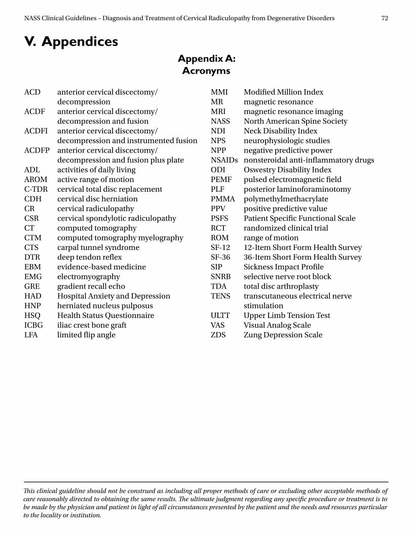
NASS Clinical Guidelines – Diagnosis and Treatment of Cervical Radiculopathy from Degenerative Disorders 72
This clinical guideline should not be construed as including all proper methods of care or excluding other acceptable methods of care reasonably directed to obtaining the same results. The ultimate judgment regarding any specific procedure or treatment is to be made by the physician and patient in light of all circumstances presented by the patient and the needs and resources particular to the locality or institution.
V. Appendices
ACD anterior cervical discectomy/decompression
ACDF anterior cervical discectomy/decompression and fusion
ACDFI anterior cervical discectomy/decompression and instrumented fusion
ACDFP anterior cervical discectomy/decompression and fusion plus plate
ADL activities of daily livingAROM active range of motionC-TDR cervical total disc replacementCDH cervical disc herniationCR cervical radiculopathyCSR cervical spondylotic radiculopathyCT computed tomographyCTM computed tomography myelography CTS carpal tunnel syndromeDTR deep tendon reflexEBM evidence-based medicineEMG electromyographyGRE gradient recall echoHAD Hospital Anxiety and DepressionHNP herniated nucleus pulposusHSQ Health Status QuestionnaireICBG iliac crest bone graftLFA limited flip angle
MMI Modified Million IndexMR magnetic resonanceMRI magnetic resonance imagingNASS North American Spine SocietyNDI Neck Disability IndexNPS neurophysiologic studiesNPP negative predictive powerNSAIDs nonsteroidal anti-inflammatory drugsODI Oswestry Disability IndexPEMF pulsed electromagnetic fieldPLF posterior laminoforaminotomyPMMA polymethylmethacrylatePPV positive predictive valuePSFS Patient Specific Functional ScaleRCT randomized clinical trialROM range of motionSF-12 12-Item Short Form Health SurveySF-36 36-Item Short Form Health Survey SIP Sickness Impact ProfileSNRB selective nerve root blockTDA total disc arthroplastyTENS transcutaneous electrical nerve
stimulationULTT Upper Limb Tension TestVAS Visual Analog ScaleZDS Zung Depression Scale
Appendix A:Acronyms
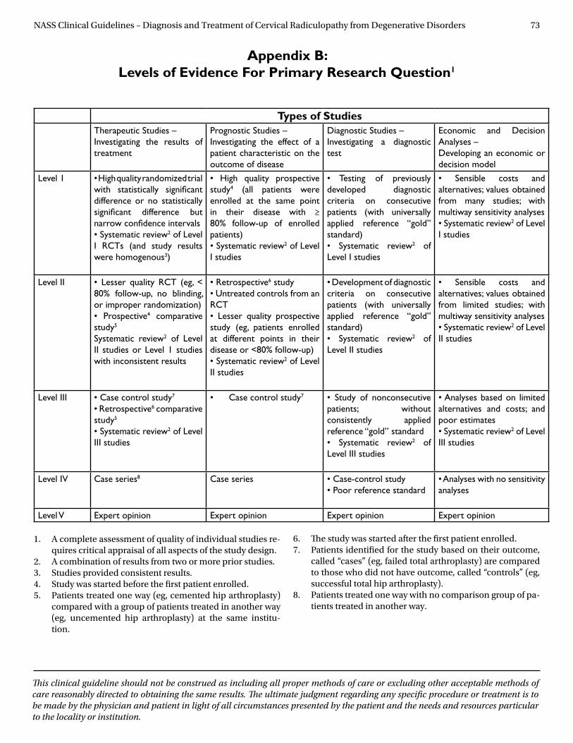
NASS Clinical Guidelines – Diagnosis and Treatment of Cervical Radiculopathy from Degenerative Disorders 73
This clinical guideline should not be construed as including all proper methods of care or excluding other acceptable methods of care reasonably directed to obtaining the same results. The ultimate judgment regarding any specific procedure or treatment is to be made by the physician and patient in light of all circumstances presented by the patient and the needs and resources particular to the locality or institution.
Appendix B: Levels of Evidence For Primary Research Question1
Types of StudiesTherapeutic Studies – Investigating the results of treatment
Prognostic Studies –Investigating the effect of a patient characteristic on the outcome of disease
Diagnostic Studies –Investigating a diagnostic test
Economic and Decision Analyses –Developing an economic or decision model
Level 1 • High quality randomized trial with statistically significant difference or no statistically significant difference but narrow confidence intervals• Systematic review2 of Level I RCTs (and study results were homogenous3)
• High quality prospective study4 (all patients were enrolled at the same point in their disease with ≥ 80% follow-up of enrolled patients)• Systematic review2 of Level I studies
• Testing of previously developed diagnostic criteria on consecutive patients (with universally applied reference “gold” standard) • Systematic review2 of Level I studies
• Sensible costs and alternatives; values obtained from many studies; with multiway sensitivity analyses • Systematic review2 of Level I studies
Level II • Lesser quality RCT (eg, < 80% follow-up, no blinding, or improper randomization)• Prospective4 comparative study5
Systematic review2 of Level II studies or Level 1 studies with inconsistent results
• Retrospective6 study• Untreated controls from an RCT• Lesser quality prospective study (eg, patients enrolled at different points in their disease or <80% follow-up) • Systematic review2 of Level II studies
• Development of diagnostic criteria on consecutive patients (with universally applied reference “gold” standard)• Systematic review2 of Level II studies
• Sensible costs and alternatives; values obtained from limited studies; with multiway sensitivity analyses • Systematic review2 of Level II studies
Level III • Case control study7
• Retrospective6 comparative study5
• Systematic review2 of Level III studies
• Case control study7 • Study of nonconsecutive patients; without consistently applied reference “gold” standard• Systematic review2 of Level III studies
• Analyses based on limited alternatives and costs; and poor estimates • Systematic review2 of Level III studies
Level IV Case series8 Case series • Case-control study• Poor reference standard
• Analyses with no sensitivity analyses
Level V Expert opinion Expert opinion Expert opinion Expert opinion
1. A complete assessment of quality of individual studies re-quires critical appraisal of all aspects of the study design.
2. A combination of results from two or more prior studies.3. Studies provided consistent results.4. Study was started before the first patient enrolled.5. Patients treated one way (eg, cemented hip arthroplasty)
compared with a group of patients treated in another way (eg, uncemented hip arthroplasty) at the same institu-tion.
6. The study was started after the first patient enrolled.7. Patients identified for the study based on their outcome,
called “cases” (eg, failed total arthroplasty) are compared to those who did not have outcome, called “controls” (eg, successful total hip arthroplasty).
8. Patients treated one way with no comparison group of pa-tients treated in another way.
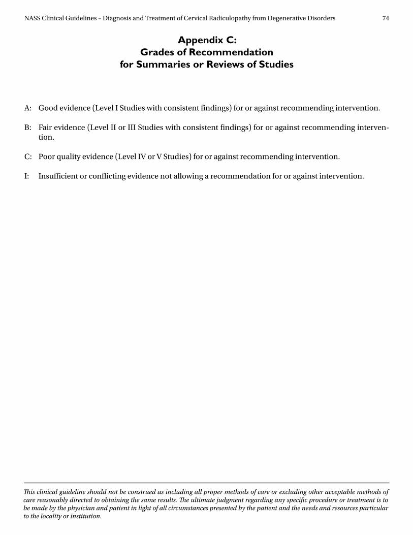
NASS Clinical Guidelines – Diagnosis and Treatment of Cervical Radiculopathy from Degenerative Disorders 74
This clinical guideline should not be construed as including all proper methods of care or excluding other acceptable methods of care reasonably directed to obtaining the same results. The ultimate judgment regarding any specific procedure or treatment is to be made by the physician and patient in light of all circumstances presented by the patient and the needs and resources particular to the locality or institution.
Appendix C:Grades of Recommendation
for Summaries or Reviews of Studies
A: Good evidence (Level I Studies with consistent findings) for or against recommending intervention.
B: Fair evidence (Level II or III Studies with consistent findings) for or against recommending interven-tion.
C: Poor quality evidence (Level IV or V Studies) for or against recommending intervention.
I: Insufficient or conflicting evidence not allowing a recommendation for or against intervention.
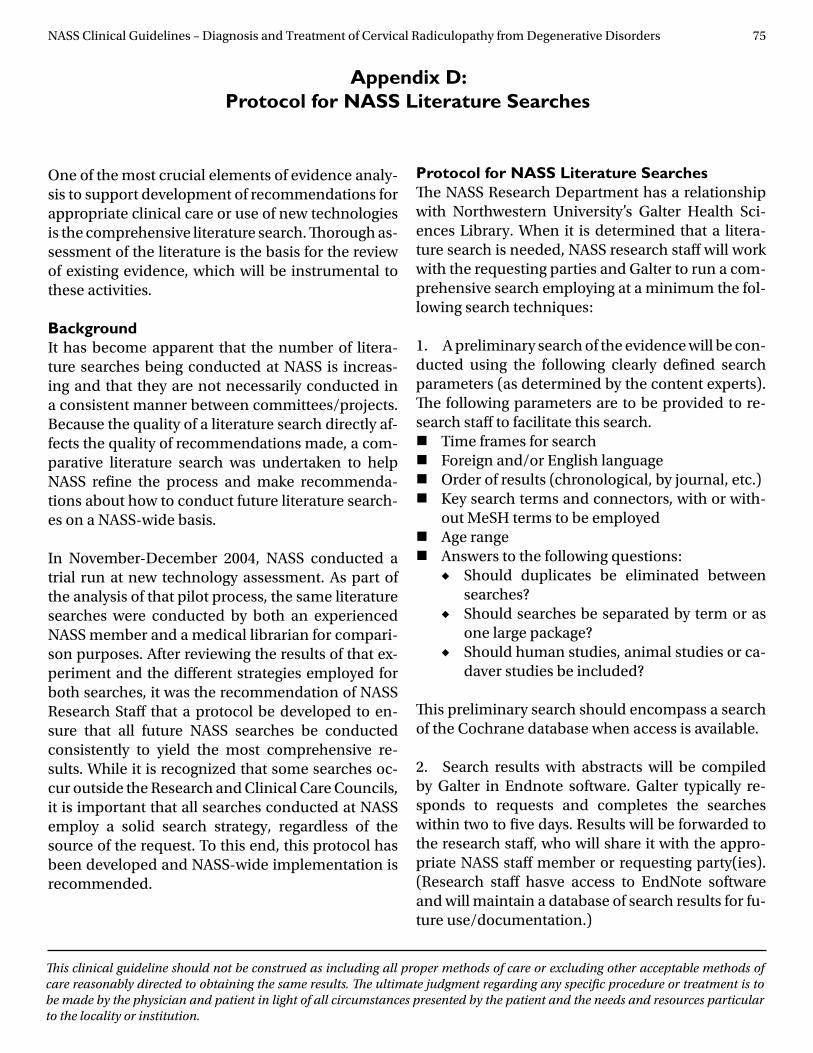
NASS Clinical Guidelines – Diagnosis and Treatment of Cervical Radiculopathy from Degenerative Disorders 75
This clinical guideline should not be construed as including all proper methods of care or excluding other acceptable methods of care reasonably directed to obtaining the same results. The ultimate judgment regarding any specific procedure or treatment is to be made by the physician and patient in light of all circumstances presented by the patient and the needs and resources particular to the locality or institution.
Appendix D:Protocol for NASS Literature Searches
One of the most crucial elements of evidence analy-sis to support development of recommendations for appropriate clinical care or use of new technologies is the comprehensive literature search. Thorough as-sessment of the literature is the basis for the review of existing evidence, which will be instrumental to these activities.
BackgroundIt has become apparent that the number of litera-ture searches being conducted at NASS is increas-ing and that they are not necessarily conducted in a consistent manner between committees/projects. Because the quality of a literature search directly af-fects the quality of recommendations made, a com-parative literature search was undertaken to help NASS refine the process and make recommenda-tions about how to conduct future literature search-es on a NASS-wide basis.
In November-December 2004, NASS conducted a trial run at new technology assessment. As part of the analysis of that pilot process, the same literature searches were conducted by both an experienced NASS member and a medical librarian for compari-son purposes. After reviewing the results of that ex-periment and the different strategies employed for both searches, it was the recommendation of NASS Research Staff that a protocol be developed to en-sure that all future NASS searches be conducted consistently to yield the most comprehensive re-sults. While it is recognized that some searches oc-cur outside the Research and Clinical Care Councils, it is important that all searches conducted at NASS employ a solid search strategy, regardless of the source of the request. To this end, this protocol has been developed and NASS-wide implementation is recommended.
Protocol for NASS Literature SearchesThe NASS Research Department has a relationship with Northwestern University’s Galter Health Sci-ences Library. When it is determined that a litera-ture search is needed, NASS research staff will work with the requesting parties and Galter to run a com-prehensive search employing at a minimum the fol-lowing search techniques:
1. A preliminary search of the evidence will be con-ducted using the following clearly defined search parameters (as determined by the content experts). The following parameters are to be provided to re-search staff to facilitate this search. Time frames for search Foreign and/or English language Order of results (chronological, by journal, etc.) Key search terms and connectors, with or with-
out MeSH terms to be employed Age range Answers to the following questions:
◆ Should duplicates be eliminated between searches?
◆ Should searches be separated by term or as one large package?
◆ Should human studies, animal studies or ca-daver studies be included?
This preliminary search should encompass a search of the Cochrane database when access is available.
2. Search results with abstracts will be compiled by Galter in Endnote software. Galter typically re-sponds to requests and completes the searches within two to five days. Results will be forwarded to the research staff, who will share it with the appro-priate NASS staff member or requesting party(ies). (Research staff hasve access to EndNote software and will maintain a database of search results for fu-ture use/documentation.)
NASS Clinical Guidelines – Diagnosis and Treatment of Cervical Radiculopathy from Degenerative Disorders 76
This clinical guideline should not be construed as including all proper methods of care or excluding other acceptable methods of care reasonably directed to obtaining the same results. The ultimate judgment regarding any specific procedure or treatment is to be made by the physician and patient in light of all circumstances presented by the patient and the needs and resources particular to the locality or institution.
3. NASS staff shares the search results with an ap-propriate content expert (NASS Committee member or other) to assess relevance of articles and identify appropriate articles to review and on which to run a “related articles” search.
4. Based on content expert’s review, NASS research staff will then coordinate with the Galter medical librarian the second level searching to identify rel-evant “related articles.”
5. Galter will forward results to Research Staff to share with appropriate NASS staff.
6. NASS staff share related articles search results with an appropriate content expert (NASS Commit-tee member or other) to assess relevance of this sec-ond set of articles, and identify appropriate articles to review and on which to run a second “related ar-ticles” search.
7. NASS research staff will work with Galter library to obtain the 2nd related articles search results and any necessary full-text articles for review.
8. NASS members reviewing full-text articles should also review the references at the end of each article to identify additional articles which should be reviewed, but may have been missed in the search.
Protocol for Expedited SearchesAt a minimum, numbers 1, 2 and 3 should be fol-lowed for any necessary expedited search. Follow-ing #3, depending on the time frame allowed, deeper searching may be conducted as described by the full protocol or request of full-text articles may occur. If full-text articles are requested, #8 should also be in-cluded. Use of the expedited protocol or any devia-tion from the full protocol should be documented with explanation.
Following these protocols will help ensure that NASS recommendations are (1) based on a thorough re-view of relevant literature; (2) are truly based on a uniform, comprehensive search strategy; and (3) represent the current best research evidence avail-able. Research staff will maintain a search history in EndNote for future use or reference.
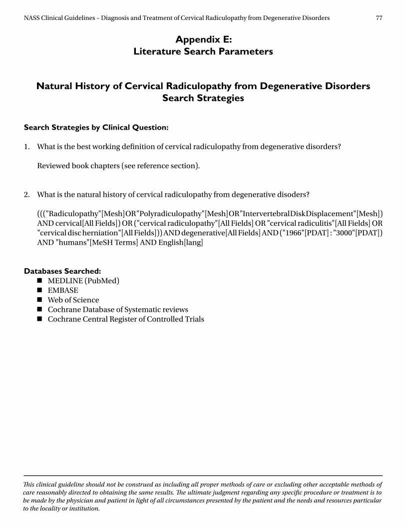
NASS Clinical Guidelines – Diagnosis and Treatment of Cervical Radiculopathy from Degenerative Disorders 77
This clinical guideline should not be construed as including all proper methods of care or excluding other acceptable methods of care reasonably directed to obtaining the same results. The ultimate judgment regarding any specific procedure or treatment is to be made by the physician and patient in light of all circumstances presented by the patient and the needs and resources particular to the locality or institution.
Appendix E:Literature Search Parameters
Natural History of Cervical Radiculopathy from Degenerative DisordersSearch Strategies
Search Strategies by Clinical Question:
1. What is the best working definition of cervical radiculopathy from degenerative disorders?
Reviewed book chapters (see reference section).
2. What is the natural history of cervical radiculopathy from degenerative disoders?
((("Radiculopathy"[Mesh] OR "Polyradiculopathy"[Mesh] OR "Intervertebral Disk Displacement"[Mesh]) AND cervical[All Fields]) OR ("cervical radiculopathy"[All Fields] OR "cervical radiculitis"[All Fields] OR "cervical disc herniation"[All Fields])) AND degenerative[All Fields] AND ("1966"[PDAT] : "3000"[PDAT]) AND "humans"[MeSH Terms] AND English[lang]
Databases Searched: MEDLINE (PubMed) EMBASE Web of Science Cochrane Database of Systematic reviews Cochrane Central Register of Controlled Trials
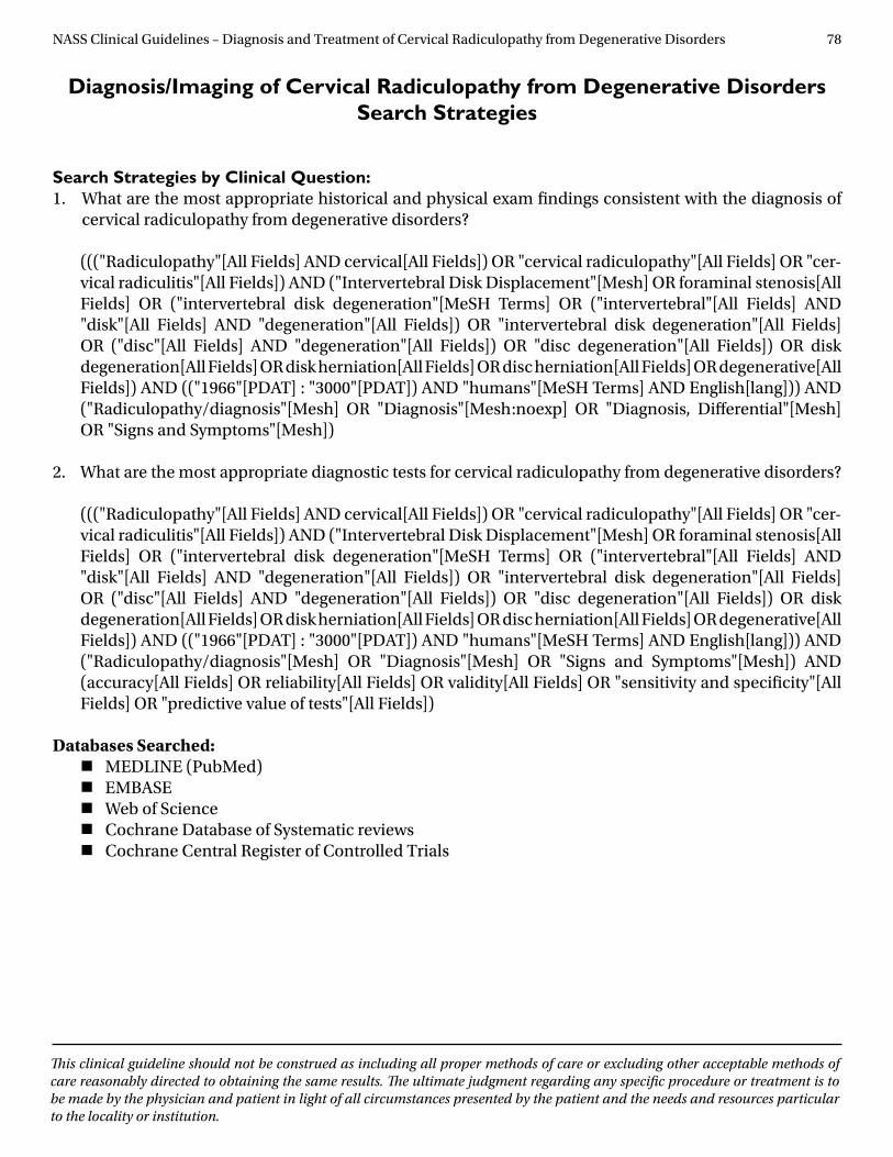
NASS Clinical Guidelines – Diagnosis and Treatment of Cervical Radiculopathy from Degenerative Disorders 78
This clinical guideline should not be construed as including all proper methods of care or excluding other acceptable methods of care reasonably directed to obtaining the same results. The ultimate judgment regarding any specific procedure or treatment is to be made by the physician and patient in light of all circumstances presented by the patient and the needs and resources particular to the locality or institution.
Diagnosis/Imaging of Cervical Radiculopathy from Degenerative Disorders Search Strategies
Search Strategies by Clinical Question:1. What are the most appropriate historical and physical exam findings consistent with the diagnosis of
cervical radiculopathy from degenerative disorders?
((("Radiculopathy"[All Fields] AND cervical[All Fields]) OR "cervical radiculopathy"[All Fields] OR "cer-vical radiculitis"[All Fields]) AND ("Intervertebral Disk Displacement"[Mesh] OR foraminal stenosis[All Fields] OR ("intervertebral disk degeneration"[MeSH Terms] OR ("intervertebral"[All Fields] AND "disk"[All Fields] AND "degeneration"[All Fields]) OR "intervertebral disk degeneration"[All Fields] OR ("disc"[All Fields] AND "degeneration"[All Fields]) OR "disc degeneration"[All Fields]) OR disk degeneration[All Fields] OR disk herniation[All Fields] OR disc herniation[All Fields] OR degenerative[All Fields]) AND (("1966"[PDAT] : "3000"[PDAT]) AND "humans"[MeSH Terms] AND English[lang])) AND ("Radiculopathy/diagnosis"[Mesh] OR "Diagnosis"[Mesh:noexp] OR "Diagnosis, Differential"[Mesh] OR "Signs and Symptoms"[Mesh])
2. What are the most appropriate diagnostic tests for cervical radiculopathy from degenerative disorders?
((("Radiculopathy"[All Fields] AND cervical[All Fields]) OR "cervical radiculopathy"[All Fields] OR "cer-vical radiculitis"[All Fields]) AND ("Intervertebral Disk Displacement"[Mesh] OR foraminal stenosis[All Fields] OR ("intervertebral disk degeneration"[MeSH Terms] OR ("intervertebral"[All Fields] AND "disk"[All Fields] AND "degeneration"[All Fields]) OR "intervertebral disk degeneration"[All Fields] OR ("disc"[All Fields] AND "degeneration"[All Fields]) OR "disc degeneration"[All Fields]) OR disk degeneration[All Fields] OR disk herniation[All Fields] OR disc herniation[All Fields] OR degenerative[All Fields]) AND (("1966"[PDAT] : "3000"[PDAT]) AND "humans"[MeSH Terms] AND English[lang])) AND ("Radiculopathy/diagnosis"[Mesh] OR "Diagnosis"[Mesh] OR "Signs and Symptoms"[Mesh]) AND (accuracy[All Fields] OR reliability[All Fields] OR validity[All Fields] OR "sensitivity and specificity"[All Fields] OR "predictive value of tests"[All Fields])
Databases Searched: MEDLINE (PubMed) EMBASE Web of Science Cochrane Database of Systematic reviews Cochrane Central Register of Controlled Trials
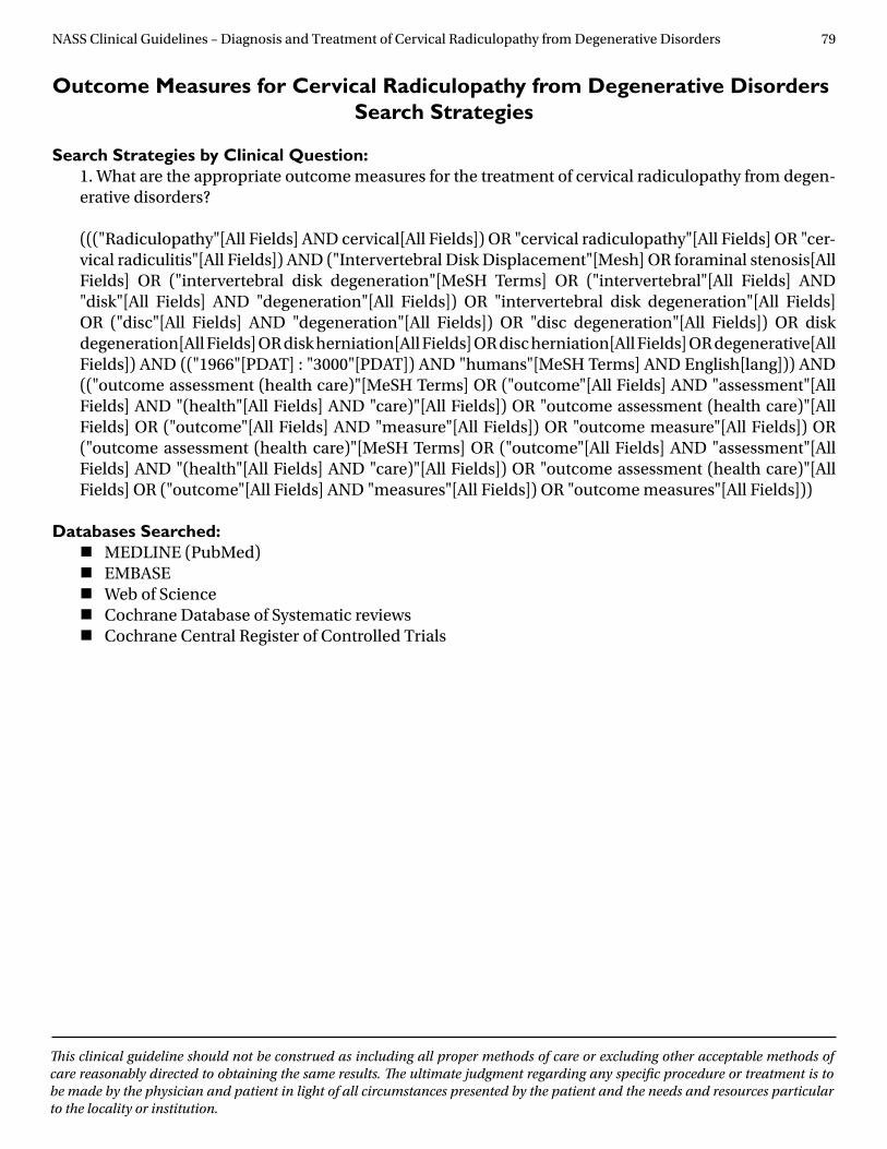
NASS Clinical Guidelines – Diagnosis and Treatment of Cervical Radiculopathy from Degenerative Disorders 79
This clinical guideline should not be construed as including all proper methods of care or excluding other acceptable methods of care reasonably directed to obtaining the same results. The ultimate judgment regarding any specific procedure or treatment is to be made by the physician and patient in light of all circumstances presented by the patient and the needs and resources particular to the locality or institution.
Outcome Measures for Cervical Radiculopathy from Degenerative Disorders Search Strategies
Search Strategies by Clinical Question: 1. What are the appropriate outcome measures for the treatment of cervical radiculopathy from degen-
erative disorders?
((("Radiculopathy"[All Fields] AND cervical[All Fields]) OR "cervical radiculopathy"[All Fields] OR "cer-vical radiculitis"[All Fields]) AND ("Intervertebral Disk Displacement"[Mesh] OR foraminal stenosis[All Fields] OR ("intervertebral disk degeneration"[MeSH Terms] OR ("intervertebral"[All Fields] AND "disk"[All Fields] AND "degeneration"[All Fields]) OR "intervertebral disk degeneration"[All Fields] OR ("disc"[All Fields] AND "degeneration"[All Fields]) OR "disc degeneration"[All Fields]) OR disk degeneration[All Fields] OR disk herniation[All Fields] OR disc herniation[All Fields] OR degenerative[All Fields]) AND (("1966"[PDAT] : "3000"[PDAT]) AND "humans"[MeSH Terms] AND English[lang])) AND (("outcome assessment (health care)"[MeSH Terms] OR ("outcome"[All Fields] AND "assessment"[All Fields] AND "(health"[All Fields] AND "care)"[All Fields]) OR "outcome assessment (health care)"[All Fields] OR ("outcome"[All Fields] AND "measure"[All Fields]) OR "outcome measure"[All Fields]) OR ("outcome assessment (health care)"[MeSH Terms] OR ("outcome"[All Fields] AND "assessment"[All Fields] AND "(health"[All Fields] AND "care)"[All Fields]) OR "outcome assessment (health care)"[All Fields] OR ("outcome"[All Fields] AND "measures"[All Fields]) OR "outcome measures"[All Fields]))
Databases Searched: MEDLINE (PubMed) EMBASE Web of Science Cochrane Database of Systematic reviews Cochrane Central Register of Controlled Trials
NASS Clinical Guidelines – Diagnosis and Treatment of Cervical Radiculopathy from Degenerative Disorders 80
This clinical guideline should not be construed as including all proper methods of care or excluding other acceptable methods of care reasonably directed to obtaining the same results. The ultimate judgment regarding any specific procedure or treatment is to be made by the physician and patient in light of all circumstances presented by the patient and the needs and resources particular to the locality or institution.
Medical/Interventional Treatment of Cervical Radiculopathy from Degenerative Disorders
Search Strategies
Search Strategies by Clinical Question: 1. What is the role of pharmacological treatment in the management of cervical radiculopathy from de-
generative disorders? ((("Radiculopathy"[All Fields] AND cervical[All Fields]) OR "cervical radiculopathy"[All Fields] OR "cer-
vical radiculitis"[All Fields]) AND ("Intervertebral Disk Displacement"[Mesh] OR foraminal stenosis[All Fields] OR ("intervertebral disk degeneration"[MeSH Terms] OR ("intervertebral"[All Fields] AND "disk"[All Fields] AND "degeneration"[All Fields]) OR "intervertebral disk degeneration"[All Fields] OR ("disc"[All Fields] AND "degeneration"[All Fields]) OR "disc degeneration"[All Fields]) OR disk degeneration[All Fields] OR disk herniation[All Fields] OR disc herniation[All Fields] OR degenerative[All Fields]) AND (("1966"[PDAT] : "3000"[PDAT]) AND "humans"[MeSH Terms] AND English[lang])) AND (("Drug Therapy"[Mesh] OR "drug therapy "[Subheading] OR ("pharmacology"[MeSH Terms] OR "pharmacology"[All Fields] OR "pharmacological"[All Fields]) OR ("pharmaceuti-cal preparations"[MeSH Terms] OR ("pharmaceutical"[All Fields] AND "preparations"[All Fields]) OR "pharmaceutical preparations"[All Fields] OR "medication"[All Fields])) AND (("1966"[PDAT] : "3000"[PDAT]) AND "humans"[MeSH Terms] AND English[lang])) AND (("1966"[PDAT] : "3000"[PDAT]) AND "humans"[MeSH Terms] AND English[lang])
2. What is the role of physical therapy/exercise in the treatment of cervical radiculopathy from degenera-tive disorders?
((("Radiculopathy"[All Fields] AND cervical[All Fields]) OR "cervical radiculopathy"[All Fields] OR "cer-vical radiculitis"[All Fields]) AND ("Intervertebral Disk Displacement"[Mesh] OR foraminal stenosis[All Fields] OR ("intervertebral disk degeneration"[MeSH Terms] OR ("intervertebral"[All Fields] AND "disk"[All Fields] AND "degeneration"[All Fields]) OR "intervertebral disk degeneration"[All Fields] OR ("disc"[All Fields] AND "degeneration"[All Fields]) OR "disc degeneration"[All Fields]) OR disk degeneration[All Fields] OR disk herniation[All Fields] OR disc herniation[All Fields] OR degenerative[All Fields]) AND (("1966"[PDAT] : "3000"[PDAT]) AND "humans"[MeSH Terms] AND English[lang])) AND ("Physical Therapy Modalities"[Mesh] OR "Exercise"[Mesh] OR "Physical Exertion"[Mesh] OR "Physical Fitness"[Mesh] OR "Rehabilitation"[Mesh] OR "rehabilitation "[Subheading] OR exercise[title] OR phys-ical therapy[title] OR rehabilitation[title] AND (("1966"[PDAT] : "3000"[PDAT]) AND "humans"[MeSH Terms] AND English[lang]))
NASS Clinical Guidelines – Diagnosis and Treatment of Cervical Radiculopathy from Degenerative Disorders 81
This clinical guideline should not be construed as including all proper methods of care or excluding other acceptable methods of care reasonably directed to obtaining the same results. The ultimate judgment regarding any specific procedure or treatment is to be made by the physician and patient in light of all circumstances presented by the patient and the needs and resources particular to the locality or institution.
3. What is the role of manipulation/chiropractics in the treatment of cervical radiculopathy from degen-erative disorders?
((("Radiculopathy"[All Fields] AND cervical[All Fields]) OR "cervical radiculopathy"[All Fields] OR "cer-vical radiculitis"[All Fields]) AND ("Intervertebral Disk Displacement"[Mesh] OR foraminal stenosis[All Fields] OR ("intervertebral disk degeneration"[MeSH Terms] OR ("intervertebral"[All Fields] AND "disk"[All Fields] AND "degeneration"[All Fields]) OR "intervertebral disk degeneration"[All Fields] OR ("disc"[All Fields] AND "degeneration"[All Fields]) OR "disc degeneration"[All Fields]) OR disk degeneration[All Fields] OR disk herniation[All Fields] OR disc herniation[All Fields] OR degenerative[All Fields]) AND (("1966"[PDAT] : "3000"[PDAT]) AND "humans"[MeSH Terms] AND English[lang])) AND ("Manipula-tion, Chiropractic"[Mesh] OR "Manipulation, Spinal"[Mesh] OR "Manipulation, Orthopedic"[Mesh] OR "Musculoskeletal Manipulations"[Mesh] OR "Chiropractic"[Mesh] OR manipulation[All Fields] OR ("chiropractic"[MeSH Terms] OR "chiropractic"[All Fields]))
4. What is the role of epidural steroid injections for the treatment of cervical radiculopathy from degenera-tive disorders?
((("Radiculopathy"[All Fields] AND cervical[All Fields]) OR "cervical radiculopathy"[All Fields] OR "cer-vical radiculitis"[All Fields]) AND ("Intervertebral Disk Displacement"[Mesh] OR foraminal stenosis[All Fields] OR ("intervertebral disk degeneration"[MeSH Terms] OR ("intervertebral"[All Fields] AND "disk"[All Fields] AND "degeneration"[All Fields]) OR "intervertebral disk degeneration"[All Fields] OR ("disc"[All Fields] AND "degeneration"[All Fields]) OR "disc degeneration"[All Fields]) OR disk degeneration[All Fields] OR disk herniation[All Fields] OR disc herniation[All Fields] OR degenerative[All Fields]) AND (("1966"[PDAT] : "3000"[PDAT]) AND "humans"[MeSH Terms] AND English[lang])) AND ("Injections"[Mesh] OR injections[title] OR injection[title])
5. What is the role of ancillary treatments such as bracing, traction, electrical stimulation, acupuncture and transcutaneous electrical stimulation (TENS) in the treatment of cervical radiculopathy from de-generative disorders?
((("Radiculopathy"[All Fields] AND cervical[All Fields]) OR "cervical radiculopathy"[All Fields] OR "cervi-cal radiculitis"[All Fields]) AND ("Intervertebral Disk Displacement"[Mesh] OR foraminal stenosis[All Fields] OR ("intervertebral disk degeneration"[MeSH Terms] OR ("intervertebral"[All Fields] AND "disk"[All Fields] AND "degeneration"[All Fields]) OR "intervertebral disk degeneration"[All Fields] OR ("disc"[All Fields] AND "degeneration"[All Fields]) OR "disc degeneration"[All Fields]) OR disk degeneration[All Fields] OR disk herniation[All Fields] OR disc herniation[All Fields] OR degenerative[All Fields]) AND (("1966"[PDAT] : "3000"[PDAT]) AND "humans"[MeSH Terms] AND English[lang])) AND ("Braces"[Mesh] OR "Traction"[Mesh] OR "Electric Stimulation"[Mesh] OR "Transcutane-ous Electric Nerve Stimulation"[Mesh] OR "Acupuncture Therapy"[Mesh] OR "Acupuncture"[Mesh] OR bracing[All Fields] OR ("braces"[MeSH Terms] OR "braces"[All Fields] OR "brace"[All Fields]) OR ("braces"[MeSH Terms] OR "braces"[All Fields]) OR ("traction"[MeSH Terms] OR "traction"[All Fields]) OR "electrical stimulation"[All Fields] OR ("transcutaneous electric nerve stimulation"[MeSH Terms] OR ("transcutaneous"[All Fields] AND "electric"[All Fields] AND "nerve"[All Fields] AND "stimulation"[All Fields]) OR "transcutaneous electric nerve stimulation"[All Fields] OR "tens"[All Fields]))
NASS Clinical Guidelines – Diagnosis and Treatment of Cervical Radiculopathy from Degenerative Disorders 82
This clinical guideline should not be construed as including all proper methods of care or excluding other acceptable methods of care reasonably directed to obtaining the same results. The ultimate judgment regarding any specific procedure or treatment is to be made by the physician and patient in light of all circumstances presented by the patient and the needs and resources particular to the locality or institution.
Databases Searched: MEDLINE (PubMed) EMBASE Web of Science Cochrane Database of Systematic reviews Cochrane Central Register of Controlled Trials
Surgical Treatment of Cervical Radiculopathy from Degenerative DisordersSearch Strategies
Search Strategies by Clinical Question1. Does surgical treatment (with or without preoperative medical/interventional treatment) result in bet-
ter outcomes than medical/interventional treatment for cervical radiculopathy from degenerative dis-orders?
((("Radiculopathy"[All Fields] AND cervical[All Fields]) OR "cervical radiculopathy"[All Fields] OR "cer-vical radiculitis"[All Fields]) AND ("Intervertebral Disk Displacement"[Mesh] OR foraminal stenosis[All Fields] OR ("intervertebral disk degeneration"[MeSH Terms] OR ("intervertebral"[All Fields] AND "disk"[All Fields] AND "degeneration"[All Fields]) OR "intervertebral disk degeneration"[All Fields] OR ("disc"[All Fields] AND "degeneration"[All Fields]) OR "disc degeneration"[All Fields]) OR disk degeneration[All Fields] OR disk herniation[All Fields] OR disc herniation[All Fields] OR degenerative[All Fields]) AND (("1966"[PDAT] : "3000"[PDAT]) AND "humans"[MeSH Terms] AND English[lang])) AND ("Surgical Procedures, Operative"[Mesh] OR "surgery "[Subheading] OR surgery[title] OR surgical[title] OR operative[title] OR operation[title] AND (("1966"[PDAT] : "3000"[PDAT]) AND "humans"[MeSH Terms] AND English[lang])) AND (("1966"[PDAT] : "3000"[PDAT]) AND "humans"[MeSH Terms] AND English[lang])
2. Does anterior cervical decompression with fusion result in better outcomes (clinical or radiographic) than anterior cervical decompression alone?
((("Radiculopathy"[All Fields] AND cervical[All Fields]) OR "cervical radiculopathy"[All Fields] OR "cervical radiculitis"[All Fields]) AND ("Intervertebral Disk Displacement"[Mesh] OR foraminal stenosis[All Fields] OR ("intervertebral disk degeneration"[MeSH Terms] OR ("intervertebral"[All Fields] AND "disk"[All Fields] AND "degeneration"[All Fields]) OR "intervertebral disk degeneration"[All Fields] OR ("disc"[All Fields] AND "degeneration"[All Fields]) OR "disc degeneration"[All Fields]) OR disk degeneration[All Fields] OR disk herniation[All Fields] OR disc herniation[All Fields] OR degenerative[All Fields]) AND (("1966"[PDAT] : "3000"[PDAT]) AND "humans"[MeSH Terms] AND English[lang])) AND (("Decompression, Surgical"[Mesh] AND anterior[All Fields] AND cervical[All Fields]) OR (anterior[All Fields] AND cervical[All Fields] AND ("decompression"[MeSH Terms] OR "decompression"[All Fields]) OR (anterior[All Fields] AND cervical[All Fields] AND ("diskectomy"[MeSH Terms] OR "diskectomy"[All Fields] OR "discectomy"[All Fields]))) AND (("1966"[PDAT] : "3000"[PDAT]) AND "humans"[MeSH Terms] AND English[lang])) AND (("1966"[PDAT] : "3000"[PDAT]) AND "humans"[MeSH Terms] AND English[lang]) AND (("Nucl Eng Des/Fusion"[Journal] OR "fusion"[All Fields]) OR ("arthrodesis"[MeSH Terms] OR "arthrodesis"[All Fields]))

NASS Clinical Guidelines – Diagnosis and Treatment of Cervical Radiculopathy from Degenerative Disorders 83
This clinical guideline should not be construed as including all proper methods of care or excluding other acceptable methods of care reasonably directed to obtaining the same results. The ultimate judgment regarding any specific procedure or treatment is to be made by the physician and patient in light of all circumstances presented by the patient and the needs and resources particular to the locality or institution.
3. Does anterior cervical decompression and fusion with instrumentation result in better outcomes (clini-cal or radiographic) than anterior cervical decompression and fusion without instrumentation?
((("Radiculopathy"[All Fields] AND cervical[All Fields]) OR "cervical radiculopathy"[All Fields] OR "cer-vical radiculitis"[All Fields]) AND ("Intervertebral Disk Displacement"[Mesh] OR foraminal stenosis[All Fields] OR ("intervertebral disk degeneration"[MeSH Terms] OR ("intervertebral"[All Fields] AND "disk"[All Fields] AND "degeneration"[All Fields]) OR "intervertebral disk degeneration"[All Fields] OR ("disc"[All Fields] AND "degeneration"[All Fields]) OR "disc degeneration"[All Fields]) OR disk degeneration[All Fields] OR disk herniation[All Fields] OR disc herniation[All Fields] OR degenerative[All Fields]) AND (("1966"[PDAT] : "3000"[PDAT]) AND "humans"[MeSH Terms] AND English[lang])) AND (("Decompression, Surgical"[Mesh] AND anterior[All Fields] AND cervical[All Fields]) OR (anterior[All Fields] AND cervical[All Fields] AND ("decompression"[MeSH Terms] OR "decompression"[All Fields]) OR ((anterior[All Fields] AND cervical[All Fields] AND ("diskectomy"[MeSH Terms] OR "diskectomy"[All Fields] OR "discectomy"[All Fields])) OR (anterior[All Fields] AND cervical[All Fields] AND ("diskectomy"[MeSH Terms] OR "diskectomy"[All Fields]))) AND (("1966"[PDAT] : "3000"[PDAT]) AND "humans"[MeSH Terms] AND English[lang])) AND (("1966"[PDAT] : "3000"[PDAT]) AND "humans"[MeSH Terms] AND English[lang]) AND (("Nucl Eng Des/Fusion"[Journal] OR "fusion"[All Fields]) OR ("arthrodesis"[MeSH Terms] OR "arthrodesis"[All Fields]))) AND ("instrumentation "[Subheading] OR "Bone Plates"[Mesh] OR ("bone plates"[MeSH Terms] OR ("bone"[All Fields] AND "plates"[All Fields]) OR "bone plates"[All Fields] OR "plate"[All Fields]) OR plates[All Fields] OR plating[All Fields] OR instrumentation[title] OR ("computers"[MeSH Terms] OR "computers"[All Fields] OR "hardware"[All Fields]))
4. Does anterior surgery result in better outcomes (clinical or radiographic) than posterior surgery in the treatment of cervical radiculopathy from degenerative disorders?
((("Radiculopathy"[All Fields] AND cervical[All Fields]) OR "cervical radiculopathy"[All Fields] OR "cer-vical radiculitis"[All Fields]) AND ("Intervertebral Disk Displacement"[Mesh] OR foraminal stenosis[All Fields] OR ("intervertebral disk degeneration"[MeSH Terms] OR ("intervertebral"[All Fields] AND "disk"[All Fields] AND "degeneration"[All Fields]) OR "intervertebral disk degeneration"[All Fields] OR ("disc"[All Fields] AND "degeneration"[All Fields]) OR "disc degeneration"[All Fields]) OR disk degeneration[All Fields] OR disk herniation[All Fields] OR disc herniation[All Fields] OR degenerative[All Fields]) AND (("1966"[PDAT] : "3000"[PDAT]) AND "humans"[MeSH Terms] AND English[lang])) AND (("Decompression, Surgical"[Mesh] OR "Laminectomy"[Mesh] OR "cervical decompression"[All Fields] OR "laminectomy"[title] OR "laminotomy"[title] OR foraminotomy[title] OR laminoplasty[title] OR (cervical[All Fields] AND ("diskectomy"[MeSH Terms] OR "diskectomy"[All Fields] OR "discectomy"[All Fields])) OR (cervical[All Fields] AND ("diskectomy"[MeSH Terms] OR "diskectomy"[All Fields]))) AND (anterior[All Fields] AND posterior[All Fields]))
5. Does posterior decompression with fusion result in better outcomes (clinical or radiographic) than pos-terior decompression alone in the treatment of cervical radiculopathy from degenerative disorders?
NASS Clinical Guidelines – Diagnosis and Treatment of Cervical Radiculopathy from Degenerative Disorders 84
This clinical guideline should not be construed as including all proper methods of care or excluding other acceptable methods of care reasonably directed to obtaining the same results. The ultimate judgment regarding any specific procedure or treatment is to be made by the physician and patient in light of all circumstances presented by the patient and the needs and resources particular to the locality or institution.
((("Radiculopathy"[All Fields] AND cervical[All Fields]) OR "cervical radiculopathy"[All Fields] OR "cer-vical radiculitis"[All Fields]) AND ("Intervertebral Disk Displacement"[Mesh] OR foraminal stenosis[All Fields] OR ("intervertebral disk degeneration"[MeSH Terms] OR ("intervertebral"[All Fields] AND "disk"[All Fields] AND "degeneration"[All Fields]) OR "intervertebral disk degeneration"[All Fields] OR ("disc"[All Fields] AND "degeneration"[All Fields]) OR "disc degeneration"[All Fields]) OR disk degeneration[All Fields] OR disk herniation[All Fields] OR disc herniation[All Fields] OR degenerative[All Fields]) AND (("1966"[PDAT] : "3000"[PDAT]) AND "humans"[MeSH Terms] AND English[lang])) AND ("Decompression, Surgical"[Mesh] OR "Laminectomy"[Mesh] OR "cervical decompression"[All Fields] OR "laminectomy"[title] OR "laminotomy"[title] OR foraminotomy[title] OR laminoplasty[title] OR (cervical[All Fields] AND ("diskectomy"[MeSH Terms] OR "diskectomy"[All Fields] OR "discectomy"[All Fields])) OR (cervical[All Fields] AND ("diskectomy"[MeSH Terms] OR "diskectomy"[All Fields]))) AND posterior[All Fields] AND (("Nucl Eng Des/Fusion"[Journal] OR "fusion"[All Fields]) OR ("arthrodesis"[MeSH Terms] OR "arthrodesis"[All Fields]))
6. Does anterior cervical decompression and reconstruction with total disc replacement result in better outcomes (clinical or radiographic) than anterior cervical decompression and fusion in the treatment of cervical radiculopathy from degenerative disorders?
((("Radiculopathy"[All Fields] AND cervical[All Fields]) OR "cervical radiculopathy"[All Fields] OR "cer-vical radiculitis"[All Fields]) AND ("Intervertebral Disk Displacement"[Mesh] OR foraminal stenosis[All Fields] OR ("intervertebral disk degeneration"[MeSH Terms] OR ("intervertebral"[All Fields] AND "disk"[All Fields] AND "degeneration"[All Fields]) OR "intervertebral disk degeneration"[All Fields] OR ("disc"[All Fields] AND "degeneration"[All Fields]) OR "disc degeneration"[All Fields]) OR disk degeneration[All Fields] OR disk herniation[All Fields] OR disc herniation[All Fields] OR degenerative[All Fields]) AND (("1966"[PDAT] : "3000"[PDAT]) AND "humans"[MeSH Terms] AND English[lang])) AND (("Arthroplasty"[Mesh] AND disc[All Fields]) OR disc arthroplasty[All Fields] OR disk arthroplasty[All Fields] OR disc replacement[All Fields] OR disk replacement[All Fields] AND (("1966"[PDAT] : "3000"[PDAT]) AND "humans"[MeSH Terms] AND English[lang])) AND (("Nucl Eng Des/Fusion"[Journal] OR "fusion"[All Fields]) OR ("arthrodesis"[MeSH Terms] OR "arthrodesis"[All Fields])) AND (("Decom-pression, Surgical"[Mesh] AND anterior[All Fields] AND cervical[All Fields]) OR (anterior[All Fields] AND cervical[All Fields] AND ("decompression"[MeSH Terms] OR "decompression"[All Fields]) OR (anterior[All Fields] AND cervical[All Fields] AND ("diskectomy"[MeSH Terms] OR "diskectomy"[All Fields] OR "discectomy"[All Fields]))) AND (("1966"[PDAT] : "3000"[PDAT]) AND "humans"[MeSH Terms] AND English[lang]))
7. What is the long-term result (four+ years) of surgical management of cervical radiculopathy from de-generative disorders?
("Longitudinal Studies"[Mesh:noexp] OR (long[All Fields] AND term[All Fields]) OR long-term[All Fields] AND (("1966"[PDAT] : "3000"[PDAT]) AND "humans"[MeSH Terms] AND English[lang])) AND (((("Radiculopathy"[All Fields] AND cervical[All Fields]) OR "cervical radiculopathy"[All Fields] OR "cer-vical radiculitis"[All Fields]) AND ("Intervertebral Disk Displacement"[Mesh] OR foraminal stenosis[All Fields] OR ("intervertebral disk degeneration"[MeSH Terms] OR ("intervertebral"[All Fields] AND "disk"[All Fields] AND "degeneration"[All Fields]) OR "intervertebral disk degeneration"[All Fields]
NASS Clinical Guidelines – Diagnosis and Treatment of Cervical Radiculopathy from Degenerative Disorders 85
This clinical guideline should not be construed as including all proper methods of care or excluding other acceptable methods of care reasonably directed to obtaining the same results. The ultimate judgment regarding any specific procedure or treatment is to be made by the physician and patient in light of all circumstances presented by the patient and the needs and resources particular to the locality or institution.
OR ("disc"[All Fields] AND "degeneration"[All Fields]) OR "disc degeneration"[All Fields]) OR disk degeneration[All Fields] OR disk herniation[All Fields] OR disc herniation[All Fields] OR degenerative[All Fields]) AND (("1966"[PDAT] : "3000"[PDAT]) AND "humans"[MeSH Terms] AND English[lang])) AND ("Surgical Procedures, Operative"[Mesh] OR "surgery "[Subheading] OR surgery[title] OR surgical[title] OR operative[title] OR operation[title] AND (("1966"[PDAT] : "3000"[PDAT]) AND "humans"[MeSH Terms] AND English[lang])) AND (("1966"[PDAT] : "3000"[PDAT]) AND "humans"[MeSH Terms] AND English[lang])) AND (("1966"[PDAT] : "3000"[PDAT]) AND "humans"[MeSH Terms] AND English[lang])
8. How do long-term results of single-level compare with multilevel surgical decompression for cervical radiculopathy from degenerative disorders?
(((("Radiculopathy"[All Fields] AND cervical[All Fields]) OR "cervical radiculopathy"[All Fields] OR "cer-vical radiculitis"[All Fields]) AND ("Intervertebral Disk Displacement"[Mesh] OR foraminal stenosis[All Fields] OR ("intervertebral disk degeneration"[MeSH Terms] OR ("intervertebral"[All Fields] AND "disk"[All Fields] AND "degeneration"[All Fields]) OR "intervertebral disk degeneration"[All Fields] OR ("disc"[All Fields] AND "degeneration"[All Fields]) OR "disc degeneration"[All Fields]) OR disk degeneration[All Fields] OR disk herniation[All Fields] OR disc herniation[All Fields] OR degenerative[All Fields]) AND (("1966"[PDAT] : "3000"[PDAT]) AND "humans"[MeSH Terms] AND English[lang])) AND ("Decompression, Surgical"[Mesh] OR "Laminectomy"[Mesh] OR "cervical decompression"[All Fields] OR "laminectomy"[All Fields] OR "laminotomy"[All Fields] OR foraminotomy[All Fields] OR laminoplasty[All Fields] OR (cervical[All Fields] AND ("diskectomy"[MeSH Terms] OR "diskectomy"[All Fields] OR "discectomy"[All Fields])) OR (cervical[All Fields] AND ("diskectomy"[MeSH Terms] OR "diskectomy"[All Fields])))) AND ("Longitudinal Studies"[Mesh:noexp] OR (long[All Fields] AND term[All Fields]) OR long-term[All Fields] AND (("1966"[PDAT] : "3000"[PDAT]) AND "humans"[MeSH Terms] AND English[lang]))
Databases Searched: MEDLINE (PubMed) EMBASE Web of Science Cochrane Database of Systematic reviews Cochrane Central Register of Controlled Trials
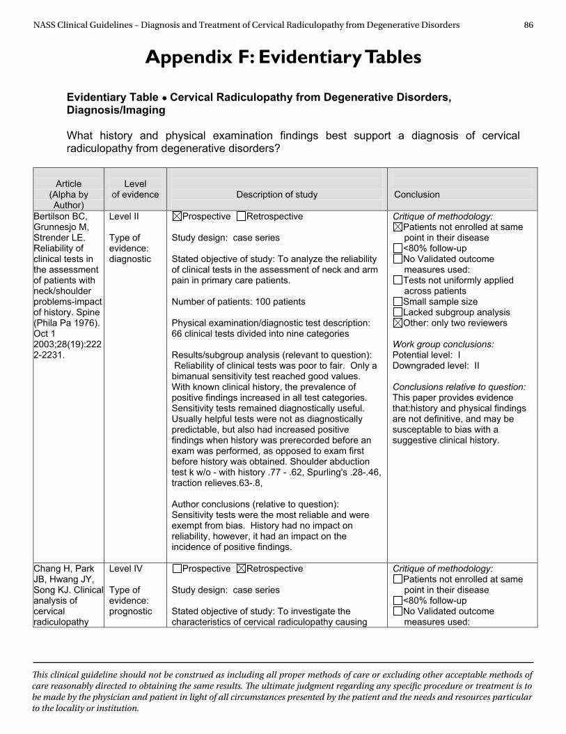
NASS Clinical Guidelines – Diagnosis and Treatment of Cervical Radiculopathy from Degenerative Disorders 86
This clinical guideline should not be construed as including all proper methods of care or excluding other acceptable methods of care reasonably directed to obtaining the same results. The ultimate judgment regarding any specific procedure or treatment is to be made by the physician and patient in light of all circumstances presented by the patient and the needs and resources particular to the locality or institution.
Appendix F: Evidentiary Tables
Evidentiary Table ● Cervical Radiculopathy from Degenerative Disorders, Diagnosis/Imaging What history and physical examination findings best support a diagnosis of cervical radiculopathy from degenerative disorders?
Article (Alpha by Author)
Level
of evidence
Description of study
Conclusion
Bertilson BC, Grunnesjo M, Strender LE. Reliability of clinical tests in the assessment of patients with neck/shoulder problems-impact of history. Spine (Phila Pa 1976). Oct 1 2003;28(19):2222-2231.
Level II Type of evidence: diagnostic
Prospective Retrospective Study design: case series Stated objective of study: To analyze the reliability of clinical tests in the assessment of neck and arm pain in primary care patients. Number of patients: 100 patients Physical examination/diagnostic test description: 66 clinical tests divided into nine categories Results/subgroup analysis (relevant to question): Reliability of clinical tests was poor to fair. Only a bimanual sensitivity test reached good values. With known clinical history, the prevalence of positive findings increased in all test categories. Sensitivity tests remained diagnostically useful. Usually helpful tests were not as diagnostically predictable, but also had increased positive findings when history was prerecorded before an exam was performed, as opposed to exam first before history was obtained. Shoulder abduction test k w/o - with history .77 - .62, Spurling's .28-.46, traction relieves.63-.8, Author conclusions (relative to question): Sensitivity tests were the most reliable and were exempt from bias. History had no impact on reliability, however, it had an impact on the incidence of positive findings.
Critique of methodology: Patients not enrolled at same point in their disease <80% follow-up No Validated outcome measures used: Tests not uniformly applied across patients Small sample size Lacked subgroup analysis Other: only two reviewers
Work group conclusions: Potential level: I Downgraded level: II Conclusions relative to question: This paper provides evidence that:history and physical findings are not definitive, and may be susceptable to bias with a suggestive clinical history.
Chang H, Park JB, Hwang JY, Song KJ. Clinical analysis of cervical radiculopathy
Level IV Type of evidence: prognostic
Prospective Retrospective Study design: case series Stated objective of study: To investigate the characteristics of cervical radiculopathy causing
Critique of methodology: Patients not enrolled at same point in their disease <80% follow-up No Validated outcome measures used:
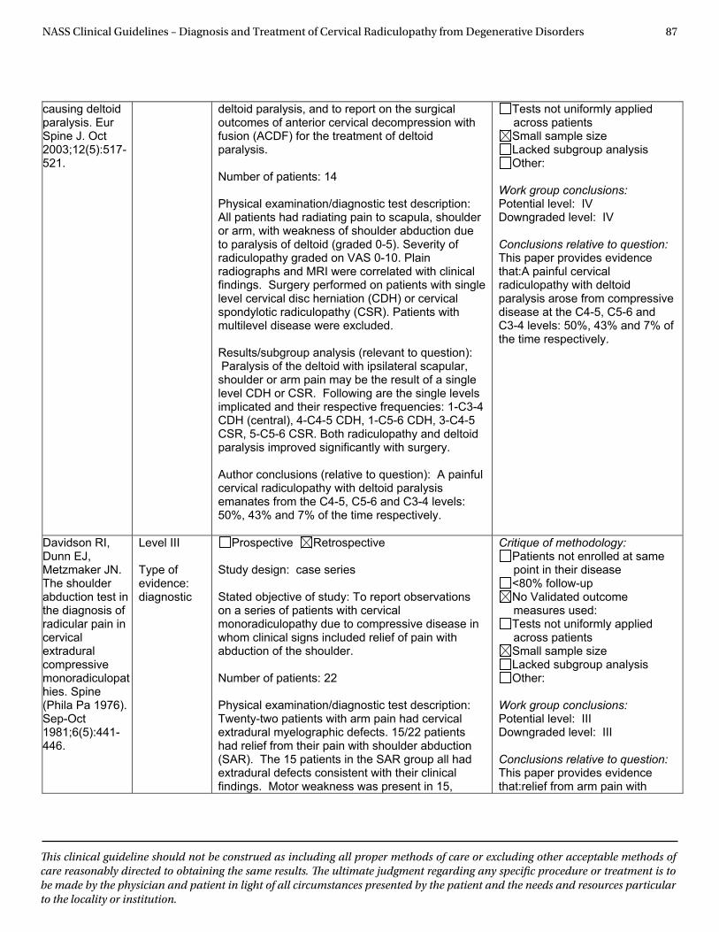
NASS Clinical Guidelines – Diagnosis and Treatment of Cervical Radiculopathy from Degenerative Disorders 87
This clinical guideline should not be construed as including all proper methods of care or excluding other acceptable methods of care reasonably directed to obtaining the same results. The ultimate judgment regarding any specific procedure or treatment is to be made by the physician and patient in light of all circumstances presented by the patient and the needs and resources particular to the locality or institution.
causing deltoid paralysis. Eur Spine J. Oct 2003;12(5):517-521.
deltoid paralysis, and to report on the surgical outcomes of anterior cervical decompression with fusion (ACDF) for the treatment of deltoid paralysis.
Number of patients: 14
Physical examination/diagnostic test description: All patients had radiating pain to scapula, shoulder or arm, with weakness of shoulder abduction due to paralysis of deltoid (graded 0-5). Severity of radiculopathy graded on VAS 0-10. Plain radiographs and MRI were correlated with clinical findings. Surgery performed on patients with single level cervical disc herniation (CDH) or cervical spondylotic radiculopathy (CSR). Patients with multilevel disease were excluded.
Results/subgroup analysis (relevant to question): Paralysis of the deltoid with ipsilateral scapular, shoulder or arm pain may be the result of a single level CDH or CSR. Following are the single levels implicated and their respective frequencies: 1-C3-4 CDH (central), 4-C4-5 CDH, 1-C5-6 CDH, 3-C4-5 CSR, 5-C5-6 CSR. Both radiculopathy and deltoid paralysis improved significantly with surgery.
Author conclusions (relative to question): A painful cervical radiculopathy with deltoid paralysis emanates from the C4-5, C5-6 and C3-4 levels: 50%, 43% and 7% of the time respectively.
Tests not uniformly applied across patients Small sample size Lacked subgroup analysis Other:
Work group conclusions:Potential level: IV Downgraded level: IV
Conclusions relative to question: This paper provides evidence that:A painful cervical radiculopathy with deltoid paralysis arose from compressive disease at the C4-5, C5-6 and C3-4 levels: 50%, 43% and 7% of the time respectively.
Davidson RI, Dunn EJ, Metzmaker JN. The shoulder abduction test in the diagnosis of radicular pain in cervical extradural compressive monoradiculopathies. Spine (Phila Pa 1976). Sep-Oct 1981;6(5):441-446.
Level III
Type of evidence: diagnostic
Prospective Retrospective
Study design: case series
Stated objective of study: To report observations on a series of patients with cervical monoradiculopathy due to compressive disease in whom clinical signs included relief of pain with abduction of the shoulder.
Number of patients: 22
Physical examination/diagnostic test description: Twenty-two patients with arm pain had cervical extradural myelographic defects. 15/22 patients had relief from their pain with shoulder abduction (SAR). The 15 patients in the SAR group all had extradural defects consistent with their clinical findings. Motor weakness was present in 15,
Critique of methodology: Patients not enrolled at same point in their disease <80% follow-up No Validated outcome measures used: Tests not uniformly applied across patients Small sample size Lacked subgroup analysis Other:
Work group conclusions: Potential level: III Downgraded level: III
Conclusions relative to question: This paper provides evidence that:relief from arm pain with
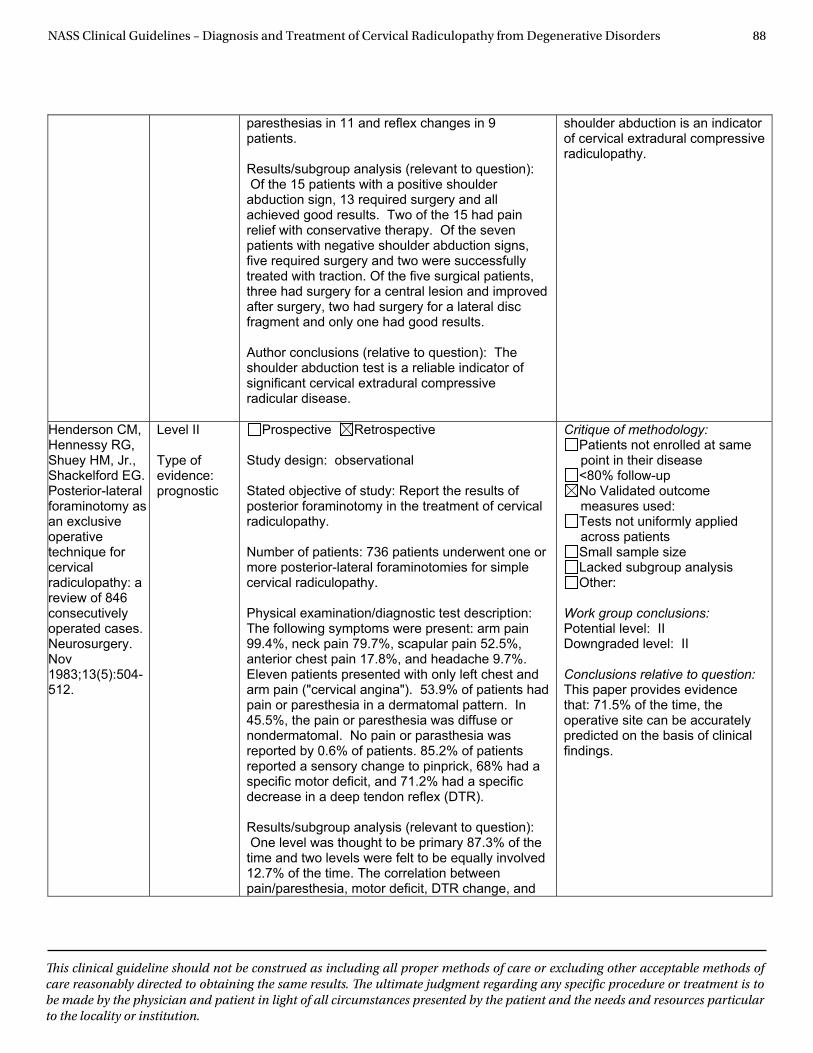
NASS Clinical Guidelines – Diagnosis and Treatment of Cervical Radiculopathy from Degenerative Disorders 88
This clinical guideline should not be construed as including all proper methods of care or excluding other acceptable methods of care reasonably directed to obtaining the same results. The ultimate judgment regarding any specific procedure or treatment is to be made by the physician and patient in light of all circumstances presented by the patient and the needs and resources particular to the locality or institution.
paresthesias in 11 and reflex changes in 9 patients.
Results/subgroup analysis (relevant to question): Of the 15 patients with a positive shoulder abduction sign, 13 required surgery and all achieved good results. Two of the 15 had pain relief with conservative therapy. Of the seven patients with negative shoulder abduction signs, five required surgery and two were successfully treated with traction. Of the five surgical patients, three had surgery for a central lesion and improved after surgery, two had surgery for a lateral disc fragment and only one had good results.
Author conclusions (relative to question): The shoulder abduction test is a reliable indicator of significant cervical extradural compressive radicular disease.
shoulder abduction is an indicator of cervical extradural compressive radiculopathy.
Henderson CM, Hennessy RG, Shuey HM, Jr., Shackelford EG. Posterior-lateral foraminotomy as an exclusive operativetechnique for cervical radiculopathy: a review of 846 consecutively operated cases. Neurosurgery. Nov 1983;13(5):504-512.
Level II
Type of evidence: prognostic
Prospective Retrospective
Study design: observational
Stated objective of study: Report the results of posterior foraminotomy in the treatment of cervical radiculopathy.
Number of patients: 736 patients underwent one or more posterior-lateral foraminotomies for simple cervical radiculopathy.
Physical examination/diagnostic test description: The following symptoms were present: arm pain 99.4%, neck pain 79.7%, scapular pain 52.5%, anterior chest pain 17.8%, and headache 9.7%. Eleven patients presented with only left chest and arm pain ("cervical angina"). 53.9% of patients had pain or paresthesia in a dermatomal pattern. In 45.5%, the pain or paresthesia was diffuse or nondermatomal. No pain or parasthesia was reported by 0.6% of patients. 85.2% of patients reported a sensory change to pinprick, 68% had a specific motor deficit, and 71.2% had a specific decrease in a deep tendon reflex (DTR).
Results/subgroup analysis (relevant to question): One level was thought to be primary 87.3% of the time and two levels were felt to be equally involved 12.7% of the time. The correlation between pain/paresthesia, motor deficit, DTR change, and
Critique of methodology: Patients not enrolled at same point in their disease <80% follow-up No Validated outcome measures used: Tests not uniformly applied across patients Small sample size Lacked subgroup analysis Other:
Work group conclusions: Potential level: II Downgraded level: II
Conclusions relative to question: This paper provides evidence that: 71.5% of the time, the operative site can be accurately predicted on the basis of clinical findings.
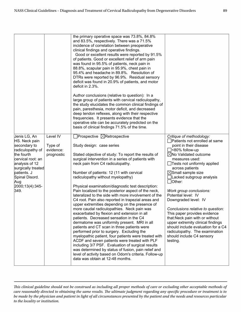
NASS Clinical Guidelines – Diagnosis and Treatment of Cervical Radiculopathy from Degenerative Disorders 89
This clinical guideline should not be construed as including all proper methods of care or excluding other acceptable methods of care reasonably directed to obtaining the same results. The ultimate judgment regarding any specific procedure or treatment is to be made by the physician and patient in light of all circumstances presented by the patient and the needs and resources particular to the locality or institution.
the primary operative space was 73.8%, 84.8% and 83.5%, respectively. There was a 71.5% incidence of correlation between preoperative clinical findings and operative findings. Good or excellent results were reported by 91.5% of patients. Good or excellent relief of arm pain was found in 95.5% of patients, neck pain in 88.8%, scapular pain in 95.9%, chest pain in 95.4% and headache in 89.8%. Resolution of DTRs were reported by 96.9%. Residual sensory deficit was found in 20.9% of patients, and motor deficit in 2.3%.
Author conclusions (relative to question): In a large group of patients with cervical radiculopathy, the study elucidates the common clinical findings of pain, paresthesia, motor deficit, and decreased deep tendon reflexes, along with their respective frequencies. It presents evidence that the operative site can be accurately predicted on the basis of clinical findings 71.5% of the time.
Jenis LG, An HS. Neck pain secondary to radiculopathy of the fourth cervical root: an analysis of 12 surgically treated patients. J Spinal Disord. Aug2000;13(4):345-349.
Level IV
Type of evidence: prognostic
Prospective Retrospective
Study design: case series
Stated objective of study: To report the results of surgical intervention in a series of patients with neck pain from C4 radiculopathy.
Number of patients: 12 (11 with cervical radiculopathy without myelopathy)
Physical examination/diagnostic test description: Pain localized to the posterior aspect of the neck, lateralized to the side with more involvement of the C4 root. Pain also reported in trapezial areas and upper extremities depending on the presence of more caudal radiculopathies. Neck pain was exacerbated by flexion and extension in all patients. Decreased sensation in the C4 dermatome was uniformily present. MRI in all patients and CT scan in three patients were performed prior to surgery. Excluding the myelopathic patient, four patients were treated with ACDF and seven patients were treated with PLF including 3/7 PSF. Evaluation of surgical results was determined by status of fusion, pain relief and level of activity based on Odom's criteria. Follow-up data was obtain at 12-48 months.
Critique of methodology: Patients not enrolled at same point in their disease <80% follow-up No Validated outcome measures used: Tests not uniformly applied across patients Small sample size Lacked subgroup analysis Other:
Work group conclusions: Potential level: IV Downgraded level: IV
Conclusions relative to question: This paper provides evidence that:Neck pain with or without upper extremity clinical findings should include evaluation for a C4 radiculopathy. The examination should include C4 sensory testing.
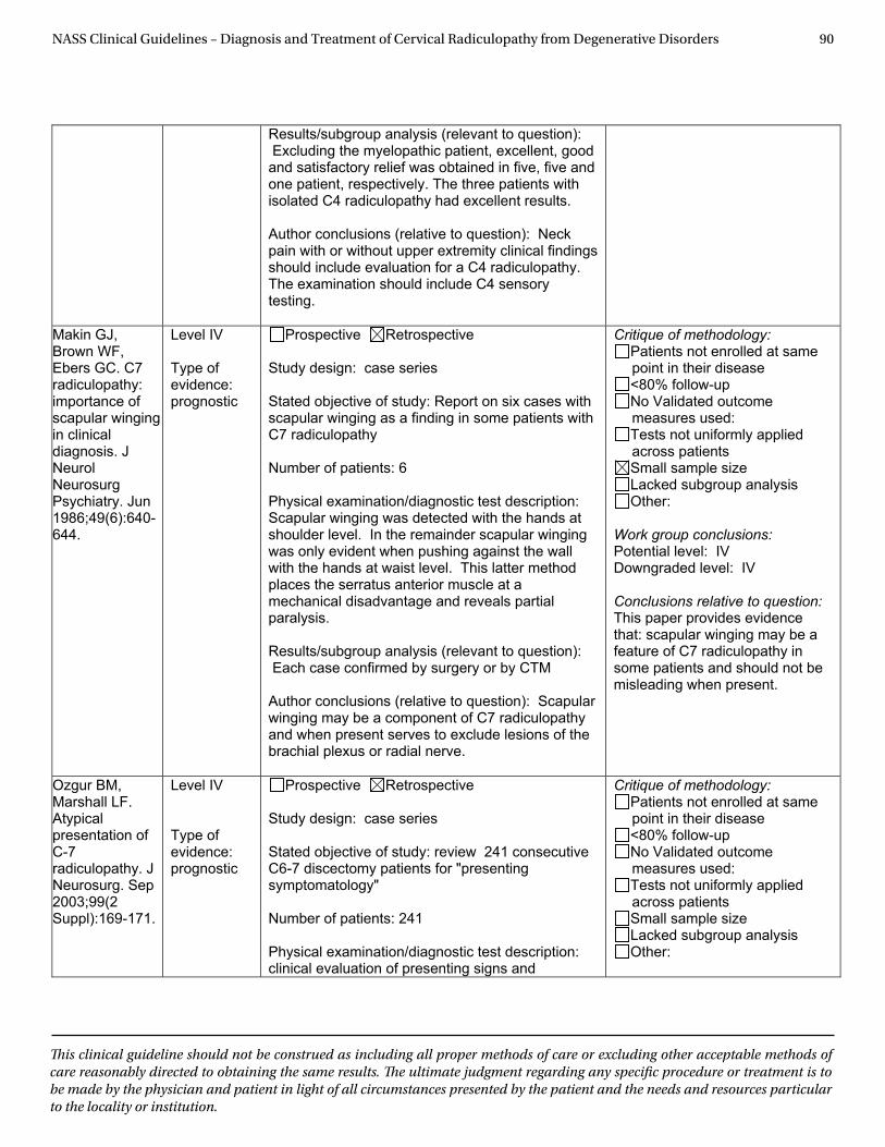
NASS Clinical Guidelines – Diagnosis and Treatment of Cervical Radiculopathy from Degenerative Disorders 90
This clinical guideline should not be construed as including all proper methods of care or excluding other acceptable methods of care reasonably directed to obtaining the same results. The ultimate judgment regarding any specific procedure or treatment is to be made by the physician and patient in light of all circumstances presented by the patient and the needs and resources particular to the locality or institution.
Results/subgroup analysis (relevant to question): Excluding the myelopathic patient, excellent, good and satisfactory relief was obtained in five, five and one patient, respectively. The three patients with isolated C4 radiculopathy had excellent results.
Author conclusions (relative to question): Neck pain with or without upper extremity clinical findings should include evaluation for a C4 radiculopathy. The examination should include C4 sensory testing.
Makin GJ, Brown WF, Ebers GC. C7 radiculopathy: importance of scapular winging in clinical diagnosis. J Neurol Neurosurg Psychiatry. Jun 1986;49(6):640-644.
Level IV
Type of evidence: prognostic
Prospective Retrospective
Study design: case series
Stated objective of study: Report on six cases with scapular winging as a finding in some patients with C7 radiculopathy
Number of patients: 6
Physical examination/diagnostic test description: Scapular winging was detected with the hands at shoulder level. In the remainder scapular winging was only evident when pushing against the wall with the hands at waist level. This latter method places the serratus anterior muscle at a mechanical disadvantage and reveals partial paralysis.
Results/subgroup analysis (relevant to question): Each case confirmed by surgery or by CTM
Author conclusions (relative to question): Scapular winging may be a component of C7 radiculopathy and when present serves to exclude lesions of the brachial plexus or radial nerve.
Critique of methodology: Patients not enrolled at same point in their disease <80% follow-up No Validated outcome measures used: Tests not uniformly applied across patients Small sample size Lacked subgroup analysis Other:
Work group conclusions: Potential level: IV Downgraded level: IV
Conclusions relative to question: This paper provides evidence that: scapular winging may be a feature of C7 radiculopathy in some patients and should not be misleading when present.
Ozgur BM, Marshall LF. Atypical presentation of C-7radiculopathy. J Neurosurg. Sep 2003;99(2 Suppl):169-171.
Level IV
Type of evidence: prognostic
Prospective Retrospective
Study design: case series
Stated objective of study: review 241 consecutive C6-7 discectomy patients for "presenting symptomatology"
Number of patients: 241
Physical examination/diagnostic test description: clinical evaluation of presenting signs and
Critique of methodology: Patients not enrolled at same point in their disease <80% follow-up No Validated outcome measures used: Tests not uniformly applied across patients Small sample size Lacked subgroup analysis Other:

NASS Clinical Guidelines – Diagnosis and Treatment of Cervical Radiculopathy from Degenerative Disorders 91
This clinical guideline should not be construed as including all proper methods of care or excluding other acceptable methods of care reasonably directed to obtaining the same results. The ultimate judgment regarding any specific procedure or treatment is to be made by the physician and patient in light of all circumstances presented by the patient and the needs and resources particular to the locality or institution.
symptoms for usual and unusual findings
Results/subgroup analysis (relevant to question): Most patients had usual C7 traditional radicular signs (dermatomal distribution), with 12% reporting sole complaint of subscapular pain, 5% having deep breast or chest pain. None of these 17% had the "typical" C7 presenting symptoms.
Author conclusions (relative to question): Patients presenting with unusual symptoms had their complaints validated by surgical findings and 93% experienced symptom relief
Work group conclusions: Potential level: IV Downgraded level: IV
Conclusions relative to question: This paper provides evidence that:a significant percentage of patients may present with atypical symptoms, in addition to or without standard symptoms (eg, scapular pain only). These patients responded well to surgical treatment.
Persson LCG, Carlsson JY, Anderberg L. Headache in patients with cervical radiculopathy: A prospective study with selective nerve root blocks in 275 patients. European Spine Journal. Jul 2007;16(7):953-959.
Level III
Type of evidence: prognostic
Prospective Retrospective
Study design: observational
Stated objective of study: To describe the frequency of headaches in patients with lower level cervical radiculopathy and its response to a selective nerve root block (SNRB).
Number of patients: Of 275 total patients, 161 complained of headaches in addition to other symptoms. These are the ones studied.
Physical examination/diagnostic test description: Of 275 patients, 161 suffered from daily or recurrent headaches, most often ipsilateral to the patients' radiculopathy. All patients underwent clinical exam and MRI. Patients with significantly compressed nerve root underwent SNRB. Effect on headache was evaluated with VAS.
Results/subgroup analysis (relevant to question): All patients with headaches had tender points in the neck/shoulder region ipsilateral to the radiculopathy. Patients with headache had significantly more limitations in daily activites and higher pain in the neck/shoulder. Immediately before the injections, 161 (59%) of patients experienced a headache exceeding 15 on the VAS. Of the 161 patients, 101 (63%) experienced >25% headache reduction following SNRB, 93 (58%) reported greater than 50% headache reduction, 66 experienced 100% relief. (C4 3%, C5 11%, C6 52%, C7 29%, C8 5%) A significant correlation was found between reduced headache and decreased pain in the neck and shoulder region.
Critique of methodology: Patients not enrolled at same point in their disease <80% follow-up No Validated outcome measures used: Tests not uniformly applied across patients Small sample size Lacked subgroup analysis Other: 50% threshold and lack of specificity of the injection
Work group conclusions: Potential level: II Downgraded level: III
Conclusions relative to question: This paper provides evidence that:Complaint of headache is also a common symptom with C4 and lower nerve compression problems. SNRB can reduce headaches in a significant percentage of patients, and this was considered significant as a diagnostic tool.
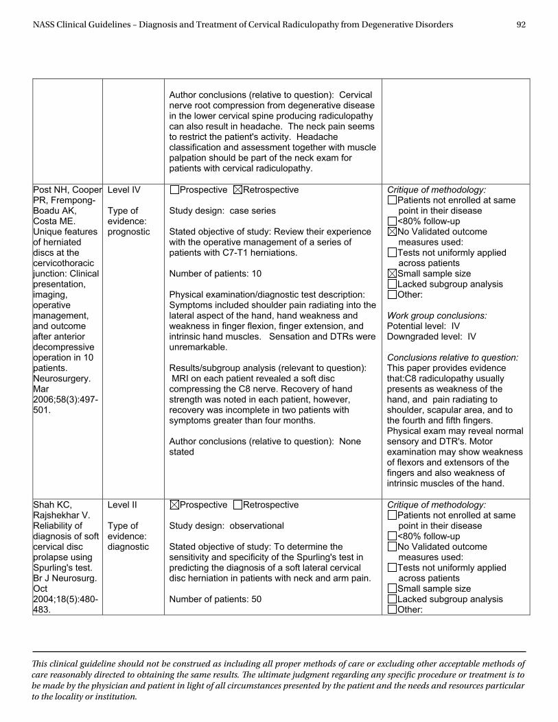
NASS Clinical Guidelines – Diagnosis and Treatment of Cervical Radiculopathy from Degenerative Disorders 92
This clinical guideline should not be construed as including all proper methods of care or excluding other acceptable methods of care reasonably directed to obtaining the same results. The ultimate judgment regarding any specific procedure or treatment is to be made by the physician and patient in light of all circumstances presented by the patient and the needs and resources particular to the locality or institution.
Author conclusions (relative to question): Cervical nerve root compression from degenerative disease in the lower cervical spine producing radiculopathy can also result in headache. The neck pain seems to restrict the patient's activity. Headache classification and assessment together with muscle palpation should be part of the neck exam for patients with cervical radiculopathy.
Post NH, Cooper PR, Frempong-Boadu AK, Costa ME. Unique features of herniated discs at the cervicothoracic junction: Clinical presentation, imaging,operativemanagement, and outcome after anterior decompressive operation in 10 patients.Neurosurgery. Mar2006;58(3):497-501.
Level IV
Type of evidence: prognostic
Prospective Retrospective
Study design: case series
Stated objective of study: Review their experience with the operative management of a series of patients with C7-T1 herniations.
Number of patients: 10
Physical examination/diagnostic test description: Symptoms included shoulder pain radiating into the lateral aspect of the hand, hand weakness and weakness in finger flexion, finger extension, and intrinsic hand muscles. Sensation and DTRs were unremarkable.
Results/subgroup analysis (relevant to question): MRI on each patient revealed a soft disc compressing the C8 nerve. Recovery of hand strength was noted in each patient, however, recovery was incomplete in two patients with symptoms greater than four months.
Author conclusions (relative to question): None stated
Critique of methodology: Patients not enrolled at same point in their disease <80% follow-up No Validated outcome measures used: Tests not uniformly applied across patients Small sample size Lacked subgroup analysis Other:
Work group conclusions: Potential level: IV Downgraded level: IV
Conclusions relative to question: This paper provides evidence that:C8 radiculopathy usually presents as weakness of the hand, and pain radiating to shoulder, scapular area, and to the fourth and fifth fingers. Physical exam may reveal normal sensory and DTR's. Motor examination may show weakness of flexors and extensors of the fingers and also weakness of intrinsic muscles of the hand.
Shah KC, Rajshekhar V. Reliability of diagnosis of soft cervical disc prolapse using Spurling's test. Br J Neurosurg. Oct2004;18(5):480-483.
Level II
Type of evidence: diagnostic
Prospective Retrospective
Study design: observational
Stated objective of study: To determine the sensitivity and specificity of the Spurling's test in predicting the diagnosis of a soft lateral cervical disc herniation in patients with neck and arm pain.
Number of patients: 50
Critique of methodology: Patients not enrolled at same point in their disease <80% follow-up No Validated outcome measures used: Tests not uniformly applied across patients Small sample size Lacked subgroup analysis Other:
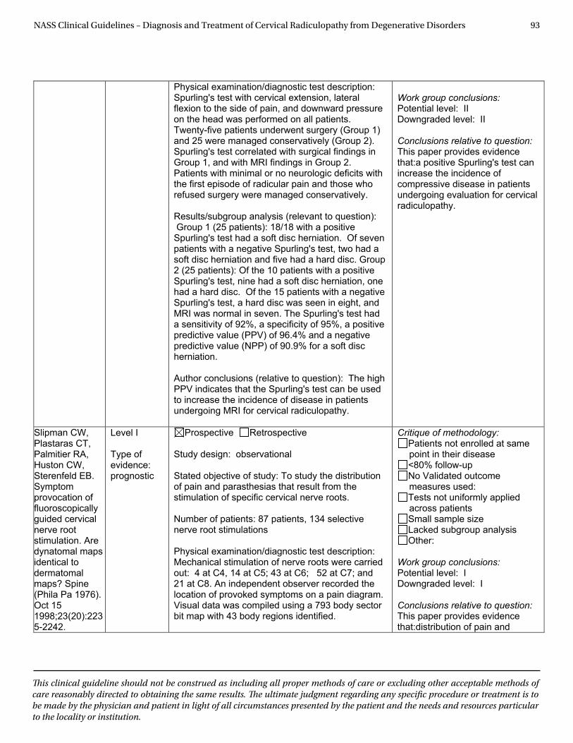
NASS Clinical Guidelines – Diagnosis and Treatment of Cervical Radiculopathy from Degenerative Disorders 93
This clinical guideline should not be construed as including all proper methods of care or excluding other acceptable methods of care reasonably directed to obtaining the same results. The ultimate judgment regarding any specific procedure or treatment is to be made by the physician and patient in light of all circumstances presented by the patient and the needs and resources particular to the locality or institution.
Physical examination/diagnostic test description: Spurling's test with cervical extension, lateral flexion to the side of pain, and downward pressure on the head was performed on all patients. Twenty-five patients underwent surgery (Group 1) and 25 were managed conservatively (Group 2). Spurling's test correlated with surgical findings in Group 1, and with MRI findings in Group 2. Patients with minimal or no neurologic deficits with the first episode of radicular pain and those who refused surgery were managed conservatively.
Results/subgroup analysis (relevant to question): Group 1 (25 patients): 18/18 with a positive Spurling's test had a soft disc herniation. Of seven patients with a negative Spurling's test, two had a soft disc herniation and five had a hard disc. Group 2 (25 patients): Of the 10 patients with a positive Spurling's test, nine had a soft disc herniation, one had a hard disc. Of the 15 patients with a negative Spurling's test, a hard disc was seen in eight, and MRI was normal in seven. The Spurling's test had a sensitivity of 92%, a specificity of 95%, a positive predictive value (PPV) of 96.4% and a negative predictive value (NPP) of 90.9% for a soft disc herniation.
Author conclusions (relative to question): The high PPV indicates that the Spurling's test can be used to increase the incidence of disease in patients undergoing MRI for cervical radiculopathy.
Work group conclusions: Potential level: II Downgraded level: II
Conclusions relative to question: This paper provides evidence that:a positive Spurling's test can increase the incidence of compressive disease in patients undergoing evaluation for cervical radiculopathy.
Slipman CW, Plastaras CT, Palmitier RA, Huston CW, Sterenfeld EB. Symptomprovocation of fluoroscopically guided cervical nerve root stimulation. Are dynatomal maps identical to dermatomal maps? Spine (Phila Pa 1976). Oct 15 1998;23(20):2235-2242.
Level I
Type of evidence: prognostic
Prospective Retrospective
Study design: observational
Stated objective of study: To study the distribution of pain and parasthesias that result from the stimulation of specific cervical nerve roots.
Number of patients: 87 patients, 134 selective nerve root stimulations
Physical examination/diagnostic test description: Mechanical stimulation of nerve roots were carried out: 4 at C4, 14 at C5; 43 at C6; 52 at C7; and 21 at C8. An independent observer recorded the location of provoked symptoms on a pain diagram. Visual data was compiled using a 793 body sector bit map with 43 body regions identified.
Critique of methodology: Patients not enrolled at same point in their disease <80% follow-up No Validated outcome measures used: Tests not uniformly applied across patients Small sample size Lacked subgroup analysis Other:
Work group conclusions: Potential level: I Downgraded level: I
Conclusions relative to question: This paper provides evidence that:distribution of pain and
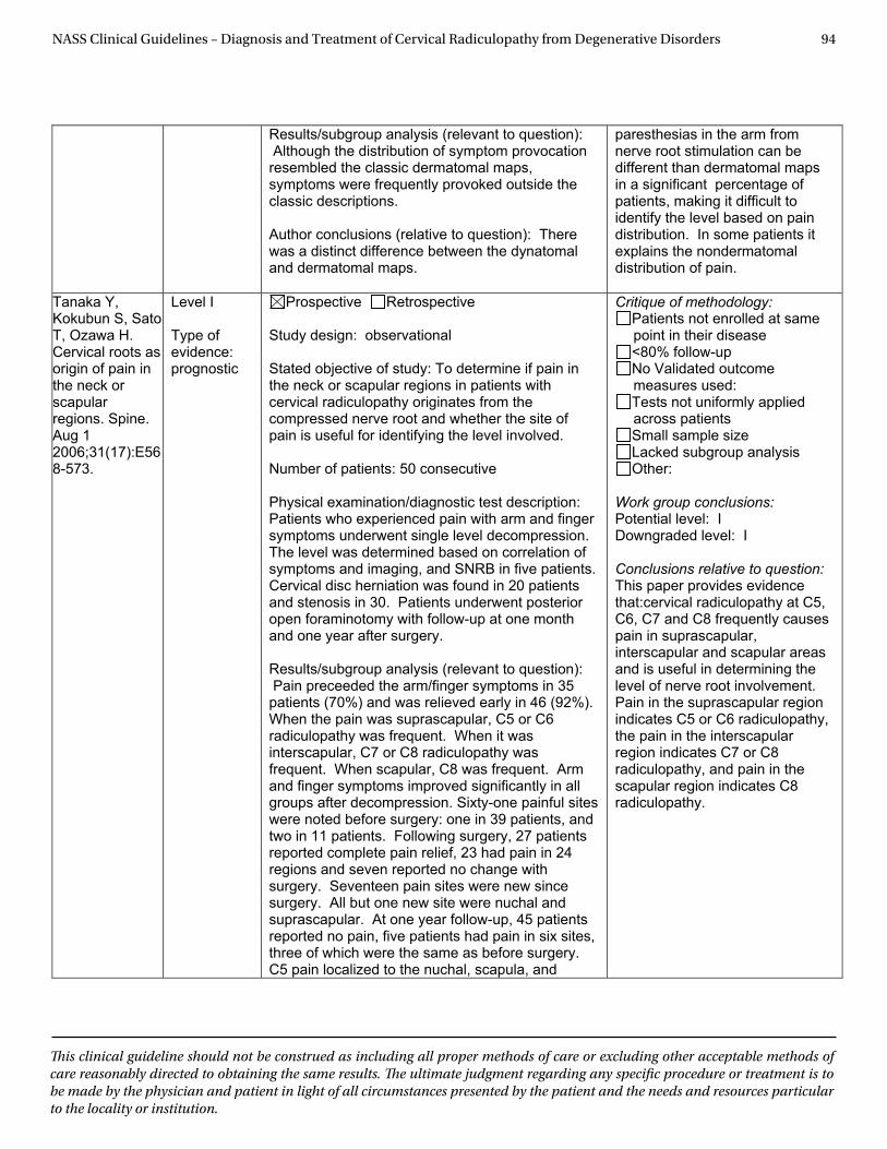
NASS Clinical Guidelines – Diagnosis and Treatment of Cervical Radiculopathy from Degenerative Disorders 94
This clinical guideline should not be construed as including all proper methods of care or excluding other acceptable methods of care reasonably directed to obtaining the same results. The ultimate judgment regarding any specific procedure or treatment is to be made by the physician and patient in light of all circumstances presented by the patient and the needs and resources particular to the locality or institution.
Results/subgroup analysis (relevant to question): Although the distribution of symptom provocation resembled the classic dermatomal maps, symptoms were frequently provoked outside the classic descriptions.
Author conclusions (relative to question): There was a distinct difference between the dynatomal and dermatomal maps.
paresthesias in the arm from nerve root stimulation can be different than dermatomal maps in a significant percentage of patients, making it difficult to identify the level based on pain distribution. In some patients it explains the nondermatomal distribution of pain.
Tanaka Y, Kokubun S, Sato T, Ozawa H. Cervical roots as origin of pain in the neck or scapular regions. Spine. Aug 1 2006;31(17):E568-573.
Level I
Type of evidence: prognostic
Prospective Retrospective
Study design: observational
Stated objective of study: To determine if pain in the neck or scapular regions in patients with cervical radiculopathy originates from the compressed nerve root and whether the site of pain is useful for identifying the level involved.
Number of patients: 50 consecutive
Physical examination/diagnostic test description: Patients who experienced pain with arm and finger symptoms underwent single level decompression. The level was determined based on correlation of symptoms and imaging, and SNRB in five patients. Cervical disc herniation was found in 20 patients and stenosis in 30. Patients underwent posterior open foraminotomy with follow-up at one month and one year after surgery.
Results/subgroup analysis (relevant to question): Pain preceeded the arm/finger symptoms in 35 patients (70%) and was relieved early in 46 (92%). When the pain was suprascapular, C5 or C6 radiculopathy was frequent. When it was interscapular, C7 or C8 radiculopathy was frequent. When scapular, C8 was frequent. Arm and finger symptoms improved significantly in all groups after decompression. Sixty-one painful sites were noted before surgery: one in 39 patients, and two in 11 patients. Following surgery, 27 patients reported complete pain relief, 23 had pain in 24 regions and seven reported no change with surgery. Seventeen pain sites were new since surgery. All but one new site were nuchal and suprascapular. At one year follow-up, 45 patients reported no pain, five patients had pain in six sites, three of which were the same as before surgery. C5 pain localized to the nuchal, scapula, and
Critique of methodology: Patients not enrolled at same point in their disease <80% follow-up No Validated outcome measures used: Tests not uniformly applied across patients Small sample size Lacked subgroup analysis Other:
Work group conclusions: Potential level: I Downgraded level: I
Conclusions relative to question: This paper provides evidence that:cervical radiculopathy at C5, C6, C7 and C8 frequently causes pain in suprascapular, interscapular and scapular areas and is useful in determining the level of nerve root involvement. Pain in the suprascapular region indicates C5 or C6 radiculopathy, the pain in the interscapular region indicates C7 or C8 radiculopathy, and pain in the scapular region indicates C8 radiculopathy.
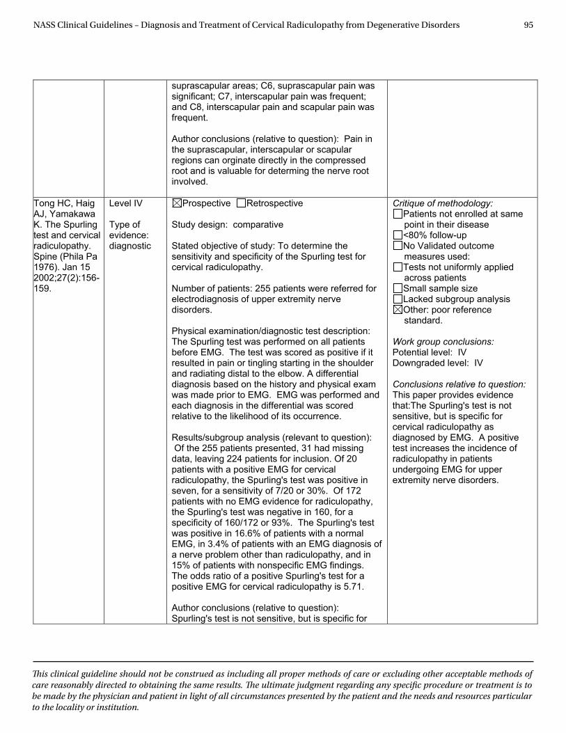
NASS Clinical Guidelines – Diagnosis and Treatment of Cervical Radiculopathy from Degenerative Disorders 95
This clinical guideline should not be construed as including all proper methods of care or excluding other acceptable methods of care reasonably directed to obtaining the same results. The ultimate judgment regarding any specific procedure or treatment is to be made by the physician and patient in light of all circumstances presented by the patient and the needs and resources particular to the locality or institution.
suprascapular areas; C6, suprascapular pain was significant; C7, interscapular pain was frequent; and C8, interscapular pain and scapular pain was frequent.
Author conclusions (relative to question): Pain in the suprascapular, interscapular or scapular regions can orginate directly in the compressed root and is valuable for determing the nerve root involved.
Tong HC, Haig AJ, Yamakawa K. The Spurling test and cervical radiculopathy. Spine (Phila Pa 1976). Jan 15 2002;27(2):156-159.
Level IV
Type of evidence: diagnostic
Prospective Retrospective
Study design: comparative
Stated objective of study: To determine the sensitivity and specificity of the Spurling test for cervical radiculopathy.
Number of patients: 255 patients were referred for electrodiagnosis of upper extremity nerve disorders.
Physical examination/diagnostic test description: The Spurling test was performed on all patients before EMG. The test was scored as positive if it resulted in pain or tingling starting in the shoulder and radiating distal to the elbow. A differential diagnosis based on the history and physical exam was made prior to EMG. EMG was performed and each diagnosis in the differential was scored relative to the likelihood of its occurrence.
Results/subgroup analysis (relevant to question): Of the 255 patients presented, 31 had missing data, leaving 224 patients for inclusion. Of 20 patients with a positive EMG for cervical radiculopathy, the Spurling's test was positive in seven, for a sensitivity of 7/20 or 30%. Of 172 patients with no EMG evidence for radiculopathy, the Spurling's test was negative in 160, for a specificity of 160/172 or 93%. The Spurling's test was positive in 16.6% of patients with a normal EMG, in 3.4% of patients with an EMG diagnosis of a nerve problem other than radiculopathy, and in 15% of patients with nonspecific EMG findings. The odds ratio of a positive Spurling's test for a positive EMG for cervical radiculopathy is 5.71.
Author conclusions (relative to question): Spurling's test is not sensitive, but is specific for
Critique of methodology: Patients not enrolled at same point in their disease <80% follow-up No Validated outcome measures used: Tests not uniformly applied across patients Small sample size Lacked subgroup analysis Other: poor reference standard.
Work group conclusions: Potential level: IV Downgraded level: IV
Conclusions relative to question: This paper provides evidence that:The Spurling's test is not sensitive, but is specific for cervical radiculopathy as diagnosed by EMG. A positive test increases the incidence of radiculopathy in patients undergoing EMG for upper extremity nerve disorders.
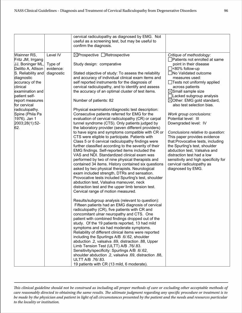
NASS Clinical Guidelines – Diagnosis and Treatment of Cervical Radiculopathy from Degenerative Disorders 96
This clinical guideline should not be construed as including all proper methods of care or excluding other acceptable methods of care reasonably directed to obtaining the same results. The ultimate judgment regarding any specific procedure or treatment is to be made by the physician and patient in light of all circumstances presented by the patient and the needs and resources particular to the locality or institution.
cervical radiculopathy as diagnosed by EMG. Not useful as a screening test, but may be useful to confirm the diagnosis.
Wainner RS, Fritz JM, Irrgang JJ, Boninger ML, Delitto A, Allison S. Reliability and diagnostic accuracy of the clinical examination and patient self-report measures for cervical radiculopathy. Spine (Phila Pa 1976). Jan 1 2003;28(1):52-62.
Level IV
Type of evidence: diagnostic
Prospective Retrospective
Study design: comparative
Stated objective of study: To assess the reliability and accuracy of individual clinical exam items and self reported instruments for the diagnosis of cervical radiculopathy, and to identify and assess the accuracy of an optimal cluster of test items.
Number of patients: 82
Physical examination/diagnostic test description: Consecutive patients referred for EMG for the evaluation of cervical radiculopathy (CR) or carpal tunnel syndrome (CTS). Only patients judged by the laboratory provider (seven different providers) to have signs and symptoms compatible with CR or CTS were eligible to participate. Patients with Class 5 or 6 cervical radiculopathy findings were further classified according to the severity of their EMG findings. Self-reported items included the VAS and NDI. Standardized clinical exam was performed by two of nine physical therapists and contained 34 items. History contained six questions asked by two physical therapists. Neurological exam included strength, DTRs and sensation. Provocative tests included Spurling's test, shoulder abduction test, Valsalva maneuver, neck distraction test and the upper limb tension test. Cervical range of motion measured.
Results/subgroup analysis (relevant to question): Fifteen patients had an EMG diagnosis of cervical radiculopathy (CR), five patients with CR and concomitant ulnar neuropathy and CTS. One patient with combined findings dropped out of the study. Of the 19 patients reported, 13 had mild symptoms and six had moderate symptoms. Reliability of different clinical items were reported including the Spurlings A/B .6/.62, shoulder abduction .2, valsalva .69, distraction .88, Upper Limb Tension Test (ULTT) A/B .76/.83. Sensitivity/specificity: Spurlings A/B .6/.62, shoulder abduction .2, valsalva .69, distraction .88, ULTT A/B .76/.83. 19 patients with CR (13 mild, 6 moderate).
Critique of methodology: Patients not enrolled at same point in their disease <80% follow-up No Validated outcome measures used: Tests not uniformly applied across patients Small sample size Lacked subgroup analysis Other: EMG gold standard, also test selection bias.
Work group conclusions: Potential level: III Downgraded level: IV
Conclusions relative to question: This paper provides evidence that:Provocative tests, including the Spurling's test, shoulder abduction test, Valsalva and distraction test had a low sensitivity and high specificity for cervical radiculopathy as diagnosed by EMG.
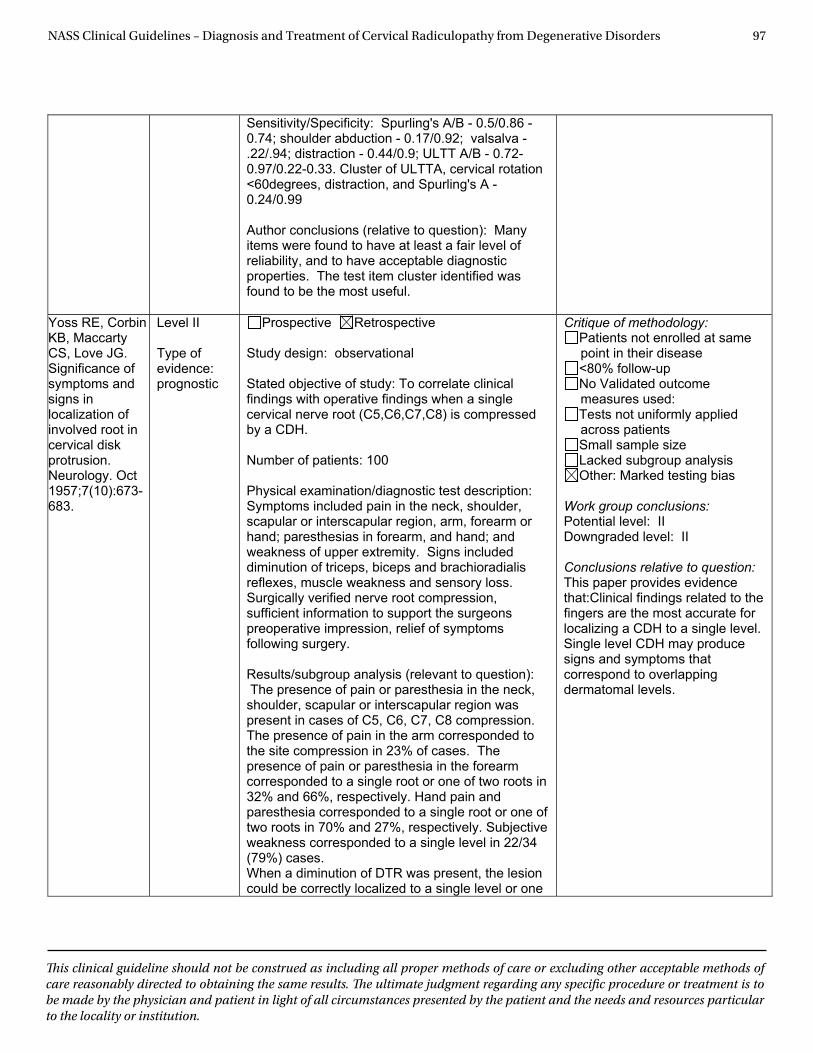
NASS Clinical Guidelines – Diagnosis and Treatment of Cervical Radiculopathy from Degenerative Disorders 97
This clinical guideline should not be construed as including all proper methods of care or excluding other acceptable methods of care reasonably directed to obtaining the same results. The ultimate judgment regarding any specific procedure or treatment is to be made by the physician and patient in light of all circumstances presented by the patient and the needs and resources particular to the locality or institution.
Sensitivity/Specificity: Spurling's A/B - 0.5/0.86 - 0.74; shoulder abduction - 0.17/0.92; valsalva - .22/.94; distraction - 0.44/0.9; ULTT A/B - 0.72-0.97/0.22-0.33. Cluster of ULTTA, cervical rotation <60degrees, distraction, and Spurling's A - 0.24/0.99
Author conclusions (relative to question): Many items were found to have at least a fair level of reliability, and to have acceptable diagnostic properties. The test item cluster identified was found to be the most useful.
Yoss RE, Corbin KB, Maccarty CS, Love JG. Significance of symptoms and signs in localization of involved root in cervical disk protrusion. Neurology. Oct 1957;7(10):673-683.
Level II
Type of evidence: prognostic
Prospective Retrospective
Study design: observational
Stated objective of study: To correlate clinical findings with operative findings when a single cervical nerve root (C5,C6,C7,C8) is compressed by a CDH.
Number of patients: 100
Physical examination/diagnostic test description: Symptoms included pain in the neck, shoulder, scapular or interscapular region, arm, forearm or hand; paresthesias in forearm, and hand; and weakness of upper extremity. Signs included diminution of triceps, biceps and brachioradialis reflexes, muscle weakness and sensory loss. Surgically verified nerve root compression, sufficient information to support the surgeons preoperative impression, relief of symptoms following surgery.
Results/subgroup analysis (relevant to question): The presence of pain or paresthesia in the neck, shoulder, scapular or interscapular region was present in cases of C5, C6, C7, C8 compression. The presence of pain in the arm corresponded to the site compression in 23% of cases. The presence of pain or paresthesia in the forearm corresponded to a single root or one of two roots in 32% and 66%, respectively. Hand pain and paresthesia corresponded to a single root or one of two roots in 70% and 27%, respectively. Subjective weakness corresponded to a single level in 22/34 (79%) cases. When a diminution of DTR was present, the lesion could be correctly localized to a single level or one
Critique of methodology: Patients not enrolled at same point in their disease <80% follow-up No Validated outcome measures used: Tests not uniformly applied across patients Small sample size Lacked subgroup analysis Other: Marked testing bias
Work group conclusions: Potential level: II Downgraded level: II
Conclusions relative to question: This paper provides evidence that:Clinical findings related to the fingers are the most accurate for localizing a CDH to a single level. Single level CDH may produce signs and symptoms that correspond to overlapping dermatomal levels.
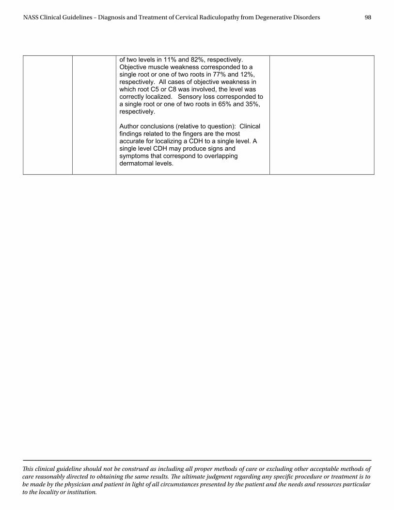
NASS Clinical Guidelines – Diagnosis and Treatment of Cervical Radiculopathy from Degenerative Disorders 98
This clinical guideline should not be construed as including all proper methods of care or excluding other acceptable methods of care reasonably directed to obtaining the same results. The ultimate judgment regarding any specific procedure or treatment is to be made by the physician and patient in light of all circumstances presented by the patient and the needs and resources particular to the locality or institution.
of two levels in 11% and 82%, respectively. Objective muscle weakness corresponded to a single root or one of two roots in 77% and 12%, respectively. All cases of objective weakness in which root C5 or C8 was involved, the level was correctly localized. Sensory loss corresponded to a single root or one of two roots in 65% and 35%, respectively.
Author conclusions (relative to question): Clinical findings related to the fingers are the most accurate for localizing a CDH to a single level. A single level CDH may produce signs and symptoms that correspond to overlapping dermatomal levels.
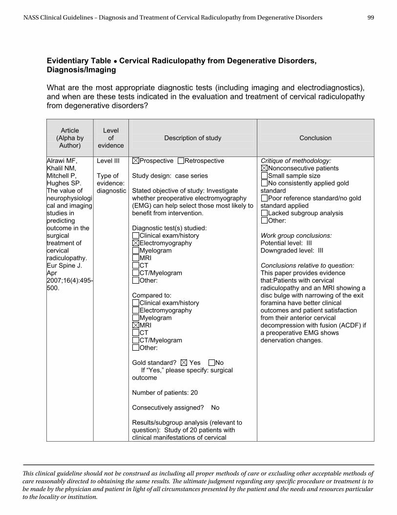
NASS Clinical Guidelines – Diagnosis and Treatment of Cervical Radiculopathy from Degenerative Disorders 99
This clinical guideline should not be construed as including all proper methods of care or excluding other acceptable methods of care reasonably directed to obtaining the same results. The ultimate judgment regarding any specific procedure or treatment is to be made by the physician and patient in light of all circumstances presented by the patient and the needs and resources particular to the locality or institution.
Evidentiary Table ● Cervical Radiculopathy from Degenerative Disorders, Diagnosis/Imaging What are the most appropriate diagnostic tests (including imaging and electrodiagnostics), and when are these tests indicated in the evaluation and treatment of cervical radiculopathy from degenerative disorders?
Article
(Alpha by Author)
Level
of evidence
Description of study
Conclusion
Alrawi MF, Khalil NM, Mitchell P, Hughes SP. The value of neurophysiological and imaging studies in predicting outcome in the surgical treatment of cervical radiculopathy. Eur Spine J. Apr 2007;16(4):495-500.
Level III Type of evidence: diagnostic
Prospective Retrospective Study design: case series Stated objective of study: Investigate whether preoperative electromyography (EMG) can help select those most likely to benefit from intervention. Diagnostic test(s) studied:
Clinical exam/history Electromyography Myelogram MRI CT CT/Myelogram Other:
Compared to:
Clinical exam/history Electromyography Myelogram MRI CT CT/Myelogram Other:
Gold standard? Yes No If “Yes,” please specify: surgical outcome Number of patients: 20 Consecutively assigned? No Results/subgroup analysis (relevant to question): Study of 20 patients with clinical manifestations of cervical
Critique of methodology: Nonconsecutive patients Small sample size No consistently applied gold
standard Poor reference standard/no gold
standard applied Lacked subgroup analysis Other:
Work group conclusions: Potential level: III Downgraded level: III
Conclusions relative to question: This paper provides evidence that:Patients with cervical radiculopathy and an MRI showing a disc bulge with narrowing of the exit foramina have better clinical outcomes and patient satisfaction from their anterior cervical decompression with fusion (ACDF) if a preoperative EMG shows denervation changes.
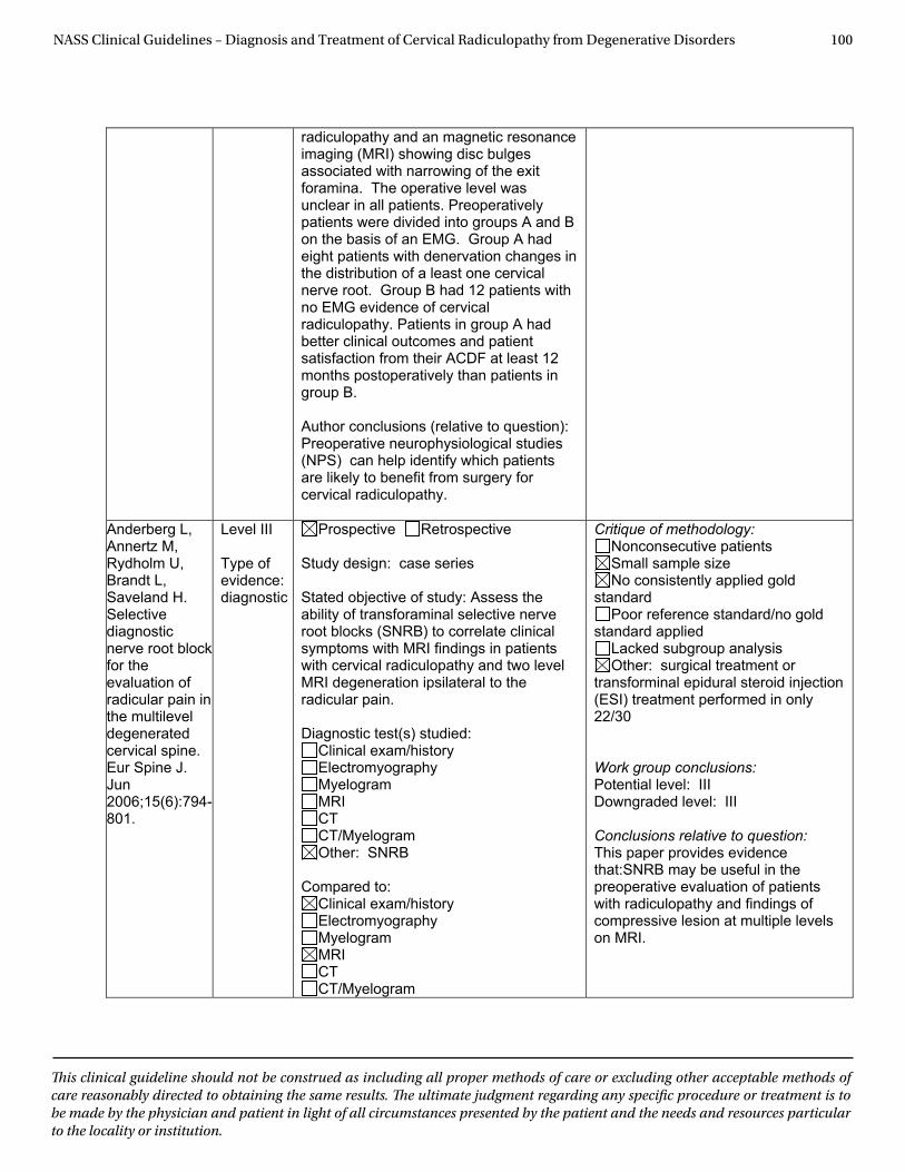
NASS Clinical Guidelines – Diagnosis and Treatment of Cervical Radiculopathy from Degenerative Disorders 100
This clinical guideline should not be construed as including all proper methods of care or excluding other acceptable methods of care reasonably directed to obtaining the same results. The ultimate judgment regarding any specific procedure or treatment is to be made by the physician and patient in light of all circumstances presented by the patient and the needs and resources particular to the locality or institution.
radiculopathy and an magnetic resonance imaging (MRI) showing disc bulges associated with narrowing of the exit foramina. The operative level was unclear in all patients. Preoperatively patients were divided into groups A and B on the basis of an EMG. Group A had eight patients with denervation changes in the distribution of a least one cervical nerve root. Group B had 12 patients with no EMG evidence of cervical radiculopathy. Patients in group A had better clinical outcomes and patient satisfaction from their ACDF at least 12 months postoperatively than patients in group B.
Author conclusions (relative to question): Preoperative neurophysiological studies (NPS) can help identify which patients are likely to benefit from surgery for cervical radiculopathy.
Anderberg L, Annertz M, Rydholm U, Brandt L, Saveland H. Selectivediagnostic nerve root block for the evaluation of radicular pain in the multilevel degenerated cervical spine. Eur Spine J. Jun2006;15(6):794-801.
Level III
Type of evidence: diagnostic
Prospective Retrospective
Study design: case series
Stated objective of study: Assess the ability of transforaminal selective nerve root blocks (SNRB) to correlate clinical symptoms with MRI findings in patients with cervical radiculopathy and two level MRI degeneration ipsilateral to the radicular pain.
Diagnostic test(s) studied: Clinical exam/history Electromyography MyelogramMRICTCT/Myelogram Other: SNRB
Compared to: Clinical exam/history Electromyography MyelogramMRICTCT/Myelogram
Critique of methodology: Nonconsecutive patients Small sample size No consistently applied gold
standard Poor reference standard/no gold
standard applied Lacked subgroup analysis Other: surgical treatment or
transforminal epidural steroid injection (ESI) treatment performed in only 22/30
Work group conclusions: Potential level: III Downgraded level: III
Conclusions relative to question: This paper provides evidence that:SNRB may be useful in the preoperative evaluation of patients with radiculopathy and findings of compressive lesion at multiple levels on MRI.
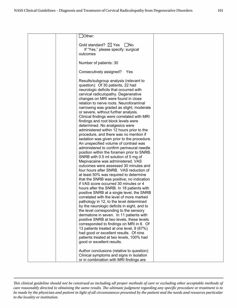
NASS Clinical Guidelines – Diagnosis and Treatment of Cervical Radiculopathy from Degenerative Disorders 101
This clinical guideline should not be construed as including all proper methods of care or excluding other acceptable methods of care reasonably directed to obtaining the same results. The ultimate judgment regarding any specific procedure or treatment is to be made by the physician and patient in light of all circumstances presented by the patient and the needs and resources particular to the locality or institution.
Other:
Gold standard? Yes No If “Yes,” please specify: surgical outcomes Number of patients: 30
Consecutively assigned? Yes
Results/subgroup analysis (relevant to question): Of 30 patients, 22 had neurologic deficits that occurred with cervical radiculopathy. Degenerative changes on MRI were found in close relation to nerve roots. Neuroforaminal narrowing was graded as slight, moderate or severe, without further analysis. Clinical findings were correlated with MRI findings and root block levels were determined. No analgesics were administered within 12 hours prior to the procedure, and there was no mention if sedation was given prior to the procedure. An unspecified volume of contrast was administered to confirm perineural needle position within the foramen prior to SNRB. SNRB with 0.5 ml solution of 5 mg of Mepivacaine was administered. VAS outcomes were assessed 30 minutes and four hours after SNRB. VAS reduction of at least 50% was required to determine that the SNRB was positive; no indication if VAS score occurred 30 minutes or 4 hours after the SNRB. In 18 patients with positive SNRB at a single level, the SNRB correlated with the level of more marked pathology in 12, to the level determined by the neurologic deficits in eight, and to the level corresponding to the sensory dermatone in seven. In 11 patients with positive SNRB at two levels, these levels corresponded to findings on MRI in 6. Of 13 patients treated at one level, 9 (67%) had good or excellent results. Of nine patients treated at two levels, 100% had good or excellent results.
Author conclusions (relative to question): Clinical symptoms and signs in isolation or in combination with MRI findings are
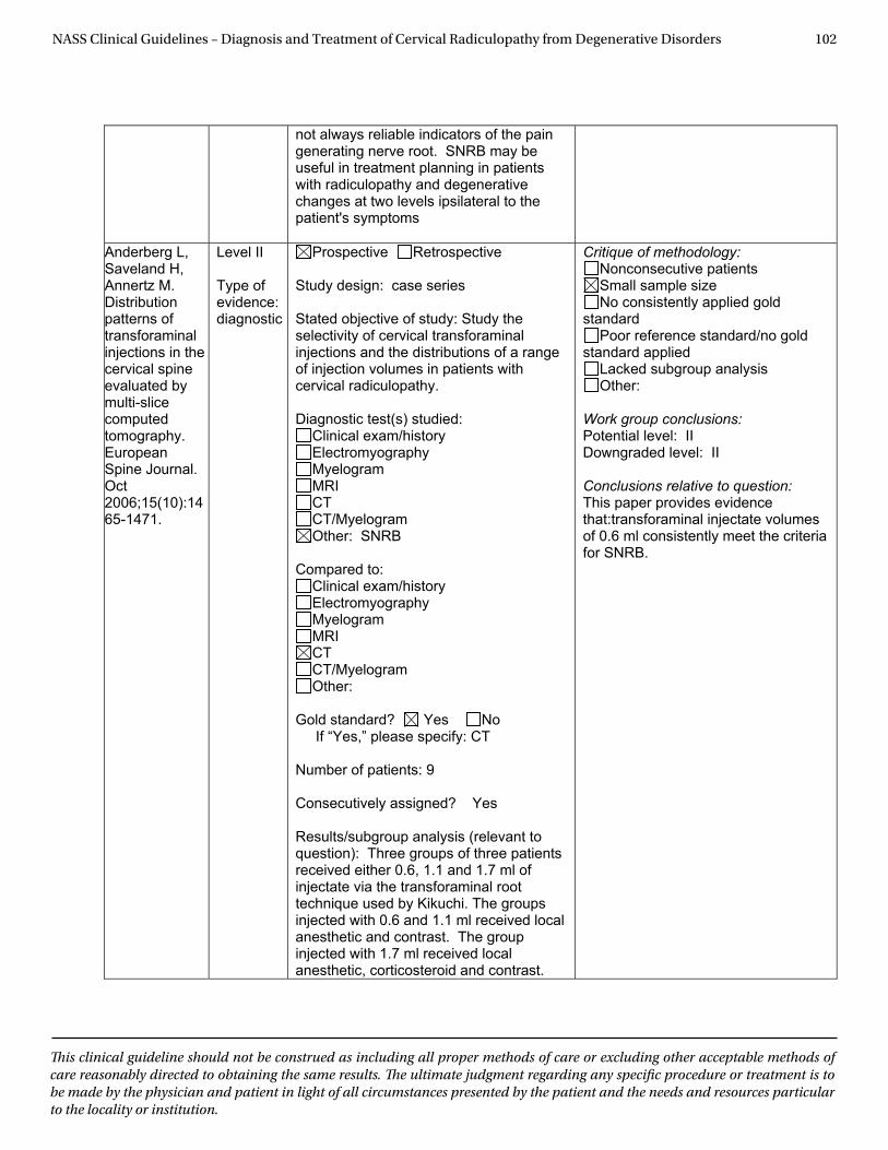
NASS Clinical Guidelines – Diagnosis and Treatment of Cervical Radiculopathy from Degenerative Disorders 102
This clinical guideline should not be construed as including all proper methods of care or excluding other acceptable methods of care reasonably directed to obtaining the same results. The ultimate judgment regarding any specific procedure or treatment is to be made by the physician and patient in light of all circumstances presented by the patient and the needs and resources particular to the locality or institution.
not always reliable indicators of the pain generating nerve root. SNRB may be useful in treatment planning in patients with radiculopathy and degenerative changes at two levels ipsilateral to the patient's symptoms
Anderberg L, Saveland H, Annertz M. Distribution patterns of transforaminal injections in the cervical spine evaluated by multi-slice computed tomography.European Spine Journal. Oct2006;15(10):1465-1471.
Level II
Type of evidence: diagnostic
Prospective Retrospective
Study design: case series
Stated objective of study: Study the selectivity of cervical transforaminal injections and the distributions of a range of injection volumes in patients with cervical radiculopathy.
Diagnostic test(s) studied: Clinical exam/history Electromyography MyelogramMRICTCT/Myelogram Other: SNRB
Compared to: Clinical exam/history Electromyography MyelogramMRICTCT/Myelogram Other:
Gold standard? Yes No If “Yes,” please specify: CT Number of patients: 9
Consecutively assigned? Yes
Results/subgroup analysis (relevant to question): Three groups of three patients received either 0.6, 1.1 and 1.7 ml of injectate via the transforaminal root technique used by Kikuchi. The groups injected with 0.6 and 1.1 ml received local anesthetic and contrast. The group injected with 1.7 ml received local anesthetic, corticosteroid and contrast.
Critique of methodology: Nonconsecutive patients Small sample size No consistently applied gold
standard Poor reference standard/no gold
standard applied Lacked subgroup analysis Other:
Work group conclusions: Potential level: II Downgraded level: II
Conclusions relative to question: This paper provides evidence that:transforaminal injectate volumes of 0.6 ml consistently meet the criteria for SNRB.
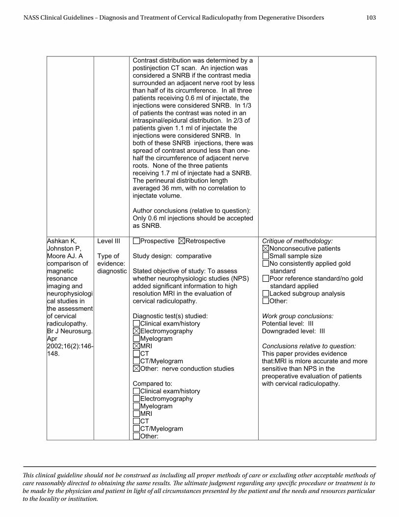
NASS Clinical Guidelines – Diagnosis and Treatment of Cervical Radiculopathy from Degenerative Disorders 103
This clinical guideline should not be construed as including all proper methods of care or excluding other acceptable methods of care reasonably directed to obtaining the same results. The ultimate judgment regarding any specific procedure or treatment is to be made by the physician and patient in light of all circumstances presented by the patient and the needs and resources particular to the locality or institution.
Contrast distribution was determined by a postinjection CT scan. An injection was considered a SNRB if the contrast media surrounded an adjacent nerve root by less than half of its circumference. In all three patients receiving 0.6 ml of injectate, the injections were considered SNRB. In 1/3 of patients the contrast was noted in an intraspinal/epidural distribution. In 2/3 of patients given 1.1 ml of injectate the injections were considered SNRB. In both of these SNRB injections, there was spread of contrast around less than one-half the circumference of adjacent nerve roots. None of the three patients receiving 1.7 ml of injectate had a SNRB. The perineural distribution length averaged 36 mm, with no correlation to injectate volume.
Author conclusions (relative to question): Only 0.6 ml injections should be accepted as SNRB.
Ashkan K, Johnston P, Moore AJ. A comparison of magnetic resonance imaging and neurophysiological studies in the assessment of cervical radiculopathy. Br J Neurosurg. Apr2002;16(2):146-148.
Level III
Type of evidence: diagnostic
Prospective Retrospective
Study design: comparative
Stated objective of study: To assess whether neurophysiologic studies (NPS) added significant information to high resolution MRI in the evaluation of cervical radiculopathy.
Diagnostic test(s) studied: Clinical exam/history Electromyography MyelogramMRICTCT/Myelogram Other: nerve conduction studies
Compared to: Clinical exam/history Electromyography MyelogramMRICTCT/Myelogram Other:
Critique of methodology: Nonconsecutive patients Small sample size No consistently applied gold standard Poor reference standard/no gold standard applied Lacked subgroup analysis Other:
Work group conclusions: Potential level: III Downgraded level: III
Conclusions relative to question: This paper provides evidence that:MRI is mlore accurate and more sensitive than NPS in the preoperative evaluation of patients with cervical radiculopathy.
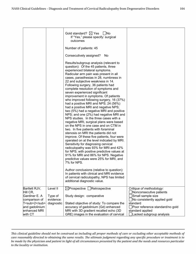
NASS Clinical Guidelines – Diagnosis and Treatment of Cervical Radiculopathy from Degenerative Disorders 104
This clinical guideline should not be construed as including all proper methods of care or excluding other acceptable methods of care reasonably directed to obtaining the same results. The ultimate judgment regarding any specific procedure or treatment is to be made by the physician and patient in light of all circumstances presented by the patient and the needs and resources particular to the locality or institution.
Gold standard? Yes No If “Yes,” please specify: surgical outcomes
Number of patients: 45
Consecutively assigned? No
Results/subgroup analysis (relevant to question): Of the 45 patients, three experienced bilateral symptoms. Radicular arm pain was present in all cases, parasthesias in 28, numbness in 22 and subjective weakness in 14. Following surgery, 36 patients had complete resolution of symptoms and seven experienced significant improvement in symptoms. Of patients who improved following surgery, 16 (37%) had a positive MRI and NPS; 24 (56%) had a positive MRI and negative NPS; two (5%) had a negative MRI and positive NPS; and one (2%) had negative MRI and NPS studies. In the three cases with a negative MRI, surgical plans were based on the NPS in one case and on CTM in two. In five patients with foraminal stenosis on MRI the patients did not improve. Of these five patients, four were operated on at the level indicated by MRI. Sensitivity for diagnosing cervical radiculopathy was 93% for MRI and 42% for NPS; with positive predictive values at 91% for MRI and 86% for NPS. Negative predictive values were 25% for MRI, and 7% for NPS.
Author conclusions (relative to question): In patients with clinical and MRI evidence of cervical radiculopathy, NPS has limited additional diagnostic value.
Bartlett RJV, Hill CR, Gardiner E. A comparison of T<sub>2</sub> and gadolinium enhanced MRI with CT
Level II
Type of evidence: diagnostic
Prospective Retrospective
Study design: comparative
Stated objective of study: To compare the accuracy of gadolinium (Gd) enhanced MRI with 3D gradient recalled echo (3D GRE) images in the evaluation of cervical
Critique of methodology: Nonconsecutive patients Small sample size No consistently applied gold
standard Poor reference standard/no gold
standard applied Lacked subgroup analysis

NASS Clinical Guidelines – Diagnosis and Treatment of Cervical Radiculopathy from Degenerative Disorders 105
This clinical guideline should not be construed as including all proper methods of care or excluding other acceptable methods of care reasonably directed to obtaining the same results. The ultimate judgment regarding any specific procedure or treatment is to be made by the physician and patient in light of all circumstances presented by the patient and the needs and resources particular to the locality or institution.
myelography in cervical radiculopathy. British Journal of Radiology. Jan1998;71(JAN.):11-19.
radiculopathy.
Diagnostic test(s) studied: Clinical exam/history Electromyography MyelogramMRICTCT/Myelogram Other:
Compared to: Clinical exam/history Electromyography MyelogramMRICTCT/Myelogram Other:
Gold standard? Yes No If “Yes,” please specify: best diagnosis reviewing all the studies Number of patients: 20
Consecutively assigned? Yes
Results/subgroup analysis (relevant to question): 3D GRE images had an accuracy of 87% for the diagnosis of foraminal encroachment. CTM had an accuracy of 90%. MRI with Gd conferred no additional benefit. Oblique reconstructions were less accurate than axial images.
Author conclusions (relative to question): MRI with 3D GRE images is an acceptable technique for the primary evaluation of cervical radiculopathy. CTM remains indicated for patients with incongruent symptoms and MRI results.
Other:
Work group conclusions: Potential level: II Downgraded level: II
Conclusions relative to question: This paper provides evidence that:MRI with 3D T2 technique has an accuracy approaching that of CT myelography for the diagnosis of a compressive lesion in patients with cervical radiculopathy.
Hedberg MC, Drayer BP, Flom RA, Hodak JA, Bird CR. Gradient echo (GRASS) MR imaging in
Level III
Type of evidence: diagnostic
Prospective Retrospective
Study design: comparative
Stated objective of study: To determine the accuracy of MRI with limited flip angle (LFA) GRE technique in patients with
Critique of methodology: Nonconsecutive patients Small sample size No consistently applied gold standard Poor reference standard/no gold standard applied
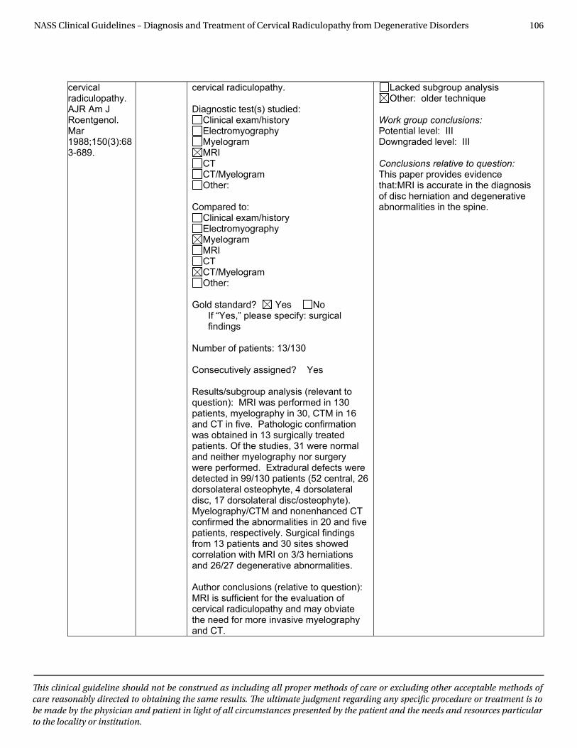
NASS Clinical Guidelines – Diagnosis and Treatment of Cervical Radiculopathy from Degenerative Disorders 106
This clinical guideline should not be construed as including all proper methods of care or excluding other acceptable methods of care reasonably directed to obtaining the same results. The ultimate judgment regarding any specific procedure or treatment is to be made by the physician and patient in light of all circumstances presented by the patient and the needs and resources particular to the locality or institution.
cervical radiculopathy. AJR Am J Roentgenol. Mar1988;150(3):683-689.
cervical radiculopathy.
Diagnostic test(s) studied: Clinical exam/history Electromyography MyelogramMRICTCT/Myelogram Other:
Compared to: Clinical exam/history Electromyography MyelogramMRICTCT/Myelogram Other:
Gold standard? Yes No If “Yes,” please specify: surgical findings
Number of patients: 13/130
Consecutively assigned? Yes
Results/subgroup analysis (relevant to question): MRI was performed in 130 patients, myelography in 30, CTM in 16 and CT in five. Pathologic confirmation was obtained in 13 surgically treated patients. Of the studies, 31 were normal and neither myelography nor surgery were performed. Extradural defects were detected in 99/130 patients (52 central, 26 dorsolateral osteophyte, 4 dorsolateral disc, 17 dorsolateral disc/osteophyte). Myelography/CTM and nonenhanced CT confirmed the abnormalities in 20 and five patients, respectively. Surgical findings from 13 patients and 30 sites showed correlation with MRI on 3/3 herniations and 26/27 degenerative abnormalities.
Author conclusions (relative to question): MRI is sufficient for the evaluation of cervical radiculopathy and may obviate the need for more invasive myelography and CT.
Lacked subgroup analysis Other: older technique
Work group conclusions: Potential level: III Downgraded level: III
Conclusions relative to question: This paper provides evidence that:MRI is accurate in the diagnosis of disc herniation and degenerative abnormalities in the spine.
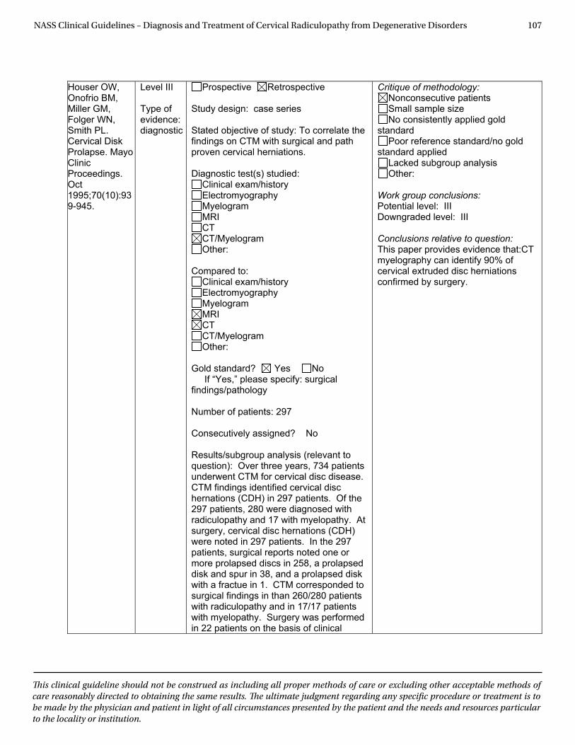
NASS Clinical Guidelines – Diagnosis and Treatment of Cervical Radiculopathy from Degenerative Disorders 107
This clinical guideline should not be construed as including all proper methods of care or excluding other acceptable methods of care reasonably directed to obtaining the same results. The ultimate judgment regarding any specific procedure or treatment is to be made by the physician and patient in light of all circumstances presented by the patient and the needs and resources particular to the locality or institution.
Houser OW, Onofrio BM, Miller GM, Folger WN, Smith PL. Cervical Disk Prolapse. Mayo Clinic Proceedings. Oct1995;70(10):939-945.
Level III
Type of evidence: diagnostic
Prospective Retrospective
Study design: case series
Stated objective of study: To correlate the findings on CTM with surgical and path proven cervical herniations.
Diagnostic test(s) studied: Clinical exam/history Electromyography MyelogramMRICTCT/Myelogram Other:
Compared to: Clinical exam/history Electromyography MyelogramMRICTCT/Myelogram Other:
Gold standard? Yes No If “Yes,” please specify: surgical findings/pathology Number of patients: 297
Consecutively assigned? No
Results/subgroup analysis (relevant to question): Over three years, 734 patients underwent CTM for cervical disc disease. CTM findings identified cervical disc hernations (CDH) in 297 patients. Of the 297 patients, 280 were diagnosed with radiculopathy and 17 with myelopathy. At surgery, cervical disc hernations (CDH) were noted in 297 patients. In the 297 patients, surgical reports noted one or more prolapsed discs in 258, a prolapsed disk and spur in 38, and a prolapsed disk with a fractue in 1. CTM corresponded to surgical findings in than 260/280 patients with radiculopathy and in 17/17 patients with myelopathy. Surgery was performed in 22 patients on the basis of clinical
Critique of methodology: Nonconsecutive patients Small sample size No consistently applied gold
standard Poor reference standard/no gold
standard applied Lacked subgroup analysis Other:
Work group conclusions: Potential level: III Downgraded level: III
Conclusions relative to question: This paper provides evidence that:CT myelography can identify 90% of cervical extruded disc herniations confirmed by surgery.
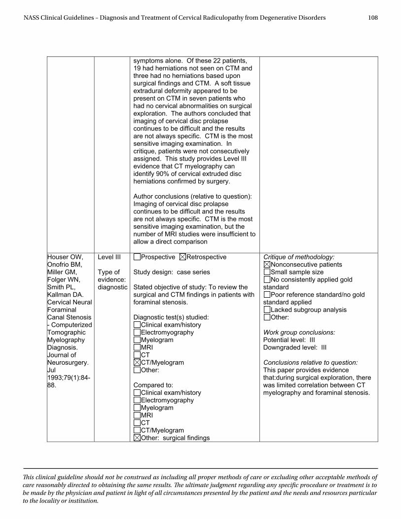
NASS Clinical Guidelines – Diagnosis and Treatment of Cervical Radiculopathy from Degenerative Disorders 108
This clinical guideline should not be construed as including all proper methods of care or excluding other acceptable methods of care reasonably directed to obtaining the same results. The ultimate judgment regarding any specific procedure or treatment is to be made by the physician and patient in light of all circumstances presented by the patient and the needs and resources particular to the locality or institution.
symptoms alone. Of these 22 patients, 19 had herniations not seen on CTM and three had no herniations based upon surgical findings and CTM. A soft tissue extradural deformity appeared to be present on CTM in seven patients who had no cervical abnormalities on surgical exploration. The authors concluded that imaging of cervical disc prolapse continues to be difficult and the results are not always specific. CTM is the most sensitive imaging examination. In critique, patients were not consecutively assigned. This study provides Level III evidence that CT myelography can identify 90% of cervical extruded disc herniations confirmed by surgery.
Author conclusions (relative to question): Imaging of cervical disc prolapse continues to be difficult and the results are not always specific. CTM is the most sensitive imaging examination, but the number of MRI studies were insufficient to allow a direct comparison
Houser OW, Onofrio BM, Miller GM, Folger WN, Smith PL, Kallman DA. Cervical Neural Foraminal Canal Stenosis - Computerized Tomographic MyelographyDiagnosis. Journal of Neurosurgery. Jul1993;79(1):84-88.
Level III
Type of evidence: diagnostic
Prospective Retrospective
Study design: case series
Stated objective of study: To review the surgical and CTM findings in patients with foraminal stenosis.
Diagnostic test(s) studied: Clinical exam/history Electromyography MyelogramMRICTCT/Myelogram Other:
Compared to: Clinical exam/history Electromyography MyelogramMRICTCT/Myelogram Other: surgical findings
Critique of methodology: Nonconsecutive patients Small sample size No consistently applied gold
standard Poor reference standard/no gold
standard applied Lacked subgroup analysis Other:
Work group conclusions: Potential level: III Downgraded level: III
Conclusions relative to question: This paper provides evidence that:during surgical exploration, there was limited correlation between CT myelography and foraminal stenosis.
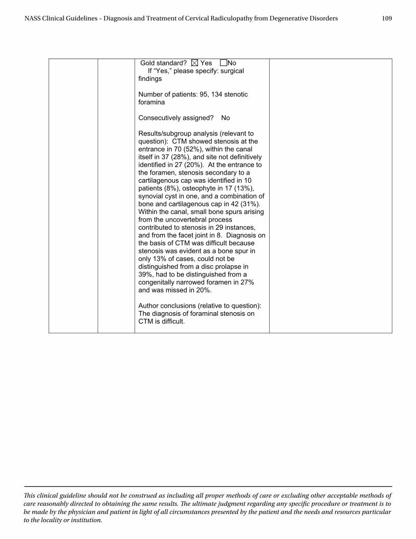
NASS Clinical Guidelines – Diagnosis and Treatment of Cervical Radiculopathy from Degenerative Disorders 109
This clinical guideline should not be construed as including all proper methods of care or excluding other acceptable methods of care reasonably directed to obtaining the same results. The ultimate judgment regarding any specific procedure or treatment is to be made by the physician and patient in light of all circumstances presented by the patient and the needs and resources particular to the locality or institution.
Gold standard? Yes No If “Yes,” please specify: surgical findings Number of patients: 95, 134 stenotic foramina
Consecutively assigned? No
Results/subgroup analysis (relevant to question): CTM showed stenosis at the entrance in 70 (52%), within the canal itself in 37 (28%), and site not definitively identified in 27 (20%). At the entrance to the foramen, stenosis secondary to a cartilagenous cap was identified in 10 patients (8%), osteophyte in 17 (13%), synovial cyst in one, and a combination of bone and cartilagenous cap in 42 (31%). Within the canal, small bone spurs arising from the uncovertebral process contributed to stenosis in 29 instances, and from the facet joint in 8. Diagnosis on the basis of CTM was difficult because stenosis was evident as a bone spur in only 13% of cases, could not be distinguished from a disc prolapse in 39%, had to be distinguished from a congenitally narrowed foramen in 27% and was missed in 20%.
Author conclusions (relative to question): The diagnosis of foraminal stenosis on CTM is difficult.
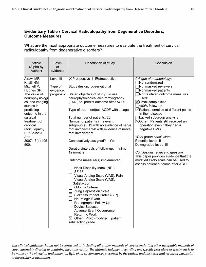
NASS Clinical Guidelines – Diagnosis and Treatment of Cervical Radiculopathy from Degenerative Disorders 110
This clinical guideline should not be construed as including all proper methods of care or excluding other acceptable methods of care reasonably directed to obtaining the same results. The ultimate judgment regarding any specific procedure or treatment is to be made by the physician and patient in light of all circumstances presented by the patient and the needs and resources particular to the locality or institution.
Evidentiary Table ● Cervical Radiculopathy from Degenerative Disorders, Outcome Measures
What are the most appropriate outcome measures to evaluate the treatment of cervical radiculopathy from degenerative disorders?
Article
(Alpha by Author)
Level
of evidence
Description of study
Conclusion
Alrawi MF, Khalil NM, Mitchell P, Hughes SP. The value of neurophysiological and imaging studies in predicting outcome in the surgical treatment of cervical radiculopathy. Eur Spine J. Apr 2007;16(4):495-500.
Level III Type of evidence: prognostic
Prospective Retrospective Study design: observational Stated objective of study: To use neurophysiological electromyography (EMG) to predict outcome after ACDF. Type of treatment(s): ACDF with a cage Total number of patients: 20 Number of patients in relevant subgroup(s): 12 with no evidence of nerve root involvement/8 with evidence of nerve root involvement Consecutively assigned? Yes Duration/intervals of follow-up: minimum 12 months Outcome measure(s) implemented
Neck Disability Index (NDI) SF-36 Visual Analog Scale (VAS), Pain Visual Analog Scale (VAS), Satisfaction Odom’s Criteria Zung Depression Scale Sickness Impact Profile (SIP) Neurologic Exam Radiographic Follow-Up Device Success Adverse Event Occurrence Return to Work Other: Prolo (modified), patient
satisfaction grade
Critique of methodology: Nonrandomized Nonmasked reviewers Nonmasked patients No Validated outcome measures used: Small sample size <80% follow-up Patients enrolled at different points in their disease Lacked subgroup analysis Other: Patients still received an operation even if they had a negative EMG.
Work group conclusions: Potential level: II Downgraded level: III Conclusions relative to question: This paper provides evidence that:the modified Prolo scale can be used to assess patient outcome after ACDF
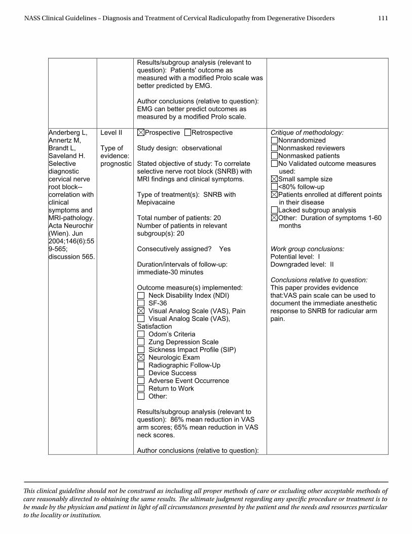
NASS Clinical Guidelines – Diagnosis and Treatment of Cervical Radiculopathy from Degenerative Disorders 111
This clinical guideline should not be construed as including all proper methods of care or excluding other acceptable methods of care reasonably directed to obtaining the same results. The ultimate judgment regarding any specific procedure or treatment is to be made by the physician and patient in light of all circumstances presented by the patient and the needs and resources particular to the locality or institution.
Results/subgroup analysis (relevant to question): Patients' outcome as measured with a modified Prolo scale was better predicted by EMG.
Author conclusions (relative to question): EMG can better predict outcomes as measured by a modified Prolo scale.
Anderberg L, Annertz M, Brandt L, Saveland H. Selectivediagnostic cervical nerve root block--correlation with clinical symptoms and MRI-pathology. Acta Neurochir (Wien). Jun 2004;146(6):559-565; discussion 565.
Level II
Type of evidence: prognostic
Prospective Retrospective
Study design: observational
Stated objective of study: To correlate selective nerve root block (SNRB) with MRI findings and clinical symptoms. Type of treatment(s): SNRB with Mepivacaine
Total number of patients: 20 Number of patients in relevant subgroup(s): 20
Consecutively assigned? Yes
Duration/intervals of follow-up: immediate-30 minutes
Outcome measure(s) implemented: Neck Disability Index (NDI) SF-36 Visual Analog Scale (VAS), Pain Visual Analog Scale (VAS),
Satisfaction Odom’s Criteria Zung Depression Scale Sickness Impact Profile (SIP) Neurologic Exam Radiographic Follow-Up Device Success Adverse Event Occurrence Return to Work Other:
Results/subgroup analysis (relevant to question): 86% mean reduction in VAS arm scores; 65% mean reduction in VAS neck scores.
Author conclusions (relative to question):
Critique of methodology: Nonrandomized Nonmasked reviewers Nonmasked patients No Validated outcome measures used: Small sample size <80% follow-up Patients enrolled at different points in their disease Lacked subgroup analysis Other: Duration of symptoms 1-60 months
Work group conclusions: Potential level: I Downgraded level: II
Conclusions relative to question: This paper provides evidence that:VAS pain scale can be used to document the immediate anesthetic response to SNRB for radicular arm pain.

NASS Clinical Guidelines – Diagnosis and Treatment of Cervical Radiculopathy from Degenerative Disorders 112
This clinical guideline should not be construed as including all proper methods of care or excluding other acceptable methods of care reasonably directed to obtaining the same results. The ultimate judgment regarding any specific procedure or treatment is to be made by the physician and patient in light of all circumstances presented by the patient and the needs and resources particular to the locality or institution.
The VAS can be used to document response to anesthetic phase of SNRB for arm (and neck) pain.
Cleland JA, Fritz JM, Whitman JM, Palmer JA. The reliability and construct validity of the Neck Disability Index and patient specific functional scale in patients with cervical radiculopathy. Spine (Phila Pa 1976). Mar 1 2006;31(5):598-602.
Level I
Type of evidence: prognostic
Prospective Retrospective
Study design: observational
Stated objective of study: Examine the test-retest reliability, construct validity and minimum levels of detectable and clinically important change for the Neck Disability Index (NDI) and Patient Specific Functional Scale (PSFS) in a cohort of patients with cervical radiculopathy. Type of treatment(s): Physical therapy
Total number of patients: 38 Number of patients in relevant subgroup(s): 38
Consecutively assigned? Yes
Duration/intervals of follow-up: 13-31 days. Mean 21.5 days
Outcome measure(s) implemented
Neck Disability Index (NDI) SF-36 Visual Analog Scale (VAS), Pain Visual Analog Scale (VAS), Satisfaction Odom’s Criteria Zung Depression Scale Sickness Impact Profile (SIP) Neurologic Exam Radiographic Follow-Up Device Success Adverse Event Occurrence Return to Work Other: PSFS
Results/subgroup analysis (relevant to question): Test-retest reliability was moderate for the NDI and high for the PSFS. The PSFS was more responsive to change than the NDI. The minimal detectable change for the NDI was 10.2 and for the PSFS was 2.1.
Critique of methodology: Nonrandomized Nonmasked reviewers Nonmasked patients No Validated outcome measures used: Small sample size <80% follow-up Patients enrolled at different points in their disease Lacked subgroup analysis Other:
Work group conclusions: Potential level: I Downgraded level: I
Conclusions relative to question: This paper provides evidence that:PSFS may be better than the NDI for the assessment of outcomes in patients with cervical radiculopathy.
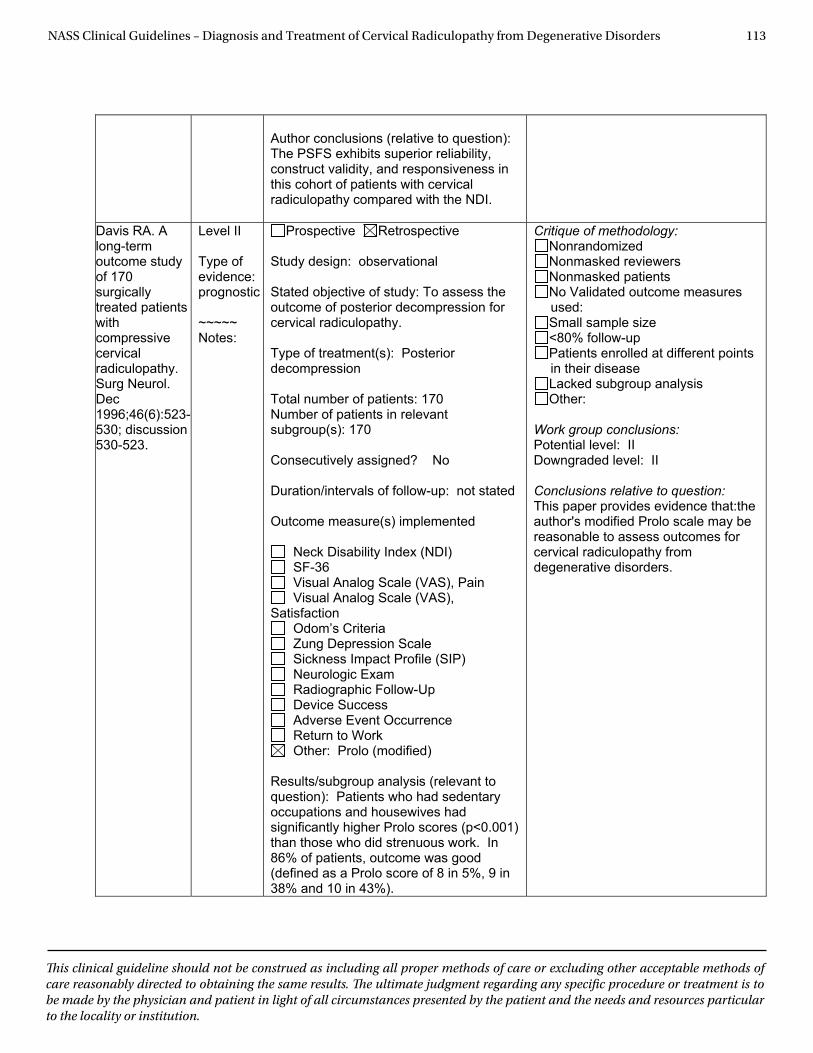
NASS Clinical Guidelines – Diagnosis and Treatment of Cervical Radiculopathy from Degenerative Disorders 113
This clinical guideline should not be construed as including all proper methods of care or excluding other acceptable methods of care reasonably directed to obtaining the same results. The ultimate judgment regarding any specific procedure or treatment is to be made by the physician and patient in light of all circumstances presented by the patient and the needs and resources particular to the locality or institution.
Author conclusions (relative to question): The PSFS exhibits superior reliability, construct validity, and responsiveness in this cohort of patients with cervical radiculopathy compared with the NDI.
Davis RA. A long-term outcome study of 170 surgicallytreated patients withcompressive cervical radiculopathy. Surg Neurol. Dec 1996;46(6):523-530; discussion 530-523.
Level II
Type of evidence: prognostic
~~~~~ Notes:
Prospective Retrospective
Study design: observational
Stated objective of study: To assess the outcome of posterior decompression for cervical radiculopathy. Type of treatment(s): Posterior decompression
Total number of patients: 170 Number of patients in relevant subgroup(s): 170
Consecutively assigned? No
Duration/intervals of follow-up: not stated
Outcome measure(s) implemented
Neck Disability Index (NDI) SF-36 Visual Analog Scale (VAS), Pain Visual Analog Scale (VAS),
Satisfaction Odom’s Criteria Zung Depression Scale Sickness Impact Profile (SIP) Neurologic Exam Radiographic Follow-Up Device Success Adverse Event Occurrence Return to Work Other: Prolo (modified)
Results/subgroup analysis (relevant to question): Patients who had sedentary occupations and housewives had significantly higher Prolo scores (p<0.001) than those who did strenuous work. In 86% of patients, outcome was good (defined as a Prolo score of 8 in 5%, 9 in 38% and 10 in 43%).
Critique of methodology: Nonrandomized Nonmasked reviewers Nonmasked patients No Validated outcome measures used: Small sample size <80% follow-up Patients enrolled at different points in their disease Lacked subgroup analysis Other:
Work group conclusions: Potential level: II Downgraded level: II
Conclusions relative to question: This paper provides evidence that:the author's modified Prolo scale may be reasonable to assess outcomes for cervical radiculopathy from degenerative disorders.
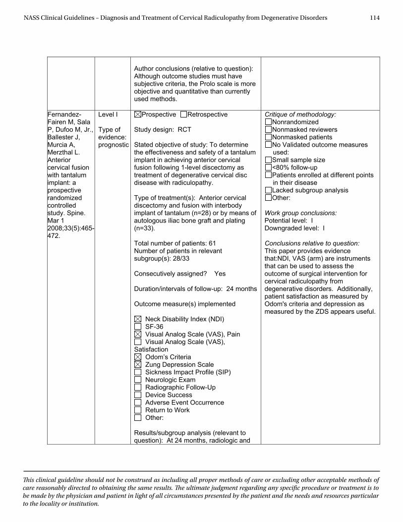
NASS Clinical Guidelines – Diagnosis and Treatment of Cervical Radiculopathy from Degenerative Disorders 114
This clinical guideline should not be construed as including all proper methods of care or excluding other acceptable methods of care reasonably directed to obtaining the same results. The ultimate judgment regarding any specific procedure or treatment is to be made by the physician and patient in light of all circumstances presented by the patient and the needs and resources particular to the locality or institution.
Author conclusions (relative to question): Although outcome studies must have subjective criteria, the Prolo scale is more objective and quantitative than currently used methods.
Fernandez-Fairen M, Sala P, Dufoo M, Jr., Ballester J, Murcia A, Merzthal L. Anteriorcervical fusion with tantalum implant: a prospective randomized controlledstudy. Spine. Mar 1 2008;33(5):465-472.
Level I
Type of evidence: prognostic
Prospective Retrospective
Study design: RCT
Stated objective of study: To determine the effectiveness and safety of a tantalum implant in achieving anterior cervical fusion following 1-level discectomy as treatment of degenerative cervical disc disease with radiculopathy. Type of treatment(s): Anterior cervical discectomy and fusion with interbody implant of tantalum (n=28) or by means of autologous iliac bone graft and plating (n=33).
Total number of patients: 61 Number of patients in relevant subgroup(s): 28/33
Consecutively assigned? Yes
Duration/intervals of follow-up: 24 months
Outcome measure(s) implemented
Neck Disability Index (NDI) SF-36 Visual Analog Scale (VAS), Pain Visual Analog Scale (VAS),
Satisfaction Odom’s Criteria Zung Depression Scale Sickness Impact Profile (SIP) Neurologic Exam Radiographic Follow-Up Device Success Adverse Event Occurrence Return to Work Other:
Results/subgroup analysis (relevant to question): At 24 months, radiologic and
Critique of methodology: Nonrandomized Nonmasked reviewers Nonmasked patients No Validated outcome measures used: Small sample size <80% follow-up Patients enrolled at different points in their disease Lacked subgroup analysis Other:
Work group conclusions: Potential level: I Downgraded level: I
Conclusions relative to question: This paper provides evidence that:NDI, VAS (arm) are instruments that can be used to assess the outcome of surgical intervention for cervical radiculopathy from degenerative disorders. Additionally, patient satisfaction as measured by Odom's criteria and depression as measured by the ZDS appears useful.
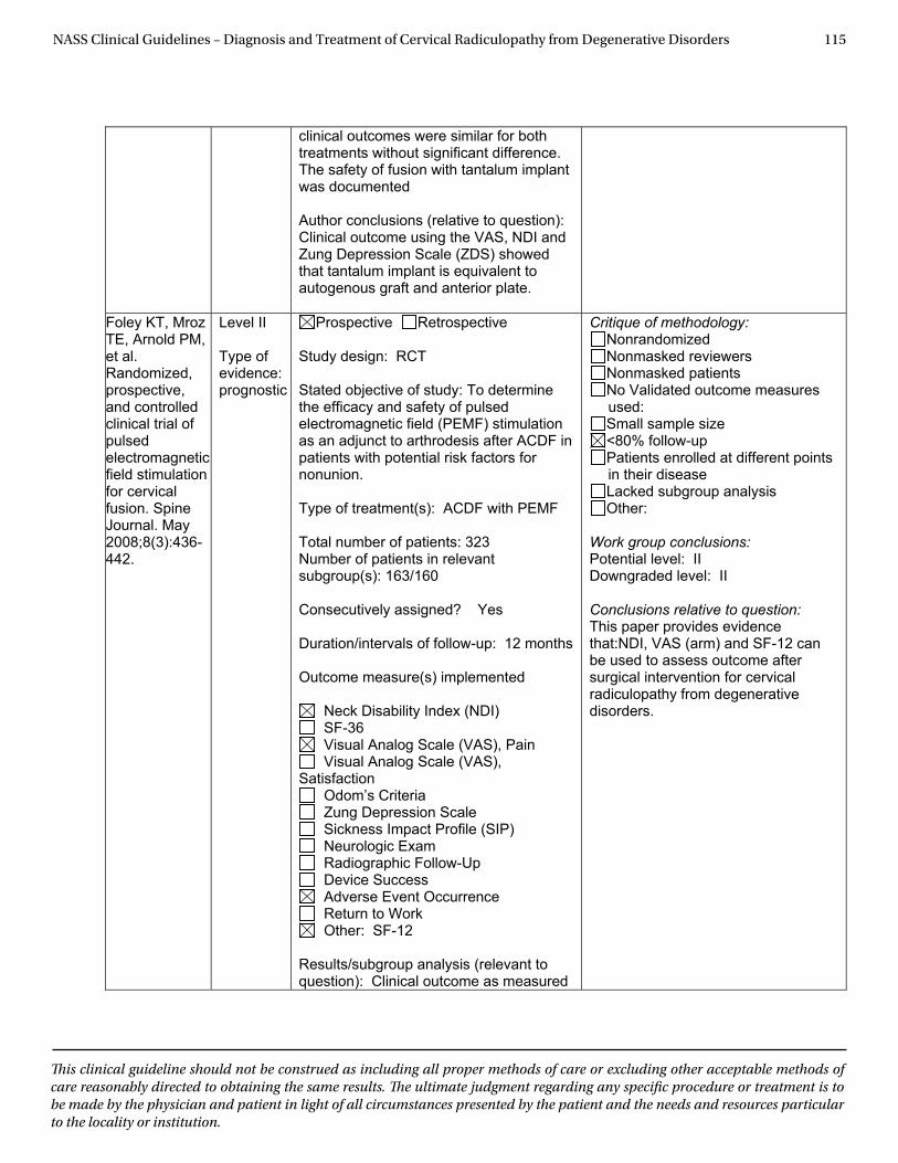
NASS Clinical Guidelines – Diagnosis and Treatment of Cervical Radiculopathy from Degenerative Disorders 115
This clinical guideline should not be construed as including all proper methods of care or excluding other acceptable methods of care reasonably directed to obtaining the same results. The ultimate judgment regarding any specific procedure or treatment is to be made by the physician and patient in light of all circumstances presented by the patient and the needs and resources particular to the locality or institution.
clinical outcomes were similar for both treatments without significant difference. The safety of fusion with tantalum implant was documented
Author conclusions (relative to question): Clinical outcome using the VAS, NDI and Zung Depression Scale (ZDS) showed that tantalum implant is equivalent to autogenous graft and anterior plate.
Foley KT, Mroz TE, Arnold PM, et al. Randomized, prospective, and controlled clinical trial of pulsedelectromagnetic field stimulation for cervical fusion. Spine Journal. May 2008;8(3):436-442.
Level II
Type of evidence: prognostic
Prospective Retrospective
Study design: RCT
Stated objective of study: To determine the efficacy and safety of pulsed electromagnetic field (PEMF) stimulation as an adjunct to arthrodesis after ACDF in patients with potential risk factors for nonunion. Type of treatment(s): ACDF with PEMF
Total number of patients: 323 Number of patients in relevant subgroup(s): 163/160
Consecutively assigned? Yes
Duration/intervals of follow-up: 12 months
Outcome measure(s) implemented
Neck Disability Index (NDI) SF-36 Visual Analog Scale (VAS), Pain Visual Analog Scale (VAS),
Satisfaction Odom’s Criteria Zung Depression Scale Sickness Impact Profile (SIP) Neurologic Exam Radiographic Follow-Up Device Success Adverse Event Occurrence Return to Work Other: SF-12
Results/subgroup analysis (relevant to question): Clinical outcome as measured
Critique of methodology: Nonrandomized Nonmasked reviewers Nonmasked patients No Validated outcome measures used: Small sample size <80% follow-up Patients enrolled at different points in their disease Lacked subgroup analysis Other:
Work group conclusions: Potential level: II Downgraded level: II
Conclusions relative to question: This paper provides evidence that:NDI, VAS (arm) and SF-12 can be used to assess outcome after surgical intervention for cervical radiculopathy from degenerative disorders.
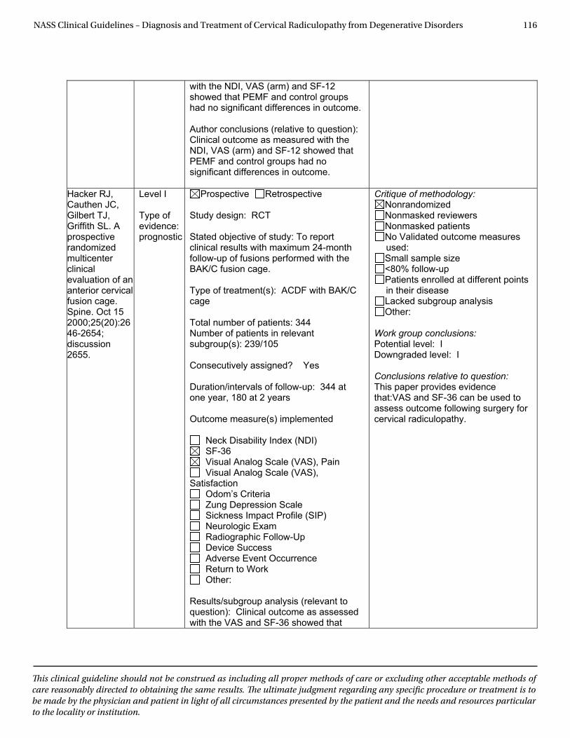
NASS Clinical Guidelines – Diagnosis and Treatment of Cervical Radiculopathy from Degenerative Disorders 116
This clinical guideline should not be construed as including all proper methods of care or excluding other acceptable methods of care reasonably directed to obtaining the same results. The ultimate judgment regarding any specific procedure or treatment is to be made by the physician and patient in light of all circumstances presented by the patient and the needs and resources particular to the locality or institution.
with the NDI, VAS (arm) and SF-12 showed that PEMF and control groups had no significant differences in outcome.
Author conclusions (relative to question): Clinical outcome as measured with the NDI, VAS (arm) and SF-12 showed that PEMF and control groups had no significant differences in outcome.
Hacker RJ, Cauthen JC, Gilbert TJ, Griffith SL. A prospective randomized multicenterclinical evaluation of an anterior cervical fusion cage. Spine. Oct 15 2000;25(20):2646-2654; discussion 2655.
Level I
Type of evidence: prognostic
Prospective Retrospective
Study design: RCT
Stated objective of study: To report clinical results with maximum 24-month follow-up of fusions performed with the BAK/C fusion cage. Type of treatment(s): ACDF with BAK/C cage
Total number of patients: 344 Number of patients in relevant subgroup(s): 239/105
Consecutively assigned? Yes
Duration/intervals of follow-up: 344 at one year, 180 at 2 years
Outcome measure(s) implemented
Neck Disability Index (NDI) SF-36 Visual Analog Scale (VAS), Pain Visual Analog Scale (VAS),
Satisfaction Odom’s Criteria Zung Depression Scale Sickness Impact Profile (SIP) Neurologic Exam Radiographic Follow-Up Device Success Adverse Event Occurrence Return to Work Other:
Results/subgroup analysis (relevant to question): Clinical outcome as assessed with the VAS and SF-36 showed that
Critique of methodology: Nonrandomized Nonmasked reviewers Nonmasked patients No Validated outcome measures used: Small sample size <80% follow-up Patients enrolled at different points in their disease Lacked subgroup analysis Other:
Work group conclusions: Potential level: I Downgraded level: I
Conclusions relative to question: This paper provides evidence that:VAS and SF-36 can be used to assess outcome following surgery for cervical radiculopathy.

NASS Clinical Guidelines – Diagnosis and Treatment of Cervical Radiculopathy from Degenerative Disorders 117
This clinical guideline should not be construed as including all proper methods of care or excluding other acceptable methods of care reasonably directed to obtaining the same results. The ultimate judgment regarding any specific procedure or treatment is to be made by the physician and patient in light of all circumstances presented by the patient and the needs and resources particular to the locality or institution.
with the NDI, VAS (arm) and SF-12 showed that PEMF and control groups had no significant differences in outcome.
Author conclusions (relative to question): Clinical outcome as measured with the NDI, VAS (arm) and SF-12 showed that PEMF and control groups had no significant differences in outcome.
Hacker RJ, Cauthen JC, Gilbert TJ, Griffith SL. A prospective randomized multicenterclinical evaluation of an anterior cervical fusion cage. Spine. Oct 15 2000;25(20):2646-2654; discussion 2655.
Level I
Type of evidence: prognostic
Prospective Retrospective
Study design: RCT
Stated objective of study: To report clinical results with maximum 24-month follow-up of fusions performed with the BAK/C fusion cage. Type of treatment(s): ACDF with BAK/C cage
Total number of patients: 344 Number of patients in relevant subgroup(s): 239/105
Consecutively assigned? Yes
Duration/intervals of follow-up: 344 at one year, 180 at 2 years
Outcome measure(s) implemented
Neck Disability Index (NDI) SF-36 Visual Analog Scale (VAS), Pain Visual Analog Scale (VAS),
Satisfaction Odom’s Criteria Zung Depression Scale Sickness Impact Profile (SIP) Neurologic Exam Radiographic Follow-Up Device Success Adverse Event Occurrence Return to Work Other:
Results/subgroup analysis (relevant to question): Clinical outcome as assessed with the VAS and SF-36 showed that
Critique of methodology: Nonrandomized Nonmasked reviewers Nonmasked patients No Validated outcome measures used: Small sample size <80% follow-up Patients enrolled at different points in their disease Lacked subgroup analysis Other:
Work group conclusions: Potential level: I Downgraded level: I
Conclusions relative to question: This paper provides evidence that:VAS and SF-36 can be used to assess outcome following surgery for cervical radiculopathy.
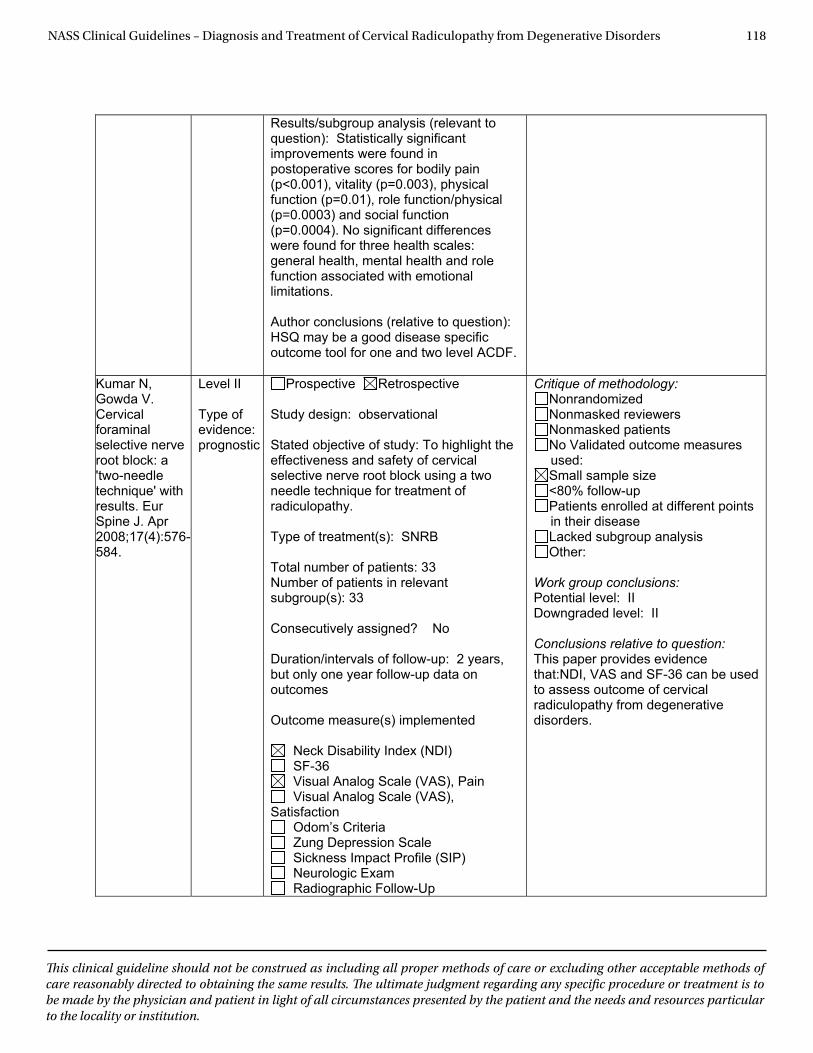
NASS Clinical Guidelines – Diagnosis and Treatment of Cervical Radiculopathy from Degenerative Disorders 118
This clinical guideline should not be construed as including all proper methods of care or excluding other acceptable methods of care reasonably directed to obtaining the same results. The ultimate judgment regarding any specific procedure or treatment is to be made by the physician and patient in light of all circumstances presented by the patient and the needs and resources particular to the locality or institution.
Results/subgroup analysis (relevant to question): Statistically significant improvements were found in postoperative scores for bodily pain (p<0.001), vitality (p=0.003), physical function (p=0.01), role function/physical (p=0.0003) and social function (p=0.0004). No significant differences were found for three health scales: general health, mental health and role function associated with emotional limitations.
Author conclusions (relative to question): HSQ may be a good disease specific outcome tool for one and two level ACDF.
Kumar N, Gowda V. Cervical foraminalselective nerve root block: a 'two-needle technique' with results. Eur Spine J. Apr 2008;17(4):576-584.
Level II
Type of evidence: prognostic
Prospective Retrospective
Study design: observational
Stated objective of study: To highlight the effectiveness and safety of cervical selective nerve root block using a two needle technique for treatment of radiculopathy. Type of treatment(s): SNRB
Total number of patients: 33 Number of patients in relevant subgroup(s): 33
Consecutively assigned? No
Duration/intervals of follow-up: 2 years, but only one year follow-up data on outcomes
Outcome measure(s) implemented
Neck Disability Index (NDI) SF-36 Visual Analog Scale (VAS), Pain Visual Analog Scale (VAS),
Satisfaction Odom’s Criteria Zung Depression Scale Sickness Impact Profile (SIP) Neurologic Exam Radiographic Follow-Up
Critique of methodology: Nonrandomized Nonmasked reviewers Nonmasked patients No Validated outcome measures used: Small sample size <80% follow-up Patients enrolled at different points in their disease Lacked subgroup analysis Other:
Work group conclusions: Potential level: II Downgraded level: II
Conclusions relative to question: This paper provides evidence that:NDI, VAS and SF-36 can be used to assess outcome of cervical radiculopathy from degenerative disorders.
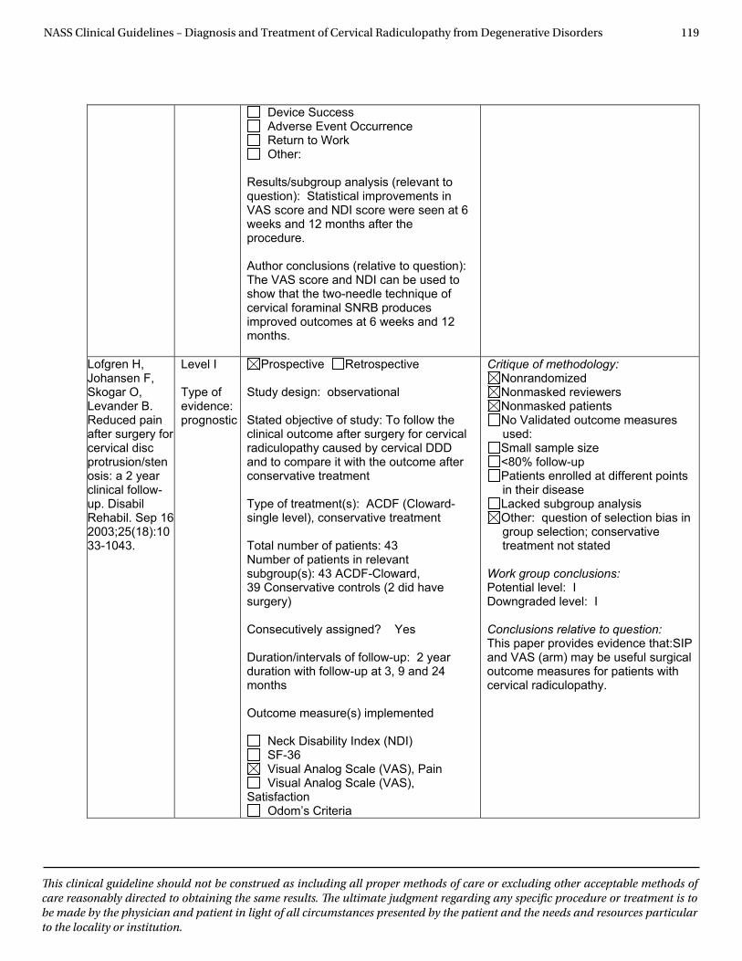
NASS Clinical Guidelines – Diagnosis and Treatment of Cervical Radiculopathy from Degenerative Disorders 119
This clinical guideline should not be construed as including all proper methods of care or excluding other acceptable methods of care reasonably directed to obtaining the same results. The ultimate judgment regarding any specific procedure or treatment is to be made by the physician and patient in light of all circumstances presented by the patient and the needs and resources particular to the locality or institution.
Device Success Adverse Event Occurrence Return to Work Other:
Results/subgroup analysis (relevant to question): Statistical improvements in VAS score and NDI score were seen at 6 weeks and 12 months after the procedure.
Author conclusions (relative to question): The VAS score and NDI can be used to show that the two-needle technique of cervical foraminal SNRB produces improved outcomes at 6 weeks and 12 months.
Lofgren H, Johansen F, Skogar O, Levander B. Reduced pain after surgery for cervical disc protrusion/stenosis: a 2 year clinical follow-up. Disabil Rehabil. Sep 16 2003;25(18):1033-1043.
Level I
Type of evidence: prognostic
Prospective Retrospective
Study design: observational
Stated objective of study: To follow the clinical outcome after surgery for cervical radiculopathy caused by cervical DDD and to compare it with the outcome after conservative treatment Type of treatment(s): ACDF (Cloward-single level), conservative treatment
Total number of patients: 43 Number of patients in relevant subgroup(s): 43 ACDF-Cloward, 39 Conservative controls (2 did have surgery)
Consecutively assigned? Yes
Duration/intervals of follow-up: 2 year duration with follow-up at 3, 9 and 24 months
Outcome measure(s) implemented
Neck Disability Index (NDI) SF-36 Visual Analog Scale (VAS), Pain Visual Analog Scale (VAS),
Satisfaction Odom’s Criteria
Critique of methodology: Nonrandomized Nonmasked reviewers Nonmasked patients No Validated outcome measures used: Small sample size <80% follow-up Patients enrolled at different points in their disease Lacked subgroup analysis Other: question of selection bias in group selection; conservative treatment not stated
Work group conclusions: Potential level: I Downgraded level: I
Conclusions relative to question: This paper provides evidence that:SIP and VAS (arm) may be useful surgical outcome measures for patients with cervical radiculopathy.
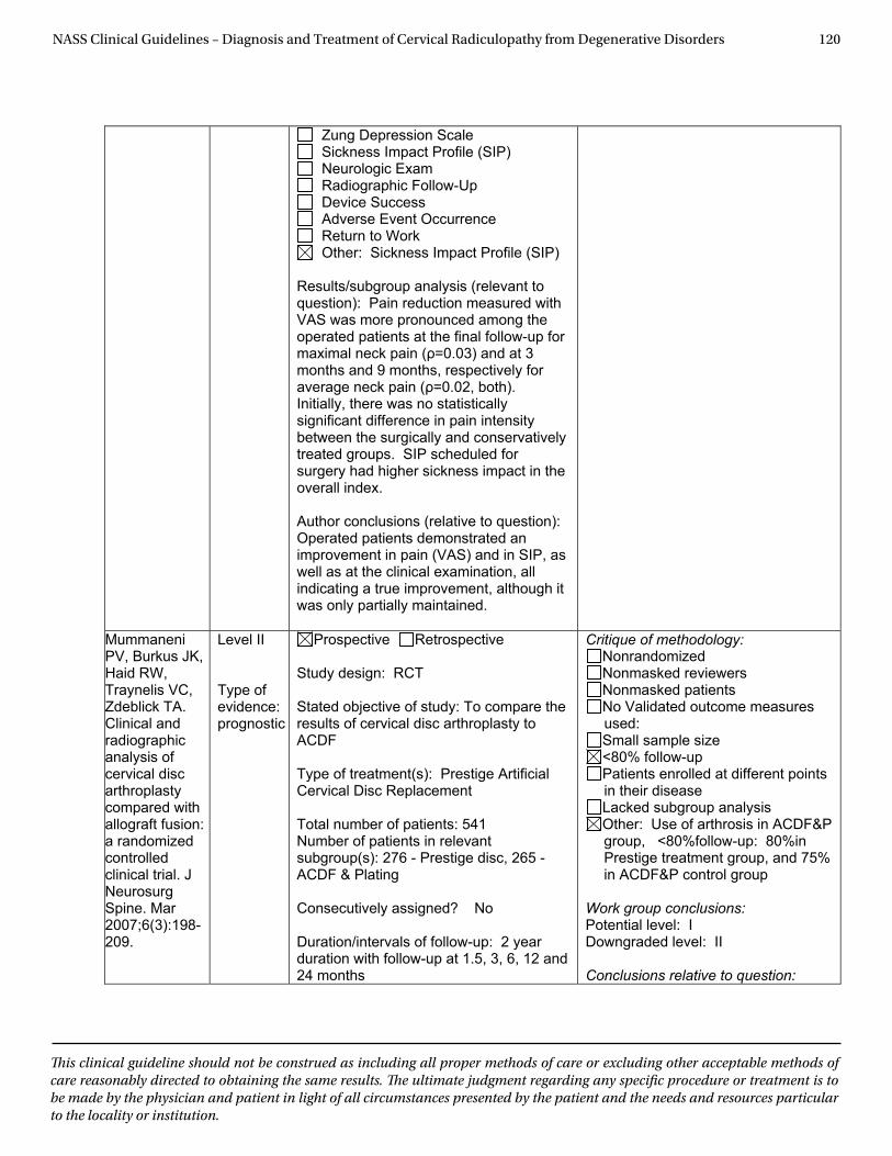
NASS Clinical Guidelines – Diagnosis and Treatment of Cervical Radiculopathy from Degenerative Disorders 120
This clinical guideline should not be construed as including all proper methods of care or excluding other acceptable methods of care reasonably directed to obtaining the same results. The ultimate judgment regarding any specific procedure or treatment is to be made by the physician and patient in light of all circumstances presented by the patient and the needs and resources particular to the locality or institution.
Zung Depression Scale Sickness Impact Profile (SIP) Neurologic Exam Radiographic Follow-Up Device Success Adverse Event Occurrence Return to Work Other: Sickness Impact Profile (SIP)
Results/subgroup analysis (relevant to question): Pain reduction measured with VAS was more pronounced among the operated patients at the final follow-up for maximal neck pain (ρ=0.03) and at 3 months and 9 months, respectively for average neck pain (ρ=0.02, both). Initially, there was no statistically significant difference in pain intensity between the surgically and conservatively treated groups. SIP scheduled for surgery had higher sickness impact in the overall index. Author conclusions (relative to question): Operated patients demonstrated an improvement in pain (VAS) and in SIP, as well as at the clinical examination, all indicating a true improvement, although it was only partially maintained.
Mummaneni PV, Burkus JK, Haid RW, Traynelis VC, Zdeblick TA. Clinical and radiographic analysis of cervical disc arthroplasty compared with allograft fusion: a randomized controlled clinical trial. J Neurosurg Spine. Mar 2007;6(3):198-209.
Level II Type of evidence: prognostic
Prospective Retrospective Study design: RCT Stated objective of study: To compare the results of cervical disc arthroplasty to ACDF Type of treatment(s): Prestige Artificial Cervical Disc Replacement Total number of patients: 541 Number of patients in relevant subgroup(s): 276 - Prestige disc, 265 - ACDF & Plating Consecutively assigned? No Duration/intervals of follow-up: 2 year duration with follow-up at 1.5, 3, 6, 12 and 24 months
Critique of methodology: Nonrandomized Nonmasked reviewers Nonmasked patients No Validated outcome measures used: Small sample size <80% follow-up Patients enrolled at different points in their disease Lacked subgroup analysis Other: Use of arthrosis in ACDF&P group, <80%follow-up: 80%in Prestige treatment group, and 75% in ACDF&P control group
Work group conclusions: Potential level: I Downgraded level: II Conclusions relative to question:
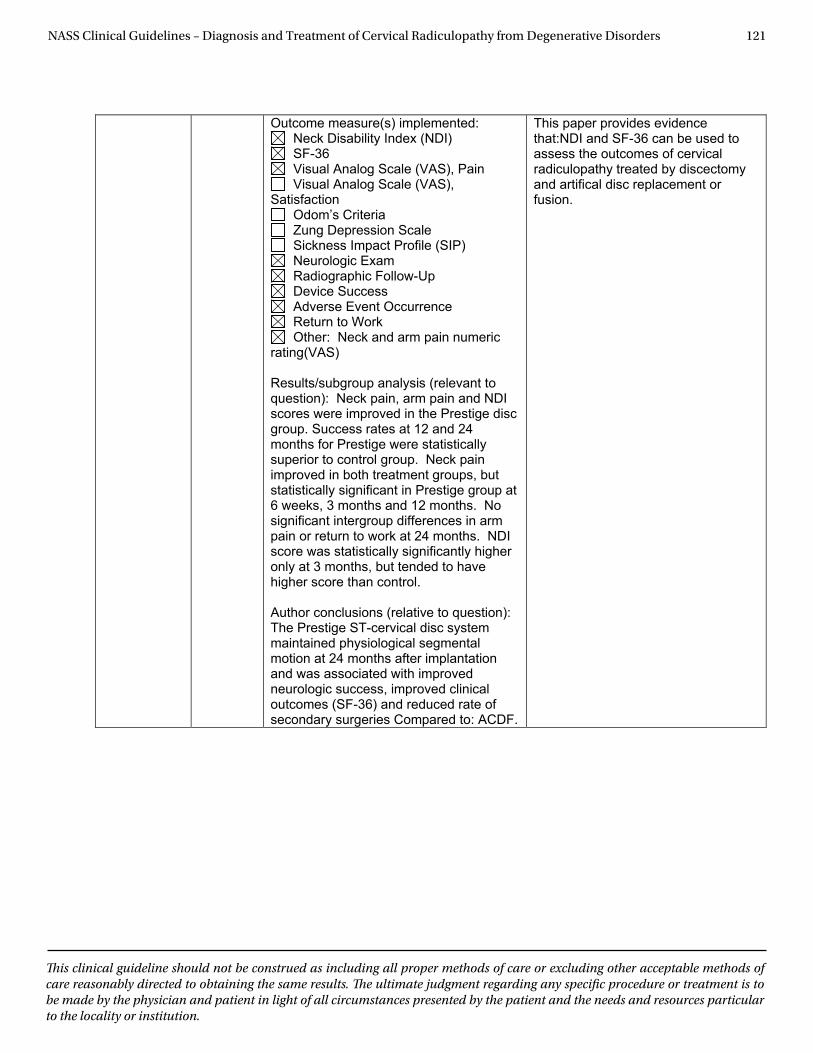
NASS Clinical Guidelines – Diagnosis and Treatment of Cervical Radiculopathy from Degenerative Disorders 121
This clinical guideline should not be construed as including all proper methods of care or excluding other acceptable methods of care reasonably directed to obtaining the same results. The ultimate judgment regarding any specific procedure or treatment is to be made by the physician and patient in light of all circumstances presented by the patient and the needs and resources particular to the locality or institution.
Outcome measure(s) implemented: Neck Disability Index (NDI) SF-36 Visual Analog Scale (VAS), Pain Visual Analog Scale (VAS),
Satisfaction Odom’s Criteria Zung Depression Scale Sickness Impact Profile (SIP) Neurologic Exam Radiographic Follow-Up Device Success Adverse Event Occurrence Return to Work Other: Neck and arm pain numeric
rating(VAS)
Results/subgroup analysis (relevant to question): Neck pain, arm pain and NDI scores were improved in the Prestige disc group. Success rates at 12 and 24 months for Prestige were statistically superior to control group. Neck pain improved in both treatment groups, but statistically significant in Prestige group at 6 weeks, 3 months and 12 months. No significant intergroup differences in arm pain or return to work at 24 months. NDI score was statistically significantly higher only at 3 months, but tended to have higher score than control.
Author conclusions (relative to question): The Prestige ST-cervical disc system maintained physiological segmental motion at 24 months after implantation and was associated with improved neurologic success, improved clinical outcomes (SF-36) and reduced rate of secondary surgeries Compared to: ACDF.
This paper provides evidence that:NDI and SF-36 can be used to assess the outcomes of cervical radiculopathy treated by discectomy and artifical disc replacement or fusion.
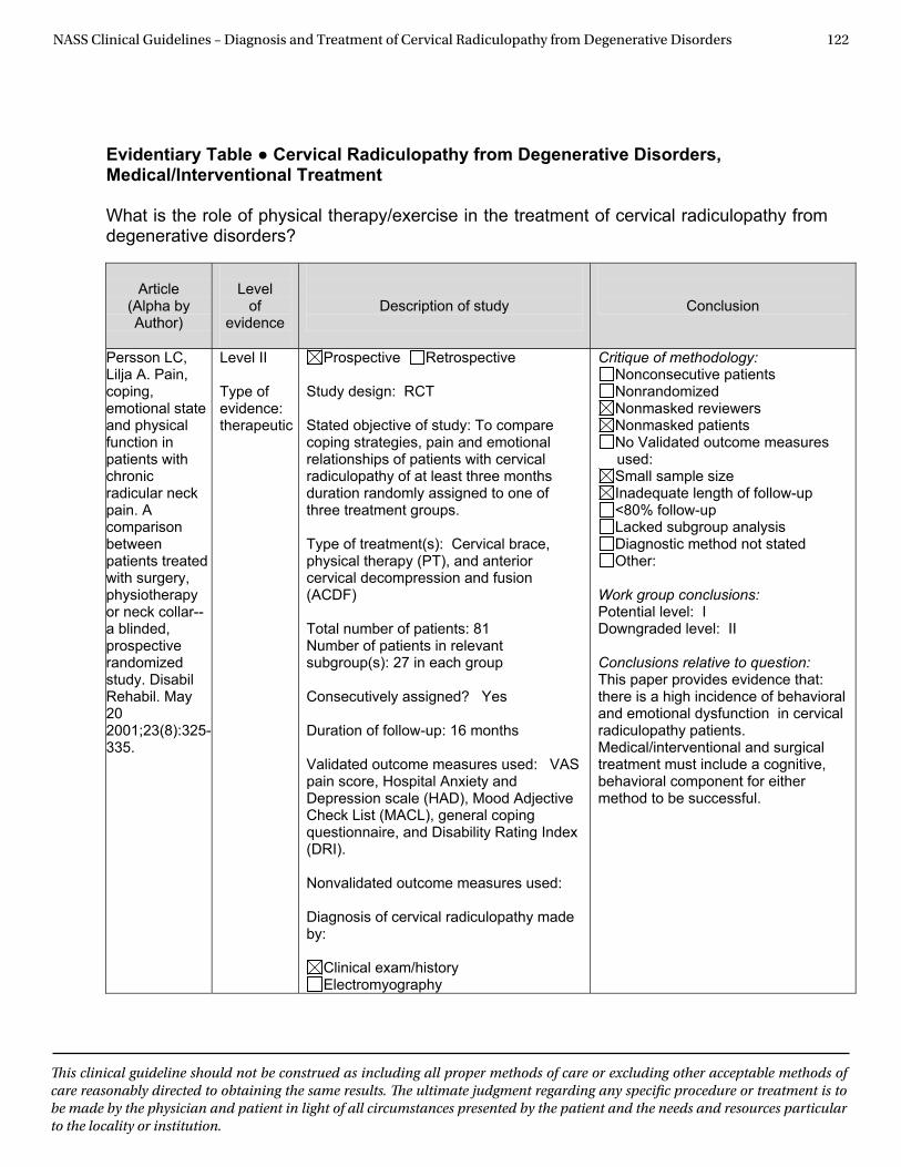
NASS Clinical Guidelines – Diagnosis and Treatment of Cervical Radiculopathy from Degenerative Disorders 122
This clinical guideline should not be construed as including all proper methods of care or excluding other acceptable methods of care reasonably directed to obtaining the same results. The ultimate judgment regarding any specific procedure or treatment is to be made by the physician and patient in light of all circumstances presented by the patient and the needs and resources particular to the locality or institution.
Evidentiary Table ● Cervical Radiculopathy from Degenerative Disorders, Medical/Interventional Treatment
What is the role of physical therapy/exercise in the treatment of cervical radiculopathy from degenerative disorders?
Article
(Alpha by Author)
Level
of evidence
Description of study
Conclusion
Persson LC, Lilja A. Pain, coping, emotional state and physical function in patients with chronic radicular neck pain. A comparison between patients treated with surgery, physiotherapy or neck collar--a blinded, prospective randomized study. Disabil Rehabil. May 20 2001;23(8):325-335.
Level II Type of evidence: therapeutic
Prospective Retrospective Study design: RCT Stated objective of study: To compare coping strategies, pain and emotional relationships of patients with cervical radiculopathy of at least three months duration randomly assigned to one of three treatment groups. Type of treatment(s): Cervical brace, physical therapy (PT), and anterior cervical decompression and fusion (ACDF) Total number of patients: 81 Number of patients in relevant subgroup(s): 27 in each group Consecutively assigned? Yes Duration of follow-up: 16 months Validated outcome measures used: VAS pain score, Hospital Anxiety and Depression scale (HAD), Mood Adjective Check List (MACL), general coping questionnaire, and Disability Rating Index (DRI). Nonvalidated outcome measures used: Diagnosis of cervical radiculopathy made by:
Clinical exam/history Electromyography
Critique of methodology: Nonconsecutive patients Nonrandomized Nonmasked reviewers Nonmasked patients No Validated outcome measures used: Small sample size Inadequate length of follow-up <80% follow-up Lacked subgroup analysis Diagnostic method not stated Other:
Work group conclusions: Potential level: I Downgraded level: II Conclusions relative to question: This paper provides evidence that: there is a high incidence of behavioral and emotional dysfunction in cervical radiculopathy patients. Medical/interventional and surgical treatment must include a cognitive, behavioral component for either method to be successful.
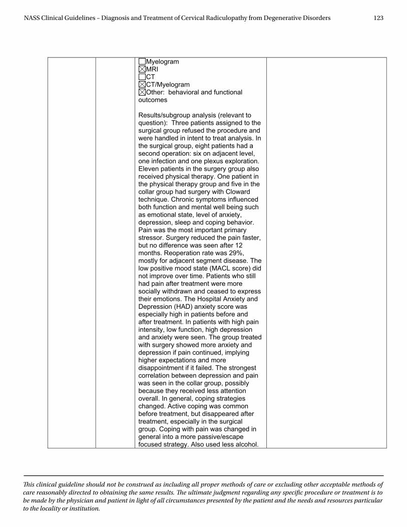
NASS Clinical Guidelines – Diagnosis and Treatment of Cervical Radiculopathy from Degenerative Disorders 123
This clinical guideline should not be construed as including all proper methods of care or excluding other acceptable methods of care reasonably directed to obtaining the same results. The ultimate judgment regarding any specific procedure or treatment is to be made by the physician and patient in light of all circumstances presented by the patient and the needs and resources particular to the locality or institution.
MyelogramMRICTCT/Myelogram Other: behavioral and functional
outcomes
Results/subgroup analysis (relevant to question): Three patients assigned to the surgical group refused the procedure and were handled in intent to treat analysis. In the surgical group, eight patients had a second operation: six on adjacent level, one infection and one plexus exploration. Eleven patients in the surgery group also received physical therapy. One patient in the physical therapy group and five in the collar group had surgery with Cloward technique. Chronic symptoms influenced both function and mental well being such as emotional state, level of anxiety, depression, sleep and coping behavior. Pain was the most important primary stressor. Surgery reduced the pain faster, but no difference was seen after 12 months. Reoperation rate was 29%, mostly for adjacent segment disease. The low positive mood state (MACL score) did not improve over time. Patients who still had pain after treatment were more socially withdrawn and ceased to express their emotions. The Hospital Anxiety and Depression (HAD) anxiety score was especially high in patients before and after treatment. In patients with high pain intensity, low function, high depression and anxiety were seen. The group treated with surgery showed more anxiety and depression if pain continued, implying higher expectations and more disappointment if it failed. The strongest correlation between depression and pain was seen in the collar group, possibly because they received less attention overall. In general, coping strategies changed. Active coping was common before treatment, but disappeared after treatment, especially in the surgical group. Coping with pain was changed in general into a more passive/escape focused strategy. Also used less alcohol.
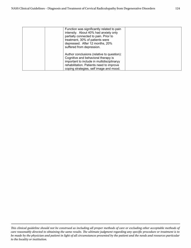
NASS Clinical Guidelines – Diagnosis and Treatment of Cervical Radiculopathy from Degenerative Disorders 124
This clinical guideline should not be construed as including all proper methods of care or excluding other acceptable methods of care reasonably directed to obtaining the same results. The ultimate judgment regarding any specific procedure or treatment is to be made by the physician and patient in light of all circumstances presented by the patient and the needs and resources particular to the locality or institution.
Function was significantly related to pain intensity. About 40% had anxiety only partially connected to pain. Prior to treatment, 30% of patients were depressed. After 12 months, 20% suffered from depression.
Author conclusions (relative to question): Cognitive and behavioral therapy is important to include in multidisciplinaryy rehabilitation. Patients need to improve coping strategies, self image and mood.
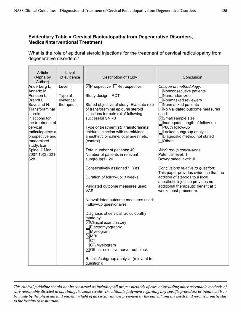
NASS Clinical Guidelines – Diagnosis and Treatment of Cervical Radiculopathy from Degenerative Disorders 125
This clinical guideline should not be construed as including all proper methods of care or excluding other acceptable methods of care reasonably directed to obtaining the same results. The ultimate judgment regarding any specific procedure or treatment is to be made by the physician and patient in light of all circumstances presented by the patient and the needs and resources particular to the locality or institution.
Evidentiary Table ● Cervical Radiculopathy from Degenerative Disorders, Medical/Interventional Treatment
What is the role of epidural steroid injections for the treatment of cervical radiculopathy from degenerative disorders?
Article
(Alpha by Author)
Level
of evidence
Description of study
Conclusion
Anderberg L, Annertz M, Persson L, Brandt L, Saveland H. Transforaminal steroid injections for the treatment of cervical radiculopathy: a prospective and randomised study. Eur Spine J. Mar 2007;16(3):321-328.
Level II Type of evidence: therapeutic
Prospective Retrospective Study design: RCT Stated objective of study: Evaluate role of transforaminal epidural steroid injections for pain relief following successful SNRB Type of treatment(s): transforaminal epidural injection with steroid/local anesthetic or saline/local anesthetic (control) Total number of patients: 40 Number of patients in relevant subgroup(s): 20 Consecutively assigned? Yes Duration of follow-up: 3 weeks Validated outcome measures used: VAS Nonvalidated outcome measures used: Follow-up questionairre Diagnosis of cervical radiculopathy made by:
Clinical exam/history Electromyography Myelogram MRI CT CT/Myelogram Other: selective nerve root block
Results/subgroup analysis (relevant to question):
Critique of methodology: Nonconsecutive patients Nonrandomized Nonmasked reviewers Nonmasked patients No Validated outcome measures
used: Small sample size Inadequate length of follow-up <80% follow-up Lacked subgroup analysis Diagnostic method not stated Other:
Work group conclusions: Potential level: I Downgraded level: II Conclusions relative to question: This paper provides evidence that:the addition of steroids to a local anesthetic injection provides no additional therapeutic benefit at 3 weeks post-procedure.
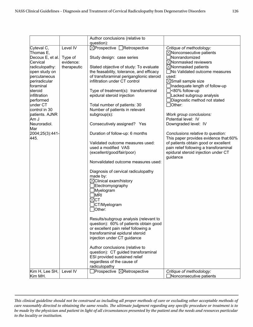
NASS Clinical Guidelines – Diagnosis and Treatment of Cervical Radiculopathy from Degenerative Disorders 126
This clinical guideline should not be construed as including all proper methods of care or excluding other acceptable methods of care reasonably directed to obtaining the same results. The ultimate judgment regarding any specific procedure or treatment is to be made by the physician and patient in light of all circumstances presented by the patient and the needs and resources particular to the locality or institution.
Author conclusions (relative to question):
Cyteval C, Thomas E, Decoux E, et al. Cervical radiculopathy: open study on percutaneous periradicular foraminalsteroid infiltrationperformed under CT control in 30 patients. AJNR Am J Neuroradiol. Mar2004;25(3):441-445.
Level IV
Type of evidence: therapeutic
Prospective Retrospective
Study design: case series
Stated objective of study: To evaluate the feasability, tolerance, and efficacy of transforaminal periganglionic steroid infiltration under CT control
Type of treatment(s): transforaminal epidural steroid injection
Total number of patients: 30 Number of patients in relevant subgroup(s):
Consecutively assigned? Yes
Duration of follow-up: 6 months
Validated outcome measures used: used a modified VAS (excellent/good/fair/poor)
Nonvalidated outcome measures used:
Diagnosis of cervical radiculopathy made by:
Clinical exam/history Electromyography MyelogramMRICTCT/Myelogram Other:
Results/subgroup analysis (relevant to question): 60% of patients obtain good or excellent pain relief following a transforaminal epidural steroid injection under CT guidance
Author conclusions (relative to question): CT guided transforaminal ESI provided sustained relief regardless of the cause of radiculopathy
Critique of methodology: Nonconsecutive patients Nonrandomized Nonmasked reviewers Nonmasked patients No Validated outcome measures
used: Small sample size Inadequate length of follow-up <80% follow-up Lacked subgroup analysis Diagnostic method not stated Other:
Work group conclusions: Potential level: IV Downgraded level: IV
Conclusions relative to question: This paper provides evidence that:60% of patients obtain good or excellent pain relief following a transforaminal epidural steroid injection under CT guidance
Kim H, Lee SH, Kim MH.
Level IV Prospective Retrospective Critique of methodology: Nonconsecutive patients
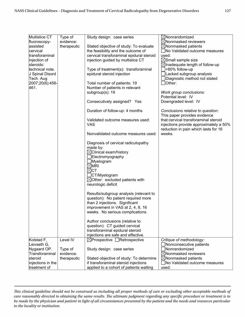
NASS Clinical Guidelines – Diagnosis and Treatment of Cervical Radiculopathy from Degenerative Disorders 127
This clinical guideline should not be construed as including all proper methods of care or excluding other acceptable methods of care reasonably directed to obtaining the same results. The ultimate judgment regarding any specific procedure or treatment is to be made by the physician and patient in light of all circumstances presented by the patient and the needs and resources particular to the locality or institution.
Multislice CT fluoroscopy-assisted cervical transforaminal injection of steroids: technical note. J Spinal Disord Tech. Aug 2007;20(6):456-461.
Type of evidence: therapeutic
Study design: case series
Stated objective of study: To evaluate the feasibility and the outcome of cervical transforaminal epidural steroid injection guided by multislice CT
Type of treatment(s): transforaminal epidural steroid injection
Total number of patients: 19 Number of patients in relevant subgroup(s): 19
Consecutively assigned? Yes
Duration of follow-up: 4 months
Validated outcome measures used: VAS
Nonvalidated outcome measures used:
Diagnosis of cervical radiculopathy made by:
Clinical exam/history Electromyography MyelogramMRICTCT/Myelogram Other: excluded patients with
neurologic deficit
Results/subgroup analysis (relevant to question): No patient required more than 2 injections. Significant improvement in VAS at 2, 4, 8, 16 weeks. No serious complications.
Author conclusions (relative to question): CT guided cervical transforaminal epidural steroid injections are safe and effective.
Nonrandomized Nonmasked reviewers Nonmasked patients No Validated outcome measures
used: Small sample size Inadequate length of follow-up <80% follow-up Lacked subgroup analysis Diagnostic method not stated Other:
Work group conclusions: Potential level: IV Downgraded level: IV
Conclusions relative to question: This paper provides evidence that:cervical transforaminal steroid injections provide approximately a 50% reduction in pain which lasts for 16 weeks.
Kolstad F, Leivseth G, Nygaard OP. Transforaminal steroid injections in the treatment of
Level IV
Type of evidence: therapeutic
Prospective Retrospective
Study design: case series
Stated objective of study: To determine if transforaminal steroid injections applied to a cohort of patients waiting
Critique of methodology: Nonconsecutive patients Nonrandomized Nonmasked reviewers Nonmasked patients No Validated outcome measures
used:
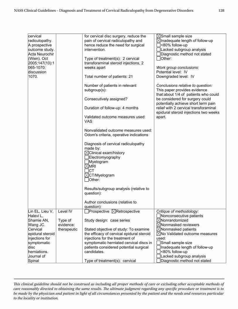
NASS Clinical Guidelines – Diagnosis and Treatment of Cervical Radiculopathy from Degenerative Disorders 128
This clinical guideline should not be construed as including all proper methods of care or excluding other acceptable methods of care reasonably directed to obtaining the same results. The ultimate judgment regarding any specific procedure or treatment is to be made by the physician and patient in light of all circumstances presented by the patient and the needs and resources particular to the locality or institution.
cervical radiculopathy. A prospective outcome study. Acta Neurochir (Wien). Oct 2005;147(10):1065-1070; discussion 1070.
for cervical disc surgery, reduce the pain of cervical radiculopathy and hence reduce the need for surgical intervention.
Type of treatment(s): 2 cervical transforaminal steroid injections, 2 weeks apart
Total number of patients: 21
Number of patients in relevant subgroup(s):
Consecutively assigned?
Duration of follow-up: 4 months
Validated outcome measures used: VAS
Nonvalidated outcome measures used: Odom's criteria, operative indications
Diagnosis of cervical radiculopathy made by:
Clinical exam/history Electromyography MyelogramMRICTCT/Myelogram Other:
Results/subgroup analysis (relative to question):
Author conclusions (relative to question):
Small sample size Inadequate length of follow-up <80% follow-up Lacked subgroup analysis Diagnostic method not stated Other:
Work group conclusions: Potential level: IV Downgraded level: IV
Conclusions relative to question: This paper provides evidence that:about 1/4 of patients who could be considered for surgery could potentially achieve short term pain relief with 2 cervical transforaminal epidural steroid injections two weeks apart.
Lin EL, Lieu V, Halevi L, Shamie AN, Wang JC. Cervical epidural steroid injections for symptomatic discherniations. Journal of Spinal
Level IV
Type of evidence: therapeutic
Prospective Retrospective
Study design: case series
Stated objective of study: To examine the efficacy of cervical epidural steroid injections for the treatment of symptomatic herniated cervical discs in patients considered potential surgical candidates.
Type of treatment(s): cervical
Critique of methodology: Nonconsecutive patients Nonrandomized Nonmasked reviewers Nonmasked patients No Validated outcome measures
used: Small sample size Inadequate length of follow-up <80% follow-up Lacked subgroup analysis Diagnostic method not stated
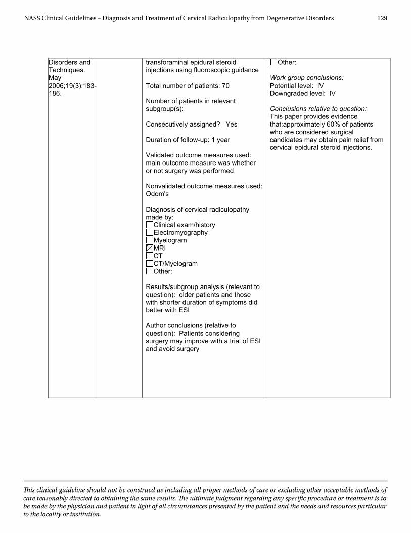
NASS Clinical Guidelines – Diagnosis and Treatment of Cervical Radiculopathy from Degenerative Disorders 129
This clinical guideline should not be construed as including all proper methods of care or excluding other acceptable methods of care reasonably directed to obtaining the same results. The ultimate judgment regarding any specific procedure or treatment is to be made by the physician and patient in light of all circumstances presented by the patient and the needs and resources particular to the locality or institution.
Disorders and Techniques. May2006;19(3):183-186.
transforaminal epidural steroid injections using fluoroscopic guidance
Total number of patients: 70
Number of patients in relevant subgroup(s):
Consecutively assigned? Yes
Duration of follow-up: 1 year
Validated outcome measures used: main outcome measure was whether or not surgery was performed
Nonvalidated outcome measures used: Odom's
Diagnosis of cervical radiculopathy made by:
Clinical exam/history Electromyography MyelogramMRICTCT/Myelogram Other:
Results/subgroup analysis (relevant to question): older patients and those with shorter duration of symptoms did better with ESI
Author conclusions (relative to question): Patients considering surgery may improve with a trial of ESI and avoid surgery
Other:
Work group conclusions: Potential level: IV Downgraded level: IV
Conclusions relative to question: This paper provides evidence that:approximately 60% of patients who are considered surgical candidates may obtain pain relief from cervical epidural steroid injections.

NASS Clinical Guidelines – Diagnosis and Treatment of Cervical Radiculopathy from Degenerative Disorders 130
This clinical guideline should not be construed as including all proper methods of care or excluding other acceptable methods of care reasonably directed to obtaining the same results. The ultimate judgment regarding any specific procedure or treatment is to be made by the physician and patient in light of all circumstances presented by the patient and the needs and resources particular to the locality or institution.
Evidentiary Table ● Cervical Radiculopathy from Degenerative Disorders, Medical/Interventional Treatment
What is the role of ancillary treatments such as bracing, traction, electrical stimulation, acupuncture and transcutaneous electrical stimulation (TENS) in the treatment of cervical radiculopathy from degenerative disorders?
Article
(Alpha by Author)
Level
of evidence
Description of study
Conclusion
Alexandre A, Coro L, Azuelos A, et al. Intradiscal injection of oxygen-ozone gas mixture for the treatment of cervical disc herniations. Acta Neurochir Suppl. 2005;92:79-82.
Level V Type of evidence: therapeutic
Prospective Retrospective Study design: case series Stated objective of study: Report the effects of intervertebral disc and paravertebral injections of ozone & oxygen in patients with cervical disc herniations Type of treatment(s): Intervertebral disc and five paravertebral injections of ozone & oxygen Total number of patients: 252 Number of patients in relevant subgroup(s): Consecutively assigned? No Duration of follow-up: possibly 7 months Validated outcome measures used: Nonvalidated outcome measures used: pain improvement, sensory dysfunction, strength improvement Diagnosis of cervical radiculopathy made by:
Clinical exam/history Electromyography Myelogram MRI CT CT/Myelogram Other:
Critique of methodology: Nonconsecutive patients Nonrandomized Nonmasked reviewers Nonmasked patients No Validated outcome measures used: Small sample size Inadequate length of follow-up <80% follow-up Lacked subgroup analysis Diagnostic method not stated Other: No specified duration of follow-up, no data tables or speed of recovery noted.
Work group conclusions: Potential level: IV Downgraded level: V Conclusions relative to question: This paper provides evidence that:Approximately 80% of patients will report symptomatic relief from cervical radiculopathy at some point following ozone and oxygen injection into the intervertebral disc and paravertebral musculature.
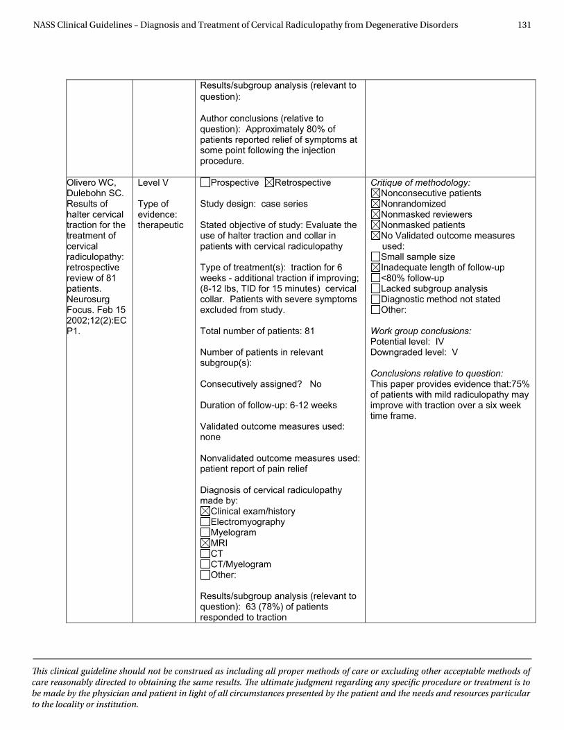
NASS Clinical Guidelines – Diagnosis and Treatment of Cervical Radiculopathy from Degenerative Disorders 131
This clinical guideline should not be construed as including all proper methods of care or excluding other acceptable methods of care reasonably directed to obtaining the same results. The ultimate judgment regarding any specific procedure or treatment is to be made by the physician and patient in light of all circumstances presented by the patient and the needs and resources particular to the locality or institution.
Results/subgroup analysis (relevant to question):
Author conclusions (relative to question): Approximately 80% of patients reported relief of symptoms at some point following the injection procedure.
Olivero WC, Dulebohn SC. Results of halter cervical traction for the treatment of cervical radiculopathy: retrospective review of 81 patients.Neurosurg Focus. Feb 15 2002;12(2):ECP1.
Level V
Type of evidence: therapeutic
Prospective Retrospective
Study design: case series
Stated objective of study: Evaluate the use of halter traction and collar in patients with cervical radiculopathy
Type of treatment(s): traction for 6 weeks - additional traction if improving; (8-12 lbs, TID for 15 minutes) cervical collar. Patients with severe symptoms excluded from study.
Total number of patients: 81
Number of patients in relevant subgroup(s):
Consecutively assigned? No
Duration of follow-up: 6-12 weeks
Validated outcome measures used: none
Nonvalidated outcome measures used: patient report of pain relief
Diagnosis of cervical radiculopathy made by:
Clinical exam/history Electromyography MyelogramMRICTCT/Myelogram Other:
Results/subgroup analysis (relevant to question): 63 (78%) of patients responded to traction
Critique of methodology: Nonconsecutive patients Nonrandomized Nonmasked reviewers Nonmasked patients No Validated outcome measures used: Small sample size Inadequate length of follow-up <80% follow-up Lacked subgroup analysis Diagnostic method not stated Other:
Work group conclusions: Potential level: IV Downgraded level: V
Conclusions relative to question: This paper provides evidence that:75% of patients with mild radiculopathy may improve with traction over a six week time frame.
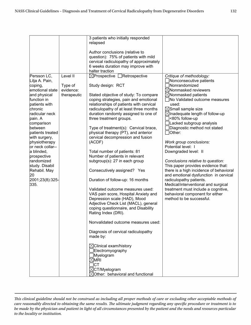
NASS Clinical Guidelines – Diagnosis and Treatment of Cervical Radiculopathy from Degenerative Disorders 132
This clinical guideline should not be construed as including all proper methods of care or excluding other acceptable methods of care reasonably directed to obtaining the same results. The ultimate judgment regarding any specific procedure or treatment is to be made by the physician and patient in light of all circumstances presented by the patient and the needs and resources particular to the locality or institution.
3 patients who initially responded relapsed
Author conclusions (relative to question): 75% of patients with mild cervical radiculopathy of approximately 6 weeks duration may improve with halter traction
Persson LC, Lilja A. Pain, coping, emotional state and physical function in patients with chronic radicular neck pain. A comparison between patients treated with surgery, physiotherapy or neck collar--a blinded, prospective randomized study. Disabil Rehabil. May 202001;23(8):325-335.
Level II
Type of evidence: therapeutic
Prospective Retrospective
Study design: RCT
Stated objective of study: To compare coping strategies, pain and emotional relationships of patients with cervical radiculopathy of at least three months duration randomly assigned to one of three treatment groups.
Type of treatment(s): Cervical brace, physical therapy (PT), and anterior cervical decompression and fusion (ACDF)
Total number of patients: 81 Number of patients in relevant subgroup(s): 27 in each group
Consecutively assigned? Yes
Duration of follow-up: 16 months
Validated outcome measures used: VAS pain score, Hospital Anxiety and Depression scale (HAD), Mood Adjective Check List (MACL), general coping questionnaire, and Disability Rating Index (DRI).
Nonvalidated outcome measures used:
Diagnosis of cervical radiculopathy made by:
Clinical exam/history Electromyography MyelogramMRICTCT/Myelogram Other: behavioral and functional
Critique of methodology: Nonconsecutive patients Nonrandomized Nonmasked reviewers Nonmasked patients No Validated outcome measures used: Small sample size Inadequate length of follow-up <80% follow-up Lacked subgroup analysis Diagnostic method not stated Other:
Work group conclusions: Potential level: I Downgraded level: II
Conclusions relative to question: This paper provides evidence that: there is a high incidence of behavioral and emotional dysfunction in cervical radiculopathy patients. Medical/interventional and surgical treatment must include a cognitive, behavioral component for either method to be successful.
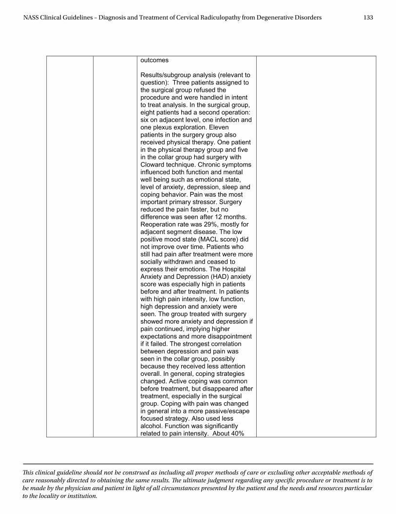
NASS Clinical Guidelines – Diagnosis and Treatment of Cervical Radiculopathy from Degenerative Disorders 133
This clinical guideline should not be construed as including all proper methods of care or excluding other acceptable methods of care reasonably directed to obtaining the same results. The ultimate judgment regarding any specific procedure or treatment is to be made by the physician and patient in light of all circumstances presented by the patient and the needs and resources particular to the locality or institution.
outcomes
Results/subgroup analysis (relevant to question): Three patients assigned to the surgical group refused the procedure and were handled in intent to treat analysis. In the surgical group, eight patients had a second operation: six on adjacent level, one infection and one plexus exploration. Eleven patients in the surgery group also received physical therapy. One patient in the physical therapy group and five in the collar group had surgery with Cloward technique. Chronic symptoms influenced both function and mental well being such as emotional state, level of anxiety, depression, sleep and coping behavior. Pain was the most important primary stressor. Surgery reduced the pain faster, but no difference was seen after 12 months. Reoperation rate was 29%, mostly for adjacent segment disease. The low positive mood state (MACL score) did not improve over time. Patients who still had pain after treatment were more socially withdrawn and ceased to express their emotions. The Hospital Anxiety and Depression (HAD) anxiety score was especially high in patients before and after treatment. In patients with high pain intensity, low function, high depression and anxiety were seen. The group treated with surgery showed more anxiety and depression if pain continued, implying higher expectations and more disappointment if it failed. The strongest correlation between depression and pain was seen in the collar group, possibly because they received less attention overall. In general, coping strategies changed. Active coping was common before treatment, but disappeared after treatment, especially in the surgical group. Coping with pain was changed in general into a more passive/escape focused strategy. Also used less alcohol. Function was significantly related to pain intensity. About 40%
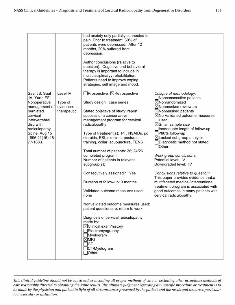
NASS Clinical Guidelines – Diagnosis and Treatment of Cervical Radiculopathy from Degenerative Disorders 134
This clinical guideline should not be construed as including all proper methods of care or excluding other acceptable methods of care reasonably directed to obtaining the same results. The ultimate judgment regarding any specific procedure or treatment is to be made by the physician and patient in light of all circumstances presented by the patient and the needs and resources particular to the locality or institution.
had anxiety only partially connected to pain. Prior to treatment, 30% of patients were depressed. After 12 months, 20% suffered from depression.
Author conclusions (relative to question): Cognitive and behavioral therapy is important to include in multidisciplinaryy rehabilitation. Patients need to improve coping strategies, self image and mood.
Saal JS, Saal JA, Yurth EF. Nonoperative management of herniated cervical intervertebral disc with radiculopathy. Spine. Aug 15 1996;21(16):1877-1883.
Level IV
Type of evidence: therapeutic
Prospective Retrospective
Study design: case series
Stated objective of study: report success of a conservative management program for cervical radiculopathy
Type of treatment(s): PT, NSAIDs, po steroids, ESI, exercise, postural training, collar, acupuncture, TENS
Total number of patients: 26; 24/26 completed program Number of patients in relevant subgroup(s):
Consecutively assigned? Yes
Duration of follow-up: 3 months
Validated outcome measures used: none
Nonvalidated outcome measures used: patient questionaire, return to work
Diagnosis of cervical radiculopathy made by:
Clinical exam/history Electromyography MyelogramMRICTCT/Myelogram Other:
Critique of methodology: Nonconsecutive patients Nonrandomized Nonmasked reviewers Nonmasked patients No Validated outcome measures used: Small sample size Inadequate length of follow-up <80% follow-up Lacked subgroup analysis Diagnostic method not stated Other:
Work group conclusions: Potential level: IV Downgraded level: IV
Conclusions relative to question: This paper provides evidence that:a multifaceted medical/interventional treatment program is associated with good outcomes in many patients with cervical radiculopathy.
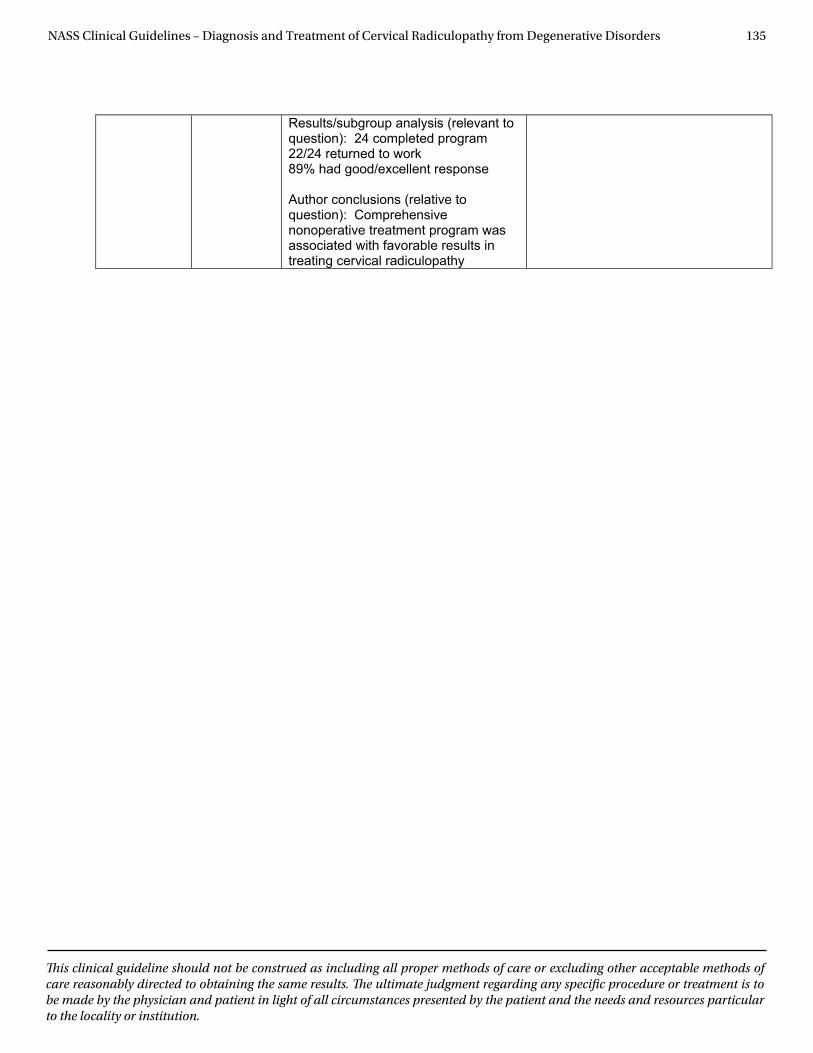
NASS Clinical Guidelines – Diagnosis and Treatment of Cervical Radiculopathy from Degenerative Disorders 135
This clinical guideline should not be construed as including all proper methods of care or excluding other acceptable methods of care reasonably directed to obtaining the same results. The ultimate judgment regarding any specific procedure or treatment is to be made by the physician and patient in light of all circumstances presented by the patient and the needs and resources particular to the locality or institution.
Results/subgroup analysis (relevant to question): 24 completed program 22/24 returned to work 89% had good/excellent response
Author conclusions (relative to question): Comprehensive nonoperative treatment program was associated with favorable results in treating cervical radiculopathy
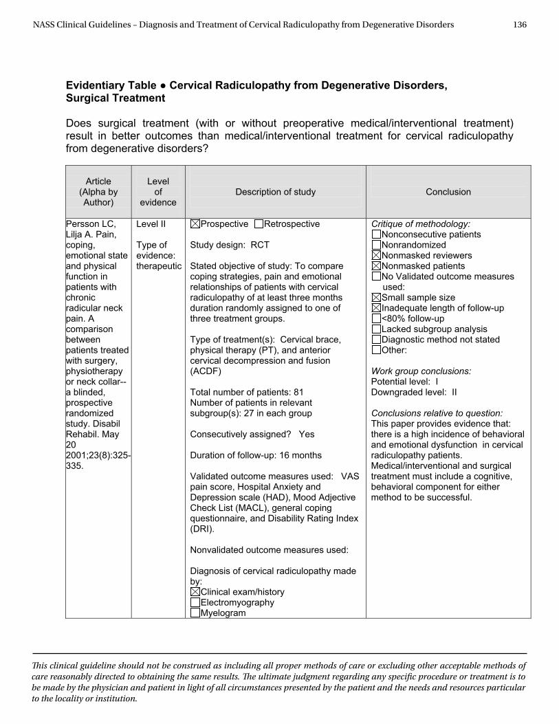
NASS Clinical Guidelines – Diagnosis and Treatment of Cervical Radiculopathy from Degenerative Disorders 136
This clinical guideline should not be construed as including all proper methods of care or excluding other acceptable methods of care reasonably directed to obtaining the same results. The ultimate judgment regarding any specific procedure or treatment is to be made by the physician and patient in light of all circumstances presented by the patient and the needs and resources particular to the locality or institution.
Evidentiary Table ● Cervical Radiculopathy from Degenerative Disorders, Surgical Treatment Does surgical treatment (with or without preoperative medical/interventional treatment) result in better outcomes than medical/interventional treatment for cervical radiculopathy from degenerative disorders?
Article
(Alpha by Author)
Level
of evidence
Description of study
Conclusion
Persson LC, Lilja A. Pain, coping, emotional state and physical function in patients with chronic radicular neck pain. A comparison between patients treated with surgery, physiotherapy or neck collar--a blinded, prospective randomized study. Disabil Rehabil. May 20 2001;23(8):325-335.
Level II Type of evidence: therapeutic
Prospective Retrospective Study design: RCT Stated objective of study: To compare coping strategies, pain and emotional relationships of patients with cervical radiculopathy of at least three months duration randomly assigned to one of three treatment groups. Type of treatment(s): Cervical brace, physical therapy (PT), and anterior cervical decompression and fusion (ACDF) Total number of patients: 81 Number of patients in relevant subgroup(s): 27 in each group Consecutively assigned? Yes Duration of follow-up: 16 months Validated outcome measures used: VAS pain score, Hospital Anxiety and Depression scale (HAD), Mood Adjective Check List (MACL), general coping questionnaire, and Disability Rating Index (DRI). Nonvalidated outcome measures used: Diagnosis of cervical radiculopathy made by:
Clinical exam/history Electromyography Myelogram
Critique of methodology: Nonconsecutive patients Nonrandomized Nonmasked reviewers Nonmasked patients No Validated outcome measures used: Small sample size Inadequate length of follow-up <80% follow-up Lacked subgroup analysis Diagnostic method not stated Other:
Work group conclusions: Potential level: I Downgraded level: II Conclusions relative to question: This paper provides evidence that: there is a high incidence of behavioral and emotional dysfunction in cervical radiculopathy patients. Medical/interventional and surgical treatment must include a cognitive, behavioral component for either method to be successful.
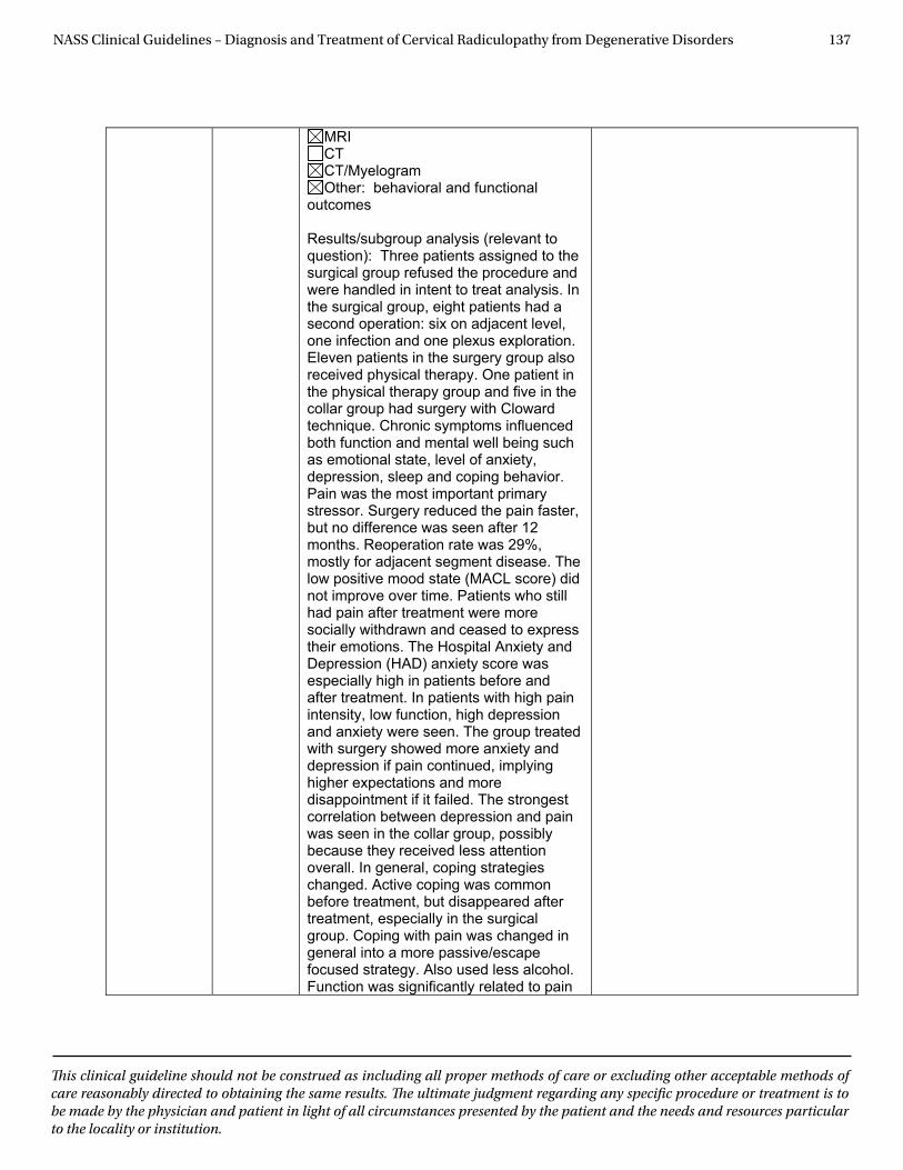
NASS Clinical Guidelines – Diagnosis and Treatment of Cervical Radiculopathy from Degenerative Disorders 137
This clinical guideline should not be construed as including all proper methods of care or excluding other acceptable methods of care reasonably directed to obtaining the same results. The ultimate judgment regarding any specific procedure or treatment is to be made by the physician and patient in light of all circumstances presented by the patient and the needs and resources particular to the locality or institution.
MRICTCT/Myelogram Other: behavioral and functional
outcomes
Results/subgroup analysis (relevant to question): Three patients assigned to the surgical group refused the procedure and were handled in intent to treat analysis. In the surgical group, eight patients had a second operation: six on adjacent level, one infection and one plexus exploration. Eleven patients in the surgery group also received physical therapy. One patient in the physical therapy group and five in the collar group had surgery with Cloward technique. Chronic symptoms influenced both function and mental well being such as emotional state, level of anxiety, depression, sleep and coping behavior. Pain was the most important primary stressor. Surgery reduced the pain faster, but no difference was seen after 12 months. Reoperation rate was 29%, mostly for adjacent segment disease. The low positive mood state (MACL score) did not improve over time. Patients who still had pain after treatment were more socially withdrawn and ceased to express their emotions. The Hospital Anxiety and Depression (HAD) anxiety score was especially high in patients before and after treatment. In patients with high pain intensity, low function, high depression and anxiety were seen. The group treated with surgery showed more anxiety and depression if pain continued, implying higher expectations and more disappointment if it failed. The strongest correlation between depression and pain was seen in the collar group, possibly because they received less attention overall. In general, coping strategies changed. Active coping was common before treatment, but disappeared after treatment, especially in the surgical group. Coping with pain was changed in general into a more passive/escape focused strategy. Also used less alcohol. Function was significantly related to pain

NASS Clinical Guidelines – Diagnosis and Treatment of Cervical Radiculopathy from Degenerative Disorders 138
This clinical guideline should not be construed as including all proper methods of care or excluding other acceptable methods of care reasonably directed to obtaining the same results. The ultimate judgment regarding any specific procedure or treatment is to be made by the physician and patient in light of all circumstances presented by the patient and the needs and resources particular to the locality or institution.
intensity. About 40% had anxiety only partially connected to pain. Prior to treatment, 30% of patients were depressed. After 12 months, 20% suffered from depression.
Author conclusions (relative to question): Cognitive and behavioral therapy is important to include in multidisciplinaryy rehabilitation. Patients need to improve coping strategies, self image and mood.
Persson LC, Moritz U, Brandt L, Carlsson CA. Cervical radiculopathy: pain, muscle weakness and sensory loss in patients with cervical radiculopathy treated with surgery, physiotherapy or cervical collar. A prospective, controlledstudy. Eur Spine J. 1997;6(4):256-266.
Level II
Type of evidence: therapeutic
Prospective Retrospective
Study design: RCT
Stated objective of study: To compare outcomes in pain, strength and sensation in three treatment groups of patients with cervical radiculopathy of a minimum of three months duration
Type of treatment(s): Cervical brace, physical therapy (PT), and anterior cervical decompression and fusion (ACDF) (Cloward technique)
Total number of patients: 81 Number of patients in relevant subgroup(s): 27 in each group.
Consecutively assigned? Yes
Duration of follow-up: 16 months
Validated outcome measures used: VAS pain scale, muscle strength assessed by a handheld dynamometer, vigorometer and pinchometer. Sensory loss recorded
Nonvalidated outcome measures used: Diagnosis of cervical radiculopathy made by:
Clinical exam/history Electromyography MyelogramMRICTCT/Myelogram Other:
Critique of methodology: Nonconsecutive patients Nonrandomized Nonmasked reviewers Nonmasked patients No Validated outcome measures used: Small sample size Inadequate length of follow-up <80% follow-up Lacked subgroup analysis Diagnostic method not stated Other:
Work group conclusions: Potential level: I Downgraded level: II
Conclusions relative to question: This paper provides evidence that:at one year, outcomes are similar for medical/interventional treatment and surgical treatment of patients with cervical radiculopathy from degenerative disorders. Due to the small sample size, one may not expect to see a difference between the groups on a statistical basis. Surgical treatment resulted in improved outcomes earlier in the postoperative treatment period when compared with the medical/interventional treatment group.

NASS Clinical Guidelines – Diagnosis and Treatment of Cervical Radiculopathy from Degenerative Disorders 139
This clinical guideline should not be construed as including all proper methods of care or excluding other acceptable methods of care reasonably directed to obtaining the same results. The ultimate judgment regarding any specific procedure or treatment is to be made by the physician and patient in light of all circumstances presented by the patient and the needs and resources particular to the locality or institution.
Results/subgroup analysis (relevant to question): Three surgical patients refused the procedure and were handled in intent to treat analysis. In the surgical group, eight patients had a second operation: six on adjacent level, one infection and one plexus exploration. Eleven patients in the surgery group also received physical therapy. One patient in the physical therapy group and five in the collar group had surgery with Cloward technique. Strength measurements were all performed by one physical therapist with standard protocol. Physical therapy was done for 15 visits and was not standardized. Several different collars were used and worn for three months. At four month follow-up, pain was improved in the surgical and physical therapy groups, and improvement in pain scores in the surgical group was significantly better than in the collar group. After another year, the pain was about the same across groups. The surgical group improved strength a little faster, but at final follow-up strength improvement was equal across groups. At final follow-up, there was no difference between groups on the sensory exam.
Author conclusions (relative to question): No difference in outcomes after one year between patients treated with a collar, physical therapy or surgery.
Sampath P, Bendebba M, Davis JD, Ducker T. Outcome in patients with cervical radiculopathy. Prospective, multicenterstudy with independent clinical review. Spine. Mar 15 1999;24(6):591-
Level III
Type of evidence: therapeutic
Prospective Retrospective
Study design: comparative
Stated objective of study: Evaluated clinical outcomes in patients with cervical radiculopathy
Type of treatment(s):Medical/interventional treatment was nonstandardized in this multicenter trial, and included medications, steroids, bed rest, exercise, traction, bracing, injections, chiropractic care, acupuncture and homeopathic medicine. Surgery included
Critique of methodology: Nonconsecutive patients Nonrandomized Nonmasked reviewers Nonmasked patients No Validated outcome measures
used: Small sample size Inadequate length of follow-up <80% follow-up Lacked subgroup analysis Diagnostic method not stated Other: high attrition rate,
medical/interventional and surgical treatment protocols were
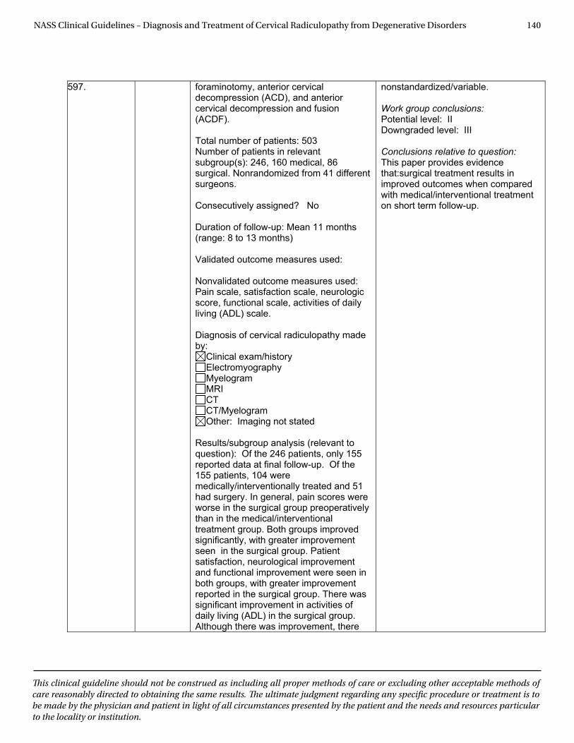
NASS Clinical Guidelines – Diagnosis and Treatment of Cervical Radiculopathy from Degenerative Disorders 140
This clinical guideline should not be construed as including all proper methods of care or excluding other acceptable methods of care reasonably directed to obtaining the same results. The ultimate judgment regarding any specific procedure or treatment is to be made by the physician and patient in light of all circumstances presented by the patient and the needs and resources particular to the locality or institution.
597. foraminotomy, anterior cervical decompression (ACD), and anterior cervical decompression and fusion (ACDF).
Total number of patients: 503 Number of patients in relevant subgroup(s): 246, 160 medical, 86 surgical. Nonrandomized from 41 different surgeons.
Consecutively assigned? No
Duration of follow-up: Mean 11 months (range: 8 to 13 months)
Validated outcome measures used:
Nonvalidated outcome measures used: Pain scale, satisfaction scale, neurologic score, functional scale, activities of daily living (ADL) scale.
Diagnosis of cervical radiculopathy made by:
Clinical exam/history Electromyography MyelogramMRICTCT/Myelogram Other: Imaging not stated
Results/subgroup analysis (relevant to question): Of the 246 patients, only 155 reported data at final follow-up. Of the 155 patients, 104 were medically/interventionally treated and 51 had surgery. In general, pain scores were worse in the surgical group preoperatively than in the medical/interventional treatment group. Both groups improved significantly, with greater improvement seen in the surgical group. Patient satisfaction, neurological improvement and functional improvement were seen in both groups, with greater improvement reported in the surgical group. There was significant improvement in activities of daily living (ADL) in the surgical group. Although there was improvement, there
nonstandardized/variable.
Work group conclusions: Potential level: II Downgraded level: III
Conclusions relative to question: This paper provides evidence that:surgical treatment results in improved outcomes when compared with medical/interventional treatment on short term follow-up.

NASS Clinical Guidelines – Diagnosis and Treatment of Cervical Radiculopathy from Degenerative Disorders 141
This clinical guideline should not be construed as including all proper methods of care or excluding other acceptable methods of care reasonably directed to obtaining the same results. The ultimate judgment regarding any specific procedure or treatment is to be made by the physician and patient in light of all circumstances presented by the patient and the needs and resources particular to the locality or institution.
was still significant pain in about 26% of surgical patients. The number returning to work did not differ before and after intervention in either group despite improved functional ability, implying that the most important factor for return to work was work status prior to treatment.
Author conclusions (relative to question): Surgery appears to have more success than medical/interventional treatment, although both help. Despite this, a substantial percentage of patients continue to have severe pain, neurologic symptoms and no work activity.
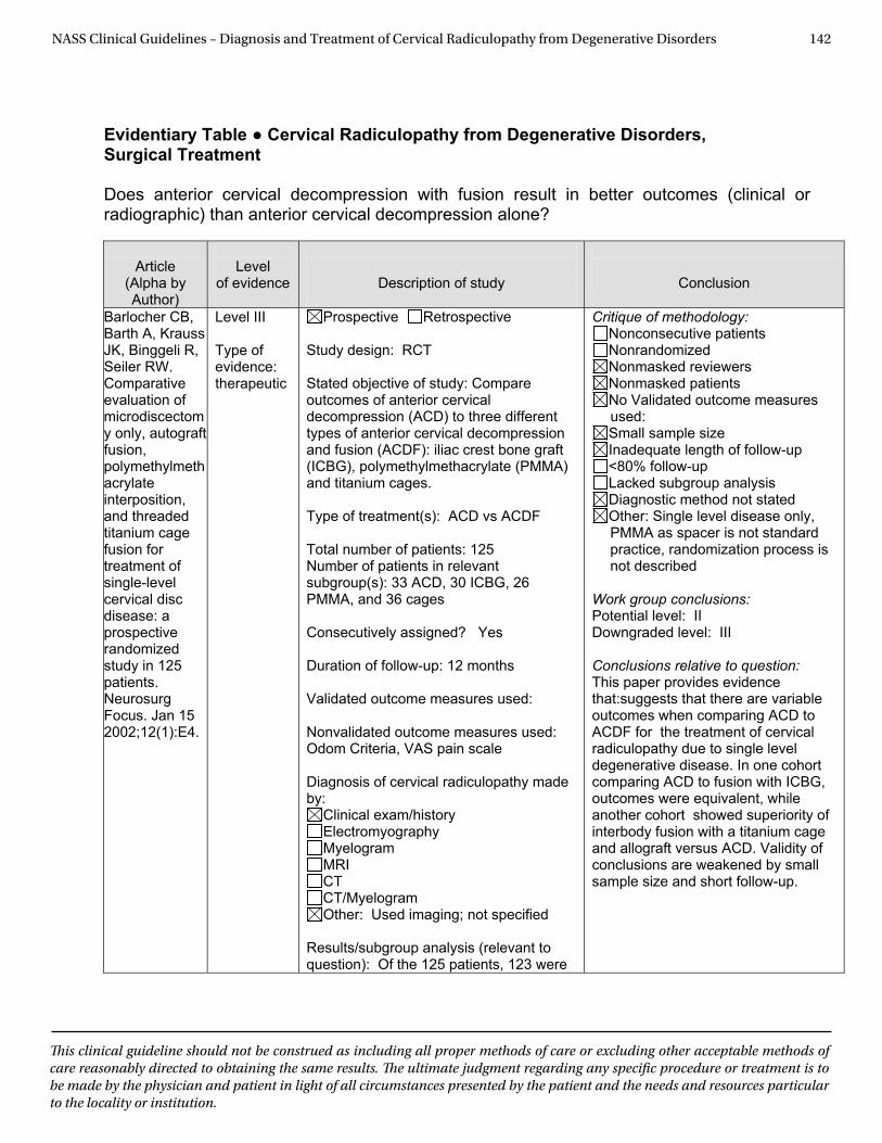
NASS Clinical Guidelines – Diagnosis and Treatment of Cervical Radiculopathy from Degenerative Disorders 142
This clinical guideline should not be construed as including all proper methods of care or excluding other acceptable methods of care reasonably directed to obtaining the same results. The ultimate judgment regarding any specific procedure or treatment is to be made by the physician and patient in light of all circumstances presented by the patient and the needs and resources particular to the locality or institution.
Evidentiary Table ● Cervical Radiculopathy from Degenerative Disorders, Surgical Treatment Does anterior cervical decompression with fusion result in better outcomes (clinical or radiographic) than anterior cervical decompression alone?
Article
(Alpha by Author)
Level
of evidence
Description of study
Conclusion
Barlocher CB, Barth A, Krauss JK, Binggeli R, Seiler RW. Comparative evaluation of microdiscectomy only, autograft fusion, polymethylmethacrylate interposition, and threaded titanium cage fusion for treatment of single-level cervical disc disease: a prospective randomized study in 125 patients. Neurosurg Focus. Jan 15 2002;12(1):E4.
Level III Type of evidence: therapeutic
Prospective Retrospective Study design: RCT Stated objective of study: Compare outcomes of anterior cervical decompression (ACD) to three different types of anterior cervical decompression and fusion (ACDF): iliac crest bone graft (ICBG), polymethylmethacrylate (PMMA) and titanium cages. Type of treatment(s): ACD vs ACDF Total number of patients: 125 Number of patients in relevant subgroup(s): 33 ACD, 30 ICBG, 26 PMMA, and 36 cages Consecutively assigned? Yes Duration of follow-up: 12 months Validated outcome measures used: Nonvalidated outcome measures used: Odom Criteria, VAS pain scale Diagnosis of cervical radiculopathy made by:
Clinical exam/history Electromyography Myelogram MRI CT CT/Myelogram Other: Used imaging; not specified
Results/subgroup analysis (relevant to question): Of the 125 patients, 123 were
Critique of methodology: Nonconsecutive patients Nonrandomized Nonmasked reviewers Nonmasked patients No Validated outcome measures used: Small sample size Inadequate length of follow-up <80% follow-up Lacked subgroup analysis Diagnostic method not stated Other: Single level disease only, PMMA as spacer is not standard practice, randomization process is not described
Work group conclusions: Potential level: II Downgraded level: III Conclusions relative to question: This paper provides evidence that:suggests that there are variable outcomes when comparing ACD to ACDF for the treatment of cervical radiculopathy due to single level degenerative disease. In one cohort comparing ACD to fusion with ICBG, outcomes were equivalent, while another cohort showed superiority of interbody fusion with a titanium cage and allograft versus ACD. Validity of conclusions are weakened by small sample size and short follow-up.

NASS Clinical Guidelines – Diagnosis and Treatment of Cervical Radiculopathy from Degenerative Disorders 143
This clinical guideline should not be construed as including all proper methods of care or excluding other acceptable methods of care reasonably directed to obtaining the same results. The ultimate judgment regarding any specific procedure or treatment is to be made by the physician and patient in light of all circumstances presented by the patient and the needs and resources particular to the locality or institution.
available for follow-up. The functional outcomes were grouped by good and excellent to poor and fair, with good/excellent results reported for 75% of the ACDF group, 80% for ICBG, 87% for PMMA and 94% for cage. Average reported kyphosis for ACD patients was 24 degrees, with one patient requiring revision surgery (31 degrees); 12 degrees for PMMA and about three degrees for the ICBG and cage groups. Twelve month fusion results were reported as 93% for the ACD patients, 93% for ICBG and 97% for cage . Fusion rate was faster in the cage group as well with 86% achieving fusion at six months compared with 61% in the ACD group and 65% in the ICBG group.
Author conclusions (relative to question): Concluded that ACDF with cage did significantly better with faster and better recovery and less kyphotic deformity than ACD. ACD Compared to: ICBG had similar outcomes at medium length follow-up.
Hauerberg J, Kosteljanetz M, Boge-Rasmussen T, et al. Anterior cervical discectomy with or without fusion with ray titanium cage: a prospective randomized clinical study. Spine. Mar 1 2008;33(5):458-464.
Level II
Type of evidence: therapeutic
Prospective Retrospective
Study design: RCT
Stated objective of study: Compare radiographic and clinical outcomes of ACD with ACDF using a titanium cage.
Type of treatment(s): anterior cervical discectomy (ACD), anterior cervical discectomy with fusion ( ACDF) at one level only in subaxial cervical spine
Total number of patients: 86 Number of patients in relevant subgroup(s): 46 ACD and 40 ACDF
Consecutively assigned? Yes
Duration of follow-up: minimum two years
Validated outcome measures used: none
Nonvalidated outcome measures used:
Critique of methodology: Nonconsecutive patients Nonrandomized Nonmasked reviewers Nonmasked patients No Validated outcome measures used: Small sample size Inadequate length of follow-up <80% follow-up Lacked subgroup analysis Diagnostic method not stated Other:
Work group conclusions: Potential level: I Downgraded level: II
Conclusions relative to question: This paper provides evidence that: for cervical radiculopathy due to single level degenerative disease, clinical outcomes are similar at two years for patients undergoing ACD and ACDF with threaded titanium cage and local
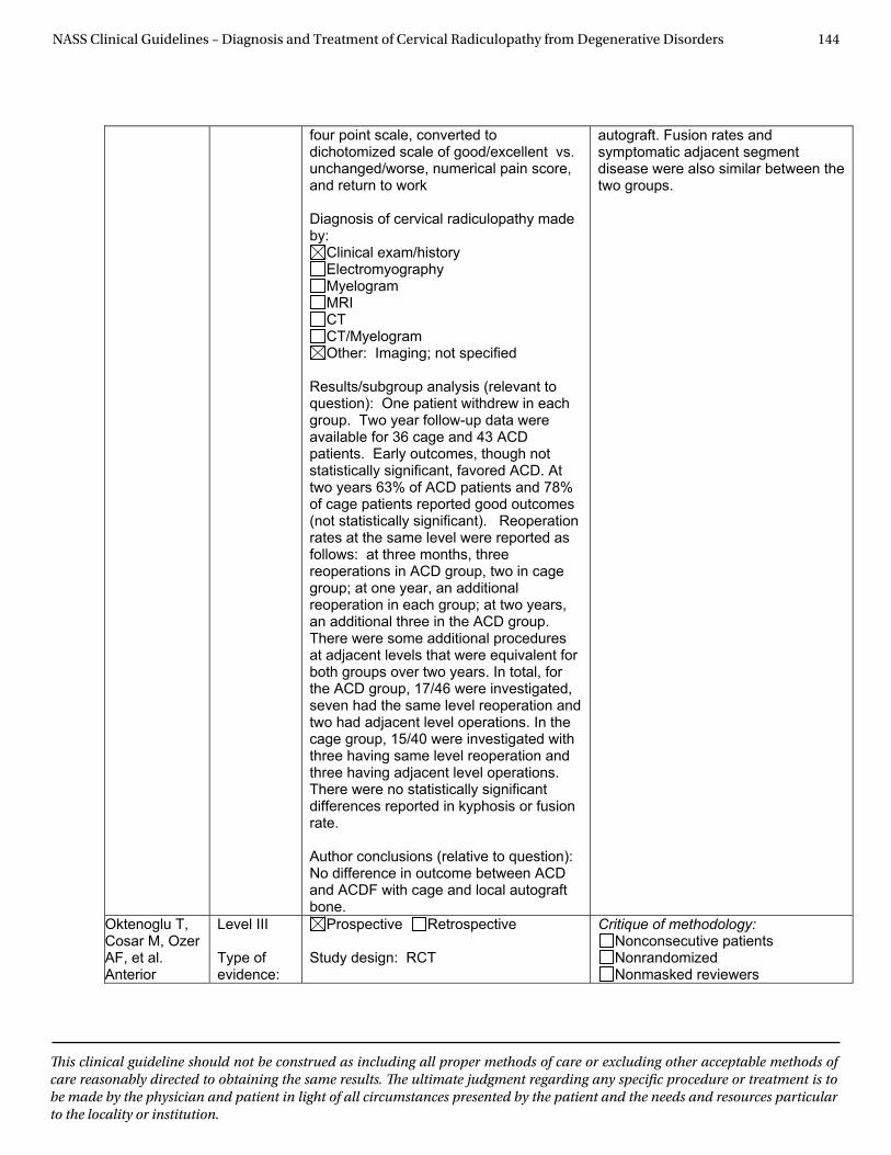
NASS Clinical Guidelines – Diagnosis and Treatment of Cervical Radiculopathy from Degenerative Disorders 144
This clinical guideline should not be construed as including all proper methods of care or excluding other acceptable methods of care reasonably directed to obtaining the same results. The ultimate judgment regarding any specific procedure or treatment is to be made by the physician and patient in light of all circumstances presented by the patient and the needs and resources particular to the locality or institution.
four point scale, converted to dichotomized scale of good/excellent vs. unchanged/worse, numerical pain score, and return to work
Diagnosis of cervical radiculopathy made by:
Clinical exam/history Electromyography MyelogramMRICTCT/Myelogram Other: Imaging; not specified
Results/subgroup analysis (relevant to question): One patient withdrew in each group. Two year follow-up data were available for 36 cage and 43 ACD patients. Early outcomes, though not statistically significant, favored ACD. At two years 63% of ACD patients and 78% of cage patients reported good outcomes (not statistically significant). Reoperation rates at the same level were reported as follows: at three months, three reoperations in ACD group, two in cage group; at one year, an additional reoperation in each group; at two years, an additional three in the ACD group. There were some additional procedures at adjacent levels that were equivalent for both groups over two years. In total, for the ACD group, 17/46 were investigated, seven had the same level reoperation and two had adjacent level operations. In the cage group, 15/40 were investigated with three having same level reoperation and three having adjacent level operations. There were no statistically significant differences reported in kyphosis or fusion rate.
Author conclusions (relative to question): No difference in outcome between ACD and ACDF with cage and local autograft bone.
autograft. Fusion rates and symptomatic adjacent segment disease were also similar between the two groups.
Oktenoglu T, Cosar M, Ozer AF, et al. Anterior
Level III
Type of evidence:
Prospective Retrospective
Study design: RCT
Critique of methodology: Nonconsecutive patients Nonrandomized Nonmasked reviewers
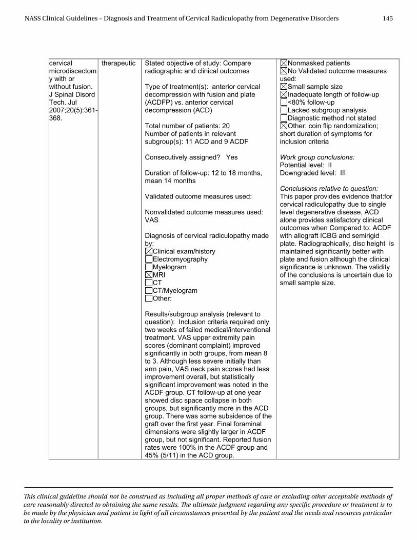
NASS Clinical Guidelines – Diagnosis and Treatment of Cervical Radiculopathy from Degenerative Disorders 145
This clinical guideline should not be construed as including all proper methods of care or excluding other acceptable methods of care reasonably directed to obtaining the same results. The ultimate judgment regarding any specific procedure or treatment is to be made by the physician and patient in light of all circumstances presented by the patient and the needs and resources particular to the locality or institution.
cervical microdiscectomy with or without fusion. J Spinal Disord Tech. Jul 2007;20(5):361-368.
therapeutic Stated objective of study: Compare radiographic and clinical outcomes
Type of treatment(s): anterior cervical decompression with fusion and plate (ACDFP) vs. anterior cervical decompression (ACD)
Total number of patients: 20 Number of patients in relevant subgroup(s): 11 ACD and 9 ACDF
Consecutively assigned? Yes
Duration of follow-up: 12 to 18 months, mean 14 months
Validated outcome measures used: Nonvalidated outcome measures used: VAS
Diagnosis of cervical radiculopathy made by:
Clinical exam/history Electromyography MyelogramMRICTCT/Myelogram Other:
Results/subgroup analysis (relevant to question): Inclusion criteria required only two weeks of failed medical/interventional treatment. VAS upper extremity pain scores (dominant complaint) improved significantly in both groups, from mean 8 to 3. Although less severe initially than arm pain, VAS neck pain scores had less improvement overall, but statistically significant improvement was noted in the ACDF group. CT follow-up at one year showed disc space collapse in both groups, but significantly more in the ACD group. There was some subsidence of the graft over the first year. Final foraminal dimensions were slightly larger in ACDF group, but not significant. Reported fusion rates were 100% in the ACDF group and 45% (5/11) in the ACD group.
Nonmasked patients No Validated outcome measures
used: Small sample size Inadequate length of follow-up <80% follow-up Lacked subgroup analysis Diagnostic method not stated Other: coin flip randomization;
short duration of symptoms for inclusion criteria
Work group conclusions: Potential level: II Downgraded level: III
Conclusions relative to question: This paper provides evidence that:for cervical radiculopathy due to single level degenerative disease, ACD alone provides satisfactory clinical outcomes when Compared to: ACDF with allograft ICBG and semirigid plate. Radiographically, disc height is maintained significantly better with plate and fusion although the clinical significance is unknown. The validity of the conclusions is uncertain due to small sample size.
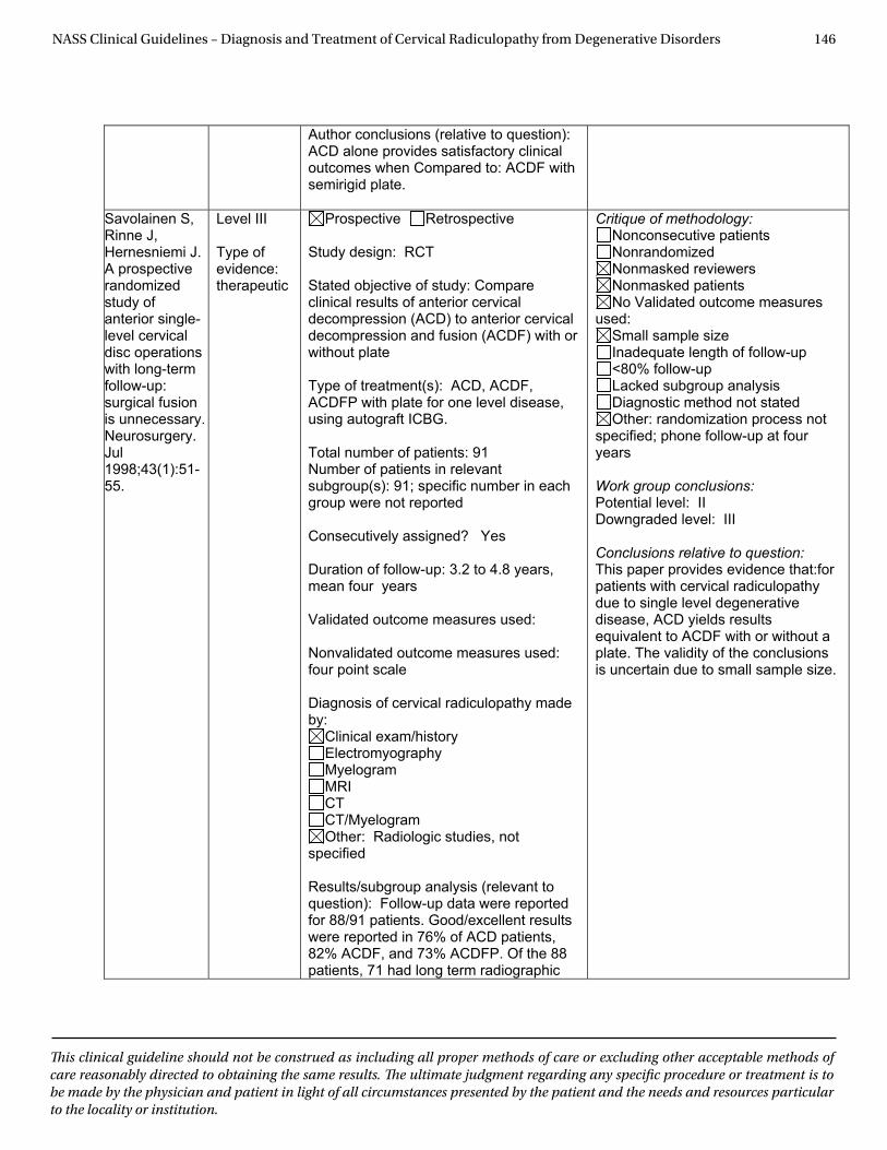
NASS Clinical Guidelines – Diagnosis and Treatment of Cervical Radiculopathy from Degenerative Disorders 146
This clinical guideline should not be construed as including all proper methods of care or excluding other acceptable methods of care reasonably directed to obtaining the same results. The ultimate judgment regarding any specific procedure or treatment is to be made by the physician and patient in light of all circumstances presented by the patient and the needs and resources particular to the locality or institution.
Author conclusions (relative to question): ACD alone provides satisfactory clinical outcomes when Compared to: ACDF with semirigid plate.
Savolainen S, Rinne J, Hernesniemi J. A prospective randomized study of anterior single-level cervical disc operations with long-term follow-up:surgical fusion is unnecessary. Neurosurgery. Jul1998;43(1):51-55.
Level III
Type of evidence: therapeutic
Prospective Retrospective
Study design: RCT
Stated objective of study: Compare clinical results of anterior cervical decompression (ACD) to anterior cervical decompression and fusion (ACDF) with or without plate
Type of treatment(s): ACD, ACDF, ACDFP with plate for one level disease, using autograft ICBG.
Total number of patients: 91 Number of patients in relevant subgroup(s): 91; specific number in each group were not reported
Consecutively assigned? Yes
Duration of follow-up: 3.2 to 4.8 years, mean four years
Validated outcome measures used: Nonvalidated outcome measures used: four point scale
Diagnosis of cervical radiculopathy made by:
Clinical exam/history Electromyography MyelogramMRICTCT/Myelogram Other: Radiologic studies, not
specified
Results/subgroup analysis (relevant to question): Follow-up data were reported for 88/91 patients. Good/excellent results were reported in 76% of ACD patients, 82% ACDF, and 73% ACDFP. Of the 88 patients, 71 had long term radiographic
Critique of methodology: Nonconsecutive patients Nonrandomized Nonmasked reviewers Nonmasked patients No Validated outcome measures
used: Small sample size Inadequate length of follow-up <80% follow-up Lacked subgroup analysis Diagnostic method not stated Other: randomization process not
specified; phone follow-up at four years
Work group conclusions: Potential level: II Downgraded level: III
Conclusions relative to question: This paper provides evidence that:for patients with cervical radiculopathy due to single level degenerative disease, ACD yields results equivalent to ACDF with or without a plate. The validity of the conclusions is uncertain due to small sample size.
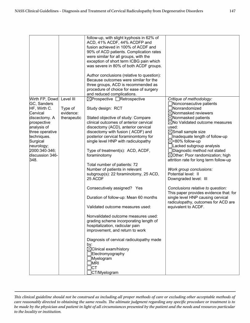
NASS Clinical Guidelines – Diagnosis and Treatment of Cervical Radiculopathy from Degenerative Disorders 147
This clinical guideline should not be construed as including all proper methods of care or excluding other acceptable methods of care reasonably directed to obtaining the same results. The ultimate judgment regarding any specific procedure or treatment is to be made by the physician and patient in light of all circumstances presented by the patient and the needs and resources particular to the locality or institution.
follow-up, with slight kyphosis in 62% of ACD, 41% ACDF, 44% ACDFP and fusion achieved in 100% of ACDF and 90% of ACD patients. Complication rates were similar for all groups, with the exception of short term ICBG pain which was severe in 80% of both ACDF groups.
Author conclusions (relative to question): Because outcomes were similar for the three groups, ACD is recommended as procedure of choice for ease of surgery and reduced complications.
Wirth FP, Dowd GC, Sanders HF, Wirth C. Cervical discectomy. A prospective analysis of three operative techniques. Surgical neurology;2000:340-346; discussion 346-348.
Level III
Type of evidence: therapeutic
Prospective Retrospective
Study design: RCT
Stated objective of study: Compare clinical outcomes of anterior cervical discectomy (ACD), anterior cervical discectomy with fusion ( ACDF) and posterior cervical foraminomtomy for single level HNP with radiculopathy
Type of treatment(s): ACD, ACDF, foraminotomy
Total number of patients: 72 Number of patients in relevant subgroup(s): 22 foraminotomy, 25 ACD, 25 ACDF
Consecutively assigned? Yes
Duration of follow-up: Mean 60 months
Validated outcome measures used: Nonvalidated outcome measures used: grading scheme incorporating length of hospitalization, radicular pain improvement, and return to work
Diagnosis of cervical radiculopathy made by:
Clinical exam/history Electromyography MyelogramMRICTCT/Myelogram
Critique of methodology: Nonconsecutive patients Nonrandomized Nonmasked reviewers Nonmasked patients No Validated outcome measures
used: Small sample size Inadequate length of follow-up <80% follow-up Lacked subgroup analysis Diagnostic method not stated Other: Poor randomization; high
attrition rate for long term follow-up
Work group conclusions: Potential level: II Downgraded level: III
Conclusions relative to question: This paper provides evidence that: for single level HNP causing cervical radiculopathy, outcomes for ACD are equivalent to ACDF.
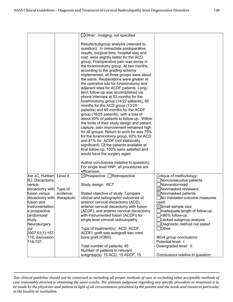
NASS Clinical Guidelines – Diagnosis and Treatment of Cervical Radiculopathy from Degenerative Disorders 148
This clinical guideline should not be construed as including all proper methods of care or excluding other acceptable methods of care reasonably directed to obtaining the same results. The ultimate judgment regarding any specific procedure or treatment is to be made by the physician and patient in light of all circumstances presented by the patient and the needs and resources particular to the locality or institution.
Other: Imaging; not specified
Results/subgroup analysis (relevant to question): In immediate postoperative results, surgical time, hospital stay and cost were slightly better for the ACD group. Postoperative pain was worse in the foraminotomy group. At two months, according to the grading scheme implemented, all three groups were about the same. Reoperations were greater at the operative site for foraminotomy and adjacent sites for ACDF patients. Long-term follow-up was accomplished via phone interview at 53 months for the foraminotomy group (14/22 patients), 56 months for the ACD group (13/25 patients) and 69 months for the ACDF group (16/25 patients), with a loss of about 40% of patients to follow-up. Within the limits of their study design and patient capture, pain improvement remained high for all groups. Return to work for was 79% for the foraminotomy group, 92% for ACD and 81% for ACDF (not statisically significant). Of the patients available at final follow-up, 100% were satisfied and would have the surgery again.
Author conclusions (relative to question): For single level HNP, all procedures are efficacious.
Xie JC, Hurlbert RJ. Discectomy versus discectomy with fusion versus discectomy with fusion and instrumentation: a prospective randomized study.Neurosurgery. Jul2007;61(1):107-116; discussion 116-107.
Level II
Type of evidence: therapeutic
Prospective Retrospective
Study design: RCT
Stated objective of study: Compare clinical and radiographic outcomes of anterior cervical discectomy (ACD), anterior cervical discectomy with fusion (ACDF), and anterior cervical discectomy with instrumented fusion (ACDFI) for single level cervical radiculopathy
Type of treatment(s): ACD, ACDF, ACDFI; graft was autograft iliac crest bone graft (ICBG)
Total number of patients: 45 Number of patients in relevant subgroup(s): 15 ACD, 15 ADCF, 15
Critique of methodology: Nonconsecutive patients Nonrandomized Nonmasked reviewers Nonmasked patients No Validated outcome measures
used: Small sample size Inadequate length of follow-up <80% follow-up Lacked subgroup analysis Diagnostic method not stated Other:
Work group conclusions: Potential level: I Downgraded level: II
Conclusions relative to question:
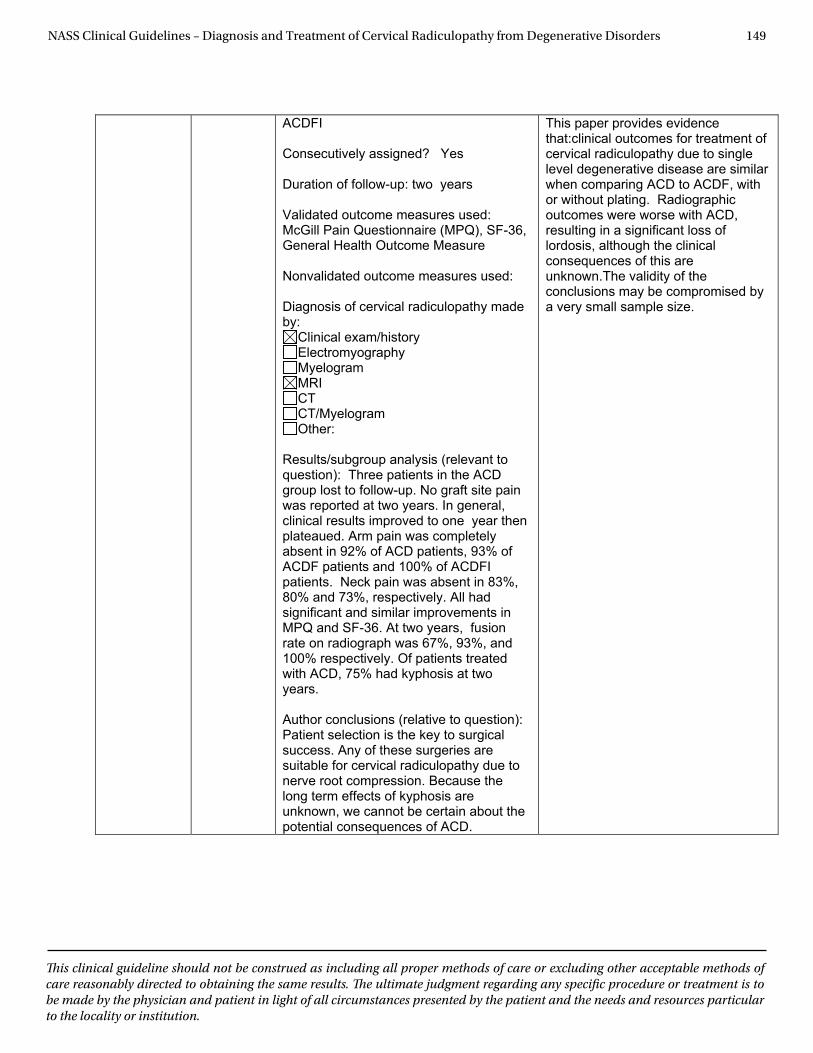
NASS Clinical Guidelines – Diagnosis and Treatment of Cervical Radiculopathy from Degenerative Disorders 149
This clinical guideline should not be construed as including all proper methods of care or excluding other acceptable methods of care reasonably directed to obtaining the same results. The ultimate judgment regarding any specific procedure or treatment is to be made by the physician and patient in light of all circumstances presented by the patient and the needs and resources particular to the locality or institution.
ACDFI
Consecutively assigned? Yes
Duration of follow-up: two years
Validated outcome measures used: McGill Pain Questionnaire (MPQ), SF-36, General Health Outcome Measure
Nonvalidated outcome measures used: Diagnosis of cervical radiculopathy made by:
Clinical exam/history Electromyography MyelogramMRICTCT/Myelogram Other:
Results/subgroup analysis (relevant to question): Three patients in the ACD group lost to follow-up. No graft site pain was reported at two years. In general, clinical results improved to one year then plateaued. Arm pain was completely absent in 92% of ACD patients, 93% of ACDF patients and 100% of ACDFI patients. Neck pain was absent in 83%, 80% and 73%, respectively. All had significant and similar improvements in MPQ and SF-36. At two years, fusion rate on radiograph was 67%, 93%, and 100% respectively. Of patients treated with ACD, 75% had kyphosis at two years.
Author conclusions (relative to question): Patient selection is the key to surgical success. Any of these surgeries are suitable for cervical radiculopathy due to nerve root compression. Because the long term effects of kyphosis are unknown, we cannot be certain about the potential consequences of ACD.
This paper provides evidence that:clinical outcomes for treatment of cervical radiculopathy due to single level degenerative disease are similar when comparing ACD to ACDF, with or without plating. Radiographic outcomes were worse with ACD, resulting in a significant loss of lordosis, although the clinical consequences of this are unknown.The validity of the conclusions may be compromised by a very small sample size.
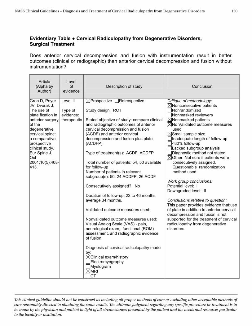
NASS Clinical Guidelines – Diagnosis and Treatment of Cervical Radiculopathy from Degenerative Disorders 150
This clinical guideline should not be construed as including all proper methods of care or excluding other acceptable methods of care reasonably directed to obtaining the same results. The ultimate judgment regarding any specific procedure or treatment is to be made by the physician and patient in light of all circumstances presented by the patient and the needs and resources particular to the locality or institution.
Evidentiary Table ● Cervical Radiculopathy from Degenerative Disorders, Surgical Treatment Does anterior cervical decompression and fusion with instrumentation result in better outcomes (clinical or radiographic) than anterior cervical decompression and fusion without instrumentation?
Article
(Alpha by Author)
Level
of evidence
Description of study
Conclusion
Grob D, Peyer JV, Dvorak J. The use of plate fixation in anterior surgery of the degenerative cervical spine: a comparative prospective clinical study. Eur Spine J. Oct 2001;10(5):408-413.
Level II Type of evidence: therapeutic
Prospective Retrospective Study design: RCT Stated objective of study: compare clinical and radiographic outcomes of anterior cervical decompression and fusion (ACDF) and anterior cervical decompression and fusion plus plate (ACDFP) Type of treatment(s): ACDF, ACDFP Total number of patients: 54, 50 available for follow-up Number of patients in relevant subgroup(s): 50: 24 ACDFP, 26 ACDF Consecutively assigned? No Duration of follow-up: 22 to 46 months, average 34 months. Validated outcome measures used: Nonvalidated outcome measures used: Visual Analog Scale (VAS) - pain, neurological exam, functional (ROM) assessment, and radiographic evidence of fusion Diagnosis of cervical radiculopathy made by:
Clinical exam/history Electromyography Myelogram MRI CT
Critique of methodology: Nonconsecutive patients Nonrandomized Nonmasked reviewers Nonmasked patients No Validated outcome measures used: Small sample size Inadequate length of follow-up <80% follow-up Lacked subgroup analysis Diagnostic method not stated Other: Not sure if patients were consecutively assigned. Questionable randomization method used.
Work group conclusions: Potential level: I Downgraded level: II Conclusions relative to question: This paper provides evidence that:use of plate in addition to anterior cervical decompression and fusion is not supported for the treatment of cervical radiculopathy from degenerative disorders.
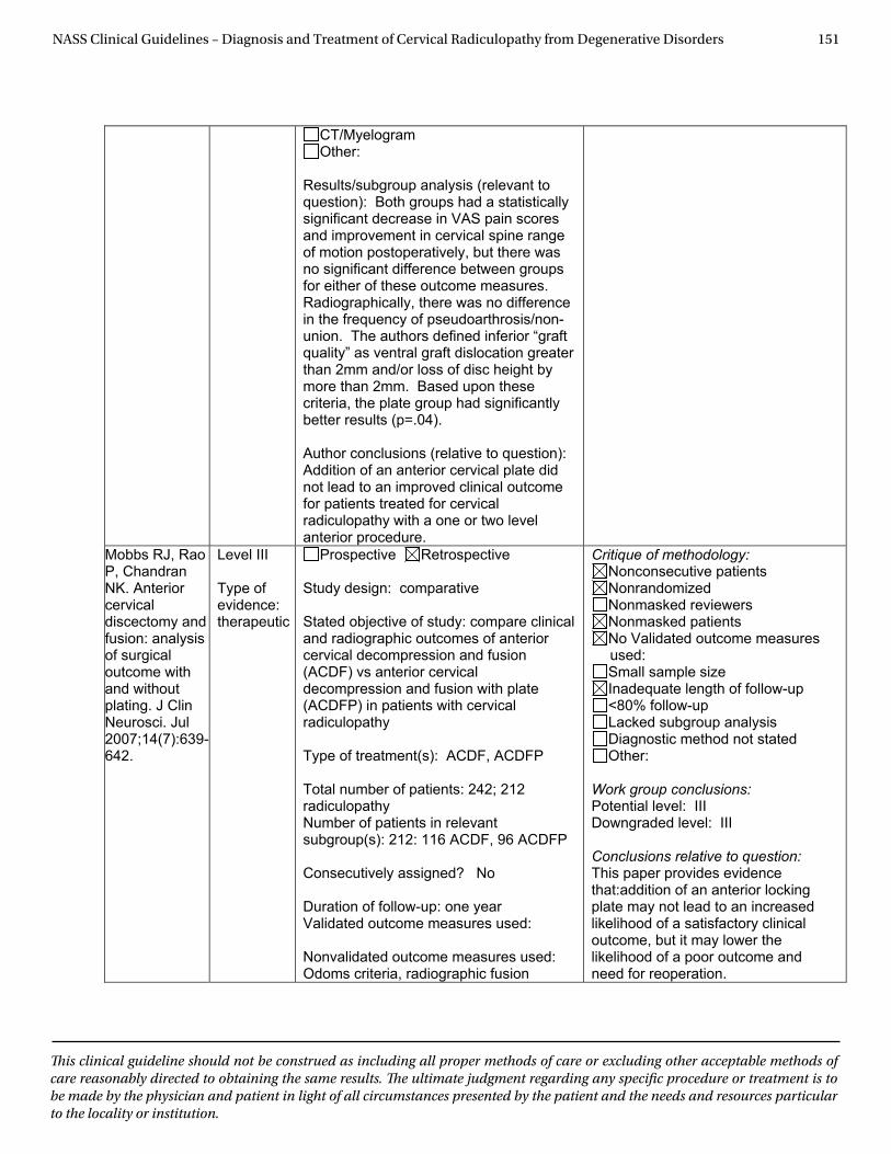
NASS Clinical Guidelines – Diagnosis and Treatment of Cervical Radiculopathy from Degenerative Disorders 151
This clinical guideline should not be construed as including all proper methods of care or excluding other acceptable methods of care reasonably directed to obtaining the same results. The ultimate judgment regarding any specific procedure or treatment is to be made by the physician and patient in light of all circumstances presented by the patient and the needs and resources particular to the locality or institution.
CT/Myelogram Other:
Results/subgroup analysis (relevant to question): Both groups had a statistically significant decrease in VAS pain scores and improvement in cervical spine range of motion postoperatively, but there was no significant difference between groups for either of these outcome measures. Radiographically, there was no difference in the frequency of pseudoarthrosis/non-union. The authors defined inferior “graft quality” as ventral graft dislocation greater than 2mm and/or loss of disc height by more than 2mm. Based upon these criteria, the plate group had significantly better results (p=.04).
Author conclusions (relative to question): Addition of an anterior cervical plate did not lead to an improved clinical outcome for patients treated for cervical radiculopathy with a one or two level anterior procedure.
Mobbs RJ, Rao P, Chandran NK. Anterior cervical discectomy and fusion: analysis of surgical outcome with and without plating. J Clin Neurosci. Jul 2007;14(7):639-642.
Level III
Type of evidence: therapeutic
Prospective Retrospective
Study design: comparative
Stated objective of study: compare clinical and radiographic outcomes of anterior cervical decompression and fusion (ACDF) vs anterior cervical decompression and fusion with plate (ACDFP) in patients with cervical radiculopathy
Type of treatment(s): ACDF, ACDFP
Total number of patients: 242; 212 radiculopathy Number of patients in relevant subgroup(s): 212: 116 ACDF, 96 ACDFP
Consecutively assigned? No
Duration of follow-up: one year Validated outcome measures used: Nonvalidated outcome measures used: Odoms criteria, radiographic fusion
Critique of methodology: Nonconsecutive patients Nonrandomized Nonmasked reviewers Nonmasked patients No Validated outcome measures used: Small sample size Inadequate length of follow-up <80% follow-up Lacked subgroup analysis Diagnostic method not stated Other:
Work group conclusions: Potential level: III Downgraded level: III
Conclusions relative to question: This paper provides evidence that:addition of an anterior locking plate may not lead to an increased likelihood of a satisfactory clinical outcome, but it may lower the likelihood of a poor outcome and need for reoperation.

NASS Clinical Guidelines – Diagnosis and Treatment of Cervical Radiculopathy from Degenerative Disorders 152
This clinical guideline should not be construed as including all proper methods of care or excluding other acceptable methods of care reasonably directed to obtaining the same results. The ultimate judgment regarding any specific procedure or treatment is to be made by the physician and patient in light of all circumstances presented by the patient and the needs and resources particular to the locality or institution.
Diagnosis of cervical radiculopathy made by:
Clinical exam/history Electromyography MyelogramMRICTCT/Myelogram Other: Imaging; not specified
Results/subgroup analysis (relevant to question): Using Odom’s criteria, there was no significant difference in good to excellent outcomes between the two groups (87% of the ACDF patient group and 92% of the ACDFP). On the other hand, the noninstrumented group had a statistically significantly higher frequency of poor outcomes at 7% (8/116) Compared to: the ACDFP group at 1% (1/96. Poor outcomes were considered to be postoperative kyphosis and nonunion.
Author conclusions (relative to question): Excellent results were similar for both groups. There was a significantly higher rate of poor outcomes in the uninstrumented group and this lead to higher rate of second surgery.
Zoega B, Karrholm J, Lind B. One-level cervical spine fusion. A randomized study, with or without plate fixation, using radiostereometry in 27 patients. Acta Orthop Scand. Aug 1998;69(4):363-368.
Level II
Type of evidence: therapeutic
Prospective Retrospective
Study design: RCT
Stated objective of study: to evaluate whether addition of a plate to a single level cervical fusion for DDD enhances fusion rate and contributes to maintaining alignment
Type of treatment(s): anterior cervical discectomy and fusion (ACDF), anterior cervical discectomy and fusion plus plate (ACDFP)
Total number of patients: 27 Number of patients in relevant subgroup(s): 15 ACDFP, 12 ACDF
Consecutively assigned? Yes
Duration of follow-up: 24 months
Critique of methodology: Nonconsecutive patients Nonrandomized Nonmasked reviewers Nonmasked patients No Validated outcome measures
used: Small sample size Inadequate length of follow-up <80% follow-up Lacked subgroup analysis Diagnostic method not stated Other:
Work group conclusions: Potential level: I Downgraded level: II
Conclusions relative to question:This paper provides evidence that:plate maintains alignment.
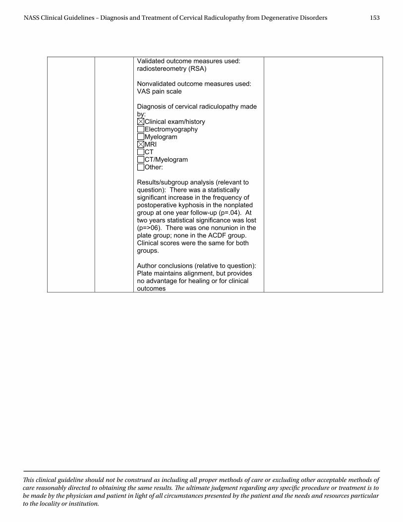
NASS Clinical Guidelines – Diagnosis and Treatment of Cervical Radiculopathy from Degenerative Disorders 153
This clinical guideline should not be construed as including all proper methods of care or excluding other acceptable methods of care reasonably directed to obtaining the same results. The ultimate judgment regarding any specific procedure or treatment is to be made by the physician and patient in light of all circumstances presented by the patient and the needs and resources particular to the locality or institution.
Validated outcome measures used: radiostereometry (RSA)
Nonvalidated outcome measures used: VAS pain scale
Diagnosis of cervical radiculopathy made by:
Clinical exam/history Electromyography MyelogramMRICTCT/Myelogram Other:
Results/subgroup analysis (relevant to question): There was a statistically significant increase in the frequency of postoperative kyphosis in the nonplated group at one year follow-up (p=.04). At two years statistical significance was lost (p=>06). There was one nonunion in the plate group; none in the ACDF group. Clinical scores were the same for both groups.
Author conclusions (relative to question): Plate maintains alignment, but provides no advantage for healing or for clinical outcomes
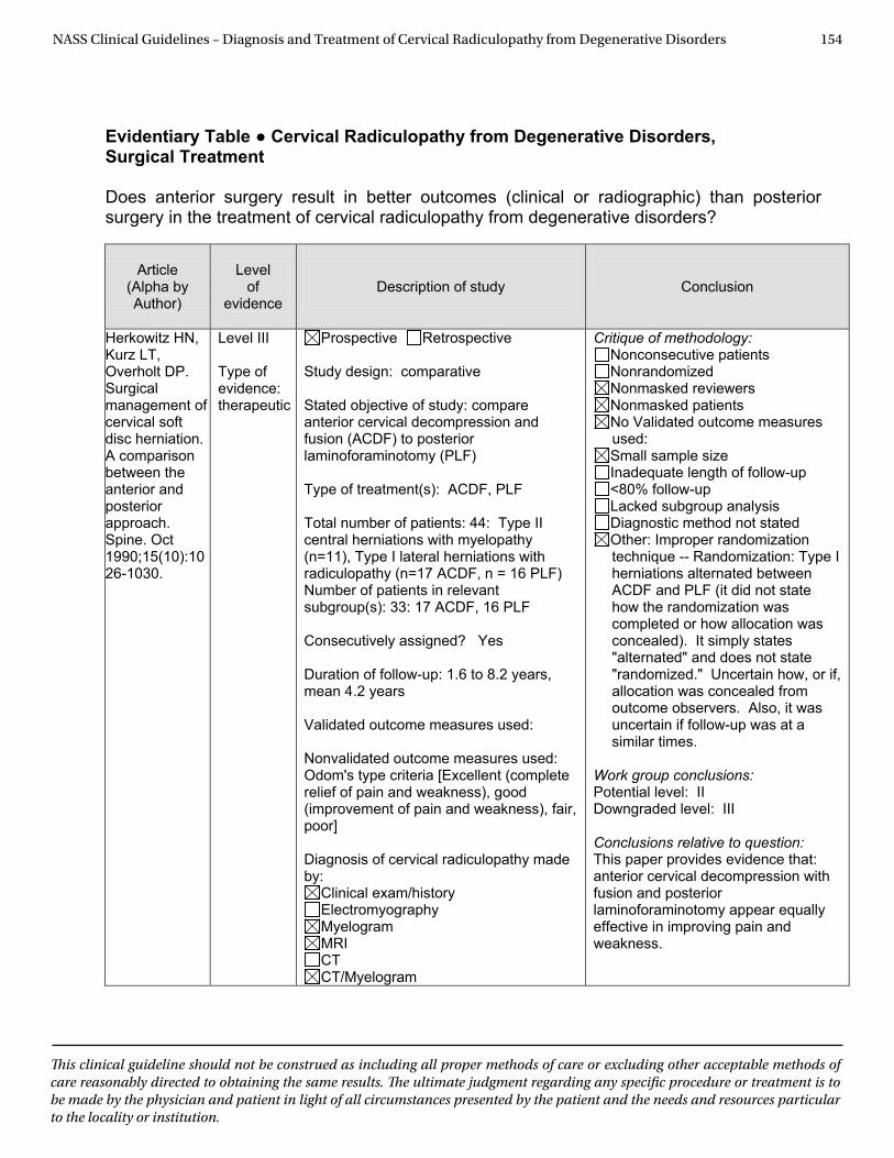
NASS Clinical Guidelines – Diagnosis and Treatment of Cervical Radiculopathy from Degenerative Disorders 154
This clinical guideline should not be construed as including all proper methods of care or excluding other acceptable methods of care reasonably directed to obtaining the same results. The ultimate judgment regarding any specific procedure or treatment is to be made by the physician and patient in light of all circumstances presented by the patient and the needs and resources particular to the locality or institution.
Evidentiary Table ● Cervical Radiculopathy from Degenerative Disorders, Surgical Treatment Does anterior surgery result in better outcomes (clinical or radiographic) than posterior surgery in the treatment of cervical radiculopathy from degenerative disorders?
Article
(Alpha by Author)
Level
of evidence
Description of study
Conclusion
Herkowitz HN, Kurz LT, Overholt DP. Surgical management of cervical soft disc herniation. A comparison between the anterior and posterior approach. Spine. Oct 1990;15(10):1026-1030.
Level III Type of evidence: therapeutic
Prospective Retrospective Study design: comparative Stated objective of study: compare anterior cervical decompression and fusion (ACDF) to posterior laminoforaminotomy (PLF) Type of treatment(s): ACDF, PLF Total number of patients: 44: Type II central herniations with myelopathy (n=11), Type I lateral herniations with radiculopathy (n=17 ACDF, n = 16 PLF) Number of patients in relevant subgroup(s): 33: 17 ACDF, 16 PLF Consecutively assigned? Yes Duration of follow-up: 1.6 to 8.2 years, mean 4.2 years Validated outcome measures used: Nonvalidated outcome measures used: Odom's type criteria [Excellent (complete relief of pain and weakness), good (improvement of pain and weakness), fair, poor] Diagnosis of cervical radiculopathy made by:
Clinical exam/history Electromyography Myelogram MRI CT CT/Myelogram
Critique of methodology: Nonconsecutive patients Nonrandomized Nonmasked reviewers Nonmasked patients No Validated outcome measures used: Small sample size Inadequate length of follow-up <80% follow-up Lacked subgroup analysis Diagnostic method not stated Other: Improper randomization technique -- Randomization: Type I herniations alternated between ACDF and PLF (it did not state how the randomization was completed or how allocation was concealed). It simply states "alternated" and does not state "randomized." Uncertain how, or if, allocation was concealed from outcome observers. Also, it was uncertain if follow-up was at a similar times.
Work group conclusions: Potential level: II Downgraded level: III Conclusions relative to question: This paper provides evidence that: anterior cervical decompression with fusion and posterior laminoforaminotomy appear equally effective in improving pain and weakness.
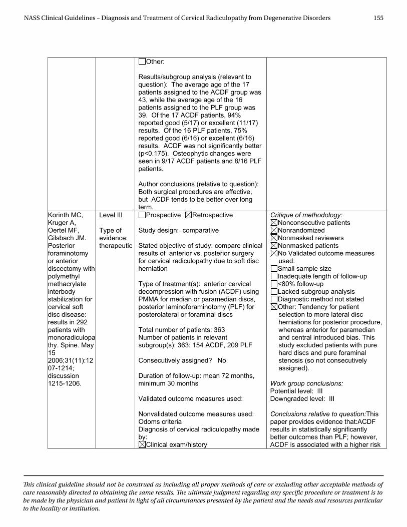
NASS Clinical Guidelines – Diagnosis and Treatment of Cervical Radiculopathy from Degenerative Disorders 155
This clinical guideline should not be construed as including all proper methods of care or excluding other acceptable methods of care reasonably directed to obtaining the same results. The ultimate judgment regarding any specific procedure or treatment is to be made by the physician and patient in light of all circumstances presented by the patient and the needs and resources particular to the locality or institution.
Other:
Results/subgroup analysis (relevant to question): The average age of the 17 patients assigned to the ACDF group was 43, while the average age of the 16 patients assigned to the PLF group was 39. Of the 17 ACDF patients, 94% reported good (5/17) or excellent (11/17) results. Of the 16 PLF patients, 75% reported good (6/16) or excellent (6/16) results. ACDF was not significantly better (p<0.175). Osteophytic changes were seen in 9/17 ACDF patients and 8/16 PLF patients.
Author conclusions (relative to question): Both surgical procedures are effective, but ACDF tends to be better over long term.
Korinth MC, Kruger A, Oertel MF, Gilsbach JM. Posterior foraminotomyor anterior discectomy with polymethylmethacrylateinterbodystabilization for cervical soft disc disease: results in 292 patients with monoradiculopathy. Spine. May 152006;31(11):1207-1214; discussion 1215-1206.
Level III
Type of evidence: therapeutic
Prospective Retrospective
Study design: comparative
Stated objective of study: compare clinical results of anterior vs. posterior surgery for cervical radiculopathy due to soft disc herniation
Type of treatment(s): anterior cervical decompression with fusion (ACDF) using PMMA for median or paramedian discs, posterior laminoforaminotomy (PLF) for posterolateral or foraminal discs
Total number of patients: 363 Number of patients in relevant subgroup(s): 363: 154 ACDF, 209 PLF
Consecutively assigned? No
Duration of follow-up: mean 72 months, minimum 30 months
Validated outcome measures used: Nonvalidated outcome measures used: Odoms criteria Diagnosis of cervical radiculopathy made by:
Clinical exam/history
Critique of methodology: Nonconsecutive patients Nonrandomized Nonmasked reviewers Nonmasked patients No Validated outcome measures used: Small sample size Inadequate length of follow-up <80% follow-up Lacked subgroup analysis Diagnostic method not stated Other: Tendency for patient selection to more lateral disc herniations for posterior procedure, whereas anterior for paramedian and central introduced bias. This study excluded patients with pure hard discs and pure foraminal stenosis (so not consecutively assigned).
Work group conclusions: Potential level: III Downgraded level: III
Conclusions relative to question:This paper provides evidence that:ACDF results in statistically significantly better outcomes than PLF; however, ACDF is associated with a higher risk
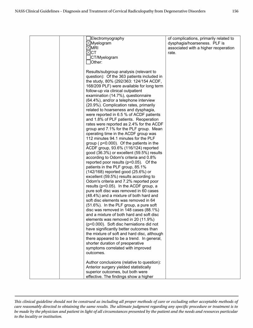
NASS Clinical Guidelines – Diagnosis and Treatment of Cervical Radiculopathy from Degenerative Disorders 156
This clinical guideline should not be construed as including all proper methods of care or excluding other acceptable methods of care reasonably directed to obtaining the same results. The ultimate judgment regarding any specific procedure or treatment is to be made by the physician and patient in light of all circumstances presented by the patient and the needs and resources particular to the locality or institution.
Electromyography MyelogramMRICTCT/Myelogram Other:
Results/subgroup analysis (relevant to question): Of the 363 patients included in the study, 80% (292/363: 124/154 ACDF, 168/209 PLF) were available for long term follow-up via clinical outpatient examination (14.7%), questionnaire (64.4%), and/or a telephone interview (20.9%). Complication rates, primarily related to hoarseness and dysphagia, were reported in 6.5 % of ACDF patients and 1.8% of PLF patients. Reoperation rates were reported as 2.4% for the ACDF group and 7.1% for the PLF group. Mean operating time in the ACDF group was 112 minutes 94.1 minutes for the PLF group ( p<0.000). Of the patients in the ACDF group, 93.6% (116/124) reported good (36.3%) or excellent (59.5%) results according to Odom's criteria and 0.8% reported poor results (p<0.05). Of the patients in the PLF group, 85.1% (142/168) reported good (25.6%) or excellent (59.5%) results according to Odom's criteria and 7.2% reported poor results (p<0.05). In the ACDF group, a pure soft disc was removed in 60 cases (48.4%) and a mixture of both hard and soft disc elements was removed in 64 (51.6%). In the PLF group, a pure soft disc was removed in 148 cases (88.1%) and a mixture of both hard and soft disc elements was removed in 20 (11.9%) (p<0.000). Soft disc herniations did not have significantly better outcomes than the mixture of soft and hard disc, although there appeared to be a trend. In general, shorter duration of preoperative symptoms correlated with improved outcomes.
Author conclusions (relative to question): Anterior surgery yielded statistically superior outcomes, but both were effective. The findings show a higher
of complications, primarily related to dysphagia/hoarseness. PLF is associated with a higher reoperation rate.
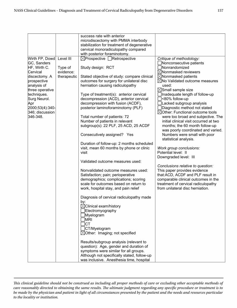
NASS Clinical Guidelines – Diagnosis and Treatment of Cervical Radiculopathy from Degenerative Disorders 157
This clinical guideline should not be construed as including all proper methods of care or excluding other acceptable methods of care reasonably directed to obtaining the same results. The ultimate judgment regarding any specific procedure or treatment is to be made by the physician and patient in light of all circumstances presented by the patient and the needs and resources particular to the locality or institution.
success rate with anterior microdiscectomy with PMMA interbody stabilization for treatment of degenerative cervical monoradiculopathy compared with posterior foraminotomy.
Wirth FP, Dowd GC, Sanders HF, Wirth C. Cervical discectomy. A prospective analysis of three operative techniques. Surg Neurol. Apr2000;53(4):340-346; discussion 346-348.
Level III
Type of evidence: therapeutic
Prospective Retrospective
Study design: RCT
Stated objective of study: compare clinical outcomes for surgery for unilateral disc herniation causing radiculopathy
Type of treatment(s): anterior cervical decompression (ACD), anterior cervical decompression with fusion (ACDF), posterior laminoforaminotomy (PLF)
Total number of patients: 72 Number of patients in relevant subgroup(s): 22 PLF, 25 ACD, 25 ACDF
Consecutively assigned? Yes
Duration of follow-up: 2 months scheduled visit, mean 60 months by phone or clinic visit
Validated outcome measures used: Nonvalidated outcome measures used: Satisfaction; pain; perioperative demographics; complications; scoring scale for outcomes based on return to work, hospital stay, and pain relief
Diagnosis of cervical radiculopathy made by:
Clinical exam/history Electromyography MyelogramMRICTCT/Myelogram Other: Imaging; not specified
Results/subgroup analysis (relevant to question): Age, gender and duration of symptoms were similar for all groups. Although not specifically stated, follow-up was inclusive. Anesthesia time, hospital
Critique of methodology: Nonconsecutive patients Nonrandomized Nonmasked reviewers Nonmasked patients No Validated outcome measures used: Small sample size Inadequate length of follow-up <80% follow-up Lacked subgroup analysis Diagnostic method not stated Other: Functional outcome tools were too broad and subjective. The initial clinical visit occurred at two months; the 60 month follow-up was poorly coordinated and varied. Numbers were small with poor statistical analysis.
Work group conclusions: Potential level: II Downgraded level: III
Conclusions relative to question: This paper provides evidence that:ACD, ACDF and PLF result in comparable clinical outcomes in the treatment of cervical radiculopathy from unilateral disc herniation.
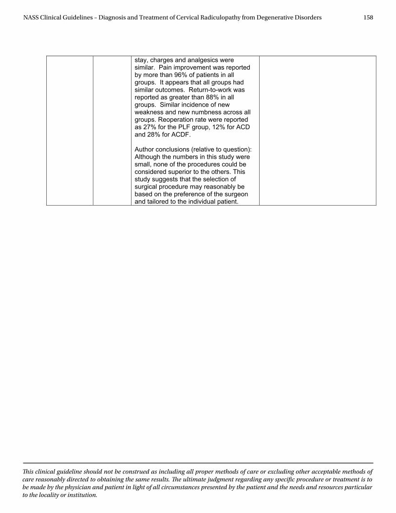
NASS Clinical Guidelines – Diagnosis and Treatment of Cervical Radiculopathy from Degenerative Disorders 158
This clinical guideline should not be construed as including all proper methods of care or excluding other acceptable methods of care reasonably directed to obtaining the same results. The ultimate judgment regarding any specific procedure or treatment is to be made by the physician and patient in light of all circumstances presented by the patient and the needs and resources particular to the locality or institution.
stay, charges and analgesics were similar. Pain improvement was reported by more than 96% of patients in all groups. It appears that all groups had similar outcomes. Return-to-work was reported as greater than 88% in all groups. Similar incidence of new weakness and new numbness across all groups. Reoperation rate were reported as 27% for the PLF group, 12% for ACD and 28% for ACDF.
Author conclusions (relative to question): Although the numbers in this study were small, none of the procedures could be considered superior to the others. This study suggests that the selection of surgical procedure may reasonably be based on the preference of the surgeon and tailored to the individual patient.
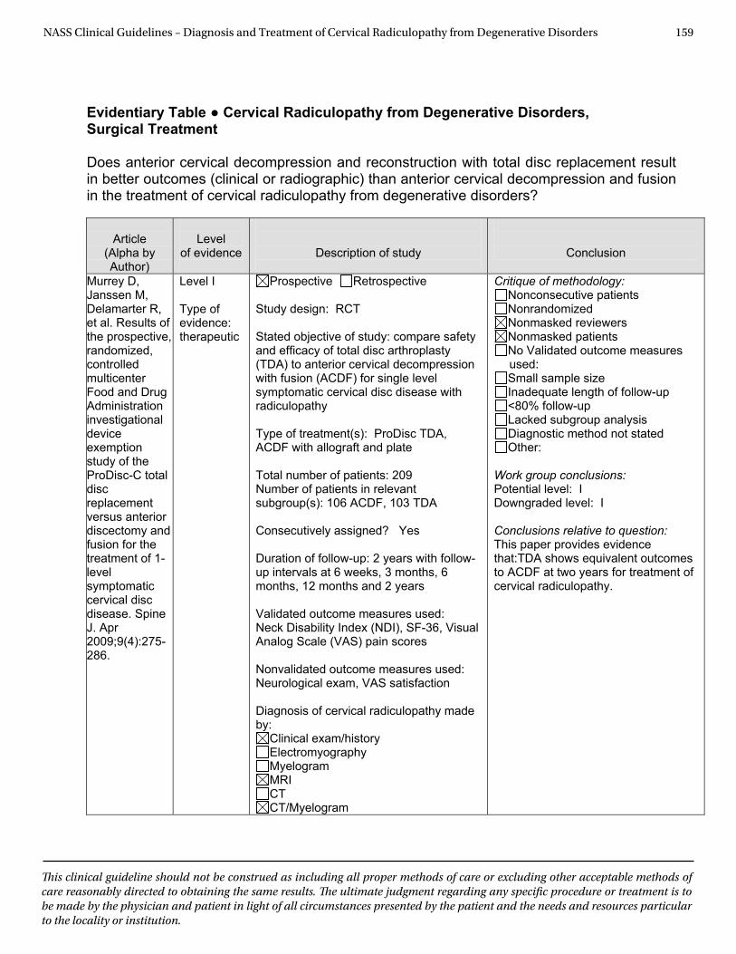
NASS Clinical Guidelines – Diagnosis and Treatment of Cervical Radiculopathy from Degenerative Disorders 159
This clinical guideline should not be construed as including all proper methods of care or excluding other acceptable methods of care reasonably directed to obtaining the same results. The ultimate judgment regarding any specific procedure or treatment is to be made by the physician and patient in light of all circumstances presented by the patient and the needs and resources particular to the locality or institution.
Evidentiary Table ● Cervical Radiculopathy from Degenerative Disorders, Surgical Treatment Does anterior cervical decompression and reconstruction with total disc replacement result in better outcomes (clinical or radiographic) than anterior cervical decompression and fusion in the treatment of cervical radiculopathy from degenerative disorders?
Article
(Alpha by Author)
Level
of evidence
Description of study
Conclusion
Murrey D, Janssen M, Delamarter R, et al. Results of the prospective, randomized, controlled multicenter Food and Drug Administration investigational device exemption study of the ProDisc-C total disc replacement versus anterior discectomy and fusion for the treatment of 1-level symptomatic cervical disc disease. Spine J. Apr 2009;9(4):275-286.
Level I Type of evidence: therapeutic
Prospective Retrospective Study design: RCT Stated objective of study: compare safety and efficacy of total disc arthroplasty (TDA) to anterior cervical decompression with fusion (ACDF) for single level symptomatic cervical disc disease with radiculopathy Type of treatment(s): ProDisc TDA, ACDF with allograft and plate Total number of patients: 209 Number of patients in relevant subgroup(s): 106 ACDF, 103 TDA Consecutively assigned? Yes Duration of follow-up: 2 years with follow-up intervals at 6 weeks, 3 months, 6 months, 12 months and 2 years Validated outcome measures used: Neck Disability Index (NDI), SF-36, Visual Analog Scale (VAS) pain scores Nonvalidated outcome measures used: Neurological exam, VAS satisfaction Diagnosis of cervical radiculopathy made by:
Clinical exam/history Electromyography Myelogram MRI CT CT/Myelogram
Critique of methodology: Nonconsecutive patients Nonrandomized Nonmasked reviewers Nonmasked patients No Validated outcome measures used: Small sample size Inadequate length of follow-up <80% follow-up Lacked subgroup analysis Diagnostic method not stated Other:
Work group conclusions: Potential level: I Downgraded level: I Conclusions relative to question: This paper provides evidence that:TDA shows equivalent outcomes to ACDF at two years for treatment of cervical radiculopathy.
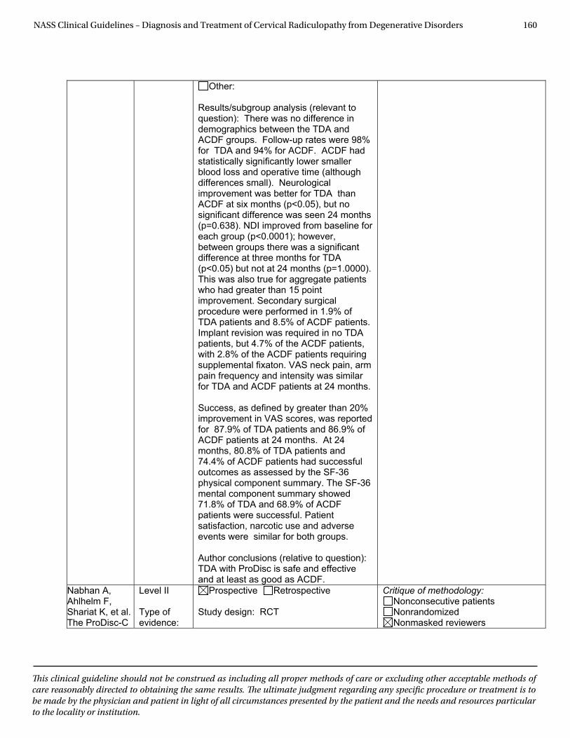
NASS Clinical Guidelines – Diagnosis and Treatment of Cervical Radiculopathy from Degenerative Disorders 160
This clinical guideline should not be construed as including all proper methods of care or excluding other acceptable methods of care reasonably directed to obtaining the same results. The ultimate judgment regarding any specific procedure or treatment is to be made by the physician and patient in light of all circumstances presented by the patient and the needs and resources particular to the locality or institution.
Other:
Results/subgroup analysis (relevant to question): There was no difference in demographics between the TDA and ACDF groups. Follow-up rates were 98% for TDA and 94% for ACDF. ACDF had statistically significantly lower smaller blood loss and operative time (although differences small). Neurological improvement was better for TDA than ACDF at six months (p<0.05), but no significant difference was seen 24 months (p=0.638). NDI improved from baseline for each group (p<0.0001); however, between groups there was a significant difference at three months for TDA (p<0.05) but not at 24 months (p=1.0000). This was also true for aggregate patients who had greater than 15 point improvement. Secondary surgical procedure were performed in 1.9% of TDA patients and 8.5% of ACDF patients. Implant revision was required in no TDA patients, but 4.7% of the ACDF patients, with 2.8% of the ACDF patients requiring supplemental fixaton. VAS neck pain, arm pain frequency and intensity was similar for TDA and ACDF patients at 24 months.
Success, as defined by greater than 20% improvement in VAS scores, was reported for 87.9% of TDA patients and 86.9% of ACDF patients at 24 months. At 24 months, 80.8% of TDA patients and 74.4% of ACDF patients had successful outcomes as assessed by the SF-36 physical component summary. The SF-36 mental component summary showed 71.8% of TDA and 68.9% of ACDF patients were successful. Patient satisfaction, narcotic use and adverse events were similar for both groups.
Author conclusions (relative to question): TDA with ProDisc is safe and effective and at least as good as ACDF.
Nabhan A, Ahlhelm F, Shariat K, et al. The ProDisc-C
Level II
Type of evidence:
Prospective Retrospective
Study design: RCT
Critique of methodology: Nonconsecutive patients Nonrandomized Nonmasked reviewers
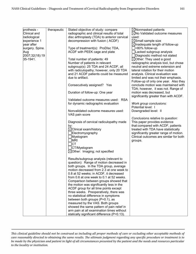
NASS Clinical Guidelines – Diagnosis and Treatment of Cervical Radiculopathy from Degenerative Disorders 161
This clinical guideline should not be construed as including all proper methods of care or excluding other acceptable methods of care reasonably directed to obtaining the same results. The ultimate judgment regarding any specific procedure or treatment is to be made by the physician and patient in light of all circumstances presented by the patient and the needs and resources particular to the locality or institution.
prothesis - Clinical and radiological experience 1 year after surgery. Spine. Aug2007;32(18):1935-1941.
therapeutic Stated objective of study: compare radiographic and clinical results of total disc arthroplasty (TDA) to anterior cervical decompression with fusion ( ACDF)
Type of treatment(s): ProDIsc TDA, ACDF with PEEK cage and plate
Total number of patients: 49 Number of patients in relevant subgroup(s): 25 TDA and 24 ACDF, all with radiculopathy; however, only 20 TDA and 21 ACDF patients could be measured due to artifact.
Consecutively assigned? Yes
Duration of follow-up: One year
Validated outcome measures used: RSA for dynamic radiographic evaluation
Nonvalidated outcome measures used: VAS pain score
Diagnosis of cervical radiculopathy made by:
Clinical exam/history Electromyography MyelogramMRICTCT/Myelogram Other: Imaging; not specified
Results/subgroup analysis (relevant to question): Range of motion decreased in both groups. In the TDA group, average motion decreased from 2.3 at one week to 0.8 at 52 weeks; in ACDF, it decreased from 0.6 at one week to 0.1 at 52 weeks. Comparison between groups showed that the motion was significantly less in the ACDF group for all time points except three weeks. Preoperatively, there was no statistical difference in symptoms between both groups (P=0.1), as measured by the VAS. Both groups showed the same pattern of pain relief in arm pain at all examination times without statically significant difference (P=0.13).
Nonmasked patients No Validated outcome measures
used: Small sample size Inadequate length of follow-up <80% follow-up Lacked subgroup analysis Diagnostic method not stated Other: They used a good
radiographic analysis tool, but chose neutral and extreme extension and lateral rotation for their motion analysis. Clinical evaluation was limited and was not their emphasis. Follow-up of only one year. Also they conclude motion was maintained with TDA; however, it was not. Range of motion was decreased, but significantly greater than with ACDF.
Work group conclusions: Potential level: I Downgraded level: II
Conclusions relative to question: This paper provides evidence that:compared with ACDF, patients treated with TDA have statistically significantly greater range of motion. Clinical outcomes are similar for both groups.
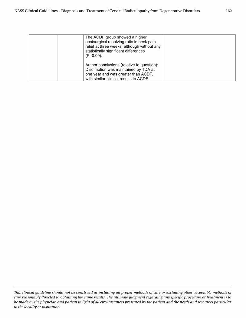
NASS Clinical Guidelines – Diagnosis and Treatment of Cervical Radiculopathy from Degenerative Disorders 162
This clinical guideline should not be construed as including all proper methods of care or excluding other acceptable methods of care reasonably directed to obtaining the same results. The ultimate judgment regarding any specific procedure or treatment is to be made by the physician and patient in light of all circumstances presented by the patient and the needs and resources particular to the locality or institution.
The ACDF group showed a higher postsurgical resolving ratio in neck pain relief at three weeks, although without any statistically significant differences (P=0.09).
Author conclusions (relative to question): Disc motion was maintained by TDA at one year and was greater than ACDF, with similar clinical results to ACDF.
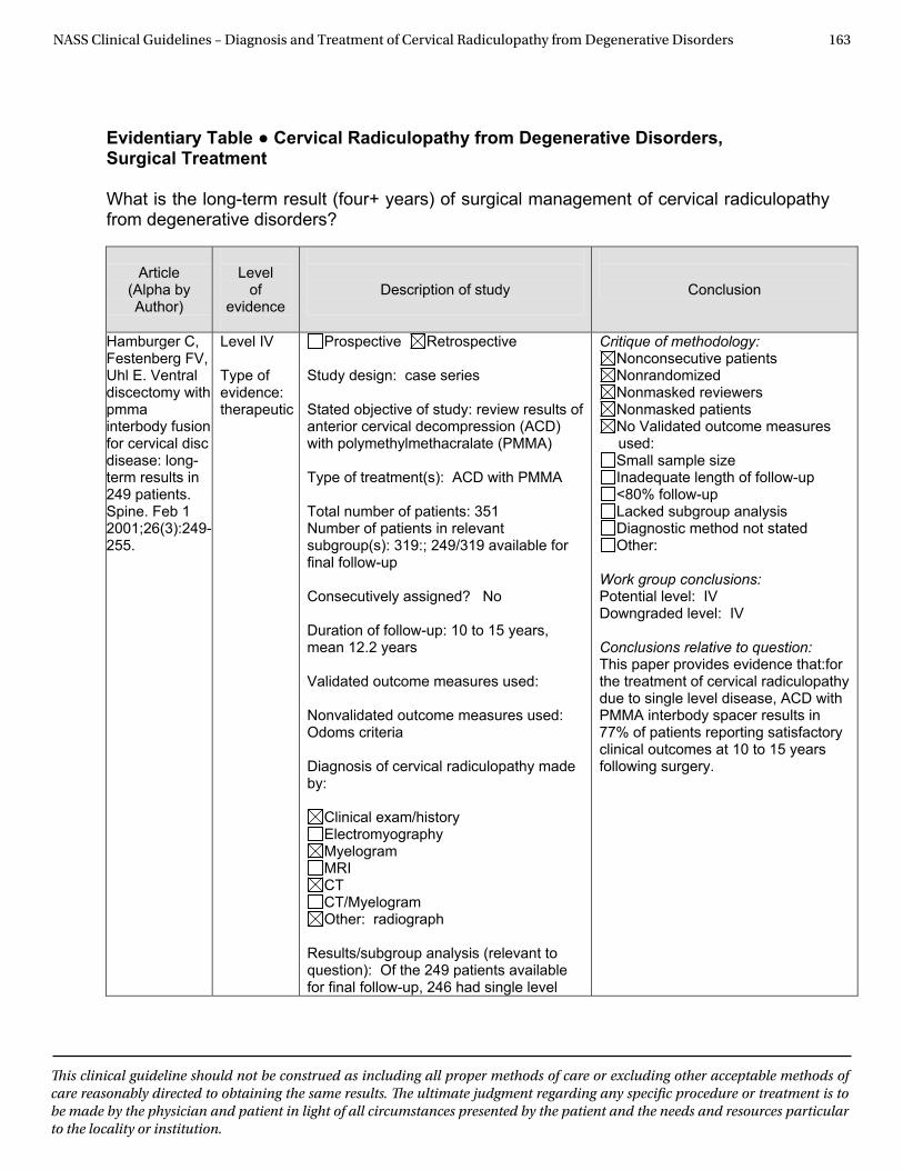
NASS Clinical Guidelines – Diagnosis and Treatment of Cervical Radiculopathy from Degenerative Disorders 163
This clinical guideline should not be construed as including all proper methods of care or excluding other acceptable methods of care reasonably directed to obtaining the same results. The ultimate judgment regarding any specific procedure or treatment is to be made by the physician and patient in light of all circumstances presented by the patient and the needs and resources particular to the locality or institution.
Evidentiary Table ● Cervical Radiculopathy from Degenerative Disorders, Surgical Treatment What is the long-term result (four+ years) of surgical management of cervical radiculopathy from degenerative disorders?
Article
(Alpha by Author)
Level
of evidence
Description of study
Conclusion
Hamburger C, Festenberg FV, Uhl E. Ventral discectomy with pmma interbody fusion for cervical disc disease: long-term results in 249 patients. Spine. Feb 1 2001;26(3):249-255.
Level IV Type of evidence: therapeutic
Prospective Retrospective Study design: case series Stated objective of study: review results of anterior cervical decompression (ACD) with polymethylmethacralate (PMMA) Type of treatment(s): ACD with PMMA Total number of patients: 351 Number of patients in relevant subgroup(s): 319:; 249/319 available for final follow-up Consecutively assigned? No Duration of follow-up: 10 to 15 years, mean 12.2 years Validated outcome measures used: Nonvalidated outcome measures used: Odoms criteria Diagnosis of cervical radiculopathy made by:
Clinical exam/history Electromyography Myelogram MRI CT CT/Myelogram Other: radiograph
Results/subgroup analysis (relevant to question): Of the 249 patients available for final follow-up, 246 had single level
Critique of methodology: Nonconsecutive patients Nonrandomized Nonmasked reviewers Nonmasked patients No Validated outcome measures used: Small sample size Inadequate length of follow-up <80% follow-up Lacked subgroup analysis Diagnostic method not stated Other:
Work group conclusions: Potential level: IV Downgraded level: IV Conclusions relative to question: This paper provides evidence that:for the treatment of cervical radiculopathy due to single level disease, ACD with PMMA interbody spacer results in 77% of patients reporting satisfactory clinical outcomes at 10 to 15 years following surgery.
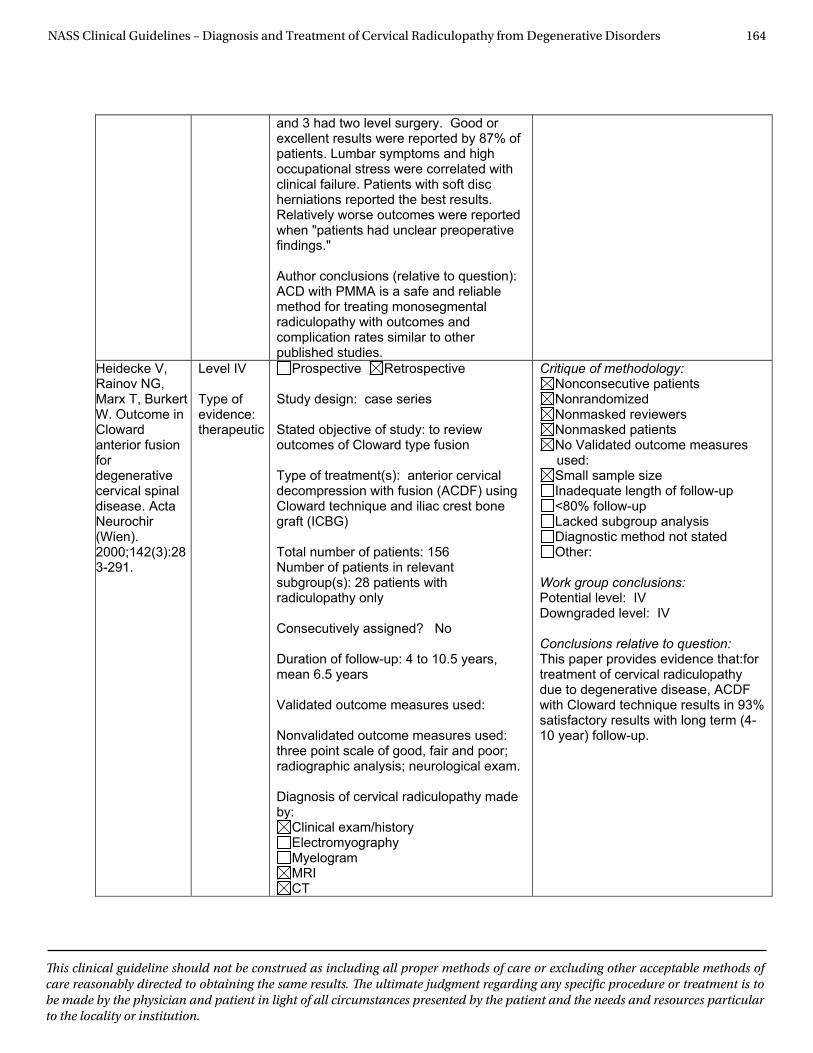
NASS Clinical Guidelines – Diagnosis and Treatment of Cervical Radiculopathy from Degenerative Disorders 164
This clinical guideline should not be construed as including all proper methods of care or excluding other acceptable methods of care reasonably directed to obtaining the same results. The ultimate judgment regarding any specific procedure or treatment is to be made by the physician and patient in light of all circumstances presented by the patient and the needs and resources particular to the locality or institution.
and 3 had two level surgery. Good or excellent results were reported by 87% of patients. Lumbar symptoms and high occupational stress were correlated with clinical failure. Patients with soft disc herniations reported the best results. Relatively worse outcomes were reported when "patients had unclear preoperative findings."
Author conclusions (relative to question): ACD with PMMA is a safe and reliable method for treating monosegmental radiculopathy with outcomes and complication rates similar to other published studies.
Heidecke V, Rainov NG, Marx T, Burkert W. Outcome in Cloward anterior fusion fordegenerative cervical spinal disease. Acta Neurochir (Wien). 2000;142(3):283-291.
Level IV
Type of evidence: therapeutic
Prospective Retrospective
Study design: case series
Stated objective of study: to review outcomes of Cloward type fusion
Type of treatment(s): anterior cervical decompression with fusion (ACDF) using Cloward technique and iliac crest bone graft (ICBG)
Total number of patients: 156 Number of patients in relevant subgroup(s): 28 patients with radiculopathy only
Consecutively assigned? No
Duration of follow-up: 4 to 10.5 years, mean 6.5 years
Validated outcome measures used: Nonvalidated outcome measures used: three point scale of good, fair and poor; radiographic analysis; neurological exam.
Diagnosis of cervical radiculopathy made by:
Clinical exam/history Electromyography MyelogramMRICT
Critique of methodology: Nonconsecutive patients Nonrandomized Nonmasked reviewers Nonmasked patients No Validated outcome measures used: Small sample size Inadequate length of follow-up <80% follow-up Lacked subgroup analysis Diagnostic method not stated Other:
Work group conclusions: Potential level: IV Downgraded level: IV
Conclusions relative to question: This paper provides evidence that:for treatment of cervical radiculopathy due to degenerative disease, ACDF with Cloward technique results in 93% satisfactory results with long term (4-10 year) follow-up.
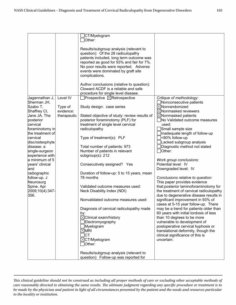
NASS Clinical Guidelines – Diagnosis and Treatment of Cervical Radiculopathy from Degenerative Disorders 165
This clinical guideline should not be construed as including all proper methods of care or excluding other acceptable methods of care reasonably directed to obtaining the same results. The ultimate judgment regarding any specific procedure or treatment is to be made by the physician and patient in light of all circumstances presented by the patient and the needs and resources particular to the locality or institution.
CT/Myelogram Other:
Results/subgroup analysis (relevant to question): Of the 28 radiculopathy patients included, long term outcome was reported as good for 93% and fair for 7%. No poor results were reported. Adverse events were dominated by graft site complications.
Author conclusions (relative to question): Cloward ACDF is a reliable and safe procedure for single level disease.
Jagannathan J, Sherman JH, Szabo T, Shaffrey CI, Jane JA. The posterior cervical foraminotomy in the treatment of cervical disc/osteophyte disease: a single-surgeon experience with a minimum of 5 years' clinical andradiographic follow-up. J Neurosurg Spine. Apr 2009;10(4):347-356.
Level IV
Type of evidence: therapeutic
Prospective Retrospective
Study design: case series
Stated objective of study: review results of posterior foraminotomy (PLF) for treatment of single level cervical radiculopathy
Type of treatment(s): PLF
Total number of patients: 973 Number of patients in relevant subgroup(s): 212
Consecutively assigned? Yes
Duration of follow-up: 5 to 15 years, mean 78 months
Validated outcome measures used: Neck Disability Index (NDI)
Nonvalidated outcome measures used: Diagnosis of cervical radiculopathy made by:
Clinical exam/history Electromyography MyelogramMRICTCT/Myelogram Other:
Results/subgroup analysis (relevant to question): Follow-up was reported for
Critique of methodology: Nonconsecutive patients Nonrandomized Nonmasked reviewers Nonmasked patients No Validated outcome measures used: Small sample size Inadequate length of follow-up <80% follow-up Lacked subgroup analysis Diagnostic method not stated Other:
Work group conclusions: Potential level: IV Downgraded level: IV
Conclusions relative to question: This paper provides evidence that:posterior laminoforaminotomy for the treatment of cervical radiculopathy due to degenerative disease results in significant improvement in 93% of cases at 5-15 year follow-up. There may be a trend for patients older than 60 years with initial lordosis of less than 10 degrees to be more vulnerable to development of postoperative cervical kyphosis or translational deformity, though the clinical significance of this is uncertain.
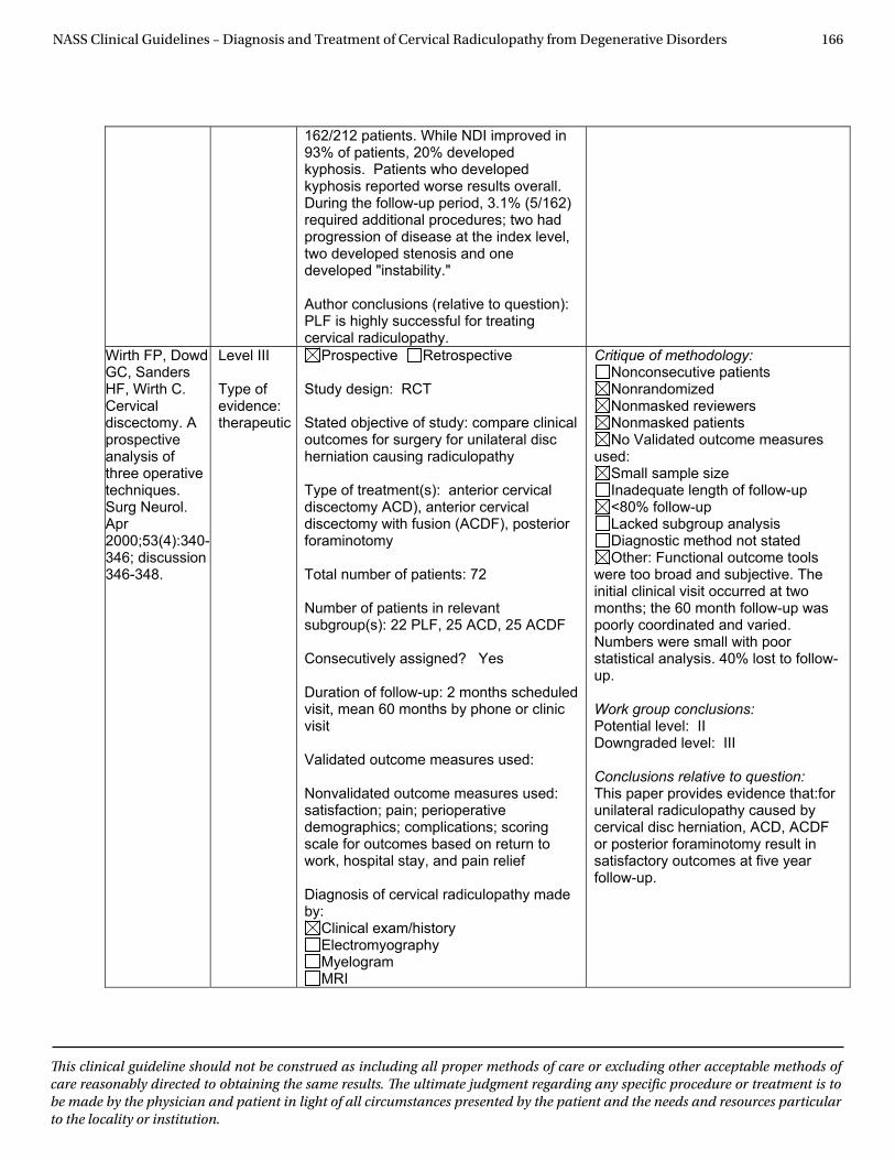
NASS Clinical Guidelines – Diagnosis and Treatment of Cervical Radiculopathy from Degenerative Disorders 166
This clinical guideline should not be construed as including all proper methods of care or excluding other acceptable methods of care reasonably directed to obtaining the same results. The ultimate judgment regarding any specific procedure or treatment is to be made by the physician and patient in light of all circumstances presented by the patient and the needs and resources particular to the locality or institution.
162/212 patients. While NDI improved in 93% of patients, 20% developed kyphosis. Patients who developed kyphosis reported worse results overall. During the follow-up period, 3.1% (5/162) required additional procedures; two had progression of disease at the index level, two developed stenosis and one developed "instability."
Author conclusions (relative to question): PLF is highly successful for treating cervical radiculopathy.
Wirth FP, Dowd GC, Sanders HF, Wirth C. Cervical discectomy. A prospective analysis of three operative techniques. Surg Neurol. Apr2000;53(4):340-346; discussion 346-348.
Level III
Type of evidence: therapeutic
Prospective Retrospective
Study design: RCT
Stated objective of study: compare clinical outcomes for surgery for unilateral disc herniation causing radiculopathy
Type of treatment(s): anterior cervical discectomy ACD), anterior cervical discectomy with fusion (ACDF), posterior foraminotomy
Total number of patients: 72
Number of patients in relevant subgroup(s): 22 PLF, 25 ACD, 25 ACDF
Consecutively assigned? Yes
Duration of follow-up: 2 months scheduled visit, mean 60 months by phone or clinic visit
Validated outcome measures used:
Nonvalidated outcome measures used: satisfaction; pain; perioperative demographics; complications; scoring scale for outcomes based on return to work, hospital stay, and pain relief
Diagnosis of cervical radiculopathy made by:
Clinical exam/history Electromyography MyelogramMRI
Critique of methodology: Nonconsecutive patients Nonrandomized Nonmasked reviewers Nonmasked patients No Validated outcome measures
used: Small sample size Inadequate length of follow-up <80% follow-up Lacked subgroup analysis Diagnostic method not stated Other: Functional outcome tools
were too broad and subjective. The initial clinical visit occurred at two months; the 60 month follow-up was poorly coordinated and varied. Numbers were small with poor statistical analysis. 40% lost to follow-up.
Work group conclusions: Potential level: II Downgraded level: III
Conclusions relative to question: This paper provides evidence that:for unilateral radiculopathy caused by cervical disc herniation, ACD, ACDF or posterior foraminotomy result in satisfactory outcomes at five year follow-up.
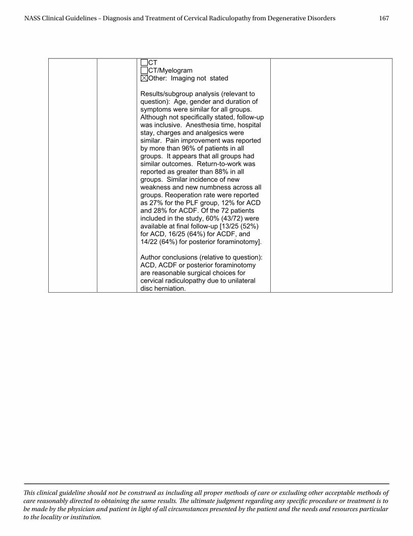
NASS Clinical Guidelines – Diagnosis and Treatment of Cervical Radiculopathy from Degenerative Disorders 167
This clinical guideline should not be construed as including all proper methods of care or excluding other acceptable methods of care reasonably directed to obtaining the same results. The ultimate judgment regarding any specific procedure or treatment is to be made by the physician and patient in light of all circumstances presented by the patient and the needs and resources particular to the locality or institution.
CTCT/Myelogram Other: Imaging not stated
Results/subgroup analysis (relevant to question): Age, gender and duration of symptoms were similar for all groups. Although not specifically stated, follow-up was inclusive. Anesthesia time, hospital stay, charges and analgesics were similar. Pain improvement was reported by more than 96% of patients in all groups. It appears that all groups had similar outcomes. Return-to-work was reported as greater than 88% in all groups. Similar incidence of new weakness and new numbness across all groups. Reoperation rate were reported as 27% for the PLF group, 12% for ACD and 28% for ACDF. Of the 72 patients included in the study, 60% (43/72) were available at final follow-up [13/25 (52%) for ACD, 16/25 (64%) for ACDF, and 14/22 (64%) for posterior foraminotomy].
Author conclusions (relative to question): ACD, ACDF or posterior foraminotomy are reasonable surgical choices for cervical radiculopathy due to unilateral disc herniation.
NASS Clinical Guidelines – Diagnosis and Treatment of Cervical Radiculopathy from Degenerative Disorders 168
This clinical guideline should not be construed as including all proper methods of care or excluding other acceptable methods of care reasonably directed to obtaining the same results. The ultimate judgment regarding any specific procedure or treatment is to be made by the physician and patient in light of all circumstances presented by the patient and the needs and resources particular to the locality or institution.
VI. Cervical Radiculopathy from Degenerative Disorders Guideline References 1. Abbed KM, Coumans JV. Cervical radiculopathy:
pathophysiology, presentation, and clinical evaluation. Neurosurgery. Jan 2007;60(1 Supp1 1):S28-34.
2. Abd-Alrahman N, Dokmak AS, Abou-Madawi A. Anterior cervical discectomy (ACD) versus anterior cervical fusion (ACF), clinical and radiological outcome study. Acta Neu-rochir (Wien). 1999;141(10):1089-1092.
3. Ahmed M, Modic MT. Neck and Low Back Pain: Neuroim-aging. Neurologic Clinics. May 2007;25(2):439-471.
4. Alexandre A, Coro L, Azuelos A, et al. Intradiscal injection of oxygen-ozone gas mixture for the treatment of cervical disc herniations. Acta Neurochir Suppl. 2005;92:79-82.
5. Al-Hami S. Cervical monosegmental interbody fusion us-ing titanium implants in degenerative, intervertebral disc disease. Minim Invasive Neurosurg. Mar 1999;42(1):10-17.
6. Alrawi MF, Khalil NM, Mitchell P, Hughes SP. The value of neurophysiological and imaging studies in predicting out-come in the surgical treatment of cervical radiculopathy. Eur Spine J. Apr 2007;16(4):495-500.
7. Alvarez JA, Hardy RW. Anterior cervical discectomy for one- and two-level cervical disc disease: the controversy surrounding the question of whether to fuse, plate, or both. Crit Rev Neurosurg. Jul 1999;9(4):234-251.
8. An HS. Cervical root entrapment. Hand Clin. Nov 1996;12(4):719-730.
9. An HS, Ahn NU. Posterior decompressive procedures for the cervical spine. Instr Course Lect. 2003;52:471-477.
10. Anderberg L, Annertz M, Brandt L, Saveland H. Selec-tive diagnostic cervical nerve root block--correlation with clinical symptoms and MRI-pathology. Acta Neurochir (Wien). Jun 2004;146(6):559-565; discussion 565.
11. Anderberg L, Annertz M, Persson L, Brandt L, Saveland H. Transforaminal steroid injections for the treatment of cer-vical radiculopathy: a prospective and randomised study. Eur Spine J. Mar 2007;16(3):321-328.
12. Anderberg L, Annertz M, Rydholm U, Brandt L, Saveland H. Selective diagnostic nerve root block for the evaluation of radicular pain in the multilevel degenerated cervical spine. Eur Spine J. Jun 2006;15(6):794-801.
13. Anderberg L, Saveland H, Annertz M. Distribution pat-terns of transforaminal injections in the cervical spine evaluated by multi-slice computed tomography. Euro Spine J. Oct 2006;15(10):1465-1471.
14. Anderson PA, Sasso RC, Riew KD. Comparison of adverse events between the Bryan artificial cervical disc and ante-rior cervical arthrodesis. Spine. 2008:1305-1312.
15. Anderson PA, Subach BR, Riew KD. Predictors of outcome
after anterior cervical discectomy and fusion: a multivari-ate analysis. Spine. Jan 15 2009;34(2):161-166.
16. Andrews NB, Lawson HL, Odjidja TL. Elective non-instru-mented anterior cervical diskectomy and fusion in Ghana: a preliminary report. West Afr J Med. Jun 2003;22(2):128-132.
17. Arnasson O, Carlsson CA, Pellettieri L. Surgical and con-servative treatment of cervical spondylotic radiculopathy and myelopathy. Acta Neurochir (Wien). 1987;84(1-2):48-53.
18. Arnold P, Boswell S, McMahon J. Threaded interbody fu-sion cage for adjacent segment degenerative disease af-ter previous anterior cervical fusion. Surg Neurol. Oct 2008;70(4):390-397.
19. Ashkan K, Johnston P, Moore AJ. A comparison of mag-netic resonance imaging and neurophysiological studies in the assessment of cervical radiculopathy. Br J Neuro-surg. Apr 2002;16(2):146-148.
20. Balasubramanian C, Price R, Brydon H. Anterior cervical microforaminotomy for cervical radiculopathy--results and review. Minim Invasive Neurosurg. Oct 2008;51(5):258-262.
21. Barlocher CB, Barth A, Krauss JK, Binggeli R, Seiler RW. Comparative evaluation of microdiscectomy only, au-tograft fusion, polymethylmethacrylate interposition, and threaded titanium cage fusion for treatment of single-level cervical disc disease: a prospective randomized study in 125 patients. Neurosurg Focus. Jan 15 2002;12(1):E4.
22. Baron EM, Loftus CM, Ducker TB, Nakagawa H. Dynamic computed tomography myelography for the investigation of cervical degenerative disease: Commentary. Neurol Med Chir (Tokyo). 2006;46(4):215-216.
23. Bartels RH, Donk R, van der Wilt GJ, Grotenhuis JA, Ven-derink D. Design of the PROCON trial: a prospective, ran-domized multi-center study comparing cervical anterior discectomy without fusion, with fusion or with arthro-plasty. BMC Musculoskelet Disord. 2006;7:85.
24. Bartels RH, Verbeek AL, Grotenhuis JA. Design of Lami-fuse: a randomised, multi-centre controlled trial com-paring laminectomy without or with dorsal fusion for cervical myeloradiculopathy. BMC Musculoskelet Disord. 2007;8:111.
25. Bartleson JD. Spine Disorder Case Studies. Neurologic Clinics. May 2006;24(2):309-330.
26. Bartlett RJ, Hill CA, Devlin R, Gardiner ED. Two-dimen-sional MRI at 1.5 and 0.5 T versus CT myelography in the diagnosis of cervical radiculopathy. Neuroradiology. Feb 1996;38(2):142-147.
27. Bartlett RJV, Hill CR, Gardiner E. A comparison of
NASS Clinical Guidelines – Diagnosis and Treatment of Cervical Radiculopathy from Degenerative Disorders 169
This clinical guideline should not be construed as including all proper methods of care or excluding other acceptable methods of care reasonably directed to obtaining the same results. The ultimate judgment regarding any specific procedure or treatment is to be made by the physician and patient in light of all circumstances presented by the patient and the needs and resources particular to the locality or institution.
T<sub>2</sub> and gadolinium enhanced MRI with CT myelography in cervical radiculopathy. Br J Radiol. Jan 1998;71(JAN.):11-19.
28. Beatty RM, Fowler FD, Hanson EJ, Jr. The abducted arm as a sign of ruptured cervical disc. Neurosurgery. Nov 1987;21(5):731-732.
29. Bell GR. The anterior approach to the cervical spine. Neu-roimaging Clin N Am. 1995;5(3):465-479.
30. Bell GR, Ross JS. Diagnosis of nerve root compression: Myelography, computed tomography, and MRI. Orthop Clin North Am. 1992;23(3):405-419.
31. Ben-Eliyahu DJ. Thermographic imaging of pathoneuro-physiology due to cervical disc herniation. J Manipulative Physiol Ther. 1989;12(6):482-490.
32. Benyamin R, Singh V, Parr AT, Conn A, Diwan S, Abdi S. Systematic Review of the Effectiveness of Cervical Epidur-als in the Management of Chronic Neck Pain. Pain Physi-cian. Jan-Feb 2009;12(1):137-157.
33. Bertalanffy H, Eggert HR. Clinical long-term results of an-terior discectomy without fusion for treatment of cervical radiculopathy and myelopathy. A follow-up of 164 cases. Acta Neurochir (Wien). 1988;90(3-4):127-135.
34. Bertilson BC, Grunnesjo M, Strender LE. Reliability of clin-ical tests in the assessment of patients with neck/shoulder problems-impact of history. Spine (Phila Pa 1976). Oct 1 2003;28(19):2222-2231.
35. Birchall D, Connelly D, Walker L, Hall K. Evaluation of magnetic resonance myelography in the investigation of cervical spondylotic radiculopathy. Br J Radiol. Aug 2003;76(908):525-531.
36. Boden SD, McCowin PR, Davis DO, Dina TS, Mark AS, Wi-esel S. Abnormal magnetic-resonance scans of the cervi-cal spine in asymptomatic subjects. A prospective investi-gation. J Bone Joint Surg Am. Sep 1990;72(8):1178-1184.
37. Boehm H, Greiner-Perth R, El-Saghir H, Allam Y. A new minimally invasive posterior approach for the treat-ment of cervical radiculopathy and myelopathy: surgi-cal technique and preliminary results. Eur Spine J. Jun 2003;12(3):268-273.
38. Bolesta MJ, Rechtine GR, 2nd, Chrin AM. One- and two-level anterior cervical discectomy and fusion: the effect of plate fixation. Spine J. May-Jun 2002;2(3):197-203.
39. Bolton JE, Humphreys BK. The Bournemouth Question-naire: a short-form comprehensive outcome measure. II. Psychometric properties in neck pain patients. J Manipu-lative Physiol Ther. Mar-Apr 2002;25(3):141-148.
40. Bommireddy R, Kamat A, Smith ET, et al. Magnetic res-onance image findings in the early post-operative pe-riod after anterior cervical discectomy. Eur Spine J. Jan 2007;16(1):27-31.
41. Boswell MV, Hansen HC, Trescot AM, Hirsch JA. Epidural steroids in the management of chronic spinal pain and ra-diculopathy. Pain Physician. Jul 2003;6(3):319-334.
42. Brodke DS, Zdeblick TA. Modified Smith-Robinson pro-cedure for anterior cervical discectomy and fusion. Spine. Oct 1992;17(10 Suppl):S427-430.
43. Brouillette DL, Gurske DT. Chiropractic treatment of cer-vical radiculopathy caused by a herniated cervical disc. J Manipulative Physiol Ther. Feb 1994;17(2):119-123.
44. Brown BM, Schwartz RH, Frank E, Blank NK. Preopera-tive evaluation of cervical radiculopathy and myelopathy by surface-coil MR imaging. AJR Am J Roentgenol. Dec 1988;151(6):1205-1212.
45. Bruneau M, Nisolle JF, Gilliard C, Gustin T. Anterior cer-vical interbody fusion with hydroxyapatite graft and plate system. Neurosurg Focus. 2001;10(4):E8.
46. Bucciero A. Myeloradicular damage in traumatic cervical disc herniation. J Neurosurg Sci. 1998;42(4):203-211.
47. Bucciero A, Vizioli L, Cerillo A. Soft cervical disc her-niation. An analysis of 187 cases. J Neurosurg Sci. Sep 1998;42(3):125-130.
48. Bush K, Hillier S. Outcome of cervical radiculopathy treat-ed with periradicular/epidural corticosteroid injections: a prospective study with independent clinical review. Eur Spine J. 1996;5(5):319-325.
49. Caglar YS, Bozkurt M, Kahilogullari G, et al. Keyhole ap-proach for posterior cervical discectomy: experience on 84 patients. Minim Invasive Neurosurg. Feb 2007;50(1):7-11.
50. Carette S, Fehlings MG. Clinical practice. Cervical radicu-lopathy. N Engl J Med. Jul 28 2005;353(4):392-399.
51. Caroli E, Orlando ER, D'Andrea G, Ferrante L. Anterior cervical fusion with interbody titanium cage containing surgical bone site graft: our institution's experience in 103 consecutive cases of degenerative spondylosis. J Spinal Disord Tech. May 2007;20(3):216-220.
52. Carragee EJ, Hurwitz EL, Cheng I, et al. Treatment of neck pain - Injections and surgical interventions: Results of the bone and joint decade 2000-2010 task force on neck pain and its associated disorders. Spine. Feb 2008;33(4):S153-S169.
53. Caspar W, Geisler FH, Pitzen T, Johnson TA. Anterior cer-vical plate stabilization in one- and two-level degenera-tive disease: overtreatment or benefit? J Spinal Disord. Feb 1998;11(1):1-11.
54. Castagnera L, Maurette P, Pointillart V, Vital JM, Erny P, Senegas J. Long-term results of cervical epidural steroid injection with and without morphine in chronic cervical radicular pain. Pain. Aug 1994;58(2):239-243.
55. Cauthen JC, Kinard RE, Vogler JB, et al. Outcome analysis of noninstrumented anterior cervical discectomy and in-terbody fusion in 348 patients. Spine (Phila Pa 1976). Jan 15 1998;23(2):188-192.
56. Chang H, Park JB, Hwang JY, Song KJ. Clinical analysis of cervical radiculopathy causing deltoid paralysis. Eur Spine J. Oct 2003;12(5):517-521.
NASS Clinical Guidelines – Diagnosis and Treatment of Cervical Radiculopathy from Degenerative Disorders 170
This clinical guideline should not be construed as including all proper methods of care or excluding other acceptable methods of care reasonably directed to obtaining the same results. The ultimate judgment regarding any specific procedure or treatment is to be made by the physician and patient in light of all circumstances presented by the patient and the needs and resources particular to the locality or institution.
57. Chen TY. The clinical presentation of uppermost cervical disc protrusion. Spine. 15 2000;25(4):439-442.
58. Chesnut RM, Abitbol JJ, Garfin SR. Surgical management of cervical radiculopathy. Indication, techniques, and re-sults. Orthop Clin North Am. Jul 1992;23(3):461-474.
59. Chiba K, Ogawa Y, Ishii K, et al. Long-term results of expansive open-door laminoplasty for cervical myel-opathy - Average 14-year follow-up study. Spine. Dec 2006;31(26):2998-3005.
60. Chiles BW, Leonard MA, Choudhri HF, Cooper PR. Cervi-cal spondylotic myelopathy: Patterns of neurological defi-cit and recovery after anterior cervical decompression. Neurosurgery. Apr 1999;44(4):762-769.
61. Cho DY, Lee WY, Sheu PC. Treatment of multilevel cervical fusion with cages. Surgical Neurology. Nov 2004;62(5):378-386.
62. Cleland JA, Fritz JM, Whitman JM, Palmer JA. The reli-ability and construct validity of the Neck Disability In-dex and patient specific functional scale in patients with cervical radiculopathy. Spine (Phila Pa 1976). Mar 1 2006;31(5):598-602.
63. Connell MD, Wiesel SW. Natural history and patho-genesis of cervical disk disease. Orthop Clin North Am. 1992;23(3):369-380.
64. Connolly PJ, Esses SI, Kostuik JP. Anterior cervical fusion: outcome analysis of patients fused with and without an-terior cervical plates. J Spinal Disord. Jun 1996;9(3):202-206.
65. Constantoyannis C, Konstantinou D, Kourtopoulos H, Papadakis N. Intermittent cervical traction for cervical radiculopathy caused by large-volume herniated disks. J Manipulative Physiol Ther. Mar-Apr 2002;25(3):188-192.
66. Cornelius JF, Bruneau M, George B. Microsurgical cervical nerve root decompression via an anterolateral approach: Clinical outcome of patients treated for spondylotic radic-ulopathy. Neurosurgery. Nov 2007;61(5):972-980.
67. Cyteval C, Thomas E, Decoux E, et al. Cervical radiculopa-thy: open study on percutaneous periradicular foraminal steroid infiltration performed under CT control in 30 pa-tients. AJNR Am J Neuroradiol. Mar 2004;25(3):441-445.
68. Dai LY, Jiang LS. Anterior cervical fusion with interbody cage containing beta-tricalcium phosphate augmented with plate fixation: a prospective randomized study with 2-year follow-up. Eur Spine J. May 2008;17(5):698-705.
69. Daniels DL, Grogan JP, Johansen JG. Cervical radiculopa-thy: Computed tomography and myelography compared. Radiology. 1984;151(1):109-113.
70. Daniels DL, Grogan JP, Johansen JG, Meyer GA, Williams AL, Haughton VM. Cervical radiculopathy: computed to-mography and myelography compared. Radiology. Apr 1984;151(1):109-113.
71. Davidson RI, Dunn EJ, Metzmaker JN. The shoulder ab-duction test in the diagnosis of radicular pain in cervical
extradural compressive monoradiculopathies. Spine (Phi-la Pa 1976). Sep-Oct 1981;6(5):441-446.
72. Davis RA. A long-term outcome study of 170 surgically treated patients with compressive cervical radiculopathy. Surg Neurol. Dec 1996;46(6):523-530; discussion 530-523.
73. Deshmukh VR, Rekate HL, Sonntag VKH. High cervi-cal disc herniation presenting with C-2 radiculopathy: Case report and review of the literature. J Neurosurg. Mar 2004;100(3 SUPPL.):303-306.
74. Devereaux M. Neck Pain. Med Clin N Am. March 2009;93(2):273-284.
75. Donaldson JW, Nelson PB. Anterior cervical discectomy without interbody fusion. Surg Neurol. Apr 2002;57(4):219-224; discussion 224-215.
76. Dowd GC, Wirth FP. Anterior cervical discectomy: is fu-sion necessary? J Neurosurg. Jan 1999;90(1 Suppl):8-12.
77. Dubuisson A, Lenelle J, Stevenaert A. Soft cervical disc herniation: A retrospective study of 100 cases. Acta Neuro-chir (Wien). 1993;125(1-4):115-119.
78. Duggal N, Pickett GE, Mitsis DK, Keller JL. Early clinical and biomechanical results following cervical arthroplasty. Neurosurg Focus. Sep 15 2004;17(3):E9.
79. Ellenberg MR, Honet JC, Treanor WJ. Cervical radiculopa-thy. Arch Phys Med Rehabil. Mar 1994;75(3):342-352.
80. Epstein NE. Technical note: unilateral posterior resection of cervical disc and spondylostenosis with contralateral fusion for instability. Surg Neurol. Oct 2001;56(4):256-258.
81. Eriksen K. Management of cervical disc herniation with upper cervical chiropractic care. J Manipulative Physiol Ther. Jan 1998;21(1):51-56.
82. Farmer JC, Wisneski RJ. Cervical spine nerve root com-pression: An analysis of neuroforaminal pressures with varying head and arm positions. Spine. 1994;19(16):1850-1855.
83. Fernandez-Fairen M, Sala P, Dufoo M, Jr., Ballester J, Mur-cia A, Merzthal L. Anterior cervical fusion with tantalum implant: a prospective randomized controlled study. Spine. Mar 1 2008;33(5):465-472.
84. Fessler RG, Khoo LT. Minimally invasive cervical microen-doscopic foraminotomy: an initial clinical experience. Neurosurgery. Nov 2002;51(5 Suppl):S37-45.
85. Fish DE, Kobayashi HW, Chang TL, Pham Q. MRI predic-tion of therapeutic response to epidural steroid injection in patients with cervical radiculopathy. Am J Phys Med Re-habil. March 2009;88(3):239-246.
86. Fisher MA. Electrophysiology of radiculopathies. Clin Neurophysiol. Mar 2002;113(3):317-335.
87. Foley KT, Mroz TE, Arnold PM, et al. Randomized, pro-spective, and controlled clinical trial of pulsed electro-magnetic field stimulation for cervical fusion. Spine J. May 2008;8(3):436-442.
88. Fouyas IP, Sandercock PA, Statham PF, Lynch C. WITH-

NASS Clinical Guidelines – Diagnosis and Treatment of Cervical Radiculopathy from Degenerative Disorders 171
This clinical guideline should not be construed as including all proper methods of care or excluding other acceptable methods of care reasonably directed to obtaining the same results. The ultimate judgment regarding any specific procedure or treatment is to be made by the physician and patient in light of all circumstances presented by the patient and the needs and resources particular to the locality or institution.
DRAWN: Surgery for cervical radiculomyelopathy. Co-chrane Database Syst Rev. 2006(2):CD001466.
89. Frederic S, Benedict R, Payer M. Implantation of an empty carbon fiber cage or a tricortical iliac crest autograft af-ter cervical discectomy for single-level disc herniation: a prospective comparative study. J Neurosurg Spine. Apr 2006;4(4):292-299.
90. Gaetani P, Tancioni F, Spanu G, Rodriguez y Baena R. Ante-rior cervical discectomy: an analysis on clinical long-term results in 153 cases. J Neurosurg Sci. Dec 1995;39(4):211-218.
91. Garvey TA, Eismont FJ. Diagnosis and treatment of cer-vical radiculopathy and myelopathy. Orthop Rev. Jul 1991;20(7):595-603.
92. Gifford L. Acute low cervical nerve root conditions: symp-tom presentations and pathobiological reasoning. Man Ther. May 2001;6(2):106-115.
93. Goffin J, Geusens E, Vantomme N, et al. Long-term follow-up after interbody fusion of the cervical spine. J Spinal Disord Tech. Apr 2004;17(2):79-85.
94. Goffin J, Van Calenbergh F, van Loon J, et al. Intermedi-ate follow-up after treatment of degenerative disc disease with the Bryan Cervical Disc Prosthesis: single-level and bi-level. Spine (Phila Pa 1976). Dec 15 2003;28(24):2673-2678.
95. Goldberg EJ, Singh K, Van U, Garretson R, An HS. Com-paring outcomes of anterior cervical discectomy and fu-sion in workman's versus non-workman's compensation population. Spine J. Nov-Dec 2002;2(6):408-414.
96. Goldstein B. Anatomic issues related to cervical and lum-bosacral radiculopathy. Phys Med Rehabil Clin N Am. Aug 2002;13(3):423-437.
97. Gore DR, Carrera GF, Glaeser ST. Smoking and degen-erative changes of the cervical spine: a roentgenographic study. Spine J. Sep-Oct 2006;6(5):557-560.
98. Gore DR, Sepic SB, Gardner GM. Roentgenographic find-ings of the cervical spine in asymptomatic people. Spine (Phila Pa 1976). Jul-Aug 1986;11(6):521-524.
99. Gore DR, Sepic SB, Gardner GM, Murray MP. Neck pain: a long-term follow-up of 205 patients. Spine. Jan-Feb 1987;12(1):1-5.
100. Grieve JP, Kitchen ND, Moore AJ, Marsh HT. Results of posterior cervical foraminotomy for treatment of cer-vical spondylitic radiculopathy. Br J Neurosurg. Feb 2000;14(1):40-43.
101. Grisoli F, Graziani N, Fabrizi AP, Peragut JC, Vincentelli F, Diaz-Vasquez P. Anterior discectomy without fusion for treatment of cervical lateral soft disc extrusion: A follow-up of 120 cases. Neurosurgery. 1989;24(6):853-859.
102. Grob D, Peyer JV, Dvorak J. The use of plate fixation in anterior surgery of the degenerative cervical spine: a comparative prospective clinical study. Eur Spine J. Oct 2001;10(5):408-413.
103. Grundy CR, Fritts HM. Magnetic resonance imaging of the musculoskeletal system .8. The spine, section 1. Clin Or-thop Relat Res. May 1997(338):275-287.
104. Gudavalli S, Kruse RA. Foraminal stenosis with radiculop-athy from a cervical disc herniation in a 33-year-old man treated with flexion distraction decompression manipula-tion. J Manipulative Physiol Ther. Jun 2008;31(5):376-380.
105. Hacker RJ. A randomized prospective study of an ante-rior cervical interbody fusion device with a minimum of 2 years of follow-up results. J Neurosurg. Oct 2000;93(2 Suppl):222-226.
106. Hacker RJ. Cervical disc arthroplasty: a controlled ran-domized prospective study with intermediate follow-up results. J Neurosurg Spine. Dec 2005;3(6):424-428.
107. Hacker RJ, Cauthen JC, Gilbert TJ, Griffith SL. A prospec-tive randomized multicenter clinical evaluation of an an-terior cervical fusion cage. Spine. Oct 15 2000;25(20):2646-2654; discussion 2655.
108. Hamalainen O, Toivakka-Hamalainen SK, Kuronen P. +Gz associated stenosis of the cervical spinal canal in fighter pilots. Aviat Space Environ Med. Apr 1999;70(4):330-334.
109. Hamburger C, Festenberg FV, Uhl E. Ventral discectomy with pmma interbody fusion for cervical disc disease: long-term results in 249 patients. Spine. Feb 1 2001;26(3):249-255.
110. Haneline MT, Lewkovich G. Malone D G, Baldwin N G, Tomecek F J, et al: Complications of cervical spine ma-nipulation therapy: 5-year retrospective study in a single-group practice. Neurosurg Focus 13 (6):Clinical Pearl, 2002. Neurosurg Focus. Mar 15 2003;14(3):e10; author re-ply e10.
111. Hardin JG, Halla JT. Cervical spine and radicular pain syn-dromes. Curr Opin Rheumatol. Mar 1995;7(2):136-140.
112. Harrop JS, Hanna A, Silva MT, Sharan A. Neurological manifestations of cervical spondylosis: an overview of signs, symptoms, and pathophysiology. Neurosurgery. Jan 2007;60(1 Supp1 1):S14-20.
113. Harrop JS, Silva MT, Sharan AD, Dante SJ, Simeone FA. Cervicothoracic radiculopathy treated using posterior cer-vical foraminotomy/discectomy. J Neurosurg. 01 2003;98(2 SUPPL.):131-136.
114. Hauerberg J, Kosteljanetz M, Boge-Rasmussen T, et al. An-terior cervical discectomy with or without fusion with ray titanium cage: a prospective randomized clinical study. Spine. Mar 1 2008;33(5):458-464.
115. Healy JF, Healy BB, Wong WH, Olson EM. Cervical and lumbar MRI in asymptomatic older male lifelong athletes: frequency of degenerative findings. J Comput Assist To-mogr. Jan-Feb 1996;20(1):107-112.
116. Heckmann JG, Lang CJ, Zobelein I, Laumer R, Druschky A, Neundorfer B. Herniated cervical intervertebral discs with radiculopathy: an outcome study of conserva-tively or surgically treated patients. J Spinal Disord. Oct
NASS Clinical Guidelines – Diagnosis and Treatment of Cervical Radiculopathy from Degenerative Disorders 172
This clinical guideline should not be construed as including all proper methods of care or excluding other acceptable methods of care reasonably directed to obtaining the same results. The ultimate judgment regarding any specific procedure or treatment is to be made by the physician and patient in light of all circumstances presented by the patient and the needs and resources particular to the locality or institution.
1999;12(5):396-401.117. Heckmann JG, Lang CJG, Zobelein I, Laumer R, Druschky
A, Neundorfer B. Herniated cervical intervertebral discs with radiculopathy: An outcome study of conservatively or surgically treated patients. J Spinal Disord. 1999;12(5):396-401.
118. Hedberg MC, Drayer BP, Flom RA, Hodak JA, Bird CR. Gradient echo (GRASS) MR imaging in cervical radicul-opathy. AJR Am J Roentgenol. Mar 1988;150(3):683-689.
119. Heidecke V, Burkert W, Brucke M, Rainov NG. Interverte-bral disc replacement for cervical degenerative disease--clinical results and functional outcome at two years in pa-tients implanted with the Bryan cervical disc prosthesis. Acta Neurochir (Wien). May 2008;150(5):453-459; discus-sion 459.
120. Heidecke V, Rainov NG, Marx T, Burkert W. Outcome in Cloward anterior fusion for degenerative cervical spinal disease. Acta Neurochir (Wien). 2000;142(3):283-291.
121. Heller JG. The syndromes of degenerative cervical disease. Orthop Clin North Am. 1992;23(3):381-394.
122. Henderson CM, Hennessy RG, Shuey HM, Jr., Shackel-ford EG. Posterior-lateral foraminotomy as an exclusive operative technique for cervical radiculopathy: a review of 846 consecutively operated cases. Neurosurgery. Nov 1983;13(5):504-512.
123. Hendriksen IJ, Holewijn M. Degenerative changes of the spine of fighter pilots of the Royal Netherlands Air Force (RNLAF). Aviat Space Environ Med. Nov 1999;70(11):1057-1063.
124. Herkowitz HN, Kurz LT, Overholt DP. Surgical manage-ment of cervical soft disc herniation. A comparison be-tween the anterior and posterior approach. Spine (Phila Pa 1976). Oct 1990;15(10):1026-1030.
125. Herzog J. Use of cervical spine manipulation under anes-thesia for management of cervical disk herniation, cer-vical radiculopathy, and associated cervicogenic head-ache syndrome. J Manipulative Physiol Ther. Mar-Apr 1999;22(3):166-170.
126. Hida K, Iwasaki Y, Yano S, Akino M, Seki T. Long-term follow-up results in patients with cervical disk disease treated by cervical anterior fusion using titanium cage im-plants. Neurol Med Chir (Tokyo). Oct 2008;48(10):440-446; discussion 446.
127. Hilibrand AS, Carlson GD, Palumbo MA, Jones PK, Bohl-man HH. Radiculopathy and myelopathy at segments ad-jacent to the site of a previous anterior cervical arthrod-esis. J Bone Joint Surg Am. Apr 1999;81(4):519-528.
128. Hitselberger WE, Witten RM. Abnormal myelograms in asymptomatic patients. J Neurosurg. Mar 1968;28(3):204-206.
129. Houser OW, Onofrio BM, Miller GM, Folger WN, Smith PL. Cervical Disk Prolapse. Mayo Clinic Proceedings. Oct 1995;70(10):939-945.
130. Houser OW, Onofrio BM, Miller GM, Folger WN, Smith PL, Kallman DA. Cervical Neural Foraminal Canal Steno-sis - Computerized Tomographic Myelography Diagnosis. J Neurosurg. Jul 1993;79(1):84-88.
131. Hubka MJ, Phelan SP, Delaney PM, Robertson VL. Rotary manipulation for cervical radiculopathy: observations on the importance of the direction of the thrust. J Manipula-tive Physiol Ther. Nov-Dec 1997;20(9):622-627.
132. Humphreys SC, Hodges SD, Patwardhan A, Eck JC, Coving-ton LA, Sartori M. The natural history of the cervical fora-men in symptomatic and asymptomatic individuals aged 20-60 years as measured by magnetic resonance imaging. A descriptive approach. Spine. Oct 15 1998;23(20):2180-2184.
133. Husag L, Costabile G, Vanloffeld W, Keller RJD, Landolt H. Anterior cervical discectomy without fusion: A com-parison with Cloward's procedure. J Clin Neurosci. Jul 1997;4(3):331-340.
134. Hwang SL, Hwang YF, Lieu AS, et al. Outcome analyses of interbody titanium cage fusion used in the anterior dis-cectomy for cervical degenerative disc disease. J Spinal Disord Tech. Aug 2005;18(4):326-331.
135. Ilkko E, Lahde S, Heiskari M. Thin-section CT in the ex-amination of cervical disc herniation. A prospective study with 1-mm axial and helical images. Acta Radiol. Mar 1996;37(2):148-152.
136. Jacobs WC, Anderson PG, Limbeek J, Willems PC, Pavlov P. Single or double-level anterior interbody fusion tech-niques for cervical degenerative disc disease. Cochrane Database Syst Rev. 2004(4):CD004958.
137. Jagannathan J, Shaffrey CI, Oskouian RJ, et al. Radio-graphic and clinical outcomes following single-level an-terior cervical discectomy and allograft fusion without plate placement or cervical collar. J Neurosurg Spine. May 2008;8(5):420-428.
138. Jagannathan J, Sherman JH, Szabo T, Shaffrey CI, Jane JA. The posterior cervical foraminotomy in the treatment of cervical disc/osteophyte disease: a single-surgeon ex-perience with a minimum of 5 years' clinical and radio-graphic follow-up Clinical article. J Neurosurg Spine. Apr 2009;10(4):347-356.
139. Jagannathan J, Sherman JH, Szabo T, Shaffrey CI, Jane JA. The posterior cervical foraminotomy in the treatment of cervical disc/osteophyte disease: a single-surgeon experi-ence with a minimum of 5 years' clinical and radiographic follow-up. J Neurosurg Spine. Apr 2009;10(4):347-356.
140. Jenis LG, An HS. Neck pain secondary to radiculopathy of the fourth cervical root: an analysis of 12 surgically treated patients. J Spinal Disord. Aug 2000;13(4):345-349.
141. Jho HD, Jho DH. Ventral uncoforaminotomy. J Neurosurg Spine. Nov 2007;7(5):533-535.
142. Jho HD, Kim WK, Kim MH. Anterior microforaminotomy for treatment of cervical radiculopathy: part 1–disc-pre-
NASS Clinical Guidelines – Diagnosis and Treatment of Cervical Radiculopathy from Degenerative Disorders 173
This clinical guideline should not be construed as including all proper methods of care or excluding other acceptable methods of care reasonably directed to obtaining the same results. The ultimate judgment regarding any specific procedure or treatment is to be made by the physician and patient in light of all circumstances presented by the patient and the needs and resources particular to the locality or institution.
serving functional cervical disc surgery. Neurosurgery. Nov 2002;51(5 Suppl):S46-53.
143. Kadoya S, Iizuka H, Nakamura T. Long-term outcome for surgically treated cervical spondylotic radiculopathy and myelopathy. Neurol Med Chir (Tokyo). May 2003;43(5):228-240; discussion 241.
144. Kaech DL, Elsig JPJ. Functional magnetic resonance imag-ing of the spine. Rivista Medica. Sep 2006;12(3-4):69-73.
145. Kaiser JA, Holland BA. Imaging of the cervical spine. Spine (Phila Pa 1976). Dec 15 1998;23(24):2701-2712.
146. Kaiser MG, Haid RW, Jr., Subach BR, Barnes B, Rodts GE, Jr. Anterior cervical plating enhances arthrodesis after dis-cectomy and fusion with cortical allograft. Neurosurgery. Feb 2002;50(2):229-236; discussion 236-228.
147. Kang JD, Stefanovic-Racic M, McIntyre LA, Georgescu HI, Evans CH. Toward a biochemical understanding of human intervertebral disc degeneration and herniation. Contributions of nitric oxide, interleukins, prostaglan-din E2, and matrix metalloproteinases. Spine. May 15 1997;22(10):1065-1073.
148. Kent DL, Haynor DR, Longstreth WT, Larson EB. Clinical Efficacy of Magnetic-Resonance-Imaging in Neuroimag-ing. Ann Intern Med. May 1994;120(10):856-871.
149. Kim H, Lee SH, Kim MH. Multislice CT fluoroscopy-assist-ed cervical transforaminal injection of steroids: technical note. J Spinal Disord Tech. Aug 2007;20(6):456-461.
150. Kim SH, Shin HC, Shin DA, Kim KN, Yoon do H. Early clinical experience with the mobi-C disc prosthesis. Yon-sei Med J. Jun 30 2007;48(3):457-464.
151. Kim SW, Limson MA, Kim SB, et al. Comparison of radio-graphic changes after ACDF versus Bryan disc arthroplasty in single and bi-level cases. European Spine Journal. Feb 2009;18(2):218-231.
152. Klaiber RD, Vonammon K, Sarioglu AC. Anterior Micro-surgical Approach for Degenerative Cervical Disk Disease. Acta Neurochirurgica. 1992;114(1-2):36-42.
153. Klein GR, Vaccaro AR, Albert TJ. Health outcome assess-ment before and after anterior cervical discectomy and fu-sion for radiculopathy: a prospective analysis. Spine. Apr 1 2000;25(7):801-803.
154. Koc RK, Menku A, Tucer B, Gocmez C, Akdemir H. Anteri-or cervical foraminotomy for unilateral spondylotic radic-ulopathy. Minim Invasive Neurosurg. Jun 2004;47(3):186-189.
155. Kolstad F, Leivseth G, Nygaard OP. Transforaminal steroid injections in the treatment of cervical radiculopathy. A prospective outcome study. Acta Neurochir (Wien). Oct 2005;147(10):1065-1070; discussion 1070.
156. Korinth MC, Kruger A, Oertel MF, Gilsbach JM. Posterior foraminotomy or anterior discectomy with polymethyl methacrylate interbody stabilization for cervical soft disc disease: results in 292 patients with monoradiculopathy. Spine. May 15 2006;31(11):1207-1214; discussion 1215-
1206.157. Kotil K, Bilge T. Prospective study of anterior cervical mi-
croforaminotomy for cervical radiculopathy. J Clin Neuro-sci. Jul 2008;15(7):749-756.
158. Krupp W, Schattke H, Muke R. Clinical-Results of the Foraminotomy as Described by Frykholm for the Treat-ment of Lateral Cervical Disk Herniation. Acta Neurochir (Wien). 1990;107(1-2):22-29.
159. Kruse RA, Imbarlina F, De Bono VF. Treatment of cervi-cal radiculopathy with flexion distraction. J Manipulative Physiol Ther. Mar-Apr 2001;24(3):206-209.
160. Kuijper B, Tans JTJ, Schimsheimer RJ, et al. Degenerative cervical radiculopathy: Diagnosis and conservative treat-ment. A review. Euro J Neurol. January 2009;16(1):15-20.
161. Kumar GRV, Maurice-Williams RS, Bradford R. Cervical foraminotomy: an effective treatment for cervical spon-dylotic radiculopathy. Br J Neurosurg. Dec 1998;12(6):563-568.
162. Kumar N, Gowda V. Cervical foraminal selective nerve root block: a 'two-needle technique' with results. Eur Spine J. Apr 2008;17(4):576-584.
163. Kwon JW, Lee JW, Kim SH, et al. Cervical interlaminar epi-dural steroid injection for neck pain and cervical radicu-lopathy: Effect and prognostic factors. Skeletal Radiology. May 2007;36(5):431-436.
164. LaBan MM, Macy JA, Meerschaert JR. Intermittent cervi-cal traction: a progenitor of lumbar radicular pain. Arch Phys Med Rehabil. Mar 1992;73(3):295-296.
165. Larsson EM, Holtas S, Cronqvist S, Brandt L. Comparison of myelography, CT myelography and magnetic resonance imaging in cervical spondylosis and disk herniation. Pre- and postoperative findings. Acta Radiol. May-Jun 1989;30(3):233-239.
166. Lauder TD. Physical examination signs, clinical symp-toms, and their relationship to electrodiagnostic findings and the presence of radiculopathy. Phys Med Rehabil Clin N Am. Aug 2002;13(3):451-467.
167. Leblhuber F, Reisecker F. Diagnostic efficacy of DSER in patients with spine pain and radiculopathy in the cer-vical and lumbar region. Clin Electroencephalogr. Jan 1990;21(1):VII-IX.
168. Lees F, Turner JW. Natural history and prognosis of cervi-cal spondylosis. Br Med J. Dec 28 1963;2(5373):1607-1610.
169. Li J, Yan DL, Zhang ZH. Percutaneous cervical nucleo-plasty in the treatment of cervical disc herniation. Eur Spine J. Dec 2008;17(12):1664-1669.
170. Lin EL, Lieu V, Halevi L, Shamie AN, Wang JC. Cervical epidural steroid injections for symptomatic disc hernia-tions. J Spinal Disord Tech. May 2006;19(3):183-186.
171. Lin HL, Cho DY, Liu YF, Lee WY, Lee HC, Chen CC. Change of cervical balance following single to multi-level inter-body fusion with cage. Br J Neurosurg. Dec 2008;22(6):758-763.
NASS Clinical Guidelines – Diagnosis and Treatment of Cervical Radiculopathy from Degenerative Disorders 174
This clinical guideline should not be construed as including all proper methods of care or excluding other acceptable methods of care reasonably directed to obtaining the same results. The ultimate judgment regarding any specific procedure or treatment is to be made by the physician and patient in light of all circumstances presented by the patient and the needs and resources particular to the locality or institution.
172. Lipetz JS, Misra N, Silber JS. Resolution of pronounced painless weakness arising from radiculopathy and disk extrusion. Am J Phys Med Rehabil. Jul 2005;84(7):528-537.
173. Lofgren H, Johansen F, Skogar O, Levander B. Reduced pain after surgery for cervical disc protrusion/steno-sis: a 2 year clinical follow-up. Disabil Rehabil. Sep 16 2003;25(18):1033-1043.
174. Makin GJ, Brown WF, Ebers GC. C7 radiculopathy: impor-tance of scapular winging in clinical diagnosis. J Neurol Neurosurg Psychiatry. Jun 1986;49(6):640-644.
175. Malone DG, Baldwin NG, Tomecek FJ, et al. Complications of cervical spine manipulation therapy: 5-year retrospec-tive study in a single-group practice. Neurosurg Focus. Dec 15 2002;13(6):ecp1.
176. Manchikanti L, Pampati V, Damron KS, et al. The effect of sedation on diagnostic validity of facet joint nerve blocks: an evaluation to assess similarities in population with involvement in cervical and lumbar regions (ISRCTNo: 76376497). Pain Physician. Jan 2006;9(1):47-51.
177. Matge G. Cervical cage fusion with 5 different implants: 250 cases. Acta Neurochir (Wien). Jun 2002;144(6):539-549; discussion 550.
178. Matge G, Leclercq TA. Rationale for interbody fusion with threaded titanium cages at cervical and lumbar levels. Re-sults on 357 cases. Acta Neurochir (Wien). 2000;142(4):425-434.
179. Matsumoto M, Chiba K, Ishikawa M, Maruiwa H, Fujimura Y, Toyama Y. Relationships between outcomes of conser-vative treatment and magnetic resonance imaging find-ings in patients with mild cervical myelopathy caused by soft disc herniations. Spine. Jul 15 2001;26(14):1592-1598.
180. Maurice-Williams RS, Elsmore A. Extended anterior cervi-cal decompression without fusion: a long-term follow-up study. Br J Neurosurg. Oct 1999;13(5):474-479.
181. McClure P. The degenerative cervical spine: pathogen-esis and rehabilitation concepts. J Hand Ther. Apr-Jun 2000;13(2):163-174.
182. McCormack BM, Weinstein PR. Cervical spondylosis. An update. West J Med. Jul-Aug 1996;165(1-2):43-51.
183. McLaughlin MR, Purighalla V, Pizzi FJ. Cost advantages of two-level anterior cervical fusion with rigid internal fixation for radiculopathy and degenerative disease. Surg Neurol. Dec 1997;48(6):560-565.
184. Mobbs RJ, Rao P, Chandran NK. Anterior cervical discec-tomy and fusion: analysis of surgical outcome with and without plating. J Clin Neurosci. Jul 2007;14(7):639-642.
185. Modic MT, Masaryk TJ, Mulopulos GP. Cervical radicul-opathy: prospective evaluation with surface coil MR imag-ing, CT with metrizamide, and metrizamide myelography. Radiology. 1986;161(3):753-759.
186. Muhle C, Bischoff L, Weinert D, et al. Exacerbated pain in cervical radiculopathy at axial rotation, flexion, extension, and coupled motions of the cervical spine: evaluation by
kinematic magnetic resonance imaging. Invest Radiol. May 1998;33(5):279-288.
187. Muhle C, Metzner J, Weinert D, et al. Kinematic MR imag-ing in surgical management of cervical disc disease, spon-dylosis and spondylotic myelopathy. Acta Radiol. Mar 1999;40(2):146-153.
188. Mummaneni PV, Burkus JK, Haid RW, Traynelis VC, Zde-blick TA. Clinical and radiographic analysis of cervical disc arthroplasty compared with allograft fusion: a ran-domized controlled clinical trial. J Neurosurg Spine. Mar 2007;6(3):198-209.
189. Murphey F, Simmons JC, Brunson B. Chapter 2. Ruptured cervical discs, 1939 to 1972. Clin Neurosurg. 1973;20:9-17.
190. Murphy DR. Herniated disc with radiculopathy following cervical manipulation: nonsurgical management. Spine J. Jul-Aug 2006;6(4):459-463.
191. Murphy DR, Beres JL. Cervical myelopathy: a case report of a "near-miss" complication to cervical manipulation. J Manipulative Physiol Ther. Sep 2008;31(7):553-557.
192. Murphy DR, Beres JL. Is treatment in extension contrain-dicated in the presence of cervical spinal cord compres-sion without myelopathy? A case report. Man Ther. Oct 2008;13(5):468-472.
193. Murphy DR, Hurwitz EL, Gregory A, Clary R. A nonsurgi-cal approach to the management of patients with cervical radiculopathy: A prospective observational cohort study. J Manipulative Physiol Ther. May 2006;29(4):279-287.
194. Murphy MA, Trimble MB, Piedmonte MR, Kalfas IH. Changes in the cervical foraminal area after anterior discectomy with and without a graft. Neurosurgery. Jan 1994;34(1):93-96.
195. Murrey D, Janssen M, Delamarter R, et al. Results of the prospective, randomized, controlled multicenter Food and Drug Administration investigational device exemp-tion study of the ProDisc-C total disc replacement ver-sus anterior discectomy and fusion for the treatment of 1-level symptomatic cervical disc disease. Spine J. Apr 2009;9(4):275-286.
196. Nabhan A, Ahlhelm F, Pitzen T, et al. Disc replacement us-ing Pro-Disc C versus fusion: a prospective randomised and controlled radiographic and clinical study. Euro Spine J. Mar 2007;16(3):423-430.
197. Nabhan A, Ahlhelm F, Shariat K, et al. The ProDisc-C pro-thesis - Clinical and radiological experience 1 year after surgery. Spine. Aug 2007;32(18):1935-1941.
198. Nabhan A, Pape D, Pitzen T, et al. Radiographic analysis of fusion progression following one-level cervical fusion with or without plate fixation. Zentralbl Neurochir. Aug 2007;68(3):133-138.
199. Nakstad PH, Hald JK, Bakke SJ, Skalpe IO, Wiberg J. MRI in cervical disk herniation. Neuroradiology. 1989;31(5):382-385.
200. Nardin RA, Patel MR, Gudas TF, Rutkove SB, Raynor
NASS Clinical Guidelines – Diagnosis and Treatment of Cervical Radiculopathy from Degenerative Disorders 175
This clinical guideline should not be construed as including all proper methods of care or excluding other acceptable methods of care reasonably directed to obtaining the same results. The ultimate judgment regarding any specific procedure or treatment is to be made by the physician and patient in light of all circumstances presented by the patient and the needs and resources particular to the locality or institution.
EM. Electromyography and magnetic resonance imag-ing in the evaluation of radiculopathy. Muscle Nerve. Feb 1999;22(2):151-155.
201. Neuhold A, Stiskal M, Platzer C, Pernecky G, Brainin M. Combined use of spin-echo and gradient-echo MR-imaging in cervical disk disease. Comparison with my-elography and intraoperative findings. Neuroradiology. 1991;33(5):422-426.
202. Nunley PD, Jawahar A, Kerr EJ, 3rd, Cavanaugh DA, How-ard C, Brandao SM. Choice of plate may affect outcomes for single versus multilevel ACDF: results of a prospective randomized single-blind trial. Spine J. Feb 2009;9(2):121-127.
203. Odom GL, Finney W, Woodhall B. Cervical disk lesions. J Am Med Assoc. 1958;166(1):23-28.
204. Oktenoglu T, Cosar M, Ozer AF, et al. Anterior cervical microdiscectomy with or without fusion. J Spinal Disord Tech. Jul 2007;20(5):361-368.
205. Olivero WC, Dulebohn SC. Results of halter cervical trac-tion for the treatment of cervical radiculopathy: retro-spective review of 81 patients. Neurosurg Focus. Feb 15 2002;12(2):ECP1.
206. Oppenheim JS, Spitzer DE, Segal DH. Nonvascular com-plications following spinal manipulation. Spine J. Nov 2005;5(6):660-666.
207. Ozgur BM, Marshall LF. Atypical presentation of C-7 ra-diculopathy. J Neurosurg. Sep 2003;99(2 Suppl):169-171.
208. Padua L, Padua R, LoMonaco M, Tonali PA. Radiculom-edullary complications of cervical spinal manipulation. Spinal Cord. Aug 1996;34(8):488-492.
209. Park JH, Roh KH, Cho JY, Ra YS, Rhim SC, Noh SW. Com-parative analysis of cervical arthroplasty using Mobi-C and anterior cervical discectomy and husion using the Solis-cage. J Korean Neurosurg Soc. 2008;44(4):217-221.
210. Pawl RP, Matz M, Wissinger JP, Vacca DF, Goldfarb RP. Epi-dural steroids for cervical and lumbar radiculopathy. Surg Neurol. Nov 1996;46(5):455-457.
211. Pechlivanis I, Brenke C, Scholz M, Engelhardt M, Harders A, Schmieder K. Treatment of degenerative cervical disc disease with uncoforaminotomy--intermediate clinical outcome. Minim Invasive Neurosurg. Aug 2008;51(4):211-217.
212. Peloso Paul Michael J, Gross A, Haines T, et al. Medicinal and injection therapies for mechanical neck disorders. Cochrane Database of Systematic Reviews. 2007.
213. Peng B, Hao J, Hou S, et al. Possible pathogenesis of painful intervertebral disc degeneration. Spine. Mar 1 2006;31(5):560-566.
214. Peolsson A, Peolsson M. Predictive factors for long-term outcome of anterior cervical decompression and fusion: a multivariate data analysis. Eur Spine J. Mar 2008;17(3):406-414.
215. Perneczky G, Bock FW, Neuhold A, Stiskal M. Diagnosis
of cervical disc disease. MRI versus cervical myelography. Acta Neurochir (Wien). 1992;116(1):44-48.
216. Persson LC, Lilja A. Pain, coping, emotional state and physical function in patients with chronic radicular neck pain. A comparison between patients treated with surgery, physiotherapy or neck collar--a blinded, prospective ran-domized study. Disabil Rehabil. May 20 2001;23(8):325-335.
217. Persson LC, Moritz U, Brandt L, Carlsson CA. Cervical ra-diculopathy: pain, muscle weakness and sensory loss in patients with cervical radiculopathy treated with surgery, physiotherapy or cervical collar. A prospective, controlled study. Eur Spine J. 1997;6(4):256-266.
218. Persson LCG, Carlsson JY, Anderberg L. Headache in pa-tients with cervical radiculopathy: A prospective study with selective nerve root blocks in 275 patients. Euro Spine J. Jul 2007;16(7):953-959.
219. Petren-Mallmin M, Linder J. MRI cervical spine findings in asymptomatic fighter pilots. Aviat Space Environ Med. Dec 1999;70(12):1183-1188.
220. Petren-Mallmin M, Linder J. Cervical spine degeneration in fighter pilots and controls: a 5-yr follow-up study. Aviat Space Environ Med. May 2001;72(5):443-446.
221. Pimenta L, McAfee PC, Cappuccino A, Cunningham BW, Diaz R, Coutinho E. Superiority of multilevel cer-vical arthroplasty outcomes versus single-level out-comes: 229 consecutive PCM prostheses. Spine. May 20 2007;32(12):1337-1344.
222. Pointillart V, Cernier A, Vital JM, Senegas J. Anterior dis-cectomy without interbody fusion for cervical disc hernia-tion. Eur Spine J. 1995;4(1):45-51.
223. Pollard H, Tuchin P. Cervical radiculopathy: a case for ancillary therapies? J Manipulative Physiol Ther. May 1995;18(4):244-249.
224. Post NH, Cooper PR, Frempong-Boadu AK, Costa ME. Unique features of herniated discs at the cervicothoracic junction: Clinical presentation, imaging, operative man-agement, and outcome after anterior decompressive op-eration in 10 patients. Neurosurgery. Mar 2006;58(3):497-501.
225. Radhakrishnan K, Litchy WJ, O'Fallon WM, Kurland LT. Epidemiology of cervical radiculopathy. A population-based study from Rochester, Minnesota, 1976 through 1990. Brain. Apr 1994;117 ( Pt 2):325-335.
226. Ramzi N, Ribeiro-Vaz G, Fomekong E, Lecouvet FE, Rafto-poulos C. Long term outcome of anterior cervical discec-tomy and fusion using coral grafts. Acta Neurochir (Wien). Dec 2008;150(12):1249-1256; discussion 1256.
227. Rao PJ, Christie JG, Ghahreman A, Cartwright CA, Ferch RD. Clinical and functional outcomes of anterior cervi-cal discectomy without fusion. J Clin Neurosci. December 2008;15(12):1354-1359.
228. Rao R. Neck pain, cervical radiculopathy, and cervical my-
NASS Clinical Guidelines – Diagnosis and Treatment of Cervical Radiculopathy from Degenerative Disorders 176
This clinical guideline should not be construed as including all proper methods of care or excluding other acceptable methods of care reasonably directed to obtaining the same results. The ultimate judgment regarding any specific procedure or treatment is to be made by the physician and patient in light of all circumstances presented by the patient and the needs and resources particular to the locality or institution.
elopathy: pathophysiology, natural history, and clinical evaluation. J Bone Joint Surg Am. Oct 2002;84-A(10):1872-1881.
229. Rao R. Neck pain, cervical radiculopathy, and cervical myelopathy: pathophysiology, natural history, and clini-cal evaluation. Instr Course Lect. 2003;52:479-488.
230. Rao RD, Currier BL, Albert TJ, et al. Degenerative cervical spondylosis: clinical syndromes, pathogenesis, and man-agement. J Bone Joint Surg Am. Jun 2007;89(6):1360-1378.
231. Rechtine IGR, Bolesta MJ. Cervical radiculopathy. Semin Spine Surg. 1999;11(4):363-372.
232. Resnick DK, Trost GR. Use of ventral plates for cervical arthrodesis. Neurosurgery. Jan 2007;60(1 Supp1 1):S112-117.
233. Rhee JM, Yoon T, Riew KD. Cervical radiculopathy. J Am Acad Orthop Surg. Aug 2007;15(8):486-494.
234. Riew KD, Cheng I, Pimenta L, Taylor B. Posterior cervi-cal spine surgery for radiculopathy. Neurosurgery. Jan 2007;60(1 Supp1 1):S57-63.
235. Rocchi G, Caroli E, Salvati M, Delfini R. Multilevel oblique corpectomy without fusion: our experience in 48 patients. Spine. Sep 1 2005;30(17):1963-1969.
236. Rodrigues MA, Hanel RA, Prevedello DM, Antoniuk A, Araujo JC. Posterior approach for soft cervical disc herniation: a neglected technique? Surg Neurol. Jan 2001;55(1):17-22; discussion 22.
237. Romner B, Due-Tonnessen BJ, Egge A, Anke IM, Trumpy JH. Modified Robinson-Smith procedure for the treat-ment of cervical radiculopathy. Acta Neurol Scand. Sep 1994;90(3):197-200.
238. Rosenkranz M, Grzyska U, Niesen W, et al. Anterior spinal artery syndrome following periradicular cervical nerve root therapy. J Neurol. Feb 2004;251(2):229-231.
239. Rosomoff HL, Fishbain D, Rosomoff RS. Chronic cervi-cal pain: radiculopathy or brachialgia. Noninterventional treatment. Spine. Oct 1992;17(10 Suppl):S362-366.
240. Ross JS, Modic MT, Masaryk TJ, Carter J, Marcus RE, Bohlman H. Assissment of extradural degenerative dis-ease with Gd-DTPA-enhanced MR imaging: Correlation with surgical and pathologic findings. Am J Neurorad. 1989;10(6):1243-1249.
241. Ross JS, Modic MT, Masaryk TJ, Carter J, Marcus RE, Bohl-man H. Assessment of extradural degenerative disease with Gd-DTPA-enhanced MR imaging: correlation with surgical and pathologic findings. AJR Am J Roentgenol. Jan 1990;154(1):151-157.
242. Ross JS, Ruggieri PM, Glicklich M, et al. 3d Mri of the Cer-vical-Spine - Low Flip Angle Fisp Vs Gd-Dtpa Turboflash in Degenerative Disk Disease. Journal of Computer As-sisted Tomography. Jan-Feb 1993;17(1):26-33.
243. Rubin D. Cervical radiculitis: diagnosis and treatment. Arch Phys Med Rehabil. Dec 1960;41:580-586.
244. Rubinstein SM, Pool JJM, van Tulder MW, Riphagen I, de
Vet HCW. A systematic review of the diagnostic accuracy of provocative tests of the neck for diagnosing cervical ra-diculopathy. Euro Spine J. Mar 2007;16(3):307-319.
245. Ruetten S, Komp M, Merk H, Godolias G. A new full-endo-scopic technique for cervical posterior foraminotomy in the treatment of lateral disc herniations using 6.9-mm en-doscopes: prospective 2-year results of 87 patients. Minim Invasive Neurosurg. Aug 2007;50(4):219-226.
246. Ruggieri PM. Cervical radiculopathy. Neuroimag Clin N Am. 1995;5(3):349-366.
247. Russell EJ, D'Angelo CM, Zimmerman RD. Cervical disk herniation: CT demonstration after contrast enhance-ment. Radiology. 1984;152(3):703-712.
248. Saal JS, Saal JA, Yurth EF. Nonoperative management of herniated cervical intervertebral disc with radiculopathy. Spine. Aug 1996;21(16):1877-1883.
249. Sabbahi MA, Khalil M. Segmental H-reflex studies in up-per and lower limbs of patient with radiculopathy. Arch Phys Med Rehabil. 1990;71(3):223-227.
250. Samartzis D, Shen FH, Lyon C, Phillips M, Goldberg EJ, An HS. Does rigid instrumentation increase the fusion rate in one-level anterior cervical discectomy and fusion? Spine J. Nov-Dec 2004;4(6):636-643.
251. Sambrook PN, MacGregor AJ, Spector TD. Genetic influ-ences on cervical and lumbar disc degeneration: a mag-netic resonance imaging study in twins. Arthritis Rheum. Feb 1999;42(2):366-372.
252. Sampath P, Bendebba M, Davis JD, Ducker T. Outcome in patients with cervical radiculopathy. Prospective, multi-center study with independent clinical review. Spine. Mar 15 1999;24(6):591-597.
253. Sasso RC, Smucker JD, Hacker RJ, Heller JG. Artificial disc versus fusion: a prospective, randomized study with 2-year follow-up on 99 patients. Spine. Dec 15 2007;32(26):2933-2940; discussion 2941-2932.
254. Savolainen S, Rinne J, Hernesniemi J. A prospective ran-domized study of anterior single-level cervical disc opera-tions with long-term follow-up: surgical fusion is unnec-essary. Neurosurgery. Jul 1998;43(1):51-55.
255. Scheufler KM, Kirsch E. Percutaneous multilevel decom-pressive laminectomy, foraminotomy, and instrumented fusion for cervical spondylotic radiculopathy and myel-opathy: assessment of feasibility and surgical technique. J Neurosurg Spine. Nov 2007;7(5):514-520.
256. Schneeberger AG, Boos N, Schwarzenbach O, Aebi M. Anterior cervical interbody fusion with plate fixation for chronic spondylotic radiculopathy: a 2- to 8-year follow-up. J Spinal Disord. Jun 1999;12(3):215-220; discussion 221.
257. Schoggl A, Reddy M, Saringer W, Ungersbock K. Social and economic outcome after posterior microforamino-tomy for cervical spondylotic radiculopathy. Wien Klin Wochenschr. Mar 28 2002;114(5-6):200-204.
NASS Clinical Guidelines – Diagnosis and Treatment of Cervical Radiculopathy from Degenerative Disorders 177
This clinical guideline should not be construed as including all proper methods of care or excluding other acceptable methods of care reasonably directed to obtaining the same results. The ultimate judgment regarding any specific procedure or treatment is to be made by the physician and patient in light of all circumstances presented by the patient and the needs and resources particular to the locality or institution.
258. Scotti G, Scialfa G, Pieralli S, Boccardi E, Valsecchi F, Tonon C. Myelopathy and radiculopathy due to cervical spondy-losis: myelographic-CT correlations. AJNR Am J Neurora-diol. May-Jun 1983;4(3):601-603.
259. Shad A, Leach JC, Teddy PJ, Cadoux-Hudson TA. Use of the Solis cage and local autologous bone graft for anterior cervical discectomy and fusion: early technical experi-ence. J Neurosurg Spine. Feb 2005;2(2):116-122.
260. Shafaie FF, Wippold FJ, 2nd, Gado M, Pilgram TK, Riew KD. Comparison of computed tomography myelography and magnetic resonance imaging in the evaluation of cer-vical spondylotic myelopathy and radiculopathy. Spine. Sep 1 1999;24(17):1781-1785.
261. Shah KC, Rajshekhar V. Reliability of diagnosis of soft cer-vical disc prolapse using Spurling's test. Br J Neurosurg. Oct 2004;18(5):480-483.
262. Shapiro S, Connolly P, Donnaldson J, Abel T. Cadaveric fibula, locking plate, and allogeneic bone matrix for ante-rior cervical fusions after cervical discectomy for radicu-lopathy or myelopathy. J Neurosurg. Jul 2001;95(1 Sup-pl):43-50.
263. Shen FH, Samartzis D, Khanna N, Goldberg EJ, An HS. Comparison of clinical and radiographic outcome in in-strumented anterior cervical discectomy and fusion with or without direct uncovertebral joint decompression. Spine J. Nov 2004;4(6):629-635.
264. Siivola SM, Levoska S, Tervonen O, Ilkko E, Vanharanta H, Keinanen-Kiukaanniemi S. MRI changes of cervical spine in asymptomatic and symptomatic young adults. Eur Spine J. Aug 2002;11(4):358-363.
265. Slipman CW, Chow DW. Therapeutic spinal corticosteroid injections for the management of radiculopathies. Phys Med Rehabil Clin N Am. Aug 2002;13(3):697-711.
266. Slipman CW, Plastaras CT, Palmitier RA, Huston CW, Sterenfeld EB. Symptom provocation of fluoroscopically guided cervical nerve root stimulation. Are dynatomal maps identical to dermatomal maps? Spine (Phila Pa 1976). Oct 15 1998;23(20):2235-2242.
267. Strobel K, Pfirrmann CW, Schmid M, Hodler J, Boos N, Za-netti M. Cervical nerve root blocks: indications and role of MR imaging. Radiology. Oct 2004;233(1):87-92.
268. Strub WM, Brown TA, Ying J, Hoffmann M, Ernst RJ, Bu-las RV. Translaminar cervical epidural steroid injection: short-term results and factors influencing outcome. J Vasc Interv Radiol. Sep 2007;18(9):1151-1155.
269. Suetsuna F, Yokoyama T, Kenuka E, Harata S. Anterior cer-vical fusion using porous hydroxyapatite ceramics for cer-vical disc herniation. a two-year follow-up. Spine J. Sep-Oct 2001;1(5):348-357.
270. Sugawara T, Itoh Y, Hirano Y, Higashiyama N, Mizoi K. Long term outcome and adjacent disc degeneration after ante-rior cervical discectomy and fusion with titanium cylin-drical cages. Acta Neurochir (Wien). Apr 2009;151(4):303-
309.271. Swezey RL. Conservative treatment of cervical radiculop-
athy. J Clin Rheumatol. Apr 1999;5(2):65-73.272. Tan J, Zheng Y, Gong L, Liu X, Li J, Du W. Anterior cervical
discectomy and interbody fusion by endoscopic approach: a preliminary report. J Neurosurg Spine. Jan 2008;8(1):17-21.
273. Tanaka Y, Kokubun S, Sato T. Mini-symposium: Cervical spine: (i) Cervical radiculopathy and its unsolved prob-lems. Curr Orthop. Jan 1998;12(1):1-6.
274. Tanaka Y, Kokubun S, Sato T, Ozawa H. Cervical roots as origin of pain in the neck or scapular regions. Spine. Aug 1 2006;31(17):E568-573.
275. Tegos S, Rizos K, Papathanasiu A, Kyriakopulos K. Re-sults of anterior discectomy without fusion for treatment of cervical radiculopathy and myelopathy. Eur Spine J. 1994;3(2):62-65.
276. Teresi LM, Lufkin RB, Reicher MA, et al. Asymptomatic degenerative disk disease and spondylosis of the cervical spine: MR imaging. Radiology. Jul 1987;164(1):83-88.
277. Thorell W, Cooper J, Hellbusch L, Leibrock L. The long-term clinical outcome of patients undergoing anterior cervical discectomy with and without intervertebral bone graft placement. Neurosurgery. Aug 1998;43(2):268-273; discussion 273-264.
278. Tiso RL, Cutler T, Catania JA, Whalen K. Adverse cen-tral nervous system sequelae after selective transforam-inal block: the role of corticosteroids. Spine J. Jul-Aug 2004;4(4):468-474.
279. Tong HC, Haig AJ, Yamakawa K. The Spurling test and cervical radiculopathy. Spine (Phila Pa 1976). Jan 15 2002;27(2):156-159.
280. Topuz K, Colak A, Kaya S, et al. Two-level contiguous cer-vical disc disease treated with peek cages packed with de-mineralized bone matrix: results of 3-year follow-up. Eur Spine J. Feb 2009;18(2):238-243.
281. Troyanovich SJ, Stroink AR, Kattner KA, Dornan WA, Gu-bina I. Does anterior plating maintain cervical lordosis versus conventional fusion techniques? A retrospective analysis of patients receiving single-level fusions. J Spinal Disord Tech. Feb 2002;15(1):69-74.
282. Truumees E, Herkowitz HN. Cervical spondylotic myel-opathy and radiculopathy. Instr Course Lect. 2000;49:339-360.
283. Tsao B. The Electrodiagnosis of Cervical and Lumbosacral Radiculopathy. Neurologic Clinics. May 2007;25(2):473-494.
284. Tseng SH, Lin SM, Chen Y, Wang CH. Ruptured cervical disc after spinal manipulation therapy: report of two cas-es. Spine. Feb 1 2002;27(3):E80-82.
285. Van de Kelft E, van Vyve M. Diagnostic imaging algorithm for cervical soft disc herniation. J Neurol Neurosurg Psy-chiatry. Jun 1994;57(6):724-728.
NASS Clinical Guidelines – Diagnosis and Treatment of Cervical Radiculopathy from Degenerative Disorders 178
This clinical guideline should not be construed as including all proper methods of care or excluding other acceptable methods of care reasonably directed to obtaining the same results. The ultimate judgment regarding any specific procedure or treatment is to be made by the physician and patient in light of all circumstances presented by the patient and the needs and resources particular to the locality or institution.
286. Van de Kelft E, van Vyve M. Diagnostic imaging algorithm for cervical soft disc herniation. Acta Chir Belg. May-Jun 1995;95(3):152-156.
287. van den Bent MJ, Oosting J, Laman DM, van Duijn H. EMG before and after cervical anterior discectomy. Acta Neurol Scand. Oct 1995;92(4):332-336.
288. Van Zundert J, Harney D, Joosten EA, et al. The role of the dorsal root ganglion in cervical radicular pain: diagnosis, pathophysiology, and rationale for treatment. Reg Anesth Pain Med. Mar-Apr 2006;31(2):152-167.
289. Vandekelft E, Vanvyve M. Diagnostic-Imaging Algorhythm for Cervical Soft Disc Herniation. Acta Chirurgica Belgica. May-Jun 1995;95(3):152-156.
290. Verbiest H. Chapter 23. The management of cervical spon-dylosis. Clin Neurosurg. 1973;20:262-294.
291. Viikari-Juntura E, Porras M, Laasonen EM. Validity of clin-ical tests in the diagnosis of root compression in cervical disc disease. Spine (Phila Pa 1976). Mar 1989;14(3):253-257.
292. Villas C, Collia A, Aquerreta JD, et al. Cervicobrachialgia and pancoast tumor: Value of standard anteroposterior cervical radiographs in early diagnosis. Orthopedics. Oct 2004;27(10):1092-1095.
293. Wainner RS, Fritz JM, Irrgang JJ, Boninger ML, Delitto A, Allison S. Reliability and diagnostic accuracy of the clinical examination and patient self-report measures for cervical radiculopathy. Spine (Phila Pa 1976). Jan 1 2003;28(1):52-62.
294. Wainner RS, Gill H. Diagnosis and nonoperative manage-ment of cervical radiculopathy. J Orthop Sports Phys Ther. Dec 2000;30(12):728-744.
295. Waldrop MA. Diagnosis and treatment of cervical radicu-lopathy using a clinical prediction rule and a multimodal intervention approach: a case series. J Orthop Sports Phys Ther. Mar 2006;36(3):152-159.
296. Wang JC, McDonough PW, Endow K, Kanim LE, Delama-rter RB. The effect of cervical plating on single-level ante-rior cervical discectomy and fusion. J Spinal Disord. Dec 1999;12(6):467-471.
297. Wang JC, McDonough PW, Endow KK, Delamarter RB. Increased fusion rates with cervical plating for two-lev-el anterior cervical discectomy and fusion. Spine. Jan 2000;25(1):41-45.
298. Wang JC, McDonough PW, Kanim LE, Endow KK, Dela-marter RB. Increased fusion rates with cervical plating for three-level anterior cervical discectomy and fusion. Spine. Mar 15 2001;26(6):643-646; discussion 646-647.
299. Wang MY, Liu CY. Resorbable polylactic acid interbody spacers with vertebral autograft for anterior cervical dis-cectomy and fusion. Neurosurgery. Jul 2005;57(1):135-140; discussion 135-140.
300. Watters WC, 3rd, Levinthal R. Anterior cervical discectomy with and without fusion. Results, complications, and long-
term follow-up. Spine. Oct 15 1994;19(20):2343-2347.301. White BD, Fitzgerald JJ. To graft or not to graft: rationaliz-
ing choice in anterior cervical discectomy. Br J Neurosurg. Apr 2005;19(2):148-154.
302. Wiesel SW, Tsourmas N, Feffer HL, Citrin CM, Patronas N. A study of computer-assisted tomography. I. The inci-dence of positive CAT scans in an asymptomatic group of patients. Spine (Phila Pa 1976). Sep 1984;9(6):549-551.
303. Wilson DW, Pezzuti RT, Place JN. Magnetic resonance im-aging in the preoperative evaluation of cervical radicul-opathy. Neurosurgery. Feb 1991;28(2):175-179.
304. Wirth FP, Dowd GC, Sanders HF, Wirth C. Cervical discec-tomy. A prospective analysis of three operative techniques. Surg Neurol. Apr 2000;53(4):340-346; discussion 346-348.
305. Wirth FP, Dowd GC, Sanders HF, Wirth C. Cervical discec-tomy. A prospective analysis of three operative techniques. Surgical Neurol. 2000:340-346; discussion 346-348.
306. Witzmann A, Hejazi N, Krasznai L. Posterior cervical foraminotomy. A follow-up study of 67 surgically treated patients with compressive radiculopathy. Neurosurg Rev. Dec 2000;23(4):213-217.
307. Xie JC, Hurlbert RJ. Discectomy versus discectomy with fusion versus discectomy with fusion and instrumenta-tion: a prospective randomized study. Neurosurgery. Jul 2007;61(1):107-116; discussion 116-107.
308. Yamamoto I, Ikeda A, Shibuya N, Tsugane R, Sato O. Clinical long-term results of anterior discectomy with-out interbody fusion for cervical disc disease. Spine. Mar 1991;16(3):272-279.
309. Yoo K, Origitano TC. Familial cervical spondylosis. Case report. J Neurosurg. Jul 1998;89(1):139-141.
310. Yoshida M, Tamaki T, Kawakami M, Hayashi N, Ando M. Indication and clinical results of laminoplasty for cervical myelopathy caused by disc herniation with developmen-tal canal stenosis. Spine. Nov 1998;23(22):2391-2397.
311. Yoss RE, Corbin KB, Maccarty CS, Love JG. Significance of symptoms and signs in localization of involved root in cer-vical disk protrusion. Neurology. Oct 1957;7(10):673-683.
312. Yousem DM, Atlas SW, Hackney DB. Cervical-Spine Disk Herniation - Comparison of Ct and 3dft Gradient Echo Mr Scans. J Comput Assist Tomog. May-Jun 1992;16(3):345-351.
313. Zeidman SM, Ducker TB. Posterior cervical laminoforami-notomy for radiculopathy: review of 172 cases. Neurosur-gery. Sep 1993;33(3):356-362.
314. Zejda JE, Stasiow B. Cervical spine degenerative changes (narrowed intervertebral disc spaces and osteophytes) in coal miners. Int J Occup Med Environ Health. 2003;16(1):49-53.
315. Zoega B, Karrholm J, Lind B. One-level cervical spine fu-sion. A randomized study, with or without plate fixation, using radiostereometry in 27 patients. Acta Orthop Scand. Aug 1998;69(4):363-368.
NASS Clinical Guidelines – Diagnosis and Treatment of Cervical Radiculopathy from Degenerative Disorders 179
This clinical guideline should not be construed as including all proper methods of care or excluding other acceptable methods of care reasonably directed to obtaining the same results. The ultimate judgment regarding any specific procedure or treatment is to be made by the physician and patient in light of all circumstances presented by the patient and the needs and resources particular to the locality or institution.
316. Zoega B, Karrholm J, Lind B. Plate fixation adds stabil-ity to two-level anterior fusion in the cervical spine: a randomized study using radiostereometry. Eur Spine J. 1998;7(4):302-307.
317. Zoega B, Karrholm J, Lind B. Outcome scores in degen-erative cervical disc surgery. European Spine Journal. Apr 2000;9(2):137-143.