diagnosis and treatment of pelvic congestion syndrome ... · address for correspondence jacek kurcz...
TRANSCRIPT
Address for correspondenceJacek KurczE-mail: [email protected]
Funding sourcesnone declared
Conflict of interestnone declared
Received on October 15, 2015Revised on August 07, 2016Accepted on December 29, 2016
AbstractBackground. One of the underestimated causes of chronic pelvic pain (CPP) in women may be pelvic congestion syndrome (PCS) that is defined as the presence of varicose of ovarian and pelvic veins associated with chronic pain in the region of the pelvis. This pain is present longer than 6 months and intensifies with prolonged standing, coitus and menstruation. The disease constitutes a diagnostic as well as therapeutic problem, thus posing a challenge for the clinician. Transcatheter ovarian vein embolization might be a safe and effective option for PCS treatment.
Objectives. The objective of this study was to evaluate the efficacy of ovarian vein embolization ovarian as a method of the PCS treatment.
Material and methods. Between 2002–2012, 11 embolization procedures were performed in 10 wom-en (age range: 34–43; median age 39) with the diagnosis of PCS. One patient underwent embolization procedure twice. In 1 case the combined therapy of endovascular embolization and surgical phlebectomy of vulvar varices was performed.
Results. There were no major intrainterventional complications. In all the patients (100%) a significant improvement in the clinical status was noted. The procedure improved the quality of life in the patients. Three women (30%) had a mild recurrence of the symptoms at mid-term follow-up. Among 8 women who had complained of dyspareunia prior to embolization 6 patients reported complete pain relief, in other 2 cases the pain subsided partially. There was a significant decrease in the severity of symptoms associated with hemorrhoids.
Conclusions. We consider embolization of insufficient ovarian veins an effective and safe way of treatment in a well-selected group of patients with PCS.
Key words: chronic pelvic pain, pelvic congestion syndrome, transcatheter embolization, ovarian vein insufficiency
DOI10.17219/acem/68158
Copyright© 2017 by Wroclaw Medical University This is an article distributed under the terms of the Creative Commons Attribution Non-Commercial License(http://creativecommons.org/licenses/by-nc-nd/4.0/)
Original papers
Diagnosis and treatment of pelvic congestion syndrome: Single-centre experiences
Tadeusz A. Dorobisz1, A, B, F, Jerzy S. Garcarek2, A–C, E, F, Jacek Kurcz2, B, C, E, F, Krzysztof Korta3, B, C, E, F, Andrzej T. Dorobisz3, A–F, Przemysław Podgórski2, B, C, Jan Skóra3, A–C, E, F, Piotr Szyber3, A, B, E, F
1 4th Military Clinical Hospital, Wrocław, Poland2 Department of Radiology, Wroclaw Medical University, Poland3 Chair and Department of Vascular, General and Transplantation Surgery, Wroclaw Medical University, Poland
A – research concept and design; B – collection and/or assembly of data; C – data analysis and interpretation; D – writing the article; E – critical revision of the article; F – final approval of article
Advances in Clinical and Experimental Medicine, ISSN 1899-5276 (print), ISSN 2451-2680 (online) Adv Clin Exp Med. 2017;26(2):269–276

T. Dorobisz, et al. Pelvic congestion syndrome: Own experiences270
The syndrome of chronic pelvic pain (CPP) in women has been defined as “non-menstrual pain” of more than 6 months in duration.1,2 Pain within the region of pelvis may be caused by many different conditions, thus making the diagnosis difficult. The incidence of CPP in women aged between 18 and 50 has been estimated as ca. 15%. It constitutes 10–40% of all outpatient gynecological vis-its.2 In the USA, 35% of diagnostic laparoscopies and 15% of all hysterectomies have been performed because of the reported chronic pelvic pain.1 The economic significance of the CPP cannot be underestimated: in the USA, in 15% of women with chronic pelvic pain, labor efficiency is re-duced by ca. 14.8 working hours per month, thus causing the financial losses of ca. 14 billion dollars per year.1,3 It is estimated that the total cost of medical care of chronic pelvic pain patients makes 39 billion dollars per year.1,3–6
There are many different conditions that can result in chronic pelvic pain. These might range from alimentary tract disorders through urological to gynecological dis-eases. Recent rapid advances in both diagnostic imaging modalities and interventional therapeutic procedures enhanced the ability of physicians to precisely diagnose and to treat effectively anatomical lesions responsible for chronic pelvic pain in most women.
Pelvic congestion syndrome (PCS) was first described by Richet in 1857 as a chronic dull pelvic pain caused by increased venous pressure, and the severity of the illness is associated with the number and tortuosity of dilated veins. The incidence of chronic pelvic pain is high; varicose veins have been observed in 10% of all women and 60% of them report CPP.4 In spite of this relatively high incidence of CPP, PCS is correctly diagnosed in rare cases, possibly due to the limited diagnostic value of traditional exami-nation methods. Pelvic congestion syndrome may present with a variety of other symptoms, e.g. dyspareunia, tender lower abdomen, vaginal discharge, vulvar swelling, neu-ropathy, rectal discomfort or frequent urination. Extensive pelvic varices are not only the cause of the CPP but poten-tially can also have a fatal outcome, due to a pulmonary embolism resulting from deep vein thrombosis.5
PCS results from pathological blood reflux seen typical-ly in left ovarian vein, most frequently during pregnancy (Fig. 1).
The pathomechanism is similar as in varicose veins of the lower limbs and can be induced by hormonal chang-es as well as by the mechanical resistance and pressure. The cause of PCS may also be associated with anatomical abnormalities, such as compression of the left renal vein by the superior mesenteric artery (the nutcracker syn-drome), its retroaortic course or a venous hypertension secondary to anomalies of the inferior vena cava (IVC). The literature provides for cases of pelvic varices caused due to compression of the left common iliac vein by the right common iliac artery (May-Thurner syndrome), which may result in venous thrombosis of the lower limbs (Fig. 2). Concomitant vulvar varicose veins may develop
also externally through the great saphenous vein (GSV) and in particular through the collateral medial marginal vein, which is the junction between the venous circulation of the lower limb and the internal iliac vein (hypogastric vein) (Fig. 3). Some role may also be attributed to the vein that drains the muscles of the thigh, the branch that arises from the common femoral vein.
Most frequently, the female patients see a general prac-titioner or a surgeon because of the chronic pain in the lower abdomen, frequently associated with exertion.
Fig. 1. Venogram demonstrating insufficient, dilated left ovarian vein
Fig. 2. Venogram presenting dilated left pelvic veins due to May-Thurner syndrome

Adv Clin Exp Med. 2017;26(2):269–276 271
A physical examination sometimes reveals that the ten-derness in localized in the ovaries. Symptoms aiding the diagnosis might also be the lower extremities varicose veins disease, but, above all, the vulvar varices and to some lesser degree the hemorrhoidal varices.
Imaging methods are the most critical in the evaluation of pelvic varicose veins. The recently developed non-inva-sive or minimally invasive procedures, as the US (including Color Doppler, duplex-Doppler, Power Doppler imaging), CT or MRI techniques replaced the traditional phlebogra-phy in diagnosing the pelvic varices, with the transvaginal US imaging being a preliminary imaging modality.7–10
Traditional phlebography allows direct visualization of the tortuous and dilated ovarian veins and is still con-sidered the gold standard reference for an accurate in-patient diagnosis of PCS. There are several methods for direct opacification of the dilated varicose veins includ-ing selective venous catheterization of the ovarian veins, injection of contrast medium into vulvar varices. These methods are rarely used now because they are invasive and expose the patients, especially those of child-bearing age, to irradiation.7,11,12
There are 3 diagnostic criteria for establishing the di-agnosis of pelvic congestion: a tortuous pelvic vein with a diameter more than 4 mm, slow blood flow (about 3 cm/s), and the dilated communicating arcuate veins.11 Pelvic varices are visualized as dilated, tortuous, opacified tubular structures around the uterus and the ovary, with a possible extension into the broad pelvic ligament. They can also involve the paravaginal venous plexus.13
Based on above mentioned clinical presentation and re-sults of imaging modalities we established a diagnosis of
PCS in a relatively small group of patients and qualified them for predominantly endovascular treatment. The pur-pose of our study was to evaluate the efficacy of ovarian vein embolization as a method of the treatment of PCS.
Material and methodsThis retrospective analysis includes 10 multiparous
women (n = 10) aged between 34 and 43 (mean age 39) with clinical suspicion of PCS that were referred to our institution in order to confirm the diagnosis and to un-dergo the treatment. All ten patients (100%) suffered from chronic pelvic pain. Eight women (80%) complained of dyspareunia and tender lower abdomen. Five women (50%) complained of rectal discomfort and/or frequent urination. Three of the 10 patients (30%) suffered from neuropathy of pubic region. In 2 patients (20%) vaginal discharge was observed.
The pre-hospitalization examinations allowed us to ex-clude some other potential conditions causing pelvic pain, especially those associated with the digestive system and endometriosis.
Non-invasive portion of diagnostic protocol of PCS consisted of both lower-extremity venous duplex scan and a transvaginal duplex scan. In the case of inconclusive or ambiguous results of the ultrasound scan, magnetic resonance imaging angiography (MRA) was additionally performed. Finally, the minimally invasive venography of pelvic veins was carried out.
Eleven procedures of ovarian vein (OV) embolization were carried out in 10 women under local anesthesia. Transfemoral approach was used in all the cases. After the puncture of the right femoral vein cobra catheter was advanced into the left renal vein (LRV). Subsequently, the left ovarian vein (OV) was catheterized and a veno-gram was obtained to identify the gonadal vein tribu-taries and pelvic collaterals. Venograms were obtained in a semi-upright position of the patients and during Valsalva maneuver. A selective venography of right OV was performed with direct cannulation from the IVC. The indication for embolization included the following: an OV diameter of more than 10 mm, moderate to se-vere congestion within the ovarian plexus, uterine ve-nous engorgement, and filling of the contralateral pelvic veins. All the 10 cases were treated with a transcatheter coil embolization using Gianturco 0.035-inch MReye embolization coils (Cook Medical) of various diameter and various length depending on individual anatomical conditions. Three patients needed an additional embo-lization with the use of embolization glue – the mixture of histoacryl glue (n-butyl-2-cyanoacrylate – NBCA) (Histoacryl; B. Brown, Melsungen, Germany) with oil iodinated contrast medium (Lipiodol® UltraFluide; Guerbet, USA Guerbet) in concentration 17–25%. Next, a follow-up venogram was obtained to confirm both the
Fig. 3. Venogram demonstrating varicose veins of the left perineal region that communicate with varicosities within minor pelvis

T. Dorobisz, et al. Pelvic congestion syndrome: Own experiences272
occlusion of OV and the concomitant parallel trunks as well as the patency of the LRV. Finally, the catheter was removed.
In 1 patient the 2nd procedure was performed 12 months following the first embolization due to the manifestation of accessory ovarian vein insufficiency. One patient un-derwent a combination of endovascular embolization and surgical phlebectomy of vulvar varices. The diag-nosed pathology, the type of intervention and the results of the treatment are summarized in Table 1. The follow-up included an evaluation of a clinical presentation every 2 months and a Doppler-US examination was carried out in each patient every 6 months.
ResultsThe ovarian vein embolization was performed with no
major general or local complications. Only in 1 case some embolization glue migrated into the pulmonary circula-tion, thus causing a clinically insignificant right lower-lobe embolism that was controlled immediately during the procedure. In 1 case, a paroxysmal tachyarrhythmia was observed during the procedure and it was controlled intraprocedurally without any long-term consequences.
In most of the cases, the ovarian vein embolization using coils proved sufficient and in 3 cases it was neces-sary to additionally use histoacrylic glue. In 1 case the
Fig. 4. Descending venography showing partially double insufficient ovarian vein
Table 1. Diagnosed pathology, type of intervention and results of treatment of patients with diagnosed PCS
No. Pathology Number of embolizations/ /embolization agent
Follow-up (months)
Clinical outcomes (regression of symptoms)
1. insufficiency of left ovarian vein (OV) 1/coils 13.9 complete regression
2. insufficiency of left OV 1/coils 22.5 significant regression
3. insufficiency of left OV 1/coils 14 complete regression
4. insufficiency of left OV 1/coils 26 complete regression
5. insufficiency of left internal iliac vein and left OV 2/coils; coils and glue 16 significant regression
6. insufficiency of left OV 1/coils 12 complete regression
7. insufficiency of duplicated left OVsvulvar varicosities, lower limb varicosities
combined treatment:3/surgery; coils; coils + glue
8 significant regression
8. insufficiency of left OV 1/coils 13.9 complete regression
9. insufficiency of left OV 1/coils 13.9 complete regression
10. insufficiency of duplicated left OV 1/coils + glue 2 complete regression
Adv Clin Exp Med. 2017;26(2):269–276 273
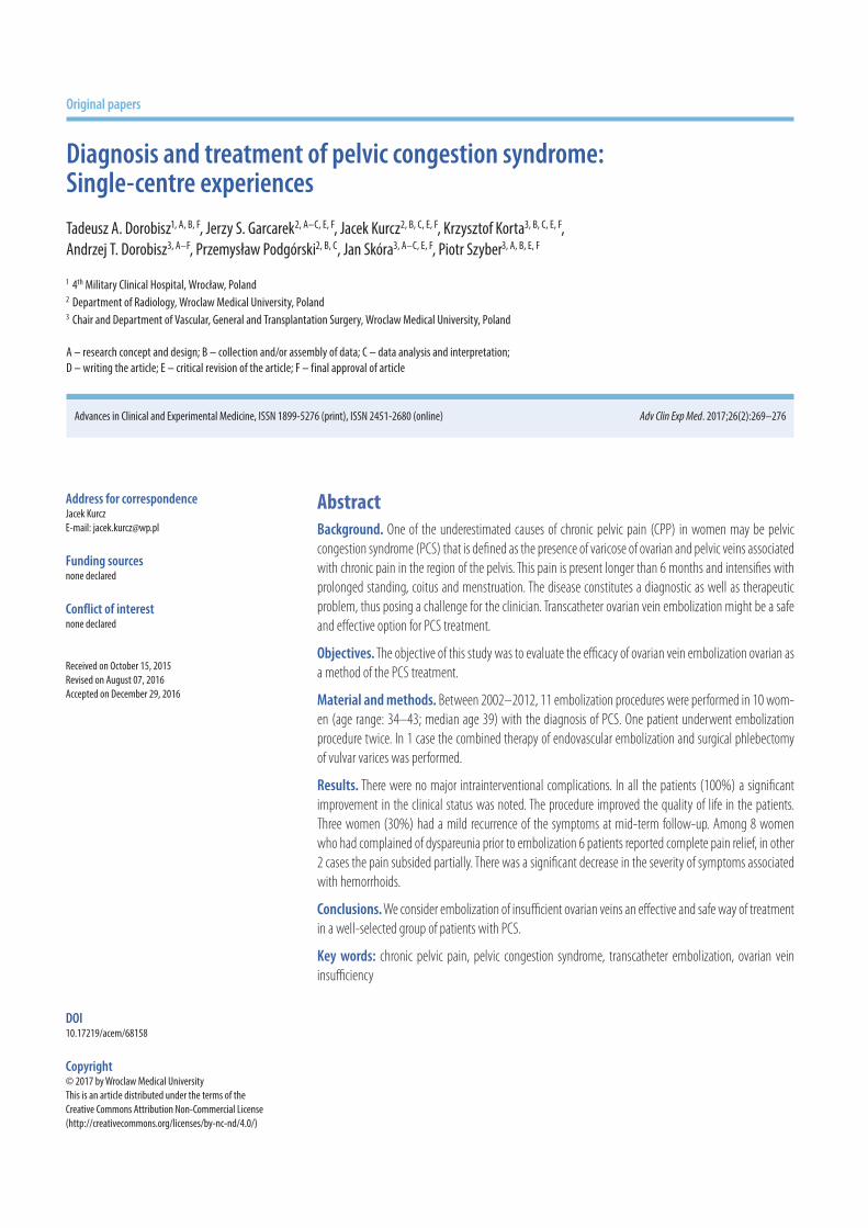
combination of surgical and endovascular approach was implemented. The 1st stage of the treatment included the surgical removal of varicose veins in the lower limbs and partially the vulvar varicose veins. The 2nd stage consisted
of 2 embolizations of ovarian veins that appeared to be double, with one of them seemingly unchanged. Eventu-ally, the additional sclerotherapy of vulvar varicosities was performed with the total pain relief.
Fig. 5a. Second procedure with embolization of duplicated left ovarian vein
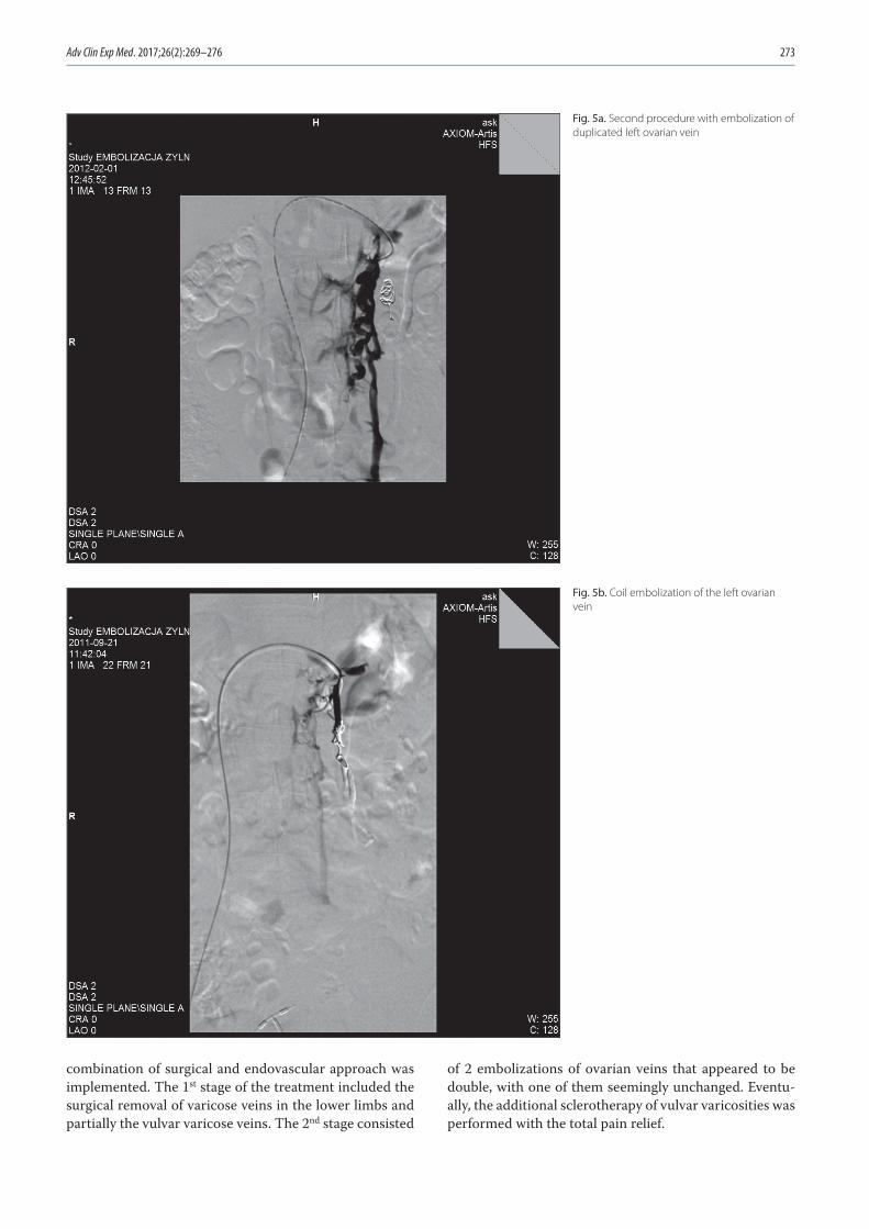
Fig. 5b. Coil embolization of the left ovarian vein

T. Dorobisz, et al. Pelvic congestion syndrome: Own experiences274
We did not observe a contrast extravasation during the embolization procedure. There were no major morbidity rates and no mortality rate neither intrainterventionally nor during the follow-up.
The mean follow-up period amounted to 16.4 months (range: 2–26 months). In all the patients the significant im-provement of the clinical status was observed. In 7 cases (70%) total pain relief was observed. In 3 women (30%) a mild recurrence of the symptoms was noted after 8–12 months postinterventionally; however, it was not necessary to repeat the embolization procedure in those patients after an initial 100% symptoms relief.
Among 8 women who had complained of dyspareunia prior to embolization, 6 patients reported complete pain relief, in other 2 cases pain subsided partially. There was a significant decrease in the severity of hemorrhoids as-sociated symptoms.
DiscussionIn 1950 Topolanski-Sierra coined the term of chronic
pelvic congestion and demonstrated the relationship be-tween chronic abdominal pain and pelvic varicose veins, and stressed the necessity of its treatment.14 One of the suggested methods of treatment included chemical dam-age of the fallopian tubes by the administration of gosere-lin acetate.15 In 1980, Rundquist described a new method of surgical treatment, with extraperitoneal ligation of the left ovarian vein. This method proved effective in the
treatment of the lower abdominal pain.16,17 It found many followers, especially in the context of the negative evalu-ation of hysterectomy. In 33% of patients after hysterec-tomy no relief of symptoms was observed and in 20% of patients symptoms reoccurred.18–20 Overall, each kind of surgical approach is associated with the risk of anesthesia, laparotomy or laparoscopic treatment.
First introduced in 1993 by Edwards et al., the tech-nique that truly revolutionized PCS treatment involves catheterization of the ovarian veins.21 The procedure is usually performed during the diagnostic phlebography and it involves venous obliteration with a foam or embo-lization agent.
There is no data from randomized trials relating to PCS treatment. Several published results of trial series do not attempt to compare the embolization of incompetent pel-vic veins with the other, alternative methods. Random-ized trials are of particular significance especially when the main symptom of the disease is the pain, because pain is highly subjective. The aforementioned published trial results relate to the retrospective cases. The search of the literature on the subject has been conducted with the use of the MEDLINE database and covered the period to March 2012 included. The largest series of clinical trials and case studies discussed by various authors are listed in the Table 2.25–30
In addition to the evaluation of clinical efficacy of the embolization procedure for the treatment of PCS, at-tempts have been made to develop the uniform diagnos-tic criteria and to determine if the chronic pain is caused
Fig. 5c. Postembolization follow-up venogram showing the proper patency of the left renal vein

Adv Clin Exp Med. 2017;26(2):269–276 275
solely by vascular lesions. The significance of B-mode US, duplex Doppler imaging, CT and magnetic resonance imaging for the differential diagnostic considerations has been stressed.31,33 The authors note that the published material hardly suggests guidelines for the diagnosis of PCS; moreover, the definitions of symptoms and of pel-vic pain vary greatly among studies. Furthermore, in the majority of trials no statistical methods have been applied for the objective presentation of the results. In his pub-lication, Ball argues whether an acute pain in the lower abdomen is indeed caused by pelvic varices.34 The author demonstrates that not all patients with vascular lesions suffer from pelvic pain.
So far there have been no unambiguous randomized trials or criteria that would qualify women for PCS treat-ment. However, the long history of research on the es-sence of this condition allowed for the development of ef-fective and minimally invasive treatment for women with this chronic illness. There are many different conditions that can directly cause the main symptom: dull pain in the lower abdomen. Anatomic abnormalities, pregnan-cies, hormonal changes, body weight gain or the lower extremities varicose vein disease can all contribute to the unidirectional course and intensification of PCS. Venous valve incompetence can result in pelvic engorgement and resultant edema thus causing pelvic pain. This pain is ex-acerbated after prolonged sitting, at the end of a day, dur-ing or directly after sexual activity (dyspareunia), during menstruation. There are also other unspecific symptoms of varying intensity. Women affected with these symp-toms can experience nausea, depression, tender lower abdomen, vaginal discharge, vulvar swelling, neuropa-thy, rectal discomfort or frequent urination. Most often affected women are in the pre-menopausal age; therefore, the suggested causes include the estrogen influence that significantly weakens venous walls. Pelvic vein thrombo-sis is a potentially fatal condition and is seen as a compli-cation of the abovementioned condition.
Contemporary medical imaging techniques have broad-ened diagnostic procedures. Pelvic ultrasound scans, trans-vaginal US and CT have become the first imaging diag-nostic procedures in patients with chronic pelvic pain. They all visualize the uterus and its vicinity. It is, however, the computed tomography with the use of contrast media
that demonstrated the highest efficacy in visualizing the vascular lesions. Then again, the US in the duplex Dop-pler mode allows for the dynamic evaluation of the blood flow, the detection of reversed flow in the left ovarian vein. In chronic pelvic pain the transvaginal ultrasound can be especially useful for the preliminary diagnoses of tumors and cysts, and endometric cysts. However, the proper di-agnosis is often missed because women lie down for a pel-vic examination. In this position, the ovarian veins will not fill enough with blood to reveal the vascular changes. It should be remembered that a standard transvaginal US will not reveal vascular changes and is reliable only when duplex Doppler modality is implemented.
In their advanced diagnostic management most diag-nosticians rely on MRI. This procedure can be carried out as an outpatient procedure, it is non-invasive, does not require ionizing radiation and is of particular value in diagnosing the pelvic varicose veins. Typical findings in patients with PCS on MRI include dilated, enhancing tubular structures near the ovaries and uterus, with pos-sible extension into the broad pelvic ligament. As indicat-ed by the clinical experience, MRI has become the most valuable diagnostic modality, particularly in T1-weighted scans and above all with volumetric 3D imaging following intravenous injection of gadolinium.
Traditional phlebography still seems to be the golden standard reference for the best visualization of the venous circulation and above all it allows for therapeutic proce-dures. However, it should be remembered that it is an invasive procedure and, therefore, should be undertaken after appropriate non-invasive demonstration of lesions as both a diagnostic and therapeutic procedure. In this procedure, it is possible to plan and perform the occlu-sion of insufficient veins.
Laparoscopy is the most commonly used diagnostic technique used in women with chronic pelvic pain. This direct visualization is an excellent tool to exclude other pelvic pathologic conditions such as, e.g. endometriosis. It is, however, not useful for varicose vein diagnosis be-cause it requires women to lie down for the procedure and involves insufflations of carbon dioxide that conceals path-ological vessels and, as a rule, yields unsatisfactory results.
In our material the patients reported substantial (30%) or complete relief (70%) of their pain following trans-
Table 2. Outcomes of clinical trials trials
Study Country Number of patients Mean follow-up(months)
Clinical outcomes (regression of symptoms)
Maleux et al., 20001 Belgium 41 19.9 significant: 58.9%
Venbrux et al., 20022 USA 56 22.1 significant or partial: 96%
Pieri et al., 20033 Italy 33 12.0 significant: 100%
Kim et al., 20064 USA 127 45.0 significant: 83%
Kwon et al., 20075 Korea 67 ~44.8 significant or partial: 82%
Gandini et al., 20086 Italy 38 12.0 significant: 100%

T. Dorobisz, et al. Pelvic congestion syndrome: Own experiences276
catheter ovarian vein embolization. In the present study, 8 (80%) patients underwent only one embolization. One patient required a multistage treatment, involving em-bolization of ovarian veins and varicose veins surgery (including vulvar varicosities). There was significant re-lief of the following symptoms after embolization: pelvic pain; pain prior to and during menstruation, pain during and after sexual activity and moderate reduction of com-plaints that were associated with maintaining a standing position. The procedures performed did improve the pa-tient’s quality of life. No major local or general compli-cations were observed. The present study was based on a limited number of patients, so all the results have to be interpreted with caution. However, there is still a lack of randomized controlled trials estimating ovarian vein em-bolization as a treatment of choice of PCS. Despite the limitations of our study we believe that venography with ovarian vein embolization could be the optimal therapeu-tic approach for pelvic congestion syndrome.
References 1. APGO Educational Series on Women’s Health Issues. Chronic pelvic
pain: An integrated approach Crofton, Md: APGO, January 2000. 2. Harris R, Holtzman S, Poppe A. Clinical outcome in female patients
with pelvic pain and normal pelvic US findings. Radiology. 2000;216: 440–443.
3. Francica G, Giardiello C, Angelone G, et al. Abdominal wall endo-metriomas near cesarean delivery scars. J Ultrasound Med. 2003;22: 1041–1047.
4. Kuligowska E, Deeds L, Lu K. Pelvic pain: Overlooked and underdi-agnosed gynecologic conditions. Radiographics. 2005;25(1):3–20.
5. Waliszewski P. Masywny zator tętnicy płucnej w przebiegu zakrzepi-cy splotów żylnych miednicy mniejszej. Przew Lek. 2000;9:105–108.
6. Belenky A, Bartal G, Atar E, et al. Ovarian varices in healthy female kidney donors: Incidence, morbidity, and clinical outcome. Am J Roentgenol. 2002;179(3):625–627.
7. Coakley F, Varghese S, Hricak H. CT and MRI of pelvic varices in women. J Comput Assist Tomogr. 1999;23:429–434.
8. Capasso P, Simons C, Trotteur G, et al. Treatment of symptomatic pelvic varices by ovarian vein embolization. Cardiovasc Intervent Radiol. 1997;20:107–111.
9. Tarazov P, Prozorovskij K, Ryzhkov V. Pelvic pain syndrome caused by ovarian varices: Treatment by transcatheter embolization. Acta Radiol. 1997;38:1023–1025.
10. Gargiulo T, Mais V, Brokaj L, et al. Bilateral laparoscopic transperi-toneal ligation of ovarian veins for treatment of pelvic congestion syndrome. J Am Assoc Gynecol Laparosc. 2003;10:501–504.
11. Beard R, Highman J, Pearce S, et al. Diagnosis of pelvic varicosities in women with chronic pelvic pain. Lancet. 1984;2:946–949.
12. Maleux G, Stockx L, Wilms G, et al. Ovarian vein embolization for the treatment of pelvic congestion syndrome: Long-term techni-cal and clinical results. J Vasc Interv Radiol. 2000;11:859–864.
13. Desimpelaere J, Seynaeve P, Hagers Y, et al. Pelvic congestion syn-drome: Demonstration and diagnosis by helical CT. Abdom Imag-ing. 1999;24:100–102.
14. Topolanski-Sierra R. Pelvic phlebography. J Obstet Gynecol. 1958; 76(1):44–52.
15. Rundqvist E, Sandholm L, Larsson G. Treatment of pelvic varicosi-ties causing lower abdominal pain with extraperitoneal resection of the left ovarian vein. Ann Chir Gynaecol. 1984;73 :339–341.
16. Soysal M, Soysal S, Vidcan K, et al. A randomized controlled trial of goserelin and medroxyprogesterone acetate in the treatment of pelvic congestion. Reprod Hum. 2001;16(5):931–939.
17. Hiromura T, Nishioka T, Nishioka S, et al. Reflux in the left ovarian vein: Analysis of MDCT findings in asymptomatic women. Am J Roent genol. 2004;183(5):1411–1415.
18. Beard R, Kennedy R, Gangar K, et al. PubMed Bilateral oophorec-tomy and hysterectomy in the treatment of intractable pelvic pain associated with pelvic congestion. Br J Obstet Gynaecol. 1991;98(10): 988–992.
19. Carter J. Review Surgical treatment for chronic pelvic pain. JSLS. 1998;2(2):129–139.
20. Takeuchi K, Mochizuki M, Kitagaki S. Laparoscopic varicocele liga-tion for pelvic congestion syndrome. Int J Gynaecol Obstet. 1996; 55:177–178.
21. Edwards R, Robertson J, MacLean A, et al. Case report: Pelvic pain syndrome- successful treatment of a case by ovarian vein emboli-zation. Clin Radiol. 1993;47:429–431.
22. Kim H, Malhotra A, Rowe P, Lee J, Venbrux A. Embolotherapy for pelvic congestion syndrome: Long-term results. J Vasc Interv Radiol. 2006;17:289–297.
23. Nascimento A, Mitchell D, Holland G. Ovarian veins: Magnetic res-onance imaging findings in an asymptomatic population. J Magn Reson Imaging 2002;15(5): 551–556.
24. Ignatio E, Dua R, Sarin S, et al. Pelvic congestion syndrome: Diagno-sis and treatment. Semin Intervent Radiol. 2008;25(4):361–368.
25. Rozenblit A, Ricci Z, Tuvia J, et al. Incompetent and dilated ovarian veins: A common CT finding in asymptomatic parous women. Am J Roentgenol. 2001;176(1):119–122.
26. Venbrux A, Chang A, Kim H. Pelvic congestion syndrome (pelvic venous incompetence): Impact of ovarian and internal iliac vein embolotherapy on menstrual cycle and chronic pelvic pain. J Vasc Interv Radiol. 2002;13:171–178
27. Pieri S, Agresti P, Morucci M. Percutaneous treatment of pelvic con-gestion syndrome. Radiol Med. 2003;105:76–82.
28. Kim H, Malhotra A, Rowe P, et al. Embolotherapy for pelvic congestion syndrome: Long-term results. J Vasc Interv Radiol. 2006;17:289–297.
29. Kwon S, Oh J, Ko K, et al. Transcatheter ovarian vein embolization using coils for the treatment of pelvic congestion syndrome. Car-diovasc Intervent Radiol. 2007;30(4):655–661.
30. Gandini R, Chiocchi M, Konda D, et al. Transcatheter foam sclero-therapy of symptomatic female varicocele with sodium-tetradec-yl-sulfate foam. Cardiovasc Intervent Radiol. 2008;31(4):778–784.
31. Naoum J. Endovascular therapy for pelvic congestion syndrome. Methodist Debakey Cardiovasc J. 2009;5(4):36–38.
32. Kies D, Kim H. Pelvic congestion syndrome: A review of current diagnostic and minimally invasive treatment modalities. Phlebol-ogy. 2012;27(Suppl 1):52–57.
33. Tu F, Hahn D, Steege J. Pelvic congestion syndrome-associated pel-vic pain: A systematic review of diagnosis and management. Obstet Gynecol Surv. 2010;65(5):332–340
34. Ball E, Khan K, Meads C. Does pelvic congestion syndrome exist and can it be treated? Acta Obstet Gynecol Scand. 2012;91:525–528.