diagnosis of gout - effective health care program effectiveness review number 158 diagnosis of gout...
TRANSCRIPT

Comparative Effectiveness ReviewNumber 158
Diagnosis of Gout

Comparative Effectiveness Review Number 158
Diagnosis of Gout
Prepared for: Agency for Healthcare Research and Quality U.S. Department of Health and Human Services 5600 Fishers Lane Rockville, MD 20857 www.ahrq.gov
Contract No. 290-2012-00006-I
Prepared by: Southern California Evidence-based Practice Center Santa Monica, CA
Investigators: Sydne J. Newberry, Ph.D. John FitzGerald, M.D., Ph.D. Margaret A. Maglione, M.P.P. Claire E. O’Hanlon, M.P.P. Dan Han, M.P.A. Marika Booth, M.S. Aneesa Motala, B.A. Abdul Tariq, B.S. Whitney Dudley, B.S. Roberta Shanman, M.L.S. Paul G. Shekelle, M.D, Ph.D.
AHRQ Publication No. 15(16)-EHC026-EF February 2016Addendum October 2016
Addendum – October 2016
An update search was conducted to prepare a manuscript based on the report (end date of update search February 29, 2016) and identified six new studies that met inclusion criteria. Five addressed Key Question (KQ) 1 (validity) (three reported on algorithms [Jatuworapruk et al., 2016; Taylor et al., 2015; Neogi et al., 2016] and two reported on imaging [Löffler et al., 2015; Pascal et al., 2015]). One addressed KQ2 (adverse events [Taylor et al., 2016]).
KQ1 Validity of Diagnostic Methods One study reported on the development and validation of the new American College of
Rheumatology/European League Against Rheumatism (ACR/EULAR) gout classification criteria (Neogi et al., 2016). Using monosodium urate (MSU) crystal analysis as the reference standard, the classification criteria had a sensitivity of 92 percent and a specificity of 89 percent (including clinical and imaging domains) or 85 percent and 78 percent (excluding imaging). Thus, imaging findings improved both the sensitivity and specificity of clinical and laboratory criteria (study quality high).
Two studies further validated existing classification and diagnostic algorithms in different populations (Jatuworapruk, et al., 2015; Taylor et al., 2015). Based on these additional studies, the strength of evidence for the use of Janssens’ Diagnostic Rule (the Netherlands criteria) or the Clinical Gout Diagnosis criteria was raised from low to moderate.
Two studies further validated the use of the ultrasound double contour sign to diagnose gout (Löffler et al., 2015; Pascal et al., 2015). The pooled sensitivity and specificity for the double contour sign from the three studies in the original report and one of the two new studies (464 joints) were 74 percent (95% confidence interval [CI] 52, 88) and 88 percent (95% CI 68, 96), respectively. The strength of evidence supporting the use of ultrasound for gout diagnosis remains low. No new studies were identified that report on the use of dual energy computerized tomography or x ray.
KQ2. Adverse Events Associated with Gout Diagnostic Methods One new study identified one serious adverse event after synovial fluid aspiration (septic
arthritis 11 days post arthrocentesis, event rate 0.1%, 95% CI 0, 0.34) and 11 nonserious adverse events (mostly mild pain following the procedure; event rate 1.4%, 95% CI 0.6–2.1) (Taylor et al., 2016). More information is located in the Annals of Internal Medicine manuscript: http://annals.org/aim/article/doi/10.7326/M16-0462.
References Jatuworapruk K, Lhakum P, Pattamapaspong N, et al. Performance of the existing classification criteria for gout in Thai patients presenting with acute arthritis. Medicine (Baltimore). 2016;95(5):e27jatu30. Epub 6 Feb 2016. doi: 10.1097/md.0000000000002730. PMID: 2684451.
Löffler C, Sattler H, Peters L, et al. Distinguishing gouty arthritis from calcium pyrophosphate disease and other arthritides. Journal of Rheumatology. 2015;42(3):513-20. PMID: 2015796981. http://dx.doi.org/10.3899/jrheum.140634.

Neogi T, Jansen TL, Dalbeth N, et al. 2015 Gout classification criteria: an American College of Rheumatology/European League Against Rheumatism collaborative initiative. Annals of the Rheumatic Diseases. 2015;74(10):1789-98. Epub 12 Sept 2015. doi: 10.1136/annrheumdis-2015-208237. PubMed PMID: 26359487; PMCID: PMCPmc4602275.
Pascal Z, Valcov R, Fabreguet I, et al. A prospective evaluation of ultrasound as a diagnostic tool in acute microcrystalline arthritis. Arthritis research & therapy. 2015;17:188. Epub 23 July 2015. doi: 10.1186/s13075-015-0701-7. PMID: 26198435; PMCID: PMCPmc4511437.
Taylor WJ, Fransen J, Dalbeth N, et al. Diagnostic arthrocentesis for suspicion of gout is safe and well tolerated. The Journal of rheumatology. 2016;43(1):150-3. Epub 2015 Dec 3. doi: 10.3899/jrheum.150684. PMID: 26628602.
Taylor WJ, Fransen J, Jansen TL, et al. Study for Updated Gout Classification Criteria (SUGAR): identification of features to classify gout. Arthritis care & research. 2015. Epub 18 Mar 2015. doi: 10.1002/acr.22585. PMID: 25777045.

ii
This report is based on research conducted by the Southern California Evidence-based Practice Center (EPC) under contract to the Agency for Healthcare Research and Quality (AHRQ), Rockville, MD (Contract No. 290-2012-00006-I). The findings and conclusions in this document are those of the authors, who are responsible for its contents; the findings and conclusions do not necessarily represent the views of AHRQ. Therefore, no statement in this report should be construed as an official position of AHRQ or of the U.S. Department of Health and Human Services.
None of the investigators have any affiliations or financial involvement that conflicts with the material presented in this report.
The information in this report is intended to help health care decisionmakers—patients and clinicians, health system leaders, and policymakers, among others—make well informed decisions and thereby improve the quality of health care services. This report is not intended to be a substitute for the application of clinical judgment. Anyone who makes decisions concerning the provision of clinical care should consider this report in the same way as any medical reference and in conjunction with all other pertinent information, i.e., in the context of available resources and circumstances presented by individual patients.
This report is made available to the public under the terms of a licensing agreement between the author and the Agency for Healthcare Research and Quality. This report may be used and reprinted without permission except those copyrighted materials that are clearly noted in the report. Further reproduction of those copyrighted materials is prohibited without the express permission of the copyright holders.
AHRQ or U.S. Department of Health and Human Services endorsement of any derivative products that may be developed from this report, such as clinical practice guidelines, other quality enhancement tools, or reimbursement or coverage policies, may not be stated or implied.
This report may periodically be assessed for the currency of conclusions. If an assessment is done, the resulting surveillance report describing the methodology and findings will be found on the Effective Health Care Program Web site at www.effectivehealthcare.ahrq.gov. Search on the title of the report.
Persons using assistive technology may not be able to fully access information in this report. For assistance contact [email protected].
Suggested citation: Newberry SJ, FitzGerald J, Maglione MA, O’Hanlon CE, Han D, Booth M, Motala A, Tariq A, Dudley W, Shanman R, Shekelle PG. Diagnosis of Gout. Comparative Effectiveness Review No. 158. (Prepared by the Southern California Evidence-based Practice Center under Contract No. 290-2012-00006-I.) AHRQ Publication No. 15(16)-EHC026-EF. Rockville, MD: Agency for Healthcare Research and Quality; February 2016. Addendum October 2016. www.effectivehealthcare.ahrq.gov/reports/final.cfm.

iii
Preface The Agency for Healthcare Research and Quality (AHRQ), through its Evidence-based
Practice Centers (EPCs), sponsors the development of systematic reviews to assist public- and private-sector organizations in their efforts to improve the quality of health care in the United States. These reviews provide comprehensive, science-based information on common, costly medical conditions, and new health care technologies and strategies.
Systematic reviews are the building blocks underlying evidence-based practice; they focus attention on the strength and limits of evidence from research studies about the effectiveness and safety of a clinical intervention. In the context of developing recommendations for practice, systematic reviews can help clarify whether assertions about the value of the intervention are based on strong evidence from clinical studies. For more information about AHRQ EPC systematic reviews, see www.effectivehealthcare.ahrq.gov/reference/purpose.cfm.
AHRQ expects that these systematic reviews will be helpful to health plans, providers, purchasers, government programs, and the health care system as a whole. Transparency and stakeholder input are essential to the Effective Health Care Program. Please visit the Web site (www.effectivehealthcare.ahrq.gov) to see draft research questions and reports or to join an email list to learn about new program products and opportunities for input.
We welcome comments on this systematic review. They may be sent by mail to the Task Order Officer named below at: Agency for Healthcare Research and Quality, 5600 Fishers Lane, Rockville, MD 20857, or by email to [email protected]. Richard G. Kronick, Ph.D. Director Agency for Healthcare Research and Quality Stephanie Chang, M.D., M.P.H. Director, EPC Program Center for Evidence and Practice Improvement Agency for Healthcare Research and Quality
Arlene S. Bierman, M.D., M.S. Director Center for Evidence and Practice Improvement Agency for Healthcare Research and Quality Aysegul Gozu, M.D., M.P.H Task Order Officer Center for Evidence and Practice Improvement Agency for Healthcare Research and Quality
iv
Acknowledgments We wish to acknowledge the help of the Key Informants; Technical Expert Panel members;
Peer Reviewers; our AHRQ Task Order Officer, Aysegul Gozu; and the Associate Editor, Kathy Lohr, in conducting this review and completing the report. We also wish to thank Patty Smith for her assistance with administrative tasks.
Key Informants In designing the study questions, the EPC consulted several Key Informants who represent
the end-users of research. The EPC sought the Key Informant input on the priority areas for research and synthesis. Key Informants are not involved in the analysis of the evidence or the writing of the report. Therefore, in the end, study questions, design, methodological approaches, and/or conclusions do not necessarily represent the views of individual Key Informants.
Key Informants must disclose any financial conflicts of interest greater than $10,000 and any other relevant business or professional conflicts of interest. Because of their role as end-users, individuals with potential conflicts may be retained. The TOO and the EPC work to balance, manage, or mitigate any conflicts of interest.
The list of Key Informants who participated in developing this report follows: Hyon K. Choi, M.D., Ph.D. Professor of Medicine, Harvard Medical
School Director, Gout and Crystal Arthropathy
Center Director, Clinical Epidemiology and Health
Outcomes Division of Rheumatology, Allergy,
and Immunology Department of Medicine Massachusetts General Hospital Boston, MA Russell Harris, M.D., M.P.H. Sheps Center for Health Services Research University of North Carolina Professor, Division of General Medicine University of North Carolina–Chapel Hill Chapel Hill, NC David Hoelting, M.D. Pender Community Hospital Pender, NE Sanford S. Kaplan, D.D.S. Patient Advocate Los Angeles, CA
Gerald D. Levy, M.D. Rheumatologist Kaiser Foundation Hospital Bellflower, CA Shari Ling, M.D Deputy Chief Medical Officer Office of Clinical Standards and Quality Centers for Medicare & Medicaid Services Baltimore, MD Ted Mikuls, M.D. Associate Professor, Department of Internal
Medicine Division of Rheumatology University of Nebraska Medical Center Omaha, NE Tuhina Neogi, M.D. Associate Professor of Medicine,
Epidemiology Boston University School of Medicine Boston, MA

v
Richard Treger, M.D. Board-Certified Nephrologist Greater Los Angeles VA Health Care
System Los Angeles, CA
Lee Ann Weintraub, M.S., R.D. Dietitian Private Practice Los Angeles, CA
Technical Expert Panel In designing the study questions and methodology at the outset of this report, the EPC
consulted several technical and content experts. Broad expertise and perspectives were sought. Divergent and conflicted opinions are common and perceived as healthy scientific discourse that results in a thoughtful, relevant systematic review. Therefore, in the end, study questions, design, methodologic approaches, and/or conclusions do not necessarily represent the views of individual technical and content experts.
Technical Experts must disclose any financial conflicts of interest greater than $10,000 and any other relevant business or professional conflicts of interest. Because of their unique clinical or content expertise, individuals with potential conflicts may be retained. The TOO and the EPC work to balance, manage, or mitigate any potential conflicts of interest identified.
The list of Technical Experts who participated in developing this report follows: Hyon K. Choi, M.D., Ph.D.* Professor of Medicine, Harvard Medical
School Director, Gout and Crystal Arthropathy
Center Director, Clinical Epidemiology and Health
Outcomes Division of Rheumatology, Allergy,
and Immunology Department of Medicine Massachusetts General Hospital Boston, MA Nicola Dalbeth, M.D.* Bone and Joint Research Group Department of Medicine Faculty of Medical and Health Sciences University of Auckland Aucland, New Zealand Russell Harris, M.D., M.P.H. Sheps Center for Health Services Research University of North Carolina Professor, Division of General Medicine University of North Carolina–Chapel Hill Chapel Hill, NC
Gerald D. Levy, M.D.* Rheumatologist Kaiser Permanente Bellflower, CA Shari Ling, M.D. Deputy Chief Medical Officer Office of Clinical Standards and Quality Centers for Medicare & Medicaid Services Baltimore, MD Ted Mikuls, M.D. Associate Professor, Department of Internal
Medicine Division of Rheumatology University of Nebraska Medical Center Omaha, NE
Esther Myers, Ph.D, R.D. Director, Scientific Affairs and Research Academy of Nutrition and Dietetics Trenton, IL

vi
Tuhina Neogi, M.D.* Associate Professor of Medicine,
Epidemiology Boston University School of Medicine Boston, MA Savvas Nicolaou, M.D.* Vice-Chair, Undergraduate Education
and Continuing Professional Development University of British Columbia
and Vancouver General Hospital Vancouver, British Columbia, Canada
H. Ralph Schumacher, Jr., M.D.* Professor of Medicine University of Pennsylvania Philadelphia, PA Ralf G. Thiele, M.D.* Associate Professor Department of Medicine, Allergy/
Immunology and Rheumatology School of Medicine and Dentistry University of Rochester Medical Center Rochester, NY Richard Treger, M.D. Board-Certified Nephrologist Greater Los Angeles VA Health Care
System Los Angeles, CA
Peter Tugwell, M.D. Institute of Population Health University of Ottawa Director, Centre for Global Health Ottawa, Ontario, Canada Daniel Waxman, M.D., Ph.D.* Natural Scientist and Visiting Associate
Professor of Emergency Medicine RAND and University of California,
Los Angeles Los Angeles, CA Neil Wenger, M.D. General Internal Medicine Physician and
Senior Researcher RAND and University of California, Los
Angeles Los Angeles, CA *Technical Expert Panel members who were also Peer Reviewers

vii
Peer Reviewers Prior to publication of the final evidence report, EPCs sought input from independent Peer
Reviewers without financial conflicts of interest. However, the conclusions and synthesis of the scientific literature presented in this report do not necessarily represent the views of individual reviewers.
Peer Reviewers must disclose any financial conflicts of interest greater than $10,000 and any other relevant business or professional conflicts of interest. Because of their unique clinical or content expertise, individuals with potential nonfinancial conflicts may be retained. The TOO and the EPC work to balance, manage, or mitigate any potential nonfinancial conflicts of interest identified.
The list of Peer Reviewers follows: Alan Baer, M.D. Good Samaritan Hospital Director, Johns Hopkins University Clinical
Practice Johns Hopkins University School
of Medicine Baltimore, MD Jasvinder Singh, M.D., M.P.H. Professor of Medicine and Staff
Rheumatologist University of Alabama at Birmingham
and VA Medical Center Birmingham, AL
William Taylor, M.B.Ch.B, Ph.D. Department of Medicine University of Otago Wellington, New Zealand Penny Whiting, Ph.D. Senior Research Fellow School of Social and Community Medicine University of Bristol Bristol, United Kingdom

viii
Diagnosis of Gout Structured Abstract Objectives. The aim of this review is to assess the evidence for the accuracy and safety of tests to diagnose gout in patients with no prior diagnosis of gout. The review also assesses factors that affect accuracy of diagnostic tests. Tests include algorithms that combine clinical signs and symptoms, dual-energy computed tomography (DECT), ultrasound, and plain x ray, with particular emphasis on tests that can be conducted in primary and acute (urgent and emergent) care settings. Data sources. We searched Medline® (from 1946), Embase® (from 1972), the Cochrane Library (from 1945), and the Web of Science™ (from 1980) to November 7, 2014, for published studies. We also searched ClinicalTrials.gov and the Web of Science and contacted manufacturers of imaging equipment and test kits for unpublished data on gout diagnosis. Review methods. We reviewed published and unpublished prospective cohort, cross-sectional, and case-control studies, as well as prior systematic reviews on the accuracy (sensitivity and specificity) of diagnostic tests for gout compared with a validated reference standard in patients without a prior gout diagnosis. We also reviewed studies and prior reviews of factors affecting the accuracy of monosodium urate crystal assessment in synovial fluid. We reviewed prospective cohort, cross-sectional, and case-control studies; case reports of any size; and systematic reviews that reported adverse events associated with diagnostic tests for gout and outcomes of gout misdiagnosis. A standardized protocol with predefined criteria was used to extract details on study design, interventions, outcomes, and study quality, and to assess the strength of evidence for each conclusion. Results. Six clinical algorithms comprising clinical signs and symptoms have been tested for diagnostic accuracy against the presence of monosodium urate crystals in synovial fluid aspirated from affected joints. Most studies were conducted with small groups of patients in academic rheumatology departments. Two recently developed clinical algorithms, the Diagnostic Rule, which is the only one developed and validated with primary care physicians and patients, and the Clinical Gout Diagnosis (CGD), demonstrated sensitivities of 88 percent and 97 percent, respectively, and specificities of 75 percent and 96 percent, respectively, in patients with shorter (2 years or less) and longer durations of symptoms, and they are simple to administer. However, the strength of evidence supporting their use is low, as validation of these tools remains limited. Three studies of DECT that enrolled patients without a previous gout diagnosis revealed sensitivities ranging from 85 percent to 100 percent and specificities ranging from 83 percent to 92 percent in diagnosing gout; the strength of evidence regarding the use of DECT for gout diagnosis is low. Four studies of ultrasound that enrolled patients without a previous diagnosis showed sensitivities ranging from 37 percent to 100 percent and specificities ranging from 68 percent to 97 percent, depending on the ultrasound signs assessed; the strength of evidence is low for the utility of ultrasound in diagnosing gout. A small number of studies examined factors that affected the accuracy of tests for the diagnosis of gout. The accuracy of monosodium urate

ix
analysis in synovial fluid varies widely among practitioners, but evidence on the effects of skill and experience is insufficient. No studies examined differences among practitioners in the rate of successful joint aspiration. No studies reported adverse events directly associated with techniques used to diagnose gout. However in one small study, missed gout diagnosis resulted in unnecessary surgery, longer hospital stays, and delay in appropriate treatment. Conclusions. Promising diagnostic clinical algorithms such as the Diagnostic Rule and CGD need to be validated more broadly in primary and urgent care settings. A clinical algorithm with high diagnostic accuracy ideally can form part of a diagnostic decision tree, with referral of more clinically challenging cases to rheumatologists for more invasive tests or imaging. Research is needed to assess the incremental value of synovial fluid monosodium urate crystal analysis and imaging over that of a diagnostic clinical algorithm.

x
Contents Executive Summary .................................................................................................................ES-1 Introduction ....................................................................................................................................1
Background ................................................................................................................................1 Condition..............................................................................................................................1 Etiology of Gout ..................................................................................................................1 Diagnostic Strategies ...........................................................................................................2
Scope and Key Questions ..........................................................................................................3 Scope of the Review ............................................................................................................3 Key Questions ......................................................................................................................4
Organization of This Report ......................................................................................................5 Methods ...........................................................................................................................................6
Criteria for Inclusion/Exclusion of Studies in the Review ........................................................6 PICOTS of Included Studies ................................................................................................6
Literature Search Strategies for Identification of Relevant Studies To Answer the Key Questions ................................................................................................................8
Data Abstraction and Data Management ...................................................................................9 Assessment of Methodological Quality of Individual Studies ..................................................9 Data Synthesis/Analysis.............................................................................................................9 Grading the Strength of the Body of Evidence for Each Key Question ....................................9 Applicability ............................................................................................................................10 Peer Review and Public Commentary .....................................................................................10
Results ...........................................................................................................................................11 Introduction ..............................................................................................................................11 Results of Literature Searches .................................................................................................11 Key Question 1 ........................................................................................................................13
a. What is the accuracy of clinical signs and symptoms and other diagnostic tests (such as serum uric acid, ultrasound, CT scan, DECT, and plain x-ray), alone or in combination, compared with synovial fluid analysis in the diagnosis of acute gouty arthritis, and how does the accuracy affect clinical decisionmaking, clinical outcomes and complications, and patient centered outcomes?
b. How does the diagnostic accuracy of clinical signs and symptoms and other tests vary by affected joint site and number of joints?
c. Does the accuracy of diagnostic tests for gout vary by duration of symptoms (i.e., time from the beginning of a flare)?
Key Points ..........................................................................................................................13 Description of Included Studies .........................................................................................14 Detailed Synthesis ..............................................................................................................14
KQ1d. Does the accuracy of synovial fluid aspiration and crystal analysis differ by i) the type of practitioner who is performing the aspiration and ii) the type of practitioner who is performing the crystal analysis? .....................................................42 Key Points ..........................................................................................................................42 Description of Included Studies .........................................................................................42 Detailed Synthesis ..............................................................................................................42
xi
Key Question 2. What are the adverse effects (including pain, infection at the aspiration site, radiation exposure) or harms (related to false positives, false negatives, indeterminate results) associated with tests used to diagnose gout? ................43 Key Points ..........................................................................................................................43 Description of Included Studies .........................................................................................44 Detailed Synthesis ..............................................................................................................44
Discussion......................................................................................................................................45 Key Findings and Strength of Evidence ..................................................................................45
Accuracy of Tests for the Diagnosis of Gout....................................................................45 Adverse Events Associated With Testing for Gout ..........................................................45
Findings in Relationship to What Is Already Known ..............................................................46 Monosodium Urate Crystal Assessment ............................................................................46 Accuracy of Algorithms Comprising Clinical Signs and Symptoms
for the Diagnosis of Gout .............................................................................................47 Accuracy of DECT for the Diagnosis of Gout...................................................................47 Accuracy of Ultrasound for the Diagnosis of Gout ...........................................................48
Applicability ............................................................................................................................49 Implications for Clinical and Policy Decisionmaking .............................................................49 Limitations of the Comparative Effectiveness Review Process ..............................................49 Limitations of the Evidence Base ............................................................................................50 Research Gaps ..........................................................................................................................50 Conclusions ..............................................................................................................................52
References .....................................................................................................................................54 Abbreviations/Acronyms .............................................................................................................58
Tables Table A. Comparison of components among clinical algorithms for diagnosis of gout .........ES-14 Table B. Summary of findings and strength of evidence.........................................................ES-23 Table C. Summary of findings on comparative accuracy and safety of gout
diagnostic methods.............................................................................................................ES-25 Table 1. Summary of included studies of algorithms comprising clinical signs
and symptoms used to diagnose gout ......................................................................................20 Table 2. Comparison of components among clinical algorithms for diagnosis of gout ................26 Table 3. Studies assessing the accuracy of DECT to diagnose gout .............................................30 Table 4. Summary of studies reporting on the use of ultrasound for diagnosis of gout ................35 Table 5. Risk of bias assessment by QUADAS-2..........................................................................40 Table 6. Summary of findings and strength of evidence ...............................................................52
Figures Figure A. Analytic framework ...................................................................................................ES-4 Figure B. Literature flow diagram ...........................................................................................ES-11 Figure 1. Analytic framework ..........................................................................................................4 Figure 2. Literature flow diagram ..................................................................................................12

xii
Appendixes Appendix A. Search Strategy Appendix B. List of Excluded Studies Appendix C. Evidence Table Appendix D. Data Abstraction Tools

ES-1
Executive Summary Background
Condition Gout is a form of inflammatory arthritis characterized by acute intermittent episodes of
synovitis presenting with joint swelling and pain; the episodes are referred to as acute gouty arthritis flares or attacks. The condition may progress to a chronic and persistent condition, with development of tophi (solid deposits of monosodium urate [MSU] crystals in joints, cartilage, tendons, bursae, bone, and soft tissue), a condition called chronic tophaceous gout. There is no clear distinction between acute intermittent and chronic intermittent conditions, whereas the advanced stage of gout is characterized by more persistent joint manifestations and tophi (either clinically evident or hidden within the joint).
Gout is the most common form of inflammatory arthritis, and the prevalence has been increasing. The most recent estimate of prevalence among adults in the United States, based on data from the 2007–08 National Health and Nutrition Examination Survey (NHANES), is 3.9 percent (8.3 million individuals), ranging from 2.0 percent in women to 5.9 percent in men,1 an increase over that of previous NHANES data cycles. The rise in the prevalence of gout has paralleled the increase in prevalence of comorbid conditions associated with hyperuricemia (the primary risk factor for gout), including obesity, hypertension, hypertriglyceridemia, hypercholesterolemia, type 2 diabetes, metabolic syndrome, chronic kidney disease, and renal insufficiency. Increased use of medications that increase the risk for developing hyperuricemia (e.g., thiazide diuretics, low-dose aspirin, or their combination) may further explain the increasing prevalence of gout.
In a 2013 study that analyzed data from several national surveys administered from 2002 to 2008, the number of ambulatory care visits attributable to gout was estimated to be 7 million visits annually, with 2 million attributable to acute attacks. (The rate more than doubled from 2002 to 2008.) The total annual ambulatory care costs associated with gout (visits and medications) were estimated at $933 million (in 2009 dollars). Drug expenditures accounted for 61 percent of the total costs.2
In addition to gout, the types of inflammatory arthritis include rheumatoid arthritis, septic arthritis, inflammatory episodes of osteoarthritis, and calcium pyrophosphate dihydrate crystal deposition disease (CPPD, formerly known as pseudogout). Patients with any of these types of arthritis can present with clinically similar signs and symptoms, but the conditions have different treatments, and incorrect diagnosis can have serious outcomes. For example, missing a case of septic arthritis can lead to joint damage and septic shock. A major challenge for effective gout management, particularly in the primary care and urgent/emergent care setting where most gout patients are managed, is distinguishing gout from these other conditions. Inappropriate or delayed treatment can incur serious complications.
Etiology of Gout The driving force behind acute episodes of gout is hyperuricemia, defined as an elevated
serum uric acid (more accurately referred to as “serum urate” for the salt form that occurs in the serum) concentration greater than 6.8 mg per deciliter in men and greater than 6.0 in women. Hyperuricemia is most commonly the result of inadequate renal excretion of uric acid or, less

ES-2
commonly, uric acid overproduction. (Uric acid is a breakdown product of dietary or endogenous purines.) Hyperuricemia leads to formation and deposition of MSU crystals, which preferentially deposit in joints, tendons, and bursa spaces. For reasons that remain unclear, only a small proportion of individuals with hyperuricemia go on to develop gout. For others, hyperuricemia remains asymptomatic.3 The prevalence of hyperuricemia ranges from 21.2 percent in men to 21.6 percent in women, 4 to 10 times as high as the prevalence of gout.4
The causes of gout are multifactorial, including a combination of genetic, hormonal, metabolic, pharmacologic, comorbid (renal disease), and dietary factors. Family history, advancing age, male sex, or, in women, early menopause have been associated with a higher risk of gout and/or gout flares.5 Dietary risk factors for gout include consumption of purine-rich foods or drinks, including alcohol, meat, and seafood, and consumption of sugar-sweetened soft drinks and foods high in fructose. Dairy foods and coffee have been associated with a lower risk of incident gout and in some cases a lower rate of gout flares. However, the role of diet in the etiology and treatment of gout is a topic of considerable research and will be reviewed in a separate systematic review.
Diagnostic Strategies The majority of individuals with gout are initially seen, diagnosed, and treated in primary and
urgent care settings. Thus primary care physicians (PCPs) and emergency medicine physicians are the most likely practitioners to see patients with symptoms suggestive of an acute attack of gout but with no prior diagnosis. Such patients may be experiencing a first attack (early-stage gout) or may have experienced numerous attacks and have more advanced gout.
Some researchers have argued the need for laboratory assessment of synovial (joint) fluid MSU crystals in the presence of an acute inflammatory arthritis for a definitive diagnosis of gout, and MSU crystal analysis has been regarded as the gold standard against which other potential diagnostic methods are measured. However, joint aspiration can be technically difficult to perform and painful to the patient, and is often deferred in primary and urgent care settings, to be conducted by a specialist (e.g., a rheumatologist or orthopedic surgeon).6 In addition, the accuracy of synovial fluid analysis may be affected by a number of factors (patient, practitioner, and analyst related).7,8 A 2009 study found that unguided needle insertion in the toe is often inaccurate.9 At least three studies have found wide variation in the accuracy of assessment of synovial fluid crystals (both MSU and calcium pyrophosphate) and white blood cells across hospital laboratories,10-12 which could potentially be caused by patient differences, differences in skill levels of the practitioners drawing or analyzing the samples, or differences in sample handling. A 1999 systematic review on the accuracy of MSU crystal analysis in synovial fluid13 concluded that MSU analysis had poor sensitivity, specificity, and reproducibility. A 2013 systematic review of the accuracy of methods for detecting MSU in synovial fluid concluded that storage of samples at room temperature resulted in a decrease in MSU concentration over time compared with refrigeration14 but could not draw any conclusions about the role of personnel. Evidence from a 2011 survey of rheumatologists suggests that synovial fluid analysis is underused in the rheumatology setting as well.15
Instead of analyzing MSU crystals in synovial fluid, PCPs and emergency medicine physicians tend to rely on clinical algorithms comprising some combination of clinical signs and symptoms to diagnose an acute episode of gout. These clinical signs and symptoms include rapid development of inflammation and pain, erythema, monoarthritis, response to administration of the drug colchicine, and symptoms in the first metatarsophalangeal joint, among others (with

ES-3
synovial fluid culture sometimes used to rule out septic arthritis and other potential causes for inflammatory arthritis).
Attempts to standardize and validate such clinical diagnostic algorithms date back to the 1960s.16 Most of these algorithms were not developed for diagnostic purposes but for classification of gout. Concurrent with this review, the American College of Rheumatology (ACR) and the European League Against Rheumatism (EULAR) are collaborating to update and evaluate classification criteria for gout. Distinct from diagnostic criteria, classification criteria are intended to ensure the correct identification and staging of patients with a particular disease condition (especially patients in the early stages of the disease) for the purpose of enrollment in studies of disease management.17
Therefore, a question of importance is whether any combination of clinical signs and symptoms and laboratory tests accessible in the primary or acute care setting (which we refer to as a “clinical algorithm” or “clinical diagnostic algorithm”) will have good predictive value compared with tests such as joint aspiration and synovial fluid analysis for MSU, both to correctly diagnose gout and to rule out other causes of joint inflammation, particularly septic arthritis and calcium pyrophosphate deposition disease, for patients presenting with an acute episode of inflammatory arthritis.
Imaging modalities have also been assessed for both diagnosis and classification of gout. These techniques include plain radiographs and newer techniques such as ultrasound and dual-energy computed tomography (DECT), which are just beginning to be used to diagnose gout in some settings.18 Therefore, another question of importance for gout diagnosis is how these newer methods compare with joint aspiration and synovial fluid MSU analysis in their predictive value for the initial diagnosis of gout and whether they provide any additive value over the use of MSU analysis or clinical signs and symptoms alone.
The safety of tests used to diagnose gout also needs to be considered. Potential safety concerns include acute physical discomfort from joint aspiration and long-term effects (e.g., from accumulated radiation exposure). Other concerns are the potential effects of misdiagnosis. These effects could include delay in initiating or failure to initiate appropriate treatment for gout, delay in initiating treatment for the actual disorder if it is not gout, or incorrect initiation of treatment for another disorder (e.g., hospitalization and administration of intravenous antibiotics for suspected joint sepsis) when the patient has gout.
Therefore, we have undertaken a systematic review of studies examining the accuracy and safety of tests used to diagnose gout—including algorithms combining physical signs and symptoms, serum urate, ultrasound, plain radiography, and DECT—compared with synovial fluid MSU analysis. The primary focus of this review is on tests that can be used in the primary care or urgent/emergent care setting for an initial diagnosis of gout.
The aim of this review is to help inform clinical decisionmaking for patients and providers and to improve the quality of care for patients who present with previously undiagnosed gout in the primary and acute care setting.
Scope and Key Questions
Scope of the Review The purpose of this review is to assess the evidence on the validity and safety of tests for
diagnosing gout—including clinical signs and symptoms (individually and in combination as a clinical diagnostic algorithm), DECT, ultrasound, and other imaging methods—compared with
ES-4
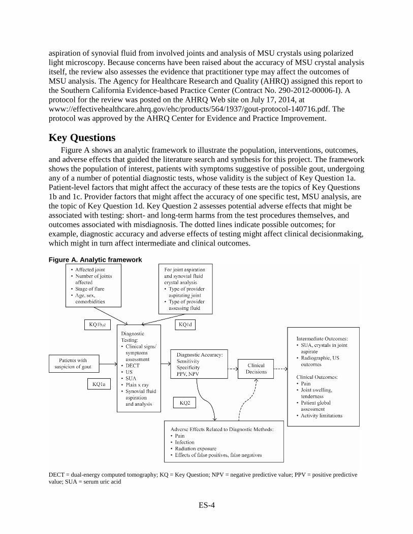
aspiration of synovial fluid from involved joints and analysis of MSU crystals using polarized light microscopy. Because concerns have been raised about the accuracy of MSU crystal analysis itself, the review also assesses the evidence that practitioner type may affect the outcomes of MSU analysis. The Agency for Healthcare Research and Quality (AHRQ) assigned this report to the Southern California Evidence-based Practice Center (Contract No. 290-2012-00006-I). A protocol for the review was posted on the AHRQ Web site on July 17, 2014, at www://effectivehealthcare.ahrq.gov/ehc/products/564/1937/gout-protocol-140716.pdf. The protocol was approved by the AHRQ Center for Evidence and Practice Improvement.
Key Questions Figure A shows an analytic framework to illustrate the population, interventions, outcomes,
and adverse effects that guided the literature search and synthesis for this project. The framework shows the population of interest, patients with symptoms suggestive of possible gout, undergoing any of a number of potential diagnostic tests, whose validity is the subject of Key Question 1a. Patient-level factors that might affect the accuracy of these tests are the topics of Key Questions 1b and 1c. Provider factors that might affect the accuracy of one specific test, MSU analysis, are the topic of Key Question 1d. Key Question 2 assesses potential adverse effects that might be associated with testing: short- and long-term harms from the test procedures themselves, and outcomes associated with misdiagnosis. The dotted lines indicate possible outcomes; for example, diagnostic accuracy and adverse effects of testing might affect clinical decisionmaking, which might in turn affect intermediate and clinical outcomes.
Figure A. Analytic framework
DECT = dual-energy computed tomography; KQ = Key Question; NPV = negative predictive value; PPV = positive predictive value; SUA = serum uric acid

ES-5
Key Question 1. a. What is the accuracy of clinical signs and symptoms and other
diagnostic tests (such as serum uric acid, ultrasound, computed tomography (CT) scan, DECT, and plain x ray), alone or in combination, compared witha synovial fluid analysis in the diagnosis of acute gouty arthritis, and how does the accuracy affect clinical decisionmaking, clinical outcomes and complications, and patient-centered outcomes?
b. How does the diagnostic accuracy of clinical signs and symptoms and other tests vary by affected joint site and number of joints?
c. Does the accuracy of diagnostic tests for gout vary by duration of symptoms (i.e., time from the beginning of a flare)?
d. Does the accuracy of synovial fluid aspiration and crystal analysis differ by (i) the type of practitioner who is performing the aspiration and (ii) the type of practitioner who is performing the crystal analysis?
Key Question 2. What are the adverse effects (including pain, infection at the aspiration site, radiation exposure) or harms (related to false positives, false negatives, indeterminate results) associated with tests used to diagnose gout?
Methods
Criteria for Inclusion/Exclusion of Studies in the Review This report is based on a systematic search for prospective or cross-sectional studies that
compared the sensitivity and specificity of tests used to diagnose gout, preferably against joint aspiration and synovial fluid assessment for MSU crystals, in populations of adults 18 years of age or older suspected of having gout but not previously diagnosed. (See Table A in the Results section.) We also included studies that assessed patient and practitioner factors that affect the diagnostic accuracy of these tests or assessed harms associated with the tests, and studies that examined particular factors that potentially affect the sensitivity or specificity of tests (joints involved, duration of symptoms).
Tests of interest included algorithms comprising clinical or laboratory examination for physical signs, symptoms, and history; serum uric acid; US; DECT; and plain radiography. The comparator of primary interest was synovial fluid analysis of MSU crystals using polarized light microscopy. However, if no such studies could be identified for a diagnostic test of interest, studies were also included if some or all of the participants were diagnosed using the ACR criteria for gout diagnosis and classification or another validated set of diagnostic or classification criteria as a reference standard (comparator).
a Using monosodium urate crystal analysis of synovial fluid as the reference standard.
ES-6
Studies were excluded if participants had already been definitively diagnosed with gout prior to enrollment (to ensure that the patient populations were as similar as possible to patients who would be seen in the primary or urgent/emergent care setting), or if the comparator was individual physician opinion or was not identified. Inclusion criteria are further described in terms of PICOTs (populations, interventions, comparators, outcomes, timing, and settings), a framework used in systematic reviews to categorize inclusion and exclusion criteria).
Outcomes of interest were the comparative accuracy of the test results (as measured by the sensitivity and specificity or the positive and negative predictive value of the test in question), intermediate outcomes such as lab and radiographic test results, clinical decisionmaking that resulted from a diagnosis, short-term clinical (patient-centered) outcomes such as a change in pain and joint swelling that resulted from a diagnosis, and any adverse events (including adverse patient experiences such as pain or infection at the aspiration site, effects of radiation exposure, and the results of a false-positive or false-negative diagnosis) associated with the test. Prospective cohort, cross-sectional, and case-control (if needed) studies were included to address Key Question 1 (accuracy of test and factors that affect accuracy). Prospective cohort, cross-sectional, and case-control studies, as well as case series of any size and case reports of rare adverse events, were included if they addressed Key Question 2 (adverse events or other negative outcomes in individuals undergoing testing). The PICOTS for studies included in this review are as follows. Population(s) (Key Questions 1 and 2):
• Adults (18 years and over) presenting with symptoms (e.g., an acute episode of joint inflammation) suggestive of gout but without a prior gout diagnosis, including the following subgroups:
o Male and female patients o Patients with longer versus shorter duration of symptoms o Patients with comorbidities, including hypertension, type 2 diabetes, and kidney
disease (renal insufficiency) o Patients with osteoarthritis, septic arthritis, calcium pyrophosphate deposition
disease, or previous joint trauma o Individuals with a family history of gout
Interventions (index tests) (Key Questions 1 and 2): • Clinical history and physical exam • Serum urate assessment • US • DECT • Plain x ray • Joint aspiration by physicians and synovial fluid analysis using polarizing microscopy
(by physicians or laboratory personnel) • Combinations of these tests as identified in the literature
Comparators (reference tests): • Joint synovial fluid aspiration and microscopic assessment for MSU crystals (Key
Questions 1a–c and 2) • Joint synovial fluid aspiration and microscopic assessment for MSU crystals performed
by a practitioner with a different level of expertise or experience, such as rheumatologist, laboratory personnel (Key Question 1d)

ES-7
Outcomes: • Diagnostic accuracy of clinical signs and symptoms, ultrasound, DECT, and plain
radiographs compared with joint aspiration and synovial fluid analysis (Key Question 1) o Sensitivity/specificity, true positives/true negatives, area under the curve o Positive and negative predictive value, positive/negative likelihood ratios
• Clinical decisionmaking (Key Question 1) o Additional testing o Pharmacologic/dietary management
• Intermediate outcomes (Key Question 1) o Serum urate o Synovial fluid crystals o Radiographic or ultrasound changes
• Clinical outcomes (Key Question 1) o Pain, joint swelling, and tenderness o Patient global assessment and activity limitations (Key Questions 1 and 2)
• Adverse effects of the tests, including— o Pain, infection, and radiation exposure o Effects of false positives or false negatives (Key Question 2)
Timing: • For clinical outcomes of symptom relief: 1–2 days minimum (Key Question 1) • Early in an attack versus later or post-attack (Key Question 1c) • For adverse events: immediate
Settings: • Primary care (outpatient) or acute care settings preferred • Outpatient rheumatology practices/academic medical centers also accepted
Literature Search Strategies for Identification of Studies Relevant to Key Questions
The search strategy was designed by the Southern California Evidence-based Practice Center (EPC) reference librarian in collaboration with our local content expert, who has participated in two systematic reviews on gout;19,20 it appears in Appendix A of the full report. As recommended by the AHRQ “Methods Guide for Medical Test Reviews,”21 the searches were conducted without filters specific for diagnostic tests; instead, we used the term “gout” combined with the terms for the diagnostic tests.
We searched PubMed® (January 1, 1946, to November 7 , 2014), Embase® (January 1, 1972, to November 7, 2014), the Cochrane Library (January 1, 1945, to November 7, 2014, for the Cochrane Central Registry of Controlled Trials and January 1, 1996, to November 7, 2014, for the Cochrane Database of Systematic Reviews), and the Web of Science™ (January 1, 1980, to November 7, 2014); these dates were selected to replicate the searches conducted as the basis for the 2006 EULAR Guidelines on Diagnosis and Management of Gout.22 We also included any relevant studies identified in the searches we conducted for a simultaneous review on management of gout if they were not already identified in the searches for this review. Finally, we asked the Technical Expert Panel (TEP) to assess our list of included studies and to provide references for any studies they believed should also be included.
ES-8
We searched ClinicalTrials.gov and the Web of Science for recently completed studies and unpublished or non–peer-reviewed study findings. Searches were not limited by language of publication: non–English-language studies that met the inclusion/exclusion criteria based on a review of an English-language abstract were screened further in full text if translators could be identified with reasonable effort. We also contacted manufacturers of diagnostic equipment (polarizing microscopes, sonography equipment, DECT, and serum uric acid test kits) for unpublished data specific to the use of their equipment or tests for gout diagnosis.
An update search was conducted on November 7, 2014, after submission of the draft report for peer review. We transferred the output of the literature searches to DistillerSR™ for screening. Article titles and abstracts identified by the searches were independently screened by two literature reviewers using the predetermined inclusion and exclusion criteria, and those selected by either reviewer were accepted without reconciliation for further full-text review.
Two reviewers independently conducted full-text review to exclude articles that provided no usable data, reported the same data as another article, or enrolled participants with established gout diagnoses. Disagreements regarding inclusion at the full-text stage were reconciled with the input of the project lead when necessary.
We identified a small number of relatively recent systematic reviews on various aspects of gout diagnosis. In most cases, we used these reviews to identify references we had missed; however, if the review was of high quality, addressed a subquestion of interest, and included all the literature on the topic, we included it as a data source after assessing its quality. We also searched the reference lists of included studies for additional titles that appeared to meet our inclusion criteria and screened these articles for inclusion. For studies of apparent interest reported in meeting abstracts (conference proceedings), we searched for peer-reviewed publications of the findings. If findings had not yet been published in a peer-reviewed journal, we reserved them and cited them in the Discussion in suggestions for future research.
Data Abstraction and Data Management Two reviewers independently abstracted study-level details from articles accepted for
inclusion in DistillerSR, and any disagreements were reconciled with the input of the project leader, Southern California EPC director, or local subject-matter expert, if needed. Studies provided by manufacturers or suggested by peer reviewers underwent the same process, as did studies identified in update searches.
Assessment of Methodological Quality of Individual Studies The risk of bias (study quality) of individual included studies was assessed independently by
two reviewers using the Quality Assessment of Diagnostic Accuracy Studies (QUADAS)-2 tool,23,24 and assessments were reconciled, with any disagreements mediated by the project lead. We used AMSTAR (A Measurement Tool to Assess Systematic Reviews) to assess the quality of existing systematic reviews that we included;25 AMSTAR assessments were also conducted independently by two reviewers and reconciled.
Data Synthesis/Analysis For studies that assessed ultrasound, DECT, or another radiographic method, we extracted
and reported sensitivity, specificity, positive and negative predictive value, and area under the curve/receiver-operating characteristics, if reported.

ES-9
Studies were considered for meta-analysis if the number of true positives, true negatives, false positives, and false negatives was reported or could be calculated; studies were similar enough with respect to outcome measures, participants, and tests; and they assessed the validity of an alternative diagnostic method against that of analysis of MSU crystals in synovial fluid. The number of studies we identified precluded pooling; therefore, outcomes are described narratively in the full report, stratified by test comparisons of interest and study design. All included studies are also described in summary tables in the full report.
Grading the Strength of the Body of Evidence for Each Key Question
We assessed the overall strength of evidence for each conclusion using guidance suggested by AHRQ for its Effective Health Care Program.26 This method is based on a method developed by the GRADE (Grading of Recommendations, Assessment, Development and Evaluations) Working Group. The evidence grade is usually based on five required domains:
• Study limitations were assessed based on the risk-of-bias assessments for all studies that contribute to a conclusion.
• Consistency was determined by comparing the relative sensitivities and specificities because we did not pool studies.
• Directness is a measure of whether the evidence being assessed reflects a single direct link between the interventions of interest and the ultimate health outcome under consideration.
• Precision, a measure of the confidence intervals in a pooled analysis, also was not assessed in this review.
• Publication bias was assessed only for studies for which data were pooled.
Based on the domains we included, we classified the strength (grade) of evidence as follows: • High = Further research is unlikely to change our confidence in the estimate of effect. • Moderate = Further research is likely to have an important impact on our confidence in
the estimate of effect and may change the estimate. • Low = Further research is very likely to have an important impact on our confidence in
the estimate of effect and is likely to change the estimate. • Very low/insufficient = Any estimate of effect is very uncertain.
Applicability Applicability is a measure of the extent to which the participants, interventions, and outcome
measures are similar to those of the population of interest and care settings for which the outcomes are intended. We assessed applicability based on the inclusion and exclusion criteria described in the PICOTS, which included the study population age, sex, health profiles (including comorbidities as well as duration of symptoms and number of affected joints, when relevant), tests, gold standards, study settings, and provider types.27 Thus we would assign higher priority to studies of adult populations being seen in primary/urgent/emergent care settings for first or subsequent episodes of symptoms suggestive of gout than to studies of patients in an academic rheumatology department.
ES-10
Peer Review and Public Commentary A draft version of the report was posted for peer review on November 4, 2014, and revised in
response to reviewer comments.
Results This section first describes the results of the literature searches, followed by descriptions of
the studies that met inclusion criteria for each of the Key Questions and the key points (conclusions).
Results of Literature Searches Our searches identified 3,646 titles/abstracts, of which 3,391 were excluded for the following
reasons: participants not human (129); diagnostic methods beyond the scope of the review (129); not gout diagnosis or management (1,801); no original data or nonsystematic reviews (374); conference proceedings, presentations, or abstracts (11); case reports with sample sizes of fewer than 10 (415); population under age 18 (5); renal transplant or end-stage renal disease patients (12); titles with no abstracts (based on a survey of a random sample of 10% of these titles, for which full-text articles or reports were obtained and all were rejected as letters, commentaries, or nonsystematic reviews with no original data) (252); and gout management only (263). (See the PRISMA [Preferred Reporting Items for Systematic Reviews and Meta-Analyses] diagram, Figure B.)
We reviewed 255 full-text articles, of which 234 were excluded for the following reasons: participants not human (2); diagnostic methods beyond the scope of the review (44); not gout diagnosis or management (69); no original data (29); conference proceedings, presentations, or abstracts not identified as such by title and abstract review (38); case reports with sample size fewer than 10 (17); gout management only (13); no reference standard reported or not all patients received the reference standard (7). We were unable to obtain articles for 15 studies.
Our search of ClinicalTrials.gov for gout-related research identified 152 entries, none of which were relevant to this review.
None of the manufacturers of imaging equipment or laboratory test kits used in the diagnosis of gout who were contacted for information responded to requests. A notice placed in the Federal Register requesting such information also received no responses.
We include the results of 17 original studies16,18,28-42 and 4 systematic reviews43-46 in our evidence synthesis. Seventeen studies answer Key Question 1, and two studies answer Key Question 2. Results are shown by Key Question.

ES-11
Figure B. Literature flow diagram
KQ = Key Question; SR = systematic review

ES-12
The findings of the review are summarized below and in Tables B and C.
Key Question 1. a. What is the accuracy of clinical signs and symptoms and other
diagnostic tests (such as serum uric acid, ultrasound, CT scan, DECT, and plain x ray), alone or in combination, compared with synovial fluid analysis, in the diagnosis of acute gouty arthritis, and how does the accuracy affect clinical decisionmaking, clinical outcomes and complications, and patient-centered outcomes?
b. How does the diagnostic accuracy of clinical signs and symptoms and other tests vary by affected joint site and number of joints?
c. Does the accuracy of diagnostic tests for gout vary by duration of symptoms (i.e., time from the beginning of a flare)?
Description of Included Studies We identified 15 original studies that met our inclusion criteria for studies on the
comparative effectiveness of methods for the diagnosis of gout: 9 studies assessed the sensitivity and specificity of combinations of clinical signs and symptoms (clinical algorithms),16,31-34,39-42 3 assessed the use of DECT,18,28,30 and 4 assessed the use of ultrasound (1 study compared ultrasound and DECT).30,35,36,38 We also identified four prior systematic reviews: one that addressed a clinical algorithm,46 two that assessed the use of imaging for diagnosis of gout,43,45 and one on sex differences in gout diagnosis.44
The nine studies that assessed the use of clinical algorithms compared the predictions based on six clinical algorithms (Table A) with assessment of synovial fluid MSU crystals in all or most enrolled patients, or at least in those believed to have gout. (In the latter case, patients who were considered not to have gout had to have another condition confirmed by a validated diagnostic criterion.) These studies, which dated from 1977 or later, enrolled from 82 to 983 adult patients, both male and female. All studies were conducted in academic rheumatology departments, although several of the studies purposely enrolled patients who were referred by PCPs.
The three studies that assessed the use of DECT compared the predictions based on these imaging studies with assessment of synovial fluid MSU crystals, with a validated clinical algorithm, or with some combination of the two reference standards. These studies dated from 2011 to 2014 and enrolled from 31 to 94 patients with suspected gout. All studies were conducted in academic rheumatology departments.
The four studies that assessed the use of ultrasound compared the predictions based on ultrasound signs with assessment of synovial fluid MSU crystals, with a validated clinical algorithm, or some combination. The studies dated from 2008 to 2014 and enrolled from 54 to 105 patients with suspected gout.

ES-13
Key Points The key points for Key Questions 1a–c are as follows: • Few studies that assessed the accuracy of diagnostic clinical algorithms consistently
applied the same reference standard (either analysis of MSU crystals in synovial fluid or a single clinical algorithm) to all participants with suspected gout.
• Studies that assessed the use of diagnostic clinical algorithms compared with synovial fluid analysis for MSU crystals reported widely varying sensitivities and specificities. However, two recently developed algorithms (the Diagnostic Rule and the Clinical Gout Diagnosis), the former developed from clinical signs and symptoms used by primary care physicians, reported sensitivities of 88 percent and 97 percent, respectively, and specificities of 75 percent and 96 percent, respectively. The strength of evidence for this conclusion is low; it is based on the identification of three studies that assessed one of the clinical algorithms and two studies that assessed the other one, all in single clinics.
• In three studies that enrolled only patients not previously diagnosed with gout, the sensitivities and specificities of DECT for predicting gout ranged from 85 percent to 100 percent compared with synovial fluid analysis for MSU crystals and from 83 percent to 92 percent compared with a validated clinical algorithm. The strength of evidence for this conclusion is low.
• Ultrasound was more variable than DECT in its ability to detect gout. Four studies of ultrasound showed sensitivities ranging from 37 percent to 100 percent and specificities ranging from 68 percent to 97 percent, depending on the signs assessed and probably related to the duration of the disease. The strength of evidence for this conclusion is low.
• No studies were identified that assessed the validity of serum urate, CT scan, or plain x ray for diagnosing gout. The strength of evidence for these tests is insufficient.
• No studies were identified that directly assessed the effect of joint site or number of affected joints on diagnostic accuracy, although several studies indirectly addressed this question for imaging techniques. The strength of evidence for this question is insufficient for all diagnostic methods.
• No studies were identified that directly assessed the effect of duration of symptoms on the accuracy of diagnostic tests. The strength of evidence for this question is insufficient for all diagnostic methods.

ES-14
Table A. Comparison of components among clinical algorithms for diagnosis of gout
Components Rome, 1963a,16
New York,
1966b,16
Wallace, 1977 (ARA/ ACR)42
EULAR, 200622
Janssens’ Diagnostic Rule, 201031
CGD, 2010c,41 3e Initiative, 2014d,47
Richette Surveye,39
Clinical characteristics >1 attack of acute arthritis Maximum inflammation developed within 1 day
Painful joint
swelling, abrupt onset, clearing
1–2 weeks
Monoarthritis/oligoarthritis attack
Redness observed over joints 1st MTP joint painful or swollen
Podagra
Podagra
Unilateral 1st MTP joint attack Unilateral tarsal joint attack Abrupt onset and remission in 1–2 weeks initially
Response to colchicine—major reduction in inflammation within 48 hours
Pain intensity ≥9/10 Involvement of toes, foot, or ankle
Treatment with corticosteroids
Treatment with NSAIDs Resolution of pain <15 days after onset
Tophi (proven or suspected) Radiographic Asymmetric swelling within a joint on radiograph
Subcortical cysts without erosions on radiograph

ES-15
Components Rome, 1963a,16
New York,
1966b,16
Wallace, 1977 (ARA/ ACR)42
EULAR, 200622
Janssens’ Diagnostic Rule, 201031
CGD, 2010c,41 3e Initiative, 2014d,47
Richette Surveye,39
Joint fluid Joint fluid culture negative MSU crystals in synovial fluid or tissuesf
Comorbid or risk factors Hyperuricemia Male sex Hypertension or ≥1 CVD Hypertriglyceridemia
3e = Evidence, Expertise, Exchange; ACR = American College of Rheumatology; ARA = American Rheumatology Association; CGD = Clinical Gout Diagnosis; CVD = cardiovascular disease; EULAR = European League Against Rheumatism; MSU = monosodium urate; MTP = metatarsophalangeal; NSAIDs = nonsteroidal anti-inflammatory drugs Note: Podagra is gout that involves the big toe. a Meets 2 of the criteria. b MSU crystals in joint fluid or tophus or tissue OR meets 2 of the criteria. c ≥4/8 of the criteria checked. d Guideline 1states that MSU is required for a definitive diagnosis but in its absence, clinical criteria such as those checked can be used or characteristic imaging findings may substitute. e Designed to be administered telephonically by nonphysicians to assess prevalence of gout via patient self-report; treatment questions refer to most prominent episode. f Several algorithms specified presence of MSU crystals as definitive in lieu of other signs.
ES-16
Key Question 1d. Does the accuracy of synovial fluid aspiration and crystal analysis differ by (i) the type of practitioner who is performing the aspiration and (ii) the type of practitioner who is performing the crystal analysis?
Description of Included Studies We identified two original studies that addressed this question directly.29,37 A 2014 study was
identified that retrospectively audited medical records of two Korean academic medical centers to assess factors associated with false-negative synovial fluid MSU results; it focused on the personnel performing the analysis and several other factors.37 A 1989 study compared the accuracy of an experienced rheumatologist, several medical residents, and several technicians in identifying MSU and calcium pyrophosphate crystals suspended in synovial fluid using polarizing microscopy.29
Key Points The key point for Key Question 1d is as follows: • Agreement among medical and ancillary health personnel examining synovial fluid using
polarizing microscopy for detection of MSU crystals appears to be poor, but it is unclear whether the experience and training of analysts are factors. No studies examined the effect of the type of practitioner performing fluid aspiration on the ability to obtain a sample for analysis. Because of the relatively small number of studies identified, the strength of evidence for definitive influential factors is insufficient.
Key Question 2. What are the adverse effects (including pain, infection at the aspiration site, radiation exposure) or harms (related to false positives, false negatives, indeterminate results) associated with tests used to diagnose gout?
Description of Included Studies One study was identified that assessed adverse effects associated with tests used to diagnose
gout.28 This study reported no adverse events associated with aspiration of synovial fluid for MSU analysis or the use of DECT.
One study examined the outcomes of delayed diagnosis or misdiagnosis of gout in two academic medical centers in South Korea.37
Key Points The key points for Key Question 2 are as follows: • Potential adverse effects that might be associated with diagnostic tests for gout include
pain, infection at the aspiration site, or the short- or long-term effects of radiation exposure. No studies were identified that documented any adverse events associated with diagnostic tests included in this report. The strength of evidence for this conclusion is low, based on one study that reported no adverse events associated with joint fluid aspiration for MSU analysis or DECT, and no studies that reported on adverse events associated with ultrasound or clinical examination.
ES-17
• Missed diagnosis or delayed diagnosis of acute gout (failure to find MSU crystals in synovial fluid) was reported in a retrospective two-center study to be associated with a longer interval between the onset of attack and joint aspiration. A negative MSU finding was associated with higher risk for undergoing arthroscopic drainage, longer hospital stays, and delays in anti-inflammatory treatment. The strength of evidence for this conclusion is insufficient.
Discussion
Findings in Relation to What Is Already Known Over the past 25 to 30 years, gout diagnosis has been an area of some controversy. Efforts
have been aimed at determining whether the assessment of MSU crystals in synovial fluid aspirated from joints is really the gold standard, validating algorithms comprising various combinations of clinical and laboratory criteria, and validating the use of ultrasound and DECT imaging.
The focus of this report is on evaluating the validity and safety of existing diagnostic methods for use in primary, urgent, and emergency care settings, where the majority of gout patients are first seen and diagnosed. Patients who present in these settings with an inflamed joint and who have not had a prior diagnosis of gout (or another rheumatic condition) are almost certainly having an acute attack, which may be the first or the latest of a number of attacks. Thus, they may be in an early stage of the disease, or at least will be less advanced in the disease process than patients seen in the rheumatology setting. Important considerations in diagnosing gout in these patients include ensuring that criteria are sensitive enough to diagnose less advanced disease and specific enough to rule out other conditions, such as septic arthritis and calcium pyrophosphate deposition disease.
Monosodium Urate Crystal Assessment The assessment of MSU crystals in synovial fluid for the diagnosis of gout has problems, as
noted in the Background section and confirmed by several studies we reviewed, suggesting that it is a suboptimal gold standard against which to measure potential diagnostic methods.29,37 Further confirming these findings, an abstract presented at the 2013 EULAR meetings on a study that tested the competence of a group of rheumatologists, lab technicians, and rheumatology residents in identifying MSU and calcium pyrophosphate crystals found that fewer than half identified all samples correctly and that rheumatologist, resident, and technician performance was fairly comparable, although residents performed much more poorly on identification of calcium pyrophosphate crystals.48
Nevertheless, recent guidelines continue to recommend the use of MSU assessment for definitive diagnosis. For example, the 2011 Postgraduate Medicine guidelines for diagnosis of gout (which aimed to update the EULAR 2006 guidelines) emphasize that diagnosis based on clinical signs and symptoms alone has reasonable accuracy when patients have typical presentation of gout but that MSU constitutes the definitive diagnosis.49 (Neither the 2011 Postgraduate Medicine guidelines nor the EULAR 2006 guidelines have been clinically validated.) The 2014 3e (Evidence, Expertise, Exchange) initiative is a multinational effort to promote evidence-based practice. The 3e recommendations on the diagnosis and treatment of gout recognize the use of MSU as the gold standard but also note the difficulty in performing this

ES-18
test under some circumstances, asserting that if MSU cannot be performed, the diagnosis “can be supported by classical clinical features, and/or characteristic imaging findings.”47
At the 2014 ACR Meeting, new ACR/EULAR diagnostic criteria were presented (updating the 2006 EULAR diagnostic criteria). Based on a systematic review (yet to be published) and consensus panel, the new guidelines advocate the use of MSU for any patient with suspected gout. However, the authors of these latest guidelines also acknowledge the difficulty of assessing MSU and note that, in its absence, a combination of clinical signs and symptoms is suggestive of, but not definitive for, gout.50
Accuracy of Algorithms Comprising Clinical Signs and Symptoms for the Diagnosis of Gout
This review identified a series of algorithms, some intended for classification of gout for research purposes (but used in diagnosis as well) and some intended for diagnosis. Comparing the more recent diagnostic algorithms with the earlier algorithms highlights the likely importance of patient population and duration of disease in determining diagnostic criteria. The Diagnostic Rule and the Clinical Gout Diagnosis were developed and validated on patients first identified in primary care; these patients were likely to be in an earlier stage of the disease than the patients on whom earlier diagnostic criteria, such as the ACR criteria, were based. The patients in the earlier validation studies were hand-picked by rheumatologists, which would have increased the sensitivity of the tests compared with their use on a more typical population with a less certain diagnosis.
The incremental utility of MSU over clinical diagnostic criteria alone was recently assessed and compared in patients with shorter (2 years or less) and longer durations of symptoms (history of attacks). This study compared the sensitivities of the classification criteria that include the use of MSU (the Rome, New York, American Rheumatology Association, and Clinical Gout Diagnosis criteria) with and without the MSU findings. They found that, in patients with shorter symptom duration, inclusion of MSU assessment improved sensitivity considerably over the same criteria without MSU. Nevertheless, the sensitivities of the CGD criteria without including an MSU assessment and the Diagnostic Rule (which does not include MSU) were still fairly high (87.2% and 87.9%, respectively). The sensitivities of all clinical diagnostic and classification criteria are greater for patients with symptom duration longer than 2 years than for newer patients. In addition, omission of MSU and reliance on the clinical diagnostic criteria alone resulted in a much smaller decrease in sensitivity for these more advanced patients. None of the studies we identified limited inclusion to patients having a first attack.
Accuracy of DECT for the Diagnosis of Gout DECT is a noninvasive study method that can detect urate deposits in joints, tendons, bursa,
and soft tissues. The radiographic signature of urate can be distinguished from that of calcium. DECT requires special machines and software to process the images and currently is not widely available. Radiation exposure is not greater than standard CT scanning and is limited to extremities, which are not radio-sensitive organs.
Studies assessing the diagnostic utility of DECT are promising, generally demonstrating high sensitivity and specificity for gout. However, we identified only a small number of studies on patients without previous diagnoses of gout.
A recent publication28 sought to determine the additive value of DECT to a clinically unclear presentation among 30 patients. Of these 30, 14 had a positive DECT, and of those 14, 11 of 12

ES-19
(2 patients refused aspiration) had crystal confirmation of gout using ultrasound-guided aspiration. In a group of 40 patients seen in the same clinic whose gout was confirmed with MSU assessment, all 4 patients with false-negative DECT had new-onset gout (first attack and symptom duration <6 months). A 2011 study prospectively studied inflammatory monoarthritis patients, demonstrating high sensitivity and specificity for crystal-confirmed gout cases.18
The summary of the literature demonstrates that DECT can be both specific and sensitive for gout. Utility of DECT may be best for evaluating urate burden in established gout patients. Limited data suggest that for patients with recurrent attacks of inflammatory monoarthrities or oligoarthritis for whom the question of gout is unresolved (for example, no fluid available for aspiration or negative study), DECT should demonstrate good diagnostic value. However, for patients with a first inflammatory monoarticular attack (due to gout), DECT may not be sensitive. The lack of availability of DECT machines in most regions also may limit application of this technology.
Accuracy of Ultrasound for the Diagnosis of Gout Although we identified only a small number of studies assessing the accuracy of ultrasound
for diagnosis of gout in patients without a previous diagnosis, its use as a diagnostic test appears to be promising. Sensitivity and specificity for specific ultrasound characteristics or signals (such as the “double contour sign,” characteristic intra-articular findings [bright spots or “snow”], and tophaceous findings, or combinations of these signals) were typically high, with one exception. In addition, it is relatively inexpensive, noninvasive, and well accepted by patients.
However, several challenges must be overcome prior to ultrasound being accepted as a standard diagnostic tool. The various signals can present in many different joints, and the analyses we reviewed each used different methodology for identifying which joints they studied. The number of joints studied ranged from a single target (inflamed) joint to 26 joints. Additionally, up to 20 tendon areas and 6 bursae were examined. Such exhaustive scanning is not practical. Some authors36 described limited systematic evaluation of inflammatory monoarthritis patients with sensitivities and likelihood ratios for specific findings. Nevertheless, even this focused methodology (4 to 6 joints) may be beyond what would be available from most radiology centers, which typically focus on more comprehensive examinations of single joints. The tendency to conduct multisite scans to diagnose and characterize gout appears to be greatest in the rheumatology community.
The low sensitivity reported for the knee double contour sign by Lai and colleagues was attributed to the shorter duration of disease in the included patients,35 suggesting better diagnostic value in patients with more advanced disease, although another study reported no differences between patients having their first attack and those having had several attacks.36 Furthermore, we did not find any studies that evaluated the marginal utility of using ultrasound data to diagnose gout above that of using clinical criteria alone or in lieu of joint aspiration.
Thus, the present review confirms the results of several relatively recent systematic reviews on the validity and potential superiority of DECT (and ultrasound) for the diagnosis of gout. However, as the 3e recommendations note, the “availability, cost, and the need for trained personnel and specific equipment” might limit their use in routine clinical practice. Thus, these guidelines seem to suggest that in primary care settings, diagnosis can be based on a set of clinical criteria.51

ES-20
Applicability Two factors may reduce the applicability of this review. First, of the studies we identified that assessed the validity of clinical diagnostic algorithms
and imaging for the diagnosis of gout, most included at least some participants who had already had a definitive diagnosis. Relatively few studies enrolled only participants with an inflamed joint or even with suspected gout but no established diagnosis. Although the present review excluded studies of individuals with a prior gout diagnosis, we identified no studies that limited inclusion only to patients presenting with a first attack, and few studies considered the duration of the disease or the number of prior attacks in their assessments.
Second, all imaging studies were conducted in a rheumatology setting, usually an academic rheumatology department. Patients seen in this setting may have more advanced disease than those seen in a primary care setting or may have comorbidities that add complexity to their treatment.
Implications for Clinical and Policy Decisionmaking The findings of this review provide some evidence to support the further development and
validation of clinical diagnostic algorithms based on a combination of clinical signs and symptoms for the diagnosis of gout in the primary care setting. The review further supports the use of imaging modalities (ultrasound and DECT) in cases in which a definitive diagnosis cannot be made from signs and symptoms alone.
Limitations of the Comparative Effectiveness Review Process Assessing the comparative validity of diagnostic tests in systematic reviews presents a
number of challenges that are not faced with comparative effectiveness reviews of treatment strategies. These limitations are magnified by several issues surrounding tests for gout and the natural history of the disease itself. To increase applicability to the specific patient population and health care settings of interest, we limited included studies to those that enrolled previously undiagnosed patients. In doing so, we excluded a number of studies on the use of ultrasound and DECT for monitoring gout or hyperuricemia. Previous systematic reviews on the use of ultrasound and DECT included studies of patients with asymptomatic hyperuricemia and studies of patients with definitive gout diagnoses in various stages of the disease, along with studies of patients with suspected gout but without definitive diagnoses.
Our searches were aimed at identifying studies on gout diagnosis. Searches that identified studies on gout would be expected to identify studies on the differential diagnosis of gout, septic arthritis, calcium pyrophosphate deposition disease, and other such conditions. If a study were aimed at diagnosing patients with a monoarthritis or oligoarthritis, there is a nearly 100-percent chance that the word “gout” would appear, as that would be one possible diagnosis. However, we might have overlooked an occasional study on differential diagnosis of inflammatory joint conditions that was applicable to gout.
In addition, our consideration of unpublished literature was limited. We were unable to obtain information from manufacturers of microscopes and imaging equipment used to diagnose gout. In addition, we did not include conference proceedings as sources of data but cited them in discussing our findings in the context of what is known about gout diagnosis.

ES-21
Limitations of the Evidence Base The literature that addresses the diagnosis of gout has numerous limitations that make it
difficult to draw firm conclusions. These limitations can be divided into three categories: study volume, design, and reporting quality. We have already addressed some of the issues in the previous discussion. Few studies have attempted to address the diagnosis of gout. Almost no studies have examined the impact of diagnostic test accuracy on decisionmaking (decisions to order further testing or to initiate particular treatments) or any clinical or patient-centered outcomes, and almost no studies addressed adverse events potentially associated with diagnostic testing. Most studies of gout address management issues or monitoring of patients with chronic gout. Of the diagnostic studies we identified, few limited enrollment to patients suspected of having gout or patients with a monoarthritis or some other clinical signs or symptoms that might suggest gout. Many studies enrolled only patients with known gout and included no control group.
Even studies that enrolled patients suspected to have gout or included a control group and employed blinded assessment systematically failed to limit enrollment to patients in their first attack or with recent onset, or did not stratify findings by duration of the condition (as would be ascertained by asking, “How long have you been having these attacks?”). The lack of stratification by duration of condition affects the sensitivity and specificity of both clinical diagnostic algorithms and imaging techniques. Most studies also failed to stratify by other relevant factors, such as time since the onset of the current or most recent flare, sex, and comorbidities. The time since onset of the current flare definitely affects the presence of crystals, as well as clinical signs and symptoms.
No studies tested the validity of combining a clinical diagnostic algorithm comprising clinical signs and symptoms with an imaging test compared with a clinical algorithm or imaging alone. And, as described previously, issues concerning the use of synovial fluid MSU crystal identification as the reference standard abound.
Finally, failure to report important study design details in publications is a further limitation. Studies tended to be vague regarding blinding of assessors and the time lapse between implementation of the index test and reference standard (and the sequence of tests), a critical detail considering the short duration of gout attacks.
Research Gaps In a 2013 commentary, Dalbeth17 noted that, thus far, none of the current diagnostic
(classification) criteria have been adequately validated: efforts to validate the existing classification criteria have either failed to enroll patients prospectively (i.e., before a definitive diagnosis has been made) or have been limited to very small numbers of patients. The ongoing Study for Updated Gout clAssification cRiteria (SUGAR) project is validating gout classification criteria to improve case ascertainment for recruitment into research studies and for epidemiological purposes. As we suggested in describing limitations of the research base, promising algorithms for diagnosis in the primary care setting, such as the Diagnostic Rule and the Clinical Gout Diagnosis, have limited validation; additional validation is needed in larger, broader populations.
In addition, specific elements of the criteria, such as hyperuricemia, require additional testing. Most clinical diagnostic and classification criteria for gout include hyperuricemia as a criterion.16,22,31,39,41,42 However, a 1994 study concluded that serum urate was not a valid criterion

ES-22
for diagnosing gout, as there is no lower level below which gout is not a possibility (and no upper limit beyond which it is a certainty).52 The 2011 Postgraduate Medicine criteria also excluded hyperuricemia as an element for that reason,49 and the new 2014 ACR/EULAR criteria include hyperuricemia but state that it should not be the sole criterion on which a diagnosis of gout is made.50 Thus, further assessment of the effect of hyperuricemia on the sensitivity and specificity of the clinical diagnostic algorithms may be needed.
Patient-level factors that influence test behavior have also been understudied. These include the influence of duration of a flare; number and identity of joints involved; and patient age, sex, and comorbidities. A 2010 systematic review on the diagnosis of gout in women noted that clinical features and risk factors of gout in women differ from those in men.44 Women have later onset, are more likely to be taking diuretics, have more cardiovascular disease and renal comorbidity, are less likely to drink alcohol, are less likely to have podagra (more involvement of other joints), are more likely to have polyarticular gout, and have less frequent recurrent attacks. These findings suggest the need for different clinical diagnostic criteria for women. Likewise, a number of the clinical diagnostic criteria, including the Diagnostic Rule and the 2014 ACR/EULAR criteria, include cardiovascular comorbidities as a criterion. The sensitivity and specificity of this criterion may need to be established across a broad group of populations.
The findings of Park and colleagues on the effects of gout misdiagnosis37 suggest that studies are needed on differential diagnosis of gout and other inflammatory joint conditions, particularly septic arthritis and calcium pyrophosphate deposition disease. We identified two recent studies that assessed the validity of a simple laboratory test for the differential diagnosis of gout from septic arthritis. Neither study met our inclusion criteria because the gout diagnosis was made prior to the studies. A 2014 study conducted in Germany analyzed multiple inflammatory markers in serum and synovial fluid drawn from patients seen in a hospital emergency room; gout and septic arthritis were ascertained by synovial fluid aspiration with MSU crystal identification and culture, respectively. Among the markers assayed (e.g., serum uric acid, synovial fluid white blood cells, synovial fluid total protein), synovial fluid lactate had the greatest diagnostic potential to differentiate septic arthritis from gout, followed by glucose and serum uric acid concentrations.53 A 2014 study conducted in an academic orthopedics department in China found that serum and synovial fluid procalcitonin can both discriminate between septic arthritis and the noninfectious forms of arthritis (gout, rheumatoid arthritis, and osteoarthritis) in the knee, but that synovial fluid procalcitonin is much more sensitive;54 unfortunately, this assessment would still require joint aspiration. Response to colchicine, which has been suggested as a diagnostic criterion for gout, also does not distinguish gout from other crystal arthopathies. Ultrasound and DECT show some evidence of distinguishing gout from calcium pyrophosphate deposition disease, but further work is needed. Finally, studies are needed that assess the incremental value of ultrasound and DECT imaging over the use of a clinical diagnostic algorithm or even MSU analysis alone. One study assessed the potential additive value of DECT in patients with uncertain diagnosis: the findings suggested that DECT may be a useful adjunct to clinical algorithms among patients with disease of longer duration but not those with new-onset gout (first attack and symptom duration ˂6 months).28 Another study purported to assess the added value of ultrasound in a clinical diagnostic algorithm, but this study fell short of actually achieving that outcome.36 This information will be necessary in determining the importance and the practicality of setting a guideline for referring patients for imaging in making a diagnosis of gout. Of potential utility would be an appropriateness assessment study that creates a panel of possible clinical scenarios of inflammatory joint presentation with the goal

ES-23
of eliciting the most appropriate diagnostic workup for the primary/urgent/emergency care setting.
Conclusions This review highlights the need for further, broader validation of promising clinical
diagnostic algorithms in primary care settings, where the majority of patients with signs and symptoms suggestive of gout, but no definitive gout diagnosis, are likely to be seen. A clinical algorithm with high diagnostic accuracy can ideally form part of a decision tree, with referral of more clinically challenging cases to rheumatologists for more invasive tests or imaging. Research is needed to assess the incremental value of synovial fluid MSU crystal analysis and imaging over that of a diagnostic clinical algorithm. Table B summarizes findings and strength of evidence. Table C summarizes findings on comparative accuracy and safety of gout diagnostic methods.
Table B. Summary of findings and strength of evidence Key Question Number/Type of Studies Strength of
Evidence Findings
1a. Diagnostic accuracy Clinical signs and symptoms (algorithms)
9 observational16,31-34,39-42 Low Tests vary in accuracy compared with synovial fluid aspiration and MSU crystal analysis. Two algorithms based on primary care patients had sensitivities of 88% and 97% and specificities of 75% and 96% but have undergone limited validation.31,41
DECT 3 observational 1 systematic review
Low Sensitivities ranged from 85% to 100% and specificities ranged from 83% to 92% in diagnosing gout.
Ultrasound 4 observational 2 systematic reviews
Low Sensitivities ranged from 37% to 100% and specificities ranged from 68% to 97%, depending on the ultrasound signs assessed; sensitivity may be lower in patients with early disease.
Other tests 0 studies Insufficient None 1b. Influence of number and types of joints involved
0 studies Insufficient None
1c. Influence of symptom duration
0 studies Insufficient None
1d. Influence of factors on analysis of MSU crystals
2 observational 1 systematic review
Insufficient Agreement among personnel examining synovial fluid using polarizing microscopy for detection of MSU crystals appears to be poor, but the role of training and experience is unclear. No studies examined the effect of the type of practitioner performing fluid aspiration on the ability to obtain a sample.
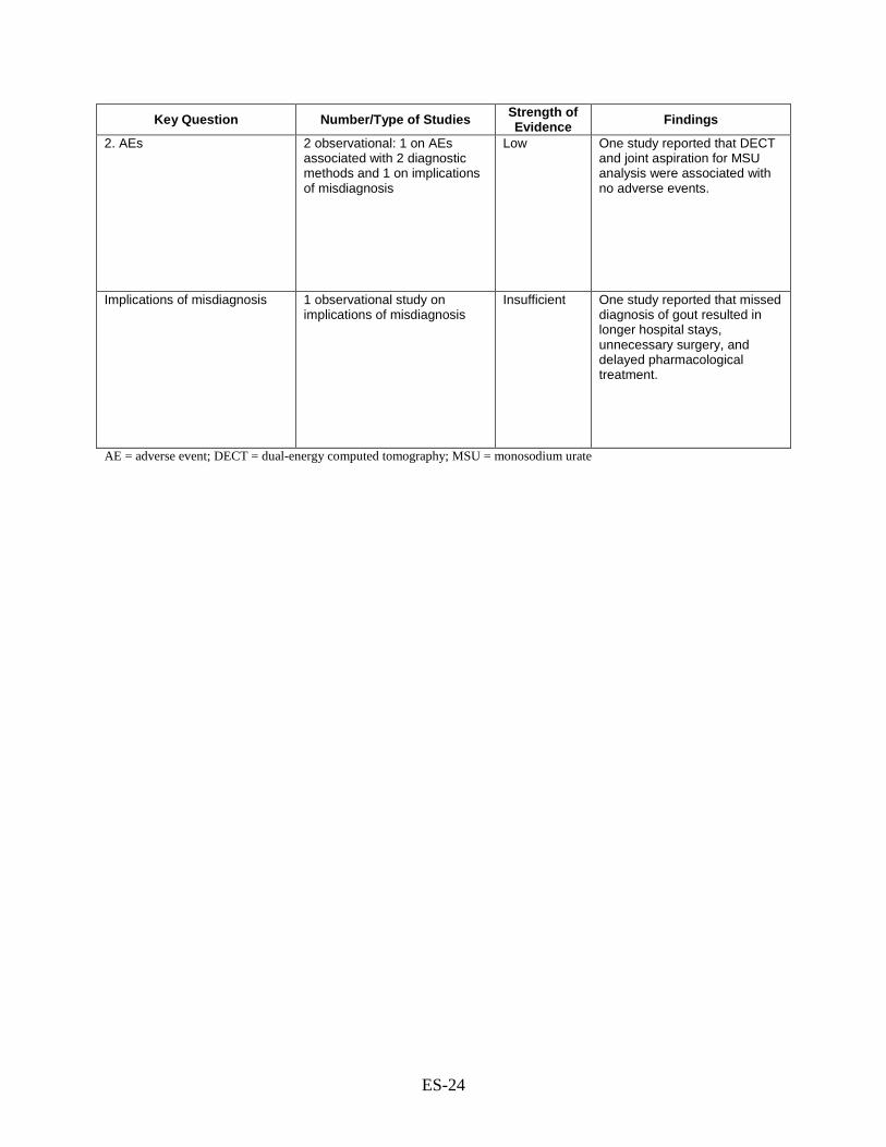
ES-24
Key Question Number/Type of Studies Strength of Evidence Findings
2. AEs 2 observational: 1 on AEs associated with 2 diagnostic methods and 1 on implications of misdiagnosis
Low One study reported that DECT and joint aspiration for MSU analysis were associated with no adverse events.
Implications of misdiagnosis 1 observational study on implications of misdiagnosis
Insufficient One study reported that missed diagnosis of gout resulted in longer hospital stays, unnecessary surgery, and delayed pharmacological treatment.
AE = adverse event; DECT = dual-energy computed tomography; MSU = monosodium urate

ES-25
Table C. Summary of findings on comparative accuracy and safety of gout diagnostic methods Outcomes Level of Evidence Findings
Accuracy of Method Clinical algorithms based on primary care patients
Low Sensitivity: 88%–97% Specificity: 75%–96%
US Low Sensitivity: 37%–100% Specificity: 68%–97%
DECT Low Sensitivity: 85%–100% Specificity: 83%–92%
MSU crystal analysis NA Reference standard Factors Potentially Affecting Accuracy
Number and/or types of joints involved
Insufficient No conclusion possible
Patient sex Insufficient No conclusion possible Duration of symptoms (early vs. late disease)
Insufficient No conclusion possible
Duration of current flare Insufficient No conclusion possible MSU sample handling Insufficient No conclusion possible DECT or US number of views Insufficient No conclusion possible Clinician type–examiner Insufficient No conclusion possible Clinician training or experience Insufficient No conclusion possible Practitioner performing aspiration Insufficient No conclusion possible Facility characteristics Insufficient No conclusion possible Adverse Events Adverse events associated with procedures
US Insufficient No conclusion possible DECT Low Evidence suggests that few serious
risks are associated with use of DECT for gout diagnosis
MSU crystal analysis Low Evidence suggests that few serious risks are associated with use of MSU analysis for gout diagnosis
Adverse events associated with false positives or negatives
Clinical algorithms Insufficient No conclusion possible US Insufficient No conclusion possible DECT Insufficient No conclusion possible MSU crystal analysis Insufficient No conclusion possible AE = adverse event; DECT = dual-energy computed tomography; NA = not applicable; US = ultrasound Note: Sensitivity is avoidance of false negatives; specificity is avoidance of false positives.

ES-26
References 1. Zhu Y, Pandya BJ, Choi HK. Prevalence of
gout and hyperuricemia in the US general population: the National Health and Nutrition Examination Survey 2007-2008. Arthritis Rheum. 2011 Oct;63(10):3136-41. PMID: 21800283.
2. Li C, Martin BC, Cummins DF, et al. Ambulatory resource utilization and cost for gout in United States. Am J Pharm Benefits. 2013 March/April;5(2):e46-e54.
3. Doghramji PP, Wortmann RL. Hyperuricemia and gout: new concepts in diagnosis and management. Postgrad Med. 2012 Nov;124(6):98-109. PMID: 23322143.
4. Wallace SL. Hyperuricemia in the diagnosis of gout. J Gen Intern Med. 1989 Mar-Apr;4(2):178-9. PMID: 2651605.
5. Singh JA, Reddy SG, Kundukulam J. Risk factors for gout and prevention: a systematic review of the literature. Curr Opin Rheumatol. 2011 Mar;23(2):192-202. PMID: 21285714.
6. Hamburger M, Baraf HS, Adamson TC 3rd, et al. 2011 Recommendations for the diagnosis and management of gout and hyperuricemia. Postgrad Med. 2011 Nov;123(6 Suppl 1):3-36. PMID: 22156509.
7. Harrold LR, Mazor KM, Negron A, et al. Primary care providers' knowledge, beliefs and treatment practices for gout: results of a physician questionnaire. Rheumatology (Oxford). 2013 Sep;52(9):1623-9. PMID: 23620554.
8. Owens D, Whelan B, McCarthy G. A survey of the management of gout in primary care. Ir Med J. 2008;101(5):147-9. PMID: 18624262.
9. Khosla S, Thiele R, Baumhauer JF. Ultrasound guidance for intra-articular injections of the foot and ankle. Foot Ankle Int. 2009 Sep;30(9):886-90. PMID: 19755074.
10. Hasselbacher P. Variation in synovial fluid analysis by hospital laboratories. Arthritis Rheum. 1987;30(6):637-42. PMID: 3606682.
11. Schumacher HR Jr, Sieck MS, Rothfuss S, et al. Reproducibility of synovial fluid analyses. A study among four laboratories. Arthritis Rheum. 1986 Jun;29(6):770-4. PMID: 3718565.
12. McGill NW, York HF. Reproducibility of synovial fluid examination for crystals. Aust N Z J Med. 1991 Oct;21(5):710-3. PMID: 1759919.
13. Segal JB, Albert D. Diagnosis of crystal-induced arthritis by synovial fluid examination for crystals: lessons from an imperfect test. Arthritis Care Res. 1999 Dec;12(6):376-80. PMID: 11081008.
14. Graf SW, Buchbinder R, Zochling J, et al. The accuracy of methods for urate crystal detection in synovial fluid and the effect of sample handling: a systematic review. Clin Rheumatol. 2013 Feb;32(2):225-32. PMID: 23138881.
15. Perez-Ruiz F, Carmona L, Yebenes MJG, et al. An audit of the variability of diagnosis and management of gout in the rheumatology setting: the Gout Evaluation and Management Study. J Clin Rheumatol. 2011 Oct;17(7):349-55. PMID: 21946457.
16. Malik A, Schumacher HR, Dinnella JE, et al. Clinical diagnostic criteria for gout: comparison with the gold standard of synovial fluid crystal analysis. J Clin Rheumatol. 2009 Feb;15(1):22-4. PMID: 19125136.
17. Dalbeth N, Fransen J, Jansen TL, et al. New classification criteria for gout: a framework for progress. Rheumatology (Oxford). 2013 Oct;52(10):1748-53. PMID: 23611919.
18. Glazebrook KN, Guimaraes LS, Murthy NS, et al. Identification of intraarticular and periarticular uric acid crystals with dual-energy CT: initial evaluation. Radiology. 2011 Nov;261(2):516-24. PMID: 21926378.
19. Khanna D, Fitzgerald JD, Khanna PP, et al. 2012 American College of Rheumatology guidelines for management of gout. Part 1: systematic nonpharmacologic and pharmacologic therapeutic approaches to hyperuricemia. Arthritis Care Res (Hoboken). 2012;64(10):1431-46. PMID: 23024028.

ES-27
20. Khanna D, Khanna PP, Fitzgerald JD, et al. 2012 American College of Rheumatology guidelines for management of gout. Part 2: therapy and antiinflammatory prophylaxis of acute gouty arthritis. Arthritis Care Res (Hoboken). 2012;64(10):1447-61. PMID: 23024029.
21. Whiting P, Westwood M, Beynon R, et al. Inclusion of methodological filters in searches for diagnostic test accuracy studies misses relevant studies. J Clin Epidemiol. 2011 Jun;64(6):602-7. PMID: 21075596.
22. Zhang W, Doherty M, Pascual E, et al. EULAR evidence based recommendations for gout. Part I: diagnosis. Report of a task force of the Standing Committee for International Clinical Studies Including Therapeutics (ESCISIT). Ann Rheum Dis. 2006 Oct;65(10):1301-11. PMID: 16707533.
23. Whiting PF, Rutjes AW, Westwood ME, et al. QUADAS-2: a revised tool for the quality assessment of diagnostic accuracy studies. Ann Intern Med. 2011 Oct 18;155(8):529-36. PMID: 22007046.
24. Santaguida PL, Riley CM, Matchar DB. Assessing risk of bias as a domain of quality in medical test studies. In: Methods Guide for Medical Test Reviews. AHRQ Publication No. 12-EHC017. Rockville, MD: Agency for Healthcare Research and Quality; June 2012. Chapters available at www.effectivehealthcare.ahrq.gov/reports/final.cfm.
25. Shea BJ, Grimshaw JM, Wells GA, et al. Development of AMSTAR: a measurement tool to assess the methodological quality of systematic reviews. BMC Med Res Methodol. 2007;7:10. PMID: 17302989.
26. Methods Guide for Medical Test Reviews. AHRQ Publication No. 12-EHC017. Agency for Healthcare Research and Quality. Rockville, MD: Agency for Healthcare Research and Quality; June 2012. Chapters available at www.effectivehealthcare.ahrq.gov/reports/final.cfm.
27. Atkins D, Chang S, Gartlehner G, et al. Assessing the applicability of studies when comparing medical interventions. In: Methods Guide for Effectiveness and Comparative Effectiveness Reviews. AHRQ Publication No. 10(14)-EHC063-EF. Rockville, MD: Agency for Healthcare Research and Quality. January 2014. Chapters available at www.effectivehealthcare.ahrq.gov.
28. Bongartz T, Glazebrook KN, Kavros SJ, et al. Dual-energy CT for the diagnosis of gout: an accuracy and diagnostic yield study. Ann Rheum Dis. 2015 Jun;74(6):1072-7. PMID: 24671771.
29. Gordon C, Swan A, Dieppe P. Detection of crystals in synovial fluids by light microscopy: sensitivity and reliability. Ann Rheum Dis. 1989 Sep;48(9):737-42. PMID: 2478085.
30. Huppertz A, Hermann KG, Diekhoff T, et al. Systemic staging for urate crystal deposits with dual-energy CT and ultrasound in patients with suspected gout. Rheumatol Int. 2014 Jun;34(6):763-71. PMID: 24619560.
31. Janssens HJEM, Fransen J, Van De Lisdonk EH, et al. A diagnostic rule for acute gouty arthritis in primary care without joint fluid analysis. Arch Intern Med. 2010;170(13):1120-6. PMID: 20625017.
32. Janssens HJEM, Janssen M, Van De Lisdonk EH, et al. Limited validity of the American College of Rheumatology criteria for classifying patients with gout in primary care. Ann Rheum Dis. 2010;69(6):1255-6. PMID: 19910298.
33. Kienhorst LB, Janssens HJ, Fransen J, et al. The validation of a diagnostic rule for gout without joint fluid analysis: a prospective study. Rheumatology (Oxford). 2015 Apr;54(4):609-14. PMID: 25231179.
34. Kienhorst LB, Janssens HJ, Fransen J, et al. Arthritis of the first metatarsophalangeal joint is not always gout: a prospective cohort study in primary care patients. Joint Bone Spine. 2014 Jul;81(4):342-6. PMID: 24468668.
35. Lai KL, Chiu YM. Role of ultrasonography in diagnosing gouty arthritis. J Med Ultrasound. 2011 March;19(1):7-13. http://dx.doi.org/10.1016/j.jmu.2011.01.003.
ES-28
36. Lamers-Karnebeek FB, Van Riel PL, Jansen TL. Additive value for ultrasonographic signal in a screening algorithm for patients presenting with acute mono-/oligoarthritis in whom gout is suspected. Clin Rheumatol. 2014 Apr;33(4):555-9. PMID: 24510062.
37. Park JW, Ko DJ, Yoo JJ, et al. Clinical factors and treatment outcomes associated with failure in the detection of urate crystal in patients with acute gouty arthritis. Korean J Intern Med. 2014;29(3):361-9. PMID: 24851071.
38. Rettenbacher T, Ennemoser S, Weirich H, et al. Diagnostic imaging of gout: comparison of high-resolution US versus conventional X-ray. Eur Radiol. 2008 Mar;18(3):621-30. PMID: 17994238.
39. Richette P, Clerson P, Bouee S, et al. Identification of patients with gout: elaboration of a questionnaire for epidemiological studies. Ann Rheum Dis. 2014 May 5. PMID: 24796335.
40. Taylor WJ, Fransen J, Dalbeth N, et al. Performance of classification criteria for gout in early and established disease. Ann Rheum Dis. 2014 Oct 28. PMID: 25351521.
41. Vazquez-Mellado J, Hernandez-Cuevas CB, Alvarez-Hernandez E, et al. The diagnostic value of the proposal for clinical gout diagnosis (CGD). Clin Rheumatol. 2012 Mar;31(3):429-34. PMID: 21979446.
42. Wallace SL, Robinson H, Masi AT, et al. Preliminary criteria for the classification of the acute arthritis of primary gout. Arthritis Rheum. 1977 Apr;20(3):895-900. PMID: 856219.
43. Chowalloor PV, Keen HI. A systematic review of ultrasonography in gout and asymptomatic hyperuricaemia. Ann Rheum Dis. 2013 May;72(5):638-45. PMID: 23291387.
44. Dirken-Heukensfeldt K, Teunissen TAM, van de Lisdonk EH, et al. "Clinical features of women with gout arthritis." A systematic review. Clinical Rheumatology. 2010 Jun;29(6):575-82. PMID: 20084441.
45. Ogdie A, Taylor W, Weatherall M, et al. Imaging modalities for the classification of gout: systematic literature review and meta-analysis. Ann Rheum Dis. 2014 Jun 20. [Epub ahead of print.] PMID: 24915980.
46. Sivera F, Andres M, Falzon L, et al. Diagnostic value of clinical, laboratory, and imaging findings in patients with a clinical suspicion of gout: a systematic literature review. J Rheumatol Suppl. 2014 Sep;92:3-8. PMID: 25180122.
47. Sivera F, Andres M, Carmona L, et al. Multinational evidence-based recommendations for the diagnosis and management of gout: integrating systematic literature review and expert opinion of a broad panel of rheumatologists in the 3e initiative. Ann Rheum Dis. 2014 Feb;73(2):328-35. PMID: 23868909.
48. Berendsen D, Jansen TL, Taylor W, et al. A critical appraisal of the competence of crystal identification by rheumatologists. Ann Rheum Dis. 2014;72(Suppl 3):A981-2.
49. Hamburger MI, Tesser JRP, Skosey JL, et al. Patterns of gout treatment and related outcomes in US community rheumatology practices: the relation between gout flares, time in treatment, serum uric acid level and urate lowering therapy. Arthritis and Rheumatism. 2012;64(Suppl 10):1904.
50. Richette P, Pascual E, Doherty M, et al. Updated EULAR evidence-based recommendations for the diagnosis of gout. Ann Rheum Dis. 2014;(Suppl 2):783-84.
51. Dalbeth N. Management of gout in primary care: challenges and potential solutions. Rheumatology (Oxford). 2013 Sep;52(9):1549-50. PMID: 23771950.
52. Rigby AS, Wood PH. Serum uric acid levels and gout: what does this herald for the population? Clin Exp Rheumatol. 1994 Jul-Aug;12(4):395-400. PMID: 7955603.
53. Lenski M, Scherer MA. Analysis of synovial inflammatory markers to differ infectious from gouty arthritis. Clin Biochem. 2014 Jan;47(1-2):49-55. PMID: 24177196.
54. Wang C, Zhong D, Liao Q, et al. Procalcitonin levels in fresh serum and fresh synovial fluid for the differential diagnosis of knee septic arthritis from rheumatoid arthritis, osteoarthritis and gouty arthritis. Exp Ther Med. 2014 Oct;8(4):1075-80. PMID: 25187799.

1
Introduction Background
Condition Gout is a form of inflammatory arthritis characterized by acute intermittent episodes of
synovitis presenting with joint swelling and pain (these episodes are referred to as acute gouty arthritis flares or attacks). The condition may progress to a chronic and persistent condition, with development of tophi (solid deposits of monosodium urate [MSU] crystals in joints, cartilage, tendons, bursae, bone, and soft tissue), a condition called chronic tophaceous gout. There is no clear distinction between acute intermittent and chronic intermittent conditions, whereas the advanced stage of gout is characterized by more persistent joint manifestations and tophi (either clinically evident or hidden within the joint).
Gout is the most common form of inflammatory arthritis, and the prevalence has been increasing. The most recent estimate of prevalence among adults in the United States (U.S.), based on data from the 2007-2008 National Health and Nutrition Examination Survey (NHANES), is3.9 percent (8.3 million individuals), ranging from 2.0 percent in women to 5.9 percent in men,1 an increase over that of previous NHANES data cycles. The rise in the prevalence of gout has paralleled the increase in prevalence of comorbid conditions associated with hyperuricemia (the primary risk factor for gout), including obesity, hypertension, hypertriglyceridemia, hypercholesterolemia, type 2 diabetes, metabolic syndrome, chronic kidney disease, and renal insufficiency. Increased use of medications that increase the risk for developing hyperuricemia (e.g., thiazide diuretics, low-dose aspirin, or their combination) may further explain the increasing prevalence of gout. In a 2013 study that analyzed data from several national surveys administered from 2002 to 2008, the number of ambulatory care visits attributable to gout was estimated to be 7 million visits annually, with 2 million attributable to acute attacks (the rate more than doubled from 2002 to 2008). The total annual ambulatory care (primary care, urgent care, and emergency department) costs associated with gout (visits and medications) were estimated to be $933 million (in 2009 dollars). Drug expenditures accounted for 61 percent of the total costs.2
In addition to gout, the types of inflammatory arthritis include rheumatoid arthritis, septic arthritis, inflammatory episodes of osteoarthritis, and calcium pyrophosphate dehydrate crystal deposition disease (formerly known as pseudogout). Patients with any of these types of arthritis can present with clinically similar signs and symptoms, but the conditions have very different treatments, and incorrect diagnosis can have serious outcomes (for example, missing a septic arthritis can lead to joint damage and septic shock). A major challenge then for effective gout management, particularly in the primary care and urgent/emergent care setting where most gout patients are managed, is distinguishing gout from these other conditions to prevent inappropriate or delayed treatment, which can incur severe complications.
Etiology of Gout The driving force behind acute episodes of gout is hyperuricemia (defined as an elevated
serum uric acid (more accurately referred to as serum urate, for salt form of uric acid that occurs in serum) concentration greater than 6.8 mg per deciliter [dl] in men and greater than 6.0 in women). Hyperuricemia is most commonly the result of inadequate renal excretion of uric acid

2
or, less commonly, uric acid overproduction (uric acid is a breakdown product of dietary or endogenous purines). Hyperuricemia leads to formation and deposition of MSU crystals, which preferentially deposit in joints, tendons, and bursa spaces. Despite the prevalence of hyperuricemia, for reasons that remain unclear, only a small proportion of individuals with hyperuricemia go on to develop gout. For others, hyperuricemia remains asymptomatic.3 The prevalence of hyperuricemia ranges from 21.2 percent in men to 21.6 percent in women, four- to ten-fold higher than the prevalence of gout.4
The causes of gout are multifactorial, including a combination of genetic, hormonal, metabolic, pharmacologic, comorbid (renal disease), and dietary factors. Family history, advancing age, male sex, or, in women, early menopause have been associated with a higher risk of gout and/or gout flares.5 Dietary risk factors for gout include consumption of purine-rich foods or drinks, including meat, and seafood; and consumption of sugar sweetened soft drinks, alcohol, and foods high in fructose. Dairy foods and coffee have been associated with a lower risk of incident gout and in some cases a lower rate of gout flares. However, the role of diet in the etiology and treatment of gout is a topic of considerable research and will be reviewed in a separate systematic review.
Diagnostic Strategies The majority of individuals with gout are initially seen, diagnosed, and treated in primary and
urgent care settings. Thus primary care physicians (PCPs) and emergency medicine physicians are the most likely practitioners to see patients with suggestive of an acute attack of gout, but with no prior diagnosis. Such patients may be experiencing a first attack (early-stage gout) or may actually have experienced multiple attacks and have more advanced gout.
Some research has argued the need for laboratory assessment of joint/synovial fluid MSU crystals in the setting of an acute inflammatory arthritis for a definitive diagnosis of gout. MSU crystal analysis has been regarded as the gold standard against which other potential diagnostic methods are measured. However, joint aspiration can be technically difficult and painful to the patient and is often deferred, to be conducted by a specialist (e.g., a rheumatologist or orthopedic surgeon).6 In addition, the accuracy of synovial fluid analysis may be affected by a number of factors (both patient-, practitioner-, and analyst-related).7, 8 A 2009 study found that unguided needle insertion in the toe is often inaccurate.9 At least three studies have found wide variation in the accuracy of assessing synovial fluid crystals (both MSU and calcium pyrophosphate) and white blood cells across hospital laboratories,10-12 which could potentially be due to patient differences, differences in skill levels of the practitioners drawing or analyzing the samples, or differences in sample handling. A 1999 systematic review on the accuracy of MSU crystal analysis in synovial fluid13 concluded that MSU analysis had poor sensitivity, specificity, and reproducibility. And a 2013 systematic review of the accuracy of methods for detecting MSU in synovial fluid concluded that storage of samples at room temperature resulted in a decrease in MSU concentration over time, compared with refrigeration14 but could not draw any conclusions about the role of personnel. In fact, evidence from a 2011 survey of rheumatologists suggests that synovial fluid analysis is underused in the rheumatology setting as well.15
Instead of analyzing MSU crystals in synovial fluid, PCPs and emergency medicine physicians tend to rely on algorithms comprising a combination of clinical signs and symptoms to diagnose an acute episode of gout. These clinical signs and symptoms include rapid development of inflammation and pain, erythema, monoarthritis, response to administration of

3
the drug colchicine, and symptoms in the first metatarsophalangeal (MTP) joint, among others (with synovial fluid culture sometimes used to rule out septic arthritis).
Attempts to standardize and validate such clinical diagnostic algorithms date back to the 1960s.16 Most of these algorithms were not developed for diagnostic purposes but for classification of gout. Concurrent with this review, the American College of Rheumatology (ACR) and the European League against Rheumatism (EULAR) are collaborating to update and evaluate classification criteria for gout. Distinct from diagnostic criteria, classification criteria are intended to ensure the correct identification and staging of patients with a particular disease condition (especially patients in the early stages of the disease) for the purpose of enrollment in studies of disease.17
Therefore, a question of importance is whether any combination of clinical signs and symptoms and laboratory tests accessible in the primary or acute care setting (which we will refer to as a clinical algorithm or clinical diagnostic algorithm) will have good predictive value compared with tests such as joint aspiration and synovial fluid MSU analysis, both to correctly diagnose gout and to rule out other causes of joint inflammation, particularly septic arthritis and calcium pyrophosphate deposition disease among patients presenting with an acute episode of inflammatory arthritis.
Imaging modalities have also been assessed for both diagnosis and classification of gout. These techniques include plain radiographs and newer techniques such as ultrasound and dual-energy computed tomography (DECT), which are just beginning to be used to diagnose gout in some settings.18 Therefore, another question of importance for gout diagnosis is how these newer methods compare with joint aspiration and synovial fluid analysis in their predictive value for the diagnosis of gout and whether they provide any additive value over the use of MSU analysis or clinical signs and symptoms.
The “safety” of tests used to diagnose gout also needs to be considered. Potential safety concerns include acute physical discomfort or infection from joint aspiration and long term effects (e.g., from accumulated radiation exposure). Other concerns are the potential effects of misdiagnosis. These effects could include delay in initiating or failure to initiate appropriate treatment for gout or another disorder or incorrect initiation of treatment for another disorder (e.g., hospitalization and administration of intravenous antibiotics for suspected joint sepsis).
Therefore, we have undertaken a systematic review of studies examining the accuracy and safety of tests used to diagnose gout, including algorithms combining physical signs and symptoms, serum urate, ultrasound, plain radiography, and DECT, compared with synovial fluid MSU analysis. The primary focus of this review is on tests that can be used in the primary care or urgent/emergent care setting for an initial diagnosis of gout. The aim of this review is to help inform clinical decision-making for patients and providers and to improve the quality of care for patients with gout in the primary and acute care setting.
Scope and Key Questions
Scope of the Review The purpose of this review is to assess the evidence on the validity and safety of tests for the
purpose of diagnosing gout, including clinical signs and symptoms (individually and in combination as an algorithm), DECT, ultrasound, and other imaging methods, compared with of synovial fluid from involved joints and analysis of MSU crystals using polarized light microscopy. Because evidence also suggests MSU crystal analysis, itself, may not be accurate in
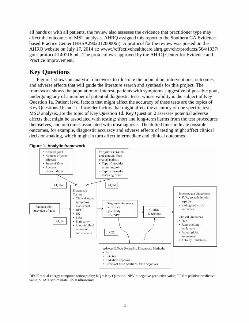
4
all hands or with all patients, the review also assesses the evidence that practitioner type may affect the outcomes of MSU analysis. AHRQ assigned this report to the Southern CA Evidence-based Practice Center (HHSA290201200006I). A protocol for the review was posted on the AHRQ website on July 17, 2014 at: www://effectivehealthcare.ahrq.gov/ehc/products/564/1937/ gout-protocol-140716.pdf. The protocol was approved by the AHRQ Center for Evidence and Practice Improvement.
Key Questions Figure 1 shows an analytic framework to illustrate the population, interventions, outcomes,
and adverse effects that will guide the literature search and synthesis for this project. The framework shows the population of interest, patients with symptoms suggestive of possible gout, undergoing any of a number of potential diagnostic tests, whose validity is the subject of Key Question 1a. Patient level factors that might affect the accuracy of these tests are the topics of Key Questions 1b and 1c. Provider factors that might affect the accuracy of one specific test, MSU analysis, are the topic of Key Question 1d. Key Question 2 assesses potential adverse effects that might be associated with testing: short and long-term harms from the test procedures themselves, and outcomes associated with misdiagnosis. The dotted lines indicate possible outcomes, for example, diagnostic accuracy and adverse effects of testing might affect clinical decision-making, which might in turn affect intermediate and clinical outcomes.
Figure 1. Analytic framework
DECT = dual energy computed tomography; KQ = Key Question; NPV = negative predictive value; PPV = positive predictive value; SUA = serum urate; US = ultrasound

5
The Key Questions that guided this review are based on questions posed by the American College of Physicians. These questions underwent revision based on input from a group of key informants, public comments, and input from a Technical Expert Panel (TEP).
Key Question 1. a. What is the accuracy of clinical signs and symptoms and other
diagnostic tests (such as serum uric acid, ultrasound, CT scan, DECT, and plain x-ray), alone or in combination, compared witha synovial fluid analysis in the diagnosis of acute gouty arthritis, and how does the accuracy affect clinical decisionmaking, clinical outcomes and complications, and patient centered outcomes?
b. How does the diagnostic accuracy of clinical signs and symptoms and other tests vary by affected joint site and number of joints?
c. Does the accuracy of diagnostic tests for gout vary by duration of symptoms (i.e., time from the beginning of a flare)?
d. Does the accuracy of synovial fluid aspiration and crystal analysis differ by i) the type of practitioner who is performing the aspiration and ii) the type of practitioner who is performing the crystal analysis?
Key Question 2. What are the adverse effects (including pain, infection at the aspiration site, radiation exposure) or harms (related to false positives, false negatives, indeterminate results) associated with tests used to diagnose gout?
Organization of This Report The remainder of this report presents the methods used to conduct the literature searches,
data abstraction, and analysis for this review; the results of the literature searches, organized by Key Question; the conclusions; and a discussion of the findings within the context of what is already known, the limitations of the review and the literature, as well as suggestions for future research.
a Using monosodium urate crystal analysis of synovial fluid as the reference standard.

6
Methods
Criteria for Inclusion/Exclusion of Studies in the Review This report is based on a systematic search for prospective or cross-sectional studies that
assessed the sensitivity and specificity of tests used to diagnose gout, preferably against the gold standard test of joint aspiration and synovial fluid assessment for MSU crystals, in populations of adults 18 years of age or older, suspected of having gout but not previously diagnosed (see Table 1). We also included studies that assessed patient and practitioner factors that affect the diagnostic accuracy of these tests, assessed harms associated with the tests, or examined particular factors that potentially affect the sensitivity or specificity of tests (joints involved and duration of symptoms).
Tests of interest included algorithms comprising clinical or laboratory examination for physical signs, symptoms, and history; serum uric acid, ultrasonography; DECT; and plain radiography. The comparator of primary interest was synovial fluid analysis of MSU crystals using polarized light microscopy. However, if no such studies could be identified for a diagnostic test of interest, studies were also included if some or all of the participants were diagnosed using the American College of Rheumatology criteria for gout diagnosis and classification or another validated set of criteria as the reference standard (comparator).
Studies were excluded if enrolled participants had already been definitively diagnosed with gout prior to enrollment (to ensure that the patient populations were as similar as possible to patients who would be seen in the primary or urgent or emergent care setting), or if the comparator was individual physician opinion or was not identified. Inclusion criteria are further described below in the section entitled PICOTs (Populations, Interventions, Comparators, Outcomes, Timeframes, a framework used in systematic reviews to categorize inclusion and exclusion criteria).
Outcomes of interest were the comparative accuracy of the test results (as measured by the sensitivity and specificity or the positive and negative predictive value of the test in question), intermediate outcomes such as lab and radiographic test results, clinical decisionmaking that resulted from diagnoses, short term clinical (patient-centered) outcomes such as a change in pain and joint swelling that resulted from a diagnosis, and any adverse events (including adverse patient experiences such as pain or infection at the aspiration site, effects of radiation exposure, and the results of a false positive or negative diagnosis) associated with the test. Outcomes are further described below.
Prospective cohort, cross-sectional, and case-control (if needed) studies were included to address Key Question 1 (accuracy of test and factors that affect accuracy). Prospective cohort, cross-sectional, and case control studies, as well as case series of any size and case reports of rare adverse events were included if they addressed Key Question 2 (adverse events or other negative outcomes in individuals undergoing testing).
PICOTS of Included Studies Population(s) (KQ1 and 2):
• Adults (18 years and over) presenting with symptoms (e.g., an acute episode of joint inflammation) suggestive of gout, but without a prior gout diagnosis, including the following subgroups:

7
o Male and female patients o Patients with longer vs. shorter duration of symptoms o Patients with comorbidities including hypertension, type 2 diabetes, kidney
disease (renal insufficiency) o Patients with osteoarthritis, septic arthritis, calcium pyrophosphate deposition
disease, or previous joint trauma o Individuals with a family history of gout
Interventions (Index Tests) (KQ1, 2): • Clinical history and physical exam • Serum urate assessment • Ultrasound • DECT • Plain x-ray • Joint aspiration by physicians and synovial fluid analysis using polarizing microscopy
(by physicians or laboratory personnel) • Combinations of these tests as identified in the literature
Comparators (Reference Tests): • Joint synovial fluid aspiration and microscopic assessment for monosodium urate crystals
(KQ1a-c, 2) • Joint synovial fluid aspiration and microscopic assessment for monosodium urate crystals
as performed by a practitioner with a different level of expertise or experience, e.g. rheumatologist, laboratory personnel (KQ1d)
Outcomes: • Diagnostic accuracy of clinical signs and symptoms, US, DECT, plain radiographs
compared with joint aspiration and synovial fluid analysis (KQ1) o Sensitivity/specificity, true positives/true negatives, area under the curve o Positive predictive value (PPV), negative predictive value (NPV),
positive/negative likelihood ratios (if prevalence known) • Clinical decisionmaking
o Additional testing o Pharmacologic/dietary management
• Intermediate outcomes o Serum urate o Synovial fluid crystals o Radiographic or ultrasound changes
• Clinical outcomes o Pain, joint swelling and tenderness, o Patient global assessment, and activity limitations (KQ1,2)
• Adverse effects of the tests, including o Pain, infection, radiation exposure and o Effects of false positive or false negative (KQ2)
Timing: • For clinical outcomes of symptom relief: 1-2 days minimum (KQ1) • Early in an attack versus later or post-attack (KQ1c) • For adverse events: immediate
8
Settings: • Primary care (outpatient) or acute care settings, preferentially; • Outpatient rheumatology practices/academic medical centers
Literature Search Strategies for Identification of Relevant Studies To Answer the Key Questions
The search strategy was designed by the Southern California Evidence-based Practice Center (EPC) reference librarian in collaboration with our local content expert, who has participated in two systematic reviews on gout;19, 20 the search strategy appears in Appendix A. As recommended by the AHRQ EPC Methods Guide for Medical Test Reviews,21 the searches were conducted without filters specific for diagnostic tests; instead we used the terms “gout” combined with the terms for the diagnostic tests. We searched PubMed (1/1/46 to 11/07/14), EMBASE (1/1/72 to 11/07/14), the Cochrane Library (1/1/45 to 11/07/14 for the Cochrane Central Registry of Controlled Trials and 1/1/96 to 11/07/14 for the Cochrane Database of Systematic Reviews), and the Web of Science (from 1/1/80 to 11/07/14); these dates were selected to replicate the searches conducted as the basis for the 2006 EULAR Guidelines on Diagnosis and Management of Gout.22 We also included any relevant studies identified in the searches we conducted for a simultaneous review on management of gout if not already identified in the searches for this review. Finally, we asked the TEP to assess our list of included studies and to provide references for any studies they believe should also be included.
We searched Clinicaltrials.gov and the Web of Science for recently completed studies and unpublished or non-peer-reviewed study findings. Searches were not limited by language of publication: Non-English language studies that met the inclusion/exclusion criteria based on a review of an English-language abstract were screened further in full text if translators could be identified with reasonable effort. We also contacted manufacturers of diagnostic equipment (polarizing microscopes, sonography equipment, DECT, and serum uric acid test kits) were contacted for unpublished data specific to the use of the equipment or tests for gout diagnosis.
An update search was conducted on 11/07/14 after submission of the draft report for peer review.
We transferred the output of the literature searches to DistillerSR™ for screening. Article titles and abstracts identified by the searches were independently screened by two literature reviewers using the predetermined inclusion and exclusion criteria, and those selected by either reviewer were accepted without reconciliation for further, full-text review.
Two reviewers independently conducted full-text review to exclude articles that provided no usable data, reported the same data as another article, or enrolled participants with established gout diagnoses. Disagreements regarding inclusion at the full-text stage were reconciled, with the input of the project lead when necessary.
We identified a small number of relatively recent systematic reviews on various aspects of gout diagnosis; in most cases we used these reviews to identify references we had missed, however if the review was of high quality, addressed a subquestion of interest, and included all the literature on the topic, we included it as a data source after assessing its quality and how it assessed risk of bias and strength of evidence. We also searched the reference lists of included studies for additional titles that appeared to fit our inclusion criteria and screened these articles for inclusion. For studies of apparent interest reported in meeting abstracts (conference proceedings), we searched for peer-reviewed publications of the findings; if findings had not yet
9
been published in a peer reviewed journal, we reserved them and cited them in the last chapter in our discussion of suggestions for future research.
Data Abstraction and Data Management Two reviewers independently abstracted study level details from articles accepted for
inclusion in DistillerSR. Any disagreements were reconciled with the input of the project leader, Southern California EPC director, or the local subject matter expert if needed. Studies provided by manufacturers or suggested by peer reviewers underwent the same process, as did studies identified in update searches.
Assessment of Methodological Quality of Individual Studies Risk of bias (study quality) of individual included studies was assessed independently by two
reviewers using the Quality Assessment of Diagnostic Accuracy Studies (QUADAS)-2 tool,23, 24 and assessments were reconciled, with any disagreements mediated by the project lead. We used the Assessing the Quality of Systematic Reviews (AMSTAR) tool to assess the quality of existing systematic reviews that we included;25 AMSTAR assessments were also conducted independently by two reviewers and reconciled.
Data Synthesis/Analysis For studies that assessed US, DECT, or another radiographic method, we extracted and
reported sensitivity, specificity, positive and negative predictive value, and area under the curve (AUC)/receiver-operating characteristics (ROC) if reported.
We considered studies for meta-analysis if the number of true positives, true negatives, false positives, and false negatives were reported or could be calculated; and if studies were similar enough with respect to outcome measures, participants, and tests, and assessed the validity of an alternative diagnostic method against that of analysis of MSU crystals in synovial fluid. Because the studies we identified precluded pooling, outcomes are described narratively, stratified by test comparisons of interest and study design. All included studies are also described in summary tables.
If any prior SRs were identified that directly addressed a KQ of interest and were deemed of high enough quality to include, we assessed whether any subsequent (or contemporaneous) original studies were sufficiently homogeneous with the review to consider conducting new quantitative synthesis for a particular outcome, based on whether the new study represents a potential pivotal finding in terms of size and effect size and the availability of the needed data. If it was determined that the newer studies could not be combined with the prior SR and could not, themselves, be pooled, we described the newer studies narratively.
Grading the Strength of the Body of Evidence for Each Key Question
We assessed the overall strength of evidence for each conclusion using guidance suggested by AHRQ for its Effective Health Care Program.26 This method is based on a method developed by the GRADE Working Group. The evidence grade is usually based on five required domains:
• Study limitations: assessed based on the risk-of-bias assessments for all studies that contribute to a conclusion;

10
• Consistency: because we did not pool studies, we compared the relative sensitivities and specificities;
• Directness: a measure of whether the evidence being assessed reflects a single, direct link between the interventions of interst and the ultimate health outcome under consideration;
• Precision: a measure of the confidence intervals in a pooled analysis, also not assessed in this report;
• Publication bias: also assessed only for studies for which data are pooled Based on the domains we included, we classified the strength (grade) of evidence as follows:
High = Further research is unlikely to change our confidence in the estimate of effect. Moderate = Further research is likely to have an important impact on our confidence in the estimate of effect and may change the estimate. Low = Further research is very likely to have an important impact on our confidence in the estimate of effect and is likely to change the estimate. Very low/Insufficient = Any estimate of effect is very uncertain.
Applicability To be useful, comparative effectiveness research (or in this case research assessing the
comparative validity of a diagnostic test) must use real world interventions and settings to the extent possible. Applicability is a measure of the degree to which participants, intervventions, and outcome measures used in studies included in a review reflect the population and care settings for whom the outcomes are intended. We assessed applicability based on the inclusion and exclusion criteria described in the PICOTS, which included the study population age, sex, health profiles (including comorbidities as well as duration of symptoms and number of affected joints, when relevant), tests, gold standards, study settings, and provider types.27 Thus we would assign higher priority to studies of adult populations being seen in primary/urgent/emergent care settings for first or subsequent episodes of symptoms suggestive of gout than to studies of patients with established gout diagnoses in an academic rheumatology department.
Peer Review and Public Commentary A draft version of the report was posted for peer review on 11/4/14 and revised in response to
reviewer comments.

11
Results Introduction
This chapter first describes the results of the literature searches and then provides the results for each Key Question, including key points, an overview of the studies identified for that question, and a detailed synthesis of the studies. The results of Key Question1, parts a through c, are presented together for each diagnostic method. The results for Key Question 1d are described following that section.
Results of Literature Searches Our searches identified 3,646 titles/abstracts, of which 3,391were excluded for the following
reasons: not human (129), diagnostic methods beyond the scope of the review (129), not gout diagnosis or management (1,801), no original data or non-systematic reviews (374), conference proceedings or presentations or abstracts (11), case reports with sample size of fewer than 10 (415), population under age 18 (5), renal transplant or end-stage renal disease patients (12), titles with no abstracts (based on a survey of a random sample of 10 percent of these titles, for which full-text articles or reports were obtained, and all were rejected as letters, commentaries, or non-systematic reviews with no original data,) (252), gout management only (236) (see PRISMA diagram, Figure 2).
We reviewed 255 full text articles, of which 234 were further excluded for the following reasons: participants not human (2), diagnostic methods beyond the scope of the review (44), not gout diagnosis or management (69), no original data (29), conference proceedings or presentations or abstracts not identified by title and abstract review (38), case reports with sample size fewer than 10 (17), gout management only (13), no reference standard reported or not all patients received the reference standard (7). We were unable to obtain articles for 15 studies.
Our search of Clinicaltrials.gov identified 152 entries, none of which were relevant to this review.
None of the manufacturers of imaging equipment or laboratory test kits used in the diagnosis of gout who were contacted for information responded to requests. A notice placed in the Federal Register requesting such information also generated no responses.
We include the results of 17 original studies16, 18, 28-42 and four systematic reviews43-46 in our evidence synthesis. Seventeen of the original studies answer Key Question 1, and 2 studies answer Key Question 2. Results for these studies can be found below by Key Question.
Appendix B contains the list of the studies excluded at full text review, and Appendix D contains our data abstraction tool and QUADAS-2 tools, which were used on the included studies, and the AMSTAR tool, which was used to assess the quality of the included systematic reviews.
The findings are organized as follows. For Key Question 1a through c, we present first the key points, followed by a brief overview of the studies and then detailed narrative descriptions of each study and prior systematic review that addresses that question or subquestion. The studies that address Key Question 1d and Key Question 2 are described separately, with the key points followed by the study details.

12
Figure 2. Literature flow diagram
KQ = Key Question; SR(s) = Systematic Review(s)

13
Key Question 1 a. What is the accuracy of clinical signs and symptoms and other
diagnostic tests (such as serum uric acid, ultrasound, CT scan, DECT, and plain x-ray), alone or in combination, compared withb synovial fluid analysis in the diagnosis of acute gouty arthritis, and how does the accuracy affect clinical decisionmaking, clinical outcomes and complications, and patient centered outcomes?
b. How does the diagnostic accuracy of clinical signs and symptoms and other tests vary by affected joint site and number of joints?
c. Does the accuracy of diagnostic tests for gout vary by duration of symptoms (i.e., time from the beginning of a flare)?
Key Points • Few studies that assessed the accuracy of algorithms comprising clinical signs and
symptoms for the diagnosis or classification of gout consistently applied the same reference standard to all participants with suspected gout and usually did not limit participation to individuals experiencing a first attack.
• Studies that assessed the use of clinical algorithms reported widely varying sensitivities and specificities; however, two recent diagnostic algorithms, one developed from clinical signs and symptoms used by primary care physicians, reported relatively good sensitivity and specificity but need further validation in larger, more varied populations. The strength of evidence for this conclusion is low.
• In small numbers of studies that enrolled only patients not previously diagnosed with gout, DECT had good sensitivity and specificity for predicting gout compared with synovial fluid analysis for MSU crystals or a validated diagnostic clinical algorithm. Three studies revealed sensitivities that ranged from 85% to 100% and specificities that ranged from 83% to 92%.
• Ultrasound was more varied in its ability to detect gout: Four studies of ultrasound showed sensitivities that ranged from 37% to 100% and specificities that ranged from 68% to 97%, depending on the signs assessed. The strength of evidence for this conclusion is low.
• No studies were identified that assessed the validity of serum uric acid, CT scan, or plain x-ray for diagnosing gout. The strength of evidence for these tests is insufficient.
• No studies were identified that directly assessed the effect of joint site or number of affected joints on diagnostic accuracy. The strength of evidence for this question is insufficient for all diagnostic methods.
• No studies were identified that directly assessed the effect of duration of symptoms on the accuracy of diagnostic tests. The strength of evidence for this question is insufficient for all diagnostic methods.
bUsing monosodium urate crystal analysis of synovial fluid as the reference standard.

14
Description of Included Studies We identified 15 original studies that met our inclusion criteria for studies on the
comparative effectiveness (validity) of methods for the diagnosis or classification of gout: 9 studies assessed the sensitivity and specificity of algorithms comprising combinations of clinical signs and symptoms,16, 31-34, 39-42 3 assessed the use of DECT,18, 28, 30 and 4 assessed the use of ultrasound (one study compared ultrasound and DECT).30, 35, 36, 38 We also identified four prior systematic reviews: one that addressed a clinical algorithm,46 two that assessed the use of imaging for diagnosis of gout43, 45 and one on sex differences in gout diagnosis.44
The nine studies that assessed the use of clinical diagnostic or classification algorithms each compared the predictions based on six clinical algorithms to assessment of synovial fluid MSU crystals in all or most enrolled patients or at least in those presumed to have gout (in the latter case, patients who were considered not to have gout had to have another condition confirmed by a validated diagnostic criterion). These studies, which dated from 1977, enrolled 82 to 983 adult patients, both male and female. All studies were conducted in academic rheumatology departments, although several of the studies purposefully enrolled patients who were recruited by primary care physicians. Table 1 describes the studies and Table 2 compares the components of each algorithm.
The three studies that assessed the use of DECT compared the predictions based on these imaging studies to assessment of synovial fluid MSU crystals or to a clinical diagnostic algorithm or some combination of the two reference standards. These studies dated from 2011 to 2014 and enrolled 31 to 94 patients with suspected gout. All studies were conducted in academic rheumatology departments.
The four studies that assessed the use of ultrasound compared the predictions based on these imaging studies to assessment of synovial fluid MSU crystals or to a clinical diagnostic algorithm or some combination of the two reference standards. These studies dated from 2008 to 2014 and enrolled 54 to 105 patients with monoarthritis or suspected gout.
Detailed Synthesis This section describes the included studies and the findings for each diagnostic method.
Studies Assessing Algorithms Comprising Clinical Signs and Symptoms To Diagnose Gout
Comparative Accuracy Nine studies assessed the validity of diagnostic and classification algorithms comprising
clinical signs and symptoms of gout against synovial fluid MSU analysis (Table 1). Table 2 compares the criteria assessed in the studies as well as additional guideline-based criteria. The QUADAS scores for risk of bias for each of the included studies are shown in Table 5 at the end of this chapter.
ARA/ACR Criteria In 1977, the American Rheumatism Association (ARA, forerunner of the American College
of Rheumatology [ACR]) Subcommittee on Classification Criteria for Gout, compiled a survey questionnaire comprising 53 proposed criteria for classifying acute gout, for epidemiology and for research, although the criteria are used for diagnosis and clinical decisionmaking.42 Thirty
15
eight rheumatologists from 38 academic and private rheumatology clinics throughout the U.S. were invited to contribute data on up to 5 patients in each of five diagnostic categories: gout, rheumatoid arthritis of less than 2 years’ duration, rheumatoid arthritis of more than 2 years duration, pseudogout, and septic arthritis. The draft diagnostic criteria were tested on 706 of the patients (average age: 61): 178 patients provisionally diagnosed with gout (average duration 10 years), 299 with rheumatoid arthritis, and the remainder with acute septic arthritis or pseudogout. The proportion of females was 13.6 percent for gout patients and ranged from 51 percent to 64 percent for the other disorders. Among the 178 patients presumed to have gout, 90 underwent joint aspiration (compared with 25 percent of the rheumatoid arthritis patients, over 80 percent of the pseudogout patients, and 70 percent of the septic arthritis patients); of those patients with clinical gout diagnoses tested for MSU, 85 percent had positive findings (no MSU-positive findings were seen among patients with any of the other diagnoses, indicating absolute specificity for MSU crystals). The 53 criteria included in the survey were narrowed to the 12 that best discriminated the study groups and showed the greatest balance of sensitivity (the proportion of MSU-positive patients who fulfilled the proposed criterion) and specificity (the proportion of control patients who did not fulfill the proposed criterion) (Table 1). The sensitivity of three different cutpoints—5 or more, 6 or more, and 7 or more positive criteria—were 95.5%, 87.6%, and 74.1%, respectively. The specificities for these cutpoints were 72.7%-93.3%, 89.1%-98.7%, and 97.3%-100%, respectively; in each case, the criteria showed the poorest discrimination between gout and pseudogout. The authors noted that in spite of the perfect specificity of MSU for diagnosing gout, few suspected gout patients underwent the test and the sensitivity was only moderate.
In 2009, researchers compared the ARA criteria42 with two briefer algorithms (see the Rome Criteria and the NY Criteria, below). They enrolled 82 patients (mean age 64.5, 6 percent female) at a Veterans Administration rheumatology clinic. The criterion for enrollment was having had synovial fluid aspirated and analyzed for MSU crystals (by two highly experienced rheumatologists) at some prior time; participants were surveyed but did not receive new physical exams for the study. Of the participants, 75.6 percent had undergone aspiration of a knee joint; the remainder had had the MTP, wrist, elbow, ankle, or proximal interphalangeal joint aspirated. Thirty patients of 82 were MSU crystal-positive. This study reported lower sensitivity (70%) and specificity (79%) for 6 of 12 of the ARA criteria than did Wallace and colleagues.
From 2004 to 2007, family physicians in the Netherlands consecutively recruited 381 patients with monoarthritis; the patients were sent, along with their blinded diagnosis, to an academic rheumatology clinic for MSU crystal analysis, clinical exam, and followup of at least 1 year. Of the 381, 328 patients (mean age 58.0; 20.4 percent female) had a diagnosis of gout by their primary care physician (PCP). The academic rheumatology researchers, led by Janssens, analyzed the patients’ MSU to validate the PCP diagnoses and examined the patients to validate the ARA/ACR criteria against the presence of MSU crystals in synovial fluid. Increasing the cutoff points from 4 or more positive criteria (out of 11) to a maximum of 9 or more decreased the sensitivity from 100% to 7% and increased the specificity from 24% to 99%. The cutoff point of 6 or more criteria showed a sensitivity of 80% and a specificity of 64% (and a PPV of 0.80 and NPV of 0.65), leading the authors to conclude in a 2010 report that the ACR criteria had limited validity for classifying gout patients in primary care,32 most of whom presented with monoarthritis, usually in the MTP joint.

16
The Rome Criteria and New York Criteria The New York Criteria and the Rome Criteria were the forerunners of the ARA Preliminary
Criteria (the Rome criteria were first published in 1963 and the New York criteria were published in 1966) (Table 2).16 The New York criteria required crystals in synovial fluid, tissue, or tophus OR meeting two of five clinical criteria. The Rome criteria required meeting two of four criteria, one of which was MSU crystals in synovial fluid or tissue. As described above, the criterion for enrollment in the validation study was having had synovial fluid aspirated and analyzed for MSU crystals (by two highly experienced rheumatologists) at some prior time; participants were surveyed but did not receive new physical exams for the study. Of the participants, 75.6 percent had undergone aspiration of a knee joint; the remainder had had the MTP, wrist, elbow, ankle, or proximal interphalangeal joint aspirated. Thirty patients of 82 were MSU crystal-positive. Considering only the clinical signs and symptoms in the three sets of criteria (i.e., excluding MSU crystal findings), none of the clinical criteria sets had a sensitivity of more than 70.0% or a specificity of more than 88.5%. Having two of the three (non-MSU) Rome criteria had the highest specificity (88.55) but the sensitivity was 66.7%. Assessing the positive likelihood ratio (LR+) of individual criteria, they found that proven or suspected tophus and response to colchicine within 48 hours had the highest LR+ (15.56 [95%CI 2.11, 114.71] and 4.33[95%CI 1.16, 16.16], respectively) and positive predictive value (PPV) (91% and 86%, respectively) of any of the criteria in the three criteria sets. Although the study suggested basing future criterion sets on the Rome criteria because of its high specificity, patients were not necessarily having an acute attack and did not undergo physical exams, and MSU assessment had been conducted any time in the past.
The Diagnostic Rule (Netherlands Criteria) Using the clinical signs and symptoms, along with the lab values (serum urate) and MSU
assessments for the 381 consecutively enrolled primary care patients described above,32 Janssens and colleagues aimed to test the validity of PCP diagnosis of gout and to develop a diagnostic rule for use in primary care settings. Employing the criteria used by PCPs, they developed a set of multivariate predictive models (six clinical signs and symptoms, with and without the addition of serum urate testing). Again, of the 381 patients, 328 had a PCP diagnosis of gout; 64 percent were MSU crystal-positive (the PCPs’ diagnosis had a PPV of 0.64 and a negative predictive value [NPV] of 0.87). All had monoarticular gout. The model with the highest area under the curve (AUC) (0.85, 95% CI 0.81, 0.90) contains the following elements: male sex, previous patient-reported arthritis attack, onset within 1 day, joint redness, first metatarsophalangeal joint (MTP1) involvement, hypertension or one or more cardiovascular disease, and serum urate level exceeding 5.88mg/dL (Figure 2). Omitting serum urate testing decreased the AUC only slightly (to 0.82).
In 2013, Janssens’ group conducted additional analysis of their 2010 dataset to assess the sensitivity and specificity of monoarthritis of the MTP1 joint as a diagnostic criterion for gout, as this clinical sign was often the sole criterion used by Dutch PCPs to diagnose and initiate treatment of gout.34 The population comprised 159 of the initially recruited 381 patients, who had monoarthritis of MTP1. All patients underwent MSU-crystal assessment: At baseline, 118 (74.2 percent) of the patients had MSU crystals. The patients who did not have MSU crystals underwent a series of additional tests. Ten were diagnosed with a different rheumatic disease, and 31 received the diagnosis of unspecified monoarthritis. All of these patients were followed for at least 1 year; during this followup period, 5 additional patients tested positive for MSU crystals and were then classified as having gout, 12 fulfilled the criteria for a different rheumatic

17
disease (3 osteoarthritis, 2 rheumatoid arthritis, 1 psoriatic arthritis, 1 calcium pyrophosphate deposition disease, 4 other types of arthritis, 1 bunion) and 24 were given the diagnosis of an unspecified monoarthritis (during a subsequent 5-year followup, 19 died). The PCP diagnosis based on MTP1 monoarthritis had a sensitivity of 99% and a specificity of 8%, (PPV was 0.79, and NPV was 0.75 in this population). The authors concluded that this clinical sign was a promising predictor of gout but not entirely reliable (as 20 percent of patients were incorrectly diagnosed with gout and 25 percent were missed).
Janssens’ group also validated their criteria set on a group of 390 patients seen in a rheumatology clinic for the first time between 2011 and 2013 with signs and symptoms suggestive of gout.33 These patients were referred by PCPs for suspicion of gout based on clinical exam. Of the 390 patients, 56 percent had a positive finding of MSU crystals (diagnoses of MSU-negative patients were not further described). At cutoff scores of 4 or fewer positive criteria, 4 to 7 positive criteria, and 8 or more positive criteria, the likelihood of a positive MSU finding was 6 percent, 46 percent, and 88 percent, respectively. The AUC was 0.85 and the Hosmer-Lemeshow goodness of fit was non-significant (0.64), suggesting that the criteria set developed for use in a primary care population works equally well in the rheumatology setting.
The Clinical Gout Diagnosis Criteria In 2010, Vazquez-Mellado and colleagues developed an 8-item set of criteria, the Clinical
Gout Diagnosis (CGD) criteria, which were a subset of the ACR criteria, to diagnose chronic gout in the primary care setting without reliance on joint fluid analysis of MSU. They sought to validate the criteria on a population of patients suspected of having gout or another inflammatory arthritis.41 They recruited 167 consecutive patients (mean age 45; 24 percent of gout patients were female, compared with 90 percent of osteoarthritis patients) seen in a Mexico City academic rheumatology clinic; 75 had MSU crystal-positive+ gout (many were in the intercritical period, i.e., between flares, suggesting MSU analysis might have taken place at some time prior to the study), and the remainder met diagnostic criteria for rheumatoid arthritis (30), osteoarthritis (31), or spondyloarthritis (31). At a cutoff point of 4 or more positive items out of 8, the sensitivity was 97.3% and the specificity was 95.6% (3 percent of rheumatoid arthritis patients, 7 percent of septic arthritis patients, and 3 percent of osteoarthritis patients fulfilled 4 or more of the 8 criteria); the LRP was 22.1 and the LRN was 0.03. The mean score for all MSU crystal-positive patients was 6.27 (SD1.39). In comparison, the mean score for female gout patients was 5.22(SD 1.35) and the mean score for patients with earlier onset of gout symptoms was 7.0(SD 1.22). All patients with early-onset gout and 94.4 percent of female gout patients fulfilled 4 or more of the CGD criteria.
Comparison of Diagnostic/Classification Criteria As part of a joint ACR/EULAR effort to update gout classification criteria called The Study
for Updated Gout classification cRiteria (SUGAR), gout researchers compared the performance of the major gout diagnostic/classification clinical algorithms in early and established gout (the Rome, New York, ARA, Netherlands Diagnostic Rule, and Mexican CGD).40 The researchers recruited 983 consecutive patients attending rheumatology clinics in 16 countries with a joint swelling or subcutaneous nodule within the previous 2 weeks that was suggestive of gout. Clinical assessments and provisional diagnoses were made on all patients prior to MSU analysis. MSU crystals were identified in 509 (52 percent) patients, 144 of whom self-described early disease (less than 2 years); 228 were determined to be in the early stages of another inflammatory arthritis). Across all criteria sets, sensitivity was higher in longer-term disease, and

18
specificity was higher in those with shorter duration disease. Because four of the criteria allowed for a diagnosis based on MSU crystals, researchers assessed the sensitivity and specificity with and without MSU. Among recent onset patients, the Diagnostic Rule and the CGD had the highest sensitivity but lower specificity (88% and 87%/75% and 66%, respectively); the New York had the lowest sensitivity but highest specificity (58%/88%). Among patients with longer duration disease, the CGD had the highest sensitivity and lowest specificity (99%/34%) (the highest specificity in this group was shown by the New York criteria (88% sensitivity/70% specificity). All of the criteria sets had somewhat poor specificity in longer-duration disease and the Diagnostic Rule and CGD had poorer specificity in early disease. The authors concluded that the Rome clinical criteria had the best overall performance for both early and established disease (60% and 84% sensitivity/86% and 64% specificity) but that without performing MSU analysis, none of the criteria have adequate specificity.
French Telephone Survey Questionnaire In 2014, a group of French rheumatologists published the results of their attempt to develop
and validate a telephone survey/questionnaire to be used by non-medical personnel to elicit self-reports of gout for epidemiological studies (most immediately, to establish the prevalence of gout in France).39 They recruited a cohort of 246 patients with inflammatory arthritis who had undergone synovial fluid aspiration and analysis (102 had MSU crystal-confirmed gout and 142 had no MSU crystals) from 14 rheumatology departments throughout France (patients were age 18 or over, mean age 60, 15.7 percent females among gout patients and 58.5 percent without gout). The questionnaire contained 62 items; based on the odds ratios of individual items for predicting gout, the researchers developed two logistic regression models, one with 9 of the most predictive items and one with 5 items, and a classification and regression tree (CART) model that contained the 11 items included in the two regression models (Table 2). As shown in Table 1, the three models had similar performance, correctly classifying 90.0 percent, 88.8 percent, and 88.5 percent of patients.
Effect of Test Accuracy on Clinical Decisionmaking, Clinical Outcomes and Complications, and Patient-Centered Outcomes
No studies were identified that tested the effects of the accuracy of gout diagnoses based on clinical signs and symptoms on clinical decisionmaking (treatment decisions or decisions to conduct further testing), clinical outcomes, or patient centered outcomes.
Effect of Affected Joint Site(s) and Number of Joints on the Diagnostic Accuracy of Clinical Signs and Symptoms
No studies were identified that directly tested the effects of the joint site affected or the number of joints affected on the accuracy of diagnostic tests based on clinical signs and symptoms. One study described above assessed the accuracy of a PCP diagnosis based on MTP1 arthritis compared with synovial fluid analysis of MSU in 159 patients, of whom 118 (74.2 percent) had MSU crystals at baseline.34 The patients who did not have MSU crystals at baseline underwent a series of additional tests: 10 were diagnosed with a different rheumatic disease, and 31 received the diagnosis of unspecified monoarthritis. All of these patients were followed for at least 1 year; during this followup period, 5 additional patients tested positive for MSU crystals and were then classified as having gout, 12 fulfilled the criteria for a different rheumatic disease (3 osteoarthritis, 2 rheumatoid arthritis, 1 psoriatic arthritis, 1 calcium pyrophosphate deposition

19
disease, 4 other types of arthritis, 1 bunion) and 24 were given the diagnosis of an unspecified monoarthritis (during a subsequent 5-year followup,19 died). The PCP diagnosis based on MTP1 monoarthritis had a sensitivity of 99% and a specificity of 8%, (PPV 0.79, NPV 0.75). The authors concluded that this clinical sign was a promising predictor of gout but not entirely reliable (as at least 20 percent of patients with MTP1 arthritis were incorrectly diagnosed with gout).
Effect of Duration of Symptoms on the Accuracy of Gout Diagnoses Based on Clinical Signs and Symptoms
No studies tested the effect of duration of symptoms (time since beginning of flare) on the accuracy of gout diagnoses based on clinical signs and symptoms.
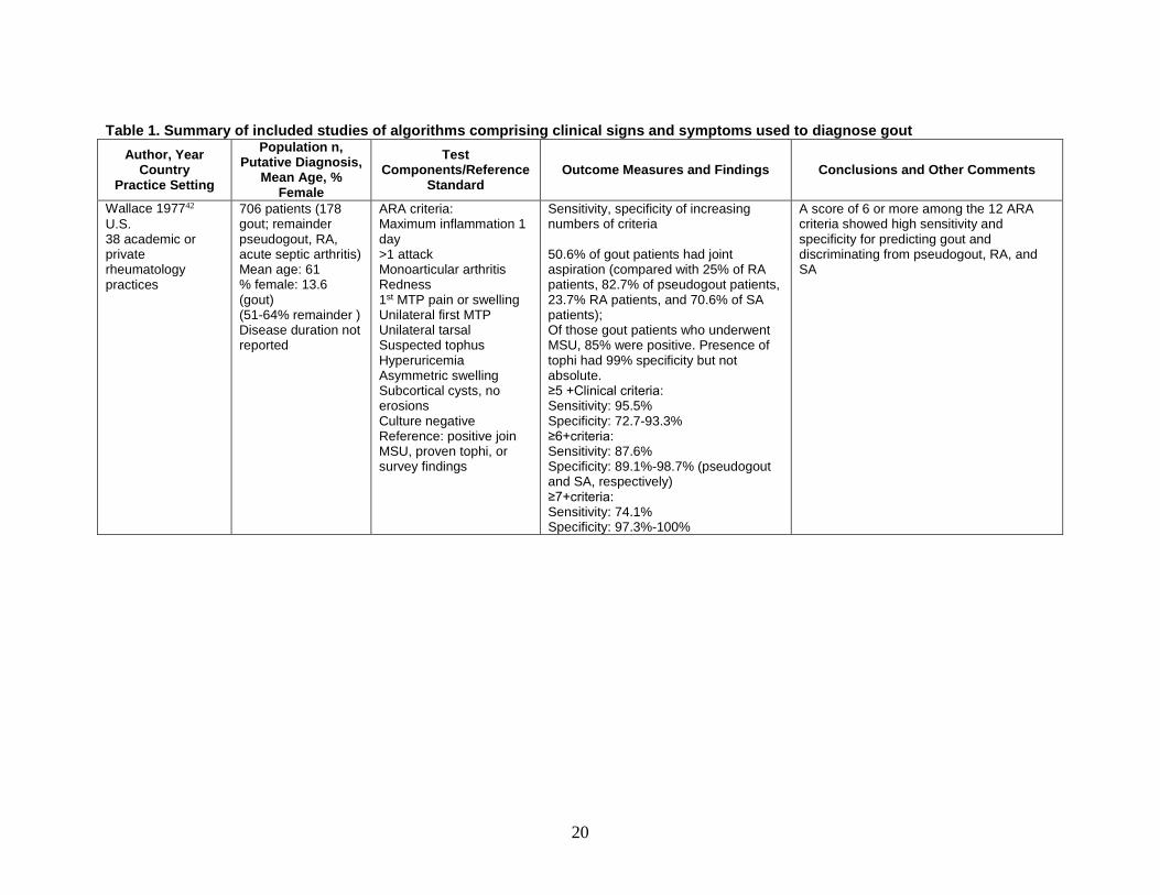
20
Table 1. Summary of included studies of algorithms comprising clinical signs and symptoms used to diagnose gout Author, Year
Country Practice Setting
Population n, Putative Diagnosis,
Mean Age, % Female
Test Components/Reference
Standard Outcome Measures and Findings Conclusions and Other Comments
Wallace 197742 U.S. 38 academic or private rheumatology practices
706 patients (178 gout; remainder pseudogout, RA, acute septic arthritis) Mean age: 61 % female: 13.6 (gout) (51-64% remainder ) Disease duration not reported
ARA criteria: Maximum inflammation 1 day >1 attack Monoarticular arthritis Redness 1st MTP pain or swelling Unilateral first MTP Unilateral tarsal Suspected tophus Hyperuricemia Asymmetric swelling Subcortical cysts, no erosions Culture negative Reference: positive join MSU, proven tophi, or survey findings
Sensitivity, specificity of increasing numbers of criteria 50.6% of gout patients had joint aspiration (compared with 25% of RA patients, 82.7% of pseudogout patients, 23.7% RA patients, and 70.6% of SA patients); Of those gout patients who underwent MSU, 85% were positive. Presence of tophi had 99% specificity but not absolute. ≥5 +Clinical criteria: Sensitivity: 95.5% Specificity: 72.7-93.3% ≥6+criteria: Sensitivity: 87.6% Specificity: 89.1%-98.7% (pseudogout and SA, respectively) ≥7+criteria: Sensitivity: 74.1% Specificity: 97.3%-100%
A score of 6 or more among the 12 ARA criteria showed high sensitivity and specificity for predicting gout and discriminating from pseudogout, RA, and SA
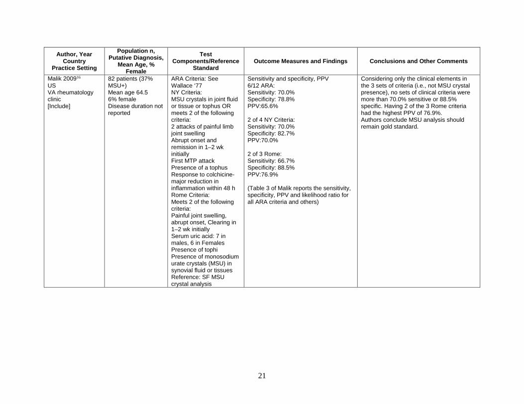
21
Author, Year Country
Practice Setting
Population n, Putative Diagnosis,
Mean Age, % Female
Test Components/Reference
Standard Outcome Measures and Findings Conclusions and Other Comments
Malik 200916 US VA rheumatology clinic [Include]
82 patients (37% MSU+) Mean age 64.5 6% female Disease duration not reported
ARA Criteria: See Wallace ‘77 NY Criteria: MSU crystals in joint fluid or tissue or tophus OR meets 2 of the following criteria: 2 attacks of painful limb joint swelling Abrupt onset and remission in 1–2 wk initially First MTP attack Presence of a tophus Response to colchicine-major reduction in inflammation within 48 h Rome Criteria: Meets 2 of the following criteria: Painful joint swelling, abrupt onset, Clearing in 1–2 wk initially Serum uric acid: 7 in males, 6 in Females Presence of tophi Presence of monosodium urate crystals (MSU) in synovial fluid or tissues Reference: SF MSU crystal analysis
Sensitivity and specificity, PPV 6/12 ARA: Sensitivity: 70.0% Specificity: 78.8% PPV:65.6% 2 of 4 NY Criteria: Sensitivity: 70.0% Specificity: 82.7% PPV:70.0% 2 of 3 Rome: Sensitivity: 66.7% Specificity: 88.5% PPV:76.9% (Table 3 of Malik reports the sensitivity, specificity, PPV and likelihood ratio for all ARA criteria and others)
Considering only the clinical elements in the 3 sets of criteria (i.e., not MSU crystal presence), no sets of clinical criteria were more than 70.0% sensitive or 88.5% specific. Having 2 of the 3 Rome criteria had the highest PPV of 76.9%. Authors conclude MSU analysis should remain gold standard.

22
Author, Year Country
Practice Setting
Population n, Putative Diagnosis,
Mean Age, % Female
Test Components/Reference
Standard Outcome Measures and Findings Conclusions and Other Comments
Janssens 2010a31 Netherlands Primary care clinics
328 patients with monoarthritis (but otherwise blinded PCP diagnosis) Mean age: 58.0 (13.) years, % female: 20.4% Recruited consecutively from primary care clinics with Disease duration not reported
Family physician diagnosis, patient-level factors (e.g., age, sex, CVD, onset within one day or hour, family history, medication use; Table 1 of Janssens includes all variables considered) Reference: SF MSU crystal analysis
PPV, NPV, AUC Family physician diagnosis: PPV: 0.64 NPV: 0.87 3 multivariate models were constructed. The most appropriate model contained the following predefined variables: Male sex Previous patient-reported arthritis attack Onset within 1 day Joint redness MTP 1 joint involvement Hypertension or1or more CVD, and sUA > 5.88mg/dL* AUC 0.85 (95% CI 0.81-0.90).
Performance did not change after transforming the regression coefficients to easy-to-use scores and was almost equal to that of the statistically optimal model (AUC 0.87; 95% CI,0.83-0.91)
Janssens 2010b32 Netherlands
381 monoarthritis patients recruited from primary care clinics (328 with PCP diagnosis of gout) (same population as Janssens 2010a)
ARA/ACR criteria Reference Standard: SF MSU crystal analysis
ARA/ACR criteria (≥4 +) Sensitivity: 1.00, Specificity: 0.24 PPV:0.70 NPV: 0.97 Overall fraction correct: 72% ARA/ACR criteria (≥6 +) Sensitivity: 0.80 Specificity: 0.64 PPV:0.80 NPV: 0.65 Overall fraction correct: 74% ARA/ACR criteria (≥ 9 +) Sensitivity: 0.07 (15/209) Specificity: 0.99 (118/119) PPV: 0.94 (15/16) NPV: 0.38 (118/312) Overall fraction correct: 41% (133/328)
Increasing stringency of ARA criteria beyond the cutoff point of 6 or more criteria did not improve usability of the criteria greatly: Patients referred from primary care with suspected gout (monoarthritis) assessed with MSU showed that the ARA/ACR criteria not particularly useful in diagnosis.

23
Author, Year Country
Practice Setting
Population n, Putative Diagnosis,
Mean Age, % Female
Test Components/Reference
Standard Outcome Measures and Findings Conclusions and Other Comments
Vazquez-Mellado 201241 Mexico Academic rheumatology departments
167 consecutive patients with diagnoses of RA, OA, SpA, or gout (75) Mean age: 45 % female: 24% (gout); 90% (OA) Disease duration not reported
Clinical gout diagnostic [CGD] criteria= ≥4/8 of the following: Current/past history of >1 attack of acute arthritis Mono- or oligoarthritis Rapid progression of pain/swelling (<24 hours) Podagra Erythema Unilateral tarsitis Tophi Hyperuricemia Reference standard: MSU crystal analysis performed in all gout patients
Sensitivity, specificity, AUC, PPV, positive likelihood ratio of CGD criteria and effects of patient characteristics (obesity, HTN, CVD, dyslipidemia, hyperglycemia, metabolic syndrome, chronic renal failure) ≥3 CGD items: Sensitivity: 99.1 Specificity: 79.8 +LR: 4.90 -LR: 0.01 PPV: 85.6 NPV: 48.7 AUC: 0.90 ≥4 CGD items: Sensitivity: 97.3 (95% CI 90.8, 99.3) Specificity: 95.6 (89.2, 98.3) +LR: 22.1 -LR: 0.03 PPV: 94.8 NPV: 97.8 AUC: 0.96
Authors state that study is limited by setting (rheumatology clinic vs. primary care setting) and the patient population likely to be seen (chronic vs. acute) but high concordance with other criteria, e.g., ACR suggests these criteria may be useful in primary care settings. Radiographic data not included because not specific. The mean CGD criteria in women: 5.22 (SD1.35) The mean CGD criteria in early-onset gout: 7.0(1.22) The kappa value comparing the CGD and the Janssens criteria was 0.85.
Kienhorst 201333 Netherlands Academic rheumatology department
390 patients with suspected gout referred to rheumatology clinic by PCPs; patients with crystal proven gout excluded (duration not reported)
Janssens diagnostic criteria31: Male sex (2*) Previous patient-reported arthritis attack (2)Onset within 1 day (0.5) Joint redness (1) MTP 1 involvement (2.5) HTN or ≥1CVD (1.5) sUA >0.35 mmol/L (5.88 mg/dL) (3.5) Reference standard: MSU crystal analysis performed in all patients *Points assigned to criterion
Prevalence of gout by cut-off scores (out of a possible 12): ≤4: PPV 5%; NPV 95% (4 of 69) >4 to <8: 46% (76 of 163) ≥8: PPV 87% (138 of 157) AUC: 0.86 (95% CI 0.82, 0.89)
Diagnostic criteria developed for primary care settings validated in secondary care population (i.e., patients referred by PCP to rheumatology for confirmation of suspected diagnosis). During followup, 28 patients who had been MSU- at baseline became MSU+.
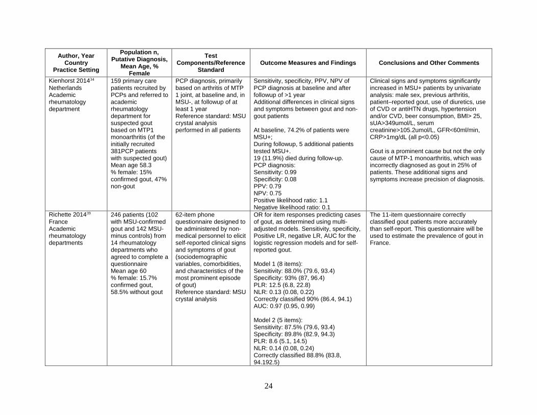
24
Author, Year Country
Practice Setting
Population n, Putative Diagnosis,
Mean Age, % Female
Test Components/Reference
Standard Outcome Measures and Findings Conclusions and Other Comments
Kienhorst 201434 Netherlands Academic rheumatology department
159 primary care patients recruited by PCPs and referred to academic rheumatology department for suspected gout based on MTP1 monoarthritis (of the initially recruited 381PCP patients with suspected gout) Mean age 58.3 % female: 15% confirmed gout, 47% non-gout
PCP diagnosis, primarily based on arthritis of MTP 1 joint, at baseline and, in MSU-, at followup of at least 1 year Reference standard: MSU crystal analysis performed in all patients
Sensitivity, specificity, PPV, NPV of PCP diagnosis at baseline and after followup of >1 year Additional differences in clinical signs and symptoms between gout and non-gout patients At baseline, 74.2% of patients were MSU+; During followup, 5 additional patients tested MSU+. 19 (11.9%) died during follow-up. PCP diagnosis: Sensitivity: 0.99 Specificity: 0.08 PPV: 0.79 NPV: 0.75 Positive likelihood ratio: 1.1 Negative likelihood ratio: 0.1
Clinical signs and symptoms significantly increased in MSU+ patients by univariate analysis: male sex, previous arthritis, patient–reported gout, use of diuretics, use of CVD or antiHTN drugs, hypertension and/or CVD, beer consumption, BMI> 25, sUA>349umol/L, serum creatinine>105.2umol/L, GFR<60ml/min, CRP>1mg/dL (all p<0.05) Gout is a prominent cause but not the only cause of MTP-1 monoarthritis, which was incorrectly diagnosed as gout in 25% of patients. These additional signs and symptoms increase precision of diagnosis.
Richette 201439 France Academic rheumatology departments
246 patients (102 with MSU-confirmed gout and 142 MSU-minus controls) from 14 rheumatology departments who agreed to complete a questionnaire Mean age 60 % female: 15.7% confirmed gout, 58.5% without gout
62-item phone questionnaire designed to be administered by non-medical personnel to elicit self-reported clinical signs and symptoms of gout (sociodemographic variables, comorbidities, and characteristics of the most prominent episode of gout) Reference standard: MSU crystal analysis
OR for item responses predicting cases of gout, as determined using multi-adjusted models. Sensitivity, specificity, Positive LR, negative LR, AUC for the logistic regression models and for self-reported gout. Model 1 (8 items): Sensitivity: 88.0% (79.6, 93.4) Specificity: 93% (87, 96.4) PLR: 12.5 (6.8, 22.8) NLR: 0.13 (0.08, 0.22) Correctly classified 90% (86.4, 94.1) AUC: 0.97 (0.95, 0.99) Model 2 (5 items): Sensitivity: 87.5% (79.6, 93.4) Specificity: 89.8% (82.9, 94.3) PLR: 8.6 (5.1, 14.5) NLR: 0.14 (0.08, 0.24) Correctly classified 88.8% (83.8, 94.192.5)
The 11-item questionnaire correctly classified gout patients more accurately than self-report. This questionnaire will be used to estimate the prevalence of gout in France.
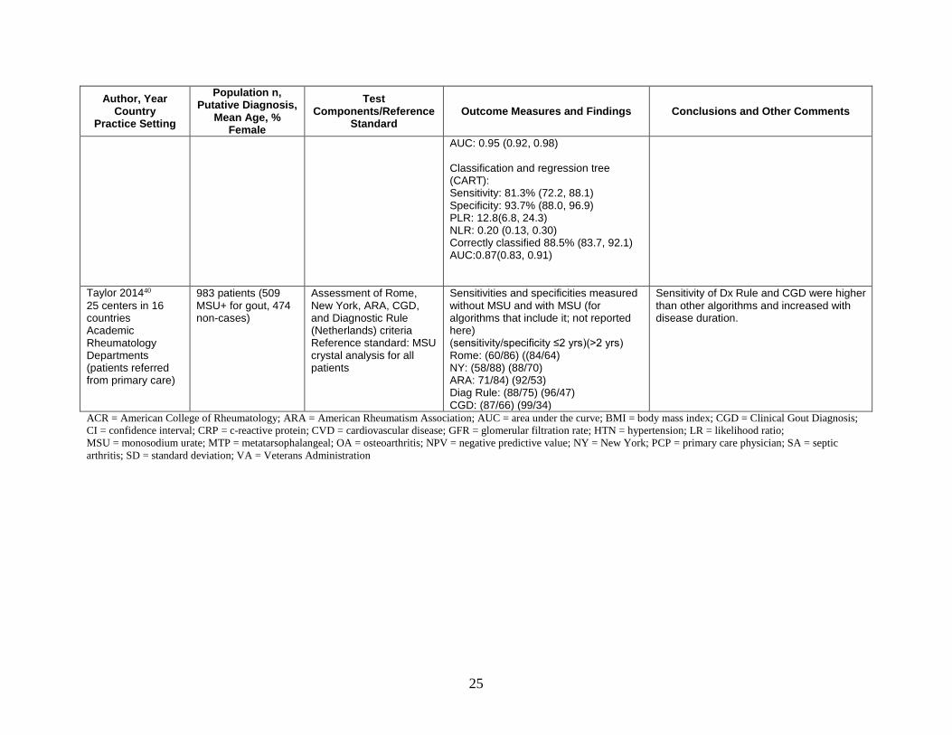
25
Author, Year Country
Practice Setting
Population n, Putative Diagnosis,
Mean Age, % Female
Test Components/Reference
Standard Outcome Measures and Findings Conclusions and Other Comments
AUC: 0.95 (0.92, 0.98) Classification and regression tree (CART): Sensitivity: 81.3% (72.2, 88.1) Specificity: 93.7% (88.0, 96.9) PLR: 12.8(6.8, 24.3) NLR: 0.20 (0.13, 0.30) Correctly classified 88.5% (83.7, 92.1) AUC:0.87(0.83, 0.91)
Taylor 201440 25 centers in 16 countries Academic Rheumatology Departments (patients referred from primary care)
983 patients (509 MSU+ for gout, 474 non-cases)
Assessment of Rome, New York, ARA, CGD, and Diagnostic Rule (Netherlands) criteria Reference standard: MSU crystal analysis for all patients
Sensitivities and specificities measured without MSU and with MSU (for algorithms that include it; not reported here) (sensitivity/specificity ≤2 yrs)(>2 yrs) Rome: (60/86) ((84/64) NY: (58/88) (88/70) ARA: 71/84) (92/53) Diag Rule: (88/75) (96/47) CGD: (87/66) (99/34)
Sensitivity of Dx Rule and CGD were higher than other algorithms and increased with disease duration.
ACR = American College of Rheumatology; ARA = American Rheumatism Association; AUC = area under the curve; BMI = body mass index; CGD = Clinical Gout Diagnosis; CI = confidence interval; CRP = c-reactive protein; CVD = cardiovascular disease; GFR = glomerular filtration rate; HTN = hypertension; LR = likelihood ratio; MSU = monosodium urate; MTP = metatarsophalangeal; OA = osteoarthritis; NPV = negative predictive value; NY = New York; PCP = primary care physician; SA = septic arthritis; SD = standard deviation; VA = Veterans Administration

26
Table 2. Comparison of components among clinical algorithms for diagnosis of gout
Components Rome
(1963)a16 NY
(1966)b16 Wallace 1977
(ARA/ ACR)42
EULAR 200622
Janssens Diagnostic Rule 201031
Clinical Gout Diagnosis
(CGD) 201041 3e Initiative
2014d47 Richette Surveye39
Clinical characteristics >1 attack of acute arthritis Maximum inflammation developed within 1 day
painful joint
swelling, abrupt onset, clearing 1-2
wk
Monoarthritis/oligoarthritis attack
Redness observed over joints
1st MTP joint painful or swollen
podagra
podagra
Unilateral 1st MTP joint attack
Unilateral tarsal joint attack Abrupt onset and remission in 1-2 weeks initially
Response to colchicine – major reduction in inflammation within 48h
Pain intensity≥9/10 Involvement of toes, foot, or ankles
Treatment with corticosteroids
Treatment with NSAIDS Resolution of pain<15 days after onset
Tophi (proven or suspected)
Radiograph Asymmetric swelling within a joint on radiograph
Subcortical cysts without erosions on radiograph

27
Components
Rome (1963)a16
NY (1966)b16
Wallace 1977 (ARA/ ACR)42
EULAR 200622
Janssens Diagnostic Rule 201031
Clinical Gout Diagnosis
(CGD) 201041 3e Initiative
2014d47 Richette Surveye39
Joint fluid Joint fluid culture negative MSU crystals in synovial fluid or tissuesf
Comorbid or risk factors Hyperuricemia Male sex Hypertension or ≥1 CVD Hypertriglyceridemia 3e = Evidence , Expertise, Exchange; ACR = American College of Rheumatology; ARA = Amereican Rheumatology Association; CGD = clinical gout diagnosis; CVD = cardiovascular disease; EULAR = European League Against Rheumatism; MTP = metatarsophalangeal Note: Podagra is gout that involves the big toe. a Meets 2 of the following criteria. b MSU crystals in joint fluid or tophus or tissue OR meets 2 of the following criteria. c ≥4/8 of the following criteria. d Guideline 1 states that MSU is required for a definitive diagnosis but in its absence, clinical criteria such as the following can be used or characteristic imaging findings may substitute. e Designed to be administered telephonically by non-physicians to assess prevalence of gout via patient self-report; treatment questions refer to most prominent episode. f Several algorithms specified presence of MSU crystals as definitive in lieu of other signs.

28
Studies Assessing DECT To Diagnose Gout
Comparative Accuracy We identified three original studies that compared the use of DECT for gout diagnosis to a
reference standard and enrolled only patients suspected of having gout.18, 28, 30 We also identified one 2014 systematic review comparing DECT and ultrasound for the classification of gout.45 The study details are described in Table 3.
A 2011 U.S. study used DECT to assess 94 consecutive patients with suspected gout (in each case, the most painful joint was studied). Of the 94 patients, 31 subsequently underwent successful joint aspiration and assessment for MSU crystals: 12 tested positive for uric acid crystals in the aspirated joint and 19 were MSU-negative (the final diagnoses for the latter patients were not revealed). Two blinded radiologists read the images, with a resulting sensitivity of 100% and specificities of 89% and 79% (K=0.87). For several patients whose DECT findings were initially thought to be false positives based on their MSU analysis, clinicians determined that the DECT findings agreed with diagnoses of gout based on clinical algorithms (which increased the specificity to 100%and 88% for the two readers, respectively. On the basis of these findings, the authors concluded that DECT is a sensitive and reproducible method for uric acid detection in multiple anatomical sites, including several atypical sites for gout and that DECT discriminated calcium pyrophosphate from MSU crystals. As a side note, 5 of the 12 MSU crystal-positive patients had elevated serum urate and the remaining 7 had serum urate within normal limits. Of the MSU crystal-negative patients, 6 had elevated serum urate, and 12 had serum urate within normal limits. Thus the concordance index for a logistic regression model configured with only serum urate as a predictor was poor (c=0.509); however, adding the DECT results to the regression model improved the concordance index to 0.98 (p<0.001).18
A 2014 U.S. study by the same group to determine the diagnostic accuracy of DECT examined 81consecutive patients with suspected gout (joint pain and swelling), 40 of whom were diagnosed with acute gout, and 41 with other arthritic conditions, at a single center.28 As in the previous study, only the most painful joint underwent aspiration and imaging. This assessment showed a sensitivity of 90% and a specificity of 83%. All false-negative DECT scans were patients with acute, recent-onset gout; all false positives had advanced knee osteoarthritis. In a separate study the authors called a diagnostic yield study, they then studied 30 patients with inflammatory arthritis but an unclear diagnosis; 14 of the 30 showed signs of gout on DECT. Of the 14, two refused subsequent joint aspiration, and 11 of the 12 who underwent aspiration were positive for MSU crystals, for a specificity of 92%. The DECT-negative patients did not undergo aspiration, so sensitivity could not be determined for this group. The authors concluded that while DECT is diagnostically useful overall, it has limited sensitivity among patients with acute gout and no prior history of gouty arthritis (i.e., a first flare).
A 2014 study conducted in Germany at a single center comparatively assessed DECT and ultrasound in multiple joints in 60 patients undergoing DECT for suspected gout. Of the 60, 39 were subsequently diagnosed with gout either through polarization microscopic detection of MSU crystals or use of a validated clinical algorithm, and the diagnostic outcomes were compared with those of DECT and of ultrasound.30 They found that DECT gave a sensitivity of 85% and a specificity of 86% (positive predictive value [PPV] 92%, negative predictive value [NPV] 75%). The remaining patients were eventually diagnosed with rheumatoid arthritis, psoriatic arthritis, undifferentiated oligoarthritis, calcium pyrophosphate deposition disease, osteoarthritis, or ankylosing spondylitis, undifferentiated connective tissue disease, Lyme

29
Disease, or hydroxyapatite deposition disease. In addition, 16 of the 39 patients diagnosed with gout had a concomitant inflammatory rheumatic disease (RA, PSA, AS, SLE, systematic sclerosis, and calcium pyrophosphate deposition disease). The comparison of DECT with ultrasound is described below.
A 2014 systematic review (AMSTAR score: 6 of 11)45 pooled the results of two of our included studies,18, 28 and one additional, case-control, study (i.e., enrolled only confirmed gout patients). The pooled sensitivity and specificity for the three DECT studies were 0.87 (95% CI, 0.79, 0.93) and 0.84 (0.75, 0.90), respectively.
Effect of Test Accuracy on Clinical Decisionmaking, Clinical Outcomes and Complications, and Patient Centered Outcomes
No studies were identified that tested the effects of the accuracy of gout diagnoses based on DECT on clinical decisionmaking (treatment decisions or decisions to conduct further testing), clinical outcomes, or patient centered outcomes.
Effect of Affected Joint Site(s) and Number of Joints on the Diagnostic Accuracy of DECT
No studies were identified that tested the effects of the joint site affected or the number of joints affected on the accuracy of DECT.
Effect of Duration of Symptoms on the Accuracy of Gout Diagnoses Based on DECT
No studies tested the effect of duration of symptoms (time since beginning of flare) on the accuracy of gout diagnoses based on DECT.

30
Table 3. Studies assessing the accuracy of DECT to diagnose gout Author, Year
Country Practice Setting
Population n, Putative Diagnosis,
Mean Age, % Female
Test Components/Reference
Standard Outcome Measures and Findings Conclusions and Other Comments
Huppertz 201430 Germany 1 academic rheumatology department
60 consecutive patients with suspected gout (gout (n=39); remainder: rheumatoid arthritis (n=6); psoriatic arthritis (n=4); undifferentiated oligoarthritis (n=3); chondrocalcinosis (n=2); osteoarthritis (n=2); ankylosing spondylitis, undifferentiated tissue disease, Lyme disease, hydroxyapatite deposition disease (n=1, respectively) Mean age: 62 %female: 18%
Gout suspects
39 diagnosed with gout:
Polarization microscopy of synovial fluid (n=18/39 = 46%)
OR
Positive Janssens Score by rheumatologist (≥8 out of a maximum of13 points) incorporating serum uric acid level (>350micromol/L; 3.5 points), first MTP joint involvement (2.5 points), gender (male, 2 points), previous patient-reported arthritis attack (2 points), cardiovascular diseases (1.5 points), joint redness and onset within 1 day (0.5 points) (n=21/39 = 54%)
Sensitivity, Specificity, Positive Predictive Value, Negative Predictive Value of DECT and US
For gout diagnosis: DECT Sensitivity: 84.6% DECT Specificity: 85.7%% US Sensitivity: 100% US Specificity: 76.2% DECT Pos. Predictive Val.: 91.7% DECT Neg. Predictive Val.: 75.0% US Pos. Predictive Val.: 88.6% US Neg. Predictive Val.: 100% All patients with false-positive DECT were negative in US; all patients with false-positive US were negative in DECT. Comparing 18 MSU+ positive gout patients and 21 patients with other rheumatic disease: DECT Sensitivity: 83.3% US Sensitivity: 100% DECT Pos. Predictive Val.: 83.3% US Pos. Predictive Val.: 78.3% DECT Neg. Predictive Val.: 85.7% US Neg. Predictive Val.: 100% Authors conclude that DECT provides better differential diagnosis than US Data provided on detection of crystals by joint (KQ1b?)
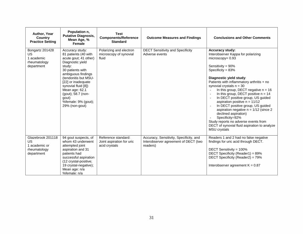
31
Author, Year Country
Practice Setting
Population n, Putative Diagnosis,
Mean Age, % Female
Test Components/Reference
Standard Outcome Measures and Findings Conclusions and Other Comments
Bongartz 201428 US 1 academic rheumatology department
Accuracy study: 81 patients (40 with acute gout; 41 other) Diagnostic yield study: 30 patients with ambiguous findings (tendonitis but MSU- [22] or inadequate synovial fluid [8]) Mean age: 62.1 (gout); 58.7 (non-gout) %female: 9% (gout); 29% (non-gout)
Polarizing and electron microscopy of synovial fluid
DECT Sensitivity and Specificity Adverse events
Accuracy study: Interobserver Kappa for polarizing microscopy= 0.93 Sensitivity = 90% Specificity = 83% Diagnostic yield study: Patients with inflammatory arthritis + no synovial crystals n = 30 - In this group, DECT negative n = 16 - In this group, DECT positive n = 14 - In DECT positive group, US guided
aspiration positive n = 11/12 - In DECT positive group, US guided
aspiration negative n = 1/12 (since 2 declined aspiration)
- Specificity=92% Study reports no adverse events from DECT of synovial fluid aspiration to analyze MSU crystals
Glazebrook 201118 US 1 academic or rheumatology department
94 gout suspects, of whom 43 underwent attempted joint aspiration and 31 patients had successful aspiration (12 crystal-positive; 19 crystal-negative); Mean age: n/a %female: n/a
Reference standard: Joint aspiration for uric acid crystals
Accuracy, Sensitivity, Specificity, and Interobserver agreement of DECT (two readers)
Readers 1 and 2 had no false negative findings for uric acid through DECT. DECT Sensitivity = 100% DECT Specificity (Reader1) = 89% DECT Specificity (Reader2) = 79% Interobserver agreement K = 0.87

32
ACR = American College of Rheumatology; ARA = American Rheumatism Association; AUC = area under the curve; BMI = body mass index; CGD = clinical gout diagnosis; CI = confidence interval; CRP = c-reactive protein; CVD = cardiovascular disease; GFR = glomerular filtration rate; HTN = hypertension; LR = likelihood ratio; MSU = monosodium urate; MTP = metatarsophalangeal; OA = osteoarthritis; NPV = negative predictive value; NY = New York; PCP = primary care physician; SA = septic arthritis; SD = standard deviation; US = ultrasound; VA = Veterans Administration
Author, Year Country
Practice Setting
Population n, Putative Diagnosis,
Mean Age, % Female
Test Components/Reference
Standard Outcome Measures and Findings Conclusions and Other Comments
Ogdie 201445 Systematic review of gout diagnosis in primary care settings
Inclusion criteria: studies comparing diagnostic performance of X-Ray, MRI, US, CT or DECT in gout; presence of non-gout control group; gout confirmation through synovial fluid aspiration. Exclusion criteria: use of clinical criteria or physician or patient reports instead of MSU crystal presence; lack of control or comparison group; cases with asymptomatic hyperuricemia; not enough information to calculate sensitivity and specificity
Joint aspiration for uric acid crystals
Sensitivity and Specificity of: Double-Contour Sign (DCS) on Ultrasound; Ultrasound of Tophus; DECT.
Ultrasound DCS Sensitivity (pooled) = 83% Ultrasound DCS specificity (pooled) = 76% Ultrasound tophus sensitivity (pooled) = 65% Ultrasound tophus specificity (pooled) = 80% DECT sensitivity (pooled) = 87% DECT specificity (pooled)
33
Studies Assessing the Accuracy of Ultrasound To Diagnose Gout
Comparative Accuracy We identified four original prospective or cross-sectional studies that assessed the accuracy of
ultrasound to diagnose gout in patients suspected of having gout; the reference standards adopted in these studies were either MSU crystal analysis, the use of a diagnostic clinical algorithm, or a combination of the two.30, 35, 36, 38 These studies are described in Table 4. The QUADAS ratings for each of these studies are presented in Table 5. We also identified two recent systematic reviews on the use of ultrasound to diagnose gout.43, 45
A 2008 study conducted in Austria with a population of 86 consecutive patients with clinical suspicion of gout compared ultrasound with plain x-ray and with a reference standard of MSU assessment (30 patients) or a diagnostic clinical algorithm (56 patients).38 The authors assessed the sensitivity and specificity for a number of ultrasound indicators, included bright stippled foci (BSF, which is hyperechoic deposition in the synovium), hyperechoic areas, at least one of the former two, bone erosions, and hyperechoic streaks. BSF and hyperechoic areas alone had comparable sensitivity (80%, 79%), whereas the specificity of BSF was relatively low (75%) compared with that of hyperechoic areas (95%). The presence of at least one positive sign (BSF or hyperechoic areas) had a sensitivity of 96% and a specificity of 73%. Compared with ultrasound, plain x-ray was much less sensitive but comparable in specificity (31%/93%). Bony erosions detected by ultrasound or plain x-ray had equally poor sensitivity but widely varying specificity (69%-95%). The patients in the study had clinical involvement at a number of different sites (primarily the MTP1 joint and the knee); the authors did not attempt to estimate the effect of site or number of sites involved in the sensitivity or specificity of the test. Patients determined not to have gout had osteoarthritis, rheumatoid arthritis, psoriatic arthritis, septic arthritis, calcium pyrophosphate deposition disease, chondrocalcinosis, or no conclusive diagnosis.
A 2011 study conducted in a Taiwan hospital enrolled 80 patients with acute monoarthritis or oligoarthritis who underwent ultrasound-guided joint aspiration (recruitment method not described). Ultrasound of the target joint was compared with ultrasound-guided joint aspiration and fluid analysis for MSU and a diagnostic clinical algorithm.35 Of the 80 patients, 34 (representing 52 joints) were MSU crystal-positive. Images were assessed for double contour sign (DCS, hyperechoic deposition on the surface of the articular hyaline cartilage), BSF, snow storm, tophi, and bony erosion. The DCS alone had a sensitivity of 36.8% and a specificity of 97.3% (PPV 93; NPV 60); BSF alone had a sensitivity of 76.9% and specificity of 65.4%. The presence of either DCS or BSF had a sensitivity of 86.5% and a specificity of 63.5% (PPV 70 and NPV 82); whereas the DCS combined with the BSF had a sensitivity of 23.7 (NPV 56.1) and a specificity and PPV of 100. Patients who were MSU crystal-negative met criteria for calcium pyrophosphate deposition disease (5), septic arthritis (14), rheumatoid arthritis (4), osteoarthritis (4), and other inflammatory arthritis (18). Interobserver correlations were excellent for the DCS (K=0.86) and good for BSF (0.73).
A 2014 study conducted in the Netherlands enrolled all 54 patients referred to an academic hospital outpatient department with monoarthritis but no further diagnosis.36 Patients underwent ultrasound of the affected joint and five additional joints (knees, MTP joints, and any additional target joint of interest, bilaterally) for a total of six images; the images were examined for DCS, snow storm, tophi, and any other ultrasound finding. Assessments were compared with the results of joint aspiration and MSU analysis in all but two patients or the Janssens diagnostic clinical algorithm31 in the remaining two patients. The finding of a DCS had a sensitivity of 77% and a
34
specificity of 75% (PPV 74%, NPV 78%; LR+ 3.08, LR- 0.31); inclusion of any sign of abnormality had a sensitivity of 96% and a specificity of 68% (PPV 74%, NPV 95%’ LR+2.99, LR- 0.06); snowstorm had a sensitivity of 38% and a specificity of 86% (PPV 71%, NPV 60%); indication of a tophus had a sensitivity of 19% and a specificity of 93%. The inter-rater reliability improved with the number of patients (kappa increased from 0.44 to 0.67 for the DCS).
A 2014 study conducted in Germany compared the findings of ultrasound with those of DECT30 (DECT findings described above) among 60 gout suspects. The standard of reference was MSU and the Janssens diagnostic clinical algorithm. The assessment of hyperechoic findings and DCS had a sensitivity of 100% and specificity of 76% (PPV 89, NPV 100). The comparison of ultrasound with DECT showed that ultrasound is more sensitive and less specific than DECT for the diagnosis of gout and is particularly useful for patients with inconclusive joint aspiration, other co-occuring rheumatic diseases, and ambivalent findings. In a joint-based evaluation, ultrasound was more sensitive than DECT for gout diagnosis; however, differentiating between calcium pyrophosphate deposition disease and gout at the wrist is challenging with ultrasound.
Two systematic reviews assessed studies of ultrasound for gout. A 2012 systematic review by Chowalloor (AMSTAR score: 4 of 11)43 reviewed the results of 18 studies (1975 to 2012) on the use of ultrasound for management (and secondarily, diagnosis) of gout and hyperuricemia. Only one of these studies was included in the present review;38 the remaining studies were excluded from our analysis either because they included only patients with definitive gout diagnoses, included only patients with chronic gout in the intercritical period, or included primarily patients with asymptomatic hyperuricemia. The review, which did not include a meta-analysis, concluded that the DCS is specific for the diagnosis of gout, and that ultrasound can track changes in joint architecture, e.g., development of tophi.
A 2014 systematic review by Ogdie (AMSTAR score: 6 of 11)45 on the use of ultrasound (and DECT) for gout classification pooled the results of five studies on the use of ultrasound, one of which was included in the present review 36 (the remaining four studies were excluded from our analysis because the populations consisted of patients with prior gout diagnoses). For the DCS, the review reported a pooled sensitivity of 83% (95% CI 72%, 91%) and a specificity of 76% (68%, 83%). For tophus detection, the pooled sensitivity was 65% (34%, 87%) and the pooled specificity was 87% (79%, 93%).
Effect of Test Accuracy on Clinical Decisionmaking, Clinical Outcomes and Complications, and Patient Centered Outcomes
No studies were identified that tested the effects of the accuracy of gout diagnoses based on ultrasound on clinical decisionmaking (treatment decisions or decisions to conduct further testing), clinical outcomes, or patient centered outcomes.
Effect of Affected Joint Site(s) and Number of Joints on the Diagnostic Accuracy of Ultrasound
No studies were identified that tested the effects of the joint site affected or the number of joints affected on the accuracy of diagnosis by ultrasound.
Effect of Duration of Symptoms on the Accuracy of Gout Diagnoses Based on Ultrasound
No studies tested the effect of duration of symptoms (time since beginning of flare) on the accuracy of gout diagnoses based on ultrasound.
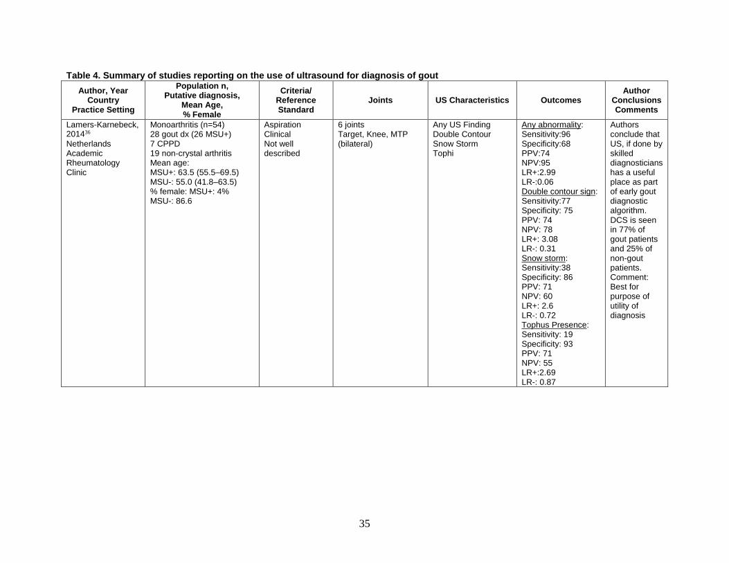
35
Table 4. Summary of studies reporting on the use of ultrasound for diagnosis of gout Author, Year
Country Practice Setting
Population n, Putative diagnosis,
Mean Age, % Female
Criteria/ Reference Standard
Joints US Characteristics Outcomes Author
Conclusions Comments
Lamers-Karnebeck, 201436 Netherlands Academic Rheumatology Clinic
Monoarthritis (n=54) 28 gout dx (26 MSU+) 7 CPPD 19 non-crystal arthritis Mean age: MSU+: 63.5 (55.5–69.5) MSU-: 55.0 (41.8–63.5) % female: MSU+: 4% MSU-: 86.6
Aspiration Clinical Not well described
6 joints Target, Knee, MTP (bilateral)
Any US Finding Double Contour Snow Storm Tophi
Any abnormality: Sensitivity:96 Specificity:68 PPV:74 NPV:95 LR+:2.99 LR-:0.06 Double contour sign: Sensitivity:77 Specificity: 75 PPV: 74 NPV: 78 LR+: 3.08 LR-: 0.31 Snow storm: Sensitivity:38 Specificity: 86 PPV: 71 NPV: 60 LR+: 2.6 LR-: 0.72 Tophus Presence: Sensitivity: 19 Specificity: 93 PPV: 71 NPV: 55 LR+:2.69 LR-: 0.87
Authors conclude that US, if done by skilled diagnosticians has a useful place as part of early gout diagnostic algorithm. DCS is seen in 77% of gout patients and 25% of non-gout patients. Comment: Best for purpose of utility of diagnosis

36
Author, Year Country
Practice Setting
Population n, Putative diagnosis,
Mean Age, % Female
Criteria/ Reference Standard
Joints US Characteristics Outcomes Author
Conclusions Comments
Lai, 201135 Taiwan Hospital rheumatology department
Monoarthritis (n=80) 34 (52 joints) MSU+ 46 (52 joints) MSU- Mean age: 69.4 (range 52.8-80.8) % female: 17.6% (gout) 52.2% (non-gout)
Aspiration Clinical criteria (joint swelling, heat, tenderness)
Target Joint Double Contour Snow Storm Tophi Bony erosion PDUS
Double contour sign (DCS): Sensitivity: 36.8 Specificity: 97.3 PPV: 93.3 NPV: 60 LR+: 13.63 LR-: 0.65 Bright Stippled foci (BSF): Sensitivity: 76.9 Specificity: 65.4 PPV: 69 NPV: 73.9 LR+: 2.22 LR-: 0.35 DCS OR BSF: Sensitivity: 86.5 Specificity: 63.5 PPV: 70.3 NPV: 82.5 LR+:2.37 LR-: 0.21 DCS AND BSF: Sensitivity: 23.7 Specificity: 100 PPV: 100 NPV: 56.1 LR+: LR-: 0.76
Analysis was at the joint level, not patient Authors conclude that presence of both DCS and BSF is indicative of gout. Hyperechoic aggregations, bone erosion, and synovial vascularity did not distinguish gout from non-gout arthritis.
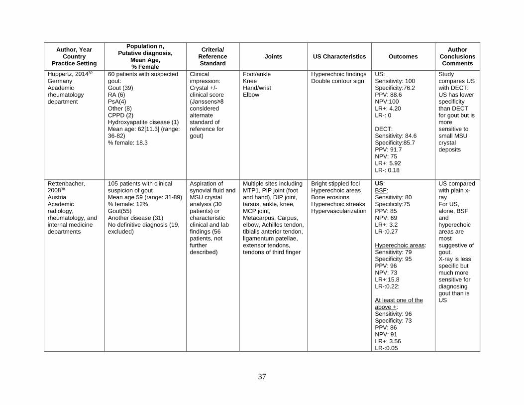
37
Author, Year Country
Practice Setting
Population n, Putative diagnosis,
Mean Age, % Female
Criteria/ Reference Standard
Joints US Characteristics Outcomes Author
Conclusions Comments
Huppertz, 201430 Germany Academic rheumatology department
60 patients with suspected gout: Gout (39) RA (6) PsA(4) Other (8) CPPD (2) Hydroxyapatite disease (1) Mean age: 62[11.3] (range: 36-82) % female: 18.3
Clinical impression: Crystal +/- clinical score (Janssens≥8 considered alternate standard of reference for gout)
Foot/ankle Knee Hand/wrist Elbow
Hyperechoic findings Double contour sign
US: Sensitivity: 100 Specificity:76.2 PPV: 88.6 NPV:100 LR+: 4.20 LR-: 0 DECT: Sensitivity: 84.6 Specificity:85.7 PPV: 91.7 NPV: 75 LR+: 5.92 LR-: 0.18
Study compares US with DECT: US has lower specificity than DECT for gout but is more sensitive to small MSU crystal deposits
Rettenbacher, 200838 Austria Academic radiology, rheumatology, and internal medicine departments
105 patients with clinical suspicion of gout Mean age 59 (range: 31-89) % female: 12% Gout(55) Another disease (31) No definitive diagnosis (19, excluded)
Aspiration of synovial fluid and MSU crystal analysis (30 patients) or characteristic clinical and lab findings (56 patients, not further described)
Multiple sites including MTP1, PIP joint (foot and hand), DIP joint, tarsus, ankle, knee, MCP joint, Metacarpus, Carpus, elbow, Achilles tendon, tibialis anterior tendon, ligamentum patellae, extensor tendons, tendons of third finger
Bright stippled foci Hyperechoic areas Bone erosions Hyperechoic streaks Hypervascularization
US: BSF: Sensitivity: 80 Specificity:75 PPV: 85 NPV: 69 LR+: 3.2 LR-:0.27 Hyperechoic areas: Sensitivity: 79 Specificity: 95 PPV: 96 NPV: 73 LR+:15.8 LR-:0.22: At least one of the above +: Sensitivity: 96 Specificity: 73 PPV: 86 NPV: 91 LR+: 3.56 LR-:0.05
US compared with plain x-ray For US, alone, BSF and hyperechoic areas are most suggestive of gout. X-ray is less specific but much more sensitive for diagnosing gout than is US

38
Author, Year Country
Practice Setting
Population n, Putative diagnosis,
Mean Age, % Female
Criteria/ Reference Standard
Joints US Characteristics Outcomes Author
Conclusions Comments
Bone erosions: Sensitivity: 24 Specificity:69 PPV: 57 NPV: 34 LR+:0.77 LR-:1.10 Hyperechoic streaks: Sensitivity: 80 Specificity:49 PPV: 1.57 NPV: 0.41 Hypervascularization Sensitivity: 94 Specificity:53 PPV: 77 NPV: 84 LR+: 2.00 LR-: 0.11 X-ray: Opacification: Sensitivity: 26 Specificity: 97 PPV: 93 NPV:43 LR+: 8.67 LR-: 0.76 Bone erosions: Sensitivity: 20 Specificity: 95 PPV: 87 NPV:41 LR+: 4.00 LR-: 0.84

39
Author, Year Country
Practice Setting
Population n, Putative diagnosis,
Mean Age, % Female
Criteria/ Reference Standard
Joints US Characteristics Outcomes Author
Conclusions Comments
Osseous Apposition: Sensitivity: 5 Specificity: 100 PPV: 100 NPV: 38 LR+: LR-: 0.95 At least one positive sign: Sensitivity: 31 Specificity: 93 PPV: 89 NPV: 44 LR+: 4.43 LR-: 0.74
AH = asymptomatic hyperuricemia; BSF = bright stippled foci; CPPD = calcium pyrophosphate deposition (formerly pseudogout); DECT = dual-energy computed tomography; DCS = double-contour sign; LR = likelihood ratio; MSU = monosodium urate; MTP = metatarsophalangeal; NPV = negative predictive value; PPV = positive predictive value; PsA = psoriatic arthritis; RA = rheumatoid arthritis; US = ultrasound.

40
Table 5. Risk of bias assessment by QUADAS-2
Author, Year
Domain 1: Patient Selection Domain 2: Index Test(s) Domain 3: Reference Standard Domain 4: Flow and Timing
Con
secu
tive/
rand
om
sam
ple
Cas
e-co
ntro
l des
ign
avoi
ded?
App
ropr
iate
exc
lusi
ons
avoi
ded?
Ove
rall
risk
of b
ias
Inde
x te
st re
sults
read
w
ithou
t kno
win
g re
fere
nce
stan
dard
?
Pres
peci
fied
thre
shol
d?
Ove
rall
risk
of b
ias
Ref
eren
ce s
tand
ard
corr
ectly
cla
ssifi
es ta
rget
co
nditi
on?
Ref
eren
ce s
tand
ard
read
w
ithou
t kno
win
g th
e in
dex
test
?
Ove
rall
risk
of b
ias
App
ropr
iate
inte
rval
be
twee
n in
dex
test
(s) a
nd
refe
renc
e st
anda
rd?
All
patie
nts
rece
ived
re
fere
nce
stan
dard
?
Sam
e re
fere
nce
stan
dard
?
All
patie
nts
incl
uded
in
the
anal
ysis
?
Ove
rall
risk
of b
ias
Bongartz et al., 201428
Yes Yes Unclear Low Yes Yes Low Yes Yes Low Yes Yes Yes Yes Low
Glazebrook et al., 201118
Yes Yes Yes Low Yes N/A Low Yes Unclear Unclear No Yes Yes No Unclear
Huppertz et al., 201430
Yes No Unclear High Yes N/A Low Yes Yes Low Yes Yes Yes Yes Low
Janssens, et al., 201032
Yes Yes Unclear Low Unclear Yes Unclear Yes Unclear Unclear Yes Yes Yes Yes Low
Janssens et al., 201031
Yes Yes Yes Low Yes Yes Low Yes Yes Low Yes Yes Yes Yes Low
Kienhorst et al., 201333
Yes Yes Yes Low Yes Yes Low Yes Yes Low Yes Yes Yes Yes Low
Kienhorst et al., 201434
Yes Yes No Low Yes N/A Low Yes No Unclear Yes Yes Yes Yes Low
Lai et al., 201135
Yes Yes Unclear Low Yes N/A Low Yes Yes Low Unclear Yes Yes Yes Low
Lamers-Karnebeek et al., 201436
Yes Yes No Low Yes N/A Low Yes Yes Low Yes Yes Yes Yes Low
Malik et al., 200916
Yes Yes Yes Low Yes Yes Low Yes Yes Low Unclear Yes Yes Yes Low
Park et al., 201437
Yes Yes No Low N/A N/A N/A Yes N/A Low N/A Yes Yes No Unclear
Rettenbacher et al., 200838
Yes Yes Yes Low Yes N/A Low Yes Unclear Unclear Unclear Yes No No High
Richette et al., 201439
Unclear No Unclear High Yes No Unclear Yes Yes Low Unclear Yes Yes Yes Unclear

41
Author, Year
Domain 1: Patient Selection Domain 2: Index Test(s) Domain 3: Reference Standard Domain 4: Flow and Timing
Con
secu
tive/
rand
om
sam
ple
Cas
e-co
ntro
l des
ign
avoi
ded?
App
ropr
iate
exc
lusi
ons
avoi
ded?
Ove
rall
risk
of b
ias
Inde
x te
st re
sults
read
w
ithou
t kno
win
g re
fere
nce
stan
dard
?
Pres
peci
fied
thre
shol
d?
Ove
rall
risk
of b
ias
Ref
eren
ce s
tand
ard
corr
ectly
cla
ssifi
es ta
rget
co
nditi
on?
Ref
eren
ce s
tand
ard
read
w
ithou
t kno
win
g th
e in
dex
test
?
Ove
rall
risk
of b
ias
App
ropr
iate
inte
rval
be
twee
n in
dex
test
(s) a
nd
refe
renc
e st
anda
rd?
All
patie
nts
rece
ived
re
fere
nce
stan
dard
?
Sam
e re
fere
nce
stan
dard
?
All
patie
nts
incl
uded
in
the
anal
ysis
?
Ove
rall
risk
of b
ias
Taylor et al., 201440
Yes Yes Yes Low Yes Yes Low Yes Yes Low Unclear Yes Yes Yes Low
Vazquez-Mellado et al., 201241
Yes No Unclear High No Yes High Yes Yes Low Unclear Yes Yes Yes Low
Wallace et al., 197742
Unclear No Unclear High Unclear Yes High Yes Yes Low No No Yes Unclear
High
N/A = not available

42
KQ1d. Does the accuracy of synovial fluid aspiration and crystal analysis Differ by (i) the type of practitioner who is performing the aspiration and (ii) the type of practitioner who is performing the crystal analysis?
The question we were asked to answer was whether the accuracy of gout detection by synovial fluid aspiration and crystal analysis differs by (i) the type of practitioner who is performing the aspiration and (ii) the type of practitioner or technologist who is performing the crystal analysis.
Key Points • Agreement among medical and ancillary health personnel examining synovial fluid using
polarizing microscopy for detection of MSU crystals appears to be poor, but it is unclear whether the experience and training of analysts are a factor. No studies examined the effect of the type of practitioner performing fluid aspiration on the ability to obtain a sample for analysis. Because of the small number of studies identified, the strength of evidence for definitive influential factors is insufficient.
Description of Included Studies We identified two original studies that addressed the question of whether the results of MSU
analysis are affected by the type of practitioner or training. We identified no studies that addressed the question of how MSU assessment is affected by the person performing the joint aspiration. A 1989 study compared the accuracy of an experienced rheumatologist, several medical residents, and several technicians in identifying MSU and calcium pyrophosphate crystals suspended in synovial fluid using polarizing microscopy. A 2014 study retrospectively audited medical records of two Korean academic medical centers to assess factors associated with false negative synovial fluid MSU results and focused on the personnel performing the analysis and several other factors.37
Detailed Synthesis A 1989 study conducted in a United Kingdom academic rheumatology department compared
the ability of a small number of technicians and clinicians to conduct crystal detection.29 Synovial fluid samples were aspirated from the knee or shoulder of 13 patients with clinical diagnoses of rheumatoid arthritis or osteoarthritis (the person conducting the aspiration was not described). Purified MSU, calcium pyrophosphate, and basic calcium phosphate crystals prepared according to published methods were suspended in the fluid samples to achieve a range of concentrations that would be visualized as occasional crystals seen in a microscopic field, 10 MSU or calcium pyrophosphate crystals, or 5 phosphate crystals per microscopic field. The individuals whose performances were compared included two lab technicians (one with specialty training in crystal identification), an inexperienced physician, two rheumatology residents, and an experienced rheumatology attending physician. At the beginning of the exercise, all were shown microscopic examples of each type of crystals. Each observer was then tested on 82 slides. The sensitivities for MSU ranged from 65% to 76% (mean 69%) and the specificities ranged from 93% to 100% (mean 97%): The trained technician and the inexperienced physician’s assessments showed the highest sensitivity 73% and 76%), whereas one of the residents and the experienced attending had the highest specificity (100%). For calcium pyrophosphate crystal detection, the performance varied far more. The sensitivity ranged from 60% to 93% (mean 82%), and the specificity ranged from

43
61% to 98% (mean 78%): The experienced attending had by far the lowest—and the inexperienced physician had the highest—sensitivity, whereas one of the residents had by far the lowest—and the experienced attending had the highest—specificity.
A 2014 study in South Korea aimed to assess the rate of MSU detection and factors associated with false-negative findings. The researchers examined retrospective data from 179 patients (94.4 percent male, average age at diagnosis 62.6[16.4], 43.9 percent were experiencing their first attack) seen in two South Korean academic rheumatology departments.37 The charts of patients who underwent synovial fluid analysis for presumed acute gout between 1999 and 2011 were audited; patients were excluded if they were diagnosed with intercritical gout without acute symptoms, pseudogout, osteoarthritis, or both septic arthritis and gout. Patients were classified as having acute gout based on ACR criteria. MSU analysis at both hospitals was conducted either by lab medicine personnel (34.3 percent), by Rheumatology Department personnel (3.5 percent), or both (62.1 percent), at both hospitals. Of 198 samples analyzed for the 179 patients, 78.8 percent were crystal-positive, and 21.2 percent were negative. The detection rate for samples examined by the lab medicine departments was 51.8 percent, compared with 93.8 percent for the rheumatologists. When agreement was assessed for samples analyzed by both departments, the detection rate was 93.5 percent for rheumatologists and 51.2 percent for lab medicine, for a kappa of 0.108. Examination by lab medicine was the most significant variable associated with crystal-negative synovial fluid analysis (OR 36.996, 95% CI 9.731, 140.648). In addition, multivariate analysis showed that the time interval from attack onset to arthrocentesis was the only factor significantly associated with the likelihood of negative crystal findings (4.5±5.1 days for negative findings vs. 3.0±2.8 days for positive findings; OR 1.105, 95% CI 1.004, 1.216; p=0.042). Analysis by laboratory medicine was thought to be associated with a longer lag time than analysis by the Rheumatology departments.
Key Question 2. What are the adverse effects (including pain, infection at the aspiration site, radiation exposure) or harms (related to false positives, false negatives, indeterminate results) associated with tests used to diagnose gout?
Key Points • Considering potential adverse effects that might be associated with diagnostic tests for
gout, including pain, infection (at the aspiration site), or the short- or long-term effects of radiation exposure, no studies documented any adverse events associated with diagnostic tests for gout in any studies included in this report. The strength of evidence for this conclusion is low, based on only one study that reported no adverse events associated with joint fluid aspiration for MSU analysis or DECT and no studies that reported on adverse events associated with ultrasound or clinical examinations.
• Missed diagnosis or delayed diagnosis of acute gout (failure to find MSU crystals in synovial fluid) was reported in a retrospective two-center study to be associated with a longer interval between the onset of attack and joint aspiration. A negative MSU finding was associated with higher risk for undergoing arthroscopic drainage, longer hospital stay, and delays in antiinflammatory treatment. The strength of evidence for this conclusion is insufficient.

44
Description of Included Studies One study was identified that assessed adverse effects associated with tests used to diagnose
gout.28 This study reported no adverse events associated with aspiration of synovial fluid for MSU analysis or the use of DECT.
One study examined the outcomes of delayed diagnosis or misdiagnosis of gout in two academic medical centers in South Korea.37
Detailed Synthesis A 2014 study described earlier that examined the accuracy of DECT for diagnosis of gout in
81 patients noted in passing that neither aspiration of synovial fluid for MSU assessment nor DECT were associated with any adverse events.28
A 2014 study conducted retrospective medical chart review to examine factors associated with failure to diagnose gout and also compared treatment outcomes between those who had a positive finding for MSU crystals and those who did not.37 This study, described above, examined retrospective data from 179 patients (all meeting ACR criteria, 94.4 percent male, average age at diagnosis 62.6[16.4], 43.9 percent experiencing their first attack) seen in two South Korean academic rheumatology departments. They found no differences in the proportions of MSU crystal-negative and MSU crystal-positive patients who eventually received NSAIDs or colchicine, but MSU crystal-negative patients were less likely than MSU crystal-positive patients to receive intra-articular glucocorticoid injections (7.1 percent vs. 21.8 percent, p=0.043). In addition, MSU crystal-negative patients underwent emergency arthroscopic surgery and drainage more frequently than MSU crystal-positive patients (26.2 percent vs. 3.8 percent, p=4.48x10⁻⁶, chi-squared test), based on a suspicion of septic arthritis, in spite of negative cultures and visible MSU deposits in the joints. A first episode of monoarthritis (OR 6.954, 95% CI 1.577, 20622, p=0.01), negative findings for MSU crystal analysis (OR 23.760, 95% CI 4.451, 126.843 p=2.10x10⁻⁴), and fever (OR 15.123, 95% CI 2.739, 83.503, p=0.002) were independent risk factors for surgery. Patients undergoing arthroscopic surgery and drainage based on suspected septic arthritis had a significantly longer hospital stay than the 19 newly diagnosed gout patients hospitalized for medical treatment (18.4±12.6 days vs. 8.9±6.7 days, p=0.007), and patients undergoing arthroscopic surgery received anti-inflammatory drugs significantly later than those who received only medical treatment (5.0±3.4 days vs. 0.4±0.8 days, p=8.74x10⁻⁵).
45
Discussion Key Findings and Strength of Evidence
The key findings and strength of evidence are summarized below and in Table 6.
Accuracy of Tests for the Diagnosis of Gout • Few studies that assessed the accuracy of clinical signs and symptoms consistently applied
the same reference standard (either analysis of MSU crystals in synovial fluid or a single clinical algorithm) to all participants with suspected gout.
• Studies that assessed the use of diagnostic clinical algorithms compared with synovial fluid analysis for MSU crystals reported widely varying sensitivities and specificities; however, an algorithm developed from clinical signs and symptoms used by primary care physicians reported good positive and negative predictive value and was validated in a small secondary care population but needs further validation. The strength of evidence for this conclusion is low, based on the identification of only two studies that assessed this particular clinical algorithm.
• In three studies that enrolled only patients not previously diagnosed with gout, the sensitivities and specificities of DECT for predicting gout ranged from 85% to 100% and 83% to 92%, respectively, compared with synovial fluid analysis for MSU crystals or a validated clinical algorithm.
• Ultrasound was more variable than DECT in its ability to detect gout: Four studies of ultrasound showed sensitivities that ranged from 37% to 100% and specificities that ranged from 68% to 97%, depending on the signs assessed. The strength of evidence for this conclusion is low.
• No studies were identified that assessed the validity of serum urate, CT scan, or plain x-ray for diagnosing gout. The strength of evidence for these tests is insufficient.
• No studies were identified that directly assessed the effect of joint site or number of affected joints on diagnostic accuracy. The strength of evidence for this question is insufficient for all diagnostic methods.
• No studies were identified that directly assessed the effect of duration of symptoms on the accuracy of diagnostic tests. The strength of evidence for this question is insufficient for all diagnostic methods.
• Agreement among personnel examining synovial fluid using polarizing microscopy for detection of MSU crystals appears to be poor, but it is unclear whether the experience and training of analysts are a factor. No studies examined the effect of the type of practitioner performing fluid aspiration on the ability to obtain a sample. Because of the relatively small number of studies identified, the strength of evidence for definitive influential factors is insufficient.
Adverse Events Associated With Testing for Gout • Considering potential adverse effects that might be associated with diagnostic tests for
gout, including pain, infection (at the aspiration site), or the short- or long-term effects of radiation exposure, no studies documented any adverse events associated with diagnostic tests in any studies included in this report. The strength of evidence for this conclusion is

46
low, based on one study that reported no adverse events associated with joint fluid aspiration for MSU analysis or DECT, and no studies that reported on adverse events associated with ultrasound or clinical examination.
• Missed diagnosis or delayed diagnosis of acute gout (failure to find MSU crystals in synovial fluid) was reported in a retrospective two-center study to be associated with a longer interval between the onset of attack and joint aspiration. A negative MSU finding was associated with higher risk for undergoing arthroscopic drainage, longer hospital stay, and delays in anti-inflammatory treatment. The strength of evidence for this conclusion is insufficient.
Findings in Relation to What Is Already Known Over the past 25 to 30 years, gout diagnosis has been an area of some controversy. Efforts
have been aimed at determining whether the assessment of MSU crystals in synovial fluid aspirated from joints is really the gold standard, validating algorithms comprising various combinations of clinical and laboratory criteria, and validating the use of ultrasound and DECT imaging.
The focus of this report was on evaluating the validity and safety of existing diagnostic methods for use in a primary, urgent, and emergent care setting, where the majority of gout patients are first seen and diagnosed. Patients who present in these settings with an inflamed joint and who have not had a prior diagnosis of gout (or another rheumatic condition) are almost certainly having an acute attack, which may be the first or the latest of a number of attacks. Thus they may be in an early stage of the disease or at least will be less advanced in the disease process than patients seen in the rheumatology setting. Important considerations in diagnosing gout in these patients include ensuring criteria are sensitive enough to diagnose less advanced disease and specific enough to rule out other conditions, including septic arthritis and calcium pyrophosphate deposition disease.
Monosodium Urate Crystal Assessment The validity of assessment of MSU crystals in synovial fluid for the diagnosis of gout has been
questioned, as noted in the introduction to this report and confirmed by several studies we reviewed, suggesting its suboptimal nature as the gold standard against which potential diagnostic methods are measured.37 Further confirming these findings, an abstract presented at the 2013 EULAR meetings that tested the MSU and calcium pyrophosphate crystal identification competence of a group of rheumatologists, lab technicians, and rheumatology residents found that fewer than half identified all samples correctly, that rheumatologist, resident, and technician performance was fairly comparable, although residents performed much more poorly on identification of calcium pyrophosphate crystals.48
Nevertheless, recent guidelines continue to recommend the use of MSU assessment for definitive diagnosis. For example, the 2011 Postgraduate Medicine guidelines for diagnosis of gout (which aimed to update the EULAR 2006 guidelines, neither of which have been clinically validated) emphasize that diagnosis based on clinical signs and symptoms alone has reasonable accuracy when patients have typical presentation of gout but that that MSU constitutes the definitive diagnosis.49 The 2014 3e (Evidence, Expertise, Exchange) initiative (3ei is a multinational effort to promote evidence-based practice) recommendations on the diagnosis and treatment of gout recognizes the use of MSU as the gold standard but also notes the difficulty in

47
performing this test under some circumstances, asserting that if MSU cannot be performed , the diagnosis “can be supported by classical clinical features, and/or characteristic imaging findings.47
At the 2014 ACR Meeting, new ACR/EULAR diagnostic criteria were presented (updating the 2006 EULAR diagnostic criteria). Based on a systematic review (yet to be published) and consensus panel, the new guidelines advocate the use of MSU for any patient with suspected gout. However, the authors of these latest guidelines also acknowledge the difficulty of assessing MSU, and note that in its absence, a combination of clinical signs and symptoms are suggestive of, but not definitive for gout.50
Accuracy of Algorithms Comprising Clinical Signs and Symptoms for the Diagnosis of Gout
This report identified a series of algorithms, some intended for classification of gout for research purposes (but used in diagnosis as well) and some intended for diagnosis.
Comparing the more recent diagnostic algorithms with the earlier algorithms highlights the likely importance of patient population and duration of disease in determining diagnostic criteria. Janssens’ Diagnostic Rule and the CGD were developed and validated on patients first identified in primary care; these patients were likely to be in an earlier stage of the disease than the patients on whom earlier diagnostic criteria, such as the ACR criteria, were based. The patients in the earlier validation studies were hand-picked by rheumatologists (which would have increased the sensitivity of the tests compared with their use on a more typical population with a less certain diagnosis.
The incremental utility of MSU over clinical diagnostic criteria alone was recently assessed and compared in patients with shorter (2 years or less) and longer durations of symptoms (history of attacks). This study compared the sensitivities of the classification criteria that include the use of MSU (the Rome, New York, ARA, and CGD criteria) with and without the MSU findings. They found that in patients with shorter symptom duration, inclusion of MSU assessment improved sensitivity considerably over those same criteria without MSU. Nevertheless, the sensitivities of the CGD criteria without including an MSU assessment and the Diagnostic Rule (which does not include MSU) were still fairly high (87.2% and 87.9%, respectively). In patients with symptom duration longer than 2 years, the sensitivities of all clinical diagnostic and classification criteria is greater than for newer patients. In addition, omission of MSU and reliance on the clinical diagnostic criteria alone resulted in a much smaller decrease in sensitivity for these more advanced patients. None of the studies we identified limited inclusion to patients having a first attack.
Accuracy of DECT for the Diagnosis of Gout DECT is a non-invasive study method that can detect urate deposits in joints, tendons, bursa,
and soft tissues. The radiographic signature of urate can be distinguished from that of calcium. DECT requires special machines and software to process the images and currently is not widely available. Radiation exposure is not greater than standard CT scanning and is limited to extremities, which are not radio-sensitive organs.
Studies looking at diagnostic utility of DECT are promising, generally demonstrating good sensitivity and specificity for gout.
A recent (2014) study 28 sought to determine the additive value of DECT to a clinically unclear presentation among 30 patients without clear diagnoses. Of these 30, 14 had a positive DECT, and

48
of those 14, 11 of 12 of those with positive DECT findings (2 patients refused aspiration) had crystal confirmation of gout using ultrasound guided aspiration, suggesting DECT may be a useful adjunct to clinical algorithms. However, among another group of 40 patients with newly diagnosed gout (confirmed with MSU assessment), all four patients with false negative DECT had new onset gout (first attack and symptom duration less than 6 months). This finding suggests DECT may be less useful in very early cases than in patients with disease of longer duration. The low sensitivity reported for the knee DCS in a 2011 study by Lai and colleagues was also thought to be attributable to the (shorter) duration of disease in the included patients.35 Another 2011 study had also prospectively studied inflammatory mono-arthritis patients, demonstrating high sensitivity and specificity for crystal-confirmed gout cases.18
The summary of the literature demonstrates that DECT is both specific and sensitive for gout. Utility of DECT may be best for evaluating urate burden in established gout patients. Limited data suggest that for patients with recurrent attacks of inflammatory mono- or oligo- arthritis where the question of gout is unresolved (for example, no fluid available for aspiration or negative study), DECT should demonstrate good diagnostic value. However, for patients with a first inflammatory mono-articular attack (due to gout), DECT may not be sensitive. The availability of DECT machines in most regions also may limit application of this technology.
Accuracy of Ultrasound for the Diagnosis of Gout Use of ultrasound as a diagnostic test for gout has promising potential. Sensitivity and
specificity for specific ultrasound characteristics or signals (such as the “double contour sign” or combinations of these signals)were typically high, with one exception. In addition, it is relatively inexpensive, non-invasive, and well accepted by patients.
However, several challenges must be overcome prior to ultrasound being accepted as a standard diagnostic technique for gout. The various signals, which include the “double contour sign,” characteristic intra-articular findings (bright spots or “snow”), and tophaceous findings, can present in many different joints, and the analyses we reviewed each used different methodology for identifying which joints they studied. The number of joints studied ranged from a single target (inflamed) joint to 26 joints. Additionally, up to 20 tendon areas and 6 bursae were also examined. Such exhaustive scanning is not practical. Some authors (notably Lamers-Karnebeck and colleagues)36 described limited systematic evaluation of inflammatory mono-arthritis patients with sensitivities and likelihood ratios for specific findings. Nevertheless, even this focused methodology (4 to 6 joints) may be beyond what would be available from most radiology centers, which typically focus on more comprehensive examinations of single joints. The tendency to conduct multisite scans to diagnose and characterize gout appears to be greatest in the rheumatology community.
The low sensitivity reported for the knee double contour sign by Lai and colleagues was attributed to the (shorter) duration of disease in the included patients,35 suggesting better diagnostic value in patients with more advanced disease (although another study reported no differences between patients having their first attack and those having had several attacks).36
Furthermore, we did not find any studies that evaluated the marginal utility of using ultrasound data to diagnose gout, above that of clinical criteria or in lieu of joint aspiration.
Thus, the present review also confirms the results of several relatively recent systematic reviews on the validity and potential superiority of both DECT and ultrasound for the diagnosis of gout. However, as the 3e Recommendations note, the “availability, cost, and the need for trained personnel and specific equipment...” might limit their use in routine clinical practice. Thus, these

49
guidelines seem to suggest that in primary care settings, diagnosis can be based on a set of clinical criteria.51
Applicability Two factors may reduce the applicability of this review. First, of the studies we identified that assessed the validity of clinical diagnostic algorithms
and imaging for the diagnosis of gout, most included at least some participants who had already had a definitive diagnosis. Relatively few studies enrolled only participants with an inflamed joint or even suspected gout but no definitive diagnosis. The present review excluded studies of individuals with a prior gout diagnosis; however, we identified no studies that limited inclusion only to patients presenting with a first attack, and almost no studies considered the duration of the disease or the number of prior attacks in their assessments.
Second, all imaging studies were conducted in a rheumatology setting, usually an academic rheumatology department. Patients seen in this setting may have more advanced disease than those seen in a primary care setting, or may have comorbidities that add complexity to their treatment.
Implications for Clinical and Policy Decisionmaking The findings of this review provide some evidence to support the further development and
validation of diagnostic algorithms based on a combination of clinical signs and symptoms for the diagnosis of gout in the primary care setting. The review further supports the use of imaging modalities (ultrasound and DECT) in cases where a definitive diagnosis cannot be made from signs and symptoms alone.
Limitations of the Comparative Effectiveness Review Process Assessing the comparative validity of diagnostic tests in systematic reviews presents a number
of challenges that are not faced with comparative effectiveness reviews of treatment strategies. These limitations are magnified by several issues surrounding tests for gout and the natural history of the disease itself. To increase applicability to the specific patient population and health care settings of interest, we limited included studies to those that enrolled previously undiagnosed patients; in doing so, we excluded a number of studies on the use of ultrasound and DECT for monitoring patients with chronic gout or hyperuricemia. Previous systematic reviews on the use of ultrasound and DECT included studies that enrolled patients with asymptomatic hyperuricemia and studies of patients with definitive gout diagnoses in various stages of the disease, as well as studies of patients with suspected gout but without a definitive diagnosis (their findings were similar to ours).
Our searches were aimed at identifying studies on gout diagnosis. Searches that identified studies on gout would be expected to identify studies on the differential diagnosis of gout, septic arthritis, calcium pyrophosphate deposition disease, and other such conditions. If a study was aimed at diagnosing patients with a mono- or oligo-arthritis, the chance that the word “gout” would appear is nearly 100%, as that would be one possible diagnosis. However, we might have overlooked an occasional study on differential diagnosis of inflammatory joint conditions that was applicable to gout.
In addition, our consideration of unpublished literature was limited. We were unable to obtain information from manufacturers of microscopes and imaging equipment used to diagnose gout. In

50
addition, we did not include conference proceedings as sources of data but cited several in discussing our findings in the context of what is known about gout diagnosis.
Limitations of the Evidence Base The literature that addresses the diagnosis of gout has numerous limitations that make it
difficult to draw firm conclusions. These limitations can be divided into three categories: study volume, design, and reporting quality. We have already addressed some of the issues in the discussion above. Few studies have attempted to address the diagnosis of gout. Almost no studies have examined the impact of diagnostic test accuracy on decision-making (decisions to order further testing or to initiate particular treatments) or any clinical or patient centered outcomes, and almost no studies addressed adverse events potentially associated with diagnostic testing. Most studies of gout address management issues or monitoring of patients with chronic gout.
Of the diagnostic studies we did identify, few studies limited enrollment to gout suspects or patients with a monoarthritis or some other clinical signs or symptoms that might suggest gout. Many studies enrolled only patients with known gout and included no control group.
Even studies that enrolled patients who were gout suspects or included a control group and employed blinded assessment systematically failed to limit enrollment to patients in their first attack or with recent onset or did not stratify findings by duration of the condition (as would be ascertained by asking, “How long have you been having these attacks?”). The lack of stratification by duration of condition affects the sensitivity and specificity of both clinical diagnostic criteria and imaging techniques.
Most studies also fail to stratify by other relevant factors, such as time since the onset of the current or most recent flare, sex, and comorbidities. The time since onset of the current flare definitely affects the presence of crystals as well as clinical signs and symptoms.
No studies tested the validity of combining a diagnostic algorithm comprising clinical signs and symptoms with an imaging test, compared with clinical signs and symptoms or imaging alone.
As described above, issues concerning the use of synovial fluid MSU crystal identification as the reference standard abound. Taking the validity of the reference standard at face value, some studies assessed MSU in a fraction of participants only (e.g., those for whom synovial fluid could be aspirated, those most suspected of having gout, or those willing to undergo the test), using the ACR criteria or individual clinical judgment as the reference standard for the remaining participants. The technical problems with aspiration and analysis have been assessed and described extensively and include inconsistencies introduced by patient factors (e.g., the time lapse from the start of the flare to aspiration), sample handling factors (storage duration and temperature), and practitioner skills in aspiration and analysis. Finally, failure to report important study design details in publications is a further limitation. Studies tended to be vague regarding blinding of assessors and the time lapse between implementation of the index test and reference standard (and the sequence of tests), a critical detail considering the short duration of gout attacks.
Research Gaps In a 2013 commentary, Dalbeth,17 noted that thus far, none of the current diagnostic
(classification) criteria have been adequately validated. Efforts to validate the existing classification criteria have either failed to enroll patients prospectively (i.e., before a definitive diagnosis has been made) or have been limited to very small numbers of patients. The ongoing Study for Updated Gout classification cRiteria (SUGAR) project is validating gout classification criteria to improve case ascertainment for recruitment into research studies and for

51
epidemiological purposes. As we suggested above in describing limitations of the research base, promising algorithms for diagnosis in the primary care setting, such as the Diagnostic Rule and the CGD have been validated, but need additional validation in larger, broader populations.
In addition, specific elements of the criteria, such as hyperuricemia, require additional testing. Most clinical diagnostic and classification criteria for gout include hyperuricemia as a criterion.16,
22, 31, 39, 41, 42 However, a 1994 study concluded that serum urate was not a valid criterion for diagnosing gout, as there was no lower level below which gout was not a possibility (and no upper limit beyond which it is a certainty).52 The 2011 Postgraduate Medicine criteria also excluded hyperuricemia as an element of its clinical diagnostic criteria for that reason,49 and the new 2014 ACR/EULAR criteria include hyperuricemia but state that it should not be the sole criterion on which a diagnosis of gout is made.50 Thus, further assessment of the effect of hyperuricemia on the sensitivity and specificity of the clinical diagnostic algorithms may be needed.
Patient-level factors that influence test behavior have also been understudied: These include the influence of duration of a flare; number and identity of joints involved; and patient age, sex, and comorbidities. A 2010 systematic review on the diagnosis of gout in women noted that clinical features and risk factors of gout in women differ from those in men.44 Women have later onset, are more likely to be taking diuretics, have more CVD and renal comorbidity, are less likely to drink alcohol, are less likely to have podagra (more involvement of other joints), are more likely to have polyarticular gout, and have less frequent recurrent attacks. These findings suggest the need for different clinical diagnostic criteria for women. Likewise, a number of the clinical diagnostic criteria, including the Diagnostic Rule and the new 2014 ACR/EULAR criteria, include cardiovascular comorbidities as a criterion. The sensitivity and specificity of this criterion may need to be established across a broad group of populations.
The findings of Park and colleagues on the effects of gout misdiagnosis37 suggest that studies are needed on differential diagnosis of gout and other inflammatory joint conditions, particularly septic arthritis and calcium pyrophosphate deposition disease. We identified two recent studies that assessed the validity of a simple laboratory test for the differential diagnosis of gout from septic arthritis. Neither study met our inclusion criteria because the gout diagnosis was made prior to the studies. A 2014 study conducted in Germany analyzed multiple inflammatory markers in serum and synovial fluid drawn from patients seen in a hospital emergency room; gout and septic arthritis were ascertained by synovial fluid aspiration with MSU crystal identification and culture, respectively. Among the markers assayed (e.g., serum UA, synovial fluid white blood cells, synovial fluid total protein), synovial fluid lactate had the greatest diagnostic potential to differentiate septic arthritis from gout, followed by glucose and serum uric acid concentrations.53 A 2014 study conducted in an academic orthopedics department in China found that serum and synovial fluid procalcitonin can discriminate between septic arthritis and the non-infectious forms of arthritis (gout, rheumatoid arthritis, and osteoarthritis) in the knee, but that synovial fluid procalcitonin is much more sensitive;54 unfortunately, this assessment would still require joint aspiration. Response to colchicine, which has been suggested as a diagnostic criterion for gout, also does not distinguish gout from other crystal arthopathies. Ultrasound and DECT show some evidence of distinguishing gout from calcium pyrophosphate deposition disease, but further work is needed.
Finally, studies are needed that assess the incremental value of ultrasound and DECT imaging over the use of a clinical diagnostic algorithm or even MSU analysis alone. One study assessed the potential additive value of DECT in patients with uncertain diagnosis: the findings suggested DECT may be a useful adjunct to clinical algorithms among patients with disease of longer
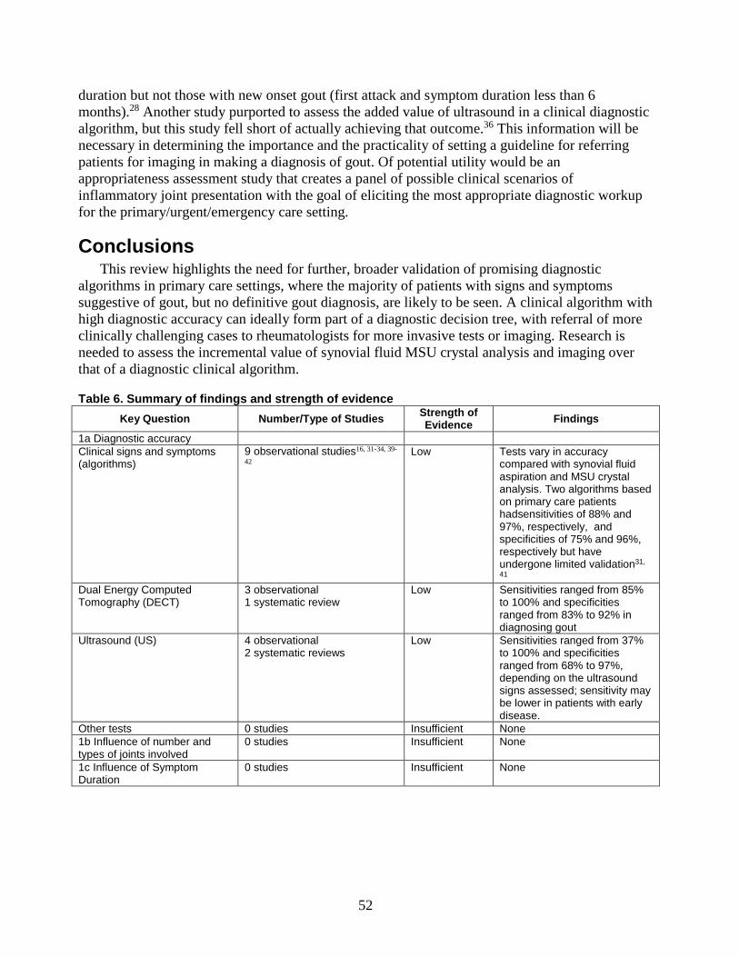
52
duration but not those with new onset gout (first attack and symptom duration less than 6 months).28 Another study purported to assess the added value of ultrasound in a clinical diagnostic algorithm, but this study fell short of actually achieving that outcome.36 This information will be necessary in determining the importance and the practicality of setting a guideline for referring patients for imaging in making a diagnosis of gout. Of potential utility would be an appropriateness assessment study that creates a panel of possible clinical scenarios of inflammatory joint presentation with the goal of eliciting the most appropriate diagnostic workup for the primary/urgent/emergency care setting.
Conclusions This review highlights the need for further, broader validation of promising diagnostic
algorithms in primary care settings, where the majority of patients with signs and symptoms suggestive of gout, but no definitive gout diagnosis, are likely to be seen. A clinical algorithm with high diagnostic accuracy can ideally form part of a diagnostic decision tree, with referral of more clinically challenging cases to rheumatologists for more invasive tests or imaging. Research is needed to assess the incremental value of synovial fluid MSU crystal analysis and imaging over that of a diagnostic clinical algorithm.
Table 6. Summary of findings and strength of evidence Key Question Number/Type of Studies Strength of
Evidence Findings
1a Diagnostic accuracy Clinical signs and symptoms (algorithms)
9 observational studies16, 31-34, 39-
42 Low Tests vary in accuracy
compared with synovial fluid aspiration and MSU crystal analysis. Two algorithms based on primary care patients hadsensitivities of 88% and 97%, respectively, and specificities of 75% and 96%, respectively but have undergone limited validation31,
41 Dual Energy Computed Tomography (DECT)
3 observational 1 systematic review
Low Sensitivities ranged from 85% to 100% and specificities ranged from 83% to 92% in diagnosing gout
Ultrasound (US) 4 observational 2 systematic reviews
Low Sensitivities ranged from 37% to 100% and specificities ranged from 68% to 97%, depending on the ultrasound signs assessed; sensitivity may be lower in patients with early disease.
Other tests 0 studies Insufficient None 1b Influence of number and types of joints involved
0 studies Insufficient None
1c Influence of Symptom Duration
0 studies Insufficient None

53
Key Question Number/Type of Studies Strength of Evidence Findings
1d. Influence of factors on analysis of monosodium urate crystals (MSU)
2 observational studies 1 systematic review
Insufficient Agreement among personnel examining synovial fluid using polarizing microscopy for detection of MSU crystals appears to be poor, but the role of training and experience are unclear. No studies examined the effect of the type of practitioner performing fluid aspiration on the ability to obtain a sample
2. Adverse events and implications of misdiagnosis
2 observational studies: 1 on AEs associated with two diagnostic methods and 1 on implications of misdiagnosis
Low One study reported DECT and joint aspiration for MSU analysis were associated with no adverse events.
Implications of misdiagnosis 1 observational study on implications of misdiagnosis
Insufficient One study reported that missed diagnosis of gout resulted in longer hospital stays, unnecessary surgery, and delayed pharmacological treatment.
AE = adverse event; DECT = dual-energy computed tomography; MSU = monosodium urate; US = ultrasound

54
References 1. Zhu Y, Pandya BJ, Choi HK. Prevalence of
gout and hyperuricemia in the US general population: the National Health and Nutrition Examination Survey 2007-2008. Arthritis Rheum. 2011 Oct;63(10):3136-41. PMID: 21800283.
2. Li C, Martin BC, Cummins DF, et al. Ambulatory resource utilization and cost for gout in United States. Am J Pharm Benefits. 2013 March/April;5(2):e46-e54. PMID:
3. Doghramji PP, Wortmann RL. Hyperuricemia and gout: new concepts in diagnosis and management. Postgrad Med. 2012 Nov;124(6):98-109. PMID: 23322143.
4. Wallace SL. Hyperuricemia in the diagnosis of gout. J Gen Intern Med. 1989 Mar-Apr;4(2):178-9. PMID: 2651605.
5. Singh JA, Reddy SG, Kundukulam J. Risk factors for gout and prevention: a systematic review of the literature. Curr Opin Rheumatol. 2011 Mar;23(2):192-202. PMID: 21285714.
6. Hamburger M, Baraf HS, Adamson TC 3rd, et al. 2011 Recommendations for the diagnosis and management of gout and hyperuricemia. Postgrad Med. 2011 Nov;123(6 Suppl 1):3-36. PMID: 22156509.
7. Harrold LR, Mazor KM, Negron A, et al. Primary care providers' knowledge, beliefs and treatment practices for gout: results of a physician questionnaire. Rheumatology (Oxford). 2013 Sep;52(9):1623-9. PMID: 23620554.
8. Owens D, Whelan B, McCarthy G. A survey of the management of gout in primary care. Ir Med J. 2008;101(5):147-9. PMID: 18624262.
9. Khosla S, Thiele R, Baumhauer JF. Ultrasound guidance for intra-articular injections of the foot and ankle. Foot Ankle Int. 2009 Sep;30(9):886-90. PMID: 19755074.
10. Hasselbacher P. Variation in synovial fluid analysis by hospital laboratories. Arthritis and Rheumatism. 1987;30(6):637-42. PMID: 3606682.
11. Schumacher HR, Jr., Sieck MS, Rothfuss S, et al. Reproducibility of synovial fluid analyses. A study among four laboratories. Arthritis Rheum. 1986 Jun;29(6):770-4. PMID: 3718565.
12. McGill NW, York HF. Reproducibility of synovial fluid examination for crystals. Aust N Z J Med. 1991 Oct;21(5):710-3. PMID: 1759919.
13. Segal JB, Albert D. Diagnosis of crystal-induced arthritis by synovial fluid examination for crystals: lessons from an imperfect test. Arthritis Care Res. 1999 Dec;12(6):376-80. PMID: 11081008.
14. Graf SW, Buchbinder R, Zochling J, et al. The accuracy of methods for urate crystal detection in synovial fluid and the effect of sample handling: a systematic review. Clin Rheumatol. 2013 Feb;32(2):225-32. PMID: 23138881.
15. Perez-Ruiz F, Carmona L, Yebenes MJG, et al. An audit of the variability of diagnosis and management of gout in the rheumatology setting: the Gout Evaluation and Management Study. J Clin Rheumatol. 2011 Oct;17(7):349-55. PMID: 21946457.
16. Malik A, Schumacher HR, Dinnella JE, et al. Clinical diagnostic criteria for gout: comparison with the gold standard of synovial fluid crystal analysis. J Clin Rheumatol. 2009 Feb;15(1):22-4. PMID: 19125136.
17. Dalbeth N, Fransen J, Jansen TL, et al. New classification criteria for gout: a framework for progress. Rheumatology (Oxford). 2013 Oct;52(10):1748-53. PMID: 23611919.
18. Glazebrook KN, Guimaraes LS, Murthy NS, et al. Identification of intraarticular and periarticular uric acid crystals with dual-energy CT: initial evaluation. Radiology. 2011 Nov;261(2):516-24. PMID: 21926378.

55
19. Khanna D, Fitzgerald JD, Khanna PP, et al. 2012 American College of Rheumatology guidelines for management of gout. Part 1: systematic nonpharmacologic and pharmacologic therapeutic approaches to hyperuricemia. Arthritis Care Res (Hoboken). 2012;64(10):1431-46. PMID: 23024028.
20. Khanna D, Khanna PP, Fitzgerald JD, et al. 2012 American College of Rheumatology guidelines for management of gout. Part 2: therapy and antiinflammatory prophylaxis of acute gouty arthritis. Arthritis Care Res (Hoboken). 2012;64(10):1447-61. PMID: 23024029.
21. Whiting P, Westwood M, Beynon R, et al. Inclusion of methodological filters in searches for diagnostic test accuracy studies misses relevant studies. J Clin Epidemiol. 2011 Jun;64(6):602-7. PMID: 21075596.
22. Zhang W, Doherty M, Pascual E, et al. EULAR evidence based recommendations for gout. Part I: diagnosis. Report of a task force of the Standing Committee for International Clinical Studies Including Therapeutics (ESCISIT). Ann Rheum Dis. 2006 Oct;65(10):1301-11. PMID: 16707533.
23. Whiting PF, Rutjes AW, Westwood ME, et al. QUADAS-2: a revised tool for the quality assessment of diagnostic accuracy studies. Ann Intern Med. 2011 Oct 18;155(8):529-36. PMID: 22007046.
24. Santaguida PL, Riley CM, Matchar DB. Assessing risk of bias as a domain of quality in medical test studies. In: Methods Guide for Medical Test Reviews. AHRQ Publication No. 12-EHC017. Rockville, MD: Agency for Healthcare Research and Quality; June 2012. Chapters available at www.effectivehealthcare.ahrq.gov/reports/final.cfm.
25. Shea BJ, Grimshaw JM, Wells GA, et al. Development of AMSTAR: a measurement tool to assess the methodological quality of systematic reviews. BMC Med Res Methodol. 2007;7:10. PMID: 17302989.
26. Methods Guide for Medical Test Reviews. AHRQ Publication No. 12-EHC017. Agency for Healthcare Research and Quality. Rockville, MD: Agency for Healthcare Research and Quality; June 2012. Chapters available at www.effectivehealthcare.ahrq.gov/reports/final.cfm.
27. Atkins D, Chang S, Gartlehner G, et al. Assessing the applicability of studies when comparing medical interventions. In: Methods Guide for Effectiveness and Comparative Effectiveness Reviews. AHRQ Publication No. 10(14)-EHC063-EF. Rockville, MD: Agency for Healthcare Research and Quality. January 2014. Chapters available at www.effectivehealthcare.ahrq.gov.
28. Bongartz T, Glazebrook KN, Kavros SJ, et al. Dual-energy CT for the diagnosis of gout: an accuracy and diagnostic yield study. Ann Rheum Dis. 2015 Jun;74(6):1072-7. PMID: 24671771.
29. Gordon C, Swan A, Dieppe P. Detection of crystals in synovial fluids by light microscopy: sensitivity and reliability. Ann Rheum Dis. 1989 Sep;48(9):737-42. PMID: 2478085.
30. Huppertz A, Hermann KG, Diekhoff T, et al. Systemic staging for urate crystal deposits with dual-energy CT and ultrasound in patients with suspected gout. Rheumatol Int. 2014 Jun;34(6):763-71. PMID: 24619560.
31. Janssens HJEM, Fransen J, Van De Lisdonk EH, et al. A diagnostic rule for acute gouty arthritis in primary care without joint fluid analysis. Arch Intern Med. 2010;170(13):1120-6. PMID: 20625017.
32. Janssens HJEM, Janssen M, Van De Lisdonk EH, et al. Limited validity of the American College of Rheumatology criteria for classifying patients with gout in primary care. Ann Rheum Dis. 2010;69(6):1255-6. PMID: 19910298.
33. Kienhorst LB, Janssens HJ, Fransen J, et al. The validation of a diagnostic rule for gout without joint fluid analysis: a prospective study. Rheumatology (Oxford). 2015 Apr;54(4):609-14. PMID: 25231179.

56
34. Kienhorst LB, Janssens HJ, Fransen J, et al. Arthritis of the first metatarsophalangeal joint is not always gout: a prospective cohort study in primary care patients. Joint Bone Spine. 2014 Jul;81(4):342-6. PMID: 24468668.
35. Lai KL, Chiu YM. Role of ultrasonography in diagnosing gouty arthritis. J Med Ultrasound. 2011 March;19(1):7-13. http://dx.doi.org/10.1016/j.jmu.2011.01.003.
36. Lamers-Karnebeek FB, Van Riel PL, Jansen TL. Additive value for ultrasonographic signal in a screening algorithm for patients presenting with acute mono-/oligoarthritis in whom gout is suspected. Clin Rheumatol. 2014 Apr;33(4):555-9. PMID: 24510062.
37. Park JW, Ko DJ, Yoo JJ, et al. Clinical factors and treatment outcomes associated with failure in the detection of urate crystal in patients with acute gouty arthritis. Korean J Intern Med. 2014;29(3):361-9. PMID: 24851071..
38. Rettenbacher T, Ennemoser S, Weirich H, et al. Diagnostic imaging of gout: comparison of high-resolution US versus conventional X-ray. Eur Radiol. 2008 Mar;18(3):621-30. PMID: 17994238.
39. Richette P, Clerson P, Bouee S, et al. Identification of patients with gout: elaboration of a questionnaire for epidemiological studies. Ann Rheum Dis. 2014 May 5. PMID: 24796335.
40. Taylor WJ, Fransen J, Dalbeth N, et al. Performance of classification criteria for gout in early and established disease. Ann Rheum Dis. 2014 Oct 28. PMID: 25351521.
41. Vazquez-Mellado J, Hernandez-Cuevas CB, Alvarez-Hernandez E, et al. The diagnostic value of the proposal for clinical gout diagnosis (CGD). Clin Rheumatol. 2012 Mar;31(3):429-34. PMID: 21979446.
42. Wallace SL, Robinson H, Masi AT, et al. Preliminary criteria for the classification of the acute arthritis of primary gout. Arthritis Rheum. 1977 Apr;20(3):895-900. PMID: 856219.
43. Chowalloor PV, Keen HI. A systematic review of ultrasonography in gout and asymptomatic hyperuricaemia. Ann Rheum Dis. 2013 May;72(5):638-45. PMID: 23291387.
44. Dirken-Heukensfeldt K, Teunissen TAM, van de Lisdonk EH, et al. "Clinical features of women with gout arthritis." A systematic review. Clinical Rheumatology. 2010 Jun;29(6):575-82. PMID: 20084441.
45. Ogdie A, Taylor W, Weatherall M, et al. Imaging modalities for the classification of gout: systematic literature review and meta-analysis. Ann Rheum Dis. 2014 Jun 20. [Epub ahead of print.] PMID: 24915980.
46. Sivera F, Andres M, Falzon L, et al. Diagnostic value of clinical, laboratory, and imaging findings in patients with a clinical suspicion of gout: a systematic literature review. J Rheumatol Suppl. 2014 Sep;92:3-8. PMID: 25180122.
47. Sivera F, Andres M, Carmona L, et al. Multinational evidence-based recommendations for the diagnosis and management of gout: integrating systematic literature review and expert opinion of a broad panel of rheumatologists in the 3e initiative. Ann Rheum Dis. 2014 Feb;73(2):328-35. PMID: 23868909.
48. Berendsen D, Jansen TL, Taylor W, et al. A critical appraisal of the competence of crystal identification by rheumatologists. Ann Rheum Dis. 2014;72(Suppl 3):A981-2.
49. Hamburger MI, Tesser JRP, Skosey JL, et al. Patterns of gout treatment and related outcomes in US community rheumatology practices: the relation between gout flares, time in treatment, serum uric acid level and urate lowering therapy. Arthritis and Rheumatism. 2012;64(Suppl 10):1904.
50. Richette P, Pascual E, Doherty M, et al. Updated EULAR evidence-based recommendations for the diagnosis of gout. Ann Rheum Dis. 2014;(Suppl 2):783-84.
51. Dalbeth N. Management of gout in primary care: challenges and potential solutions. Rheumatology (Oxford). 2013 Sep;52(9):1549-50. PMID: 23771950.
52. Rigby AS, Wood PH. Serum uric acid levels and gout: what does this herald for the population? Clin Exp Rheumatol. 1994 Jul-Aug;12(4):395-400. PMID: 7955603.
57
53. Lenski M, Scherer MA. Analysis of synovial inflammatory markers to differ infectious from gouty arthritis. Clin Biochem. 2014 Jan;47(1-2):49-55. PMID: 24177196.
54. Wang C, Zhong D, Liao Q, et al. Procalcitonin levels in fresh serum and fresh synovial fluid for the differential diagnosis of knee septic arthritis from rheumatoid arthritis, osteoarthritis and gouty arthritis. Exp Ther Med. 2014 Oct;8(4):1075-80. PMID: 25187799.

58
Abbreviations/Acronyms
3e
Evidence, Expertise, Exchange
3ei Evidence, Expertise, Exchange initiative ACA American College of Rheumatology ACP American College of Physicians AHRQ Agency for Healthcare Research and Quality ARA American Rheumatism Association (formerly American College of
Rheumatology) AUC Area under the curve BSF Bright stippled foci CART Classification and regression tree CGD Clinical Gout Disease CI Confidence interval CPPD Calcium Pyrophosphate Deposition Disease (formerly pseudogout) CT (Scan) Computed tomography CVD Cardiovascular disease DCS Double-Contour Sign DECT Dual-energy computed tomography EPC California Evidence-based Practice Center FN False negative FP False positive HTN Hypertension KQ Key Question LDH Lactate dehydrogenase LR Likelihood ratio LRN Negative likelihood ratio LRP Positive likelihood ratio MRI Magnetic resonance imaging MSU Monosodium urate MTP Metatarsophalangeal NHANES National Health and Nutrition Examination Survey NLR Nonlikelihood ratio NPV Negative predictive value NSAIDS Nonsteroidal anti-inflammatory drugs NY New York OA Osteoarthritis PCPs Primary care physicians PICOTS Populations, Interventions, Comparators, Outcomes, and Timing PLM Polarized light microscopy PLR Positive likelihood ratio

59
PPV Positive predictive value RA Rheumatoid arthritis ROC Receiver-operating characteristics SA Septic arthritis SCEPC Southern California Evidence-based Practice Center SFWBC Synovial Fluid White Blood Cell Count SpA Spondyloarthritis SRs Systematic Reviews sUA Serum uric acid TEP Technical Expert Panel US Ultrasound US DHHS U.S. Department of Health and Human Services VA Veterans Administration
A-1
Appendix A. Search Strategy PubMed DATABASE SEARCHED & TIME PERIOD COVERED: PubMed - ~1946 - 11/7/2014 SEARCH STRATEGY: gout OR gouty AND X-ray* OR radiograph* OR erosion OR diagnostic imaging[mh] OR radiography [Subheading] OR Computed tomography OR Computer tomography OR dual energy CT OR DECT OR Radiography, Dual-Energy Scanned Projection[mh] OR Ultrasound OR Ultrasonography[mh] OR Ultrasonography[sh] OR double contour OR radionuclide imaging [Subheading] OR (polariz* AND microscop*) OR Joint aspiration OR Serum urate OR Uric acid OR Crystal* OR Tophi OR tophus OR tophaceous OR Synovial fluid OR Urate OR kidney stones OR Kidney Calculi[mh] OR urate stones OR gouty nephropathy OR Hyperuricemia OR clinical symptom* AND Accura* OR Sensitivity and specificity[mh] OR Sensitivity[tiab] OR Specificity[tiab] OR False positive reactions[mh] OR false positive* OR False negative reactions[mh] OR False negative* OR Predictive value OR predictive value of tests[mh] OR Distinguish* OR Differential* OR Identif* OR Detect* OR valid* OR reliab* OR reproducibility of results Web of Science SCI-EXPANDED, SSCI, A&HCI, CPCI-S, CPCI-SSH DATABASE SEARCHED & TIME PERIOD COVERED: Web of Science SCI-EXPANDED, SSCI, A&HCI, CPCI-S, CPCI-SSH – 1/1/1980 - 11/7/2014 SEARCH STRATEGY: ts=(gout OR gouty) AND ts=(x-ray* or radiograph* or erosion or diagnostic imaging or Computed tomography or Computer tomography or dual energy CT or DECT or Ultrasound or Ultrasonograph* or double contour or radionuclide or (polariz* and microscop*) or Joint aspiration or Serum urate or Uric acid or Crystal* or Tophi or tophus or tophaceous or Synovial fluid or Urate or kidney stones or Kidney Calculi or urate stones or gouty nephropathy or Hyperuricemia or clinical symptom*) AND ts=(Accura* or Sensitivity or Specificity or false positive* or False negative* or Predictive value or Distinguish* or Differential* or Identif* or Detect* or valid* or reliab* or reproducibility of results)
A-2
Cochrane DATABASE SEARCHED & TIME PERIOD COVERED: Cochrane – Earliest - 11/7/2014 SEARCH STRATEGY: gout or gouty AND x-ray* or radiograph* or erosion or diagnostic imaging or Computed tomography or Computer tomography or dual energy CT or DECT or Ultrasound or Ultrasonograph* or double contour or radionuclide or (polariz* and microscop*) or Joint aspiration or Serum urate or Uric acid or Crystal* or Tophi or tophus or tophaceous or Synovial fluid or Urate or kidney stones or Kidney Calculi or urate stones or gouty nephropathy or Hyperuricemia or clinical symptom* IN TITLE, ABSTRACT, KEYWORD AND Accura* or Sensitivity or Specificity or false positive* or False negative* or Predictive value or Distinguish* or Differential* or Identif* or Detect* or valid* or reliab* or reproducibility of results Embase DATABASE SEARCHED & TIME PERIOD COVERED: Embase 1/1/1972-11/7/14 LIMITERS: Humans SEARCH STRATEGY: 'gout' OR 'gout'/exp OR gout OR gouty AND 'x ray'/exp OR 'x ray' OR 'x rays'/exp OR 'x rays' OR 'x-ray'/exp OR 'x-ray' OR 'x-rays'/exp OR 'x-rays' OR radiograph* OR 'erosion'/exp OR erosion OR 'diagnostic imaging'/exp OR 'diagnostic imaging' OR 'computed tomography'/exp OR 'computed tomography' OR 'computer tomography'/exp OR 'computer tomography' OR 'dual energy ct' OR dect OR 'ultrasound'/exp OR ultrasound OR ultrasonograph* OR 'double contour' OR 'radionuclide'/exp OR radionuclide OR microscop* OR 'joint aspiration'/exp OR 'joint aspiration' OR 'serum urate'/exp OR 'serum urate' OR 'uric acid'/exp OR 'uric acid' OR crystal* OR tophi OR tophus OR tophaceous OR 'synovial fluid'/exp OR 'synovial fluid' OR 'urate'/exp OR urate OR ('kidney'/exp OR kidney AND stones) OR ('kidney'/exp OR kidney AND ('calculi'/exp OR calculi)) OR 'urate stones'/exp OR 'urate stones' OR 'gouty nephropathy' OR 'hyperuricemia'/exp OR hyperuricemia OR 'hyperuricaemia'/exp OR hyperuricaemia OR 'clinical symptom' OR 'clinical symptoms' AND accura* OR sensitivity OR specificity OR 'false positive' OR 'false positives' OR 'false negative' OR 'false negatives' OR 'predictive value'/exp OR 'predictive value' OR distinguish* OR differential* OR identif* OR detect* OR valid* OR reliab* OR 'reproducibility of results'/exp OR 'reproducibility of results'

A-3
Web of Science DATABASE SEARCHED & TIME PERIOD COVERED: Web of Science – 1/1/2006-11/7/2014 SEARCH STRATEGY: Forward search on the following article: EULAR evidence based recommendations for gout. Part I: Diagnosis. Report of a task force of the standing committee for international clinical studies including therapeutics (ESCISIT) W Zhang, M Doherty, E Pascual, T Bardin, V Barskova, P Conaghan, J Gerster, J Jacobs, B Leeb, F Liote´, G McCarthy, P Netter, G Nuki, F Perez-Ruiz, A Pignone, J Pimenta˜o, L Punzi, E Roddy, T Uhlig, I Zimmermann-Go` rska Ann Rheum Dis 2006;65:1301–1311. doi: 10.1136/ard.2006.055251 SCOPUS DATABASE SEARCHED & TIME PERIOD COVERED: SCOPUS – 1/1/2006-11/7/2014 SEARCH STRATEGY: Forward search on the following article: EULAR evidence based recommendations for gout. Part I: Diagnosis. Report of a task force of the standing committee for international clinical studies including therapeutics (ESCISIT) W Zhang, M Doherty, E Pascual, T Bardin, V Barskova, P Conaghan, J Gerster, J Jacobs, B Leeb, F Liote´, G McCarthy, P Netter, G Nuki, F Perez-Ruiz, A Pignone, J Pimenta˜o, L Punzi, E Roddy, T Uhlig, I Zimmermann-Go` rska Ann Rheum Dis 2006;65:1301–1311. doi: 10.1136/ard.2006.055251 Grey Literature Report DATABASE SEARCHED & TIME PERIOD COVERED: Grey Literature Report – no date limitation SEARCH STRATEGY: Gout OR gouty NUMBER OF RESULTS: 0 Embase (Rerun) DATABASE SEARCHED & TIME PERIOD COVERED: Embase (rerun of 4/22/2014 search) – 1/1/1972-11/7/2014 LIMITERS: Humans SEARCH STRATEGY:

A-4
'gout' OR 'gout'/exp OR gout OR gouty AND 'x ray'/exp OR 'x ray' OR 'x rays'/exp OR 'x rays' OR 'x-ray'/exp OR 'x-ray' OR 'x-rays'/exp OR 'x-rays' OR radiograph* OR 'erosion'/exp OR erosion OR 'diagnostic imaging'/exp OR 'diagnostic imaging' OR 'computed tomography'/exp OR 'computed tomography' OR 'computer tomography'/exp OR 'computer tomography' OR 'dual energy ct' OR dect OR 'ultrasound'/exp OR ultrasound OR ultrasonograph* OR 'double contour' OR 'radionuclide'/exp OR radionuclide OR microscop* OR 'joint aspiration'/exp OR 'joint aspiration' OR 'serum urate'/exp OR 'serum urate' OR 'uric acid'/exp OR 'uric acid' OR crystal* OR tophi OR tophus OR tophaceous OR 'synovial fluid'/exp OR 'synovial fluid' OR 'urate'/exp OR urate OR ('kidney'/exp OR kidney AND stones) OR ('kidney'/exp OR kidney AND ('calculi'/exp OR calculi)) OR 'urate stones'/exp OR 'urate stones' OR 'gouty nephropathy' OR 'hyperuricemia'/exp OR hyperuricemia OR 'hyperuricaemia'/exp OR hyperuricaemia OR 'clinical symptom' OR 'clinical symptoms' AND accura* OR sensitivity OR specificity OR 'false positive' OR 'false positives' OR 'false negative' OR 'false negatives' OR 'predictive value'/exp OR 'predictive value' OR distinguish* OR differential* OR identif* OR detect* OR valid* OR reliab* OR 'reproducibility of results'/exp OR 'reproducibility of results' CliicalTrials.gov CLINICALTRIALS.GOV CONDITION = "Gout" RECEIVED FROM: Earliest in database to 11/7//2014

B-1
Appendix B. List of Excluded Studies Not Human – N=2 1. Sundaram PV, Igloi MP, Wassermann R, et al. Immobilized-enzyme nylon-tube reactor for routine determination of uric acid in serum. Clin Chem. 1978 Oct;24(10):1813-7. PMID: 699291. 2. Takanabe A, Tanaka M, Taniguchi A, et al. Quantitative analysis with advanced compensated polarized light microscopy on wavelength dependence of linear birefringence of single crystals causing arthritis. Journal of Physics D-Applied Physics. 2014 Jul;47(28)PMID: WOS:000338721900011.
Diagnostic Method Beyond Scope of Review- N=44
1. Backhaus M, Schmidt WA, Mellerowicz H, et al. [Technical aspects and value of arthrosonography in rheumatologic diagnosis. 4: Ultrasound of the elbow]. Z Rheumatol. 2002 Aug;61(4):415-25. PMID: 12426847. 2. Barber C, Thompson K, Hanly JG. Impact of a rheumatology consultation service on the diagnostic accuracy and management of gout in hospitalized patients. J Rheumatol. 2009 Aug;36(8):1699-704. PMID: 19567626. 3. Chauhan N, Pundir CS. An amperometric uric acid biosensor based on multiwalled carbon nanotube-gold nanoparticle composite. Anal Biochem. 2011 Jun 15;413(2):97-103. PMID: 21315682. 4. Cheng X, Haggins DG, York RH, et al. Analysis of crystals leading to joint arthropathies by Raman spectroscopy: comparison with compensated polarized imaging. Appl Spectrosc. 2009 Apr;63(4):381-6. PMID: 19366502. 5. Chu QC, Lin M, Geng CH, et al. Determination of uric acid in human saliva and urine using miniaturized capillary electrophoresis with amperometric detection. Chromatographia. 2007 Feb;65(3-4):179-84. PMID: WOS:000244670700007. 6. Dodd LG, Major NM. Fine-needle aspiration cytology of articular and periarticular lesions. Cancer. 2002 Jun 25;96(3):157-65. PMID: 12115304. 7. Friedman RJ, Hawthorne KB, Genez BM. The use of computerized tomography in the measurement of glenoid version. J Bone Joint Surg Am. 1992 Aug;74(7):1032-7. PMID: 1522089. 8. Galvez J, Saiz E, Linares LF, et al. Delayed examination of synovial fluid by ordinary and polarised light microscopy to detect and identify crystals. Ann Rheum Dis. 2002 May;61(5):444-7. PMID: 11959769.

B-2
9. Goldenberg DL, Cohen AS. Synovial membrane histopathology in the differential diagnosis of rheumatoid arthritis, gout, pseudogout, systemic lupus erythematosus, infectious arthritis and degenerative joint disease. Medicine (Baltimore). 1978 May;57(3):239-52. PMID: 642792. 10. Heidelmann G, Thiele P, Crasselt C, et al. [Uncharacteristic symptoms of the motor apparatus in gout]. Z Rheumatol. 1975 Nov-Dec;34(11-12):381-90. PMID: 1210793. 11. Hujazi I, Ambler G, Arora A, et al. Role of Newman's classification in predicting outcomes in patients with crystal arthritis. International Orthopaedics. 2012 Jun;36(6):1287-90. PMID: WOS:000304160000027. 12. Ivorra J, Rosas J, Pascual E. Most calcium pyrophosphate crystals appear as non-birefringent. Ann Rheum Dis. 1999 Sep;58(9):582-4. PMID: 10460193. 13. Johnson JS, Freemont AJ. A 10 year retrospective comparison of the diagnostic usefulness of synovial fluid and synovial biopsy examination. Journal of Clinical Pathology. 2001;54(8):605-7. 14. Kaneko K, Maru M. Determination of urate crystal formation using flow cytometry and microarea X-ray diffractometry. Anal Biochem. 2000 May 15;281(1):9-14. PMID: 10847604. 15. Klaus R, Fischer W, Hauck HE. Qualitative and Quantitative-Analysis of Uric-Acid, Creatine and Creatinine Together with Carbohydrates in Biological-Material by Hptlc. Chromatographia. 1991 Oct;32(7-8):307-16. PMID: WOS:A1991GP25100001. 16. Koski JM, Hermunen HS, Kilponen VM, et al. Verification of palpation-guided intra-articular injections using glucocorticoid-air-saline mixture and ultrasound imaging (GAS-graphy). Clinical and Experimental Rheumatology. 2006;24(3):247-52. 17. Kratky G, Hefel P. [Differential diagnosis of knee joint fluid. Enzymatic analysis (author's transl)]. Arch Orthop Trauma Surg. 1978 Dec 12;93(1):65-73. PMID: 727932. 18. Lenz W, Klein W, Huth F. [Needle biopsy in gout and pseudogout (author's transl)]. Beitr Pathol. 1976 Apr;157(2):161-82. PMID: 179523. 19. Li X, Wang X, Yu Y, et al. Value of dual energy spectral CT for detecting uric acid deposition in tophaceous gout with base material mapping. Chinese Journal of Radiology (China). 2014;48(4):303-7. 20. Lichtenstein MJ, Pincus T. How useful are combinations of blood tests in "rheumatic panels" in diagnosis of rheumatic diseases? J Gen Intern Med. 1988 Sep-Oct;3(5):435-42. PMID: 3262732. 21. Lin J, Zhao GQ, Che CY, et al. Characteristics of ocular abnormalities in gout patients. Int J Ophthalmol. 2013;6(3):307-11. PMID: 23826523.
B-3
22. Lin X, Wang YF, Sun JY, et al. Determination of uric acid in human plasma and urine by microemulsion electrokinetic chromatography. Analytical Methods. 2013;5(19):5201-7. PMID: WOS:000324489100035. 23. Liu Y, Yu P, Sun X, et al. Metabolite target analysis of human urine combined with pattern recognition techniques for the study of symptomatic gout. Mol Biosyst. 2012 Nov;8(11):2956-63. PMID: 22932763. 24. Matsubara C, Yokoi Y, Nakamichi N, et al. [Spectrophotometric determination of uric acid in serum using a titanium (IV)-porphyrin complex]. Yakugaku Zasshi. 1994 Jan;114(1):48-53. PMID: 8133459. 25. McGill NW, McGill VG. Quality assurance for synovial fluid examination for crystals: an improved method. Ann Rheum Dis. 1997 Aug;56(8):504-6. PMID: 9306876. 26. Medellin MV, Erickson AR, Enzenauer RJ. Variability of treatment for gouty arthritis between rheumatologists and primary care physicians. J Clin Rheumatol. 1997 Feb;3(1):24-7. PMID: 19078113. 27. Menghini S, Della Corte E. [Evaluation of hyperuricemia caused by fructose in a status of altered uric acid metabolism]. Quad Sclavo Diagn. 1987 Dec;23(4):441-6. PMID: 3509875. 28. Nasonova VA, Zakharova MM, Barskova VG, et al. [Detection of sodium monourate crystals in biopsies of gastric mucosa in patients with gout]. Ter Arkh. 2004;76(6):47-51. PMID: 15332576. 29. Papanicolas LE, Hakendorf P, Gordon DL. Concomitant Septic Arthritis in Crystal Monoarthritis. Journal of Rheumatology. 2012 Jan;39(1):157-60. PMID: WOS:000299400500028. 30. Pascual E, Tovar J, Ruiz MT. The ordinary light microscope: An appropriate tool for provisional detection and identification of crystals in synovial fluid. Annals of the Rheumatic Diseases. 1989;48(12):983-5. 31. Piliaev VG, Petrov VP, Pikhlak EG, et al. [The importance of arthroscopy and guided biopsy of the knee joint in gout]. Revmatologiia (Mosk). 1989 Oct-Dec(4):15-8. PMID: 2637459. 32. Pobirci O, Bogdan F, Pobirci DD, et al. The study of synovities with articular inflammatory liquid, through clinical-statistical, histological and immunohistochemical methods. Rom J Morphol Embryol. 2011;52(1 Suppl):333-8. PMID: 21424072. 33. Schlesinger N. Response to application of ice may help differentiate between gouty arthritis and other inflammatory arthritides. J Clin Rheumatol. 2006 Dec;12(6):275-6. PMID: 17149056.

B-4
34. Shidham V, Chivukula M, Basir Z, et al. Evaluation of crystals in formalin-fixed, paraffin-embedded tissue sections for the differential diagnosis of pseudogout, gout, and tumoral calcinosis. Mod Pathol. 2001 Aug;14(8):806-10. PMID: 11504841. 35. Sivera F, Aragon R, Pascual E. First metatarsophalangeal joint aspiration using a 29-gauge needle. Ann Rheum Dis. 2008 Feb;67(2):273-5. PMID: 17557892. 36. Song JS, Choi ST, Park EH. The usefulness of procalcitonin for differentiating acute gout arthritis from infectious disease. Annals of the Rheumatic Diseases. 2014;73. 37. Strobel G, Schwarz R, Heppt P, et al. [Use of an improved transmission ultrasound camera for diagnosis of rheumatic joint diseases]. Z Rheumatol. 1993 Mar-Apr;52(2):114-20. PMID: 8517073. 38. Vasil'ev A, Obramenko IE. [Magnetic resonance imaging differential diagnosis of psoriatic polyarthropathy with gouty polyarthropathy and rheumatoid polyarthritis]. Vestn Rentgenol Radiol. 2013 Jan-Feb(1):29-33. PMID: 23700923. 39. Yamakita J, Yamamoto T, Moriwaki Y, et al. Effect of urine storage on urinary uric acid concentrations. Ann Clin Biochem. 2000 May;37 ( Pt 3):355-9. PMID: 10817251. 40. Yin L, Zhu J, Xue Q, et al. Micropure imaging for the evaluation of microcalcifications in gouty arthritis involving the first metatarsophalangeal joint: A Preliminary Study. PLoS ONE. 2014;9(5). 41. Yu MX, He ZG, Zhang NZ. [Analysis of knee arthroscopic findings in rheumatic diseases]. Zhonghua Nei Ke Za Zhi. 1990 Dec;29(12):713-6, 64. PMID: 2092955. 42. Zamani B, Jamali R, Ehteram H. Synovial fluid adenosine deaminase and high-sensitivity C-reactive protein activity in differentiating monoarthritis. Rheumatol Int. 2012 Jan;32(1):183-8. PMID: 20721560. 43. Zborovskii AB, Stazharov M, Martem'ianov VF. [Purine metabolism enzymes in diagnosis and differential diagnosis of osteoarthritis and gout arthritis]. Ter Arkh. 2000;72(4):21-4. PMID: 10833792. 44. Zsernaviczky J, Dressler D. [Gout, pseudo-gout or hyperlipoproteinemia? (author's transl)]. Z Orthop Ihre Grenzgeb. 1976 Apr;114(2):243-7. PMID: 1274401.
B-5
Not Gout Diagnosis or Management or Does Not Address Key Question - N=69 1. Antommattei O, Schumacher HR, Reginato AJ, et al. Prospective study of morphology and phagocytosis of synovial fluid monosodium urate crystals in gouty arthritis. J Rheumatol. 1984 Dec;11(6):741-4. PMID: 6520827. 2. Artmann A, Ratzenbock M, Noszian I, et al. [Dual energy CT--a new perspective in the diagnosis of gout]. Rofo. 2010 Mar;182(3):261-6. PMID: 19862652. 3. Bardin T. Epidemiology of gout. Épidémiologie de la goutte. 2007;74(2):147-9. 4. Blaauw AAM, Schuwirth LWT, Vandervleuten CPM, et al. Assessing Clinical Competence - Recognition of Case Descriptions of Rheumatic Diseases by General-Practitioners. British Journal of Rheumatology. 1995 Apr;34(4):375-9. PMID: WOS:A1995RC64700016. 5. Choi HK, Burns LC, Shojania K, et al. Dual energy CT in gout: a prospective validation study. Ann Rheum Dis. 2012 Sep;71(9):1466-71. PMID: 22387729. 6. Craig MH, Poole GV, Hauser CJ. Postsurgical gout. Am Surg. 1995 Jan;61(1):56-9. PMID: 7832383. 7. Dalbeth N, Clark B, Gregory K, et al. Computed tomography measurement of tophus volume: comparison with physical measurement. Arthritis Rheum. 2007 Apr 15;57(3):461-5. PMID: 17394233. 8. de Avila Fernandes E, Kubota ES, Sandim GB, et al. Ultrasound features of tophi in chronic tophaceous gout. Skeletal Radiol. 2011 Mar;40(3):309-15. PMID: 20676636. 9. Dihlmann W, Dihlmann A. [Osteoclastic finger arthrosis--a subtype of hand polyarthrosis]. Rofo. 1998 Feb;168(2):128-32. PMID: 9519043. 10. Dixit AK, Dey RK, Panda AK, et al. Biochemical and serological profiling of sandhi shoola (Arthralgia) patients of ayurveda hospital. International Journal of Research in Ayurveda and Pharmacy. 2013 March/April;4(2):141-4. PMID: 2013400880 FULL TEXT LINK http://dx.doi.org/10.7897/2277-4343.04210. 11. Eid AS, Burrows V, Murray JRM, et al. Are we managing acute knee effusion well? British Journal of Medical Practitioners. 2012;5(1)PMID: 2012260180. 12. Fernandes EA, Lopes MG, Mitraud SA, et al. Ultrasound characteristics of gouty tophi in the olecranon bursa and evaluation of their reproducibility. Eur J Radiol. 2012 Feb;81(2):317-23. PMID: 21237599.

B-6
13. Feydy A, Liote F, Carlier R, et al. Cervical spine and crystal-associated diseases: imaging findings. Eur Radiol. 2006 Feb;16(2):459-68. PMID: 15856241. 14. Fiechtner JJ, Simkin PA. Urate spherulites in gouty synovia. JAMA. 1981 Apr 17;245(15):1533-6. PMID: 7206161. 15. Filippucci E, Delle Sedie A, Riente L, et al. Ultrasound imaging for the rheumatologist. XLVII. Ultrasound of the shoulder in patients with gout and calcium pyrophosphate deposition disease. Clin Exp Rheumatol. 2013 Sep-Oct;31(5):659-64. PMID: 24050142. 16. Filippucci E, Scire CA, Delle Sedie A, et al. Ultrasound imaging for the rheumatologist. XXV. Sonographic assessment of the knee in patients with gout and calcium pyrophosphate deposition disease. Clin Exp Rheumatol. 2010 Jan-Feb;28(1):2-5. PMID: 20346230. 17. Gaber W, Ezzat Y, Abd El Rahman SF. Role of diagnostic ultrasonography in detecting gouty arthritis. Egyptian Rheumatologist. 2013 April;35(2):71-5. PMID: 2013171876 FULL TEXT LINK http://dx.doi.org/10.1016/j.ejr.2012.12.003. 18. Gamez-Nava JI, Gonzalez-Lopez L, Davis P, et al. Referral and diagnosis of common rheumatic diseases by primary care physicians. British Journal of Rheumatology. 1998;37(11):1215-9. 19. Gancheva RN, Kundurdjiev A, Ivanova M, et al. Ultrasonographic examination of target organs in gout. Annals of the Rheumatic Diseases. 2014;73. 20. Gordon TP, Bertouch JV, Walsh BR, et al. Monosodium urate crystals in asymptomatic knee joints. J Rheumatol. 1982 Nov-Dec;9(6):967-9. PMID: 7161792. 21. Grassi W, Filippucci E, Farina A, et al. Sonographic imaging of the distal phalanx. Semin Arthritis Rheum. 2000 Jun;29(6):379-84. PMID: 10924024. 22. Gruber M, Bodner G, Rath E, et al. Dual-energy computed tomography compared with ultrasound in the diagnosis of gout. Rheumatology (Oxford). 2014 Jan;53(1):173-9. PMID: 24136065. 23. Guggi V, Calame L, Gerster JC. Contribution of digit joint aspiration to the diagnosis of rheumatic diseases. Joint Bone Spine. 2002;69(1):58-61. 24. Gutiérrez M, Di Geso L, Rovisco J, et al. Ultrasound learning curve in gout: A disease-oriented training program. Arthritis Care and Research. 2013;65(8):1265-74. 25. Hollingworth P, Williams PL, Scott JT. Frequency of chondrocalcinosis of the knees in asymptomatic hyperuricaemia and rheumatoid arthritis: a controlled study. Ann Rheum Dis. 1982 Aug;41(4):344-6. PMID: 7114915.
B-7
26. Howard RG, Pillinger MH, Gyftopoulos S, et al. Reproducibility of musculoskeletal ultrasound for determining monosodium urate deposition: concordance between readers. Arthritis Care Res (Hoboken). 2011 Oct;63(10):1456-62. PMID: 21702086. 27. Jozsa L, Reffy A, Kannus P, et al. Pathological alterations in human tendons. Arch Orthop Trauma Surg. 1990;110(1):15-21. PMID: 2288799. 28. Kayamori Y, Katayama Y. A Sensitive Determination of Uric-Acid in Serum Using Uricase/Catalase/Formaldehyde Dehydrogenase Coupled with Formate Dehydrogenase. Clinical Biochemistry. 1994 Apr;27(2):93-7. PMID: WOS:A1994NF30600004. 29. Konatalapalli RM, Demarco PJ, Jelinek JS, et al. Gout in the axial skeleton. J Rheumatol. 2009 Mar;36(3):609-13. PMID: 19208604. 30. Kotz R, Skalova M, Friza B, et al. [Vasoconstrictor phenomenon (P factor) in the synovial fluid of chronic inflammatory joint diseases]. Wien Med Wochenschr. 1970 Apr 18;120(16):288-9. PMID: 4110747. 31. Lawry GV, 2nd, Fan PT, Bluestone R. Polyarticular versus monoarticular gout: a prospective, comparative analysis of clinical features. Medicine (Baltimore). 1988 Sep;67(5):335-43. PMID: 3412175. 32. Lequesne M, Bensasson M, Kahn MF, et al. [Gout, hyperuricemia and femur head osteonecrosis (FHON)]. Rev Rhum Mal Osteoartic. 1975 Mar;42(3):177-83. PMID: 1179124. 33. Levin RW, Park J, Ostrov B, et al. Clinical assessment of the 1987 American College of Rheumatology criteria for rheumatoid arthritis. Scand J Rheumatol. 1996;25(5):277-81. PMID: 8921919. 34. Link TM, Gaubitz M, Lenzen H, et al. [Clinical use of magnification radiography in rheumatologic differential diagnosis]. Z Rheumatol. 1993 May-Jun;52(3):161-6. PMID: 8368021. 35. Liote F, Lancrenon S, Lanz S, et al. GOSPEL: prospective survey of gout in France. Part I: design and patient characteristics (n = 1003). Joint Bone Spine. 2012 Oct;79(5):464-70. PMID: 22281230. 36. Liu W, Xue HD, Zeng XJ, et al. [Application of dual-energy computed tomography for detecting uric acid deposition in patients with gout]. Zhongguo Yi Xue Ke Xue Yuan Xue Bao. 2010 Dec;32(6):645-8. PMID: 21219793. 37. Mallinson PI, Reagan AC, Coupal T, et al. The distribution of urate deposition within the extremities in gout: a review of 148 dual-energy CT cases. Skeletal Radiol. 2014 Mar;43(3):277-81. PMID: 24337414.

B-8
38. Mohanaruban K, Joglekar M, Swift CG, et al. Significance of single serum urate estimations in acutely hospitalized elderly patients. Age Ageing. 1987 Jul;16(4):221-4. PMID: 3630844. 39. Narasaki K. [Clinical studies of gout. Problems in the areas outside the cities]. Geka Chiryo. 1970 Mar;22(3):296-300. PMID: 5467647. 40. Naredo E, Uson J, Jimenez-Palop M, et al. Ultrasound-detected musculoskeletal urate crystal deposition: which joints and what findings should be assessed for diagnosing gout? Ann Rheum Dis. 2013 May 24PMID: 23709244. 41. Oliviero F, Scanu A, Galozzi P, et al. Prevalence of calcium pyrophosphate and monosodium urate crystals in synovial fluid of patients with previously diagnosed joint diseases. Joint Bone Spine. 2013 May;80(3):287-90. PMID: 23021157. 42. Park J, Schumacher HR, Jr. The clinical significance of cytoplasmic inclusions(CPI) in synovial fluid examination. J Korean Med Sci. 1996 Aug;11(4):326-31. PMID: 8878801. 43. Pascual E, Batlle-Gualda E, Martinez A, et al. Synovial fluid analysis for diagnosis of intercritical gout. Annals of Internal Medicine. 1999;131(10):756-9. 44. Paul H, Reginato AJ, Schumacher HR. Alizarin red S staining as a screening test to detect calcium compounds in synovial fluid. Arthritis Rheum. 1983 Feb;26(2):191-200. PMID: 6186260. 45. Peiteado D, De Miguel E, Villalba A, et al. Value of a short four-joint ultrasound test for gout diagnosis: a pilot study. Clin Exp Rheumatol. 2012 Nov-Dec;30(6):830-7. PMID: 23020889. 46. Pelaez-Ballestas I, Cuevas CH, Burgos-Vargas R, et al. Diagnosis of Chronic Gout: Evaluating the American College of Rheumatology Proposal, European League Against Rheumatism Recommendations, and Clinical Judgment. Journal of Rheumatology. 2010 Aug;37(8):1743-8. PMID: WOS:000280860600028. 47. Pérez Ruiz F, Ruiz López J, Herrero Beites AM. Influence of the natural history of disease on a previous diagnosis in patients with gout. Influencia de la historia natural de la enfermedad en el diagnóstico previo en pacientes con gota. 2009;5(6):248-51. 48. Poor G. Crystal arthritis. Baillieres Clin Rheumatol. 1995 May;9(2):397-406. PMID: 7656347. 49. Popp JD, Bidgood WD, Jr., Edwards NL. Magnetic resonance imaging of tophaceous gout in the hands and wrists. Semin Arthritis Rheum. 1996 Feb;25(4):282-9. PMID: 8834016.

B-9
50. Prowse RL, Dalbeth N, Kavanaugh A, et al. A Delphi exercise to identify characteristic features of gout - Opinions from patients and physicians, the first stage in developing new classification criteria. Journal of Rheumatology. 2013;40(4):498-505. 51. Puig JG, de Miguel E, Castillo MC, et al. Asymptomatic hyperuricemia: impact of ultrasonography. Nucleosides Nucleotides Nucleic Acids. 2008 Jun;27(6):592-5. PMID: 18600510. 52. Punzi L, Cimmino MA, Frizziero L, et al. Italian Society of Rheumatology (SIR) recommendations for performing arthrocentesis. Raccomandazioni della Società Italiana di Reumatologia per l'esecuzione dell'artrocentesis. 2007;59(3):227-34. 53. Raddatz DA, Mahowald ML, Bilka PJ. Acute polyarticular gout. Ann Rheum Dis. 1983 Apr;42(2):117-22. PMID: 6847258. 54. Roddy E, Menon A, Hall A, et al. Polyarticular sonographic assessment of gout: a hospital-based cross-sectional study. Joint Bone Spine. 2013 May;80(3):295-300. PMID: 23142256. 55. Santos-Moreno P, Bello JM, Cubides M, et al. Rheumathoid arthritis misdiagnosis and osteoarthritis as the most frequent cause for diagnosis mistake. Annals of the Rheumatic Disease. 2013;71(3):2012-06. 56. Stamp LK, Zhu X, Dalbeth N, et al. Serum urate as a soluble biomarker in chronic gout-evidence that serum urate fulfills the OMERACT validation criteria for soluble biomarkers. Semin Arthritis Rheum. 2011 Jun;40(6):483-500. PMID: 21056459. 57. Stiehl P, Hantzschel H. [Importance of cytologic synovial findings for the early diagnosis, differential diagnosis and activity assessment of joint diseases]. Z Gesamte Inn Med. 1971 Sep 15;26(18):573-81. PMID: 5127648. 58. Stockman A, Darlington LG, Scott JT. Frequency of chondrocalcinosis of the knees and avascular necrosis of the femoral heads in gout: a controlled study. Ann Rheum Dis. 1980 Feb;39(1):7-11. PMID: 7377863. 59. Sun Y, Ma L, Zhou Y, et al. Features of urate deposition in patients with gouty arthritis of the foot using dual-energy computed tomography. Int J Rheum Dis. 2013 Nov 15PMID: 24238356. 60. Suteanu S, Ciobanu V, Gheorghiu M, et al. [Clinical and humoral studies of sero-negative rheumatoid arthritis. II]. Med Interna (Bucur). 1969 Nov;21(11):1377-84. PMID: 5379704. 61. Tausche AK, Gehrisch S, Panzner I, et al. A 3-Day Delay in Synovial Fluid Crystal Identification Did Not Hinder the Reliable Detection of Monosodium Urate and Calcium Pyrophosphate Crystals. Jcr-Journal of Clinical Rheumatology. 2013 Aug;19(5):241-5. PMID: WOS:000330461400003.

B-10
62. Thiele RG, Schlesinger N. Diagnosis of gout by ultrasound. Rheumatology (Oxford). 2007 Jul;46(7):1116-21. PMID: 17468505. 63. Toyoda K, Miyamoto Y, Ida M, et al. Hyperechoic medulla of the kidneys. Radiology. 1989 Nov;173(2):431-4. PMID: 2678257. 64. Weinberger A, Schoenberg O, David M, et al. Synovial fluid examination in hyperuricemic psoriatic patients. Biomed Pharmacother. 1982 Jan;36(1):47-8. PMID: 7126772. 65. Weinfeld SB, Schon LC. Hallux metatarsophalangeal arthritis. Clin Orthop Relat Res. 1998 Apr(349):9-19. PMID: 9584362. 66. Wild JH, Zvaifler NJ. An office technique for identifying crystal in synovial fluid. Am Fam Physician. 1975 Jul;12(1):72-81. PMID: 168759. 67. Wu H, Xue J, Ye L, et al. The application of dual-energy computed tomography in the diagnosis of acute gouty arthritis. Clin Rheumatol. 2014 Apr 18PMID: 24744154. 68. Zayat AS, Conaghan P, Freeston J, et al. Ultrasound detected bone erosions: Are they specific for rheumatoid arthritis? Rheumatology. 2012 May;(2012) 51 SUPPL. 3:iii69. 69. Zillessen E, Baldauf G. [The distribution of destructive polyarthritic changes in the hand skeleton]. Z Rheumatol. 1978 Jul-Aug;37(7-8):225-32. PMID: 695997.
No Original Data - N=29 1. Borner H, Ewald HO. ["Calcium gout"]. Dtsch Z Verdau Stoffwechselkr. 1967 Dec;27(5):197-204. PMID: 5595740. 2. Bousson V, Hamze B, Wybier M, et al. [Soft tissue tumors and pseudotumors of the foot and ankle]. J Radiol. 2008 Jan;89(1 Pt 1):21-34. PMID: 18288023. 3. Brower AC. Appendicular arthropathy. Orthop Clin North Am. 1990 Jul;21(3):405-22. PMID: 2367097. 4. Chaturvedi V. A decade experience of a bedside MSK Ultrasound by a clinician - Was it worth it? International Journal of Rheumatic Diseases. 2013;16:14. 5. de Miguel E. [Role of ultrasound in microcrystalline arthritis]. Reumatol Clin. 2008 Oct;4 Suppl 2:50-4. PMID: 21794565. 6. Dihlmann W. [Principles of roentgen diagnosis in inflammatory rheumatic joint diseases of the extremities]. Internist (Berl). 1973 Aug;14(8):350-61. PMID: 4586535. 7. Dimov D. Actual aspects of gout diagnostics. Revmatologiia. 2009;17(1):22-7.

B-11
8. Grobner W, Zollner N. [Gout]. Z Rheumatol. 2004 Feb;63(1):2-9. PMID: 14991273. 9. Gutierrez M, Filippucci E, Salaffi F, et al. [The current role of ultrasound in the assessment of crystal-related arthropathies]. Reumatismo. 2009 Jul-Sep;61(3):216-21. PMID: 19888507. 10. Jansen TL. Rheumatology meets radiology in the hot soup of Gutta. Arthritis Res Ther. 2012 Dec 19;14(6):126. PMID: 23256732. 11. Jiang GY, Yang XL. Diagnostic evaluation and clinical characteristics of gout. Chinese Journal of Clinical Rehabilitation. 2006 25;10(28):150-5. PMID: 2006395279. 12. Krajnc I, Pahor A. [Identification of crystals in synovial fluid]. Reumatizam. 1990;37(1-6):25-30. PMID: 1966921. 13. Leclercq P, Malaise MG. [Gout]. Rev Med Liege. 2004 May;59(5):274-80. PMID: 15264576. 14. Lioté F. Diagnosis of crystal-induced arthritis. Diagnostic d'une arthropathie microcristalline. 2011;40(9):869-76. 15. Lioté F, Bardin T. Treatment of gout. Traitement de la goutte. 2007;74(2):160-7. 16. Muller-Fassbender H, Bach GL. [Radiologic findings in gout]. Wien Med Wochenschr. 1997;147(16):377-81. PMID: 9446416. 17. Nieradko-Iwanicka B. Colchicine in treatment of acute gout attacks - Is it really necessary? Kolchicyna w leczeniu ostrego napadu dny moczanowej - Czy jest rzeczywiście potrzebna? 2008;10(1):55-8. 18. Oliviero F, Pascual E, Punzi L. [Detection and identification of crystals in synovial fluid]. Reumatismo. 2005 Jul-Sep;57(3):208-11. PMID: 16258607. 19. Paparo F, Sconfienza LM, Muda A, et al. [Multimodality imaging of chronic tophaceous gout]. Reumatismo. 2010 Oct-Dec;62(4):286-91. PMID: 21253623. 20. Raab W. Dermal gout: Pathogenesis, clinic and therapy ORIGINAL (NON-ENGLISH) TITLE Hautgicht - Pathogenese, klinik und therapie. Haut. 2009 June;20(3):97-100. PMID: 2009367804. 21. Romhild N, Treutler H. [Roentgen examinations of joints in patients with gouty arthritis]. Z Gesamte Inn Med. 1982 Jul 1;37(13):421-3. PMID: 7136083. 22. Schlesinger N. Can ultrasonography make identification of asymptomatic hyperuricemic individuals at risk for developing gouty arthritis more crystal clear? Arthritis Research and Therapy. 2010;13(2).

B-12
23. Sebesta I, Krijt J, Schneiderka P. The importance of uric acid examination. Sb Lek. 1994;95(4):383-9. PMID: 8867710. 24. Sieper J. New aspects in diagnosis of inflammatory rheumatic diseases ORIGINAL (NON-ENGLISH) TITLE NEUE ASPEKTE IN DER DIAGNOSTIK ENTZUNDLICH-RHEUMATISCHER ERKRANKUNGEN. Klinikarzt. 1998 1998;27(2):23-7. PMID: 1998087889. 25. Tausche AK, Aringer M. Hyperuricemia and gout. Diagnostic and therapeutic approach. Hyperurikämie und gicht. Welche abklärung, welche therapie? 2009;49(4):743-54. 26. Villiger PM. [Gout and its differential diagnosis]. Ther Umsch. 2004 Sep;61(9):563-6. PMID: 15493116. 27. Vock P, Bonel H. [Uric acid, a neglected molecule--the value of imaging]. Ther Umsch. 2004 Sep;61(9):557-62. PMID: 15493115. 28. Winzer M, Gräßler J, Aringer M. Crystal-induced arthritis - Old but important. Kristallarthropathien - Alt, aber wichtig. 2007;66(4):317-25. 29. Zimmermann-Górska I. Diagnosis and therapy of crystal-induced diseases. Postȩpowanie diagnostyczno-terapeutyczne w chorobach wywoływanych przez kryształy. 2012;50(2):177-80.
Conference Proceedings/Presentations/Abstracts - N=38 1. Bhamra K, Seymour M, Swales C, et al. Utility of ultrasound in the nuffield orthopaedic centre emergency rheumatology clinic: Survey of clinical effectiveness. Annals of the Rheumatic Diseases. 2014;73. 2. Bhamra KK, Seymour MW, Swales CE, et al. Utility of ultrasound in the nuffield orthopaedic centre emergency rheumatology clinic: Survey of clinical effectiveness. Rheumatology (United Kingdom). 2014;53:i109. 3. Breuer GS, Bogot N, Nesher G. Dual-energy computed tomography as a diagnostic tool for gout during intercritical periods. Arthritis and Rheumatism. 2012 October;64 SUPPL. 10:S67. 4. Choi ST, Song JS. Serum procalcitonin could be a useful serologic marker for the differential diagnosis between acute gouty attack and bacterial infection. Arthritis and Rheumatism. 2011;63(10):2011-11. 5. Chowalloor P. Prevalence and distribution of subclinical synovitis in gout. Internal Medicine Journal. 2013 May;43 SUPPL. 2:22. 6. Chowalloor P, Keen H. Subclinical synovial inflammation in gout. Internal Medicine Journal. 2012 May;42 SUPPL.1:30.

B-13
7. Dalbeth N, Kalluru R, Aati O, et al. Tendon and ligament involvement in gout: A dual energy computed tomography study. Arthritis and Rheumatism. 2012 October;64 SUPPL. 10:S1095. 8. Das S, Ghosh P, Lahiri D, et al. Ultrasonographic features of gout and their relation with serum uric acid level. Indian Journal of Rheumatology. 2012 November;7 SUPPL. 1:S4. 9. Fabreguet I, So A, Dumusc A, et al. Utility of joint ultrasonography in the diagnosis of suspected acute crystal arthritis-results of a prospective controlled study of 90 patients. Annals of the Rheumatic Diseases. 2014;73. 10. Fiene M, Karnebeck V, Rosenberg C, et al. Tendon involvement in chronic tophaceous gout detected by dual-anergy-computertomography. Annals of the Rheumatic Diseases. 2014;73. 11. Gelber AC, Lanzkron S, Maynard J, et al. Population-based association of sickle ceil disease with gout. American Journal of Hematology. 2010 August;85(8):E9-E10. 12. Gruber M, Krestan C, Schueller-Weidekamm C, et al. Dual energy CT compared to sonography in the detection of acute and chronic gout. Skeletal Radiology. 2011 June;40(6):805-6. 13. Guillen Astete CA, Medina Quinones C, Ahijon Lana M, et al. Use of procalcitonin measurement to distinguish an infectious and a microcrystalline arthritis in an urgency enviroment. Annals of the Rheumatic Diseases. 2013;72(3):2013-06. 14. Holbrook M, Hermansen P, Palmer M, et al. Additional clinical effectiveness from a full synovial cytology service. Acta Cytologica. 2013 May;57 SUPPL. 1:41. 15. Iagnocco A. Ultrasound assessment of the cartilage. Annals of the Rheumatic Diseases. 2013;72(3):2013-06. 16. Kang T. Standard scans, sonographic anatomy, and basic sonographic pathology. International Journal of Rheumatic Diseases. 2013;16:2. 17. Kienhorst L, Eijgelaar R, Janssen M. A comparison of three different types of synovial fluid preservation to study the stability of monosodium urate and calcium pyrophosphate dihydrate crystals. Annals of the Rheumatic Diseases. 2013;72(3):2013-06. 18. Kienhorst L, Janssens H, Fransen J, et al. The performance of a diagnostic rule for acute gouty arthritis without joint fluid analysis: A validation study. Arthritis and Rheumatism. 2013 October;65 SUPPL. 10:S500. 19. Kienhorst L, Janssens H, Van De Lisdonk E, et al. No exclusivity for gout in arthritis of the first metatarsophalangeal joint. Annals of the Rheumatic Diseases. 2013;72(3):2013-06.

B-14
20. Kudaeva F, Vladimirov S, Bozhieva L, et al. Usefulness of imaging techniques in mixed crystal deposition (pilot study). Annals of the Rheumatic Diseases. 2013;72(3):2013-06. 21. Kuzmanova S, Solakov P, Sapoundjiev L. Establishment of msum crystals in synovial fluid by polarized light microscopy: Detection by four different methods. Revmatologiia. 2006;14(4):37-9. 22. Lamers-Karnebeek FB, Van Lieshout AW, Van Riel PL, et al. Ultrasonographic signals have additive value for gout in patients presenting with mono-/oligoarthritis. Annals of the Rheumatic Disease. 2013;71(3):2012-06. 23. Lee K, Choi ST, Kang EJ, et al. The performance of a novel scoring system in the differential diagnosis between acute gout and septic arthritis. Annals of the Rheumatic Diseases. 2013;72(3):2013-06. 24. Mustak M, Boltuch-Sherif J, Kasper M, et al. Seronegative arthropathies: Value of dual-energy CT for differential diagnosis. Journal fur Mineralstoffwechsel. 2012 2012;19(4):175. 25. Mustak M, Boltuch-Sherif J, Kasper M, et al. Seronegative arthropathies: Value of dual-energy CT for differential diagnosis. Annals of the Rheumatic Disease. 2013;71(3):2012-06. 26. Ngoc LNT, Hoai TP. Ultrasound findings of ankle joint in gout disease. International Journal of Rheumatic Diseases. 2012 September;15 SUPPL. 1:102-3. 27. Perez-Ruiz F, Castillo E, Chinchilla SP, et al. Coexistence of gout and pyrophosphate arthritis in two large cohorts: Hyperuricemia as a factor for clinical misdiagnosis. Annals of the Rheumatic Diseases. 2014;73. 28. Petrova M, Mazurov V, Shemerovskaya T, et al. Asymptomatic hyperuricemia and markers of inflammation. Annals of the Rheumatic Diseases. 2013;72(3):2013-06. 29. Petsch C, Araujo E, Englbrecht M, et al. Gout and RA: Not such a rare coexistence after all. Arthritis and Rheumatism. 2013 October;65 SUPPL. 10:S175. 30. Reuss-Borst M, Pape C, Tausche AK. Clinical and ultrasound findings in manual workers at high risk of occupational disability and hyperuricemia. Annals of the Rheumatic Diseases. 2014;73. 31. Schmidt WA, Huppertz A. Systematic staging for uric acid deposits with dual-energy computed tomography and ultrasound in suspected gout. Arthritis and Rheumatism. 2013 October;65 SUPPL. 10:S88. 32. Sivera F, Pascual E. First metatarsophalangeal joint aspiration. Seminarios de la Fundacion Espanola de Reumatologia. 2007;8(3):127-9.

B-15
33. Slot O, Terslev L. Ultrasonographically guided dry-needle synovial tissue aspiration is a useful method to obtain material for diagnostic microscopy in patients with clinically and ultrasonographically suspected gout without synovial fluid effusion or clinical tophi. Annals of the Rheumatic Diseases. 2013;72(3):2013-06. 34. Slot O, Terslev L. The double contour sign is a consistent finding in MTP joints in gout patients. Results from an ultrasound pilot study in daily clinical practice. Annals of the Rheumatic Disease. 2013;71(3):2012-06. 35. Solakov PT, Kuzmanova SI, Andreev SA. Establishment of MSUM crystals in synovial fluid by polarized light microscopy: Detection by four different methods. Revmatologiia. 2005;13(4):48-50. 36. Song JS, Choi ST, Kang EJ, et al. Serum procalcitonin could be a useful serologic marker for the differential diagnosis between acute gouty attack and bacterial infection. International Journal of Rheumatic Diseases. 2012 September;15 SUPPL. 1:101. 37. Song JS, Lee KH, Choi ST, et al. The performance of a novel scoring system in the differential diagnosis between acute gout and septic arthritis. Arthritis and Rheumatism. 2013 October;65 SUPPL. 10:S507-S8. 38. Wang H, Glazebrook KN, Kavros SJ, et al. Gouty enthesopathy: An important pattern of uric acid deposition in difficult to diagnose gout. Arthritis and Rheumatism. 2011;63(10):2011-11.
Could Not Obtain Article - N=15 1. Chernin MM, Pitt MJ. Radiographic disease patterns at the carpus. Clin Orthop Relat Res. 1984 Jul-Aug(187):72-80. PMID: 6744739. 2. de Reus HD. [Chondrocalcinosis ("pseudogout")]. Dtsch Med Wochenschr. 1974 Feb;99(8):363-5. PMID: 4822440. 3. Dimov D. Gout in women. II. Clinical laboratory and therapeutical features. Revmatologiia. 2008;16(3-4):3-7. 4. Hu HJ, Liao MY, Tian ZX, et al. The value of using dual-energy CT in the detection of monosodium urate crystals in patients with gout. Chinese Journal of Radiology. 2012 December;(2012) 46(12):1101-4. PMID: 2013018090 FULL TEXT LINK http://dx.doi.org/10.3760/cma.j.issn.1005-1201.2012.12.011. 5. Liote F. [Diagnosis of crystal-induced arthritis]. Presse Med. 2011 Sep;40(9 Pt 1):869-76. PMID: 21816563.

B-16
6. Lioté F, Nguyen C. Gout: Diagnostic criteria, classification and the quality of life. Goutte: critères de diagnostic, de classification et de qualité de vie. 2010;77(2):144-7. 7. Ostovic KT, Kaic G, Ostovic I, et al. The importance of urgent cytological examination of synovial fluids in differentiation inflammatory and non-inflammatory joint diseases. Coll Antropol. 2010 Mar;34(1):145-52. PMID: 20432743. 8. Ottaviani S, Richette P, Allard A, et al. Ultrasonography in gout: a case-control study. Clin Exp Rheumatol. 2012 Jul-Aug;30(4):499-504. PMID: 22512867. 9. Potocki K, Babic-Naglic D, Prutki M, et al. [Radiologic imaging of crystalline arthritides]. Reumatizam. 2012;59(2):97-104. PMID: 23745465. 10. Savina LV, Pavlishchuk SA, Samsygin V, et al. [Polarization microscopy in diagnosis of metabolic disorders]. Klin Lab Diagn. 2003 Mar(3):11-4. PMID: 12715390. 11. Schlapbach P, Pfluger D, Gerber NJ. Crystal Identification in Synovial-Fluid - Joint Specific Identification and Correlation with Preliminary Diagnosis. Schweizerische Medizinische Wochenschrift. 1992 Jun;122(25):969-74. PMID: WOS:A1992JA02500002. 12. Švec V. Validated diagnostic criteria for selected rheumatic disease. Validované diagnostické kritériá vybraných reumatických chorôb. 2013;27(2):95-105. 13. Vezikova NN, Ignat'ev VK. [Radionuclide diagnosis of local inflammation in patients with rheumatoid arthritis]. Ter Arkh. 1995;67(12):43-6. PMID: 8820057. 14. Wang L, Qiu L, Zhang LY. High-frequency ultrasonographic findings of gouty arthritis. Chinese Journal of Medical Imaging Technology. 2011 February;27(2):376-9. PMID: 2012256934. 15. Zhang Y, Zhang H, Guo JH, et al. [The diagnostic value of musculoskeletal ultrasound in gouty arthritis]. Zhonghua Nei Ke Za Zhi. 2012 Apr;51(4):304-7. PMID: 22781952.
Gout Management Only - N=13 1. Carter JD, Kedar RP, Anderson SR, et al. An analysis of MRI and ultrasound imaging in patients with gout who have normal plain radiographs. Rheumatology (Oxford). 2009 Nov;48(11):1442-6. PMID: 19745028. 2. Choi HK, Al-Arfaj AM, Eftekhari A, et al. Dual energy computed tomography in tophaceous gout. Ann Rheum Dis. 2009 Oct;68(10):1609-12. PMID: 19066180. 3. Dalbeth N, Aati O, Gao A, et al. Assessment of tophus size: a comparison between physical measurement methods and dual-energy computed tomography scanning. J Clin Rheumatol. 2012 Jan;18(1):23-7. PMID: 22157268.

B-17
4. Dalbeth N, Ames R, Gamble GD, et al. Effects of skim milk powder enriched with glycomacropeptide and G600 milk fat extract on frequency of gout flares: a proof-of-concept randomised controlled trial. Ann Rheum Dis. 2012 Jun;71(6):929-34. PMID: 22275296. 5. Kang MH, Moon KW, Jeon YH, et al. Sonography of the first metatarsophalangeal joint and sonographically guided intraarticular injection of corticosteroid in acute gout attack. J Clin Ultrasound. 2014 Jul 9PMID: 25041810. 6. Maldonado BP, So A. The etiology and management of gout. Zeitschrift Fur Rheumatologie. 2012 Feb;71(2):127-36. PMID: WOS:000300854300007. 7. Ottaviani S, Allard A, Bardin T, et al. An exploratory ultrasound study of early gout. Clin Exp Rheumatol. 2011 Sep-Oct;29(5):816-21. PMID: 22011529. 8. Ottaviani S, Gill G, Aubrun A, et al. Ultrasound in gout: A useful tool for follow-up with urate-lowering therapy. Annals of the Rheumatic Diseases. 2014;73. 9. Pérez Ruiz F. Treatment of Gout: Reflections From the GEMA Study. Tratamiento de la gota: reflexiones desde el estudio GEMA. 2008;4(SUPPL. 2):41-4. 10. Perez-Ruiz F, Carmona L, Yebenes MJG, et al. An Audit of the Variability of Diagnosis and Management of Gout in the Rheumatology Setting The Gout Evaluation and Management Study. Jcr-Journal of Clinical Rheumatology. 2011 Oct;17(7):349-55. PMID: WOS:000295320000001. 11. Perez-Ruiz F, Martin I, Canteli B. Ultrasonographic measurement of tophi as an outcome measure for chronic gout. J Rheumatol. 2007 Sep;34(9):1888-93. PMID: 17659752. 12. Schueller-Weidekamm C, Schueller G, Aringer M, et al. Impact of sonography in gouty arthritis: comparison with conventional radiography, clinical examination, and laboratory findings. Eur J Radiol. 2007 Jun;62(3):437-43. PMID: 17234377. 13. Shi D, Xu JX, Wu HX, et al. Methods of assessment of tophus and bone erosions in gout using dual-energy CT: reproducibility analysis. Clinical Rheumatology. 2014.
No Reference Standard or Not All Patients Received Reference Standard - N=7 1. Choi HK, Burns LC, Shojania K, et al. Dual energy CT in gout: a prospective validation study. Ann Rheum Dis. 2012 Sep;71(9):1466-71. PMID: 22387729. 2. Filippucci E, Meenagh G, Delle Sedie A, et al. Ultrasound imaging for the rheumatologist XXXVI. Sonographic assessment of the foot in gout patients. Clin Exp Rheumatol. 2011 Nov-Dec;29(6):901-5. PMID: 22206648.

B-18
3. Filippucci E, Riveros MG, Georgescu D, et al. Hyaline cartilage involvement in patients with gout and calcium pyrophosphate deposition disease. An ultrasound study. Osteoarthritis Cartilage. 2009 Feb;17(2):178-81. PMID: 18657999. 4. Lenski M, Scherer MA. Analysis of synovial inflammatory markers to differ infectious from gouty arthritis. Clin Biochem. 2014 Jan;47(1-2):49-55. PMID: 24177196. 5. Naredo E, Uson J, Jimenez-Palop M, et al. Ultrasound-detected musculoskeletal urate crystal deposition: which joints and what findings should be assessed for diagnosing gout? Ann Rheum Dis. 2013 May 24PMID: 23709244. 6. Wang C, Zhong D, Liao Q, et al. Procalcitonin levels in fresh serum and fresh synovial fluid for the differential diagnosis of knee septic arthritis from rheumatoid arthritis, osteoarthritis and gouty arthritis. Exp Ther Med. 2014 Oct;8(4):1075-80. PMID: 25187799. 7. Wu H, Xue J, Ye L, et al. The application of dual-energy computed tomography in the diagnosis of acute gouty arthritis. Clin Rheumatol. 2014 Apr 18PMID: 24744154.

C-1
Appendix C. Evidence Table
Table C-1. Evidence table
Author, Year, Location (continent), Type of practice care setting(s) [# each]
Number of participants, Mean age, Age range, % female, Patients have gout at start of study, Mean duration of current flare, Mean duration of illness
Type of diagnostic test, Gold standard (reference test), Did all patients get the gold standard? If diagnostic test was a clinical sign, symptom or a composite of signs and symptoms, what did they include?
Inclusion and Exclusion Criteria
Outcomes, Patient Characteristics, Type of physician, Type of practitioner
Bongartz et al., 201428 Location: North America Site(s) [Num. of Sites]: University rheumatology department[1]
Number of Patients: 81 Mean Age: 60.3 [nr] Age Range: NR Percent Female: 42% Mean Duration Current Flare: 6 weeks (about half presented with a first flare of inflammatory arthritis and a symptom duration <6 weeks)
Test(s): DECT Reference Standard: Synovial fluid aspiration and crystal analysis snovial fluid analysis with electron microscopy All receive the reference standard: Yes Clinical Signs and Symptoms: NA
Inclusion Criteria: referral to rheumatology clinic for joint aspiration or injection Exclusion Criteria: tophaceous gout
Outcomes: Sensitivity or the number of false negatives Specificity or the number of false positives Adverse effects associated with testing Treatment decision resulting from diagnosis Patient Characteristics for Subgroup Analysis: Duration of current episode/flare/symptoms Duration of these kinds of flares Physician: Rheumatologist Radiologists Practitioner: Rheumatologist

C-2
Author, Year, Location (continent), Type of practice care setting(s) [# each]
Number of participants, Mean age, Age range, % female, Patients have gout at start of study, Mean duration of current flare, Mean duration of illness
Type of diagnostic test, Gold standard (reference test), Did all patients get the gold standard? If diagnostic test was a clinical sign, symptom or a composite of signs and symptoms, what did they include?
Inclusion and Exclusion Criteria
Outcomes, Patient Characteristics, Type of physician, Type of practitioner
Gordon 198929 Location: UK Site(s) [Num. of Sites]: University rheumatology department[1]
Number of Patients: 13 Mean Age: NR Age Range: NR Percent Female: NR Mean Duration Current Flare: Not relevant
Test(s): MSU and CPPD detection in synovial fluid Reference Standard: Purified crystals added to synovial fluid samples All receive the reference standard: Not relevant Clinical Signs and Symptoms: Not relevant
Inclusion Criteria: RA or OA; not having received an intra-articular injection within prior 8 weeks Exclusion Criteria: NR
Outcomes: Percent of samples correctly identified for MSU or CPP crystals Patient Characteristics for Subgroup Analysis: None Physician (persons analyzing samples): 1Rheumatologist, 2 Rheumatology residents, 2 laboratory technicians, 1 general practitioner Practitioner: Not relevant
Glazebrook et al., 201118 Location: North America Site(s) [Num. of Sites]: University rheumatology department[1] radiology dept.[ ]
Number of Patients: 94 Mean Age: 62.3 Age Range: 29-89 Percent Female: 41/94 Mean Duration Current Flare: NR Mean Duration of Disease: NR
Test(s): DECT Reference Standard: Synovial fluid aspiration and crystal analysis All receive the reference standard: Yes Clinical Signs and Symptoms: NA
Inclusion Criteria: clinical suspicion of MSU crystals in affected joint; ordering of DECT; performance of DECT according to protocol between 4/08 and 2/10 Exclusion Criteria: participation in ongoing gout trial;
Outcomes: Sensitivity or the number of false negatives Specificity or the number of false positives inter-rater reliability Patient Characteristics for Subgroup Analysis: None Physician: Radiologists Practitioner: Unclear/not specified

C-3
Author, Year, Location (continent), Type of practice care setting(s) [# each]
Number of participants, Mean age, Age range, % female, Patients have gout at start of study, Mean duration of current flare, Mean duration of illness
Type of diagnostic test, Gold standard (reference test), Did all patients get the gold standard? If diagnostic test was a clinical sign, symptom or a composite of signs and symptoms, what did they include?
Inclusion and Exclusion Criteria
Outcomes, Patient Characteristics, Type of physician, Type of practitioner
Huppertz et al., 201430 Location: Europe Site(s) [Num. of Sites]: University rheumatology department[1]
Number of Patients: 60 Mean Age: 62 [11.3] Age Range: 36-82 Percent Female: 0.18 Mean Duration Current Flare: NR Mean Duration of Disease: NR
Test(s): DECT Ultrasound Reference Standard: Synovial fluid aspiration and crystal analysis algorithm incorporating a score incorporating serum uric acid level, first MTP joint involvement, gender, previous patient-reported arthritis attack, cardiovascular diseases, joint redness and onset within 1 day All receive the reference standard: Yes Clinical Signs and Symptoms: NA
Inclusion Criteria: patients with suspicion of gout undergoing DECT Exclusion Criteria: NR
Outcomes: Sensitivity or the number of false negatives Specificity or the number of false positives The positive predictive value The negative predictive value Patient Characteristics for Subgroup Analysis: None Physician: Rheumatologist Practitioner: Unclear/not specified

C-4
Author, Year, Location (continent), Type of practice care setting(s) [# each]
Number of participants, Mean age, Age range, % female, Patients have gout at start of study, Mean duration of current flare, Mean duration of illness
Type of diagnostic test, Gold standard (reference test), Did all patients get the gold standard? If diagnostic test was a clinical sign, symptom or a composite of signs and symptoms, what did they include?
Inclusion and Exclusion Criteria
Outcomes, Patient Characteristics, Type of physician, Type of practitioner
Janssens et al., 201032 Location: Europe Site(s) [Num. of Sites]: Primary care provider’s office[NR]
Number of Patients: 328 Mean Age: 58.0±13.5 Age Range: NR Percent Female: 20% Mean Duration Current Flare: NR Mean Duration of Disease: NR
Test(s): Some combination of clinical signs, symptoms, and history Reference Standard: Synovial fluid aspiration and crystal analysis All receive the reference standard: Yes Clinical Signs and Symptoms: ACR criteria. Initial diagnosis cut-off point set at >= 6 (modifying cut-off point did not induce any changes in sensitivity or specificity)
Inclusion Criteria: Patients presenting with a monoarthritis were included if the family physician suspected they had gout Exclusion Criteria: NR
Outcomes: Sensitivity or the number of false negatives Specificity or the number of false positives The positive predictive value The negative predictive value Overall fraction correct Patient Characteristics for Subgroup Analysis: Number of positive ACR criteria Physician: Rheumatologist Practitioner: Rheumatologist

C-5
Author, Year, Location (continent), Type of practice care setting(s) [# each]
Number of participants, Mean age, Age range, % female, Patients have gout at start of study, Mean duration of current flare, Mean duration of illness
Type of diagnostic test, Gold standard (reference test), Did all patients get the gold standard? If diagnostic test was a clinical sign, symptom or a composite of signs and symptoms, what did they include?
Inclusion and Exclusion Criteria
Outcomes, Patient Characteristics, Type of physician, Type of practitioner
Janssens et al., 201031 Location: Europe Site(s) [Num. of Sites]: Primary care provider’s office[nr] University primary care department[ ]
Number of Patients: 328 Mean Age: 57.7 (13.6) Age Range: NR Percent Female: MSU+:10.5%; MSU-: 37.8% Mean Duration Current Flare: NR Mean Duration of Disease: NR
Test(s): Some combination of clinical signs, symptoms, and history Reference Standard: Synovial fluid aspiration and crystal analysis All receive the reference standard: Yes Clinical Signs and Symptoms: male sex; previous patient-reported arthritis attack; onset within 1 day; joint redness; first MTP involvement; hypertension or 1 or more CVD; SUA>5.88mg/dL (other potential factors tested as well, e.g., other comorbidities and long-term followup)
Inclusion Criteria: diagnosis of gout by family physician Exclusion Criteria: NR
Outcomes: Sensitivity or the number of false negatives Specificity or the number of false positives The positive predictive value The negative predictive value The area under the ROC curve/AUC/c-statistic/concordance statistic Patient Characteristics for Subgroup Analysis: None Physician: Family Practice Rheumatologist Practitioner: Rheumatologist
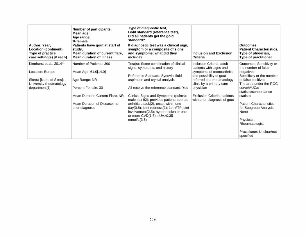
C-6
Author, Year, Location (continent), Type of practice care setting(s) [# each]
Number of participants, Mean age, Age range, % female, Patients have gout at start of study, Mean duration of current flare, Mean duration of illness
Type of diagnostic test, Gold standard (reference test), Did all patients get the gold standard? If diagnostic test was a clinical sign, symptom or a composite of signs and symptoms, what did they include?
Inclusion and Exclusion Criteria
Outcomes, Patient Characteristics, Type of physician, Type of practitioner
Kienhorst et al., 201433 Location: Europe Site(s) [Num. of Sites]: University rheumatology department[1]
Number of Patients: 390 Mean Age: 61.0[14.0] Age Range: NR Percent Female: 30 Mean Duration Current Flare: NR Mean Duration of Disease: no prior diagnosis
Test(s): Some combination of clinical signs, symptoms, and history Reference Standard: Synovial fluid aspiration and crystal analysis All receive the reference standard: Yes Clinical Signs and Symptoms (points): male sex 92); previous patient reported arthritis attack(2); onset within one day(0.5); joint redness(1); 1st MTP joint involvement(2.5); hypertension or one or more CVD(1.5), sUA>0.35 mmol/L(3.5)
Inclusion Criteria: adult patients with signs and symptoms of monoarthritis and possibility of gout referred to a rheumatology clinic by a primary care physician Exclusion Criteria: patients with prior diagnosis of gout
Outcomes: Sensitivity or the number of false negatives Specificity or the number of false positives The area under the ROC curve/AUC/c-statistic/concordance statistic Patient Characteristics for Subgroup Analysis: None Physician: Rheumatologist Practitioner: Unclear/not specified
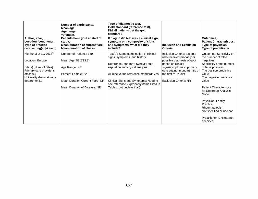
C-7
Author, Year, Location (continent), Type of practice care setting(s) [# each]
Number of participants, Mean age, Age range, % female, Patients have gout at start of study, Mean duration of current flare, Mean duration of illness
Type of diagnostic test, Gold standard (reference test), Did all patients get the gold standard? If diagnostic test was a clinical sign, symptom or a composite of signs and symptoms, what did they include?
Inclusion and Exclusion Criteria
Outcomes, Patient Characteristics, Type of physician, Type of practitioner
Kienhorst et al., 201434 Location: Europe Site(s) [Num. of Sites]: Primary care provider’s office[93] University rheumatology department[1]
Number of Patients: 159 Mean Age: 58.2[13.8] Age Range: NR Percent Female: 22.6 Mean Duration Current Flare: NR Mean Duration of Disease: NR
Test(s): Some combination of clinical signs, symptoms, and history Reference Standard: Synovial fluid aspiration and crystal analysis All receive the reference standard: Yes Clinical Signs and Symptoms: Need to see reference 2 (probably items listed in Table 1 but unclear if all)
Inclusion Criteria: patients who received probably or possible diagnosis of gout based on clinical signs/symptoms in primary care setting; monoarthritis of the first MTP joint Exclusion Criteria: NR
Outcomes: Sensitivity or the number of false negatives Specificity or the number of false positives The positive predictive value The negative predictive value Patient Characteristics for Subgroup Analysis: None Physician: Family Practice Rheumatologist Not specified or unclear Practitioner: Unclear/not specified

C-8
Author, Year, Location (continent), Type of practice care setting(s) [# each]
Number of participants, Mean age, Age range, % female, Patients have gout at start of study, Mean duration of current flare, Mean duration of illness
Type of diagnostic test, Gold standard (reference test), Did all patients get the gold standard? If diagnostic test was a clinical sign, symptom or a composite of signs and symptoms, what did they include?
Inclusion and Exclusion Criteria
Outcomes, Patient Characteristics, Type of physician, Type of practitioner
Lai et al., 201135 Location: Asia Site(s) [Num. of Sites]: University rheumatology department[1]
Number of Patients: 80 Mean Age: Gout patients: median age 74.0; Nongout patients median age 66.0 Age Range: 52.8-80.8 Percent Female: gout:17.6%; nongout: 52.2% Mean Duration Current Flare: NR Mean Duration of Disease: median: 8 years
Test(s): Ultrasound Reference Standard: Synovial fluid aspiration and crystal analysis All receive the reference standard: Yes Clinical Signs and Symptoms: NA
Inclusion Criteria: having undergone ultrasound guided joint aspiration at the authors' rheumatology division between march 2009 and March 2010 after presenting with mono or oligoarthritis with acute or subacute onset Exclusion Criteria: NR
Outcomes: Sensitivity or the number of false negatives Specificity or the number of false positives The positive predictive value The negative predictive value Patient Characteristics for Subgroup Analysis: Another arthropathy Physician: Rheumatologist Practitioner: Unclear/not specified
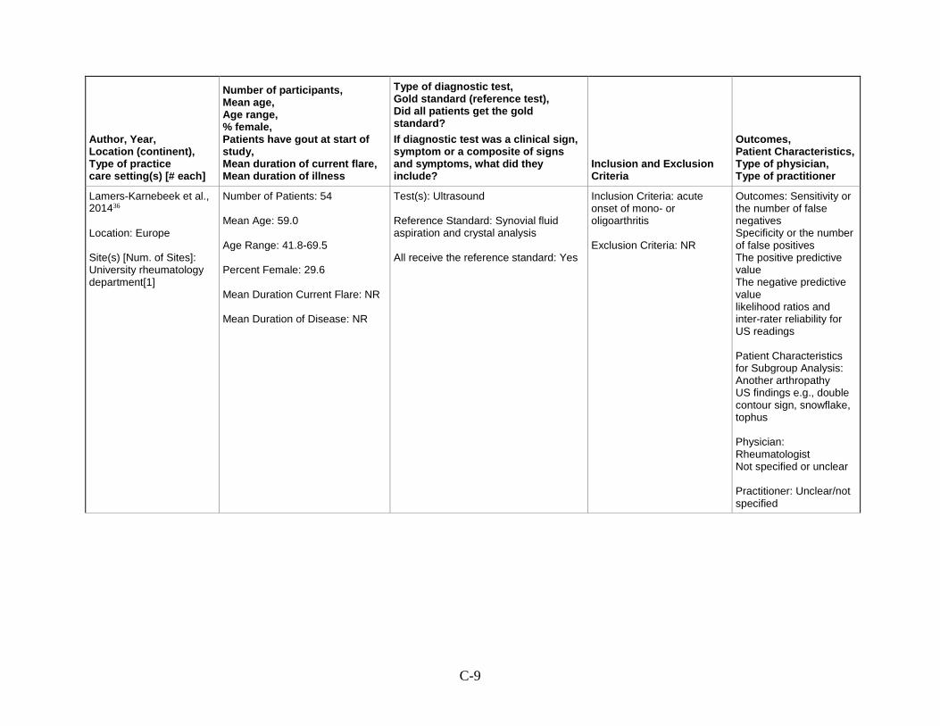
C-9
Author, Year, Location (continent), Type of practice care setting(s) [# each]
Number of participants, Mean age, Age range, % female, Patients have gout at start of study, Mean duration of current flare, Mean duration of illness
Type of diagnostic test, Gold standard (reference test), Did all patients get the gold standard? If diagnostic test was a clinical sign, symptom or a composite of signs and symptoms, what did they include?
Inclusion and Exclusion Criteria
Outcomes, Patient Characteristics, Type of physician, Type of practitioner
Lamers-Karnebeek et al., 201436 Location: Europe Site(s) [Num. of Sites]: University rheumatology department[1]
Number of Patients: 54 Mean Age: 59.0 Age Range: 41.8-69.5 Percent Female: 29.6 Mean Duration Current Flare: NR Mean Duration of Disease: NR
Test(s): Ultrasound Reference Standard: Synovial fluid aspiration and crystal analysis All receive the reference standard: Yes
Inclusion Criteria: acute onset of mono- or oligoarthritis Exclusion Criteria: NR
Outcomes: Sensitivity or the number of false negatives Specificity or the number of false positives The positive predictive value The negative predictive value likelihood ratios and inter-rater reliability for US readings Patient Characteristics for Subgroup Analysis: Another arthropathy US findings e.g., double contour sign, snowflake, tophus Physician: Rheumatologist Not specified or unclear Practitioner: Unclear/not specified

C-10
Author, Year, Location (continent), Type of practice care setting(s) [# each]
Number of participants, Mean age, Age range, % female, Patients have gout at start of study, Mean duration of current flare, Mean duration of illness
Type of diagnostic test, Gold standard (reference test), Did all patients get the gold standard? If diagnostic test was a clinical sign, symptom or a composite of signs and symptoms, what did they include?
Inclusion and Exclusion Criteria
Outcomes, Patient Characteristics, Type of physician, Type of practitioner
Malik et al., 200916 Location: North America Site(s) [Num. of Sites]: University rheumatology department[1]
Number of Patients: 82 Mean Age: 64.5 Age Range: NA Percent Female: 6 Mean Duration Current Flare: NA Mean Duration of Disease: NA
Test(s): Some combination of clinical signs, symptoms, and history Reference Standard: Synovial fluid aspiration and crystal analysis All receive the reference standard: Yes Clinical Signs and Symptoms: Crystal analysis vs. three diagnostic signs/symptoms: 1. ACR (ARA) Preliminary Criteria2. New York Criteria3. Rome Criteria
Inclusion Criteria: Synovial fluid aspirated and analyzed at some point. Exclusion Criteria: NA
Outcomes: Sensitivity or the number of false negatives Specificity or the number of false positives The positive predictive value False positivity Patient Characteristics for Subgroup Analysis: Gout criteria Physician: Rheumatologist Not specified or unclear Practitioner: Technician

C-11
Author, Year, Location (continent), Type of practice care setting(s) [# each]
Number of participants, Mean age, Age range, % female, Patients have gout at start of study, Mean duration of current flare, Mean duration of illness
Type of diagnostic test, Gold standard (reference test), Did all patients get the gold standard? If diagnostic test was a clinical sign, symptom or a composite of signs and symptoms, what did they include?
Inclusion and Exclusion Criteria
Outcomes, Patient Characteristics, Type of physician, Type of practitioner
Park et al., 201437 Location: Asia Site(s) [Num. of Sites]: University hospital departments of rheumatology and laboratory medicine[2]
Number of Patients: 179 Mean Age: 62.6 ± 16.4 (age at diagnosis) Percent Female: 5.6 Mean Duration Current Flare: 3.3 ± 3.5 days
Test(s): Synovial fluid aspiration and crystal analysis Reference Standard: Synovial fluid aspiration and crystal analysis ACR/ARA guidelines All receive the reference standard: Yes
Inclusion Criteria: patients with a diagnosis of gout, who had undergoneSF examination from October 1999 to September 2011 Exclusion Criteria: patients with unclassified acute arthritis, intercritical gout without acute symptoms, pseudogout, osteoarthritis, concomitant septic and gouty arthritis
Outcomes: Treatment decision resulting from diagnosis Misdiagnosis related harms Patient Characteristics for Subgroup Analysis: Duration of current episode/flare/symptoms Type of clinician performing tests Patient age Type 2 diabetes or metabolic syndrome Crystal positive and crystal negative gout Physician: Rheumatologist Practitioner: Technician
Rettenbacher et al., 200838 Location: Europe Site(s) [Num. of Sites]: Academic radiology department[1]
Number of Patients: 105 Mean Age: 59 Age Range: 31-89 Percent Female: 12 Mean Duration Current Flare: NR Mean Duration of Disease: NR
Test(s): Ultrasound Plain x-ray Reference Standard: Synovial fluid aspiration and crystal analysis characteristic clinical and laboratory findings All receive the reference standard: Yes Clinical Signs and Symptoms: NA
Inclusion Criteria: patients with suspected gout referred from rheumatology clinic Exclusion Criteria: inability to establish a definite diagnosis
Outcomes: Sensitivity or the number of false negatives Specificity or the number of false positives The positive predictive value The negative predictive value Physician: Radiologists Practitioner: Unclear/not specified

C-12
Author, Year, Location (continent), Type of practice care setting(s) [# each]
Number of participants, Mean age, Age range, % female, Patients have gout at start of study, Mean duration of current flare, Mean duration of illness
Type of diagnostic test, Gold standard (reference test), Did all patients get the gold standard? If diagnostic test was a clinical sign, symptom or a composite of signs and symptoms, what did they include?
Inclusion and Exclusion Criteria
Outcomes, Patient Characteristics, Type of physician, Type of practitioner
Richette et al., 201439 Location: Europe Site(s) [Num. of Sites]: University rheumatology department[14]
Number of Patients: 244 Mean Age: 59.8±12.5 years Age Range: NR Percent Female: 40.5% Mean Duration Current Flare: NR Mean Duration of Disease: NR
Test(s): Some combination of clinical signs, symptoms, and history Reference Standard: Synovial fluid aspiration and crystal analysis All receive the reference standard: Yes Clinical Signs and Symptoms: 11 items Self-reported history of gout Male sex Self-reported history of hyperuricaemiaTophus Hypertriglyceridaemia Cardiovascular disease Pain intensity Involvement of toes, foot or ankles;Treatment with corticosteroids; Treatment with NSAIDs
Inclusion Criteria: gout or other arthritis Exclusion Criteria: NR
Outcomes: Sensitivity or the number of false negatives Specificity or the number of false positives The area under the ROC curve/AUC/c-statistic/concordance statistic Patient Characteristics for Subgroup Analysis: Duration of current episode/flare/symptoms Patient age Patient sex Type 2 diabetes or metabolic syndrome Another arthropathy Current use of medication Physician: Not applicable Practitioner: Unclear/not specified

C-13
Author, Year, Location (continent), Type of practice care setting(s) [# each]
Number of participants, Mean age, Age range, % female, Patients have gout at start of study, Mean duration of current flare, Mean duration of illness
Type of diagnostic test, Gold standard (reference test), Did all patients get the gold standard? If diagnostic test was a clinical sign, symptom or a composite of signs and symptoms, what did they include?
Inclusion and Exclusion Criteria
Outcomes, Patient Characteristics, Type of physician, Type of practitioner
Taylor 201440 Location: multi-country Site(s) [Num. of Sites]: University rheumatology departments [25]
Number of Patients: 983 Mean Age: 60±15 years Age Range: NR Percent Female: 28.6% Mean Duration Current Flare: NR Mean Duration of Disease: 6 years (median, IQR 2 to 13 for MSU+)
Test(s): Rome, NY, ACR, Diagnostic Rule, CGD algorithms Reference Standard: Synovial fluid aspiration and crystal analysis All receive the reference standard: Yes Clinical Signs and Symptoms: depend on test (see Table 2 in text)
Inclusion Criteria: patient attending a participating rheumatology clinic with joint swelling or subcutaneous nodule within prior 2 weeks, possibly due to gout Exclusion Criteria: NR
Outcomes: Sensitivity Specificity Patient Characteristics for Subgroup Analysis: Duration of symptoms (time since first flare) Physician: Not applicable Practitioner: Not applicable

C-14
Author, Year, Location (continent), Type of practice care setting(s) [# each]
Number of participants, Mean age, Age range, % female, Patients have gout at start of study, Mean duration of current flare, Mean duration of illness
Type of diagnostic test, Gold standard (reference test), Did all patients get the gold standard? If diagnostic test was a clinical sign, symptom or a composite of signs and symptoms, what did they include?
Inclusion and Exclusion Criteria
Outcomes, Patient Characteristics, Type of physician, Type of practitioner
Vazquez-Mellado et al., 201241 Location: North America Site(s) [Num. of Sites]: University rheumatology department[ ]
Number of Patients: 167 Mean Age: 54[16.8] for gout, 49.6 overall Age Range: NR Percent Female: 24% for gout, 7% for RA, 42% for SA, 10% for OA Mean Duration Current Flare: NR Mean Duration of Disease: NR
Test(s): Some combination of clinical signs, symptoms, and history Reference Standard: Synovial fluid aspiration and crystal analysis All receive the reference standard: Yes Clinical Signs and Symptoms: >1 attack, mono/oligoarthritis, rapid onset of pain and swelling, podagra, erythema, tarsitis, probable tophi, hyperuricemia, and combinations of these items
Inclusion Criteria: For gout, positive synovial fluid MSU crystal analysis Exclusion Criteria: NR
Outcomes: Sensitivity or the number of false negatives Specificity or the number of false positives The positive predictive value The negative predictive value The area under the ROC curve/AUC/c-statistic/concordance statistic likelihood ratios, odds ratio Patient Characteristics for Subgroup Analysis: possibly age, sex, and secondary gout, e.g., gout and comorbidity such as CRF, hematologic condition Physician: Not specified or unclear Practitioner: Unclear/not specified

C-15
Author, Year, Location (continent), Type of practice care setting(s) [# each]
Number of participants, Mean age, Age range, % female, Patients have gout at start of study, Mean duration of current flare, Mean duration of illness
Type of diagnostic test, Gold standard (reference test), Did all patients get the gold standard? If diagnostic test was a clinical sign, symptom or a composite of signs and symptoms, what did they include?
Inclusion and Exclusion Criteria
Outcomes, Patient Characteristics, Type of physician, Type of practitioner
Wallace et al., 197742 Location: North America Site(s) [Num. of Sites]: University rheumatology department [38 (probably some private, also)]
Number of Patients: 706 Mean Age: gout: 56.2; RA: 47.6 Age Range: NR Percent Female: gout: , RA: 64.313.6 Mean Duration Current Flare: NR Mean Duration of Disease: Gout: 10.1 years
Test(s): Some combination of clinical signs, symptoms, and history Synovial fluid aspiration and crystal analysis Reference Standard: clinical opinion inferred Clinical Signs and Symptoms: >1 acute attack; maximum inflammation developed in 1 day; monoarthritis; redness over joint; first MTP painful or swollen; unilateral first MTP joint attack; unilateral tarsal joint attack; tophus (proven or suspected); hyperuricemia; asymmetric swelling within joint on x-ray; subcortical cysts without
Inclusion Criteria: NR Exclusion Criteria: NR
Outcomes: unclear Physician: Rheumatologist Practitioner: Rheumatologist
ACR (ARA) = American College of Rheumatology (American Rheumatology Association); AUC = area under the curve; CPP = calcium pyrophosphate di;hydrate; CS = corticosteroid; CVD = cardiovascular disease; DECT = dual-energy computed tomography; MSU = monosodium urate; MTP = metatarsophalangeal; NA = not applicable; NR = not reported; NSAIDS = non-steroidal anti-inflammatory drugs; OA = osteoarthritis; RA = rheumatoid arthritis; ROC = receiver-operating characteristics; SA = spondylo-arthropathy, SUA = serum uric acid.

D-1
Appendix D. Data Abstraction Tools Gout Data Abstraction Tool 1. Do you need another article to complete this form?
Yes
No Clear Response
2. Did the patient population include only patients already diagnosed or assumed to have gout?
Yes
No
Not reported Clear Response
3. Diagnostic procedure(s) being tested (check all that apply):
DECT
Ultrasound
Plain x-ray
Some combination of clinical signs/symptoms/history
Serum UA
Synovial fluid aspiration and crystal analysis (check only if some variation on the reference standard and not the standard itself)
Other [Specify and STOP]
Not reported 4. Location (country)
North America
Central/South America
Europe
Asia
Australia/New Zealand
Other (specify region) 5. Care setting and number of sites (check all that apply)
Primary care provider’s office
Urgent care clinic
Hospital emergency department
Rheumatologist in private practice
University rheumatology department
Other [specify]

D-2
Inpatient hospital/long term care only [STOP]
Not reported 6. Number of patients total [specify]
7. Mean age +/- SD [specify]
8. Age range _ to _ [specify if given]
9. Percentage of patients who are female:
10. Mean duration of current flare episode:
11. Mean duration of disease:
12. Inclusion criteria [specify]
13. Exclusion criteria [specify]
14. If this study measures the validity of a diagnostic test, what is the reference standard:
Synovial fluid aspiration and crystal analysis
ACR/ARA guidelines
Other [specify]
Not reported [Exclude, STOP] 15. Did all patients receive the reference standard test?
Yes
No [Exclude]
Unclear [Exclude] Clear Response
16. Type of physician who performed diagnostic tests: (check all that apply)
Family practice
D-3
General Internist
Rheumatologist
Emergency physician
Not specified or unclear
Radiologists
Not applicable 17. Type of practitioner who performed synovial fluid analysis:
FP/internist/ED
Rheumatologist
Technician
Unclear/not specified
Not applicable Clear Response
18. If the diagnostic procedure being tested is a clinical sign or symptom or some combination, what does it [do they] include? [specify; maybe more than one combination or may be comparison of two different signs vs. synovial fluid]
19. Were the findings stratified by any patient characteristics or comorbidities? (check all that apply)
Duration of current episode/flare/symptoms
Duration of these kinds of flares (e.g., first time patient vs. previous episodes)
Type of clinician performing tests
Type of physician
Patient age
Patient sex
Serum uric acid
Type 2 diabetes or metabolic syndrome
Another arthropathy (e.g., osteoarthritis, rheumatoid arthritis)
Current use of medication (e.g., NSAIDs, steroids, drugs associated with gout flares)
Other [specify]
None 20. Study design (select one)
Prospective

D-4
Retrospective (case control)
Other [specify] Clear Response
21. Does the study report (check all that apply):
Sensitivity or the number of false negatives (people who really have gout but who test negative when the test method is used)
Specificity or the number of false positives (people who tested negative on the reference standard but who tested positive on the test method)
The positive predictive value
The negative predictive value
The area under the ROC curve/AUC/c-statistic/concordance statistic
Adverse effects associated with testing
Pain, inflammation, or quality of life outcomes
Treatment decision resulting from diagnosis
Other [specify]
None 22. Does this article appear to report on part of a larger study? (select one)
Yes
No Clear Response
23. Does this article cite references we should get?
Yes
No Clear Response

D-5
QUADAS-2: Tool for the Quality Assessment of Diagnostic Accuracy Studies Domain 1: Patient Selection 1. Was a consecutive or random sample of patients enrolled?
Yes
No
Unclear
Not applicable Clear Response
2. Was a case-control design avoided?
Yes
No
Unclear
Not Applicable Clear Response
3. Did the study avoid inappropriate exclusions?
Yes
No
Unclear
Not Applicable Clear Response
4. Based on your answers for 1-3, could the selection of patients have introduced bias? Risk:
Low
High
Unclear
Not Applicable Clear Response
Domain 2: Index Test(s) (complete for each index test used) 1. Were the index test results interpreted without knowledge of the reference standard?
Yes
No
Unclear
Not Applicable Clear Response
2. If a threshold was used, was it pre-specified?
Yes

D-6
No
Unclear
Not Applicable Clear Response
3. Based on your answers for 1-2, could the conduct or interpretation of the index test have introduced bias? Risk:
Low
High
Unclear
Not Applicable Clear Response
Domain 3: Reference Standard 1. Is the reference standard likely to correctly classify the target condition?
Yes
No
Unclear
Not Applicable Clear Response
2. Were the reference standard results interpreted without knowledge of the results of the index test?
Yes
No
Unclear
Not Applicable Clear Response
3. Based on your answers for 1-2, could the reference standard, its conduct, or its interpretation have introduced bias? Risk:
Low
High
Unclear
Not Applicable Clear Response
Domain 4: Flow and Timing 1. Was there an appropriate interval between index test(s) and reference standard?
Yes
No
Unclear
Not Applicable Clear Response
2. Did all patients receive a reference standard?
D-7
Yes
No
Unclear
Not Applicable Clear Response
3. Did all patients receive the same reference standard?
Yes
No
Unclear
Not Applicable Clear Response
4. Were all patients included in the analysis?
Yes
No
Unclear
Not Applicable Clear Response
5. Based on your answers for 1-4, could the patient flow have introduced bias? Risk:
Low
High
Unclear
Not Applicable Clear Response

D-8
AMSTAR Assessment of Reporting Quality for Systematic Reviews 1. Was an 'a priori' design provided? The research question and inclusion criteria should be established before the conduct of the review. Note: Need to refer to a protocol, ethics approval, or pre-determined/a priori published research objectives to score a “yes.” □ Yes □ No □ Can't answer □ Not applicable 2. Was there duplicate study selection and data extraction? There should be at least two independent data extractors and a consensus procedure for disagreements should be in place. Note: 2 people do study selection, 2 people do data extraction, consensus process or one person checks the other’s work. □ Yes □ No □ Can't answer □ Not applicable 3. Was a comprehensive literature search performed? At least two electronic sources should be searched. The report must include years and databases used (e.g., Central, EMBASE, and MEDLINE). Key words and/or MESH terms must be stated and where feasible the search strategy should be provided. All searches should be supplemented by consulting current contents, reviews, textbooks, specialized registers, or experts in the particular field of study, and by reviewing the references in the studies found. Note: If at least 2 sources + one supplementary strategy used, select “yes” (Cochrane register/Central counts as 2 sources; a grey literature search counts as supplementary). □ Yes □ No □ Can't answer □ Not applicable 4. Was the status of publication (i.e. grey literature) used as an inclusion criterion? The authors should state that they searched for reports regardless of their publication type. The authors should state whether or not they excluded any reports (from the

D-9
systematic review), based on their publication status, language etc. Note: If review indicates that there was a search for “grey literature” or “unpublished literature,” indicate “yes.” SIGLE database, dissertations, conference proceedings, and trial registries are all considered grey for this purpose. If searching a source that contains both grey and non-grey, must specify that they were searching for grey/unpublished lit. □ Yes □ No □ Can't answer □ Not applicable 5. Was a list of studies (included and excluded) provided? A list of included and excluded studies should be provided. Note: Acceptable if the excluded studies are referenced. If there is an electronic link to the list but the link is dead, select “no.” □ Yes □ No □ Can't answer □ Not applicable 6. Were the characteristics of the included studies provided? In an aggregated form such as a table, data from the original studies should be provided on the participants, interventions and outcomes. The ranges of characteristics in all the studies analyzed e.g., age, race, sex, relevant socioeconomic data, disease status, duration, severity, or other diseases should be reported. Note: Acceptable if not in table format as long as they are described as above. □ Yes □ No □ Can't answer □ Not applicable 7. Was the scientific quality of the included studies assessed and documented? 'A priori' methods of assessment should be provided (e.g., for effectiveness studies if the author(s) chose to include only randomized, double-blind, placebo controlled studies, or allocation concealment as inclusion criteria); for other types of studies alternative items will be relevant. Note: Can include use of a quality scoring tool or checklist, e.g., Jadad scale, risk of bias, sensitivity analysis, etc., or a description of quality items, with some kind of result for EACH study (“low” or “high” is fine, as long as it is clear which studies scored “low” and which scored “high”; a summary score/range for all studies is not acceptable). □ Yes □ No □ Can't answer

D-10
□ Not applicable 8. Was the scientific quality of the included studies used appropriately in formulating conclusions? The results of the methodological rigor and scientific quality should be considered in the analysis and the conclusions of the review, and explicitly stated in formulating recommendations. Note: Might say something such as “the results should be interpreted with caution due to poor quality of included studies.” Cannot score “yes” for this question if scored “no” for question 7. □ Yes □ No □ Can't answer □ Not applicable 9. Were the methods used to combine the findings of studies appropriate? For the pooled results, a test should be done to ensure the studies were combinable, to assess their homogeneity (i.e., Chi-squared test for homogeneity, I2 ). If heterogeneity exists a random effects model should be used and/or the clinical appropriateness of combining should be taken into consideration (i.e., is it sensible to combine?). Note: Indicate “yes” if they mention or describe heterogeneity, i.e., if they explain that they cannot pool because of heterogeneity/variability between interventions. □ Yes □ No □ Can't answer □ Not applicable 10. Was the likelihood of publication bias assessed? An assessment of publication bias should include a combination of graphical aids (e.g., funnel plot, other available tests) and/or statistical tests (e.g., Egger regression test, Hedges-Olken). Note: If no test values or funnel plot included, score “no”. Score “yes” if mentions that publication bias could not be assessed because there were fewer than 10 included studies. □ Yes □ No □ Can't answer □ Not applicable 11. Was the conflict of interest included? Potential sources of support should be clearly acknowledged in both the systematic review and the included studies.
D-11
Note: To get a “yes,” must indicate source of funding or support for the systematic review AND for each of the included studies. □ Yes □ No □ Can't answer □ Not applicable Shea et al. BMC Medical Research Methodology 2007 7:10 doi:10.1186/1471-2288-7-10 Additional notes (in italics) made by Michelle Weir, Julia Worswick, and Carolyn Wayne based on conversations with Bev Shea and/or Jeremy Grimshaw in June and October 2008 and July and September 2010.