diagnosis of invasive fungal disease
DESCRIPTION
Diagnosis of Invasive Fungal Disease. “Gold standard,” blood cultures for the diagnosis of candidemia have been associated with a sensitivity historically ranging from 21.3 to 54% . - PowerPoint PPT PresentationTRANSCRIPT

Diagnosis of Invasive Fungal Disease


• “Gold standard,” blood cultures for the diagnosis of candidemia have been associated with a sensitivity historically ranging from 21.3 to 54% .
• The advent of lysis centrifugation has increased the diagnostic yield of blood cultures for the diagnosis of candidemia, albeit with limitations including higher rates of contamination and additional cost and required personnel
• The Peptide Nucleic Acid Fluorescent In Situ Hybridization (PNA-FISH) test has been studied and recently introduced in clinical practice for the rapid identification of Candida species. The PNA-FISH for C. albicans has had a sensitivity, specificity, positive, and negative predictive value of 99, 100, 100, and 99.3%, respectively
• More recently, a multicenter study evaluated the performance of a rapid two-color PNA-FISH assay for detection of C. albicans and C. glabrata directly from positive blood culture bottles
• Considering the relatively low sensitivity of blood cultures, PCR may prove to be a significant adjunct for the diagnosis of candidemia particularly in high risk patients, such as cancer patients. However, a major limitation of most PCR assays is their relative lack of specificity, mostly due to high rates of contamination.
Diagnosis of Hepatosplenic Candidiasis
• Definitive diagnosis requires biopsy of hepatic lesions that may reveal hyphal forms consistent with Candida species.
• The diagnosis is suggested by the presence of multiple lesions of the liver and spleen, occasionally described as “bull’s eye” appearing on abdominal CT scan or magnetic resonance imaging (MRI)

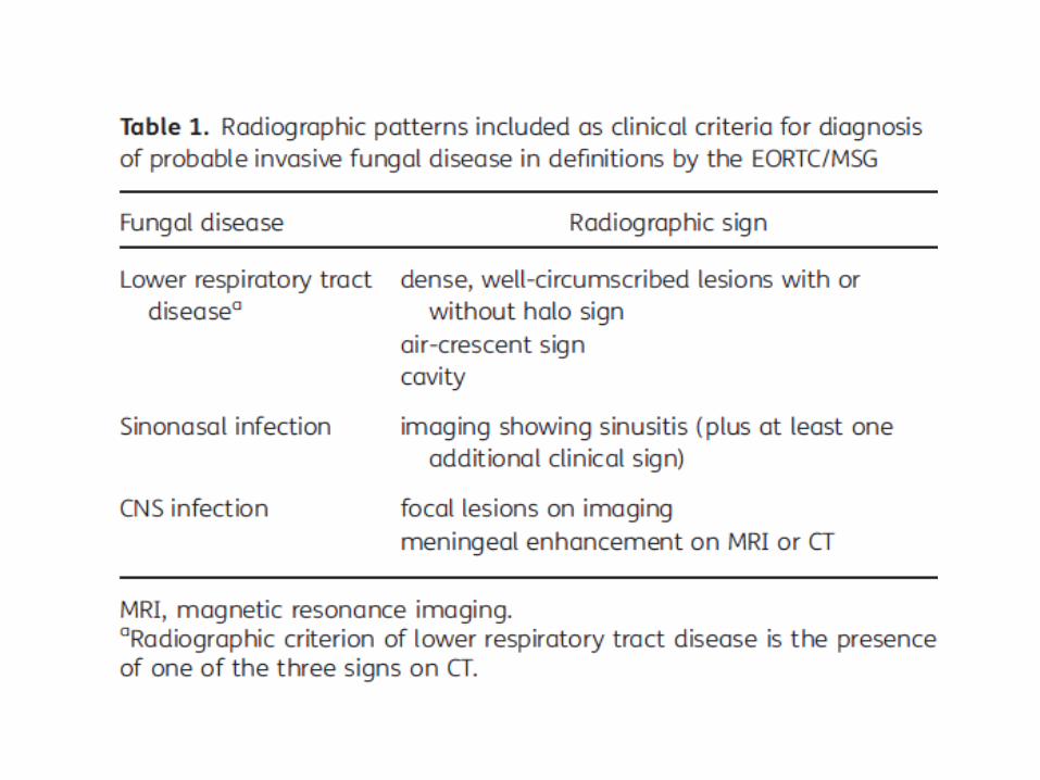
• Invasive mold infections affecting the lungs may present with different patterns on a chest CT, including small or large nodules, patchy, segmental, or wedge-shaped consolidations, peribronchial infiltrates with a tree-in-bud distribution, and cavitation .
• Two CT patterns have been associated with early and late pulmonary IA: the “halo” and the “crescent” sign, respectively
• Histopathologic confirmation of sterile tissue invasion remains the “gold standard” to establish a proven diagnosis of an invasive mold infection

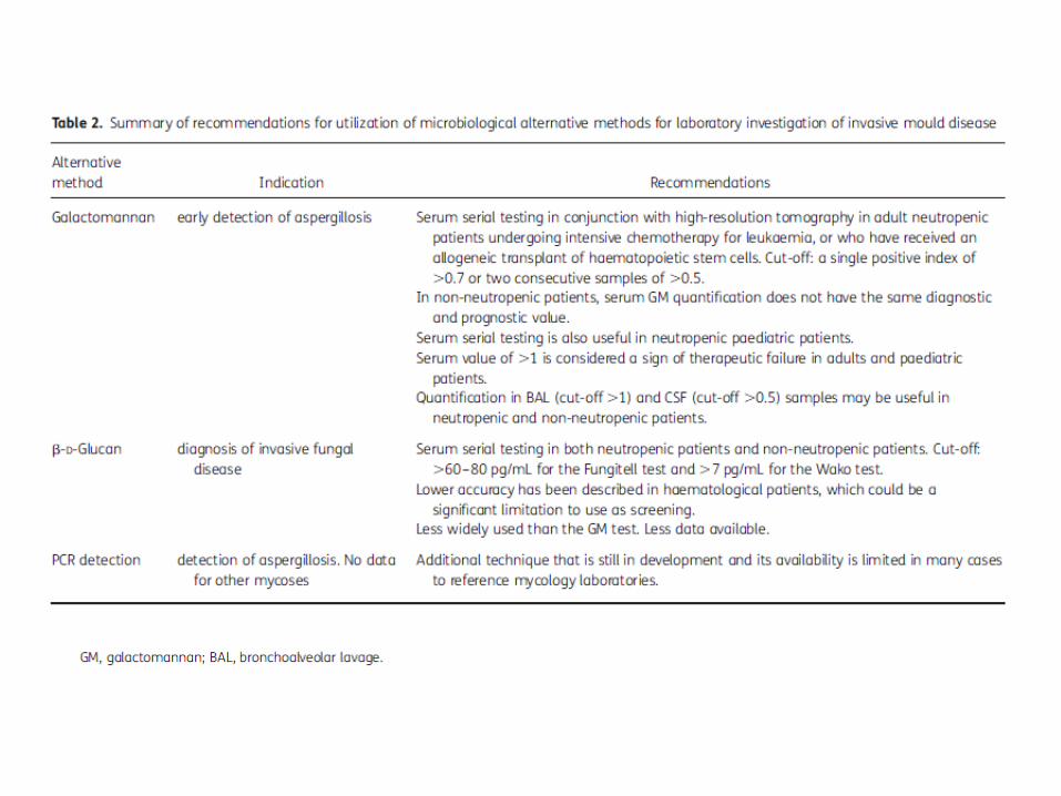
• Severe mucositis and gastrointestinal GHVD following HSCT can occasionally lead to false positive results, likely due to translocation of GM across the intestinal mucosa during periods of reduced mucosal integrity. Younger age has been associated with lower specificity rates, predominately attributed to the high concentration of GM in children’s food (e.g., cereals)
• The revised definitions retain the original classifications of “proven,” “probable,” and “possible” IMIs. For most conditions, proven infections require proof of hyphal elements in diseased tissue. To characterize a case as probable, a host factor, clinical features, and a mycologic or nonculture-based surrogate marker (e.g., galactomannan, beta-glucan, or as determined by polymerase chain reaction [PCR]) must be present. Possible invasive fungal disease is more strictly defined to include patients with the appropriate host factors and sufficient clinical evidence of invasive fungal disease, but no mycologic evidence.

• For rare molds, the isolation of fungus in respiratory secretions, skin, and blood is not synonymous with invasive disease. Most such cases represent contamination or colonization, even among high-risk patients


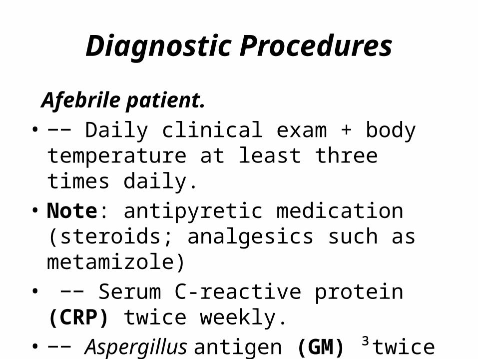
Diagnostic Procedures
Afebrile patient.• −− Daily clinical exam + body temperature at
least three times daily.• Note: antipyretic medication (steroids;
analgesics such as metamizole)• −− Serum C-reactive protein (CRP) twice
weekly.• −− Aspergillus antigen (GM) ³twice weekly..
First fever.• −− Update physical exam, blood cultures,
clinical chemistry, CRP, interleukin-6 (IL-6), and thoracic computed tomography (CT) scan; other measures according to clinical findings
Persistent fever.• −− Update physical exam, blood cultures,
clinical chemistry, CRP, IL-6, and thoracic CT scan; consider abdominal ultrasound or magnetic resonance imaging (MRI).
• −− Check results of antigen testings.

Fever + pulmonary infiltrates.• −− Bronchoscopy + bronchoalveolar lavage (BAL)
=>microscopy + culture for bacteria; • test for Mycobacterium tuberculous (MTB)• Pneumocystis,• cytomegalovirus (CMV), respiratory viruses,
adenovirus, • Aspergillus + other fungi; check for Aspergillus GM; • optional: Aspergillus-PCR and MTB/Pneumocystis-PCR.
Fever + signs of inflammation at CVC.• −− Blood cultures from peripheral vein and
from CVC.• −− Follow-up cultures in case of cultures
positive for Staphylococcus aureus and Candida spp.
• • Fever accompanied by skin lesions.• −− Blood cultures.• −− Biopsy (=>histopathology and nonfixated
=>microbiology).
Neurological symptoms ± fever.• −− Cerebrospinal fluid (CSF) =>human herpes
virus-6 (HHV-6); Aspergillus GM; CMV; HSV, VZV.
• −− Fundoscopy.• −− Cranial MRI.

Fever + abdominal symptoms.• −− Clostridium difficile toxins;
noro-/rotaviruses; CMV; adenovirus; Epstein–Barr virus (EBV).

Perianal infiltrate/abscess.• −− Beware of results from inappropriate
microbiological diagnostics suggesting monomicrobial etiology.
• Fever + increasing “liver function tests” =>viral (hepatitis B virus (HBV), varicella zoster virus (VZV); CMV, etc.), Candida?
• −− Liver ultrasound or CT or MRI (preferred) • NB: Pneumocystis jiroveci typically
accompanied by lactate dehydrogenase rise

• . They• are based on the idea that the strains that
have an MIC for an• antifungal above a certain value respond
significantly less well• to treatment with that drug, since it is
impossible to achieve• therapeutic concentrations in vivo.

• To date, breakpoints• have been established for infections by Candida spp. only, and• for some of the antifungal compounds available. For infections• with other species of yeasts and filamentous fungi, no
breakpoints• have yet been established, although it is advisable not• to treat with drugs that are inactive in vitro, or with those that• have a high MIC for the species causing the mycosis; this is• known as an epidemiological cut-off.
• In the case of Aspergillus• spp., some experts have proposed epidemiological cut-
offs• and even tentative breakpoints to interpret the results
of susceptibility• testing of those species to azole agents. An MIC value
for• itraconazole and voriconazole of ≥2 mg/L, and ≥0.5
mg/L for• posaconazole, should be taken as resistant in vitro.


• differential diagnosis• including appendicitis, ischemic colitis,
pseudomembranous• colitis, or antineoplastic drug or radiation
toxicity
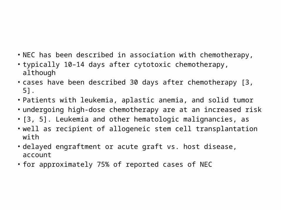
• NEC has been described in association with chemotherapy,• typically 10–14 days after cytotoxic chemotherapy, although• cases have been described 30 days after chemotherapy [3,
5].• Patients with leukemia, aplastic anemia, and solid tumor• undergoing high-dose chemotherapy are at an increased risk• [3, 5]. Leukemia and other hematologic malignancies, as• well as recipient of allogeneic stem cell transplantation with• delayed engraftment or acute graft vs. host disease, account• for approximately 75% of reported cases of NEC
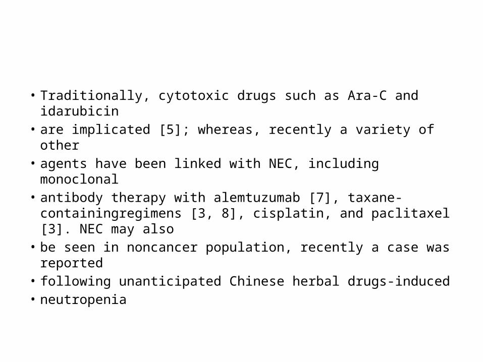
• Traditionally, cytotoxic drugs such as Ara-C and idarubicin• are implicated [5]; whereas, recently a variety of other• agents have been linked with NEC, including monoclonal• antibody therapy with alemtuzumab [7], taxane-
containingregimens [3, 8], cisplatin, and paclitaxel [3]. NEC may also
• be seen in noncancer population, recently a case was reported
• following unanticipated Chinese herbal drugs-induced• neutropenia
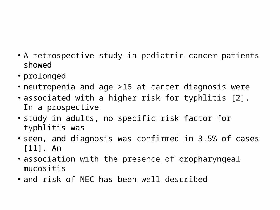
• A retrospective study in pediatric cancer patients showed• prolonged• neutropenia and age >16 at cancer diagnosis were• associated with a higher risk for typhlitis [2]. In a
prospective• study in adults, no specific risk factor for typhlitis was• seen, and diagnosis was confirmed in 3.5% of cases [11]. An• association with the presence of oropharyngeal mucositis• and risk of NEC has been well described
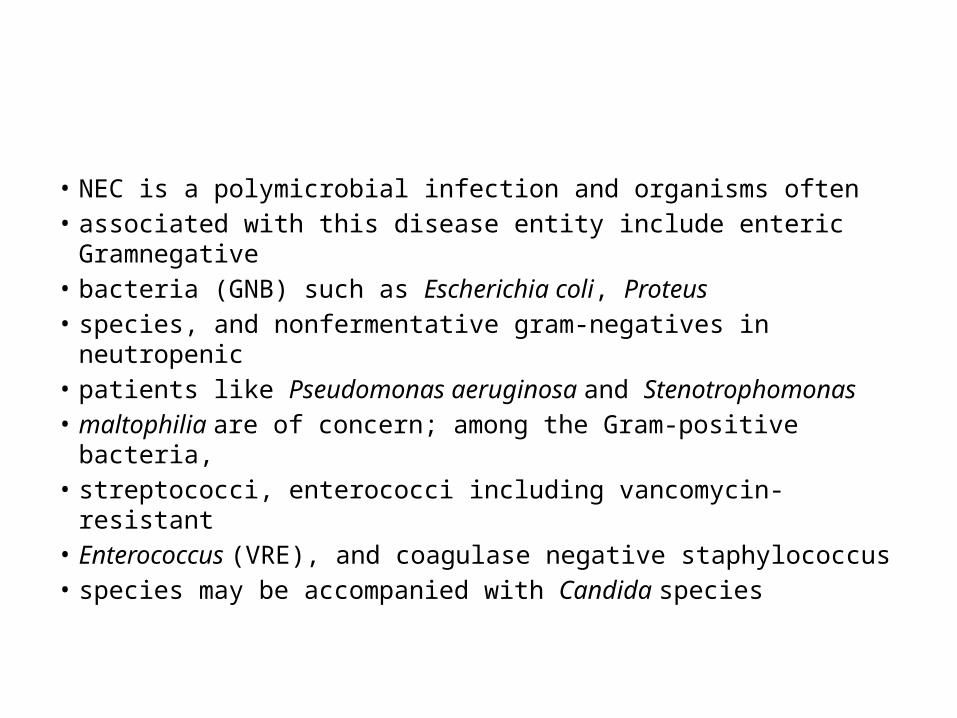
• NEC is a polymicrobial infection and organisms often• associated with this disease entity include enteric Gramnegative• bacteria (GNB) such as Escherichia coli, Proteus• species, and nonfermentative gram-negatives in neutropenic• patients like Pseudomonas aeruginosa and Stenotrophomonas• maltophilia are of concern; among the Gram-positive bacteria,• streptococci, enterococci including vancomycin-resistant• Enterococcus (VRE), and coagulase negative staphylococcus• species may be accompanied with Candida species

• 13, 14]. Cytomegalovirus or adenovirus enterocolitis may
• act as a trigger for a secondary NEC in some cases
• Antimicrobial prophylaxis may influence the time of
• onset, etiology, and possibly incidence of NEC in patients
• undergoing cancer therapy.

• . We suspect that NEC is a clinical• syndrome of various primary causes, in most patients• multiple factors appear to be responsible for this entity• including severe neutropenia, young or advanced age,
enteric• insult due to cancer, drugs or bacterial toxins such as• Clostridium difficile, subclinical viral disease, or unknown• genetic polymorphisms that predispose some individuals
to• develop this disorder.

• Patients with concomitant• bacteremia due to enteric organism(s) such as
Escherichia coli, enterococci, and streptococci give a partial
• spectrum of this polymicrobial disease, although sterile• blood cultures do not exclude a low-grade, intermittent• bacteremia• and/or fungemia in the profoundly neutropenic• susceptible patients

• Other common causes that may be• mistaken for NEC include ischemic bowel injury, C. difficile• colitis, appendicitis, or Ogilvie’s syndrome [3]. To further• complicate the diagnosis, there is a suggestion that the latter• entities can coexist, with one small pediatric study suggesting• that the combination of appendiceal thickening and• enterocolitis may more likely to result in surgical intervention• [21]. It was interesting to note that higher mortality was• seen in children with NEC without evidence of appendicitis• [21]. The frequency of NEC cases with concurrent or• preceding• C. difficile toxin-induced intestinal epithelia cell• damage remains uncertain
• A comprehensive review of adult neutropenic patients• with enterocolitis, appropriate diagnosis can be established• by (1) >4 mm of bowel wall thickening on CT or ultrasonic• abdominal scan combined with (2) clinical features such as• fever, abdominal pain, and diarrhea (Fig. 16.1a) [3]. Several• studies in pediatric and adults showed that a substantial• proportion of neutropenic patients may not exhibit fever or• abdominal pain during the early phase of the disease [2, 5,• 11]. Therefore, a high level of suspicion in febrile neutropenic• patients even in the absence of abdominal pain and/or• distention with diarrhea or clinical or radiographic features• of paralytic ileus should raise concerns for possible• enterocolitis.
• we recommend that bedside• abdominal ultrasounds should be reserved for unstable• patients in whom transport to the CT scan units is
deferred,• similarly, patients with other serious limitations for CT
scan• should than be evaluated with an abdominal X-rays and• ultrasounds
• We suggest a combination of clinical symptoms• such as abdominal pain, fever, or diarrhea in the setting of• neutropenia and possibly cytotoxic chemotherapy combined• with imaging studies (CT abdomen) that demonstrate bowel• wall thickening (3–5 mm) and in severe case pneumatosis• intestinalis may be used (Fig. 16.1b) [3, 11]. It is important• to assess other potential treatable causes that may mimic• these features such as ischemic colitis and C. difficile colitis.

• Cases of• prolonged• neutropenia may benefit from recombinant
myeloid• growth factors such as G-CSF or GM-CSF and in
select• patients with refractory neutropenia, healthy
donor-derived• granulocyte transfusions may be considered

• In neutropenic patients who are undergoing treatment for
• hematologic malignancies, the frequency of CDI was 7%
• among 875 courses of myelosuppressive chemotherapy
• CDI should be suspected in all hospitalized cancer patients
• with neutropenia who develop diarrheal illness, despite the
• fact that chemotherapy-induced oro-intestinal tract mucosal
• disruption may have indistinguishable clinical and radiologic
• features.
• Furthermore, in patients with leukemia, CDI has• been associated with secondary systemic
bacterial infections,• such as vancomycin-resistant enterococcal
intestinal colonization,• and is at a significantly higher risk for VRE
bacteremia• following CDI
• factors that increase the risk for acquiring CDI include being
• elderly, immunosuppressed or with multiple comorbidities,
• receiving tube feedings, parenteral feedings, or undergoing
• gastrointestinal surgery, and cancer chemotherapy.
• Certain• host-related factors like infection by human
immunodeficiency• virus, solid organ transplantation, or bone
marrow transplantation• render them particularly susceptible to CDI.

• This absence of the• classic risk factors seen in adults indicates that
toxigenic• strains of C. diff may in fact be part of the
normal flora in• young children
• In• patients with multiple myeloma or lymphoma,
the risk of CDI• is low although this risk increases following
autologous stem• cell transplantation to 15%
• The risk factors• associated with CDI in these patients were prior therapy
with• cephalosporins and intravenous vancomycin, On the
other• hand, patients treated with paclitaxel had a lower
incidence of• CDI when compared to those who were treated with
hematopoietic• growth factor as part of mobilization regimen

• Acute leukemia patients are exposed to higher risk of CDI
• while on chemotherapy due to probable intestinal track colonization
• and diarrheal disease [41–44]. 5-fluorouracil has been
• implicated in increasing the risk of CDI in patients with solidorgan
• cancer
• Treatment with mitoxantrone and• etoposide has also been associated with CDI in
patients with no• antibiotic exposure for over 6 months

• Low serum and/or intestinal antibody• response to C. diff toxin A is associated with
severe, prolonged,• and recurrent C. difficile diarrhea

• This was• not due to widespread humoral immune
deficiency or of• IgG subclass deficiency but due to selective
reduction in• IgG2 and IgG3 subclass responses

• In patients with acute leukemia and• in whom symptoms persist despite appropriate CDAD• therapy, diagnostic assays for CMV reactivation, such as• CMV antigenemia, serum fungal antigen like
galactomannan,• and if possible histological evaluation of tissue samples• for special viral and fungal stains, may provide• life-saving information.