diagnosis of uveitis,diagnosis relies on a process of recognition of pattern collection of findings...
TRANSCRIPT
3/16/2018
1
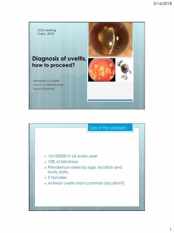
Diagnosis of uveitis,how to proceed?
Mohamed G.A Saleh
Lecturer of Ophthalmology
Assiut University
EOS meeting
Cairo, 2018
Size of the problem
15/100000 in US every year.
10% of blindness
Prevalence varies by age, location and
study date.
? Females
Anterior uveitis most common (location?)
3/16/2018
2
Challenges
• Uveitis is caused by diverse etiologies
• Often, clinical signs are shared by most
entities
• Sometimes, uveitis is a part of a disease
developing elsewhere in the body and it is
the first evidence of this disease
Failure to diagnose can lead to death
Even in the setting of isolated ocular
disease misdiagnosis can have a
devastating consequences
A patient with fungal endophthalmitis
thought to have a sterile traumatic
reaction may lose the eye if treated with
corticosteroids

3/16/2018
3
Principles of our work as
uveitis specialists
1- Distinguish infectious from non infectious
uveitis
2- Distinguish purely ocular disease from
systemic conditions
3- Obtain additional testing only if results will
influence your plan e.g. refer to the first
principle
Uveitis specialist must have thorough
knowledge of all uveitis entities and the
work up has to be complete
In addition few other challenges are
specific to practice in our locality:
1. Lack of good primary health care
2. Poor affordability
3. Poor compliance
3/16/2018
4
• Establishing an algorithmic approach in
reaching etiologic diagnosis might help
overcome these challenges by defining
the pathway to be taken
Steps in building an algorithmic approach
are:
1. Defining the problem by naming
technique
2. Review all possible causes of the problem
which fit the existing pattern
3. Proving the diagnosis by using the
diagnostic modalities in a logical matter

3/16/2018
5
General history-- Ocular examination
focused systemic history and exam-
liaise with rheumatologist/ pulmonologist.. etc now you should reach provisional
diagnosis and DDs- lab and imaging
modalities and sometimes therapeutic
response will confirm diagnosis

3/16/2018
6
Diagnosis relies on a process of recognition of
pattern
Collection of
findings from
HistoryClinical
exam
Investigations
1- Is the disease acute or chronic
2- Is inflammation granulomatous or Non granulomatous
3- Unilateral Vs. Bilateral disease?
4- Where is the inflammation located in the eye?
5- What associated signs are present of examination?
6-What systemic symptoms does the patient have?
7 -What type of patients do I deal with ?i.e. demographics
8-What is the time course of the disease and response to previous therapy?
It is helpful to ask yourself 8 questions

3/16/2018
7
By answering the previous questions you
could effectively name the uveitis e.g.
bilateral acute anterior recurrent
nongranulomatous iritis in a 28 year old
male patient with history of inflammatory
low back pain, oral ulcers and diarrhea
with marked improvement on steroid eye
drops.
Now, you should have a limited list of
possible differential diagnoses for this
particular scenario.
Then, it is time for targeted specific
investigations
3/16/2018
8
1- Is the disease acute or chronic
Onset: sudden vs insidious
Duration: limited (< 3 months) vs persistent(> 3 months)
Course:
Acute ( sudden onset+ limited duration)
Recurrent (flareups at intervals > 3 months after stopping treatment)
Chronic ( persistent OR flareups in < 3 months after stopping treatment)
SUN criteria: course
Acute disease: e.g. HLA-B27 AAU,
idiopathic AAU, some white dot
syndromes such as AMPPE, MEWDS, VKH
(early), ARN, Toxoplasmosis
Chronic E.g. JIA, birdshot, serpiginous, TB,
fungal, sympathetic ophthalmia,
lymphoma, MFC, sarcoidosis and pars
planitis

3/16/2018
9
2- Is inflammation granulomatous or Non
granulomatous Distinction based mainly on:
the appearance of KPs
Finding a choroidal granuloma
• Infections e.g. TB,
syphilis
• Sarcoidosis
• Sympathetic
ophthalmia
• Lens-induced uveitis
• Multiple sclerosis
• VKH
Finding granulomatous inflammation
suggests a unique set of possibilities
3/16/2018
10
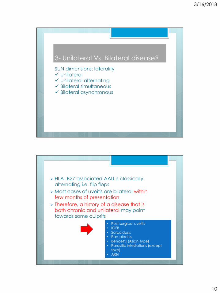
SUN dimensions: laterality
Unilateral
Unilateral alternating
Bilateral simultaneous
Bilateral asynchronous
3- Unilateral Vs. Bilateral disease?
HLA- B27 associated AAU is classically
alternating i.e. flip flops
Most cases of uveitis are bilateral within
few months of presentation
Therefore, a history of a disease that is
both chronic and unilateral may point
towards some culprits
• Post surgical uveitis
• IOFB
• Sarcoidosis
• Pars planitis
• Behcet’s (Asian type)
• Parasitic infestations (except
toxo)
• ARN

3/16/2018
11
4- Where is the inflammation located
in the eye?
Different causes for different locations
However, the diagnosis of intermediate
uveitis is the most helpful in establishing
diagnosis because it limits the possibilities
to few entities
• Sarcoidosis
• IBD
• Multiple sclerosis
• Pars planitis
• Lyme disease
SUN groupTesslerIUSG
--------Sclerouveitis---------
--------Keratouveitis--------
• Anterior uveitis
Iritis
ridocyclitis
• Anterior uveitis Iritis
Iridocyclitis
• Anterior uveitis
Iritis
Anterior Cyclitis
Anterior cyclitis--------------Iridocyclitis
• Intermediate uveitis
Pars planitis
Posterior cyclitis
Hyalitis
• Intermediate uveitis
Cyclitis
Vitritis
Pars planitis
• Intermediate uveitis Posterior cyclitis
Hyalitis
Basal retinochoroiditis
• Posterior uveitis
Focal, multifocal or
diffuse choroiditis
Chorioretinitis or
retinochoroiditis -
Retinitis
• Posterior Uveitis
Choroiditis
Retinitis
• Posterior Uveitis
Focal, multifocal or
diffuse choroiditis
Chorioretinitis or
retinochoroiditis
Neuroretinitis--------Neuroretinitis
Panuveitis----------Panuveitis
3/16/2018
12
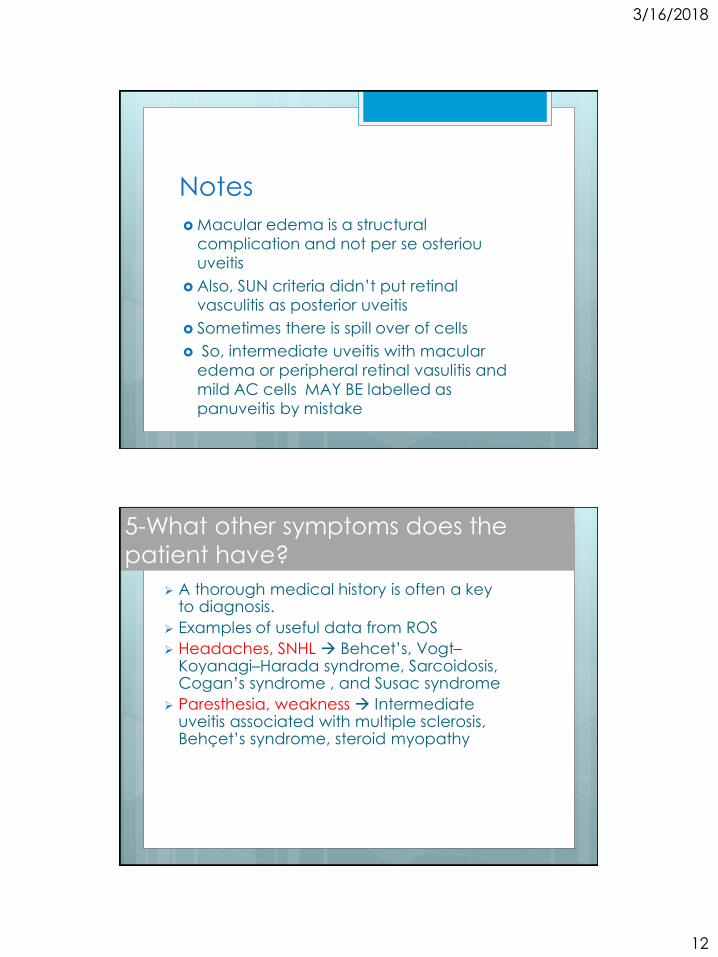
Notes
Macular edema is a structural
complication and not per se osteriou
uveitis
Also, SUN criteria didn’t put retinal
vasculitis as posterior uveitis
Sometimes there is spill over of cells
So, intermediate uveitis with macular
edema or peripheral retinal vasulitis and
mild AC cells MAY BE labelled as
panuveitis by mistake
5-What other symptoms does the
patient have?
A thorough medical history is often a key to diagnosis.
Examples of useful data from ROS
Headaches, SNHL Behcet’s, Vogt–Koyanagi–Harada syndrome, Sarcoidosis, Cogan’s syndrome , and Susac syndrome
Paresthesia, weakness Intermediate uveitis associated with multiple sclerosis, Behçet’s syndrome, steroid myopathy
3/16/2018
13
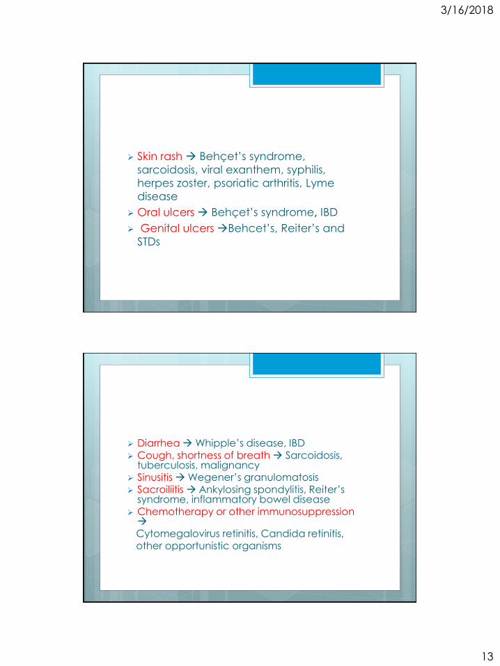
Skin rash Behçet’s syndrome,
sarcoidosis, viral exanthem, syphilis,
herpes zoster, psoriatic arthritis, Lyme
disease
Oral ulcers Behçet’s syndrome, IBD
Genital ulcers Behcet’s, Reiter’s and
STDs
Diarrhea Whipple’s disease, IBD Cough, shortness of breath Sarcoidosis,
tuberculosis, malignancy Sinusitis Wegener’s granulomatosis Sacroiliitis Ankylosing spondylitis, Reiter’s
syndrome, inflammatory bowel disease Chemotherapy or other immunosuppression
Cytomegalovirus retinitis, Candida retinitis,other opportunistic organisms

3/16/2018
14
Arthritis Behçet’s syndrome, Reiter’s
syndrome, sarcoidosis, juvenile
rheumatoid arthritis, rheumatoid arthritis,
Lyme disease, IBD, Wegener’s
granulomatosus (GPA), SLE, other
connective tissue diseases
6- What associated signs are present of
examination?
Psychosis Vogt–Koyanagi–Harada
syndrome,sarcoidosis, Behçet’s disease,
steroid psychosis, systemic lupus
erythematosus.
Cerebrospinal fluid pleocytosis Vogt–
Koyanagi–Harada syndrome, sarcoidosis,
acute posterior multifocal placoid
pigment epitheliopathy, Behçet’s
syndrome
3/16/2018
15
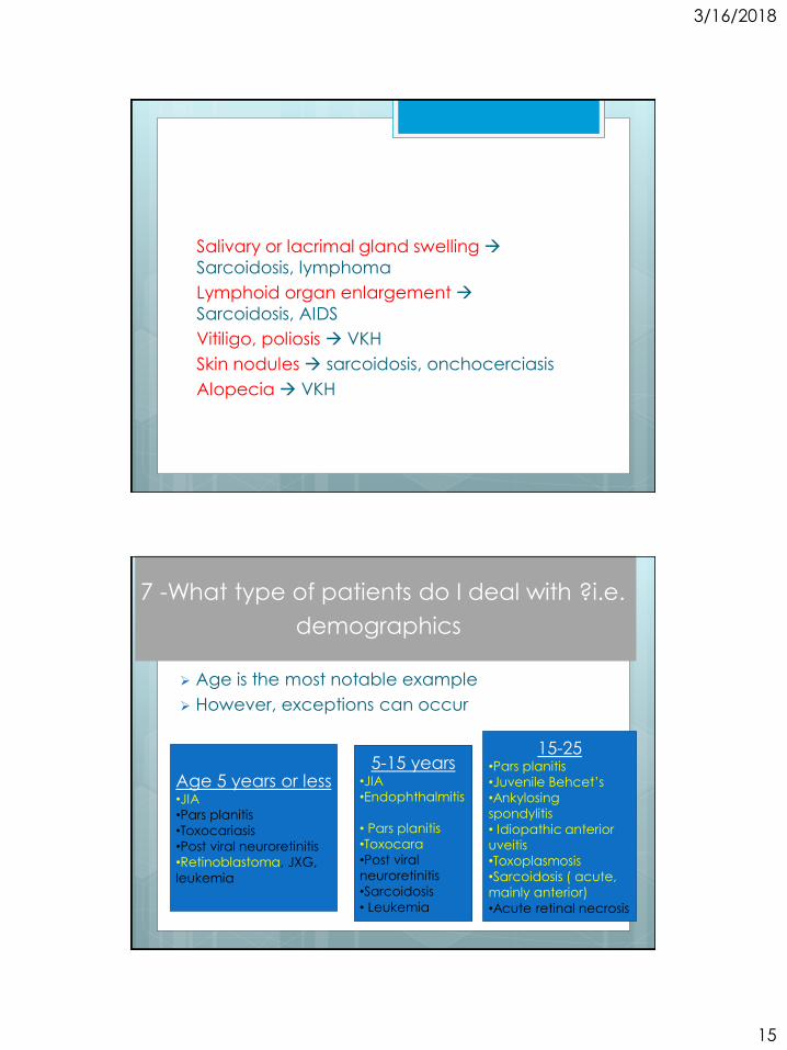
Salivary or lacrimal gland swelling
Sarcoidosis, lymphoma
Lymphoid organ enlargement
Sarcoidosis, AIDS
Vitiligo, poliosis VKH
Skin nodules sarcoidosis, onchocerciasis
Alopecia VKH
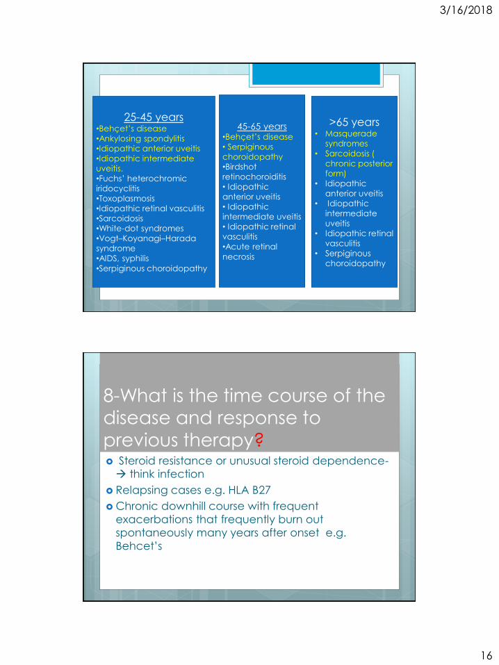
7 -What type of patients do I deal with ?i.e.
demographics
Age is the most notable example
However, exceptions can occur
Age 5 years or less•JIA
•Pars planitis
•Toxocariasis
•Post viral neuroretinitis
•Retinoblastoma, JXG,
leukemia
5-15 years•JIA
•Endophthalmitis
• Pars planitis
•Toxocara
•Post viral
neuroretinitis
•Sarcoidosis
• Leukemia
15-25•Pars planitis
•Juvenile Behcet’s
•Ankylosing
spondylitis
• Idiopathic anterior
uveitis
•Toxoplasmosis
•Sarcoidosis ( acute,
mainly anterior)
•Acute retinal necrosis
3/16/2018
16
25-45 years•Behçet’s disease
•Ankylosing spondylitis
•Idiopathic anterior uveitis
•Idiopathic intermediate
uveitis.
•Fuchs’ heterochromic
iridocyclitis
•Toxoplasmosis
•Idiopathic retinal vasculitis
•Sarcoidosis
•White-dot syndromes
•Vogt–Koyanagi–Harada
syndrome
•AIDS, syphilis
•Serpiginous choroidopathy
45-65 years•Behçet’s disease
• Serpiginous
choroidopathy
•Birdshot
retinochoroiditis
• Idiopathic
anterior uveitis
• Idiopathic
intermediate uveitis
• Idiopathic retinal
vasculitis
•Acute retinal
necrosis
>65 years• Masquerade
syndromes
• Sarcoidosis (
chronic posterior
form)
• Idiopathic
anterior uveitis
• Idiopathic
intermediate
uveitis
• Idiopathic retinal
vasculitis
• Serpiginous
choroidopathy
8-What is the time course of the
disease and response to
previous therapy? Steroid resistance or unusual steroid dependence- think infection
Relapsing cases e.g. HLA B27
Chronic downhill course with frequent
exacerbations that frequently burn out
spontaneously many years after onset e.g.
Behcet’s
3/16/2018
17
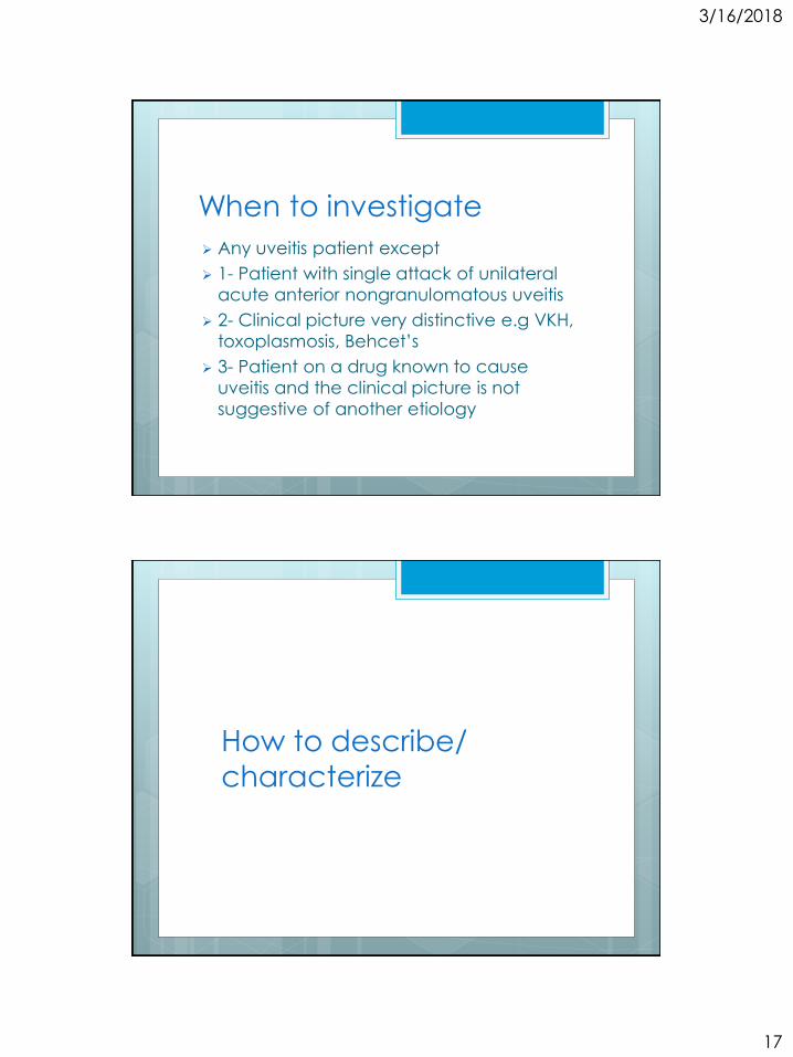
When to investigate
Any uveitis patient except
1- Patient with single attack of unilateral
acute anterior nongranulomatous uveitis
2- Clinical picture very distinctive e.g VKH,
toxoplasmosis, Behcet’s
3- Patient on a drug known to cause
uveitis and the clinical picture is not
suggestive of another etiology
How to describe/
characterize
3/16/2018
18
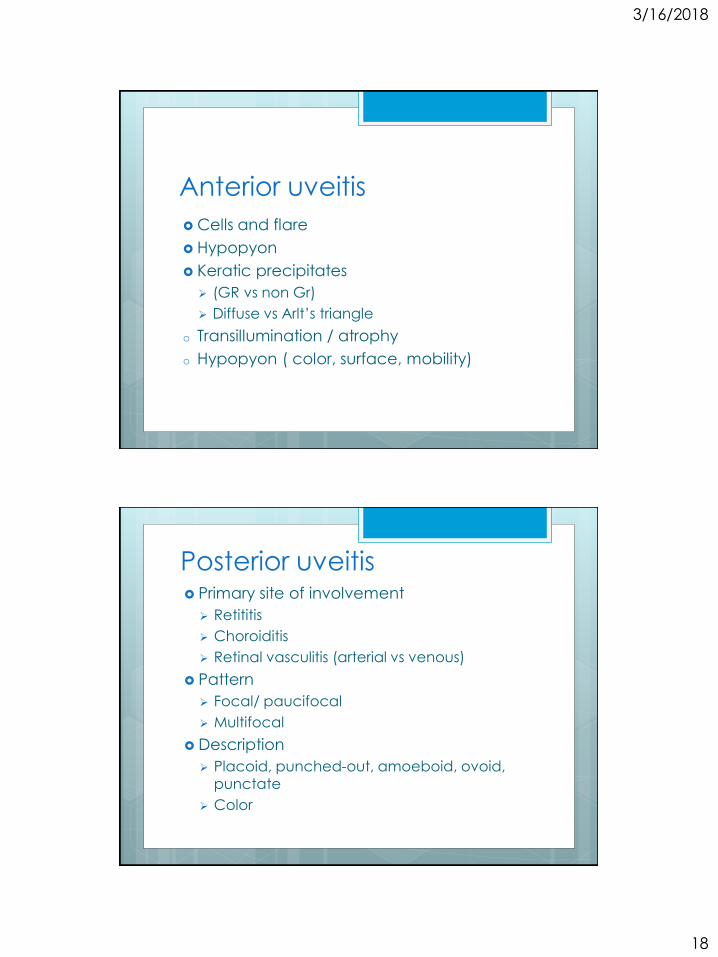
Anterior uveitis
Cells and flare
Hypopyon
Keratic precipitates
(GR vs non Gr)
Diffuse vs Arlt’s triangle
o Transillumination / atrophy
o Hypopyon ( color, surface, mobility)
Posterior uveitis Primary site of involvement
Retititis
Choroiditis
Retinal vasculitis (arterial vs venous)
Pattern
Focal/ paucifocal
Multifocal
Description
Placoid, punched-out, amoeboid, ovoid, punctate
Color

3/16/2018
19
Other characteristics
Ocular co morbidities
Response to treatment
Ethnicity
Medical conditions
Geographic location/ travel
Lab testing
Bayesian analysis
Sensitivity and specificity
Positive/ negative predictive value
Prior probability of the disease
Cost
Will it really matter( affect treatment,
prognosis)

3/16/2018
20
Rosenbaum JT, Wernick R. Arch Ophthalmol 1990;108:
1291-1293
E.g. Cost of ANA pannel is about 700
Dollars and it has low sensitivity and
specificity
TB accounts for less than 0.5% of uveitis in
the US . PPD has a sensitivity and
specificity of 75% and 85% respectively. So
if all uveitis patients in the U.S are
screened, the PPV of PPD is 1
However, in our region TB accounts for
about 15% of uveitis, so it is worth to
screen all patients with compatible uveitis
for TB
The reverse is true for syphilis

3/16/2018
21
Simplified algorithm
Anatomic location
Infectious vs non infectious
Ocular vs systemic
Possible combinations
Anterior uveitis
Infectious Non infectious
Ocular Systemic Ocular Systemic
Herpetic Syphilis FHI Behcet’s
TB UGH HLA related
Leprosy Post surgical Sarcoid
Traumatic JIA
TINU
3/16/2018
22
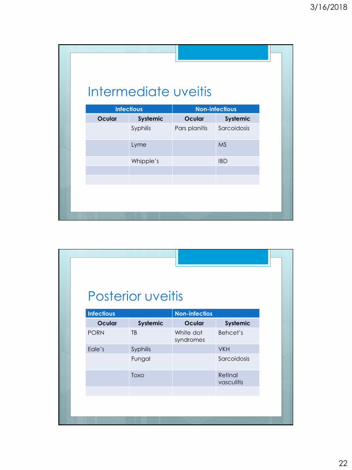
Intermediate uveitisInfectious Non-infectious
Ocular Systemic Ocular Systemic
Syphilis Pars planitis Sarcoidosis
Lyme MS
Whipple’s IBD
Posterior uveitisInfectious Non-infectios
Ocular Systemic Ocular Systemic
PORN TB White dot
syndromes
Behcet’s
Eale’s Syphilis VKH
Fungal Sarcoidosis
Toxo Retinal
vasculitis

3/16/2018
23
PanuveitisInfectious Non-infectious
Ocular Systemic Ocular Systemic
ARN Syphilis MFC-PU Behcet’s
Toxo CMV retinitis IOFB VKH
Post surgical TB Sympathetic
ophthalmia
Traumatic Metastatic
endophthal
mitis
Lymphoma
Conclusion
Ask/ examine/ think before your order
tests
Why….. To be cost effective
Consider PPV and NPV ( think of prior
probability not just specificity and
sensitivity)
Consider geography

3/16/2018
24
Thank you