dictyostelium lvsb mutants model the lysosomal defects ...656,2002.pdf · dictyostelium lvsb...
TRANSCRIPT
Molecular Biology of the CellVol. 13, 656–669, February 2002
Dictyostelium LvsB Mutants Model the LysosomalDefects Associated with Chediak-Higashi SyndromeEdward Harris,* Ning Wang,† Wei-l Wu,† Alisha Weatherford,* Arturo DeLozanne,† and James Cardelli*‡
*Department of Microbiology and Immunology, Louisiana State University Health Sciences Center,Shreveport, Louisiana 71130; and †Department of Molecular Cell and Developmental Biology andInstitute for Cell and Molecular Biology, University of Texas, Austin, Texas 78712
Submitted September 17, 2001; Revised October 29, 2001; Accepted November 13, 2001Monitoring Editor: Juan Bonifacino
Chediak-Higashi syndrome is a genetic disorder caused by mutations in a gene encoding a proteinnamed LYST in humans (“lysosomal trafficking regulator”) or Beige in mice. A prominent featureof this disease is the accumulation of enlarged lysosome-related granules in a variety of cells. Thegenome of Dictyostelium discoideum contains six genes encoding proteins that are related toLYST/Beige in amino acid sequence, and disruption of one of these genes, lvsA (large volumesphere), results in profound defects in cytokinesis. To better understand the function of this familyof proteins in membrane trafficking, we have analyzed mutants disrupted in lvsA, lvsB, lvsC, lvsD,lvsE, and lvsF. Of all these, only lvsA and lvsB mutants displayed interesting phenotypes in ourassays. lvsA-null cells exhibited defects in phagocytosis and contained abnormal looking contrac-tile vacuole membranes. Loss of LvsB, the Dictyostelium protein most similar to LYST/Beige,resulted in the formation of enlarged vesicles that by multiple criteria appeared to be acidiclysosomes. The rates of endocytosis, phagocytosis, and fluid phase exocytosis were normal inlvsB-null cells. Also, the rates of processing and the efficiency of targeting of lysosomal �-man-nosidase were normal, although lvsB mutants inefficiently retained �-mannosidase, as well as twoother lysosomal cysteine proteinases. Finally, results of pulse-chase experiments indicated that anincrease in fusion rates accounted for the enlarged lysosomes in lvsB-null cells, suggesting thatLvsB acts as a negative regulator of fusion. Our results support the notion that LvsB/LYST/Beigefunction in a similar manner to regulate lysosome biogenesis.
INTRODUCTION
Chediak-Higashi syndrome (CHS) is a rare autosomal reces-sive genetic disorder of humans that also occurs in othermammals, including cattle (Padgett, 1967), mice (Lutzner etal., 1967), rats (Nishimura et al., 1989), minks (Padgett, 1967),and killer whales (Ridgway, 1979). Patients with this disor-der suffer from partial albinism, excessive bleeding, andrecurrent bacterial infections.
The defining clinical manifestation of CHS is the pres-ence in a wide variety of cell types of enlarged lysosomesor granules that form as the result of a mutation in theLYST gene in humans (chromosome 1) or the beige gene inthe murine model (chromosome 13; Dufourcq-Lagelouseet al., 1999; Introne et al., 1999). A few models have beenproposed to explain the role of LYST in the formation ofenlarged lysosomes. The first model, based primarily onelectron microscope studies, hypothesizes that LYST nor-
mally acts as a negative regulator of homotypic and het-erotypic lysosome fusion, thus accounting for the increasein size of lysosomes in cells lacking LYST (Oliver andEssner, 1975). In addition, other studies have demon-strated that secretory lysosomes fuse to form giant lyso-somes in maturing CHS cytotoxic T lymphocytes (Stinch-combe et al., 2000). The second model proposes that LYSTis a positive regulator of fission, and in the absence ofLYST, the balance is tilted in favor of fusion, and largelysosomes accumulate (Burkhardt et al., 1993; Perou et al.,1997). In support of this model, it was observed thatoverexpression of LYST induced a more peripheral redis-tribution of smaller lysosomes. Finally, a third modelsuggests that LYST may regulate protein transport to lateendosomes; thus, trafficking defects could account for themorphological changes observed in lysosomes and lyso-some-related organelles (Faigle et al., 1998).
Although the CHS gene family is conserved in a widevariety of species, the amino acid sequence of the encodedgene products predicts little regarding the role of LYST/Beige in lysosome biogenesis or membrane trafficking.
DOI: 10.1091/mbc.01-09-0454.‡ Corresponding author. E-mail address: [email protected].
656 © 2002 by The American Society for Cell Biology
LYST/Beige comprises three distinct domains. The ami-no-terminal 80% of the protein has limited homology toother proteins in the database. This portion of the proteinis followed by the BEACH (Beige and Chediak-Higashi)domain, the defining consensus sequence for CHS-relatedproteins, and several WD-40 repeats, which are thought tobe important in protein-protein interaction. Several othermammalian gene products contain the BEACH andWD-40 domains, including FAN (factor associated withsphingomyelinase activation; Adam-Klages et al., 1996),neurobeachin (Wang et al., 2000), and CDC4L (Feuchter etal., 1992).
Dictyostelium discoideum will be a useful system to studythe function of BEACH/WD-40 domain-containing pro-teins related to LYST/Beige. First, a wide range of geneticand biochemical tools are available to explore the functionof this haploid organism (reviewed in a special issue ofBiochimica et Biophysica Acta (2001, Vol 1525). Second, theDictyostelium genome contains six genes, termed lvs (largevolume sphere) A through F, that encode proteins con-taining the BEACH and WD-40 domains that are relatedto the LYST/Beige family of proteins (Wang, N., Wu, W.,and DeLozanne, A., unpublished data ). LvsA is the firstmember of this family to be characterized and has beenfound to play an important role in cytokinesis and osmo-regulation (Kwak et al., 1999; Gerald et al., 2001). Third,the endolysosomal/phagosomal pathways in Dictyoste-lium are relatively well characterized and comparable torelated pathways in mammalian cells (reviewed byCardelli, 2001). Fluid phase enters the endosomal path-way primarily through macropinocytosis, although clath-rin-mediated micropinocytic internalization also occurs.Endosomes derived from the fission of macropinosomes,fuse to form acidic lysosomes that, in turn, fuse to formnonacidic secretory vesicles, termed postlysosomes (re-viewed by Maniak, 2001). A number of proteins have beenidentified that regulate fusion of lysosomes with eachother and with newly formed phagosomes (reviewed inRupper and Cardelli, 2001). In addition, it has been pro-posed that vesicles, containing membrane and solubleluminal proteins, form by fission and recycle from lateendosomal to earlier endocytic compartments or theplasma membrane. Proteins that regulate this process in-clude DdRab7, which may recycle membrane componentsfrom postlysosomes back to early endosomes and lyso-somes (Buczynski et al., 1997), and myosin I, which recy-cles membrane from early endosomes back to plasmamembrane (Neuhaus and Soldati, 2000).
Of the six known lvs genes in Dictyostelium, the lvsB geneencodes a protein most closely related in amino acid se-quence to the LYST/Beige protein. To determine whetherLvsB functions like LYST/Beige proteins, we biochemicallyand microscopically analyzed lvsA-null through lvsF-nullmutants. This report demonstrates that of the six proteinsonly LvsB appears to function like the LYST/Beige protein.Notably, lvsB-null mutants contain enlarged acidic lyso-somes that retain most, but not all, of the proteins found innormal lysosomes. Furthermore, these enlarged lysosomesappear to form as a result of an increase in the rate of vesiclefusion.
MATERIALS AND METHODS
Cell LinesDictyostelium strain NC4A2 was used as the control parental strain(kind gift of David Knecht). lvsA-null cell strain AD-63 (alias VIG9)was described previously (Kwak et al., 1999). All other Lvs-relatedknockout strains were made in NC4A2 as described by Wang, N.,Wu, W. and DeLozanne, A. (unpublished data). Briefly, knockoutconstructs were made using a blasticidin-resistance cassettebounded by two segments of each gene. Each construct was intro-duced into NC4A2 cells by electroporation, and clonal transfor-mants were selected in 96-well dishes. Individual clones werescreened for double-crossover insertion of the knockout constructby PCR, and the appropriate insertion was confirmed by Southernblot analysis.
MicroscopyTo visualize the entire endocytic pathway by fluorescence micros-copy, cells grown in T-25 tissue culture flasks were allowed to settleon 22- � 22-mm coverslips (Fisherbrand, Fisher Scientific, Hamp-ton, NH) in a six-well dish and were incubated for 2 h in HL-5growth medium (10.0 g proteose peptone [Oxide LTD., Basingstoke,Hampshire, United Kingdom], 10.0 g of glucose, 5.0 g yeast extract,0.19 g Na2HPO4, 0.35 g KH2PO4) containing 1.0 mg/ml fluoresceinisothiocyanate (FITC)-conjugated dextran (FD-70S, Sigma, St. Louis,MO). To view a subset of vesicles in the endocytic pathway, cellswere pulsed with FITC-dextran for 5 min, washed, and chased for 0min (to visualize macropinosomes), 20 min (lysosomes), or 60 min(postlysosomes). After the chase periods, cells were washed withfresh HL-5, fixed in 1% formaldehyde in HL-5 for 3 min, and viewedusing a fluorescence microscope (Olympus, Tokyo, Japan).
To assess acidification of vesicles, cells were allowed to settle oncoverslips in HL-5 and incubated with a 1:100 dilution of DND-189Lysosensor (Molecular Probes, Eugene, OR) in HL-5 for 2–3 min.Lysosensor fluoresces only in acidic compartments (5.2 pKa), andlive cells were visualized in the fluorescein channel.
For immunofluorescence microscopy, cells grown in T-25 flasks inlog phase were allowed to settle on 22- � 22-mm coverslips insix-well plates for 20 min, fixed, and permeabilized as described byBush et al. (1994). Cells were incubated with the primary (B832,anti–100-kDa pump) and secondary antibodies (Texas Red goatanti-mouse; Jackson Laboratories, Bar Harbor, ME), each for 1 h at4°C. Coverslips were washed and mounted on slides and visualizedusing an Olympus BX-50 fluorescence microscope.
Endocytosis, Phagocytosis, and Exocytosis AssaysFluid phase endocytosis and exocytosis rates were measured usingFITC-dextran, as described by Temesvari et al. (1996a). Phagocytosisrates were measured using fluorescent latex beads as described byTemesvari et al. (2000).
Purification of LysosomesLysosomes were purified as described before with some modifica-tions (Temesvari et al., 1994). Cells were pulsed for 15 min with anequal volume of HL-5 media containing 2.0 mg/ml iron dextran,washed twice in cold HL-5, and chased in fresh media for 15 min.Harvested cells were resuspended to 2.0 � 108 cells/ml in sucrosebuffer (5.0 mM glycine, 100 mM sucrose, pH 8.5) with a proteaseinhibitor cocktail (10 �g/ml leupeptin, 20 �g/ml chymostatin, 20�g/ml pepstatin, 5 mg/ml aprotinin, 100 mM Na-p-tosyl-l-lysinechloromethyl ketone) and were broken by passage through two5-�m pore polycarbonate filters (Poretics, Livermore, CA). Post-nuclear supernatants were pumped through the column of finemesh wire (25 �m diameter; Goodfellow, Malvern, PA) at 70 ml/h.After washing, lysosomes were recovered from the wire mesh using
A Beige-like Protein Regulates Endosome Fusion
Vol. 13, February 2002 657
a 5-ml pipette and centrifuged in 35-ml Oakridge tubes at 39,000 �g at 4°C for 30 min. The sucrose buffer was aspirated and thelysosomes were resuspended in Laemmli buffer (Laemmli, 1970),heated to 65°C for 5 min, and subjected to SDS-PAGE or stored at�80°C.
Western Blot AnalysisSDS-PAGE and Western blot analysis were done as described byBuczynski et al. (1997). The membranes were blocked overnight inTBSTG (10 mM Tris base, 150 mM NaCl, 0.1% gelatin (Knox), 0.1%Tween-20, pH 7.5) and exposed to the following antibodies thatwere diluted in TBSTG: mouse anti-100-kDa subunit of the vacuolarH�-ATPase (B832, 1:200; Fok et al., 1993); rabbit anti-41-kDa subunitof the vacuolar H�-ATPase (LSU 18-2, 1:2000; Temesvari et al.,1996b); rabbit anti–�-mannosidase (LSU 27-2, 1:5000; J. Cardelli,unpublished results); rabbit anti-D. discoideum Rab7 (LSU 7-2-4,1:200; Buczynski et al., 1997); affinity pure rabbit anti-RabB (LSU1125-1-2, 1:200; J. Cardelli, unpublished results,); rabbit anti-RabD(Rab14 homolog, LSU 4-7-2, 1:200; Bush et al., 1994); anti-cathepsinD (1:500; Journet et al., 1999); anti-CPp36, which recognizes cysteineproteinase p36, (1:1000; a kind gift from J. Garin); anti-LmpA, whichrecognizes D. discoideum LmpA, (1:1000; Karakesisoglou et al., 1999);AD7.5, which recognizes N-acetylglucose 1-phosphate–modifiedcysteine proteinases (1:1000; kind gift of Hudson Freeze); anti-acidphosphatase (5E1, 1:250); anti–�-glucosidase (AP8, 1:250). All blotswere exposed to the same mixture of alkaline phosphatase-conju-gated secondary antibodies diluted in TBSTG (both goat anti-mouse, 1:3000; catalog no. 170-6520, Bio-Rad, Hercules, CA; andgoat anti-rabbit, 1:30,000; catalog no. A-3687, Sigma). The blots weredeveloped in NBT buffer (100 mM Tris base, 100 mM NaCl, 5 mMMgCl2) containing 0.6 mM nitroblue tetrazolium (Sigma) and 1.2mM 5-bromo-4-choro-3-indolyl phosphate (ICN Biomedicals,Cleveland, OH). Densitometry was calculated using a Bio-Rad im-ager with Quantity One Quantitation software, version 4.1.0 (Bio-Rad Laboratories, Hercules, CA).
�-Mannosidase ProcessingRadiolabeling of cells, immunoprecipitation of �-mannosidase,SDS-PAGE, and fluorography were done as described by Buczynskiet al. (1997).
Endosome/Phagosome Fusion AssayTwo methods were used to determine the fusion of either endo-somes or phagosomes. To determine the rate of fusion of endo-somes, cells were allowed to settle on coverslips placed in six-wellplates and incubated for 5 min with 1.5 mg/ml rhodamine isothio-
cyanate (RITC)-conjugated dextran (catalog no. R-9379, Sigma) inHL-5. Cells were washed twice with fresh HL-5 and incubated with1.5 mg/ml FITC-dextran for 5 min. Coverslips were washed oncewith fresh HL-5 and chased for 10 min in HL-5, and cells were fixedwith 1% formaldehyde in HL-5 for 3 min. Coverslips were mountedon slides and visualized using both the fluorescein and rhodaminechannels. Images of 50–100 cells were electronically captured usingan Olympus upright fluorescence microscope equipped with a dig-ital camera (Sensy, Brussels, Belgium). Images were analyzed usingMetaView software (Universal Imaging, Dowingtown, PA).
For phagosome fusion, we used FITC-labeled bacteria (fl-bacteria)to visualize individual phagosomes. Cells were placed in shakingsuspension and allowed to phagocytose fl-bacteria for 10 min andthen washed three times with fresh HL-5. Cells were allowed tosettle on coverslips in HL-5 to initiate the 30-min chase period.Coverslips were gently immersed in fresh HL-5 to wash the remain-ing fl-bacteria off, and then the cells were fixed in 1% formaldehydein HL-5. Coverslips were immersed again in HL-5 to wash off theremaining fl-bacteria and mounted on coverslips to be examinedwith the fluorescent microscope. Cells (50–100) with �10 fl-bacteriaper cell were examined using a 100� oil objective, and the numberof fl-bacteria in each phagosome was assessed using both phase-contrast and fluorescent filters. Fusion events are typically easy toobserve because the phagosome contains some soluble fluorescentFITC that clearly marks the inner edge of the vacuole and demon-strates that a single compartment contains multiple bacteria.
RESULTS
Dictyostelium Has Six Proteins Related to theMammalian LYST/Beige ProteinsA search of the nearly complete Dictyostelium genome data-base identified six genes encoding BEACH domain-contain-ing proteins (Wang, N., Wu, W. and DeLozanne, A., unpub-lished data). Of these, lvsA was already characterized for itsessential role in cytokinesis and contractile vacuole (CV)function (Kwak et al., 1999; Gerald et al., 2001). Thus, thefamily of Dictyostelium genes was named lvsA through lvsF.All of the proteins encoded by these six genes share a highdegree of similarity to each other but only in the BEACHand WD domain regions (Wang, N., Wu, W. and DeLoz-anne, A., unpublished data).
The BEACH and WD domains from the DictyosteliumLvsA–F proteins were compared with those from relatedproteins from many other species. Blast scores and sequence
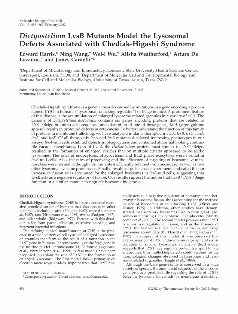
Figure 1. LvsB is the Dictyosteliumprotein most similar to mammalianLYST/Beige. Sequence comparisonof the BEACH and WD domains ofthe six Dictyostelium Lvs proteinsand proteins from other organisms.The BEACH and WD domains ofthe indicated proteins were alignedusing DNAStar with the ClustalWalgorithm. The position of LvsB isindicated with an asterisk. Note itsclose relationship to mammalianLYST proteins. Accession codes areindicated for each protein. A.t., Ara-bidopsis thaliana; C.e., Caenorhabditiselegans; D.d., Dictyostelium discoi-deum; D.m., Drosophila melanogaster.
Harris et al.
Molecular Biology of the Cell658
comparison by the Clustal method consistently indicated thesequence similarities illustrated in Figure 1. LvsB is theDictyostelium protein most closely related to the mammalianLYST/Beige proteins. LvsF is related to the mammalianprotein FAN and the other Lvs proteins are related to pre-dicted proteins of unknown function in other species.
To understand the function of this family of proteins, wedisrupted each of the Dictyostelium lvsB–F genes by homol-ogous recombination. Initial analysis of the phenotype ofeach mutant indicated that, other than lvsA-null mutants,none of them are essential for cytokinesis, cell growth, os-moregulation, or development (Wang, N., Wu, W. and De-Lozanne, A., unpublished data). Interestingly, the lvsB mu-tants displayed morphological differences that stimulatedfurther study.
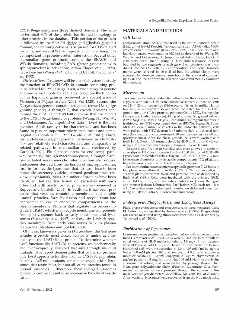
lvsB-Null Cells Accumulate Enlarged EndosomalVesiclesWild-type cells, and lvsA through lvsF-null mutants wereexamined using an inverted microscope equipped withphase-contrast optics. The most striking difference in mor-
phology observed was an increase in the number of enlargedvacuolar structures observed in lvsB-null cells (Figure 2,compare E with A and C). These vacuoles were observed inthe lvsB-null mutants at all growth densities in axenic mediaand over a culturing period of several months. The majorityof the enlarged vacuoles in lvsB-null cells were typically �2�m in size; vacuoles of this size were seldom seen in controland lvsA-null cells. The doubling times for all three strains intissue culture flasks were comparable, indicating that theenlarged vacuoles did not influence division rate of cells.
The enlarged vacuoles could be derived from the endoso-mal pathway or from the CV system of membranes. Todistinguish between these two possibilities, we incubatedcells in growth medium with the fluid phase marker FITC-dextran for 1 h to label the entire endosomal pathway,including macropinosomes, endosomes, lysosomes, andpostlysosomes; internalized FITC-dextran is not traffickedinto the CV compartments (Gabriel et al., 1999). Fluorescencemicroscopy indicated that all of the enlarged vacuoles in thelvsB-null mutant contained FITC-dextran, confirming thatthese vacuoles were part of the endosomal pathway (Figure2F). Control (Figure 2B) and lvsA-null cells (Figure 2D) con-
Figure 2. The enlarged vesicles in thelvsB-null mutant are endocytic. NC4A2control (A and B), lvsA-null (C and D),and lvsB-null (E and F) cells were incu-bated in HL-5 medium containing FITC-dextran for 1 h to illuminate the endocyticvesicles. Cells were visualized using a flu-orescence microscope. The arrows pointto enlarged endocytic vesicles in lvsB-nullcells, and the arrowheads point to vesiclesthe approximate size of normal lyso-somes. Bar, 1 �m.
A Beige-like Protein Regulates Endosome Fusion
Vol. 13, February 2002 659
tained the predicted normal range in size of fluorescentendosomal vesicles (0.2–1.0 �m).
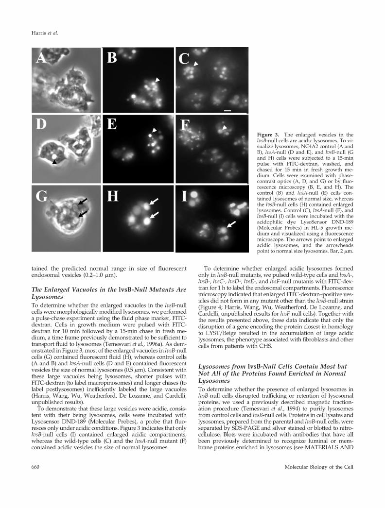
The Enlarged Vacuoles in the lvsB-Null Mutants AreLysosomesTo determine whether the enlarged vacuoles in the lvsB-nullcells were morphologically modified lysosomes, we performeda pulse-chase experiment using the fluid phase marker, FITC-dextran. Cells in growth medium were pulsed with FITC-dextran for 10 min followed by a 15-min chase in fresh me-dium, a time frame previously demonstrated to be sufficient totransport fluid to lysosomes (Temesvari et al., 1996a). As dem-onstrated in Figure 3, most of the enlarged vacuoles in lvsB-nullcells (G) contained fluorescent fluid (H), whereas control cells(A and B) and lvsA-null cells (D and E) contained fluorescentvesicles the size of normal lysosomes (0.5 �m). Consistent withthese large vacuoles being lysosomes, shorter pulses withFITC-dextran (to label macropinosomes) and longer chases (tolabel postlysosomes) inefficiently labeled the large vacuoles(Harris, Wang, Wu, Weatherford, De Lozanne, and Cardelli,unpublished results).
To demonstrate that these large vesicles were acidic, consis-tent with their being lysosomes, cells were incubated withLysosensor DND-189 (Molecular Probes), a probe that fluo-resces only under acidic conditions. Figure 3 indicates that onlylvsB-null cells (I) contained enlarged acidic compartments,whereas the wild-type cells (C) and the lvsA-null mutant (F)contained acidic vesicles the size of normal lysosomes.
To determine whether enlarged acidic lysosomes formedonly in lvsB-null mutants, we pulsed wild-type cells and lvsA-,lvsB-, lvsC-, lvsD-, lvsE-, and lvsF-null mutants with FITC-dex-tran for 1 h to label the endosomal compartments. Fluorescencemicroscopy indicated that enlarged FITC-dextran–positive ves-icles did not form in any mutant other than the lvsB-null strain(Figure 4; Harris, Wang, Wu, Weatherford, De Lozanne, andCardelli, unpublished results for lvsF-null cells). Together withthe results presented above, these data indicate that only thedisruption of a gene encoding the protein closest in homologyto LYST/Beige resulted in the accumulation of large acidiclysosomes, the phenotype associated with fibroblasts and othercells from patients with CHS.
Lysosomes from lvsB-Null Cells Contain Most butNot All of the Proteins Found Enriched in NormalLysosomesTo determine whether the presence of enlarged lysosomes inlvsB-null cells disrupted trafficking or retention of lysosomalproteins, we used a previously described magnetic fraction-ation procedure (Temesvari et al., 1994) to purify lysosomesfrom control cells and lvsB-null cells. Proteins in cell lysates andlysosomes, prepared from the parental and lvsB-null cells, wereseparated by SDS-PAGE and silver stained or blotted to nitro-cellulose. Blots were incubated with antibodies that have allbeen previously determined to recognize luminal or mem-brane proteins enriched in lysosomes (see MATERIALS AND
Figure 3. The enlarged vesicles in thelvsB-null cells are acidic lysosomes. To vi-sualize lysosomes, NC4A2 control (A andB), lvsA-null (D and E), and lvsB-null (Gand H) cells were subjected to a 15-minpulse with FITC-dextran, washed, andchased for 15 min in fresh growth me-dium. Cells were examined with phase-contrast optics (A, D, and G) or by fluo-rescence microscopy (B, E, and H). Thecontrol (B) and lvsA-null (E) cells con-tained lysosomes of normal size, whereasthe lvsB-null cells (H) contained enlargedlysosomes. Control (C), lvsA-null (F), andlvsB-null (I) cells were incubated with theacidophilic dye LysoSensor DND-189(Molecular Probes) in HL-5 growth me-dium and visualized using a fluorescencemicroscope. The arrows point to enlargedacidic lysosomes, and the arrowheadspoint to normal size lysosomes. Bar, 2 �m.
Harris et al.
Molecular Biology of the Cell660
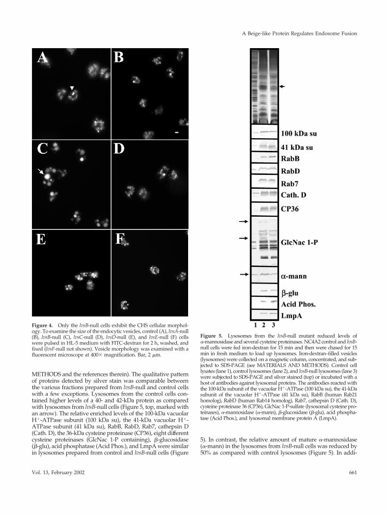
METHODS and the references therein). The qualitative patternof proteins detected by silver stain was comparable betweenthe various fractions prepared from lvsB-null and control cellswith a few exceptions. Lysosomes from the control cells con-tained higher levels of a 40- and 42-kDa protein as comparedwith lysosomes from lvsB-null cells (Figure 5, top, marked withan arrow). The relative enriched levels of the 100-kDa vacuolarH�-ATPase subunit (100 kDa su), the 41-kDa vacuolar H�-ATPase subunit (41 kDa su), RabB, RabD, Rab7, cathepsin D(Cath. D), the 36-kDa cysteine proteinase (CP36), eight differentcysteine proteinases (GlcNac 1-P containing), �-glucosidase(�-glu), acid phosphatase (Acid Phos.), and LmpA were similarin lysosomes prepared from control and lvsB-null cells (Figure
5). In contrast, the relative amount of mature �-mannosidase(�-mann) in the lysosomes from lvsB-null cells was reduced by50% as compared with control lysosomes (Figure 5). In addi-
Figure 4. Only the lvsB-null cells exhibit the CHS cellular morphol-ogy. To examine the size of the endocytic vesicles, control (A), lvsA-null(B), lvsB-null (C), lvsC-null (D), lvsD-null (E), and lvsE-null (F) cellswere pulsed in HL-5 medium with FITC-dextran for 2 h, washed, andfixed (lvsF-null not shown). Vesicle morphology was examined with afluorescent microscope at 400� magnification. Bar, 2 �m.
Figure 5. Lysosomes from the lvsB-null mutant reduced levels of�-mannosidase and several cysteine proteinases. NC4A2 control and lvsB-null cells were fed iron-dextran for 15 min and then were chased for 15min in fresh medium to load up lysosomes. Iron-dextran–filled vesicles(lysosomes) were collected on a magnetic column, concentrated, and sub-jected to SDS-PAGE (see MATERIALS AND METHODS). Control celllysates (lane 1), control lysosomes (lane 2), and lvsB-null lysosomes (lane 3)were subjected to SDS-PAGE and silver stained (top) or incubated with ahost of antibodies against lysosomal proteins. The antibodies reacted withthe 100-kDa subunit of the vacuolar H�-ATPase (100 kDa su), the 41-kDasubunit of the vacuolar H�-ATPase (41 kDa su), RabB (human Rab21homolog), RabD (human Rab14 homolog), Rab7, cathepsin D (Cath. D),cysteine proteinase 36 (CP36), GlcNac 1-P-sulfate (lysosomal cysteine pro-teinases), �-mannosidase (�-mann), �-glucosidase (�-glu), acid phospha-tase (Acid Phos.), and lysosomal membrane protein A (LmpA).
A Beige-like Protein Regulates Endosome Fusion
Vol. 13, February 2002 661
tion, two proteins detectable in control lysosomes by an anti-body (anti-GlcNac 1-P) that recognizes N-acetylglucosamine1-phosphate (Souza et al., 1997) were not detected in blots of thelysosomes from lvsB-null cells (Figure 5).
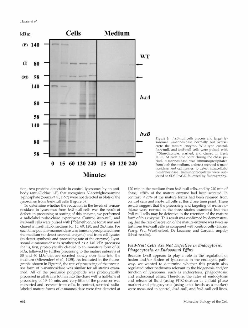
To determine whether the reduction in the levels of �-man-nosidase in lysosomes from lvsB-null cells was the result ofdefects in processing or sorting of this enzyme, we performeda radiolabel pulse-chase experiment. Control, lvsA-null, andlvsB-null cells were pulsed with [35S]methionine for 20 min andchased in fresh HL-5 medium for 15, 60, 120, and 240 min. Foreach time point, �-mannosidase was immunoprecipitated fromthe medium (to detect secreted enzyme) and from cell lysates(to detect synthesis and processing rate of the enzyme). Lyso-somal �-mannosidase is synthesized as a 140 kDa precursorthat is, first, proteolytically cleaved to an immature form of 80kDa, followed by further processing to the mature subunits of58 and 60 kDa that are secreted slowly over time into themedium (Mierendorf et al., 1985). As indicated in the fluoro-graphs shown in Figure 6, the rate of processing of the precur-sor form of �-mannosidase was similar for all strains exam-ined. All of the precursor polypeptide was proteolyticallyprocessed in all strains 60 min into the chase with a half-time ofprocessing of 10–15 min, and very little of the precursor wasmissorted and secreted from cells. In contrast, secreted radio-labeled mature forms of �-mannosidase were first detected at
120 min in the medium from lvsB-null cells, and by 240 min ofchase, �50% of the mature enzyme had been secreted. Incontrast, �25% of the mature forms had been released fromcontrol cells and lvsA-null cells at this chase time point. Theseresults suggest that the processing and targeting of �-manno-sidase were normal in the three strains examined but thatlvsB-null cells may be defective in the retention of the matureform of this enzyme. This result was confirmed by demonstrat-ing that the rate of secretion of the mature enzyme was twice asfast from lvsB-null cells as compared with control cells (Harris,Wang, Wu, Weatherford, De Lozanne, and Cardelli, unpub-lished results).
lvsB-Null Cells Are Not Defective in Endocytosis,Phagocytosis, or Endosomal EffluxBecause LvsB appears to play a role in the regulation offusion and/or fission of lysosomes in the endocytic path-way, we wanted to determine whether this protein alsoregulated other pathways relevant to the biogenesis and/orfunction of lysosomes, such as endocytosis, phagocytosis,and endosomal efflux. Therefore, the rates of endocytosisand release of fluid (using FITC-dextran as a fluid phasemarker) and phagocytosis (using latex beads as a marker)were measured in control, lvsA-null, and lvsB-null cell lines.
Figure 6. lvsB-null cells process and target ly-sosomal �-mannosidase normally but overse-crete the mature enzyme. Wild-type control,lvsA-null, and lvsB-null cells were pulsed with[35S]methionine, washed, and chased in freshHL-5. At each time point during the chase pe-riod, �-mannosidase was immunoprecipitatedfrom both the medium, to detect secreted �-man-nosidase, and cell lysates, to detect intracellular�-mannosidase. Immunoprecipitates were sub-jected to SDS-PAGE, followed by fluorography.
Harris et al.
Molecular Biology of the Cell662
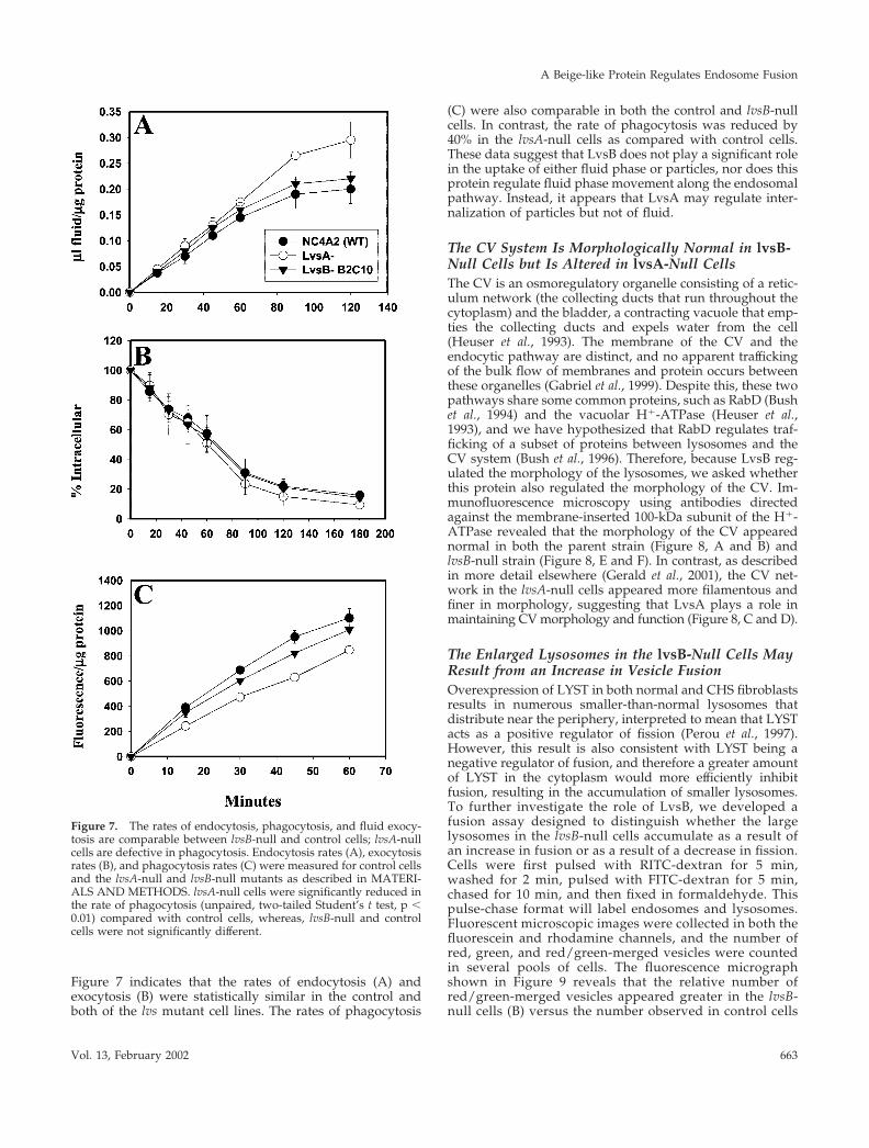
Figure 7 indicates that the rates of endocytosis (A) andexocytosis (B) were statistically similar in the control andboth of the lvs mutant cell lines. The rates of phagocytosis
(C) were also comparable in both the control and lvsB-nullcells. In contrast, the rate of phagocytosis was reduced by40% in the lvsA-null cells as compared with control cells.These data suggest that LvsB does not play a significant rolein the uptake of either fluid phase or particles, nor does thisprotein regulate fluid phase movement along the endosomalpathway. Instead, it appears that LvsA may regulate inter-nalization of particles but not of fluid.
The CV System Is Morphologically Normal in lvsB-Null Cells but Is Altered in lvsA-Null CellsThe CV is an osmoregulatory organelle consisting of a retic-ulum network (the collecting ducts that run throughout thecytoplasm) and the bladder, a contracting vacuole that emp-ties the collecting ducts and expels water from the cell(Heuser et al., 1993). The membrane of the CV and theendocytic pathway are distinct, and no apparent traffickingof the bulk flow of membranes and protein occurs betweenthese organelles (Gabriel et al., 1999). Despite this, these twopathways share some common proteins, such as RabD (Bushet al., 1994) and the vacuolar H�-ATPase (Heuser et al.,1993), and we have hypothesized that RabD regulates traf-ficking of a subset of proteins between lysosomes and theCV system (Bush et al., 1996). Therefore, because LvsB reg-ulated the morphology of the lysosomes, we asked whetherthis protein also regulated the morphology of the CV. Im-munofluorescence microscopy using antibodies directedagainst the membrane-inserted 100-kDa subunit of the H�-ATPase revealed that the morphology of the CV appearednormal in both the parent strain (Figure 8, A and B) andlvsB-null strain (Figure 8, E and F). In contrast, as describedin more detail elsewhere (Gerald et al., 2001), the CV net-work in the lvsA-null cells appeared more filamentous andfiner in morphology, suggesting that LvsA plays a role inmaintaining CV morphology and function (Figure 8, C and D).
The Enlarged Lysosomes in the lvsB-Null Cells MayResult from an Increase in Vesicle FusionOverexpression of LYST in both normal and CHS fibroblastsresults in numerous smaller-than-normal lysosomes thatdistribute near the periphery, interpreted to mean that LYSTacts as a positive regulator of fission (Perou et al., 1997).However, this result is also consistent with LYST being anegative regulator of fusion, and therefore a greater amountof LYST in the cytoplasm would more efficiently inhibitfusion, resulting in the accumulation of smaller lysosomes.To further investigate the role of LvsB, we developed afusion assay designed to distinguish whether the largelysosomes in the lvsB-null cells accumulate as a result ofan increase in fusion or as a result of a decrease in fission.Cells were first pulsed with RITC-dextran for 5 min,washed for 2 min, pulsed with FITC-dextran for 5 min,chased for 10 min, and then fixed in formaldehyde. Thispulse-chase format will label endosomes and lysosomes.Fluorescent microscopic images were collected in both thefluorescein and rhodamine channels, and the number ofred, green, and red/green-merged vesicles were countedin several pools of cells. The fluorescence micrographshown in Figure 9 reveals that the relative number ofred/green-merged vesicles appeared greater in the lvsB-null cells (B) versus the number observed in control cells
Figure 7. The rates of endocytosis, phagocytosis, and fluid exocy-tosis are comparable between lvsB-null and control cells; lvsA-nullcells are defective in phagocytosis. Endocytosis rates (A), exocytosisrates (B), and phagocytosis rates (C) were measured for control cellsand the lvsA-null and lvsB-null mutants as described in MATERI-ALS AND METHODS. lvsA-null cells were significantly reduced inthe rate of phagocytosis (unpaired, two-tailed Student’s t test, p �0.01) compared with control cells, whereas, lvsB-null and controlcells were not significantly different.
A Beige-like Protein Regulates Endosome Fusion
Vol. 13, February 2002 663
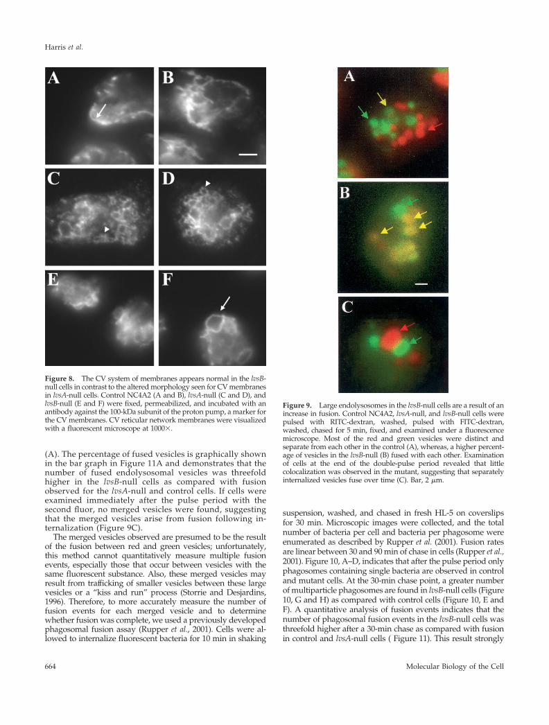
(A). The percentage of fused vesicles is graphically shownin the bar graph in Figure 11A and demonstrates that thenumber of fused endolysosomal vesicles was threefoldhigher in the lvsB-null cells as compared with fusionobserved for the lvsA-null and control cells. If cells wereexamined immediately after the pulse period with thesecond fluor, no merged vesicles were found, suggestingthat the merged vesicles arise from fusion following in-ternalization (Figure 9C).
The merged vesicles observed are presumed to be the resultof the fusion between red and green vesicles; unfortunately,this method cannot quantitatively measure multiple fusionevents, especially those that occur between vesicles with thesame fluorescent substance. Also, these merged vesicles mayresult from trafficking of smaller vesicles between these largevesicles or a “kiss and run” process (Storrie and Desjardins,1996). Therefore, to more accurately measure the number offusion events for each merged vesicle and to determinewhether fusion was complete, we used a previously developedphagosomal fusion assay (Rupper et al., 2001). Cells were al-lowed to internalize fluorescent bacteria for 10 min in shaking
suspension, washed, and chased in fresh HL-5 on coverslipsfor 30 min. Microscopic images were collected, and the totalnumber of bacteria per cell and bacteria per phagosome wereenumerated as described by Rupper et al. (2001). Fusion ratesare linear between 30 and 90 min of chase in cells (Rupper et al.,2001). Figure 10, A–D, indicates that after the pulse period onlyphagosomes containing single bacteria are observed in controland mutant cells. At the 30-min chase point, a greater numberof multiparticle phagosomes are found in lvsB-null cells (Figure10, G and H) as compared with control cells (Figure 10, E andF). A quantitative analysis of fusion events indicates that thenumber of phagosomal fusion events in the lvsB-null cells wasthreefold higher after a 30-min chase as compared with fusionin control and lvsA-null cells ( Figure 11). This result strongly
Figure 8. The CV system of membranes appears normal in the lvsB-null cells in contrast to the altered morphology seen for CV membranesin lvsA-null cells. Control NC4A2 (A and B), lvsA-null (C and D), andlvsB-null (E and F) were fixed, permeabilized, and incubated with anantibody against the 100-kDa subunit of the proton pump, a marker forthe CV membranes. CV reticular network membranes were visualizedwith a fluorescent microscope at 1000�.
Figure 9. Large endolysosomes in the lvsB-null cells are a result of anincrease in fusion. Control NC4A2, lvsA-null, and lvsB-null cells werepulsed with RITC-dextran, washed, pulsed with FITC-dextran,washed, chased for 5 min, fixed, and examined under a fluorescencemicroscope. Most of the red and green vesicles were distinct andseparate from each other in the control (A), whereas, a higher percent-age of vesicles in the lvsB-null (B) fused with each other. Examinationof cells at the end of the double-pulse period revealed that littlecolocalization was observed in the mutant, suggesting that separatelyinternalized vesicles fuse over time (C). Bar, 2 �m.
Harris et al.
Molecular Biology of the Cell664
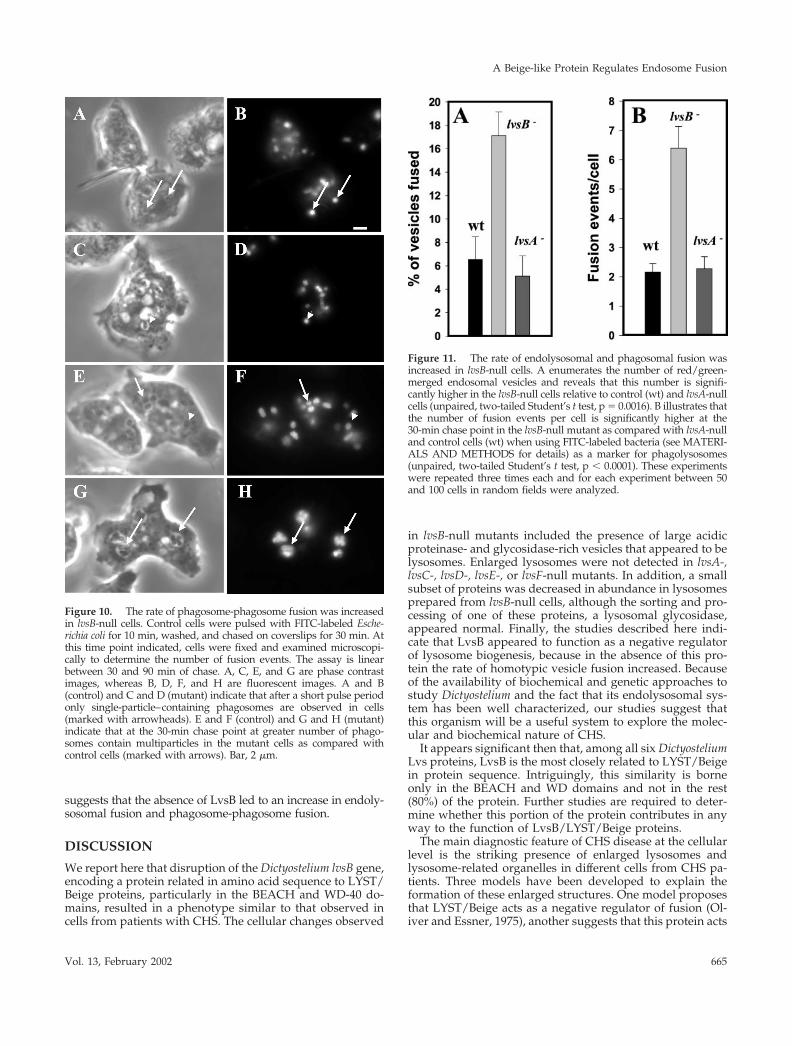
suggests that the absence of LvsB led to an increase in endoly-sosomal fusion and phagosome-phagosome fusion.
DISCUSSION
We report here that disruption of the Dictyostelium lvsB gene,encoding a protein related in amino acid sequence to LYST/Beige proteins, particularly in the BEACH and WD-40 do-mains, resulted in a phenotype similar to that observed incells from patients with CHS. The cellular changes observed
in lvsB-null mutants included the presence of large acidicproteinase- and glycosidase-rich vesicles that appeared to belysosomes. Enlarged lysosomes were not detected in lvsA-,lvsC-, lvsD-, lvsE-, or lvsF-null mutants. In addition, a smallsubset of proteins was decreased in abundance in lysosomesprepared from lvsB-null cells, although the sorting and pro-cessing of one of these proteins, a lysosomal glycosidase,appeared normal. Finally, the studies described here indi-cate that LvsB appeared to function as a negative regulatorof lysosome biogenesis, because in the absence of this pro-tein the rate of homotypic vesicle fusion increased. Becauseof the availability of biochemical and genetic approaches tostudy Dictyostelium and the fact that its endolysosomal sys-tem has been well characterized, our studies suggest thatthis organism will be a useful system to explore the molec-ular and biochemical nature of CHS.
It appears significant then that, among all six DictyosteliumLvs proteins, LvsB is the most closely related to LYST/Beigein protein sequence. Intriguingly, this similarity is borneonly in the BEACH and WD domains and not in the rest(80%) of the protein. Further studies are required to deter-mine whether this portion of the protein contributes in anyway to the function of LvsB/LYST/Beige proteins.
The main diagnostic feature of CHS disease at the cellularlevel is the striking presence of enlarged lysosomes andlysosome-related organelles in different cells from CHS pa-tients. Three models have been developed to explain theformation of these enlarged structures. One model proposesthat LYST/Beige acts as a negative regulator of fusion (Ol-iver and Essner, 1975), another suggests that this protein acts
Figure 10. The rate of phagosome-phagosome fusion was increasedin lvsB-null cells. Control cells were pulsed with FITC-labeled Esche-richia coli for 10 min, washed, and chased on coverslips for 30 min. Atthis time point indicated, cells were fixed and examined microscopi-cally to determine the number of fusion events. The assay is linearbetween 30 and 90 min of chase. A, C, E, and G are phase contrastimages, whereas B, D, F, and H are fluorescent images. A and B(control) and C and D (mutant) indicate that after a short pulse periodonly single-particle–containing phagosomes are observed in cells(marked with arrowheads). E and F (control) and G and H (mutant)indicate that at the 30-min chase point at greater number of phago-somes contain multiparticles in the mutant cells as compared withcontrol cells (marked with arrows). Bar, 2 �m.
Figure 11. The rate of endolysosomal and phagosomal fusion wasincreased in lvsB-null cells. A enumerates the number of red/green-merged endosomal vesicles and reveals that this number is signifi-cantly higher in the lvsB-null cells relative to control (wt) and lvsA-nullcells (unpaired, two-tailed Student’s t test, p � 0.0016). B illustrates thatthe number of fusion events per cell is significantly higher at the30-min chase point in the lvsB-null mutant as compared with lvsA-nulland control cells (wt) when using FITC-labeled bacteria (see MATERI-ALS AND METHODS for details) as a marker for phagolysosomes(unpaired, two-tailed Student’s t test, p � 0.0001). These experimentswere repeated three times each and for each experiment between 50and 100 cells in random fields were analyzed.
A Beige-like Protein Regulates Endosome Fusion
Vol. 13, February 2002 665
as a positive regulator of fission (Burkhardt et al., 1993;Perou et al., 1997), and a third model hypothesizes thatLYST/Beige regulates vesicle trafficking (Faigle et al., 1998),a process that contributes to lysosome morphology and size.
Early morphological and biochemical studies supportedthe first model and suggested that these large lysosomesformed from the homotypic fusion of lysosomes or the fu-sion of lysosomes with prelysosomal endocytic vesicles (Iri-majiri et al., 1992; Jones et al., 1992). Furthermore, fusion canaccount for identical and nonidentical membrane-enclosedorganelles in neutrophils, suggesting that vesicles along dif-ferent stages of maturation in the endolysosomal pathwaywere fusing together. Finally, homotypic fusion of lyso-somes and phagosomes is not a process confined to CHS orBeige cells but in fact occurs in normal cells (Tjelle et al.,2000). Together, these data can be interpreted to support arole for LYST/Beige as a negative regulator of endolysoso-mal fusion processes that occur normally in cells.
More recently, it has been proposed that the formation oflarge lysosomes in CHS cells may be the result of a reductionin the rate of fission or budding of vesicles from maturinglysosomes that have undergone fusion. First, it was demon-strated that lysosomes and late endosomes fuse to form ahybrid organelle in normal cells and that fission reactionsrestore both late endosomes and lysosomes (Luzio et al.,2000). Second, when the LYST protein is overexpressed inCHS fibroblasts, large lysosomes disappear with the emer-gence of smaller-than-normal granules (Perou et al., 1997).
The experiments described here support a role for theLvsB protein as a negative regulator of fusion of lysosomes.First, our results demonstrate that the enlarged vesicles inlvsB-null mutants were lysosomes. These enlarged struc-tures contained fluid phase markers (Figure 2), they wereacidic (Figure 3), and they contained a host of proteinspreviously demonstrated to be enriched in normal lyso-somes (Figure 5). In addition, these large vesicles receivedinternalized fluid with the same kinetics as normal lyso-somes (Figure 3). Second, we found that the rate of homo-typic fusion of phagosomes and endolysosomes was signif-icantly increased in lvsB-null cells relative to control cellsand lvsA-, lvsC-, lvsD-, lvsE-, and lvsF-null mutants (Figure9). Although our studies do not exclude the possibility thatLvsB also acts to regulate fission, they more strongly sup-port the idea that LvsB, (and by analogy, LYST/Beige),normally acts to negatively regulate the rate of fusion ofendolysosomes.
How might LvsB and LYST/Beige proteins act to down-regulate fusion of endolysosomal vesicles? One possibility isthat LYST-like proteins may inhibit the formation of tethering/docking complexes, necessary to bring vesicles close together,or SNARE complexes, necessary for membrane fusion. Noth-ing currently known about the BEACH or WD-40 domainssuggests an interaction with docking or fusion-promoting pro-teins, although this needs to be investigated.
Another possible function for LYST/Beige/LvsB proteinscould be to act as a molecular scaffold to regulate a signalingpathway that modifies the vesicle membrane to regulatefusion (Ward et al., 2000). One possible signaling pathwaymay involve the formation of ceramide. Ceramide levels areknown to increase in Beige cells, and ceramide has beendemonstrated to regulate endosome fusion. Intriguingly,FAN has BEACH and WD-40 domains related to LYST/
Beige and is known to interact with a neutral sphingomy-elinase to regulate the production of ceramide. Thus, it ispossible that LYST/Beige interacts with sphingomyelinaseto generate ceramide that in turn regulates vesicle fusion.LvsB/LYST/Beige might also alter the levels of other mem-brane-associated lipids, including phosphoinositides, whichcould alter vesicle fusion rates.
LvsB and LYST/Beige proteins may also regulate the traf-ficking of lysosomes and endosomes along cytoskeletalstructures such as microtubules and F-actin. Movement oflysosomes from the periphery of cells to a more juxtanuclearposition may increase the likelihood of docking and fusion.In fact, overexpression of the Beige protein in human fibro-blasts results in smaller-than-normal lysosomes and a moreperipheral distribution of lysosomes (Perou et al., 1997). TheLYST/Beige proteins could negatively regulate proteins likeRab7, a small GTPase implicated in the trafficking and fusionof lysosomes in a juxtanuclear position in the cell (Bucci etal., 2000; Caplan et al., 2001). Alternatively, LYST/Beige-likeproteins could positively regulate proteins like Rab27a (Ba-hadoran et al., 2001;Hume et al., 2001) or myosin V (Wu et al.,2001), both implicated in the F-actin- and microtubular-dependent trafficking of melanosomes to the periphery ofmelanocytes. Dictyostelium RabD, a Rab14-like GTPase (Har-ris, and Cardelli, unpublished results), and two phospho-inositide 3-kinases (Rupper et al., 2001) have been implicatedin the regulation of lysosome-lysosome and phagosome-phagosome fusion and potentially could be targets for theactivity of LvsB.
The study presented here also revealed only minor defects inprotein trafficking in lvsB-null cells, even although this mutantcontained enlarged lysosomes. Of �20 proteins assayed, onlymature lysosomal �-mannosidase and two cysteine proteinaseswere inefficiently retained in mutant cells. The oversecretion of�-mannosidase was not the result of missorting, because thenewly synthesized protein was processed and sorted correctlyin lvsB-null cells. Finally, the rates of endocytosis, phagocytosisand fluid phase exocytosis were also normal in lvsB-null cells,consistent with only minor defects in trafficking of membraneand protein. Changes in the rate of secretion of �-mannosidasebut not other hydrolases that reside in the same organelle havebeen observed before in clathrin-null mutants (Ruscetti et al.,1994). Lysosomal enzymes are probably retained in Dictyoste-lium by a process that involves the recycling of these enzymesfrom lysosomes and postlysosomes (a secretory compartment)back to early endosomes (Buczynski et al., 1997). In contrast,100% of the internalized fluid is released from postlysosomes.The large size of lysosomes may inhibit the efficient recyclingand retention of some lysosomal enzymes.
The observations of only subtle defects in retention or traf-ficking of lysosomal proteins in lvsB-null mutants are similar tothe conclusions reached by others in the study of CHS or Beigecells. For instance, it has been reported that acidic �-mannosi-dase levels are lower in CHS cells as compared with controls(Tanaka, 1980) and that only two lysosomal proteinases, elas-tase and cathepsin G, were deficient in Beige neutrophils(Takeuchi et al., 1986). Furthermore, it was demonstrated thatthe processing and targeting of lysosomal cathepsin D and thedelivery of proteins to lysosomes were normal in cytotoxic Tcells from patients with CHS, even though these cells containedenlarged vacuoles and were defective in cytotoxic T-lympho-cyte–mediated functions (Stinchcombe et al., 2000).
Harris et al.
Molecular Biology of the Cell666
Minor trafficking defects can, however, have profound ef-fects. For instance, peptide loading onto the major histocom-patibility complex type II molecules and antigen presentationis delayed in the B cells of CHS patients (Faigle et al., 1998). Inaddition, CTLA-4, a negative regulator of T-cell activation(Karandikar et al., 1996) is normally enriched in endocyticvesicles and the secretory granules, whereas it is found in thelarge granules of the T cells from patients with CHS. Confor-mationally altered CTLA-4 is found on the surface of cells,leading to a disruption of T-cell homeostasis and possiblycontributing to the accelerated phase in CHS patients (Barrat etal., 1999). The altered conformation of CTLA-4 may be theresult of decreased processing of this protein in lysosomesbecause of a missorted processing enzyme.
LvsA, also closely related to LYST/Beige, has been proposedto regulate membrane trafficking to the contractile ring, a pro-cess important during cytokinesis (Kwak et al., 1999). As re-ported here, lvsA-null cells, but not lvsB-null cells, were defec-tive in phagocytosis, although lvsA-null cells were normal inother endosomal processes, including lysosome biogenesis.LvsA is thus added to a growing list of Dictyostelium proteinsthat regulate phagocytosis (Cardelli, 2001).
Another known role of LvsA is in the function of the CVduring osmoregulation (Gerald et al., 2001). The CV network ofmembranes was also altered in lvsA-null mutants and ap-peared finer and more vesicular in structure as compared withthe more coarse tubular-vesicular structures observed in con-trol cells or lvsB-null mutants. The possible connection betweenthe morphology of the CV network and the regulation ofphagocytosis is intriguing, because we have observed thatoverexpression of two different dominant negative GTPases,RabDN121I (Bush et al., 1996) and Rab11N126I (Harris et al., 2001),alters the CV network and affects phagocytosis. Rates of phago-cytosis are increased in cells expressing Rab11N126I, and the CVnetwork appears “thicker” in morphology, perhaps because ofan increase in the amount of CV membrane present. In con-trast, expression of RabDN121I results in decreases in the ratesof phagocytosis and the formation of a reduced patch or clusterof vesicular CV structures next to the plasma membrane(Harris et al., 2001 ).
We have proposed that the CV network in Dictyostelium mayfunction like the recycling endosomal compartment functionsin mammalian cells. Also, as proposed for recycling endosomalcompartments, we hypothesize that the CV network of mem-branes may regulate internalization of particles by supplyingmembrane to help generate the phagocytic cup (Cardelli, 2001).The CV network and phagocytosis have also been linked inTetrahymena pyriformis. A 71-kDa protein, associated with theactin-binding proteins, localizes to both the CV and oral appa-ratus responsible for food uptake, suggesting that a connectionmay exist between the membranes involved for internalizationand osmoregularity in this single-celled organism (Watanabe etal., 1998). We are currently investigating possible traffickingpathways between the CV membranes and the phagosomes inDictyostelium.
In conclusion, we have demonstrated that LvsA and LvsB,two proteins that are related to LYST/Beige and containBEACH and WD-40 domains, play a role in the regulation ofphagocytosis and lysosome biogenesis, respectively. In con-trast, null mutants of LvsC, LvsD, LvsE, and LvsF containednormal size lysosomes, and preliminary results suggest thatendosomal processes also function normally. Future studies
will focus on defining the molecular functions of LvsB andLvsA and on investigating the possible membrane-traffick-ing defects that exist in these other lvs mutants.
ACKNOWLEDGMENTS
The authors would like to thank members of the DeLozanne andCardelli laboratories for careful reading of the manuscript. Thisresearch was supported by National Institutes of Health grant DK-39232 (to J.C.).
REFERENCES
Adam-Klages, S., Adam, D., Wiegmann, K., Struve, S., Kolanus, W.,Schneider-Mergener, J., and Kronke, M. (1996). FAN, a novel WD-repeat protein, couples the p55 TNF-receptor to neutral sphingomy-elinase. Cell 86, 937–947.
Bahadoran, P., Aberdam, E., Mantoux, F., Busca, R., Bille, K., Yal-man, N., de Saint-Basile, G., Casaroli-Marano, R., Ortonne, J.P., andBallotti, R. (2001). Rab27a: a key to melanosome transport in humanmelanocytes. J. Cell Biol. 152, 843–850.
Barrat, F.J., Le Deist, F., Benkerrou, M., Bousso, P., Feldmann, J.,Fischer, A., and de Saint, B. (1999). Defective CTLA-4 cycling path-way in Chediak-Higashi syndrome: a possible mechanism for de-regulation of T lymphocyte activation. Proc. Natl. Acad. Sci. USA 96,8645–8650.
Bucci, C., Thomsen, P., Nicoziani, P., McCarthy, J., and van Deurs,B. (2000). Rab7: a key to lysosome biogenesis. Mol. Biol. Cell 11,467–480.
Buczynski, G., Bush, J., Zhang, L., Rodriguez-Paris, J., and Cardelli,J. (1997). Evidence for a recycling role for Rab7 in regulating a latestep in endocytosis and in retention of lysosomal enzymes in Dic-tyostelium discoideum. Mol. Biol. Cell 8, 1343–1360.
Burkhardt, J.K., Wiebel, F.A., Hester, S., and Argon, Y. (1993). Thegiant organelles in beige and Chediak-Higashi fibroblasts are de-rived from late endosomes and mature lysosomes. J. Exp. Med. 178,1845–1856.
Bush, J., Nolta, K., Rodriguez-Paris, J., Kaufmann, N., O’Halloran,T., Ruscetti, T., Temesvari, L., Steck, T., and Cardelli, J. (1994). ARab4-like GTPase in Dictyostelium discoideum colocalizes withV-H(�)-ATPases in reticular membranes of the contractile vacuolecomplex and in lysosomes. J. Cell Sci. 107, 2801–2812.
Bush, J., Temesvari, L., Rodriguez-Paris, J., Buczynski, G., andCardelli, J. (1996). A role for a Rab4-like GTPase in endocytosis andin regulation of contractile vacuole structure and function in Dic-tyostelium discoideum. Mol. Biol. Cell 7, 1623–1638.
Caplan, S., Hartnell, L.M., Aguilar, R.C., Naslavsky, N., and Boni-facino, J.S. (2001). Human Vam6p promotes lysosome clusteringand fusion in vivo. J. Cell Biol. 154, 109–122.
Cardelli, J. (2001). Phagocytosis and macropinocytosis in Dictyoste-lium: phosphoinositide-based processes, biochemically distinct.Traffic 2, 311–320.
Dufourcq-Lagelouse, R., Lambert, N., Duval, M., Viot, G., Vilmer,E., Fischer, A., Prieur, M., and de Saint, B. (1999). Chediak-Higashisyndrome associated with maternal uniparental isodisomy of chro-mosome 1. Eur. J. Hum. Genet. 7, 633–637.
Faigle, W., Raposo, G., Tenza, D., Pinet, V., Vogt, A.B., Kropshofer,H., Fischer, A., de Saint-Basile, G., and Amigorena, S. (1998). Defi-cient peptide loading and MHC class II endosomal sorting in a
A Beige-like Protein Regulates Endosome Fusion
Vol. 13, February 2002 667
human genetic immunodeficiency disease: the Chediak-Higashisyndrome. J. Cell Biol. 141, 1121–1134.
Feuchter, A.E., Freeman, J.D., and Mager, D.L. (1992). Strategy fordetecting cellular transcripts promoted by human endogenous longterminal repeats: identification of a novel gene (CDC4L) with ho-mology to yeast CDC4. Genomics 13, 1237–1246.
Fok, A.K., Clarke, M., Ma, L., and Allen, R.D. (1993). VacuolarH(�)-ATPase of Dictyostelium discoideum: a monoclonal antibodystudy. J. Cell Sci. 106, 1103–1113.
Gabriel, D., Hacker, U., Kohler, J., Muller-Taubenberger, A.,Schwartz, J.M., Westphal, M., and Gerisch, G. (1999). The contractilevacuole network of Dictyostelium as a distinct organelle: its dynam-ics visualized by a GFP marker protein. J. Cell Sci. 112, 3995–4005.
Gerald, N., Siano, M., and DeLozanne, A. (2002). The DictyosteliumLusA protein is localized on the contractile vacuole and is requiredfor osmoregulation. Traffic 3, 50–60.
Harris, E., Yoshida, K., Cardelli, J., and Bush, J. (2001). Rab11-likeGTPase associates with and regulates the structure and function ofthe contractile vacuole system in Dictyostelium. J. Cell Sci. 114,3035–3045.
Heuser, J., Zhu, Q., and Clarke, M. (1993). Proton pumps populatethe contractile vacuoles of Dictyostelium amoebae. J. Cell Biol. 121,1311–1327.
Hume, A.N., Collinson, L.M., Rapak, A., Gomes, A.Q., Hopkins,C.R., and Seabra, M.C. (2001). Rab27a regulates the peripheral dis-tribution of melanosomes in melanocytes. J. Cell Biol. 152, 795–808.
Introne, W., Boissy, R.E., and Gahl, W.A. (1999). Clinical, molecular,and cell biological aspects of Chediak-Higashi syndrome. Mol.Genet. Metab. 68, 283–303.
Irimajiri, K., Iwamoto, I., Kawanishi, K., Tsuji, K., Morita, S., Koyama,A., Hamazaki, H., Horiuchi, F., Horiuchi, A., and Akiyama, T. (1992).Studies on pseudo-Chediak-Higashi granules formation in acute pro-myelocytic leukemia. Rinsho Ketsueki 33, 1057–1065.
Jones, K., Steward, R., Fowler, M., Fukuda, M., and Holcombe, R.(1992). Chediak-Higashi lymphoblastoid cell lines: granule charac-teristics and expression of lysosome-associated membrane proteins.Clin. Immunol. Immunopathol. 65, 219–226.
Journet, A., Chapel, A., Jehan, S., Adessi, C., Freeze, H., Klein, G.,and Garin, J. (1999). Characterization of Dictyostelium discoideumcathepsin D. J. Cell Sci. 112, 3833–3843.
Karakesisoglou, I., Janssen, K.P., Eichinger, L., Noegel, A.A., andSchleicher, M. (1999). Identification of a suppressor of the Dictyoste-lium profilin-minus phenotype as a CD36/LIMP-II homologue.J. Cell Biol. 145, 167–181.
Karandikar, N.J., Vanderlugt, C.L., Walunas, T.L., Miller, S.D., andBluestone, J.A. (1996). CTLA-4: a negative regulator of autoimmunedisease. J. Exp. Med. 184, 783–788.
Kwak, E., Gerald, N., Larochelle, D.A., Vithalani, K.K., Niswonger,M.L., Maready, M., and De Lozanne, A. (1999). LvsA, a proteinrelated to the mouse beige protein, is required for cytokinesis inDictyostelium. Mol. Biol. Cell 10, 4429–4439.
Laemmli, U.K. (1970). Cleavage of structural proteins during theassembly of the head of bacteriophage T4. Nature 227, 680–685.
Lutzner, M.A., Lowrie, C.T., and Jordan, H.W. (1967). Giant gran-ules in leukocytes of the beige mouse. J. Hered. 58, 299–300.
Luzio, J.P., Rous, B.A., Bright, N.A., Pryor, P.R., Mullock, B.M., andPiper, R.C. (2000). Lysosome-endosome fusion and lysosome bio-genesis. J. Cell Sci. 113, 1515–1524.
Maniak, M. (2001). Fluid-phase uptake and transit in axenic Dictyo-stelium cells. Biochim. Biophys. Acta 1525, 197–204.
Mierendorf, R.C.J., Cardelli, J.A., and Dimond, R.L. (1985). Path-ways involved in targeting and secretion of a lysosomal enzyme inDictyostelium discoideum. J. Cell Biol. 100, 1777–1787.
Neuhaus, E.M., and Soldati, T. (2000). A myosin I is involved inmembrane recycling from early endosomes. J. Cell Biol. 150, 1013–1026.
Nishimura, M., Inoue, M., Nakano, T., Nishikawa, T., Miyamoto,M., Kobayashi, T., and Kitamura, Y. (1989). Beige rat: a new animalmodel of Chediak-Higashi syndrome. Blood 74, 270–273.
Oliver, C., and Essner, E. (1975). Formation of anomalous lysosomesin monocytes, neutrophils, and eosinophils from bone marrow ofmice with Chediak-Higashi syndrome. Lab. Invest. 32, 17–27.
Padgett, G.A. (1967). Neutrophilic function in animals with theChediak-Higashi syndrome. Blood 29, 906–915.
Perou, C.M., Leslie, J.D., Green, W., Li, L., Ward, D.M., and Kaplan,J. (1997). The Beige/Chediak-Higashi syndrome gene encodes awidely expressed cytosolic protein. J. Biol. Chem. 272, 29790–29794.
Ridgway, S.H. (1979). Reported causes of death of captive killerwhales (Orcinus orca). J. Wildl. Dis. 15, 99–104.
Rupper, A., and Cardelli, J. (2001). Regulation of phagocytosis andendo-phagosomal trafficking pathways in Dictyostelium discoideum.Biochim. Biophys. Acta 1525, 205–216.
Rupper, A.C., Rodriguez-Paris, J.M., Grove, B.D., and Cardelli, J.A.(2001). p110-related PI 3-kinases regulate phagosome-phagosomefusion, and phagosomal pH through a PKB/Akt dependent path-way in Dictyostelium. J. Cell Sci. 114, 1283–1295.
Ruscetti, T., Cardelli, J.A., Niswonger, M.L., and O’Halloran, T.J. (1994).Clathrin heavy chain functions in sorting and secretion of lysosomalenzymes in Dictyostelium discoideum. J. Cell Biol. 126, 343–352.
Souza, G.M., Mehta, D.P., Lammertz, M., Rodriguez-Paris, J., Wu,R., Cardelli, J.A., and Freeze, H.H. (1997). Dictyostelium lysosomalproteins with different sugar modifications sort to functionally dis-tinct compartments. J. Cell Sci. 110, 2239–2248.
Stinchcombe, J.C., Page, L.J., and Griffiths, G.M. (2000). Secretorylysosome biogenesis in cytotoxic T lymphocytes from normal andChediak Higashi syndrome patients. Traffic 1, 435–444.
Storrie, B., and Desjardins, M. (1996). The biogenesis of lysosomes:is it a kiss and run, continuous fusion and fission process? Bioessays18, 895–903.
Takeuchi, K., Wood, H., and Swank, R.T. (1986). Lysosomal elastaseand cathepsin G in beige mice: neutrophils of beige (Chediak-Higashi) mice selectively lack lysosomal elastase and cathepsin G. J.Exp. Med. 163, 665–677.
Tanaka, T. (1980). Chediak-Higashi syndrome: abnormal lysosomalenzyme levels in granulocytes of patients and family members.Pediatr. Res. 14, 901–904.
Temesvari, L., Rodriguez-Paris, J., Bush, J., Steck, T.L., and Cardelli,J. (1994). Characterization of lysosomal membrane proteins of Dic-tyostelium discoideum: a complex population of acidic integral mem-brane glycoproteins, Rab GTP-binding proteins and vacuolar AT-Pase subunits. J. Biol. Chem. 269, 25719–25727.
Temesvari, L., Zhang, L., Fodera, B., Janssen, K.P., Schleicher, M.,and Cardelli, J.A. (2000). Inactivation of lmpA, encoding a LIMPII-related endosomal protein, suppresses the internalization and en-dosomal trafficking defects in profilin-null mutants. Mol. Biol. Cell11, 2019–2031.
Temesvari, L.A., Bush, J.M., Peterson, M.D., Novak, K.D., Titus,M.A., and Cardelli, J.A. (1996a). Examination of the endosomal andlysosomal pathways in Dictyostelium discoideum myosin I mutants.J. Cell Sci. 109, 663–673.
Temesvari, L.A., Rodriguez-Paris, J.M., Bush, J.M., Zhang, L., andCardelli, J.A. (1996b). Involvement of the vacuolar proton-translo-
Harris et al.
Molecular Biology of the Cell668
cating ATPase in multiple steps of the endo-lysosomal system andin the contractile vacuole system of Dictyostelium discoideum. J. CellSci. 109, 1479–1495.
Tjelle, T.E., Lovdal, T., and Berg, T. (2000). Phagosome dynamicsand function. Bioessays 22, 255–263.
Wang, X., Herberg, F.W., Laue, M.M., Wullner, C., Hu, B., Petrasch-Parwez, E., and Kilimann, M.W. (2000). Neurobeachin: a proteinkinase A-anchoring, beige/Chediak-Higashi protein homolog im-plicated in neuronal membrane traffic. J. Neurosci. 20, 8551–8565.
Ward, D.M., Griffiths, G.M., Stinchcombe, J.C., and Kaplan, J. (2000).Analysis of the lysosomal storage disease Chediak-Higashi syn-drome. Traffic 1, 816–822.
Watanabe, A., Kurasawa, Y., Watanabe, Y., and Numata, O. (1998).A new Tetrahymena actin-binding protein is localized in the divisionfurrow. J. Biochem. (Tokyo) 123, 607–613.
Wu, X., Rao, K., Bowers, M.B., Copeland, N.G., Jenkins, N.A., and Ham-mer, J.A. (2001). Rab27a enables myosin Va-dependent melanosome cap-ture by recruiting the myosin to the organelle. J. Cell Sci. 114, 1091–1100.
A Beige-like Protein Regulates Endosome Fusion
Vol. 13, February 2002 669