diers gesamtbroschüre a4 36seitig en · 3 diers international gmbh is an innovative, family owned...
TRANSCRIPT
www.diers.de
EN
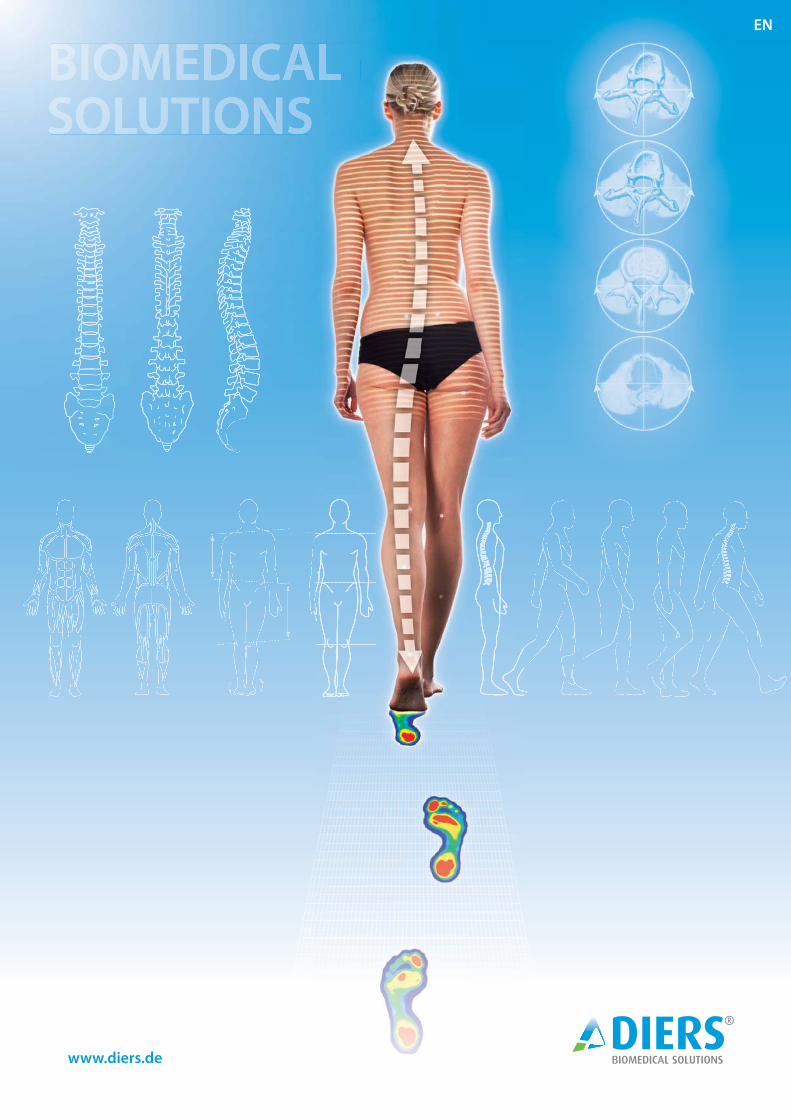
BIOMEDICAL SOLUTIONS

2
3D/4D Spine & Posture Analysis ................................ p. 04 ff .
Video Gait Analysis (Leg Axis Geometry) ...................... p.13
Foot Pressure Measurement .................................... p.16-18
Muscle Strength Measurement .............................. p. 26 / 27
Whole Body Motion Analysis ....................................... p. 20 ff .
Treatment Proposals ................ p. 30 / 31
Content:

3
DIERS International GmbH is an innovative, family owned and operated enterprise which has successfully designed, developed, produ-ced, distributed, installed and serviced biome-chanical measuring systems since 1996.
From the beginning, we have engaged in close scientifi c cooperation with German and foreign universities to guarantee continuous developments at high technical and scientifi c levels for our customers.
The objective of DIERS is to off er the market a comprehensive biomechanical product port-folio for complete head to toe analysis of the human body. During the course of product development we place great value on inter-disciplinary utilization by various professional groups such as orthopedists, orthopedic tech-nicians, physical therapists, dentists, ortho-dontists, sports medicine specialists, etc.
All DIERS products pass through a deman-ding research and development process. Their measuring accuracy and reliability has been proven in multiple clinical studies.
Meanwhile DIERS has emerged as a world-wide market leader in the fi eld of optical 3D / 4D postural and locomotion analysis. With the new product generation, DIERS 4D motion®, a new milestone has been reached in the fi eld of spine analysis.
About DIERS
For the fi rst time it is possible to measure the spine and posture while the patient is walking. In addition to the development and distribu-tion of high-quality measuring systems we strive to maintain close contact to our custo-mers and to promote the exchange of scienti-fi c expertise. To this aim we regularly organize continuing education courses, e.g. in cooperation with the Academy of German Orthopedists (ADO) which are certifi ed by the German Medical As-sociation .
A competent team consisting of engineers, sport scientists, computer scientists and eco-nomists is always available for your questions and suggestions.
With kind regards,
Your DIERS-Team
DIERS International GmbH maintains a complete quality assurance system (TOTAL QUALITY ASSURANCE) according to appendix II of the directive 93 / 42 / EEC and is certifi ed by the Germany BSI Group, also for ISO 9001 and 13485.
| CERTIFIED MEDICAL TECHNOLOGY0535

4
3D / 4D Spine & Posture
Analysis
■ radiation-free
■ contactless
■ fast & accurate
The DIERS formetric measuring technology is the most widespread system for the optical 3D Spine & Posture Analysis worldwide.
The DIERS formetric measuring procedures were developed in close collaboration with lea-ding universities and through research projects within the European Union.
The original clinical objective was the develop-ment of a radiation-free spine analysis system to reduce the high x-ray exposure of scoliosis patients during follow-ups.
The DIERS formetric allows a radiation-free and markerless surface topography scanning method including a 3D reconstruction of the spine. Varied clinical parameters from the objective and quantitative analysis of the body statics and posture, scoliosis, and various spinal deformities can be shown.
Based on the formetric method of analysis of the back, there is generally no need for markers. The anatomical landmarks (Vertebra Prominens (VP), Dimple Left/Right (DL/DR) as well as the spinal center line and spinal rotation are auto-matically detected by the system.
Clinical Applications:
■ Scoliosis & scoliotic malpositions ■ Leg length discrepancies ■ Pelvic obliquity / rotation / torsion ■ Posture-related pain symptoms ■ Posture variances ■ Hyper -/Hypo-Lordosis/-Kyphosis ■ Osteoporosis ■ Arthrosis ■ Temporomandibular joint dysfunction (TMJ) ■ Vertebral blockages ■ Neurologic symptoms (e.g. Romberg-Test) ■ Muscle defi cits/imbalances
(Matthiass-Test, Flamingo Test)
■ and many more

5
To be able to fulfi l individual demands, we off er measuring systems with various performance options at diff erent price levels.
DIERS statico 3Dfor static measurement
This is the basic system of the formetric series which allows a three-dimensional analysis of the human back. A 3D reconstruction of the spine is also possible (optional). Only for single captures.
DIERS formetric 4D for functional measurement
The 4D technology (3D + time) has expanded the clinical fi eld of applications. It allows functional testing and postural analyses up to 1 minute by recording up to 10 frames per second. In addition to the functional analysis it is also possible to gain the average data of a recording to increase measurement precision (4D averaging) needed due to postural sway of the human body.
Pric
e
Performance
3D
4D
4D motion
DIERS 4D motion®for dynamic measurement
With the new product generation DIERS 4D
motion® a new milestone has been reached in the area of spine analysis. For the fi rst time it is possible to measure the spine & posture while the patient is walking.
Altitude Map 3D Spine ReconstructionSurface Curvature
see p.21

6
Functionality
The formetric measurement technology is based on the physical principle of the Moiré Topography and optical triangulation. The latest solution is called “video-raster-stereography” for use in advanced surface topography and spinal analysis.
Accordingly, the system consists of a light projector which projects a pattern of parallel stripes onto the back of the patient, which is then recorded by a camera unit. A software analyzes the line curvature and generates from it - by means of the method of photogrammetry - a three-dimensional model of the surface.
Through the automatic detection of anatomical landmarks and a scientifi cally based correlation model, which describes the relation between the surface curvature and the orientation of the vertebra, it is possible to reconstruct a 3D-model of the spine.
As opposed to x-rays, the DIERS formetric provides comprehensive information about posture, spine and pelvis, e.g. spine curvature (lateral and frontal), vertebral rotation, and pelvic position. In certain cases even muscular dysbalances can be detected based on the curvature analysis of the back surface.
Func
al principle od surface
lel stripes onto the l he line curvature and
nal model of the surface.
correlation model, which ltebra, it is possible to , p
osture, spine and ain cases even ases e e
Camera
Light Projector Light Grid
Operating Unit
Lifting Column

7
Technological Features:
The development of the 4D-technology (3D + time) has enhanced the quality and reproducibility of the DIERS formetric system.
• Extremly short exposure times to avoid motion artifacts • Compensation for variances due to unavoidable body sway
through averaging of a series of images
Therefore even posture tests and functional studies can be taken over a period of time (e.g. Matthiass-Test, Flamingo-Test, Romberg-Test).
Based on the surface curvature analysis the system automatically determines the landmarks which are needed to reconstruct the spinal column.
As a results there is generally no need to place markers.
Based on a correlation model, which describes the relation between the surface curvature and the orientation of the vertebra, it is possible to reconstruct the curve of the spine and the rotation of each vertebra of the spinal column.
Correlation model by Turner-Smith & Drerup
Automatic Detection ofAnatomical Landmarks
4D-Technology
3D Spine Reconstruction
es es

8
Pelvic obliquity : pre and post therapy (insoles)
Hyper Lordosis
3D / 4D Spine & Posture Analysis
Clinical Examples ...

9
Osteoporosis
Scoliosis

10
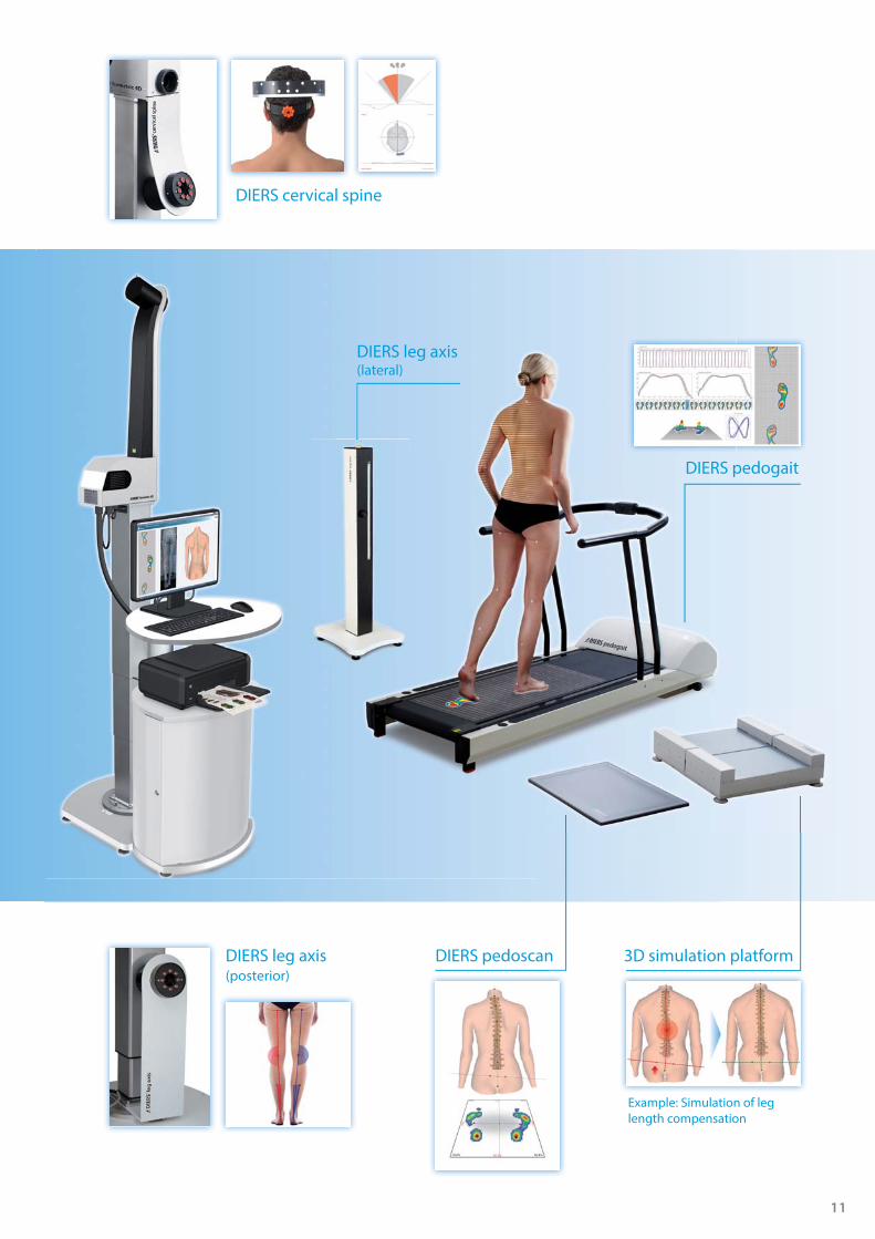
The DIERS formetric 4D can be combined with the following products to extend the range of applications on the whole musculoskeletal axis.
Additional Components:
e mususcuculoloskskeleletetalal aaxixiss.
Benefits:
° Extended range of applications
° Time saving through simultaneous measuring
° High economic efficiency
11
DIERS pedoscan 3D simulation platform
Example: Simulation of leg length compensation
DIERS leg axis (lateral)
DIERS pedogait
DIERS leg axis (posterior)
DIERS cervical spine

12
Using the cervical spine module the mobility of the cervical spine (range of motion) can be three-dimensionally recorded. The movement directions of fl exion, extension, lateral fl exion left and right, and rotation to the left and right are measured. The measurement data and asymmetries are graphically shown and can be analysed. The measurement process takes place using a light, specialized head hood. The DIERS formetric 4D system requires an addi-tional camera system for the cervical spine module.
RotationFlexion / Extension(+Rotation)
Lateral Flexion (Left/Right)(+Rotation)
AdditionalCamera ModuleAdditionaCamera M
DIERS formetric 4D + cervical spine
13
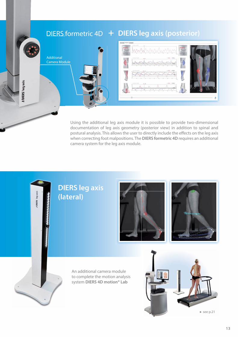
DIERS formetric 4D + DIERS leg axis (posterior)
DIERS leg axis
(lateral)
Using the additional leg axis module it is possible to provide two-dimensional documentation of leg axis geometry (posterior view) in addition to spinal and postural analysis. This allows the user to directly include the eff ects on the leg axis when correcting foot malpositions. The DIERS formetric 4D requires an additional camera system for the leg axis module.
DIERS
AdditionalCamera Module
DI
(la
An additional camera moduleto complete the motion analysis system DIERS 4D motion® Lab
see p.21

14
The simulation platform can be used to evaluate leg length discrepancies and foot malpositions. The eff ects of treatments on the spine, pelvis and posture can be simu-lated. During the examination the patient stands on two separate surfaces which can be adjusted in three directions. Correctional parameters – e.g. for insoles which are to be made – are determined with millimetric precision.
DIERS formetric 4D
The simulation platform can be
Leg length compensation (comparative measurement)
+ 3D simulation platform

15
Combining the two measuring systems DIERS formetric 4D and DIERS pedoscan
enables simultaneous analysis of the spinal form, the pelvic position, the pressure conditions under the feet and the body's center of pressure.This synchronized measurement is a valuable feature in determining optimal treatment (e.g. with posture-correcting insoles).
DIERS formetric 4D
Combining the two measuring sy
+ DIERS pedoscan

16
Clinical Applications:
■ Static foot analysis while standing
■ Asymmetries in foot pressure distribution
■ Foot malpositions and foot corrections
■ Conventional and proprioceptive/ sensomotoric insole treatment
DIERS digiscan
The DIERS digiscan system is conceived for static foot analysis using a mirror system, for controlled foot treatment (e.g. in proprioceptive insoles), for immediate documentation using a foot scanning device and to link to DIERS DICAM
with the option of electronic data transmission.
Aside from diagnostic procedures users have the option of employing a system for documentation of treatment.
Measurement of Longitudes / Angles / Area sizes
Podoscope + Foot Scanner
2 in1

17
Measuring Parameters:
■ Foot pressure reaction forces
■ Foot roll over characteristics of the feet
■ Foot rotation
■ Foot pressure distribution in the diff erent phases of walking
■ Stride length, stride time, stride width, step length
■ Movement of the body's center of pressure
Clinical Applications:
■ Foot malpositions and foot corrections
■ Diabetic foot treatment
■ Insole treatment
■ Gait imbalances / Gait disorder
■ Postural analysis
■ Treatment with orthotics or prosthetics
■ In combination with TMJ treatment
DIERS pedoscan
The foot pressure recording and gait analysis system DIERS pedoscan allows the pressure distribution on the human foot to be captured and displayed quickly and precisely while standing or while walking.Many clinical issues concerning the objective and quantitative analysis of pressure distribution, pressure peaks, and movement asymmetries as well as the roll over behavior are recorded to help diagnose foot malformations or functional limitations of the lower extremities.The precise, high-frequency measurement technology allows all users to record and document even rapid movements of the body's centre of pressure and load changes.For a time-saving dynamic measurement in both directions the walking direction is automatically identifi ed by the software.The high-frequency measurement of the body's centre of pressure (min. 100 Hz) provides additional information about neurological issues and extends the range of application to (competitive) sports.
Pressure plates are available in the length: 0.5 / 1.0 / 2.0 / 4.0 m
DIERS pedofeedback
see p.31

18
DIERS pedogait
The DIERS pedogait system allows the functional representation of the foot pressure reaction forces while walking. The integrated measuring platform is 1.0 m long with 5.376 sensors for an exact capture of the pressure values. The admission frequency is 100 Hz, which corresponds to a tact frequency of 10 ms. Measurement precision is gained and needed due to postural variances of the human body. The treadmill can also be used for static measurements of foot pressure as well as for stabilometry.
The DIERS pedogait is ready for simultaneous measurement with the DIERS 4D motion®
(dynamic spine analysis) as well as with the module DIERS leg axis (video gait analysis). These three measuring devices can be integrated to the compact motion analysis system DIERS 4D motion® Lab:
Measuring Parameters:
■ Foot pressure reaction forces
■ Foot roll-over characteristics of the feet
■ Foot Rotation
■ Foot pressure distribution while walking
■ Stride length, stride time, stride width, step length
■ Cadence (steps/min)
■ Movement of the body's center of pressure
Clinical Applications:
■ Foot malpositions and foot corrections
■ Diabetic foot treatment
■ Insole treatment
■ Gait imbalances / Gait disorders
■ Postural analysis
■ Treatment with orthotics or prosthetics
■ In combination with TMJ treatment
see p.21

19

20
DIERS
The DIERS 4D motion® system is the leading technology in the fi eld of 3D spine and surface topography. For the fi rst time it is possible to visualize the complex motion pattern of the spine and pelvis while walking and to monitor the results.This technological breakthrough is based on the innovative software and an advanced camera system (60 frames/second).
DIERS 4D motion®Dynamic Spine & Posture Analysis
Clinical Applications:
■ Postural Defi cits: Scoliosis, hyper/hypo kyphosis, hyper/hypo lordosis, blockades, pelvic obliquities, leg length discrepancies, …
■ Motion Asymmetries
■ Foot & Gait Defi cits (4D motion® Lab) Customized orthopaedic and proprioceptive insoles
■ Medical based Training Therapy
■ Follow-up Measurements: Scoliosis, pre-& post surgery, posture correcting insoles etc.
■ Physiotherapy / Rehabilitation
■ Sports Medicine & Professional Clinical Diagnostics
■ and many more
CLI
NIC
ALLY PROVEN
PAT E N T E D & C
ERT
IFIE
D
European Patent No.: EP 1718206
United States Patent No.: US 7,899,220 B2
Step into a new dimension ...
6
0 B2
2 m | 7 ft.

21
DIERS
The DIERS 4D motion® system for dynamic spine measurement is the key technology for the deve-lopment of the DIERS 4D motion® Lab.
This motion laboratory allows a synchronized measurement of the whole skeletal system and opens new fi elds of clinical applications: ranging from medical diagnosis via training therapy to sport sciences.
The dynamic spine analysis is a key measurement modality in clinical diagnostics, research and further studies.
DIERS 4D motion® LabDynamic analysis of the whole musculoskeletal system with small space requirement (8m²)
DIERS 4D motion® Dynamic measurement of the
spine, vertebra and pelvis
Components:
DIERS leg axisVideo gait analysis for the
detection and measurement of leg axis
detecct
DIERS pedogait Treadmill with integrated
pressure plate
The Compact Solution for Motion Analysis
DIERS pedogaitDwith integratedTrreadmill w
pressure platep
4 m | 13 ft.

22
DIERS 4D motion® Lab | Holistic Motion Analysis
DIERS 4D motion® | Static Measurement
DIERS pedogait | Static Foot Pressure Measurement
■ COP-Displacement right- and rearwards. ■ No toe pressure
■ Pelvic obliquity left (Indication for discharge position of the left leg) ■ Compensation by a local lateral deviation in the lumbar spine ■ Plumb alignment up until thoracic vertebrae T10
Clinical Example:
Patient: male, 46 yearsDiagnosis: Osteoarthritis of the hip (left)

23
DIERS 4D motion® | Dynamic Measurement
Signifi cant asymmetry in pelvis rotation(continuing in the rotation of the vertebral bodies)
Signifi cant lateral deviation of the thoracic spine
High rotation of the vertebras

24
DIERS leg axis (leg axis geometry): Suspicion of relieving posture (leaning to the right)
Pelvic obliquity (left) remains in every phase of walking / Suspicion of pelvis forward movement (left)
Static Measurement
Statische Vermessung
Stance Phase RightStance Phase Left
Statische Vermessung
25
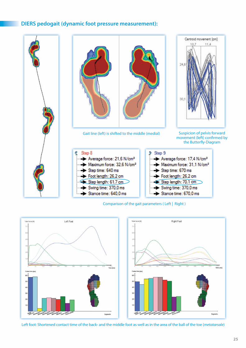
DIERS pedogait (dynamic foot pressure measurement):
Left foot: Shortened contact time of the back- and the middle foot as well as in the area of the ball of the toe (metatarsale)
Comparison of the gait parameters ( Left | Right )
Gait line (left) is shifted to the middle (medial) Suspicion of pelvis forward movement (left) confi rmed by
the Butterfl y-Diagram
26
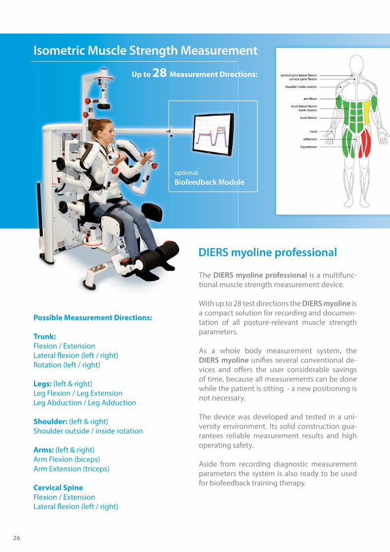
Possible Measurement Directions:
Trunk:
Flexion / ExtensionLateral fl exion (left / right)Rotation (left / right)
Legs: (left & right)Leg Flexion / Leg Extension Leg Abduction / Leg Adduction
Shoulder: (left & right)Shoulder outside / inside rotation
Arms: (left & right)Arm Flexion (biceps)Arm Extension (triceps)
Cervical Spine
Flexion / ExtensionLateral fl exion (left / right)
The DIERS myoline professional is a multifunc-tional muscle strength measurement device.
With up to 28 test directions the DIERS myoline is a compact solution for recording and documen-tation of all posture-relevant muscle strength parameters.
As a whole body measurement system, the DIERS myoline unifi es several conventional de-vices and off ers the user considerable savings of time, because all measurements can be done while the patient is sitting - a new positioning is not necessary.
The device was developed and tested in a uni-versity environment. Its solid construction gua-rantees reliable measurement results and high operating safety.
Aside from recording diagnostic measurement parameters the system is also ready to be used for biofeedback training therapy.
DIERS myoline professional
m
m
The DIERS m
tional muscl
DIERS m
Up to 28 Measurement Directions:
optional:Biofeedback Module
Isometric Muscle Strength Measurement
27
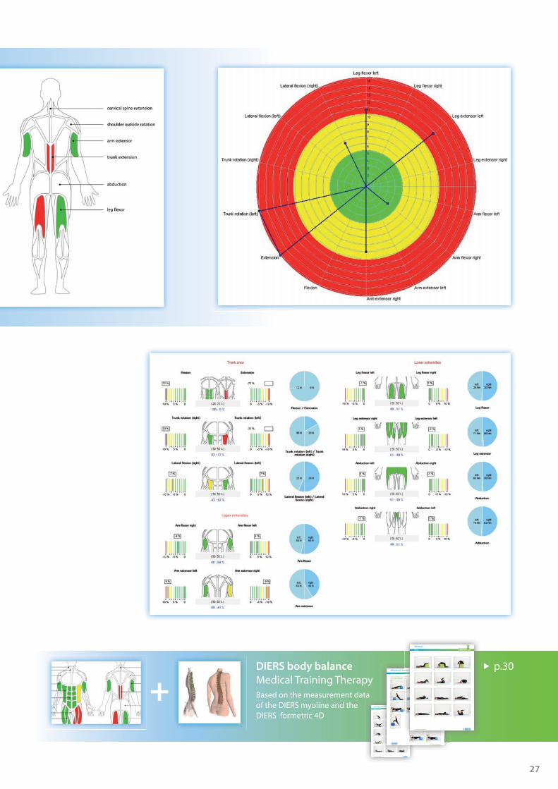
DIERS body balance
Medical Training TherapyBased on the measurement data of the DIERS myoline and the DIERS formetric 4D
+p.30
28
DIERS COMMUNICATION & APPLICATION MANAGEMENT
There are various measurement methods and tools used for biomedical analysis. It creates many problems for clinicians when more then one method is used to control and store all of the collected data, due to diff erent software structures. DICAM solves these problems:
DICAM unites the diff erent measurement de-vices in just one single software structure.
The integrated remote maintenance allows a fast software update and immediate online assistance in case of a problem.
DICAM combines the outcome of diff erent measurement devices with expert's knowledge of research and clinical case studies, which have been collected over the past 10 years. The result is the DIERS theraline software, which off ers customized treatment proposals for indi-vidual patient care.
DICAM can be plugged directly to existing soft-ware systems or networks which are connected to any biomedical device. A repeated input of patient's data can therefore be avoided.
CO N & APPDIERS CATIMMUN

29
DIA
GN
OSI
STH
ERA
PY
30
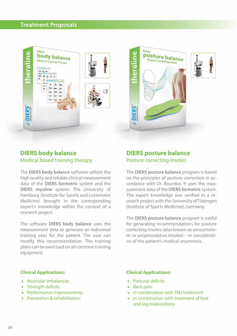
Treatment Proposals
The DIERS posture balance program is based on the principles of posture correction in ac-cordance with Dr. Bourdiol. It uses the mea-surement data of the DIERS formetric system. The expert knowledge was verifi ed in a re-search project with the University of Tübingen (Institute of Sports Medicine), Germany.
The DIERS posture balance program is useful for generating recommendations for posture correcting insoles (also known as sensomoto-ric or proprioceptive insoles) – in considerati-on of the patient’s medical anamnesis.
DIERS posture balancePosture correcting insoles
The DIERS body balance software utilizes the high quality and reliable clinical measurement data of the DIERS formetric system and the DIERS myoline system. The University of Hamburg (Institute for Sports and Locomotor Medicine) brought in the corresponding expert's knowledge within the context of a research project.
The software DIERS body balance uses the measurement data to generate an individual training plan for the patient. The user can modify this recommendation. The training plans can be exercised on all common training equipment.
DIERS body balanceMedical based training therapy
Clinical Applications:
■ Muscular imbalances ■ Strength defi cits ■ Performance improvements ■ Prevention & rehabilitation
Clinical Applications:
■ Postural defi cits ■ Back pain ■ In combination with TMJ treatment ■ In combination with treatment of foot
and leg malpositions

31
The DIERS foot balance software can be used to generate recommendations for the pro-duction of high quality insoles to correct feet and gait. This is based on the high frequency measure-ment data of the DIERS pedoscan device. The University of Tuebingen (Institute of Sports Medicine) accompanied the develop-ment of this software as part of a research project.The new software converts the dynamic foot pressure distribution into numerous strength and acceleration vectors. The additional measurement data improves the effi cacy of insoles.
DIERS foot balanceFoot correction insoles
The biofeedback training software for the foot pressure plate DIERS pedoscan off ers various modalities for specifi c training treatments.The optical feedback of the patient's activity eff ects higher motivation and infl uences the therapy progress positively.
■ Training confi guration based on an existing foot pressure measurement
and/or individual confi guration ■ 8 diff erent training procedures ■ Length of training individually adjustable
DIERS feetbackBiofeedback training
Clinical Applications:
■ Plantar foot problems ■ Foot malpositions ■ Diabetic foot disorders ■ Gait defi cits and gait asymmetries ■ Leg length discrepancies
Treatment Proposals
32
Scientifi cally Based & Clinically Proven:
In the following list you will fi nd a selection of international clinical studies, journals and publications regarding DIERS products:
3D/4D Spine & Posture Analysis (DIERS formetric 4D):
09. Frobin, W.; Hierholzer, E. (1981). Rasterstereography: A photographic method for measurement of body surfaces. Photogrammetric Engineering & Remote Sensing 47, 1717-1724 >> https://eserv.asprs.org/PERS/1981journal/dec/1981_dec_1717-1724.pdf
20. Drerup, B.; Hierholzer, E. (1985). Objective Determination of anatomical landmarks on the body surface: Measurement of the vertebra prominens from surface curvature. Journal of applied Biomechanics 18, 467-474 >> http://www.sciencedirect.com/science/article/pii/0021929085902829
28. Drerup, B.; Hierholzer, E. (1987). Automatic localization of anatomical landmarks on the back surface and construction of a body-fi xed coordinate system. Journal of applied Biomechanics 20, 961-970 >> http://www.sciencedirect.com/science/article/pii/0021929087903253
44. Drerup, B.; Hierholzer, E. (1996). Assessment of scoliotic deformity from back shape asymmetry using an improved mathematical model. Clin. Biomech. 11, 367-383 >> http://www.ncbi.nlm.nih.gov/pubmed/11415649
51. Schülein S.; Mendoza S.; Malzkorn R.; Harms J.; Skwara A. (2012). Rasterstereographic Evaluation of Inter- and Intraobserver-Reliability in Postsurgical Adolescent Idiopathic Scoliosis Patients. J. Spinal Disord Tech. ePub>> http://www.ncbi.nlm.nih.gov/pubmed/23249884
73. Mohokum, M. (2009). Reproducibility of rasterstereography for kyphotic and lordotic angles and for trunk length and trunk inclination - A reliability study. Spine 35 (14), 1713-1714 >> http://www.ncbi.nlm.nih.gov/pubmed/20505568
80. Mardjetko, S.; Knott, P.; Rollet, M.; Baute, S.; Riemenschneider, M.; Muncie, L. (2010). Evaluating the reproducibility of the formetric 4D measurements for scoliosis. Eur Spine J 21, 241-242 >> http://www.ncbi.nlm.nih.gov/pmc/articles/PMC2938639/
85. Betsch, M.; Wild, M.; Jungbluth, P.; Hakimi, M. (2011). Reliability and validity of 4D rasterstereography under dynamic condition. Computers in Biological and Medicine. 2011 06; 41 (6), 308-312 >> http://www.ncbi.nlm.nih.gov/pubmed/21489425
89. Knott, P. (2012). A comparison of automatic vs. manual detection of anatomical landmarks during surface topography evaluation using the formetric 4D system. Scoliosis 2012 >> http://www.scoliosisjournal.com/content/7/S1/O19
203. Knott, P.; Frerich, J.; Hertzler, K.; Mardjetko, S. (2011). Comparison of Radiographic and Surface topography measurements in adolescents with idiopathic scoliosis. IMAST July 2011 Proceedings >> http://www.ncbi.nlm.nih.gov/pmc/articles/PMC3414720/
Dynamic Spine & Posture Analysis (DIERS 4D motion®):
273. Betsch, M.; Wild, M.; Rapp, W. et al. (2013). Evaluation of a Novel Spine and Surface topography System for Dynamic Spinal Curvature Analysis during Gait. PLOS ONE, Jluy 2013, Vol.8, (7), 1-8 >> http://www.plosone.org/article/info%3Adoi%2F10.1371%2Fjournal.pone.0070581
Gipsman, A.; Rauschert , L.; Daneshvar, M.; Knott, P. (2013, for submission). Evaluating the Reproducibility of Motion Analysis Scanning of the Spine During Walking

33
Foot & Gait Analysis (DIERS pedoscan /pedogait):
239. Hallemanns, A.; D´Août, K.; De Clercq, D.; Aerts, P. (2003). Pressure Distribution Patterns under the feet of new walkers: The fi rst two month of independent walking. Foot and Ankle International/Vol.24, No. 5/May 2003, 444-445
262. De Cock, A.; De Clercq, D.; Willems, T.; Witvrouw, E. (2004). Temporal characteristics of foot roll-over during barefoot jogging: reference data for young adults. Gait and Posture. Elsevier, 1-8
263. Schröder, J.; Mattes, K. (2008). Posture analysis by means of pedobarography and videorasterstereography: bivariate and multiple correlation analysis. 13th Annual Congress of the European College of Sport Science (ECSS). Estorial, July 9-12, 192
270. Schröder, J. (2009). Posture Analysis: variations and reliability of biomechanical parameters in bipedal standing by means of formetric-system. Sport sciences: Nature, Nurture and Culture. 14th Annual Congress of the European College of Sport Science. Oslo/ Norway, June 24-27.
322. Willems, T. (2004). Intrinsic risk factors for sports injuries to the lower leg and ankle. University Gent, Department of rehabilitation science and physiotherapy
323. Segers, V. (2006). A biomechanical analysis of the realization of actual human gait transition. University Gent, Faculty of medicine and health science, Department of movement and sports science
324. Vereecke, E. (2006). The functional morphology and bipedal locomotion of hylobates Iar. University Antwerpen, Faculty of biology
384. Willems, T. et al. (2004). A prospective study of gait related risk factors for exerciserelated lower leg pain. Gait and Posture. Elsevier
385. Willems, T. et al. (2005). Relationship between gait biomechanics and inversion sprains: a prospective study of risk factors. Gait and Posture. Elsevier
Muscle Strength Measurement (DIERS myoline):
264. Schröder, J.; Reer, R.; Braumann, K.; Mattes, K. (2008). Evaluation of evidence based training therapy in patients with non-specifi c back pain – variability of spine shape parameters and diffi culties in short-term comparisons. J. Cabri, F. Alves, D.Araujo, J. Barreiros, J. Diniz, A. Veloso (Eds.). 13th Congress ECSS 09.-12. July 2008. Estoril, 440
Treatment Proposals (DIERS theraline):
271. Schröder, J.; Mattes, K. (2010). Spine shape changes following individualized exercise programs in back pain patients over 60 years of age. Congress abstract. ECSS, Antalya 2010
UKDUniversitätsklinikumDüsseldorf
Clinical Cooperations: Here is a selection of hospitals and universities, with whom we collaborate and maintain a scientifi c network to continuously develop our measuring systems and fi nd new product solutions to accommodate your needs.
DIERS supports the German Football Association (DFB - Deutscher Fußball-Bund) as well as some teams of the German Professional Football League (DFL - Deutsche Fußball Liga) and the European Football Leagues.
34
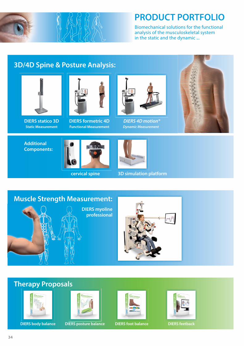
PRODUCT PORTFOLIO
3D/4D Spine & Posture Analysis:
DIERS statico 3DStatic Measurement Functional Measurement Dynamic Measurement
DIERS formetric 4D DIERS 4D motion®
Biomechanical solutions for the functional analysis of the musculoskeletal systemin the static and the dynamic ...
DIERS 4D motion® Lab
DIERS myoline
professional
Muscle Strength Measurement:
Additional
Components:
3D simulation platformcervical spine
Therapy Proposals
DIERS posture balance DIERS foot balance DIERS feetbackDIERS body balance

35
Foot & Gait Analysis
DIERS leg axis
( posterior )
DIERS leg axis
( lateral )
CompactMotion Analysis:
Components:
DIERS digiscan DIERS pedoscan DIERS pedogait
BIOMEDICAL SOLUTIONS
DIERS 4D motion® Lab
Only 8 m²
+ + +

BIOMEDICAL SOLUTIONS
© D
IERS
Inte
rnat
iona
l Gm
bH |
Nov
embe
r 20
15
HEADQUARTER:
DIERS International GmbH
Dillenbergweg 4 | 65388 Schlangenbad, GermanyPhone +49 (0) 6129 48 86 0 | Fax +49 (0) 6129 48 86 [email protected] | www.diers.de
USA:DIERS Medical Systems, Inc.1752 Capital Street, Suite 310Elgin, Illinois 60124 / USAPhone: +1 312 419-0205 Fax: +1 312 [email protected]
MIDDLE EAST:DIERS Middle East (ME) EstRoom 14 | King Faisal High WayKhobar 31952 | Kingdom of Saudi ArabiaPhone: +966 567 047 [email protected]
INDIA:DIERS Biomedical Solutions, Pvt. Ltd.Offi ce B-154, North-ex Mall, Sector 9, Rohini, New Delhi-110085, IndiaPhone: +91 11 27 56 54 [email protected] | www.diers.in
RUSSIA: DIERS Medical Co. Ltd.Vyborgskaya nab. 61, Offi ce 214 197342 Saint-PetersburgRussian FederationPhone: +7 812 924 61 [email protected] | www.diers.ru
Presented by:
www.diers.de
SERVICE is a priority for us.
Service & support by competent professionals (Engineers, sports-, computer scientists, i.a.)
Technical planning assistance
Professional installationwith fl exible scheduling
Intensive product instruction by specialists
Advanced training courses for medical assistants
Annual user meetings to promote an intensiveexchange of experience
Immediate assistance via remote maintenance
Regular maintenance of the systems according to the Medical Device Directive (MDD)