dietary blue pigments derived from genipin, attenuate ... · pdf filenf-kb, a nuclear...
TRANSCRIPT

Dietary Blue Pigments Derived from Genipin, AttenuateInflammation by Inhibiting LPS-Induced iNOS and COX-2Expression via the NF-kB InactivationQiang-Song Wang1, Yaozu Xiang2, Yuan-Lu Cui1*, Ke-Ming Lin1, Xin-Fang Zhang1
1 Tianjin State Key Laboratory of Modern Chinese Medicine, Tianjin University of Traditional Chinese Medicine, Tianjin, People’s Republic of China, 2 Department of
Medicine, Imperial College London, Hammersmith Hospital Campus, London, United Kingdom
Abstract
Background and Purpose: The edible blue pigments produced by gardenia fruits have been used as value-added colorantsfor foods in East Asia for 20 years. However, the biological activity of the blue pigments derived from genipin has not beenreported.
Methodology/Principal Findings: The anti-inflammatory effect of blue pigments was studied in lipopolysaccharide (LPS)stimulated RAW 264.7 macrophage in vitro. The secretions of nitric oxide (NO) and prostaglandin E2 (PGE2) were inhibited inconcentration-dependent manner by blue pigments. Real-time reverse-transcription polymerase chain reaction (Real-timeRT-PCR) analyses demonstrated that the mRNA expression of inducible nitric oxide synthase (iNOS), cyclooxygenase-2 (COX-2), interleukin (IL)-6, and tumor necrosis factor alpha (TNF-a) was inhibited, moreover, ELISA results showed that theproductions of IL-6 and TNF-a were inhibited. Cell-based ELISA revealed the COX-2 protein expression was inhibited. Theproteome profiler array showed that 12 cytokines and chemokines involved in the inflammatory process were down-regulated by blue pigments. Blue pigments inhibited the nuclear transcription factor kappa-B (NF-kB) activation induced byLPS, and this was associated with decreasing the DNA-binding activity of p65 and p50. Furthermore, blue pigmentssuppressed the degradation of inhibitor of kB (IkB) a, Inhibitor of NF-kB Kinase (IKK) a, IKK-b, and phosphorylation of IkB-a.The anti-inflammatory effect of blue pigments in vivo was studied in carrageenan-induced paw edema and LPS-injecting ICRmice. Finally, blue pigments significantly inhibited paw swelling and reduced plasma TNF-a and IL-6 production in vivo.
Conclusions and Implications: These results suggest that the anti-inflammatory properties of blue pigments might be theresults from the inhibition of iNOS, COX-2, IL-6, IL-1b, and TNF-a expression through the down-regulation of NF-kBactivation, which will provide strong scientific evidence for the edible blue pigments to be developed as a new health-enhancing nutritional food for the prevention and treatment of inflammatory diseases.
Citation: Wang Q-S, Xiang Y, Cui Y-L, Lin K-M, Zhang X-F (2012) Dietary Blue Pigments Derived from Genipin, Attenuate Inflammation by Inhibiting LPS-InducediNOS and COX-2 Expression via the NF-kB Inactivation. PLoS ONE 7(3): e34122. doi:10.1371/journal.pone.0034122
Editor: Joao B. Calixto, Universidad Federal de Santa Catarina, Brazil
Received September 10, 2011; Accepted February 22, 2012; Published March 30, 2012
Copyright: � 2012 Wang et al. This is an open-access article distributed under the terms of the Creative Commons Attribution License, which permitsunrestricted use, distribution, and reproduction in any medium, provided the original author and source are credited.
Funding: This work was supported by the National Natural Science Foundation of China (No. 30973967, 81173469), Program for New Century Excellent Talents inUniversity (NCET-09-0899), Program for Changjiang Scholars and Innovative Research Team in University (PCSIRT) and the Specialized Research Fund for theDoctoral Program of Higher Education (No. 20091210110003). The funders had no role in study design, data collection and analysis, decision to publish, orpreparation of the manuscript.
Competing Interests: The authors have declared that no competing interests exist.
* E-mail: [email protected]
Introduction
With growing concern on the safety of synthetic dyes, the
importance of natural colorants suitable for using in foods has gained
increasing attention. Genipin, the aglycon of geniposide, is obtained
from the fruit of Gardenia jasminoides ELLIS. Genipin itself is colorless
but it reacts spontaneously with amino acids to form blue pigments
which are used in food industry widely [1]. The edible blue pigments
produced by gardenia fruits have been widely used as a blue food
colorant in East Asia [2]. Since the blue pigments were used in the
food industry, the stability with regard to pH, temperature, and light
conditions were also investigated [3], however, very few biological
activity studies of the blue pigments are reported.
The inflammation process is crucial to defense against microorgan-
ism infection. Key events in the inflammatory process include
expression of inflammatory cytokines, chemokines, and other
mediators [4]. Macrophages play an important role in inflammatory
disease and host defense through the release of factors such as NO,
prostaglandin mediators, and cytokines involved in the immune
response [5,6,7]. LPS is one of the most powerful activators of
macrophages known, and macrophages induced by LPS are known to
be activated through the production of inflammatory mediators, such
as NO and other free radicals, in addition to numerous cytokines, such
as TNF-a, IL-1b and IL-6 [8,9,10]. NO is a major product which is
controlled by nitric oxide synthases (NOS), such as iNOS, eNOS and
nNOS [11]. Most importantly, iNOS is highly expressed in
macrophages, which leads to organ destruction in some inflammatory
and autoimmune diseases [12]. PGE2 is also another important
mediator which is produced from arachidonic acid metabolites which
are catalyzed by COX-2 in inflammatory responses [13].
PLoS ONE | www.plosone.org 1 March 2012 | Volume 7 | Issue 3 | e34122

NF-kB, a nuclear transcription factor, regulates the expression
of various genes, including cytokines, iNOS and COX-2, which
play critical roles in apoptosis, various autoimmune diseases, and
inflammation [14]. NF-kB exists in most cells as homodimeric or
heterodimeric complexes of p50 and p65 subunits and remains
inactive in the cytoplasm of cells associated with the NF-kB
inhibitory protein (I-kB) [15]. NF-kB is activated in response to
LPS, which induced NF-kB activation through increasing nuclear
p65 protein associated with decreased cytosolic I-kB protein [15].
The resulting free NF-kB is then translocated into the nucleus,
where it binds to kB binding sites in the promoter region of target
genes, and induces the transcription of pro-inflammatory media-
tors, including iNOS, COX-2, TNF-a, IL-1b, and others
[16,17,18]. Because of its ubiquitous role in the pathogenesis of
inflammatory gene expression, NF-kB is a current target for
treating various diseases [19,20].
The macrophage cell line (RAW 264.7) used in experiments has
been established as a suitable model to investigate compounds
interfering with LPS-inducible inflammatory cascades in vitro
[21,22,23,24,25]. In this study, the anti-inflammatory effects of the
blue pigments on the generation of several chemokines, cytokines
and enzymes involved in the inflammatory process, such as NO,
PGE2, TNF-a, IL-6, IL-1b, iNOS and COX-2 in LPS-induced
RAW 264.7cells were investigated. We also investigated whether
the blue pigments influence the LPS induced DNA binding
activity of NF-kB and the protein level of its subunit, p65 and p50.
Methods
Ethics statementThe approved ID of the mice experiments is TCM-2009-037-
E05.
This work was supported by the National Natural Science
Foundation of China (30973967, 81173469), which has been
inspected by the Animal Ethics Committee of Tianjin University
of Traditional Chinese Medicine. The ICR mice used in our work
were obtained from Huafukang Bio-technology Co. Ltd. (SCXK
2009-0004, Beijing, China).
ReagentsDulbecco’s modified Eagle’s medium-high glucose (DMEM), 2-
(4, 5-dimethylthiazol-2-yl)-2, 5-diphenyltetrazolium bromide
(MTT), Lipopolysaccharides from Escherichia coli 0111:B4
(LPS) were purchased from Sigma-Aldrich Co. (USA). Prosta-
glandin E2 Express EIA Monoclonal Kit was obtained from
Cayman Chemical (USA). IL-6 Mouse ELISA Kit and TNF-aMouse ELISA Kit were obtained from Invitrogen (USA). 4-amino-
5-methylamino- 29, 79-difluorofluorescein diacetate (DAF-FM
diacetate) was purchased from Invitrogen (USA). BCA Protein
Assay Kit was obtained from Pierce (USA). Mouse Cytokine Array
Panel A Array kit was purchased from R&D Systems, Inc. (USA).
Mammalian Cell Lysis Kit and UNIQ-10 column Trizol total
RNA extraction kit were bought from Sangon Biological
Engineering Technology & Services Co., Ltd. (Shanghai, China).
Improm-II Reverse Transcription System was purchased from
Promega Corporation (USA). FastStart Universal SYBR Green
Master (ROX) kit was purchased from Roche (Germany). Mouse
Anti-COX-2 Monoclonal Antibody was from BD Pharmingen
(USA) and Goat Anti-Mouse IgG Peroxidase Conjugate was from
Calbiochem (Germany). Nuclear Extract Kit was purchased from
Active Motif (Japan). Universal EZ-TFA Transcription Factor
Assay and NF-kB Family EZ-TFA Transcription Factor Assay kits
were purchased from Millipore (USA). P-IkB-a, IkB-a, IKK-a,
IKK-b monoclonal antibodies and peroxidase-conjugated second-
ary antibody were purchased from Cell Singaling Technology
(USA), and b-actin monoclonal antibody was purchased from
Sigma-Aldrich Co. (USA). NF-kB inhibitor BAY 11-7082 was
purchased from Beyotime Institute of Biotechnology (China).
Carrageenan was purchased from Sigma-Aldrich Co. (USA) and
Dexamethasone was purchased from Shanghai General Pharma-
ceutical Co., Ltd.(Shanghai, China).
Preparation of the blue pigmentsGenipin was purchased from Wako (Osaka, Japan). Dietary
blue pigments derived from genipin were prepared according to
the methods described earlier [26]. Briefly, 8.8 mmol of genipin
and amino acids (glycine, 8.8 mmol) were added respectively into
400 mL of 100 mM phosphate buffer (pH 7.0) at 80uC and stirred
for 4 h. The blue pigments were passed through Diaion HP-20
resin and ODS columns chromatography and the fractions at
595 nm were collected. The blue pigments were cold-sterilized
using a 0.22-mm pore size membrane filter (Millipore, USA) and
stored in the refrigerator for further use. The proposed formation
structure of blue pigments from genipin with glycine were shown
in Figure 1 [26]. The concentrations of blue pigments were
calculated by the molar mass of genipin.
Cells and cell cultureRAW 264.7 murine macrophages cell line was obtained from
Cell Culture Center of Chinese Academy of Medical Sciences
(Beijing, China). RAW 264.7 cells were maintained in DMEM
supplemented with 10% heat inactivated fetal bovine plasma (HI-
FBS), 100 U/mL penicillin and 100 mg/mL streptomycin at 37uCin a humidified incubator containing 5% CO2. For the
determination of cell viability, nitrite concentration as an index
for NO synthesis, PGE2 concentrations, as well as different acute
phase proteins, cytokines, and chemokines in culture medium, the
cells were plated at 56105 cells/well in 96-well plates and treated
with various concentrations of blue pigments in the presence of
0.2 mg/mL LPS for 18–24 h as indicated. Moreover, for the
determination of protein levels of COX-2, cells were treated with
various concentrations of blue pigments and in the presence of
0.2 mg/mL LPS for 24 h. For real-time RT-PCR, the cells were
pre-incubated with various concentrations of blue pigments for 2 h
and were then treated with 0.2 mg/mL LPS for an additional 6 h.
The blue pigments at various concentrations dissolved in
Phosphate buffered saline (PBS, pH 7.4) were added together
with LPS. Cells were treated with PBS as vehicle control.
Figure 1. The proposed formation structure of blue pigmentsfrom genipin with glycine.doi:10.1371/journal.pone.0034122.g001
Dietary Blue Pigments Attenuate Inflammation
PLoS ONE | www.plosone.org 2 March 2012 | Volume 7 | Issue 3 | e34122

Cell viability assay (MTT assay)RAW 264.7 cells were treated with various concentrations of blue
pigments (12.5, 25, 50, 100 and 200 mM) for 24 h. Then MTT
(stock solution of 5 mg/mL) was added to a final concentration of
0.5 mg/mL, and the cells were incubated for an additional 4 h at
37uC and 5% CO2. The medium was removed and the formazan
precipitate was solubilized in 100 mL DMSO, and the absorbance
was measured at 570 nm on a multifunctional microplate reader
(FlexStation 3, Molecular Devices, USA).
Nitrite assay, detection of intracellular NO production,PGE2 and cytokines
RAW 264.7 cells were treated with various concentrations of
blue pigments (12.5, 25, 50, 100 mM) in the presence of 0.2 mg/
mL LPS. 18 hours later, the medium was collected. The nitrite
accumulated in culture medium was measured as an indicator of
NO production based on a diazotization reaction using Griess
reagent system (Promega, USA). Nitrite concentration was
determined by a standard curve prepared with sodium nitrite
dissolved in DMEM without phenol red supplemented with 5%
HI-FBS. Intracellular NO production was evaluated by confocal
laser scanning microscopy of a fluorescent NO derivative. LPS
stimulated and blue pigments-treated RAW 264.7 cells were
seeded in 4-well chamber slides (Millicell EZ Slide, Millipore,
USA) and incubated for 1 h at 37uC with 10 mM DAF-FM
diacetate. Digitized images were generated with a Zeiss LSM 710
confocal laser scanning microscope (Carl Zeiss Microimaging,
Jena, Germany) with an excitation wavelength of 495 nm and an
emission wavelength of 515 nm. PGE2 production was measured
with the Prostaglandin E2 Express EIA Monoclonal Kit (Cayman
Chemical, USA). IL-6 and TNF-a secreted in the culture medium
were quantified by ELISA kits (Invitrogen, USA) according to the
manufacturer’s instructions. Samples were assayed after a 10-fold
dilution in the medium (DMEM without phenol red supplemented
with 5% HI-FBS).
Cell-based ELISA for COX-2 protein expression in RAW264.7 cells
RAW 264.7 cells were seeded in 96-well plates treated with
various concentrations of blue pigments (12.5, 25, 50, 100 mM) in
the presence of 0.2 mg/mL LPS. The expression of COX-2 was
determined by a slightly modified original protocol for the cell-
based ELISA [27,28]. After cultivation, cells were fixed with 4%
paraformaldehyde in phosphate-buffered saline (PBS, pH 7.4) for
20 min at room temperature and washed three times with PBS
containing 0.1% Triton X-100 (PBS/T). Endogenous peroxidase
was quenched with 0.6% H2O2 in PBS/T for 20 min, and cells
were washed three times in PBS/T. Following blocking with 10%
FCS in PBS/T for 1 h, cells were incubated for 2 h at 37uC with
the primary antibody in PBS/T containing 1% BSA. After
washing the cells four times with PBS/T for 5 min, the plate was
incubated for 1 h at room temperature with secondary antibody in
PBS/T containing 1% BSA. Subsequently, cells were washed and
incubated with TMB substrate solution (Invitrogen, USA) for
30 min at room temperature in the dark. The reaction was
stopped with 50 mL of 2 N H2SO4, and the absorbance at 450 nm
was determined on a multifunctional microplate reader. The data
were corrected for differences in cell number by staining the cells
with Janus Green B after the cell-based ELISA procedure [29].
Cytokine protein array analysesRAW 264.7 cells were seeded in 6-well plates treated with blue
pigments (100 mM) in the presence of 0.2 mg/mL LPS for 18 h.
Cells were collected by centrifugation and washed once with PBS.
The washed cell pellets were resuspended in extraction lysis buffer
(Sangon, China) and incubated with 20 min at 4uC. The protein
concentration was determined using the Pierce protein assay
reagent according to the manufacture’s instruction. Screening for
different acute phase proteins, cytokines, and chemokines in cell
lysates were performed with a Proteome profiler array (Mouse
Cytokine Array Panel A Array kit) from R&D Systems [30,31,32].
The array allows to detect the following proteins: BLC, C5a, G-
CSF, GM-CSF, I-309, Eotaxin, sICAM-1, IFN-c, IL-1a, IL- 1b,
IL-1ra, IL-2, IL-3, IL-4, IL-5, IL-6, IL-7, IL-10, IL-12p70, IL-13,
IL-16, IL-17, IL-23, IL-27, IP-10, I-TAC, KC, M-CSF, JE, MCP-
5, MIG, MIP- 1a, MIP-1b, MIP-2, RANTES, SDF-1, TARC,
TIMP-1, TNF-a and TREM-1. Horseradish peroxidase substrate
(Millipore Corporation, USA) was used to detect protein
expression and data were captured by exposure to Kodak BioMax
Light films. Films were scanned into a computer and densitometry
was performed using the Image-Pro Plus version 6.0 (Media
Cybernetics, Silver Spring, MD, USA).
Real-time RT-PCR for detecting mRNA expression of TNF-a, COX-2, iNOS, IL-6
Total RNA was isolated using Sangon UNIQ-10 column Trizol
total RNA extraction kit according to the instructions of the
manufacturer. RNA (1 mg) was reversely transcribed using
ImProm-II Reverse Transcription System cDNA synthesis kit.
The real-time RT-PCR oligonucleotide primers used for mouse
iNOS, COX-2, IL-6, TNF-a and b-actin are shown in Table 1.
The reactions were setup in duplicates in 25 mL total volumes with
1 mL of each primer (0.3 mM final concentrations), 12.5 mL of
FastStart Universal SYBR Green Master (ROX) (Roche), and
1 mL of template. The PCR cycle was as follows: 95uC for 10 min,
40 cycles of 95uC for 15 s, 60uC for 1 min, and a melt curve
analysis was performed at the end of each experiment to verify that
a single product per primer pair was amplified. The amplification
and analysis were performed using an ABI Prism 7500 Real-Time
PCR System. Samples were compared using the relative CT
method. The fold increase or decrease was determined relative to
a blank control after normalized to a housekeeping gene using
22DDCT [33,34].
NF-kB activity assayRAW 264.7 macrophages cells were plated in 60-mm dishes
(26106 cells/dish). The cells were treated with various concentra-
tions (25, 50, 100 mM) of blue pigments for 2 h, stimulated with
LPS for 30 min, washed three times with cold PBS. Cells were
collected by centrifugation and washed once with PBS. The
washed cell pellets were resuspended in extraction lysis buffer
(Sangon, China) (50 mM HEPES pH 7.0, 250 mM NaCl, 5 mM
EDTA, 0.1% Nonidet P-40, 1 mM PMSF, 0.5 mM DTT, 5 mM
NaF and 0.5 mM sodium orthovanadate) containing 5 mg/ml of
leupeptin and aprotinin, respectively, and incubated with 20 min
at 4uC. Cell debris was removed by centrifugation, and
supernatants were rapidly frozen. The protein was detected by
BCA method (Pierce, USA). Furthermore, nuclear extracts were
prepared with the manufacture’s instruction (Active Motif, Japan).
Briefly, Cell pellets were resuspended in hypotonic buffer (10 mM
HEPES, pH 7.9, 1.5 mM MgCl2, 10 mM KCl, 0.2 mM PMSF,
0.5 mM DTT, 10 mg/mL aprotinin) and incubated on ice for
15 min. They were then lysed by adding 0.1% Nonidet P-40 and
vortexing vigorously for 10 s. Nuclei were pelleted by centrifuga-
tion at 12,000 g for 10 min at 4uC and resuspended in high salt
buffer (20 mM HEPES, pH 7.9, 25% glycerol, 400 mM KCl,
1.5 mM MgCl2, 0.2 mM EDTA, 0.5 mM DTT, 1 mM NaF,
Dietary Blue Pigments Attenuate Inflammation
PLoS ONE | www.plosone.org 3 March 2012 | Volume 7 | Issue 3 | e34122

1 mM sodium orthovanadate). Cell debris was removed by
centrifugation, and supernatants were rapidly frozen. The DNA-
binding activity of NF-kB p50 and p65 was detected with universal
EZ-TFA transcription factor assay kit (Millipore, USA) as the
manufacturer’s instructions. The expressions of IKK a, IKK b,
IkB-a, P- IkB-a were analyzed by western blot. Of cellular
protein, 40 mg from treated and untreated cell extracts were
electro-blotted onto a polyvinylidene difluoride (PVDF) membrane
following separation on a 10% SDS-polyacrylamide gel electro-
phoresis. The immunoblot was incubated for 2 hours with
blocking solution (5% skim milk) at room temperature, and then
incubated overnight with a 1:1000 dilution of anti- IKK a, anti-
IKK b, anti-IkB-a, P-IkB-a, b-actin antibody at 4uC (Cell
Singaling Technology, USA). Blots were washed five times with
Tween 20/Tris-buffered saline (TTBS) and then incubated with a
1: 1000 dilution of horseradish peroxidase-conjugated secondary
antibody (Cell Singaling Technology, USA) for 1 h at room
temperature. Blots were again washed five times with TTBS and
then developed by Horseradish peroxidase substrate (Millipore
Corporation, USA) and data were captured by exposure to Kodak
BioMax Light films.
AnimalsMale ICR strain of mice (4–5 weeks of age), purchased from
Beijing Huafukang Bio-technology Co. Ltd., were group-housed
under controlled light (12-h/12-h light–dark cycle; lights on at
07:00 a.m.) in the Laboratory Animal Center of Tianjin University
of Traditional Chinese Medicine. Ambient temperature and
relative humidity were maintained at 2461uC and 5565%,
respectively. All the animals had free access to water and food in a
home cage.
Carrageenan-induced paw edema test in micePaw edema was induced by subcutaneous injection of 50 mL of
1% carrageenan into the right hind paw of mice as previously
described [35]. The mice were divided into 5 groups: vehicle
control group, 1% carrageenan+0.9% saline; 1% carrageenan+-Dexamethasone (10 mg/kg); or 1% carrageenan+blue pigments
(30, 60, 120 mg/kg) (n = 10 for each group). Groups were
pretreated intraperitoneally (i.p.) with 200 mL of 0.9% saline,
Dexamethasone or Genipin blue (30, 60, 120 mg/kg) for
30 minutes. 50 mL of 1% carrageenan was then administered in
200 mL i.p for every group. The paw thickness was measured using
a 7140 Plethysmometer (UGO BASILE, Italy) before and every
hour after edema induction for 4 h. The percent increase of paw
thickness was calculated based on the volume difference between
the paw with and without carrageenan injection.
Plasma concentrations of TNF-a and IL-6 in LPS-stimulated ICR mice
The mice were divided into 6 groups: negative control group,
injected with 0.9% saline; LPS+0.9% saline; LPS+Dexamethasone
(10 mg/kg); or LPS+blue blue pigments (30, 60, 120 mg/kg) (n = 8
for each group). Groups were pretreated intraperitoneally (i.p.)
with 200 mL of 0.9% saline, Dexamethasone or genipin blue for
30 minutes. Lipopolysaccharide (1 mg/kg) was then administered
in 200 mL i.p for 5 treatment groups, and 0.9% saline was
administered i.p. for the negative control group. Blood was
withdrawn from the animals under ether anesthesia ninety minutes
later [36]. The centrifuge tube was heparinized and blood from
mice was centrifuged at 2000 rpm for 15 minutes, and the plasma
was collected and stored at 280uC until analysis. Plasma levels of
TNF-a and IL-6 were determined using Mouse ELISA kit
(Invitrogen, USA).
Statistical analysisOne-way ANOVA or t-test was used for determining the
statistically significant differences between the values of various
experimental groups. Data were expressed as means 6 SD and a P
value,0.05 was considered statistically significant.
Results
Effect of blue pigments on RAW 264.7 cells viabilityThe cytotoxicity of blue pigments on RAW 264.7 cells was
measured with MTT assay. Cell viability was not significantly
altered by blue pigments at up to 200 mM. These results suggest
that concentrations of blue pigments below 200 mM are not toxic
to RAW 264.7 cells. Therefore, for all experiments, cells were
treated with blue pigments in the concentration range of 12.5–
100 mM. The MTT assay results of blue pigments on RAW 264.7
cells were shown in Figure S1.
Effect of blue pigments on LPS-induced NO productionThe effect of blue pigments on NO in LPS-induced RAW 264.7
cells was tested to investigate the anti-inflammatory effects.
Concentrations of nitrite accumulated in the culture medium
were estimated by Griess reagent as an index for NO. RAW 264.7
Table 1. The real-time RT-PCR oligonucleotide primers.
Gene primer sequence (59-39) PCR product (bp)
b-actin forward AGAGGGAAATCGTGCGTGAC 138
(NM_007393.3) reverse CAATAGTGATGACCTGGCCGT
iNOS forward GGCAGCCTGTGAGACCTTTG 72
(NM_010927.3) reverse GCATTGGAAGTGAAGCGTTTC
COX-2 forward TGAGTACCGCAAACGCTTCTC 151
(NM_011198.3) reverse TGGACGAGGTTTTTCCACCAG
IL-6 forward TCCAGTTGCCTTCTTGGGAC 140
(NM_031168.1) reverse GTGTAATTAAGCCTCCGACTTG
TNF-a forward TTCTGTCTACTGAACTTCGGGGTGATCGGTCC 354
(NM_013693.2) reverse GTATGAGATAGCAAATCGGCTGACGGTGTGGG
doi:10.1371/journal.pone.0034122.t001
Dietary Blue Pigments Attenuate Inflammation
PLoS ONE | www.plosone.org 4 March 2012 | Volume 7 | Issue 3 | e34122

cells were pretreated with different concentrations (12.5, 25, 50,
100 mM) of blue pigments, which were found to significantly
inhibit LPS-induced NO production in a concentration-dependent
manner (P,0.01). The NO inhibitor, L-NAME (100 mM), as a
positive control, also inhibited the production of NO in activated
RAW 264.7 cells (Figure 2A). Furthermore, confocal laser
scanning microscopy also showed blue pigments to be a stronger
inhibitor of intracellular NO production than that in single LPS
stimulation (Figure 2B).
Effect of blue pigments on LPS-induced cytokines TNF-aand IL-6
TNF-a, IL-6 are known to be pro-inflammatory cytokines that
posses a multitude of biological activities linked to the immune-
pathology of acute or chronic inflammatory diseases. After
treatment with blue pigments and activated with LPS (0.2 mg/
mL), the secretion of IL-6 and TNF-a were detected by ELISA. As
shown in Figure 3A–B, pretreatment of RAW 264.7 cells with blue
pigments (25, 50, 100 mM) significantly reduced IL-6 production
(P,0.01), whereas TNF-a production were only inhibited slightly
by 50 mM blue pigments (P,0.05).
PGE2 production and Protein expression of COX-2 onblue pigments
LPS-induced PGE2 production was detected by ELISA. As
shown in Figure 3C, blue pigments inhibited the production of
PGE2. Blue pigments (50, 100 mM) had a markedly higher
inhibitory effects (P,0.01). In order to investigate whether the
inhibition of PGE2 production was due to a decreased protein
expression of COX-2, the effect of blue pigments on COX-2
protein expression was studied by cell-based ELISA. As shown in
Figure 3D, LPS treatment significantly increased COX-2 protein
expression levels, whereas COX-2 protein expression was
suppressed the induction of blue pigments (P,0.01 or P,0.05).
Effect of blue pigments on multiple cytokinesIn Inflammatory, multiple cytokines secreted in macrophages
were activated significantly. After stimulating with 0.2 mg/mL
LPS, multiple cytokines such as, G-CSF, sICAM-1, IL-1a, IL-1b,
IL-1ra, KC, JE, MIP-1a, MIP-1b, MIP-2, RANTES and TNF-awere up-regulated. However, the expressions of G-CSF, sICAM-1,
IL-1a, IL-1ra, KC, JE, MIP-1b, RANTES could be significantly
down-regulated by the blue pigments (Figure 4B P,0.01 or
P,0.05, the optical density data are presented in Table S1),
however, the expressions of IL-1b, MIP-2, TNF-a were inhibited
by blue pigments, but no significant (P.0.05). The blue pigments
could modulate the synthesis of several cytokines which were
involved in the inflammatory process.
Effect of blue pigments on LPS-induced mRNAexpression of TNF-a, IL-6, iNOS and COX-2
Since blue pigments was found to most potently inhibit the pro-
inflammatory mediators, e.g., NO, PEG2, TNF-a, IL-6 in
supernatants, we investigated the effects of blue pigments on
LPS-induced iNOS, COX-2, TNF-a, IL-6 gene expression using
real-time RT-PCR. The inhibitions of blue pigments on LPS-
induced mRNA expression of TNF-a, IL-6, iNOS, COX-2, were
observed in Figure 5A–D. The results showed that the effect of
blue pigments on mRNA expression of TNF-a, IL-6, iNOS,
COX-2 was coincidence with the secretion of TNF-a, IL-6, NO,
PEG2 that in culture medium. Moreover, the mRNA expression of
COX-2 was significantly inhibited by the blue pigments, which
was coincidence with the expression of COX-2 protein.
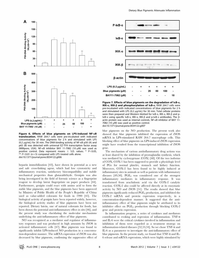
Effects of blue pigments on NF-kB activityAs the activation of NF-kB is critically required for the
activations of iNOS, COX-2, TNF-a, PEG2 and IL-6 by LPS,
we determined the DNA-binding activity of NF-kB subunits p50
and p65 using the Universal EZ-TFA transcription factor
colorimetric assay, which instead of the DNA-binding principle
of the electrophoretic mobility shift assay with the 96-well format
of an enzymelinked immunosorbent assay. Accordingly, a NF-kB
DNA binding assay was carried out using nuclear extracts from
RAW 264.7 cells stimulated with LPS in the presence or absence
of blue pigments. Treatment of RAW 264.7 cells with LPS
(0.2 mg/mL) was found to increase the expression of NF-kB
subunits p50 and p65, however, the expressions of p50 (Figure 6A)
and p65 (Figure 6B) pretreated these cells with blue pigments prior
Figure 2. The effect of blue pigments on LPS-induced NO in RAW 264.7 cells. (A) RAW 264.7 cells were incubated with the indicatedconcentrations of blue pigments and 0.2 mg/mL LPS for 18 h. The NO content of culture medium was analyzed by Griess reagent system. Datarepresent means 6S.D. values from three independent experiments. * P,0.05, ** P,0.01 (n = 6) compared with LPS treated cells alone. (B) RAW 264.7cells were incubated with the indicated concentrations of blue pigments and 0.2 mg/mL LPS for 18 h. Intracellular NO production was evaluated withDAF-FM diacetate by confocal laser scanning microscopy: (a) control (cells alone); (b) cells stimulated with LPS; (c) 100 mM blue pigments was addedunder the condition of part (b); (d) 100 mM L-NAME was added under the condition of part (b).doi:10.1371/journal.pone.0034122.g002
Dietary Blue Pigments Attenuate Inflammation
PLoS ONE | www.plosone.org 5 March 2012 | Volume 7 | Issue 3 | e34122

Figure 3. Effect of blue pigments on LPS-induced TNF-a, IL-6, PGE2 production and COX-2 protein expression. RAW 264.7 cells wereincubated with the indicated concentrations of blue pigments and 0.2 mg/mL LPS for 18 h or 24 h. TNF-a (A), IL-6 (B), PGE2 (C) in the culture mediumwere analyzed by ELISA, and the COX-2 protein expression (D) was analyzed by cell-based ELISA. Data represent means 6S.D. values from threeindependent experiments. * P,0.05, ** P,0.01 (n = 6) compared with LPS treated cells alone.doi:10.1371/journal.pone.0034122.g003
Figure 4. Effect of blue pigments on LPS-stimulated multiple cytokines produced in RAW 264.7 cells. RAW 264.7 cells were incubatedwith the concentrations of blue pigments and 0.2 mg/mL LPS for 18 h. (A) The R&D Systems Mouse Cytokine Antibody Proteome Profiler Array systemwas used to screen for activation of different acute phase proteins, cytokines, and chemokines involved in the inflammatory process in RAW264.7cells. (a) Control; (b) RAW 264.7 cells were induced by LPS (0.2 mg/mL); (c) RAW 264.7 cells were treated with blue pigments in the presence of LPS(0.2 mg/mL). The cytokines in cell lysates were analyzed by Proteome profiler array. Presented numbers on membranes mark the following targets: ‘‘1’’G-CSF; ‘‘2’’ sICAM-1; ‘‘3’’ IL-1a; ‘‘4’’ IL-1b; ‘‘5’’ IL-1ra; ‘‘6’’ KC; ‘‘7’’ JE; ‘‘8’’ MIP-1a; ‘‘9’’ MIP-1b; ‘‘10’’ MIP-2; ‘‘11’’ RANTES; ‘‘12’’ TNF-a. (B) Quantification ofcytokines optical density. Measurement was obtained with the Image-Pro Plus version 6.0.doi:10.1371/journal.pone.0034122.g004
Dietary Blue Pigments Attenuate Inflammation
PLoS ONE | www.plosone.org 6 March 2012 | Volume 7 | Issue 3 | e34122

to LPS were reduced in a concentration-dependent manner when
compared with the single LPS stimulation group (P,0.01). Taken
together, the above findings showed that blue pigments suppressed
iNOS, COX-2, TNF-a, PEG2 and IL-6 expression at least in part
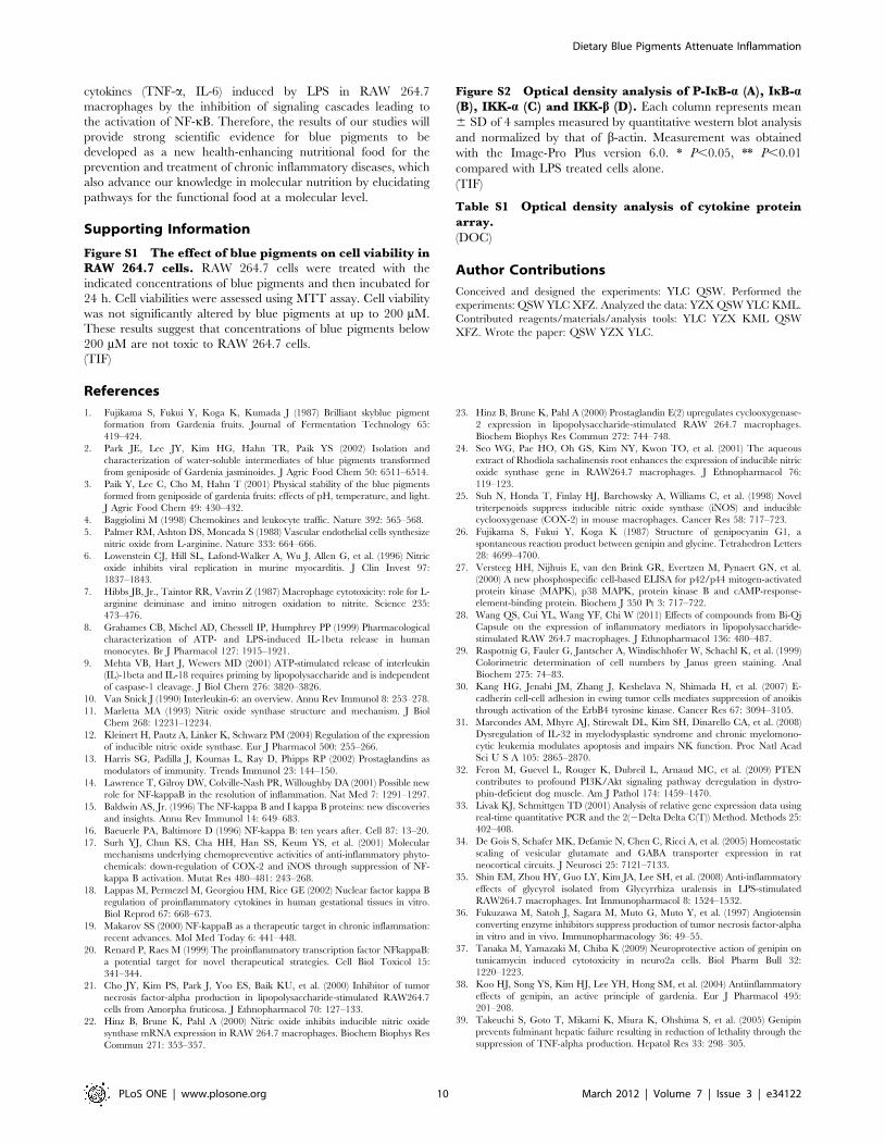
via an NF-kB-dependent mechanism. We also explored whether
blue pigments inhibited the LPS-stimulated degradation of IkB-ain RAW 264.7 cells by Western blotting with anti-IkB-a antibody.
Figure 7 shows that LPS-induced IkB-a degradation was
significantly blocked by blue pigments pretreatment. Furthermore,
to determine whether this IkB-a degradation was related to IkB-aphosphorylation, we examined the effect of blue pigments on the
LPS-induced p-IkB-a by Western blotting, and found that blue
pigments also significantly reduced LPS-induced IkB-a phosphor-
ylation. The b-actin protein was used as internal controls. Since
IKK-a and -b are upstream kinases of IkB in the NF-kB signal
pathway [17], we examined the effects of blue pigments on LPS
induced IKK-a, -b activation by immunoblotting using IKK-a, -bantibodies. Blue pigments (100 mM) inhibited the expression of
IKK-a and IKK-b. The b-actin protein was used as internal
control. (The western blot bands were quantified by densitometry
and the data are presented in Figure S2).
Effects of blue pigments on carrageenan-induced pawedema in mice
The anti-inflammatory effect of blue pigments was examined
using the carrageenan-induced paw edema model. As shown in
Figure 8, treatment with blue pigments (60, 120 mg/kg) showed
significant inhibitory effects on paw swelling, compared with the
vehicle control group. Maximal edema inhibition was observed at
1 h after edema induction. Notably, treatment with blue pigments
(120 mg/kg) reduced edema by 21.9% at 1 h, whereas the positive
control, Dexamethasone (10 mg/kg) decreased the edema rate by
34.5% at 1 h.
Effects of blue pigments on plasma concentrations ofTNF-a and IL-6 in LPS-stimulated ICR mice
Injecting mice with LPS lead to increase in plasma TNF-a and
IL-6 levels compared to untreated mice. Blue pigments signifi-
cantly reduced the plasma TNF-a and IL-6 levels in a dose-
dependent manner in the LPS-stimulated animals (Figure 9). Blue
pigments reduced the plasma TNF-a and IL-6 levels by 59.2%
and 19.5% in the LPS-stimulated animals. Pretreatment with the
anti-inflammatory steroid dexamethasone caused 81.7% and
36.1% reduction in plasma TNF-a and IL-6 in LPS-stimulated
mice.
Discussion
The pharmacological studies showed that genipin had exhibited
neuroprotective effect [37], anti-inflammatory effect [38] and
suppression of fas-induced lethal liver apoptosis in vitro [39].
Because it was a naturally occurring crosslinking reagent with
low cytotoxicity, genipin has recently been investigated as a cross-
linking regent in many biological applications. Recent explorations
into using of genipin cross-linked gelatin as a wound-dressing
membrane [40], bioadhesives [41], bone substitute [42] and
Figure 5. Effect of blue pigments on LPS-stimulated mRNA expression of TNF-a, IL-6, iNOS and COX-2. RAW 264.7 cells were pre-incubated with indicated concentrations of blue pigments for 2 h and were then treated with 0.2 mg/mL LPS for an additional 6 h. The mRNAexpression of TNF-a (A), IL-6 (B), iNOS (C) and COX-2 (D) was analyzed by real-time RT-PCR. Data represent means 6 S.D. values from threeindependent experiments. * P,0.05, ** P,0.01 (n = 6) compared with LPS treated cells alone.doi:10.1371/journal.pone.0034122.g005
Dietary Blue Pigments Attenuate Inflammation
PLoS ONE | www.plosone.org 7 March 2012 | Volume 7 | Issue 3 | e34122
heparin immobilization [43], have shown its potential as a new
and safe cross-linking agent, which had low cytotoxicity and
inflammatory reaction, satisfactory biocompatibility and stabile
mechanical properties than glutaraldehyde. Genipin was also
being investigated in the field of forensic science as a fingerprint
reagent to develop latent fingerprints on paper products [44].
Furthermore, genipin could react with amino acid to form the
stable blue pigments, and the blue pigments have been approved
by Ministry of Public Health of the People’s Republic of China
used as value-added colorants for foods in 1990 [45]. The
biological activity of genipin have been reported widely, however,
the biological activity studies of blue pigments have been not
reported. Dietary lutein, one of natural colorants for foods, has
been shown the potential anti-inflammatory effect in vitro [46], and
the present study was elucidating the molecular mechanisms
underlying the anti-inflammatory effect of blue pigments.
NO was recognized as a mediator and regulator of inflamma-
tory responses and was produced in high amounts by iNOS in
activated inflammatory cells [47]. Blue pigments was found to
significantly inhibit LPS-induced NO production in a concentra-
tion-dependent manner. The mRNA expression of iNOS was also
decreased by blue pigments, confirming the suppressive effect of
blue pigments on the NO production. The present work also
showed that blue pigments inhibited the expression of iNOS
mRNA in LPS-stimulated RAW 264.7 macrophage cells. This
blocking effect of blue pigments on LPS-induced iNOS expression
might have resulted from the transcriptional inhibition of iNOS
gene.
The mechanism of various antiinflammatory drug actions was
at least shared by the inhibition of prostaglandin synthesis, which
was mediated by cycloxygenase (COX) [48]. Of the two isoforms
of COX, COX-1 has been suggested to provide a physiologic level
of PGs for normal platelet, stomach and kidney function.
Moreover, COX-2 has been found to be highly induced at
inflammatory sites in animals as well as patients with inflammatory
diseases [49,50]. PGE2 was considered one of the strongest
inflammatory mediators in inflammatory response. It was
transformed from arachidonic acid via the COX-2 catalytic
reaction. COX-2 also could be affected directly at its enzymatic
activity by NO and iNOS [51]. The results showed that blue
pigments significantly reduced PGE2 production, and inhibited the
COX-2 mRNA and protein expression respectively, in a
concentration-dependent manner. It suggested that the anti-
inflammatory effect of blue pigments might be attributed to its
inhibitive effect on PGE2 production through blocking COX-2
gene and protein expression.
In inflammation progress, a series of cytokines and mediators
contributed to evoking and regression of inflammation. TNF-aand IL-6 were the critical cytokines involved in inflammation and
inhibition of them were regarded as a treatment strategy on
inflammation-related diseases [52,53,54]. So we chose TNF-a and
IL-6 as a parameter to investigate the anti-inflammatory effect of
blue pigments. In the present study, we found that TNF-a and IL-
6 release and mRNA expressions, which were highly stimulated by
Figure 6. Effects of blue pigments on LPS-induced NF-kBtranslocation. RAW 264.7 cells were pre-incubated with indicatedconcentrations of blue pigments for 2 h and stimulated with LPS(0.2 mg/mL) for 30 min. The DNA-binding activity of NF-kB p50 (A) andp65 (B) was detected with universal EZ-TFA transcription factor assay(Millipore, USA). NF-kB inhibitor BAY 11-7082 (10 mM) was used aspositive control. Data represent means 6 S.D. values. * P,0.05,** P,0.01 (n = 5) compared with LPS treated cells alone.doi:10.1371/journal.pone.0034122.g006
Figure 7. Effects of blue pigments on the degradation of IkB-a,IKK-a, IKK-b and phosphorylation of IkB-a. RAW 264.7 cells werepre-incubated with indicated concentrations of blue pigments for 2 hand stimulated with LPS (0.2 mg/mL) for 30 min. Total cellular proteinswere then prepared and Western blotted for IkB-a, IKK-a, IKK-b and p-IkB-a using specific IkB-a, IKK-a, IKK-b and p-IkB-a antibodies. The b-actin protein was used as internal controls. NF-kB inhibitor of BAY 11-7082 (10 mM) was used as positive control.doi:10.1371/journal.pone.0034122.g007
Dietary Blue Pigments Attenuate Inflammation
PLoS ONE | www.plosone.org 8 March 2012 | Volume 7 | Issue 3 | e34122

LPS, were inhibited by blue pigments. In particular, inhibitions of
IL-6 production and mRNA expression were more enhanced than
those of TNF-a by blue pigments, which presented the potential of
blue pigments to treat typical inflammation -related disorders. For
other cytokines involved in the inflammatory process, blue
pigments also inhibited their expressions, such as, G-CSF,
sICAM-1, IL-1a, , IL-1ra, KC, JE, MIP-1a, MIP-1b, RANTES.
It has been shown that NF-kB activation was a critical factor to
expression of various proinflammatory enzymes and cytokines,
and iNOS, COX-2, TNF-a, IL-1b and IL-6 in macrophages in
response to LPS [14,15]. NF-kB was composed mainly of two
proteins: p50 and p65. In resting cells, the NF-kB heterodimer was
held in the cytosol through interaction with IkB inhibitory proteins
[55]. NF-kB activation resulted from the phosphorylation and
proteasome- mediated degradation of inhibitory IkB proteins,
moreover, this was followed by the nuclear translocation and DNA
binding of NF-kB [56,57] where the transcription of target gene
was induced. Therefore, we examined NF-kB-DNA binding
activity to confirm that the inhibitions of the expressions of iNOS,
COX-2, TNF-a and IL-6 are influenced by the NF-kB signaling
pathway. Our results indicated that the nuclear translocations of
p65 and p50 proteins were inhibited in a concentration-dependent
manner by blue pigments and these results corresponded with its
inhibition of the expressions of iNOS, COX-2, TNF-a and IL-6.
In the cytoplasm, NF-kB is bound to tightly control by its
inhibitory subunit, IkB. In the present study, we also found that
the translocation of activated NF-kB to the nucleus was inhibited
in a concentration-dependent manner by blue pigments, and that
the degradation and phosphorylation of IkB-a were also inhibited
by blue pigments. These findings indicate that blue pigments may
inhibit NF-kB activation by suppressing the phosphorylation of
IkB-a and the translocations of the p50 and p65 subunits of NF-
kB from the cytosol to the nucleus in LPS-induced RAW 264.7
cells. IKK-a and IKK-b (known as the IkB kinases) are responsible
for phosphorylating IkBs [58]. In the present study, we observed
that blue pigments inhibited the activation of IKK-a and IKK-b.
Thus, we suggest that the inhibition of IKK-a and IKK-b by blue
pigments underlies its inhibition of NF-kB activation. Blue
pigments (60, 120 mg/kg) showed significant inhibitory effects
on carrageenan-induced paw edema in mice, compared with the
vehicle control group. Furthermore, blue pigments significantly
reduced the plasma TNF-a and IL-6 levels in a dose-dependent
manner in the LPS-stimulated animals. Although NF-kB is the
major regulator of pro-inflammatory signaling in macrophages,
other transcription factors activated by PLS, such as activating
protein-1(AP-1), cAMP response element-binding (CREB), The
nuclear factor interleukin-6 (NF-IL6) may affect the production of
inflammatory mediators[59,60,61]. The reason of the little
discrepancies in the distinct potency of genipin-derived blue
pigment against the LPS-induced activation of NF-kB and the
gene expression of some inflammatory mediator might be
associated with lack of inhibition of these transcriptional factors
(AP-1, CREB, NF-IL6).
In conclusion, although the blue pigments have been used as
value-added colorants for foods about 20 years in East Asia, its
biological activity has been first explored. The current study
demonstrated that blue pigments did not only inhibit iNOS and
COX-2 gene expression induced by LPS as well as the subsequent
production of NO and PGE2, but reduced the production of
Figure 9. Effects of blue pigments on plasma concentrations ofTNF-a and IL-6 in LPS-stimulated ICR mice. Blue pigments (30, 60,120 mg/kg), dexamethasone (10 mg/kg) or 0.9% saline were pretreatedfor 30 min. The mice were then either injected with 1 mg/kglipopolysaccharide (LPS) or PBS for 90 min. Plasma TNF-a (A) and IL-6(B) were quantified using ELISA. Data represent means 6S.D. values.* P,0.05, ** P,0.01 (n = 8) compared with LPS treated alone.doi:10.1371/journal.pone.0034122.g009
Figure 8. Effects of blue pigments on carrageenan-inducedpaw edema in mice. Blue pigments (30, 60, 120 mg/kg), dexameth-asone (10 mg/kg) or with 0.9% saline were administered 30 min beforecarrageenan injection into mice for alleviation of acute inflammation.Paw thickness was measured using Plethysmometer before and everyhour after edema induction for 4 h. The percent increase of pawthickness was calculated based on the volume difference between thepaw with and without carrageenan injection. Data represent means 6S.D. values. * P,0.05, ** P,0.01 (n = 10) indicate significant differencesfrom vehicle control.doi:10.1371/journal.pone.0034122.g008
Dietary Blue Pigments Attenuate Inflammation
PLoS ONE | www.plosone.org 9 March 2012 | Volume 7 | Issue 3 | e34122
cytokines (TNF-a, IL-6) induced by LPS in RAW 264.7
macrophages by the inhibition of signaling cascades leading to
the activation of NF-kB. Therefore, the results of our studies will
provide strong scientific evidence for blue pigments to be
developed as a new health-enhancing nutritional food for the
prevention and treatment of chronic inflammatory diseases, which
also advance our knowledge in molecular nutrition by elucidating
pathways for the functional food at a molecular level.
Supporting Information
Figure S1 The effect of blue pigments on cell viability inRAW 264.7 cells. RAW 264.7 cells were treated with the
indicated concentrations of blue pigments and then incubated for
24 h. Cell viabilities were assessed using MTT assay. Cell viability
was not significantly altered by blue pigments at up to 200 mM.
These results suggest that concentrations of blue pigments below
200 mM are not toxic to RAW 264.7 cells.
(TIF)
Figure S2 Optical density analysis of P-IkB-a (A), IkB-a(B), IKK-a (C) and IKK-b (D). Each column represents mean
6 SD of 4 samples measured by quantitative western blot analysis
and normalized by that of b-actin. Measurement was obtained
with the Image-Pro Plus version 6.0. * P,0.05, ** P,0.01
compared with LPS treated cells alone.
(TIF)
Table S1 Optical density analysis of cytokine proteinarray.
(DOC)
Author Contributions
Conceived and designed the experiments: YLC QSW. Performed the
experiments: QSW YLC XFZ. Analyzed the data: YZX QSW YLC KML.
Contributed reagents/materials/analysis tools: YLC YZX KML QSW
XFZ. Wrote the paper: QSW YZX YLC.
References
1. Fujikama S, Fukui Y, Koga K, Kumada J (1987) Brilliant skyblue pigment
formation from Gardenia fruits. Journal of Fermentation Technology 65:
419–424.
2. Park JE, Lee JY, Kim HG, Hahn TR, Paik YS (2002) Isolation and
characterization of water-soluble intermediates of blue pigments transformed
from geniposide of Gardenia jasminoides. J Agric Food Chem 50: 6511–6514.
3. Paik Y, Lee C, Cho M, Hahn T (2001) Physical stability of the blue pigments
formed from geniposide of gardenia fruits: effects of pH, temperature, and light.
J Agric Food Chem 49: 430–432.
4. Baggiolini M (1998) Chemokines and leukocyte traffic. Nature 392: 565–568.
5. Palmer RM, Ashton DS, Moncada S (1988) Vascular endothelial cells synthesize
nitric oxide from L-arginine. Nature 333: 664–666.
6. Lowenstein CJ, Hill SL, Lafond-Walker A, Wu J, Allen G, et al. (1996) Nitric
oxide inhibits viral replication in murine myocarditis. J Clin Invest 97:
1837–1843.
7. Hibbs JB, Jr., Taintor RR, Vavrin Z (1987) Macrophage cytotoxicity: role for L-
arginine deiminase and imino nitrogen oxidation to nitrite. Science 235:
473–476.
8. Grahames CB, Michel AD, Chessell IP, Humphrey PP (1999) Pharmacological
characterization of ATP- and LPS-induced IL-1beta release in human
monocytes. Br J Pharmacol 127: 1915–1921.
9. Mehta VB, Hart J, Wewers MD (2001) ATP-stimulated release of interleukin
(IL)-1beta and IL-18 requires priming by lipopolysaccharide and is independent
of caspase-1 cleavage. J Biol Chem 276: 3820–3826.
10. Van Snick J (1990) Interleukin-6: an overview. Annu Rev Immunol 8: 253–278.
11. Marletta MA (1993) Nitric oxide synthase structure and mechanism. J Biol
Chem 268: 12231–12234.
12. Kleinert H, Pautz A, Linker K, Schwarz PM (2004) Regulation of the expression
of inducible nitric oxide synthase. Eur J Pharmacol 500: 255–266.
13. Harris SG, Padilla J, Koumas L, Ray D, Phipps RP (2002) Prostaglandins as
modulators of immunity. Trends Immunol 23: 144–150.
14. Lawrence T, Gilroy DW, Colville-Nash PR, Willoughby DA (2001) Possible new
role for NF-kappaB in the resolution of inflammation. Nat Med 7: 1291–1297.
15. Baldwin AS, Jr. (1996) The NF-kappa B and I kappa B proteins: new discoveries
and insights. Annu Rev Immunol 14: 649–683.
16. Baeuerle PA, Baltimore D (1996) NF-kappa B: ten years after. Cell 87: 13–20.
17. Surh YJ, Chun KS, Cha HH, Han SS, Keum YS, et al. (2001) Molecular
mechanisms underlying chemopreventive activities of anti-inflammatory phyto-
chemicals: down-regulation of COX-2 and iNOS through suppression of NF-
kappa B activation. Mutat Res 480–481: 243–268.
18. Lappas M, Permezel M, Georgiou HM, Rice GE (2002) Nuclear factor kappa B
regulation of proinflammatory cytokines in human gestational tissues in vitro.
Biol Reprod 67: 668–673.
19. Makarov SS (2000) NF-kappaB as a therapeutic target in chronic inflammation:
recent advances. Mol Med Today 6: 441–448.
20. Renard P, Raes M (1999) The proinflammatory transcription factor NFkappaB:
a potential target for novel therapeutical strategies. Cell Biol Toxicol 15:
341–344.
21. Cho JY, Kim PS, Park J, Yoo ES, Baik KU, et al. (2000) Inhibitor of tumor
necrosis factor-alpha production in lipopolysaccharide-stimulated RAW264.7
cells from Amorpha fruticosa. J Ethnopharmacol 70: 127–133.
22. Hinz B, Brune K, Pahl A (2000) Nitric oxide inhibits inducible nitric oxide
synthase mRNA expression in RAW 264.7 macrophages. Biochem Biophys Res
Commun 271: 353–357.
23. Hinz B, Brune K, Pahl A (2000) Prostaglandin E(2) upregulates cyclooxygenase-
2 expression in lipopolysaccharide-stimulated RAW 264.7 macrophages.
Biochem Biophys Res Commun 272: 744–748.
24. Seo WG, Pae HO, Oh GS, Kim NY, Kwon TO, et al. (2001) The aqueous
extract of Rhodiola sachalinensis root enhances the expression of inducible nitric
oxide synthase gene in RAW264.7 macrophages. J Ethnopharmacol 76:
119–123.
25. Suh N, Honda T, Finlay HJ, Barchowsky A, Williams C, et al. (1998) Novel
triterpenoids suppress inducible nitric oxide synthase (iNOS) and inducible
cyclooxygenase (COX-2) in mouse macrophages. Cancer Res 58: 717–723.
26. Fujikama S, Fukui Y, Koga K (1987) Structure of genipocyanin G1, a
spontaneous reaction product between genipin and glycine. Tetrahedron Letters
28: 4699–4700.
27. Versteeg HH, Nijhuis E, van den Brink GR, Evertzen M, Pynaert GN, et al.
(2000) A new phosphospecific cell-based ELISA for p42/p44 mitogen-activated
protein kinase (MAPK), p38 MAPK, protein kinase B and cAMP-response-
element-binding protein. Biochem J 350 Pt 3: 717–722.
28. Wang QS, Cui YL, Wang YF, Chi W (2011) Effects of compounds from Bi-Qi
Capsule on the expression of inflammatory mediators in lipopolysaccharide-
stimulated RAW 264.7 macrophages. J Ethnopharmacol 136: 480–487.
29. Raspotnig G, Fauler G, Jantscher A, Windischhofer W, Schachl K, et al. (1999)
Colorimetric determination of cell numbers by Janus green staining. Anal
Biochem 275: 74–83.
30. Kang HG, Jenabi JM, Zhang J, Keshelava N, Shimada H, et al. (2007) E-
cadherin cell-cell adhesion in ewing tumor cells mediates suppression of anoikis
through activation of the ErbB4 tyrosine kinase. Cancer Res 67: 3094–3105.
31. Marcondes AM, Mhyre AJ, Stirewalt DL, Kim SH, Dinarello CA, et al. (2008)
Dysregulation of IL-32 in myelodysplastic syndrome and chronic myelomono-
cytic leukemia modulates apoptosis and impairs NK function. Proc Natl Acad
Sci U S A 105: 2865–2870.
32. Feron M, Guevel L, Rouger K, Dubreil L, Arnaud MC, et al. (2009) PTEN
contributes to profound PI3K/Akt signaling pathway deregulation in dystro-
phin-deficient dog muscle. Am J Pathol 174: 1459–1470.
33. Livak KJ, Schmittgen TD (2001) Analysis of relative gene expression data using
real-time quantitative PCR and the 2(2Delta Delta C(T)) Method. Methods 25:
402–408.
34. De Gois S, Schafer MK, Defamie N, Chen C, Ricci A, et al. (2005) Homeostatic
scaling of vesicular glutamate and GABA transporter expression in rat
neocortical circuits. J Neurosci 25: 7121–7133.
35. Shin EM, Zhou HY, Guo LY, Kim JA, Lee SH, et al. (2008) Anti-inflammatory
effects of glycyrol isolated from Glycyrrhiza uralensis in LPS-stimulated
RAW264.7 macrophages. Int Immunopharmacol 8: 1524–1532.
36. Fukuzawa M, Satoh J, Sagara M, Muto G, Muto Y, et al. (1997) Angiotensin
converting enzyme inhibitors suppress production of tumor necrosis factor-alpha
in vitro and in vivo. Immunopharmacology 36: 49–55.
37. Tanaka M, Yamazaki M, Chiba K (2009) Neuroprotective action of genipin on
tunicamycin induced cytotoxicity in neuro2a cells. Biol Pharm Bull 32:
1220–1223.
38. Koo HJ, Song YS, Kim HJ, Lee YH, Hong SM, et al. (2004) Antiinflammatory
effects of genipin, an active principle of gardenia. Eur J Pharmacol 495:
201–208.
39. Takeuchi S, Goto T, Mikami K, Miura K, Ohshima S, et al. (2005) Genipin
prevents fulminant hepatic failure resulting in reduction of lethality through the
suppression of TNF-alpha production. Hepatol Res 33: 298–305.
Dietary Blue Pigments Attenuate Inflammation
PLoS ONE | www.plosone.org 10 March 2012 | Volume 7 | Issue 3 | e34122

40. Chang WH, Chang Y, Lai PH, Sung HW (2003) A genipin-crosslinked gelatin
membrane as wound-dressing material: in vitro and in vivo studies. J BiomaterSci Polym Ed 14: 481–495.
41. Sung HW, Huang DM, Chang WH, Huang LL, Tsai CC, et al. (1999) Gelatin-
derived bioadhesives for closing skin wounds: an in vivo study. J Biomater SciPolym Ed 10: 751–771.
42. Liu BS, Yao CH, Chen YS, Hsu SH (2003) In vitro evaluation of degradationand cytotoxicity of a novel composite as a bone substitute. J Biomed Mater Res A
67: 1163–1169.
43. Tsai CC, Chang Y, Sung HW, Hsu JC, Chen CN (2001) Effects of heparinimmobilization on the surface characteristics of a biological tissue fixed with a
naturally occurring crosslinking agent (genipin): an in vitro study. Biomaterials22: 523–533.
44. Levinton-Shamuilov G, Cohen Y, Azoury M, Chaikovsky A, Almog J (2005)Genipin, a novel fingerprint reagent with colorimetric and fluorogenic activity,
part II: optimization, scope and limitations. J Forensic Sci 50: 1367–1371.
45. Zhang CH (2006) Food Colorants Data Book. Beijing: China MeasurementPress.
46. Rafi MM, Shafaie Y (2007) Dietary lutein modulates inducible nitric oxidesynthase (iNOS) gene and protein expression in mouse macrophage cells (RAW
264.7). Mol Nutr Food Res 51: 333–340.
47. Korhonen R, Lahti A, Kankaanranta H, Moilanen E (2005) Nitric oxideproduction and signaling in inflammation. Curr Drug Targets Inflamm Allergy
4: 471–479.48. Vane JR (1971) Inhibition of prostaglandin synthesis as a mechanism of action
for aspirin-like drugs. Nat New Biol 231: 232–235.49. Masferrer JL, Zweifel BS, Manning PT, Hauser SD, Leahy KM, et al. (1994)
Selective inhibition of inducible cyclooxygenase 2 in vivo is antiinflammatory
and nonulcerogenic. Proc Natl Acad Sci U S A 91: 3228–3232.50. Seibert K, Zhang Y, Leahy K, Hauser S, Masferrer J, et al. (1994)
Pharmacological and biochemical demonstration of the role of cyclooxygenase2 in inflammation and pain. Proc Natl Acad Sci U S A 91: 12013–12017.
51. Moncada S, Palmer RM, Higgs EA (1991) Nitric oxide: physiology,
pathophysiology, and pharmacology. Pharmacol Rev 43: 109–142.
52. Locksley RM, Killeen N, Lenardo MJ (2001) The TNF and TNF receptor
superfamilies: integrating mammalian biology. Cell 104: 487–501.
53. Burger D, Dayer JM, Palmer G, Gabay C (2006) Is IL-1 a good therapeutic
target in the treatment of arthritis? Best Pract Res Clin Rheumatol 20: 879–896.
54. Rose-John S, Waetzig GH, Scheller J, Grotzinger J, Seegert D (2007) The IL-6/
sIL-6R complex as a novel target for therapeutic approaches. Expert Opin Ther
Targets 11: 613–624.
55. Baeuerle PA, Henkel T (1994) Function and activation of NF-kappa B in the
immune system. Annu Rev Immunol 12: 141–179.
56. Brown K, Park S, Kanno T, Franzoso G, Siebenlist U (1993) Mutual regulation
of the transcriptional activator NF-kappa B and its inhibitor, I kappa B-alpha.
Proc Natl Acad Sci U S A 90: 2532–2536.
57. Rodriguez MS, Thompson J, Hay RT, Dargemont C (1999) Nuclear retention
of IkappaBalpha protects it from signal-induced degradation and inhibits
nuclear factor kappaB transcriptional activation. J Biol Chem 274: 9108–9115.
58. May MJ, Ghosh S (1999) IkappaB kinases: kinsmen with different crafts. Science
284: 271–273.
59. Jeon YJ, Han SH, Lee YW, Lee M, Yang KH, et al. (2000) Dexamethasone
inhibits IL-1 beta gene expression in LPS-stimulated RAW 264.7 cells by
blocking NF-kappa B/Rel and AP-1 activation. Immunopharmacology 48:
173–183.
60. Avni D, Ernst O, Philosoph A, Zor T (2010) Role of CREB in modulation of
TNFalpha and IL-10 expression in LPS-stimulated RAW264.7 macrophages.
Mol Immunol 47: 1396–1403.
61. Godambe SA, Chaplin DD, Takova T, Bellone CJ (1994) Upstream NFIL-6-like
site located within a DNase I hypersensitivity region mediates LPS-induced
transcription of the murine interleukin-1 beta gene. J Immunol 153: 143–152.
Dietary Blue Pigments Attenuate Inflammation
PLoS ONE | www.plosone.org 11 March 2012 | Volume 7 | Issue 3 | e34122