different intermediate-sized filamentsdistinguished ...proc. natl. acad.sci. usa75(1978) 5037 0a 6...
TRANSCRIPT

Proc. Natl. Acad. Sci. USAVol. 75, No. 10, pp. 5034-5038, October 1978Cell Biology
Different intermediate-sized filaments distinguished byimmunofluorescence microscopy
(vimentin/prekeratin/cytoskeleton/tonofilaments/mitotic drugs)
WERNER W. FRANKE*, ERIKA SCHMID*, MARY OSBORNt, AND KLAUS WEBERt* Division of Membrane Biology and Biochemistry, Institute of Experimental Pathology, German Cancer Research Center, D-6900 Heidelberg; andt Department of Biochemistry, Max-Planck-Institute for Biophysical Chemistry, D-3400 Gottingen, Federal Republic of Germany
Communicated by Jean Brachet, July 28,1978
ABSTRACT The major protein of intermediate-sized fila-ments in mouse 3T3 cells, for which the name vimentin is pro-posed, has a molecular weight of 57,000. Antibodies againstvimentin and antibodies against prekeratin have been used inparallel in immunofluorescence microscopy on a variety ofcultured cells as well as on frozen tissue sections. Both anti-bodies decorate extended wavy arrays of filaments that aredifferent from microfilaments and microtubules. Intermediatefilament bundles decorated by antibodies against prekeratinare predominant in many epithelial cells, including epithelia-derived tumor cells, and are not decorated by antibodies tovimentin. In contrast, intermediate filaments decorated byantibodies against vimentin are widespread among nonmusclecells of mesenchymal origin, including transformed cells, andalso occur in other cells. Perinuclear whorls of aggregates ofintermediate filaments induced by prolonged treatment withColcemid generally show strong decoration with antibodiesagainst vimentin. No significant reaction with either antiserumhas been observed in muscle structures or in brain nerve tissue.These observations show that intermediate filaments withsimilar ultrastructure and solubility characteristics can be dis-tinguished immunologically.
Most animal cells contain, in addition to microfilaments (5-6nm diameter) and microtubules (20-25 nm), a third system ofcytoskeletal filaments (6-11 nm) which are commonly desig-nated by the collective morphological term "intermediate-sizedfilaments" (intermediate filaments). These include tonofila-ments, neurofilaments, 10-nm filaments of muscle, and the 6-to 11-nm filaments present in various cultured cells (1-13). Acentral question in the studies on intermediate filaments iswhether they are identical or related to each other. Evidencefor both compositional similarities and differences among thevarious forms of intermediate filaments can be found as far aspolypeptide numbers, molecular weights of components, andimmunological crossreaction is concerned (4-18). We havefound previously that normal rabbit sera frequently decoratethe arrays of tonofilament-like filaments in rat kangaroo PtK2and other cells (8). Antibodies against bovine epidermal pre-keratin raised in guinea pigs that did not have autoantibodies'totonofilaments allowed us to conclude that the intermediatefilament arrays prominent in PtK2 and some other epithelia-derived cells contain prekeratin-related proteins (10). Thesespecific antibodies, however, did not decorate intermediatefilaments in a variety of cultured cells, especially those ofmesenchymal origin, although in such cells these fibers can bevisualized by rare autoantibodies found in rabbits (12) or inhumans (15). Since cytoskeleton preparations from such cellsare enriched both in intermediate filaments and a polypeptideof Mr 55,000-58,000 (3, 10, 13, 16, 19) [polypeptides of similarsize have been described in filaments isolated from BHK cells
(9)], we have isolated this protein from cytoskeletons of murine3T3 cells and raised antibodies against it. We report here thatthese antibodies decorate the intermediate filament arrays ofmouse 3T3 and various mesenchyme-derived cells but not thetonofilament-like fibers of epithelia-derived cells. In order toemphasize the difference between this protein and proteinsfrom other intermediate filaments, we propose the namevimentin.J
MATERIALS AND METHODSCell lines and cultures listed in Table 1 were grown by standardprocedures (for special cultures see also refs. 8 and 20). Col-cemid treatment (1 ,uM) was for 12, 24, or 48 hr. For exami-nation of frozen sections, small cubes of tissue were frozen, and3- to 4-,um thick cryostat sections were prepared.
Cytoskeletal material of mouse 3T3 cells enriched in inter-mediate filaments, as judged by electron microscopy, wasprepared by extractions in low and high salt buffers and solu-tions containing 1% Triton X-100 010, 16). The prominentpolypeptide (apparent Mr of 57,000) seen after electrophoresison sodium dodecyl sulfate/polyacrylamide gels was excised andeluted (Fig. 1). The protein was briefly crosslinked with 0.1%glutaraldehyde, freed of excess aldehyde (21), and used withcomplete adjuvant as antigen in guinea pigs (400 jig subcuta-neously at multiple sites). Booster injections (300 Mg) were givenat days 14 and 28. Animals were bled at day 34. Reaction withthe original antigen was demonstrated by immunodiffusionanalysis (see ref. 10). The guinea pig antibodies against bovineepidermal prekeratin (10) were used as antiserum or as mo-nospecific antibodies purified by affinity chromatography onprekeratin covalently bound to Sepharose 4B. For antibodiesagainst actin and tubulin, see ref. 8.
Cells grown on coverslips and frozen sections mounted onslides were processed for indirect immunofluorescence mi-croscopy as described (8, 10, 21, 22). Guinea pig sera wereroutinely used at 1:20 dilution, but reacted also at much higherdilutions (100:1-300:1). Since the intensity of decoration by theantibodies to vimentin was reduced, though not abolished, afterfixation with formaldehyde, most experiments were performedwith cells not treated with aldehyde.
RESULTSCytoskeletal material from mouse 3T3 cells highly enriched inintermediate filaments contained a prominent polypeptideband-vimentin-of apparent Mr of 57,000 (Fig. 1). Antibodiesto purified vimentin (Fig. 1) were used in indirect immu-nofluorescence microscopy on various cultured cells and onfrozen sections.
t From the Latin word vimentum, used to describe arrays of flexiblerods, both ordered ones (e.g., lattices, filigrees, and wicker-work) andnonordered ones (e.g., brushwood).
5034
The publication costs of this article were defrayed in part by pagecharge payment. This article must therefore be hereby marked "ad-vertisement" in accordance with 18 U. S. C. §1734 solely to indicatethis fact.
Dow
nloa
ded
by g
uest
on
Janu
ary
26, 2
020
Proc. Natl. Acad. Sci. USA 75 (1978) 5035
I1
23
I
:> I;W JI I
I
4
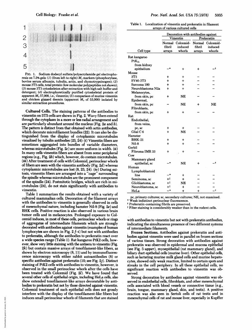
5FIG. 1. Sodium dodecyl sulfate/polyacrylamide gel electropho-
resis on 7.5% gels: (1) (from left to right) Mr markers (phosphorylase,bovine serum albumin, tubulin, actin, and chymotrypsinogen); (2)mouse 3T3 cells, total protein (low molecular polypeptides not shown);(3) mouse 3T3 cytoskeleton after extraction with high salt buffer anddetergent; (4) electrophoretically purified cytoskeletal protein ofapparent Mr 57,000, i.e., vimentin; (5) comparison of murine vimentinand chicken gizzard desmin (apparent Mr of 53,000) isolated bysimilar extraction procedures.
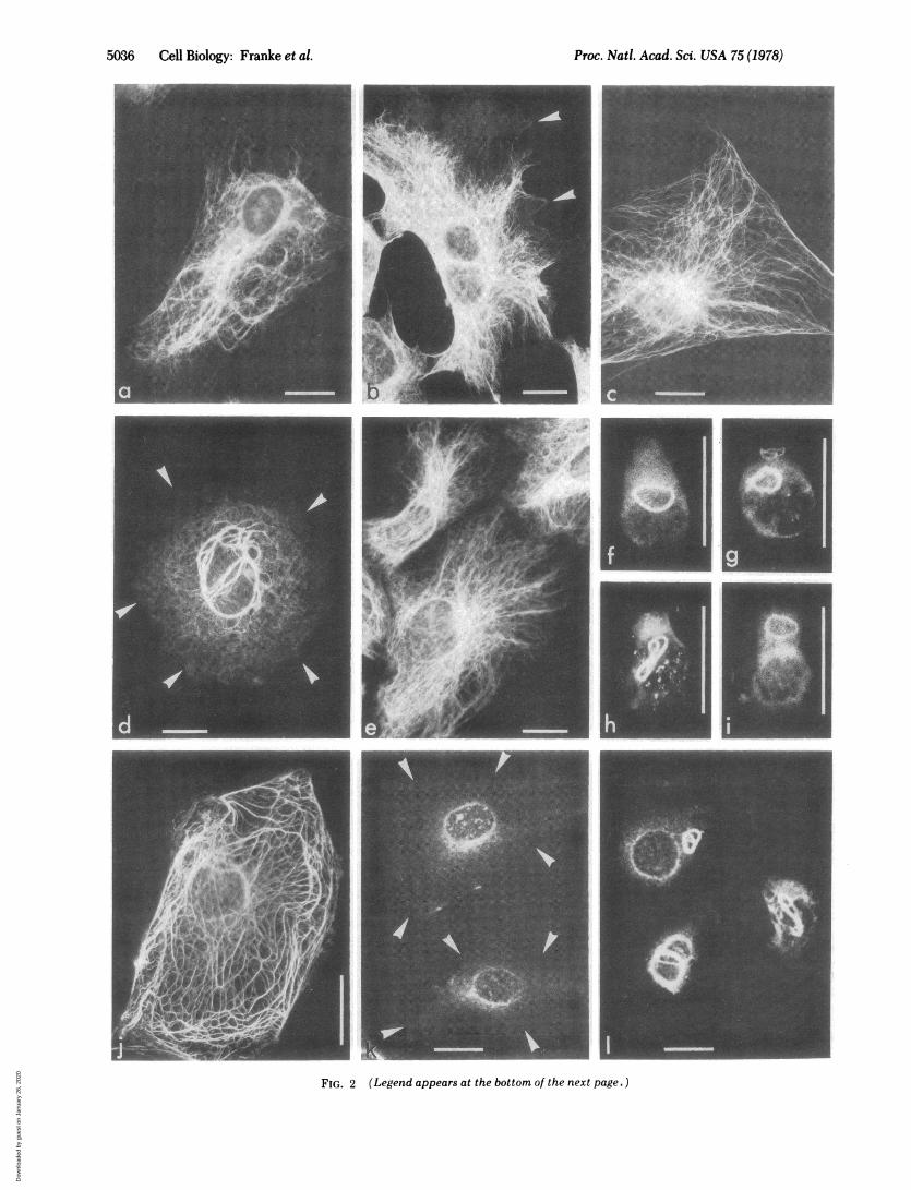
Cultured Cells. The staining patterns of the antibodies tovimentin on 3T3 cells are shown in Fig. 2. Wavy fibers extendthrough the cytoplasm in a more or less radial arrangement andare particularly abundant around the nucleus (Fig. 2a and b).The pattern is distinct from that obtained with actin antibodies,which decorate microfilament bundles (22). It can also be dis-tinguished from the display of cytoplasmic microtubulesvisualized by tubulin antibodies (23, 24): (i) Vimentin fibers aresometimes aggregated into bundles of variable diameters,whereas microtubules (Fig. 2c) are more uniform in width. (ii)In many cells vimentin fibers are absent from some peripheralregions (e.g., Fig. 2b) which, however, do contain microtubules.(iii) After treatment of cells with Colcemid, perinuclear whorlsof fibers are seen with the vimentin antibody (Fig. 2d) whereascytoplasmic microtubules are lost (8, 23, 24). (iv) During mi-tosis, vimentin fibers are arranged into a "cage" surroundingthe spindle whereas microtubules are the prominent componentof the spindle (24). Postmitotic bridges, which are rich in mi-crotubules (24), do not stain significantly with antibodies tovimentin.
Table 1 summarizes the results obtained with a variety ofcultured mammalian cells. Decoration of the filament arrayswith the antibodies to vimentin is generally observed in cellsof mesenchymal nature, including hamster Nil-8 (Fig. 2e) andBHK cells. Positive reaction is also observed in various braintumor cells and in melanocytes. Prolonged exposure to Col-cemid induces, in most of these cells, perinuclear whorls or ringsof aggregates of intermediate filaments which are stronglydecorated with antibodies against vimentin (examples of humanlymphocytes are shown in Fig. 2 f-i) but not with antibodiesto prekeratin, although the antibodies to prekeratin react overa wide species range (Table 1). Rat kangaroo PtK2 cells, how-ever, show very little staining with the antisera to vimentin (Fig.2k) but contain massive arrays of tonofilament-like fibers, asshown by electron microscopy (8, 11) and by immunofluores-cence microscopy with either rabbit autoantibodies (8) or
specific antibodies against prekeratin (10; see Fig. 2j). Distinctstaining of PtK2 cells with antibodies to vimentin, however, isobserved in the small perinuclear whorls after the cells havebeen treated with Colcemid (Fig. 21). We have found thatseveral other cells of epithelial origin, including HeLa cells, alsoshow extended tonofilament-like arrays decoratable by anti-bodies to prekeratin but not by those directed against vimentin.Colcemid treatment of such epithelial cells does not grosslyinterfere with the display of the tonofilament-like fibers butinduces small perinuclear whorls of filaments that are stained
Table 1. Localization of vimentin and prekeratin in filamentarrays of various cultured cells
Decoration with antibodies againstVimentin Prekeratin
Normal Colcemid- Normal Colcemid-fibril induced fibril induced
Cell type arrays whorls arrays whorls
Rat kangarooPtK2,from kidneyepithelium -$ + + -t
Mouse3T3 + + - -SV40-3T3 + + - _Sarcoma 180 + + -
Neuroblastoma N2a + + -
Melanocytes,fromskin,pc + NE -
Epidermal,from skin, pc - NE + NE
Fibroblasts,from skin, pc + +
RatEndothelial,
from veins,pc, sc + + - -
Glial C 6 + NE - -
HamsterBHK-21 + + - -Nil-8 + + - -
GerbilFibroma IMR 33 + + - -
CowMammary gland
epithelial, sc -* NE + -tHumanLymphoblastoid
line - + -
Seminoma, sc +-+Glioblastoma, sc +t NE -
Neuroblastoma, sc +t + -
HeLa -* + + _t
pc, primary cultures; sc, secondary cultures; NE, not examined.* Weak indistinct perinuclear fluorescence.t Prekeratin-containing fibrils are preserved.Fiber staining is considerably weaker than in the rodent cells.
with antibodies to vimentin but not with prekeratin antibodies,indicating the simultaneous presence of two different systemsof intermediate filaments.
Frozen Sections. Antibodies against prekeratin and anti-bodies against vimentin were used in parallel on frozen sectionsof various tissues. Strong decoration with antibodies againstprekeratin was observed in epidermal and mucosa epithelial(see Fig. 3 upper), myoepithelial (rat mammary gland), andbiliary duct epithelial cells (murine liver). Other epithelial cells,such as lactating murine milk gland cells and murine hepato-cytes, showed only weak reaction, limited to certain spots andstreaks in the cell periphery. In all these epithelial cells, nosignificant reaction with antibodies to vimentin was ob-served.
Strong decoration by antibodies against vimentin was ob-served in endothelial cells, fibroblasts, and other mesenchymalcells associated with blood vessels or connective tissue (e.g.,brain, tongue, mammary gland, skin, and testis). A positivereaction was also seen in Sertoli cells of rat testis and inmesenchymal cells of rat and mouse liver, especially in Kupffer
Cell Biology: Franke et al.
Dow
nloa
ded
by g
uest
on
Janu
ary
26, 2
020
Proc. Natl. Acad. Sci. USA 75 (1978)
I
k:
FIG. 2 (Legend appears at the bottom of the next page. )
5036 Cell Biology: Franke et al.
Dow
nloa
ded
by g
uest
on
Janu
ary
26, 2
020

Proc. Natl. Acad. Sci. USA 75 (1978) 5037
0A
IL 2'6
FIG. 3. Immunofluorescence micrographs of frozen sectionsof rat tongue after decoration with guinea pig antibodies againstprekeratin (Upper) and vimentin (Lower). Positive reaction is notedwith antibodies to prekeratin only in the mucosal epithelium (A) andwith antibodies to vimentin only in cells of the lamina propria (B) aswell as fibroblasts and blood vessel elements of tongue muscle. Nosignificant reaction with either antisera is observed in muscle struc-tures (C). Bars denote 30,um.
cells. All these cells were not significantly decorated by anti-bodies to prekeratin. An example of the specificity of the dif-ferential reaction with the two antibodies in different cells ofthe same organ, i.e., the rat tongue, is shown in Fig. 3. Frozensections through various types of muscle (smooth, cardiac, and
striated) did not show specific decoration by either antiserum(Fig. 3), in agreement with experiments performed on myofi-brils isolated from rat leg muscle. Brain nerve tissue also did notshow significant reaction, although strong reaction with antiserato vimentin was observed in blood vessels of brain and in lep-tomeningal cells.
DISCUSSIONOur results on various cultured cells and frozen tissue sectionsdifferentiate between different systems of intermediate fila-ments.
(i) The system typical of various cells of mesenchymalcharacter. Here vimentin represents a major cellular proteincomparable in amount to actin and tubulin (3, 9, 12, 13) andis the major protein constituent of intermediate filaments.While this work was in progress, Hynes and Destree (13) de-scribed an antibody against a protein of similar size present inthe intermediate filaments of hamster Nil-8 cells. Our resultson these cells and other cells confirm their conclusions aboutthe organization and identity of this filament system. Thesample of cells containing vimentin (Table 1) also includes cellsin which Gordon et al. (12) have recently detected intermediatefilaments by immunofluorescence microscopy using a rabbitautoimmune serum that reacted in vitro with a polypeptide ofMr of about 57,000 in extracts of gerbil fibroma and mouse 3T3cells. Intermediate filaments in BHK-21 cells, which have beenisolated and characterized by Goldman and coworkers (2, 9),also stain with vimentin antibodies. Apparently, vimentin is alsothe major protein in the whorly aggregates induced in manycells by prolonged treatment with antimitotic drugs (e.g., refs.1, 2, 4, and 9).
(ii) The type characteristic for various epithelial and epi-thelia-derived cells, including desmosome-associated tonofi-laments, contains prekeratin-related proteins and can be im-munologically distinguished from vimentin fibers. Our dataalso show that such cells can contain two different types of in-termediate filaments since their vimentin-like material isrearranged by Colcemid treatment into perinuclear whorls.
(iii) The intermediate filaments typical of muscle are notdecorated by antibodies to either vimentin or prekeratin. Thus,the major protein of muscle intermediate filaments, variouslycalled desmin (5) or skeletin (17), must be immunologicallydifferent. This agrees with the report (14) that antibodies againstchicken desmin do not crossreact with several nonmuscle cellsin culture as well as with the separation of vimentin and desminon gels (Fig. 1; in this gel electrophoretic system mammaliandesmin, prepared from smooth muscle of porcine uterus,comigrated with chicken gizzard desmin).
(iv) The relation of neurofilaments to the other classes ofintermediate filaments is somewhat difficult to evaluate. Rabbitantibodies against bovine neurofilament protein have beenreported to react not only with filament aggregates in neuro-blastoma cells (18), but also with filament bundles in endothelialand cardiac muscle cells (6), indicating an immunologicalrelation (see ref. 12). However, desmin shows peptide mapsdifferent from those of neurofilaments of the same species (7),and we found that antibodies to vimentin and to prekeratin donot react with brain nerve tissue in frozen sections, suggestingthat the neurofilament proteins are not fully identical with otherintermediate filament proteins. In addition, it seems that at least
FIG. 2 (on preceding page). Immunofluorescence microscopy on mouse 3T3 cells (a-d), hamster Nil-8 cells (e), human lymphoblastoidalcells (f-i), and rat kangaroo PtK2 cells (j-l) using antibodies against vimentin (a, b, d-i, k, 1), tubulin (c), or prekeratin (j). Cells shown in d,f-i, and I were pretreated with Colcemid (note perinuclear whorls and various forms of rings; the cell shown in i was fixed with aldehyde). Arrowsdenote cell contours. Bars indicate 20 im.
Cell Biology: Franke et al.
Dow
nloa
ded
by g
uest
on
Janu
ary
26, 2
020

5038 Cell Biology: Franke et al.
some brain tumor-derived cells (neuroblastoma) show vimentinin addition to neurofilament protein.
Intermediate filaments share a similar morphology. Theyare long flexible rods with an apparently hollow core and atendency to lateral fasciation, and are rather insoluble in variousbuffers (for references see refs. 11 and 16). The finding thatdifferent classes of polypeptides make up filamentous structuresso similar in morphology and possibly in function could indicatethat the various proteins involved may have some degree ofsimilarity in primary structure and three-dimensional order andconstitute a family of related structural proteins ("cytoskelet-ins"?), possibly derived from a common ancestral gene.
We thank S. Winter, T. Born, H. J. Koitzsch, and K. Mahler for ex-cellent technical help.
1. Ishikawa, H., Bischoff, R. & Holtzer, H. (1968) J. Cell Biol. 38,538-555.
2. Goldman, R. D. (1971) J. Cell Biol. 51, 752-762.3. Brown, S., Levinson, W. & Spudich, J. A. (1976) J. Supramol.
Struct. 5, 119-130.4. Holtzer, H., Fellini, S., Rubinstein, N., Chi, J. & Strahs, K. (1976)
in Cell Motility, eds. Goldman, R. D., Pollard, T. & Rosenbaum,J. (Cold Spring Harbor Laboratory, Cold Spring Harbor, NY),pp. 823-839.
5. Lazarides, E. & Hubbard, B. D. (1976) Proc. Natl. Acad. Sci. USA73,4344-4348.
6. Blose, S. H., Shelanski, M. L. & Chacko, S. (1977) Proc. NatI.Acad. Sci. USA 74,662-665.
7. Davison, P. F., Hong, B. S. & Cooke, P. (1977) Exp. Cell Res. 109,471-474.
8. Osborn, M., Franke, W. W. & Weber, K. (1977) Proc. Natl. Acad.Sci. USA 74, 2490z2494.
9. Starger, J. M. & Goldman, R. D. (1977) Proc. Natl. Acad. Sci. USA74, 2422-2426.
10. Franke, W. W., Weber, K., Osborn, M., Schmid, E. & Freuden-stein, C. (1978) Exp. Cell Res., in press.
11. Franke, W. W., Grund, C., Osborn, M. & Weber, K. (1978) Cy-tobiologie 17, 365-391.
12. Gordon, W. E., Bushnell, A. & Burridge, K. (1978) Cell 13,249-261.
13. Hynes, R. 0. & Destree, A. T. (1978) Cell 13, 151-163.14. Lazarides, E. (1978) Exp. Cell Res. 112,265-273.15. Kurki, P., Linder, E., Virtanen, I. & Stenman, S. (1977) Nature
(London) 268,240-241.16. Franke, W. W., Schmid, E., Osborn, M. & Weber, K. (1978)
Cytobiolpgie 17,392-411.17. Small, J. V. & Sobieszek, A. (1977) J. Cell Sci. 23,243-268.18. Jorgensen, A. O., Subrahmanyan, L., Turnbull, C. & Kalnins, V.
I. (1976) Proc. Natl. Acad. Sci. USA 73,3192-3196.19. Osborn, M. & Weber, K. (1977) Exp. Cell Res. 106,339-349.20. Dau, P. C. (1975) J. Natl. Cancer Inst. 54, 37-48.21. Franke, W. W., Fink, A. & Schmid, E. (1978) Cell Biol.-Int. Rep.,
in press.22. Lazarides, E. & Weber, K. (1974) Proc. Natl. Acad. Sci. USA 71,
2268-2272.23. Weber, K., Pollack, R. E. & Bibring, T. (1975) Proc. Natl. Acad.
Sci. USA 72,459-463.24. Weber, K., Bibring, T. & Osborn, M. (1975) Exp. Cell Res. 95,
111-120.
Proc. Natl. Acad. Sci. USA 75 (1978)
Dow
nloa
ded
by g
uest
on
Janu
ary
26, 2
020