different methods and technical considerations of ... methods and technical considerations of...
TRANSCRIPT

Different Methods and Technical Considerationsof Decompressive Craniectomy in theTreatment of Traumatic Brain Injury: A ReviewAmit Kumar Ghosh1
1Global Heath City Chennai, Chennai, Tamil Nadu, India
Indian J Neurosurg 2017;6:36–40.
Address for correspondence Amit Kumar Ghosh, DNB, EmbassyResidency, Perumbakkam, Saraswati-Rajasekhar Salai, Chennai, TamilNadu 600100, India (e-mail: [email protected]).
Introduction
The first decompressive craniectomy was presented by Kocheron 1901,1,2 followed by Cushing in 19053 and Horsley in1906.2 However, because of unpleasant aesthetic results, theprocedure lost its general acceptance.2
In traumatic brain injury (TBI), the benefit of this procedurehas been agreed as well as disagreed. In 1940, Erlich suggesteddecompressive craniectomy for all head injuries withpersistent coma for more than 24 hours.2 Rowbotham(1942) recommended decompressive craniectomy for allpatients for whom medical treatment was ineffective forfirst 12 hours.2 During 1960 to 1970, Mayfield, Moody, Lewinpresented papers noting high mortality with this procedurediscouraging its use.2
After the introduction of computed tomographic (CT)scan on 1975, Ramshoff, Morantz presented decompressivecraniectomy in series of comatose patients with traumaticacute subdural hematomas with 40% survival rate and 27%back to normal life.2 However, the method still did not getgeneral approval.
The credit of rediscovering the benefit of decompressivecraniectomy goes to Guerra et al4 in 1999 who publishedtheir 20 years results of decompressive craniectomies usingCT scan and intracranial pressure (ICP) monitoring inJournal of Neurosurgery. His evidence-based good results
allowed this technique to be accepted as a recommendedtherapy for refractory ICP. At present, the European BrainInjury Consortium and Brain Trauma Foundation guidelinesfor severe TBIs recommend decompressive craniectomy as atreatment for refractory intracranial hypertension thatdoes not respond to medical therapeutic measures.5,6
Concept of decompressive craniectomy is related to theMonro-Kellie doctrine. The brain is a soft organ housed in astiff box (the skull). Apart from the brain substance, this boxalso houses arterial and venous blood and cerebrospinalfluid (CSF). Any increase in any one of these components willresult in a shift of any other component from the box orincreased pressure within the box (ICP). Decompressivecraniectomy is performed to increase the size of the box sothat the extra volume can be accommodated. Thus “alifesaving procedure.”
Different Methods of DecompressiveCraniectomy in the Treatment of TBI
Different methods of craniectomies have been described whichinclude circular decompression, subtemporal craniectomy(Cushing), large fronto-temporoparietal decompressivecraniectomy (standard trauma craniectomy), bifrontalcraniectomy, large fronto-temporal or temporo-parietalcraniectomy, and hemispheric craniectomy.7,8
Keywords
► decompressivecraniectomy
Abstract Decompressive craniectomy, which is performed worldwide for the treatment ofsevere traumatic brain injury (TBI), is a surgical procedure in which part of the skull isremoved to allow the brain to swell without being squeezed. On 1901, Kocher was thefirst surgeon to promote surgical decompression in posttraumatic brain swelling. Inthis article, different methods of decompressive craniectomy and its technicalconsiderations have been reviewed.
receivedFebruary 7, 2016acceptedMarch 11, 2016published onlineMarch 16, 2017
DOI http://dx.doi.org/10.1055/s-0036-1584585.ISSN 2277-954X.
© 2017 Neurological Surgeons’ Societyof India
Techniques in NeurosurgeryTHIEME
36

Circular decompression was unable to take effect becauseof the limited space.9 Subtemporal craniectomy the wasintroduced by Cushing1,3 involves removing the part of theskull beneath the temporal muscle by opening the dura.10
This procedure also gives inadequate decompressioneffect.11 Furthermore, this procedure may lead to temporallobe herniation and necrosis.11 At present, the more widelyused methods include large unilateral frontotemporoparietalcraniectomy/hemisphere craniectomy for lesions or swellingconfined to one cerebral hemisphere, and bifrontalcraniectomy from the floor of the anterior cranial fossa tothe coronal suture to the pterion for diffuse swelling.7,11
L a rge decompress ive c ran iec tomies , inc lud ingfrontotemporoparietal/hemisphere craniectomy andbifrontal craniectomy, seemed to lead to better outcomesin patients with severe TBI compared with other varieties ofsurgical decompression in previous literature,4,8,12 Themost direct proof was provided by Jiang et al8: aprospective, randomized, multicenter trial suggested thatlarge frontotemporoparietal decompressive craniectomy(standard trauma craniectomy) significantly improved theoutcome in severe TBI patients with refractory intracranialhypertension, compared with routine temporoparietalcraniectomy, and had a better effect in terms of decreasingICP.8 Munch et al found that large frontotemporoparietalcraniectomy could provide as much as 92.6 cm3 additionalspace (median: 73.6 cm3).7,11
Decompressive craniectomy is sometimes combined witha simultaneous lobectomy.13,14 However, this should beperformed with caution because excessive excavation ofbrain tissue may lead to poor results, though the ICP couldbe reduced rapidly.13
Technical Considerations
Scalp IncisionsDifferent methods of scalp incisions2 have been describedsuch as classic “question mark” flap (►Fig. 1), second optionalflap (►Fig. 2), and bicoronal flap(►Fig. 3). Usually, thetemporalis muscle is dissected along with scalp in one plane(osteoplastic flap), by using monopolar cautery. According toanother technique, the temporalis muscle may be mobilizedseparately, and its fascia may be dissected and harvested forthe duraplasty.7,15,16 Superficial temporal artery and thebranches of the facial nerve always be tried to preserveduring scalp and temporalis muscle elevation.15,16
Bone RemovalThe amount of bone removal in unilateral decompressivecraniectomy has been described in RESCUEicp study,17
which is a wide craniectomy (� 12 cm in diameter)descending down to temporal fossa base and posteriorlyup to asterion, and also has been described in RomanianNeurosurgery2 and some other studies.7,16,18,19
Key point of bone removal is to remove the bone up tomiddle cranial fossa base to decompress the temporal lobeand prevent uncal herniation, and also up to asterionposteriorly.16,19
Burr holes can be placed to the pterion, temporal bone, andposterior parietal and frontal regions, as close as possible to thescalp incision, taking advantage of the whole skin flap. Then,the underlying bulging dura is carefully stripped off the bone, inall the burr holes with the use of a dissector. The burr holes areconnected by using a Gigli saw or high-speed craniotome.7,16
In bilateral hemicraniectomy, a bone ridge of approximately3 to 4 cm in width is preserved over the superior sagittalsinus.2,16
In bifrontal decompressive craniectomy,16,20 a bicoronal skinflap is performed and a frontotemporal bone flap including thebone over the superior saggital sinus is removed as a singlepiece. A key point of this procedure is the careful elevation ofthe bone flap, which requires careful dissection of theunderlying superior saggital sinus. A variant of bifrontalcraniectomy implies preserving a frontal median bone overthe superior saggital sinus.2
Fig. 1 Classic “question mark” trauma flap.2
Fig. 2 Second optional flap.2
Indian Journal of Neurosurgery Vol. 6 No. 1/2017
Decompressive Craniectomy for Treating TBI Ghosh 37
Dural Opening and DuraplastyDecompressive craniectomy, dural opening, and augmentativeduraplasty could maximize brain expansion and recommendedby most authors.7,16
It showed better outcome and lower incidences ofsecondary surgical complications such as brain herniationthrough the craniectomy defect, epilepsy, intracranialinfection, and CSF leakage through the scalp incision orcontralateral intracranial lesion compared with those whoonly underwent surgical decompression, leaving the duraopen.21 Keeping the dura open with no protection for theunderlying brain tissue may increase the risk of thesecomplications.21
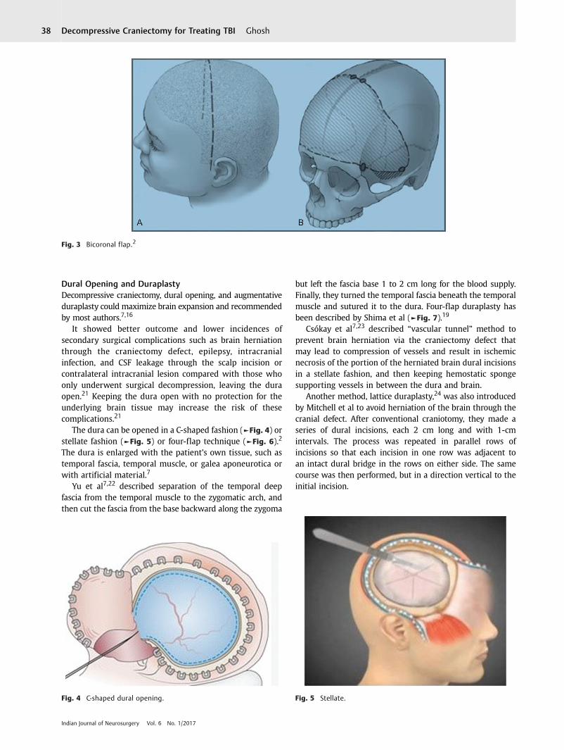
The dura can be opened in a C-shaped fashion (►Fig. 4) orstellate fashion (►Fig. 5) or four-flap technique (►Fig. 6).2
The dura is enlarged with the patient’s own tissue, such astemporal fascia, temporal muscle, or galea aponeurotica orwith artificial material.7
Yu et al7,22 described separation of the temporal deepfascia from the temporal muscle to the zygomatic arch, andthen cut the fascia from the base backward along the zygoma
but left the fascia base 1 to 2 cm long for the blood supply.Finally, they turned the temporal fascia beneath the temporalmuscle and sutured it to the dura. Four-flap duraplasty hasbeen described by Shima et al (►Fig. 7).19
Csókay et al7,23 described “vascular tunnel” method toprevent brain herniation via the craniectomy defect thatmay lead to compression of vessels and result in ischemicnecrosis of the portion of the herniated brain dural incisionsin a stellate fashion, and then keeping hemostatic spongesupporting vessels in between the dura and brain.
Another method, lattice duraplasty,24 was also introducedby Mitchell et al to avoid herniation of the brain through thecranial defect. After conventional craniotomy, they made aseries of dural incisions, each 2 cm long and with 1-cmintervals. The process was repeated in parallel rows ofincisions so that each incision in one row was adjacent toan intact dural bridge in the rows on either side. The samecourse was then performed, but in a direction vertical to theinitial incision.
Fig. 3 Bicoronal flap.2
Fig. 4 C-shaped dural opening. Fig. 5 Stellate.
Indian Journal of Neurosurgery Vol. 6 No. 1/2017
Decompressive Craniectomy for Treating TBI Ghosh38

Some Other Technical Modifications
The “Tucci flap” was suggested by Goettler and Tucci25 andsimilar technique called “in situ hinge” craniectomy wasintroduced by Ko and Segan.”26 After decompressivecraniectomy, there is theoretical risk of injury to theunprotected brain. Moreover, with the skin flap concavity, thehydrodynamic disturbance of CSF circulation and the decreasein cortical perfusion hinder patient recovery. After craniotomy,removal of the intracranial lesion, and duraplasty, the bone flapwas replaced and one side of the flap was attached to thecranium by plates. The plates act as a hinge that allows theunattached portion of the bone flap to float out with boneswelling.
Peethambaran and Valsalmony27 described a techniquefor decompressive craniectomy (►Fig. 8) to avoid revisioncranioplasty after surgery by loosely suturing four pieces ofcraniectomized bone with the skull.
Vakis et al28 introduced a method to prevent periduralfibrosis after decompressive craniectomy. Development ofmultiple adhesions among the dura, temporal muscle, andgalea would be a problem during subsequent cranioplasty,and would also be a potentially deleterious factor for patientrecovery. To prevent adhesions, the authors placed a duralsubstitute between the dural layer and galea aponeuroticaafter augmentative duraplasty with temporal muscle.
To increase the space of decompressive craniectomy,Zhang et al29 suggested a method of surgical decompressioncombined with removal of the temporal muscle part.However, survivors developed a higher rate of masticationdisability.
Bhat et al30 described multidural stabs or SKIMS-techniquethat showed that multiple incision of the dura in acute subduralhematoma drains the hematoma, relieves ICP rapidly, andavoids brain pouting and cortical lacerations during surgery.
Closure of Wound
Dural hitch stitches have been recommended to preventextradural hemorrhage. Subgaleal suction drain can begiven with low suction. Two layered (galea and skin) isalways good for subsequent healing and also to preventCSF leak.
Conclusion
A surgeon has to standardize his/her technique ofdecompressive craniectomy that is the most commonlifesaving neurosurgical procedure according to availableliterature to give the maximum therapeutic decompressioneffect by removing adequate bone, relieving refractory ICP,and restoring cerebral blood flow, and also following thetechniques to avoid subsequent complications.
Fig. 8 Four-quadrant osteoplastic decompressive craniotomy byPeethambaran and Valsalmony.27
Fig. 6 Four-flap dural opening.19
Fig. 7 Four-flap duraplasty as shown by blue line.19
Indian Journal of Neurosurgery Vol. 6 No. 1/2017
Decompressive Craniectomy for Treating TBI Ghosh 39

References1 Piek J. Decompressive surgery in the treatment of traumatic brain
injury. Curr Opin Crit Care 2002;8(2):134–1382 Balan C, Alliez B. Decompressive craniectomy from option
to standard—Part I. Romanian Neurosurgery 2009;16(2):20–263 Cushing H. The establishment of cerebral hernia as a decompressive
measure for inaccessible cerebral tumors: with the description ofintermuscular methods of making the bone defect in temporal andoccipital region. Surg Gynecol Obstet 1905;1:297–314
4 Guerra WK, Gaab MR, Dietz H, Mueller JU, Piek J, Fritsch MJ.Surgical decompression for traumatic brain swelling: indicationsand results. J Neurosurg 1999;90(2):187–196
5 Maas AI, Dearden M, Teasdale GM, et al; European Brain InjuryConsortium. EBIC-guidelines for management of severe headinjury in adults. Acta Neurochir (Wien) 1997;139(4):286–294
6 The Brain Trauma Foundation. The American Association ofNeurological Surgeons. The Joint Section on Neurotrauma andCritical Care. Management and prognosis of severe traumaticbrain injury, part 1: guidelines for the management of severetraumatic brain injury. J Neurotrauma 2000;17:451–533
7 Huang X, Wen L. Technical considerations in decompressivecraniectomy in the treatment of traumatic brain injury. Int J MedSci 2010;7(6):385–390
8 Jiang JY, Xu W, Li WP, et al. Efficacy of standard trauma craniectomyfor refractory intracranial hypertension with severe traumatic braininjury: a multicenter, prospective, randomized controlled study.J Neurotrauma 2005;22(6):623–628
9 Clark K, Nash TM, Hutchison GC. The failure of circumferentialcraniotomy in acute traumatic cerebral swelling. J Neurosurg1968;29(4):367–371
10 Alexander E, Ball MR, Laster DW. Subtemporal decompression:radiological observations and current surgical experience. Br JNeurosurg 1987;1(4):427–433
11 Kessler LA, Novelli PM, Reigel DH. Surgical treatment of benignintracranial hypertension—subtemporal decompressionrevisited. Surg Neurol 1998;50(1):73–76
12 Aarabi B, Hesdorffer DC, Ahn ES, Aresco C, Scalea TM, EisenbergHM. Outcome following decompressive craniectomy formalignant swelling due to severe head injury. J Neurosurg2006;104(4):469–479
13 Caroli M, Locatelli M, Campanella R, Balbi S, Martinelli F, ArientaC. Multiple intracranial lesions in head injury: clinicalconsiderations, prognostic factors, management, and results in95 patients. Surg Neurol 2001;56(2):82–88
14 Kohta M, Minami H, Tanaka K, Kuwamura K, Kondoh T, KohmuraE. Delayed onset massive oedema and deterioration in traumaticbrain injury. J Clin Neurosci 2007;14(2):167–170
15 Apuzzo MLJ. Complication avoidance and management. In: BrainSurgery. Vol 2, part 4. New York, NY: Churchill Livingstone; 1993:1283–1296
16 Gatos H, Kapsalaki EZ, Komnos A, Paterakis KN, Fountas KN. In:Agrawal A, ed. The Role of Decompressive Craniectomy in theManagement of Patients Suffering Severe Closed Head Injuries, Brain
Injury—Pathogenesis, Monitoring, Recovery and Management.InTech; 2012. http://www.intechopen.com/books/brain-injurypathogenesis-monitoring-recovery-and-management/the-role-of-decompressive-craniectomy-in-themanagement-of-patients-suffering-severe-closed-head-inj. Accessed March 23, 2012
17 Hutchinson PJ, Corteen E, Czosnyka M, et al. Decompressivecraniectomy in traumatic brain injury: the randomizedmulticenter RESCUEicp study (www.RESCUEicp.com). ActaNeurochir Suppl 2006;96:17–20
18 Valadka AB, Robertson CS. Surgery of cerebral traumaand associated critical care. Neurosurgery Jul 2007;61(1):203–220
19 Abdeen K. Decompressive craniectomy: rationale, indications andoutcome. http://www.surgicalneurology.org/conf7/f iles/decompressive_craniectomy.pdf. Accessed June 2015
20 Polin RS, Shaffrey ME, Bogaev CA, et al. Decompressive bifrontalcraniectomy in the treatment of severe refractory posttraumaticcerebral edema. Neurosurgery Jul 1997;41(1):84–92
21 Yang XJ, Hong GL, Su SB, Yang SY. Complications induced bydecompressive craniectomies after traumatic brain injury. Chin JTraumatol 2003;6(2):99–103
22 Yu HT, Wang B, Xia JG, et al. The application of turning down thedeep temporal fascia to mend the dura mater in the operation ofintracranial supratentorial decompression in skull trauma. Chin JNeuromed 2006;5:937–939 (In Chinese)
23 Csókay A, Együd L, Nagy L, Pataki G. Vascular tunnel creation toimprove the efficacy of decompressive craniotomy in post-traumatic cerebral edema and ischemic stroke. Surg Neurol2002;57(2):126–129
24 Mitchell P, Tseng M, Mendelow AD. Decompressive craniectomywith lattice duraplasty. Acta Neurochir (Wien) 2004;146(2):159–160
25 Goettler CE, Tucci KA. Decreasing the morbidity of decompressivecraniectomy: the Tucci flap. J Trauma 2007;62(3):777–778
26 Ko K, Segan S. In situ hinge craniectomy. Neurosurgery 2007;60(4, Suppl 2):255–258, discussion 258–259
27 Peethambaran AK, Valsalmony J. Four-quadrant osteoplasticdecompressive craniotomy: a novel technique for decompressivecraniectomy avoiding revision cranioplasty after surgery. NeurolIndia 2012;60(6):672–674
28 Vakis A, Koutentakis D, Karabetsos D, Kalostos G. Use ofpolytetrafluoroethylene dural substitute as adhesion preventivematerial during craniectomies. Clin Neurol Neurosurg 2006;108(8):798–802
29 Zhang MY, Zhao YF, Liang WB. The application of decompressivecraniectomy combined with removal of temporal muscle in thetreatment of severe traumatic brain injury. J Clin Neurosurg.2006;3:124–125
30 Bhat AR, Kirmani AR, Wani MA. Decompressive craniectomywith multi-dural stabs—a combined (SKIMS) techniqueto evacuate acute subdural hematoma with underlyingsevere traumatic brain edema. Asian J Neurosurg 2013;8;(1):15–20
Indian Journal of Neurosurgery Vol. 6 No. 1/2017
Decompressive Craniectomy for Treating TBI Ghosh40