differential diagnosis i.neck ii.back iii.extremities iv.floppy infant syndrome
TRANSCRIPT
Differential diagnosis
I. Neck
II. Back
III. Extremities
IV. Floppy infant syndrome

II. Back pain
requires careful evaluation if lasts more than 1 to 2 weeks (in child)
usually the result of a serious underlying disorder including psychogenic back pain which is often difficult to manage

II. Back pain
in the past, unlike adults, children were thought to uncommonly have back pain related to psychogenic causes
children with acute or short-lived back pain: more likely to have muscle and ligamentous strain or pain associated with systemic viral infection
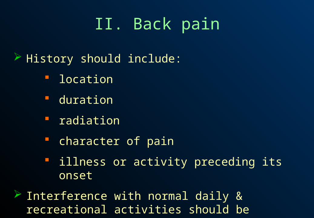
II. Back pain
History should include:
location
duration
radiation
character of pain
illness or activity preceding its onset
Interference with normal daily & recreational activities should be determined

II. Back pain
Examination should seek other signs such as :
abnormalities in gait
configuration of the back (subtle changes in contour may offer localizing clues)
tenderness on palpation

II. Back pain
Skin overlying spine should be carefully inspected for:
dimples
tufts of hair
hemangiomas
other cutaneous changes
Any cutaneous changes may denote developmental defects

II. Back pain
Lesions causing back pain may also produce neurologic changes in extremities or bladder or bowel dysfunction
Signs of neuromuscular disease should also be sought
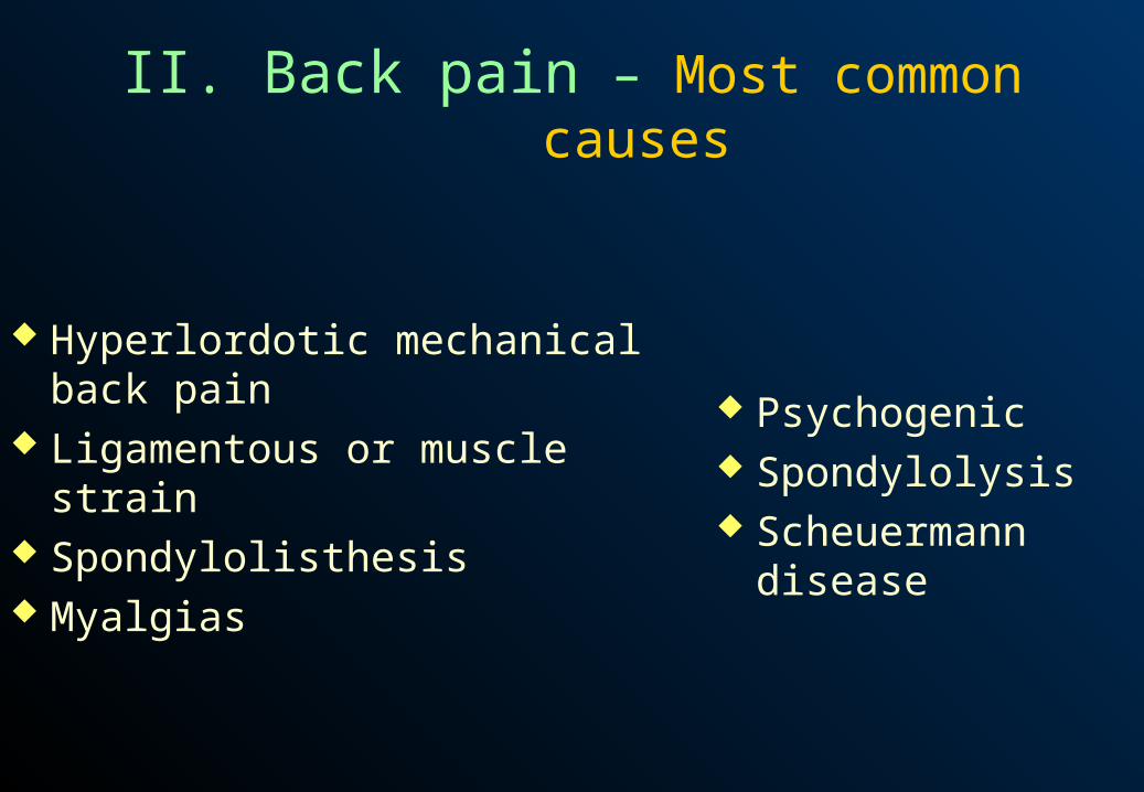
II. Back pain – Most common causes
Hyperlordotic mechanical back pain Ligamentous or muscle strain Spondylolisthesis Myalgias
Psychogenic Spondylolysis Scheuermann disease

II. Back pain – Causes not to forget
Herniated disc Spinal dysraphism Urinary tract infection Spinal cord tumors Diskitis

II. Back pain
Lordotic mechanical back pain
Reputed to be a common cause in adolescent athletes
Pain:
• only in lumbar area
• variable hyperextension or hyperflexion testing
• inability to fully flex the spine forward
Trauma

II. Back pain
Lordotic mechanical back pain
Kyphosis of thoracic spine present in compensation for decreased forward mobility of lumbar spine
Some have suggested contractures at the facet joints as site of pain
Trauma

II. Back pain
Ligamentous or muscle strain
History of fall, unusual exercise or other forms of trauma should be sought
There may be localized tenderness and paravertebral muscle spasm
Strain – probably the most common cause of back pain but it should be short-lived
Trauma
II. Back pain
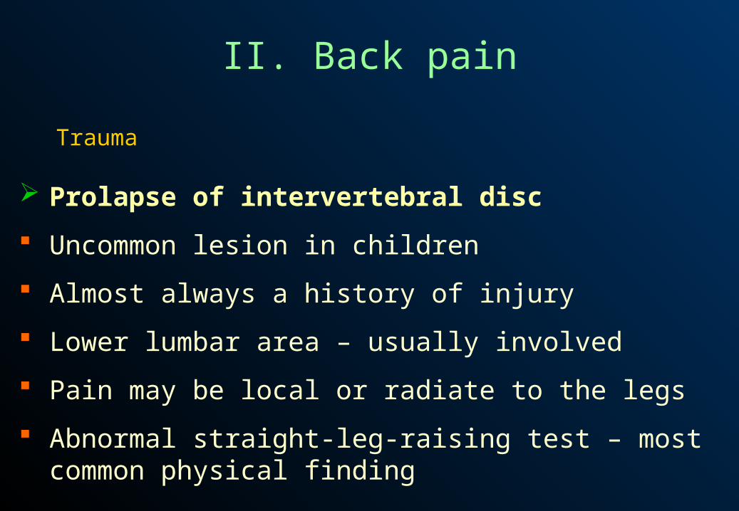
Prolapse of intervertebral disc
Uncommon lesion in children
Almost always a history of injury
Lower lumbar area – usually involved
Pain may be local or radiate to the legs
Abnormal straight-leg-raising test – most common physical finding
Trauma
II. Back pain
Slipped vertebral apophysis
May occur after strenuous activity or heavy lifting
Signs of a herniated disc
A small bone fragment, edge of ring apophysis, may be seen within spinal canal on imaging studies
Lower lumbar spine – most common site
Trauma
II. Back pain
Myalgias
Muscle pain may be associated with a multitude of viral and bacterial infections
Aches not limited to paravertebral muscles
Infections
Urinary tract infection
Back pain may be the primary complaint
A urine culture should be done

II. Back pain
Referred pain
Other infections must be considered in addition to urinary tract infections including:
• pneumonia
• appendicitis
• pancreatitis
• cholecystitis
Infections
Urinary tract infection
Back pain may be the primary complaint
A urine culture should be done

II. Back pain
Diskitis
Aching pain in lower back radiates to flanks, abdomen and lower extremities
Young child may refuse to walk
Illness may be associated with low-grade fever, irritability and lethargy
Limited back motion
Infections

II. Back pain
Osteomyelitis of vertebra
Localized tenderness present at a specific level
Spine held rigid because of muscle spasm
Systemic signs often absent
Infections
Iliac osteomyelitis, sacroiliac joint infection
Frequently confused with appendicitis or septic arthritis of hip

II. Back pain
Tuberculosis
Less common cause of back pain today
Dull local pain present over involved vertebrae
There may be a localized swelling
Destruction of vertebrae may cause pressure on spinal nerves
Stiff gait
Back held rigid
Infections
II. Back pain
Tuberculosis
Less common cause of back pain today
Dull local pain present over involved vertebrae
There may be a localized swelling
Destruction of vertebrae may cause pressure on spinal nerves
Stiff gait
Back held rigid
Infections

II. Back pain
Spinal epidural abscess
Generally exquisite pain and tenderness on palpation over the site of abscess
Rapidly developing signs of spinal cord dysfunction such as paraparesis, loss of bladder and bowel control and sensory changes
Infections
II. Back pain
Brucellosis
Small abscesses may develop in vertebrae
Generally associated with widespread lymphadenopathy
Infections
Acute transverse myelopathy
Rare disorder
Preceded by upper respiratory infection
Back pain may be an early sign
Progressive weakness develops in 2 or 2 days

II. Back pain
Osteoid osteoma
Gradual onset
Worse at night
Often relieved by aspirin
Palpation discloses localized tenderness
Radiographs reveal a small translucent area with surrounding dense bone
Neoplastic disorders – Benign tumors
II. Back pain
Benign osteoblastoma
Symptoms similar to those of osteoid osteoma, but larger lesion and less adjacent bone density seen on radiograph films
Neoplastic disorders – Benign tumors
Eosinophilic granuloma
Usually only one vertebra involved with collapse
Intervertebral disc spaces maintained
Condition may be asymptomatic
May be backache and postural change
II. Back pain
Aneurysmal bone cyst
Cystic expansile lesion in a vertebra may cause neurologic symptoms
Neoplastic disorders – Benign tumors
Neuroenteric cysts
Signs of cord dysfunction present
II. Back pain
Spinal cord tumors
Symptoms may be subacute or chronic
Most common: gliomas, neurofibromas, teratomas, lipomas
Developmental defects may be associated with cutaneous changes
Signs of cord compression with changes in gait, bladder and bowel dysfunction, localized tenderness and scoliosis
Deformity of foot such as cavus or cavovarus – frequent presenting complaint
Neoplastic disorders – Malignant tumors
II. Back pain
Ewing sarcoma Osteogenic sarcoma
Neoplastic disorders – Malignant tumors
Neuroblastoma Wilms’ tumor Leukemia and lymphoma Pain not localized and may be fleeting Rarely, spinal cord compression may occur producing typical
signs of spinal cord tumors
Neoplastic disorders – Metastatic tumors
II. Back pain
Bone abnormalities
Scheuermann disease (vertebral osteochondrosis) Produces a round-back deformity Several vertebrae may be wedged anteriorly Pathophysiologic mechanism thought to be prolapse of
nucleus pulposis into the vertebrae body, possibly due to osteoporosis
Pain – common, usually located over the apex of kyphosis

II. Back pain
Bone abnormalities
Spondylolisthesis Pain caused by anterior displacement of vertebrae Usually L5 slides forward on S1 Sciatica, increased lumbar lordosis and tight hamstrings –
often present
II. Back pain
Bone abnormalities
Spondylolisthesis Pain caused by anterior displacement of vertebrae Usually L5 slides forward on S1 Sciatica, increased lumbar lordosis and tight hamstrings –
often present

II. Back pain
Bone abnormalities
Spondylolysis Defect in pars interarticularis without vertebral slipping Probably result of a stress fracture Low-back pain – common, sometimes with radiation down
the leg Pain increased by activity

II. Back pain
Bone abnormalities
Occult fractures Trauma, sometimes minor, may result in fractures of pars
interarticularis or the transverse or spinous processes May not be seen on plain radiographs
II. Back pain
Bone abnormalities
Osteoporosis Fractures most likely to occur in osteoporotic bones present in
disorders such as Cushing synd., OI, homocystinuria, Turner synd., malabsorption and immobilization
Idiopathic juvenile osteoporosis: Onset between 8 and 14 years of age Self-limited

II. Back pain
Bone abnormalities
Scoliosis Almost always a painless disorder When back pain present, underlying problem should be
sought such as infection, diskitis or tumor

II. Back pain
Psychogenic pain
Back pain may be associated with reaction to stressful situations
Should always be considered if patient’s affect is inconsistent with symptoms or if findings are unexplainable
Careful history must be obtained Psychogenic causes as cause of back pain seem to be on the
rise

II. Back pain
Miscellaneous causes
Sickle cell disease Painful crises may be associated with back pain
Juvenile rheumatoid arthritis Occasionally, cervical pain may be a presenting complaint
II. Back pain
Miscellaneous causes
Ankylosing spondylitis Usually boys Arthritis in hips or knees and loss of mobility of the back may
be found
Chronic hemolytic anemias Signs of cord compression may result from extramedullary
hematopoiesis in extradural space
II. Back pain
Miscellaneous causes
Calcification of intervertebral discs Localized back pain Loss of mobility due to muscle spasm Cause unknown Fluffy calcification in the disc space on radiograph films may
not appear for 1 to 2 weeks following onset of pain

II. Back pain
Miscellaneous causes
Spinal dysraphism Lesions such as fibrous bands, lipomas, etc., may cause a
tethered cord => back pain in addition to neurologic findings in lower extremities and bladder problems
Clues to underlying problem should be sought by close examination of the skin over spine for cutaneous abnormalities
II. Back pain
Miscellaneous causes
Diastematomyelia Developmental defect causes a cleft in the cord by bone,
cartilage or fibrous septum Cutaneous abnormalities over affected area may be apparent Low-back pain aggravated by cough or sneeze Bladder dysfunction or slowly progressive weakness of legs –
earlier signs than back pain
II. Back pain
Miscellaneous causes
Arteriovenous malformation of cord Symptoms usually slow to develop Low-back pain – common, with progressive gait and bladder
or bowel dysfunction May be a cutaneous angioma over the cord lesion
II. Back pain
Miscellaneous causes
Limb girdle dystrophy Not a single disease entity but a group of dystrophies and
myopathies Usually with autosomal recessive inheritance pattern First symptoms usually appear during 2nd decade Early sign: difficulty in climbing stairs or rising from the floor
- low-back pain may be the source of either complaint Pseudohypertrophy sometimes present Deep tendon reflexes difficult to elicit

II. Back pain
Miscellaneous causes
Paroxysmal cold hemoglobinuria Most commonly seen after viral infections After cold exposure, child experiences back or abdominal
pain, followed by chills, fever and hemoglobinuria
Multiple epiphyseal dysplasia Most prominent symptom: painful joints – usually hips, knees
and ankles – with decreased mobility Frequent back pain Gait may be waddling

II. Scoliosis
Defined as a lateral curvature of the spine from its normal straight position
Rotational deformity of spine present as well Many children have an inconsequential curvature of less than
10° to 15 ° True scoliosis worrisome because of the possibility of
progression during growth to a degree that might affect cardiopulmonary function
Described by the direction of convexity of the curve Right thoracic and left lumbar scoliosis = most common
pattern in idiopathic scoliosis
II. Scoliosis
Prevalence of scoliosis with curves >10° in adolescents estimated to be 2% to 3%
Idiopathic scoliosis comprises 60% to 80%of cases Most children with idiopathic scoliosis require no therapy Close follow-up recommended in order to detect undue
progression of curvature Scoliosis in an adolescent is not necessarily idiopathic May be a sign of an occult neuromuscular disorder or other
pathologic conditions

II. Scoliosis
Of importance in determining possible causes: age at which scoliosis is noted rapidity of development
Painful scoliosis should never be considered idiopathic in adolescent
Adolescent with left thoracic kyphosis should be evaluated for underlying pathology
Delayed developmental milestones may suggest neuromuscular cause
II. Scoliosis – Most common causes
Idiopathic Congenital vertebral defect Leg length discrepancy
Neurofibromatosis Neuromuscular disorder

II. Scoliosis – Nonstructural causes
Primary postural scoliosis Condition most commonly seen in children between 10 and
15 years of age Shoulders may be rounded One hip may seem more prominent than the other Apparent curvature disappears on forward flexion or on lying
down
II. Scoliosis – Nonstructural causes
Secondary postural scoliosis Curvature = a result of other conditions, such as leg
discrepancy Curve disappears on forward flexion
Hysterical scoliosis Unusual type Scoliosis not present on forward flexion
II. Scoliosis – Structural causes
Idiopathic scoliosis Probably genetic cause in 90% of cases
Infantile scoliosis Noted in the first 3 years of life Rare in US More common in boys than in girls Curvature lessens with age in most cases

II. Scoliosis – Structural causes
Juvenile scoliosis defined as scoliosis appearing in the 4- to 10-year-old age
group Boys and girls equally affected
Adolescent scoliosis Most common type occurring in children > 10 years of age Girls outnumber boys ratio 5-7 : 1 Condition generally unnoticed until adolescent growth spurt
II. Scoliosis – Structural causes
Congenital scoliosis May be associated with vertebral anomalies such as
hemivertebrae, wedge vertebrae, congenital bars or failure of vertebrae segmentation
Other significant congenital defects may be present, such as of the heart of genitourinary system or other bony abnormalities
May be complicated by diastematomyelia, spinal lipomas, etc.
II. Scoliosis – Structural causes
Neurofibromatosis Accounts for approx. 2% of cases of scoliosis A slowly progressive curve similar to idiopathic variety
develops in half of these cases Significant type: with a short, sharply angular curve in the
thoracic spine Important cutaneous clues:
Café au lait spots Axillary freckling
II. Scoliosis – Structural causes Neuromuscular origin
Cerebral palsy Structural scoliosis occurs in 15% to 25% of children with CP More commonly in the more severely affected, especially
those with spastic quadriplegia
Neuropathies
II. Scoliosis – Structural causes Neuromuscular origin
Myelomeningocele Lesion may be obvious or occult May be present:
Overlying skin defects Lower extremity weakness Neurologic changes Bladder and bowel difficulties
Neuropathies
II. Scoliosis – Structural causes Neuromuscular origin
Spinal cord injury Scoliosis will develop in almost 50% of patients
Neuropathies
Syringomyelia Scoliosis may be a presenting sign before sensory changes are
noted

II. Scoliosis – Structural causes Neuromuscular origin
Diastematomyelia May be cutaneous defects or changes over the site of the bony
abnormality
Neuropathies
Friedreich ataxia Ataxia develops in 1st or 2nd decade Hypoactive deep tendon reflexes Pes cavus and kyphoscoliosis develop in almost all patients
II. Scoliosis – Structural causes Neuromuscular origin
Charcot-Marie-Tooth disease Atrophy of peroneal muscles gives a stork leg appearance Progressive weakness affects lower and, later, the upper
extremities
Neuropathies
II. Scoliosis – Structural causes Neuromuscular origin
Neuropathies
Juvenile spinal muscle atrophy Onset of weakness ranges from early childhood to late
adolescence Signs of this disorder often mistaken for muscular dystrophy
Poliomyelitis Now an uncommon cause Deformity occurs 1-2 years after the acute illness

II. Scoliosis – Structural causes Neuromuscular origin
Myopathies
Duchenne-type muscular dystrophy Scoliosis occurs later, particularly when patient is confined to
wheelchair
Nemaline myopathy Limb-girdle muscular dystrophy Onset of symptoms later than in the Duchenne type Proximal muscle weakness > distal

II. Scoliosis – Structural causes Neuromuscular origin
Myopathies
Arthrogryposis Multiple contractures present at birth Anterior horn cell loss may create muscle imbalance =>
leading to scoliosis
II. Scoliosis – Structural causes Neuromuscular origin
Mesenchymal origin
Marfan syndrome Almost 50% of affected children develop scoliosis in infancy
or early childhood Features:
dislocated lens spiderlike fingers and extremities high arched palate

II. Scoliosis – Structural causes Neuromuscular origin
Mesenchymal origin
Ehlers-Danlos syndrome Hyperlaxity of joints and skin
Congenital laxity of joints No skin hyperelasticity

II. Scoliosis – Structural causes Neuromuscular origin
Trauma
Direct vertebral trauma Fractures or wedging of vertebral bodies or nerve root
irritation may cause scoliosis
Irradiation Destruction of the vertebral growth plates especially in
treatment of Wilms’ tumor, produces curvature later
Extravertebral trauma Severe trunk burns or thoracic surgery may result in scoliosis

II. Scoliosis – Structural causes Neuromuscular origin
Tumors
Intraspinal tumors Various types of tumors may result in scoliosis Sensory and motor changes in lower extremities and bladder
and bowel incontinence may also occur
Osteoid osteoma Vertebral body tumors may cause paraspinal muscle spasm
and resultant scoliosis Pain often worse at night and relieved by aspirin
II. Scoliosis – Structural causes Neuromuscular origin
Miscellaneous causes
Vertebral body infection Scoliosis may be associated with osteomyelitis, diskitis and
TB involvement of spine
Rickets Scoliosis may develop late if condition untreated Features: epiphyseal enlargement, bowing of long bones,
growth retardation, apathy, muscle weakness
II. Scoliosis – Structural causes
Miscellaneous causes
Osteogenesis imperfecta Collapse of vertebrae following fractures may result in
scoliosis
Scheuermann disease Causes adolescent round back deformity Rarely causes scoliosis
II. Scoliosis – Structural causes
Miscellaneous causes
Achondroplasia 25% of affected children will develop scoliosis in late
childhood
Klippel-Feil syndrome Short neck with decreased movement – typical Cervicothoracic scoliosis may also be present

II. Scoliosis – Structural causes
Miscellaneous causes
Sprengel deformity Congenital high scapula almost always associated with
cervical or thoracic spine abnormalities
Cleidocranial dyotosis Features hypoplastic or absent clavicles, large head with
delayed closure of fontanel and a narrow chest
II. Scoliosis – Structural causes
Miscellaneous causes
Hyperphosphatasia Condition characterized by fever, pain and bone fragility with
frequent fractures Short stature Thickened limb bones Bluish sclerae

II. Scoliosis – Structural causes
Miscellaneous causes
Hypervitaminosis A Features include dry skin, thickened bones Often increased intracranial pressure
Hypothyroidism Congenital indifference to pain Juvenile rheumatoid arthritis`
II. Scoliosis – Structural causes
Miscellaneous causes
Mucopolysaccharidoses In type VII progressive scoliosis may be the initial presenting
sign Hepatosplenomegaly, short neck and cloudy corneae develop
gradually Type VI (Maroteaux-Lamy) also has scoliosis as a clinical
feature

II. Scoliosis – Structural causes
Syndromes associated with scoliosis
Scoliosis has been described in a number of malformation syndromes
Other features of these syndromes – more striking than scoliosis
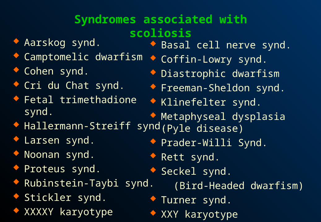
Syndromes associated with scoliosis
Aarskog synd. Camptomelic dwarfism Cohen synd. Cri du Chat synd. Fetal trimethadione synd. Hallermann-Streiff synd. Larsen synd. Noonan synd. Proteus synd. Rubinstein-Taybi synd. Stickler synd. XXXXY karyotype
Basal cell nerve synd. Coffin-Lowry synd. Diastrophic dwarfism Freeman-Sheldon synd. Klinefelter synd. Metaphyseal dysplasia (Pyle disease) Prader-Willi Synd. Rett synd. Seckel synd.
(Bird-Headed dwarfism) Turner synd. XXY karyotype

II. Scoliosis – Transient structural
Inflammation
Lateral curvature can be produced by irritation from empyema or a perinephric abscess
Torticollis
Sciatic scoliosis
Pressure of an intervertebral disk on nerve roots may produce a scoliosis
II. Kyphosis and lordosis
Curvature of spine may occur in anterior (lordosis) & posterior (kyphosis) directions
Most children with these conditions have postural deformities
Pathologic or fixed deformities may result from various disorders
Lordosis – normal in young children, but should no longer be present my mid-childhood
II. Kyphosis and lordosis
Poor posture
Accounts for most cases of kyphosis, especially in adolescence when concern about appearance is prevalent
Postural kyphosis – not fixed
Can be easily corrected by finding appropriate method of encouragement or exercises
Kyphosis

Scheuermann disease (juvenile kyphosis)
Poorly understood disorder
Usually develops around puberty
Poor posture
Apparent round back deformity
Fatigue & discomfort in area of kyphosis – common, on standing
Kyphosis
Scheuermann disease (juvenile kyphosis)
Full correction cannot voluntarily be obtained
On radiographs: wedged-shaped appearance of one or more vertebrae due to diminished anterior height
Cause – unknown
Lumbar lordosis – often accentuated
Kyphosis
Congenital kyphosis
Noted in infancy
Usually progresses with age, especially when child begins to walk & stand
Caused by a structural abnormality of spine apparent on radiographic examination
Painless in childhood
May become painful during adolescence & adulthood
Compression of spinal cord may occur
Kyphosis

Neuromuscular problems
Almost any neuromuscular disorder may cause spinal deformities in a growing child
CP, post traumatic paralysis, spinal muscular atrophy, myotonic dystrophy, poliomyelitis
Kyphosis
Myelomeningocele
Kyphotic defects may be present at birth secondary to vertebral disruption
May develop later associated with muscle weakness
Infection
Destruction of vertebrae from infectious causes may lead to kyphosis
Spasm of paravertebral musculature may be responsible for abnormality
Tuberculosis – archetypical cause, but much less common today
Tuberculosis spondylitis (Pott disease) – often insidious in onset
May affect any level of spine
Kyphosis

Skeletal dysplasias
A host of skeletal disorders may involve vertebral column & produce kyphosis
Radiographic skeletal survey helps to differentiate various types
Kyphosis

Spondyloepiphyseal dysplasia
Mucopolysaccharidoses
• Kyphosis especially likely to be a finding in Hurler synd. (type I), Morquio synd. (type IV), Maroteaux-Lamy synd. (type VI) and type VII
Kyphosis - Skeletal dysplasias
Diastrophic dwarfism
Diaphyseal dysplasia (Engelmann disease)
Kniest dwarfism
Achondroplasia
Cleidocranial dysotosis
Cockayne syndrome
Neurofibromatosis
Noonan syndrome
Kyphosis - Skeletal dysplasias

Metabolic & endocrine disorders
Hypothyroidism
Gaucher disease
Ehlers-Danlos syndrome
Marfan syndrome
Homocystinuria
O.I.
Juvenile osteoporosis
Kyphosis
Tumors
Kyphosis bay be caused by benign or malignant, either primary or metastatic tumors
Intraspinal tumors must always be considered
Kyphosis
Iatrogenic kyphosis
Radiation therapy
• damage to vertebral growth plates may follow, resulting in kyphosis
Kyphosis
Iatrogenic kyphosis
Surgery
• Surgical removal of parts of vertebral column may lead to kyphosis
Miscellaneous
Familial dysautonomia
• Scoliosis & kyphosis – common
• Other symptoms & signs predominate including unexplained fever, aspiration, other signs of autonomic nervous system dysfunction

Lordosis
Physiologic lordosis
Exaggerated lumbar lordosis – common in toddlers
Compensatory posture
Compensatory lumbar lordosis frequently accompanies kyphotic disorders such as Scheuermann disease
Pes planus
Lordosis may be an adaptive mechanism for individuals with flat feet to keep stable stance
Lordosis
Neuromuscular disorders
Lumbar lordosis - prominent & progressive in muscular dystrophy
Often accompanies CP, spinal injuries with paralysis, poliomyelitis
Lordosis
Spondylolisthesis
Slipping forward of vertebral column at lumbosacral junction can be
• secondary to congenital sacral defects
• result of trauma
• caused by developmental or acquired bone defects
Poor posture & increased lumbar lordosis may be the only complaints
Backache, often with radiation don the legs, occurs in 2nd & 3rd decades
Lordosis
Bilateral flexion contractures of hips
Increased pelvic inclination – result of hip flexion contractures – produces a compensatory lumbar lordosis
Flexion contractures may occur in juvenile rheumatoid arthritis, other hip dysplasias and CP
Myelomeningocele
Lordosis – most common spinal deformity
Compensatory in nature
Lordosis
Inflammatory processes
Spasm of paravertebral muscles from inflammatory processes in spine may cause accentuated lordosis
Common features in diskitis:
• Inflammation of intervertebral disc space
• Symptoms of backache
• Pain radiating to the legs
• Occasionally, lower extremity muscle weakness

Lordosis
Skeletal dysplasias
Achondroplasia
• Exaggerated lumbar lordosis because of fixed flexion of hips and some thoracolumbar kyphosis
Cleidocranial dysostosis
• Major features include a large head with delayed closure of anterior fontanel & hypoplastic clavicles
Spondyloepiphyseal dysplasias