digestive and liver disease - aigo di gestione... · 530 a. chauvin et al. / digestive and liver...
TRANSCRIPT
R
Mb
Aa
b
c
a
ARAA
KEFFETESCBC
1
boarfssw
2
2
y
AF
1h
Digestive and Liver Disease 45 (2013) 529– 542
Contents lists available at SciVerse ScienceDirect
Digestive and Liver Disease
j ourna l ho mepage: www.elsev ier .com/ lo cate /d ld
eview article
anagement and endoscopic techniques for digestive foreign body and foodolus impaction�
rmelle Chauvina, Jerome Vialab, Philippe Marteaua, Philippe Hermannc, Xavier Draya,∗
Emergency Endoscopy Unit, Department of Gastroenterology, APHP, Lariboisière Hospital, & iTEC Paris Diderot Paris 7 University, Paris, FranceDepartment of Pediatric Gastroenterology, APHP, Robert Debré Hospital, & Denis Diderot, Paris 7 University, Paris, FranceDepartment of Otorhinolaryngology, APHP, Lariboisière Hospital, & Denis Diderot, Paris 7 University, Paris, France
r t i c l e i n f o
rticle history:eceived 3 October 2012ccepted 2 November 2012vailable online 23 December 2012
eywords:osinophilic esophagitisoreign bodyood bolus impaction
a b s t r a c t
Ingested foreign bodies, food bolus impaction, migration or retention of medical devices are frequent,in children as well as in adults. Most of these foreign bodies will naturally pass through the gastro-intestinal tract. Complications are rare but sometimes severe (oesophageal perforations are the mostfrequent and most feared). We aimed to review the literature on therapeutic management of digestiveforeign bodies and food bolus impaction, with special focus on endoscopic indications, material, timingand techniques for removal. The role of the gastroenterologist is to recognise specific situations and toplan endoscopic removal in a timely manner with the most adequate conditions and extraction tools.Risk factors and underlying pathology, for example eosinophilic esophagitis, must be investigated and if
ndoscopyherapeutic endoscopysophageal perforationtentapsule retention
necessary treated.© 2012 Editrice Gastroenterologica Italiana S.r.l. Published by Elsevier Ltd. All rights reserved.
atteryoin
. Introduction
Foreign body ingestion (127,000 a year in the USA) and foodolus impaction frequently occur [1] and represent about 4%f all emergency endoscopies [2]. Clinical situations are variednd require a skilled operator with adapted extraction tools. Weeviewed the literature on therapeutic management of digestiveoreign body and food bolus impaction, with special focus on endo-copic indications, material, timing and techniques for removal. Thepecific cases of sword swallowers [3] and rectal foreign bodies [4]ill not be detailed herein.
. Epidemiology
.1. Children
Seventy-five percent of foreign bodies are ingested by childrenounger than 5 years old [1]. Coins, buttons, plastic pieces, batteries
� No support in the form of grants, equipment, drugs, or all of the above.∗ Corresponding author at: Department of Gastroenterology, Lariboisière HospitalPHP & Paris 7 University, 2, rue Ambroise Paré 75010 Paris,rance. Tel.: +33 1 49 95 25 45; fax: +33 1 49 95 25 77.
E-mail address: [email protected] (X. Dray).
590-8658/$36.00 © 2012 Editrice Gastroenterologica Italiana S.r.l. Published by Elsevierttp://dx.doi.org/10.1016/j.dld.2012.11.002
and bones are far more frequent than the expected toys [5,6]. Tenpercent of children experience a recurrent ingestion [7]. Underlyingoesophageal diseases are likely to be seen in children with foreignbody impaction (more than 80% in some series, including 53% ofpatients with eosinophilic oesophagitis) [8]. Only 10–20% requireendoscopic removal, while less than 1% will need surgery for foreignbody extraction or to treat a complication [9].
2.2. Adults
Foreign body ingestion is accidental in more than 95% of adultcases, and food-related (food impaction, animal bones, toothpicks)in two thirds of cases (steakhouse syndrome, Figs. 1–3) [2]. Fishbones are the most frequent in Asia [10,11]. Coins are the mostfrequent non-alimentary foreign body ingested by adults [12].Accidental ingestion also sometimes occurs during dental proce-dures. Voluntary foreign body ingestion are sometimes seen inpsychiatric patients (Figs. 4–6) [13], in prisoners (seeking some sec-ondary gain with access to a medical facility) and in drug dealers(also known as “drug-mules” or “bodypackers”). Underlying dis-
eases facilitating foreign body impactions are frequent (more than30% [2], including dentures, pre-existing oesophageal disorders(eosinophilic esophagitis in 10% [14], oesophageal motor distur-bances, stenosis and diverticulae) (Figs. 1 and 2).Ltd. All rights reserved.

530 A. Chauvin et al. / Digestive and Liver Disease 45 (2013) 529– 542
F A) Fooi meat
u
2i
an
[loarprttott
ig. 1. 25 year-old male patient with first episode of food bolus impaction (meat). (s inserted along the food bolus, (D) allowing the en-bloc removal of the 6 cm-longpheld on elective endoscopy.
.2.1. Digestive segments at risk of complication, with relation tondication and timing
Although 80% of foreign bodies can pass through naturally (indults as well as in children [15]), some will remain blocked in thearrower segments of the digestive tract.
The oesophagus is the most frequent site of blockage (50–75%)2,10]. Foreign bodies impacted in the oesophagus are particu-arly at risk of complication because of its thin wall and becausef numerous physiological (cricopharyngeal sphincter, aortic archnd diaphragmatic hiatus) and putative non-physiological nar-owings (oesophagus atresia or stenosis, cardial achalasia, cancer,ost-surgical or congenital modification of anatomy [16,17]). Theisk of complication is 25% higher in the upper oesophagushan in other sites [18] Working space is limited, particularly in
he cervical oesophagus (Fig. 3). Moreover, the vicinity of vitalrgans around the oesophagus makes many complications life-hreatening [19,20]. These anatomic and high-risk features leado remove any object without delay particularly in the followingd bolus is located at the second third of the oesophagus. (B,C) A polyp retrieval netbolus. Discharged, uneventful outcome. Diagnostic of eosinophilic esophagitis was
conditions: (i) involvement of the upper third oesophagus; (ii)symptoms of complete obstruction; (iii) at-risk objects (such assharp foreign bodies or batteries) [21]. In such emergency cases, theendoscopic removal should not be delayed whether the stomach isempty or full. Any other foreign body lodged in the oesophagusshould be managed within the 24 h following the ingestion [21],because the risk of complication increases by 2 and by 7 fold withoesophageal retention lasting 24 and 72 h, respectively [18].
Foreign bodies that have reached the stomach have a chanceto be evacuated spontaneously, while the thickness of the gastricwall limits the risk of perforation. Therefore, endoscopic removalis recommended only for dangerous foreign body to avoid thempassing the duodenal curve [21]. Blunt and small objects should beextracted only if they are still present after 3–4 weeks [21].
When a sharp foreign body has passed the pylorus, perforationmay occur especially in duodenum [22,23], or at the ileocaecal valve[24] or sometimes in a Meckel diverticulum [25–27]. A blunt objectremaining in the duodenum should be removed within 8 days to

A. Chauvin et al. / Digestive and Liver Disease 45 (2013) 529– 542 531
F ). (A)
a o breaa esoph
ldri
3
3
3
bsbd
ig. 2. 32 year-old male patient with first episode of food bolus impaction (meatppearence of eosinophilic esophagitis is noticed; (B–E) a 4-prong grasper is used t
stricture is clearly visible; (E) biopsies are performed. A diagnostic of eosinophilic
imit the risk of ischaemia [21]. Sharp objects that passed the duo-enal curve should be followed with daily radiographs, and surgicalemoval should be considered if the foreign body does not progressn 3 days [28].
. Management
.1. Initial evaluation
.1.1. Clinical evaluationThe majority of patients remain asymptomatic following foreign
ody ingestion (up to 50% in paediatric series) [29]. This diagno-is should be suspected if symptoms such as dysphagia, vomiting,lood-stained saliva, hypersialorrhea, wheezing and/or respiratoryistress are present in a patient unable to report the ingestion
Food bolus is located at the lower third of the oesophagus; a ringed trachea-likekdown and to extract the food bolus; (D) after complete removal of the food bolus,agitis was confirmed.
(nonverbal children, patients with psychiatric disorders or men-tal retardation) [21]. Patients may localise discomfort with poorcorrelation to the site of impaction [1,21]. Signs of esophagealobstruction with inability to manage secretion need an urgentendoscopic retrieval [21]. Esophageal perforation may cause cer-vical crepitus, neck swelling or pneumomediastinum. Generally,when a foreign body has passed the oesophagus, it does not causesymptoms unless a complication occurs (obstruction or perfora-tion).
3.1.2. Radiological evaluationFor patients with non-bony food impaction without complica-
tions, an endoscopy may be performed without any radiologicalwork-out [21]. In other cases, X-ray examinations are used to assessthe presence and number of radiopaque foreign body, their loca-tions, sizes and shapes (Fig. 6) and to determine if obstruction

532 A. Chauvin et al. / Digestive and Liver Disease 45 (2013) 529– 542
Fig. 3. 53 year-old male patient with food bolus impaction (meat) and sharp foreign body (toothpick). (A) Food bolus is located just below the upper oesophageal sphincter; thep raspe( derlyir
onathbirbbefase
3
3
aflcc
3
aptitoc
ointed end of the toothpick is grabbed with a reusable alligator-tooth foreign body gC) view of the food bolus with toothpick after removal. No mucosal damage or unemoval. 24 h admission, uneventful outcome.
r perforation is present (Fig. 7). Biplane neck, chest or abdomi-al radiographs, as appropriate, are often sufficient, and CT scansre rarely needed [21,30]. Interestingly, coins or batteries displayhe same circular appearance: a lateral view of the foreign bodyelps to differentiate a coin from a battery, because of a 2-steporder in the latter. However, ingestions of radiolucent foreign bod-
es are not rare (22 and 36% in adult and paediatric large cohorts,espectively [11,29]), and are related to bony and non-bony foodolus impactions in most cases [11]. Contrast studies should note performed as they delay urgent endoscopies, or interfere withndoscopic visualisation [2]. Barium is contra-indicated when per-oration is suspected, whereas hypertonic contrast agents can causecute pulmonary oedema if aspirated [2]. Thus, when needed, CTcan exploration should be preferred to X-ray contrast study forvaluation of non-radiopaque objects [21,30].
.2. Initial procedural management
.2.1. Information and consentWhen foreign body extraction is considered, then the procedure
nd the risks involved with the location, nature and shape of theoreign body have to be explained to the patient. This can be a chal-enging situation in psychiatric patients or in prisoners. Informedonsent must be obtained. In a series of 414 cases in a tertiary careentre, signed consent was refused in only three cases [2].
.2.2. SedationIn most cases, upper gastro-intestinal endoscopy can be man-
ged with conscious sedation (87% of cases in a series of 414 adultatients) [2]. The patient must be lying on their left side, withhe head slightly lowered, to reduced inhalation risks. However,
n difficult cases (younger children, poor patient tolerance, mul-iple objects, anticipation of difficult extraction, or when rigidesophagoscopy is needed), general anaesthesia with endotra-heal intubation for airway protection is more appropriate, usuallyr; (B) extraction is secured with a Macgill forceps in the hand of an otolaryngologist;ng esophageal disease was found during endoscopic inspection, immediately after
performed with a rapid sequence induction technique because ofa full stomach.
3.3. Endoscopic and ancillary equipment
3.3.1. EndoscopesIn a child of less than 1 year-old, a nasogastroscope with
an external diameter inferior to 6 mm should be used althoughit restricts the choice of operating devices. A 2 mm channelaccepts only small polypectomy retrieval nets (diameter of 20 mm),polypectomy snares, or Dormia baskets.
In adults, standard flexible endoscopes (9.8 mm diameterscope, 2.8 mm diameter single channel) or therapeutic endoscopes(>3.2 mm diameter single channel) are used in most cases. Double-channel endoscopes permit a combination of devices (Fig. 6).Small-calibre endoscopes, via a transnasal approach, are sometimesneeded [21]. Enteroscopes can be used when a sharp foreign bodyhas passed the Treitz angle or for prolonged retention of bulkyobjects (capsules for instance) [31].
Rigid hypopharyngoscope with compatible forceps can beused for foreign body extraction in a hypopharyngeal or upperoesophageal sphincter location (Fig. 8). The main benefit is a largeworking channel with stronger grasping possibilities [32]. If theforeign body has stopped further, rigid oesophagoscopy can avoidrepeated oesophageal intubations when fragmentation of a foodbolus would otherwise be difficult with a flexible endoscope. How-ever, complication rates are higher with rigid oesophagoscopy(10%) than with flexible endoscopy (5%) [33]. It is therefore rec-ommended that otolaryngologists attempt rigid esophagoscopy forforeign body retrieval only when flexible endoscopy has failed.
3.4. Retrieval devices [34]
The most frequently used retrieval devices are rat-rooth andalligator forceps, tripong graspers, Dormia baskets, retrieval nets

A. Chauvin et al. / Digestive and Liver Disease 45 (2013) 529– 542 533
Fig. 4. 23 year-old female psychotic patient with numerous episodes of voluntary ingestion of foreign bodies, including a past history of gastrotomy. (A) Endoscopic extractionof a watch using a reusable alligator-tooth foreign body grasper; (B,C) the same foreign body grasper was used for extraction of a large button impacted downstream in theo he oesa succe
adtsu
3
so
esophagus, (D) with assistance of a MacGill forceps to pass the upper sphincter of tnd opening the forceps through a hole in the key bow; (G) all foreign bodies were
nd polypectomy snares (Table 1) [34]. The choice of the retrievalevice is determined by the size and shape of the foreign body, byhe endoscope length and instrument channel and by the endo-copist’s preference and habits. Magnetic retrievers are no longersed because foreign bodies are often lost during retrieval [34].
.4.1. Retrieval forcepsStandard biopsy forceps are often inadequate because of their
mall opening width, but they can be efficient for small and softbjects (Fig. 9). There is a large variety of other jaw configurations:
ophagus. (E, F) A key in the second part of the duodenum was removed by insertingssfully removed with the same instrument.
rat-tooth, alligator-tooth or shark-tooth forceps. The rat-tooth for-ceps is the most commonly used. A rubber-tip forceps can be usefulfor small hard objects, such as pins, needles or blades. Most of thesedevices are reusable, making their use inexpensive.
3.4.2. Retrieval graspersGraspers can have 2–5 prongs. They can be useful to retrieve soft
objects, such as food bolus impaction (Fig. 2), but must not be usedfor harder or heavy objects because the grip is not secure enough.

534 A. Chauvin et al. / Digestive and Liver Disease 45 (2013) 529– 542
Fig. 5. same patient as in Fig. 4, seen a few months later, and referred for voluntary ingestion of glass fragments. Glass fragments were seen in the oesophagus, in the stomachand (A) in the duodenum; (B) a latex hood is attached to the tip of a standard gastroscope, (C) and flipped over the scope; (D,E) a glass fragment is grabbed with a rotativehemispheric basket, (F) and safely removed along the oesophagus, as the latex hood has unfurled over the basket while passing the cardia, thus protecting the digestive wallduring withdrawal; (G) a total of six glass fragments were found and removed from the upper digestive tract in the same session. 48 h admission, uneventful outcome.
Table 1Main characteristics of most widely used retrieval devices for endoscopic extraction of ingested foreign bodies and food bolus impaction.
Retrieval devices Configuration Size Length Specific aspects Usual costa
Snares -Oval-Hexagonal-Crescent-Barbed-Jumbo snare
1 cm × 1.5 cm to3 cm × 6 cm
105–240 cm Single use or reusabledevices available
+ to +++
Retrieval graspers 2-prong to 5-prong 1.3–2.5 cm 129–240 cm Single use or reusabledevices available
+ to +++
Baskets 3- to 6-wire baskets 0.5 cm × 1.3 cm to2 cm × 4.3 cm × 6 cm
120–240 cm Single use or reusabledevices available
+ to +++
Retrieval nets -Oval-Octagonal-Hexagonal
2 cm × 4.5 cm to4 cm × 8 cm
160–230 cm Single use ++
Retrieval forceps Different jaw-designs:-Rat-tooth-Alligator-tooth-Shark-tooth-Rubber tip
Breadth of opening:3–19.5 mm
120–230 cm Most specialisedforceps are reusable
+++
a +, less than 50 euros; ++, 50–100 euros; +++, 100 euros and more.
A. Chauvin et al. / Digestive and Liver Disease 45 (2013) 529– 542 535
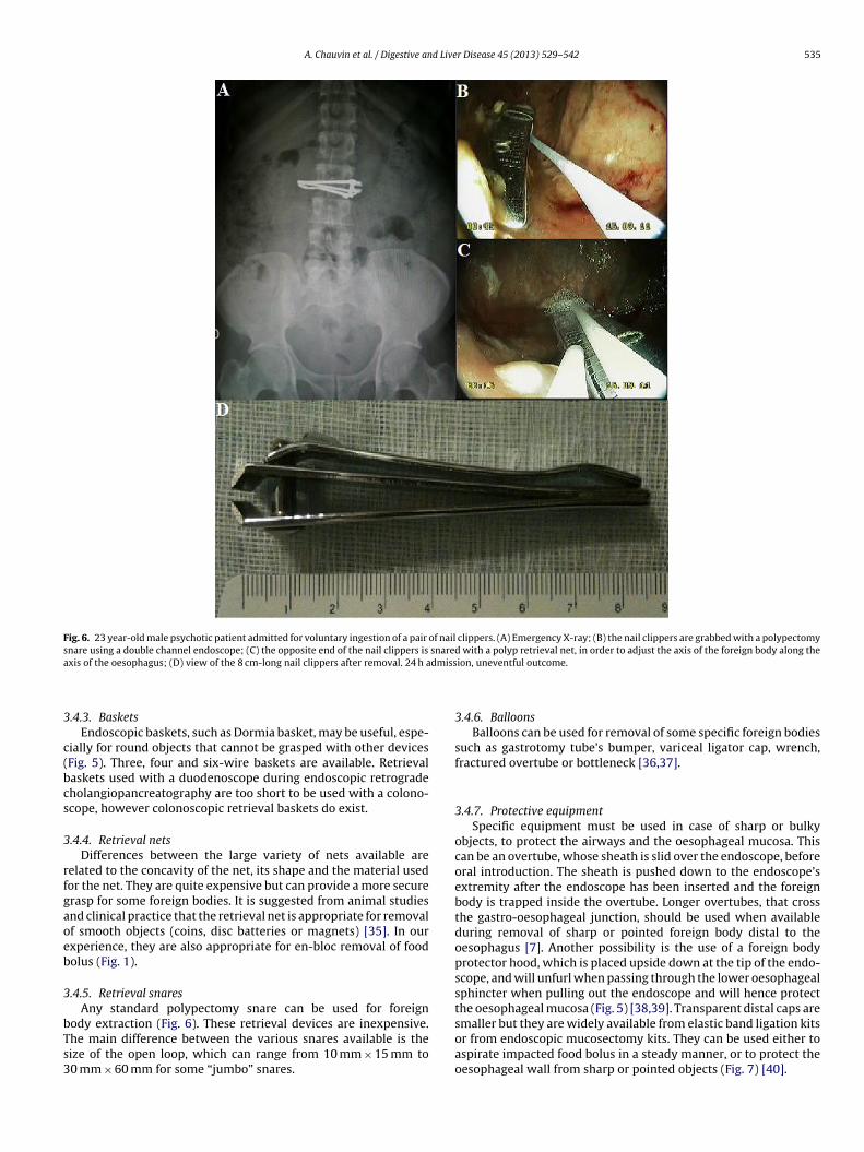
Fig. 6. 23 year-old male psychotic patient admitted for voluntary ingestion of a pair of nail clippers. (A) Emergency X-ray; (B) the nail clippers are grabbed with a polypectomysnare using a double channel endoscope; (C) the opposite end of the nail clippers is snared with a polyp retrieval net, in order to adjust the axis of the foreign body along thea dmiss
3
c(bcs
3
rfgaoeb
3
bTs3
xis of the oesophagus; (D) view of the 8 cm-long nail clippers after removal. 24 h a
.4.3. BasketsEndoscopic baskets, such as Dormia basket, may be useful, espe-
ially for round objects that cannot be grasped with other devicesFig. 5). Three, four and six-wire baskets are available. Retrievalaskets used with a duodenoscope during endoscopic retrogradeholangiopancreatography are too short to be used with a colono-cope, however colonoscopic retrieval baskets do exist.
.4.4. Retrieval netsDifferences between the large variety of nets available are
elated to the concavity of the net, its shape and the material usedor the net. They are quite expensive but can provide a more securerasp for some foreign bodies. It is suggested from animal studiesnd clinical practice that the retrieval net is appropriate for removalf smooth objects (coins, disc batteries or magnets) [35]. In ourxperience, they are also appropriate for en-bloc removal of foodolus (Fig. 1).
.4.5. Retrieval snaresAny standard polypectomy snare can be used for foreign
ody extraction (Fig. 6). These retrieval devices are inexpensive.he main difference between the various snares available is theize of the open loop, which can range from 10 mm × 15 mm to0 mm × 60 mm for some “jumbo” snares.
ion, uneventful outcome.
3.4.6. BalloonsBalloons can be used for removal of some specific foreign bodies
such as gastrotomy tube’s bumper, variceal ligator cap, wrench,fractured overtube or bottleneck [36,37].
3.4.7. Protective equipmentSpecific equipment must be used in case of sharp or bulky
objects, to protect the airways and the oesophageal mucosa. Thiscan be an overtube, whose sheath is slid over the endoscope, beforeoral introduction. The sheath is pushed down to the endoscope’sextremity after the endoscope has been inserted and the foreignbody is trapped inside the overtube. Longer overtubes, that crossthe gastro-oesophageal junction, should be used when availableduring removal of sharp or pointed foreign body distal to theoesophagus [7]. Another possibility is the use of a foreign bodyprotector hood, which is placed upside down at the tip of the endo-scope, and will unfurl when passing through the lower oesophagealsphincter when pulling out the endoscope and will hence protectthe oesophageal mucosa (Fig. 5) [38,39]. Transparent distal caps are
smaller but they are widely available from elastic band ligation kitsor from endoscopic mucosectomy kits. They can be used either toaspirate impacted food bolus in a steady manner, or to protect theoesophageal wall from sharp or pointed objects (Fig. 7) [40].
536 A. Chauvin et al. / Digestive and Liver Disease 45 (2013) 529– 542
Fig. 7. 95 year-old female patient with past history of strokes and pulmonary embolism, admitted for mediastinitis secondary to accidental ingestion of a sharp bone.Antibiotics, total parenteral nutrition and endoscopic management were proposed. (A) Esophageal perforation is diagnosed on computed-tomography scan; (B–D) successfule p andb s draiu
3m
3
Wbfeiqe
bffcn
oNi
mergency extraction is performed with a standard gastroscope, a transparent caone after removal. No esophageal stenting was performed because a spontaneouneventful outcome.
.5. Special cases with relation to therapeutic indication,ethods and timing
.5.1. Food bolus impactionMeat is the most frequent cause of impaction in adults in the
estern World (approximately 2/3 of cases) [41], whereas fish-ones are the major cause in Asia (up to 74% of cases) [42]. Riskactors associated with food impaction are use of dentures or pre-xisting oesophageal disorders [10]. Whether a fish or meat bones present in the food bolus impaction must be determined byuestioning the patient and sometimes with the help of an X-rayxamination.
Urgent treatment is required if the patient cannot swallow salivaecause of the risk of inhalation. In any case, removal must be per-ormed within 12–24 h [7,21]. As others, we recommend a timerame of 6 h [43] as the pressure exerted by a foreign body canause ischaemia, necrosis and lead to perforation or fistula [7,21] ifot treated rapidly.
Glucagon (1 mg, intravenously) induces relaxation of the distalesophagus and may ease the passage of the bolus into the stomach.evertheless it showed no significant improvement over a placebo
n a multicentre randomised trial [44]. In our opinion, it remains a
a reusable alligator-tooth foreign body grasper; (E) view of the 3 cm-long sharpnage of pus into the oesophagus during initial endoscopy. Three-week admission,
safe and acceptable option as long as it does not delay endoscopicremoval.
Most acute food impactions occur in the oesophagus, withvery limited work space and where underlying diseases (includ-ing stenosis) are frequent and sometimes unknown at the time ofthe procedure (Figs. 1, 2 and 7). Endoscopic suction of stagnantsaliva should be repeated throughout the procedure to maintainclear visibility. It is particularly mandatory in the hypopharynxto prevent aspiration. Intubation of the oesophagus must be per-formed carefully, under strict visual control, to prevent pushingthe food bolus (or any associated foreign body such as denture ortoothpick) through a diverticulum or to exert uncontrolled pres-sure on a downstream stenosis (Fig. 3). Once the bolus is reached,the possibility to evaluate the distal oesophageal anatomy is oftenlimited. It is therefore recommended to avoid a blind push of thefood bolus towards the stomach. If attempted a safe push techniqueconsists of progressive pressure on the centre of the food bolus,with partial piecemeal resection of the mass if the progression stops
[45,46]. Best endoscopic treatments include “en bloc” retrieval(Fig. 1) or piecemeal removal after fragmentation (Fig. 2), usingdifferent types of grasping forceps, polypectomy snares, retrievalnet or Dormia basket [43]. An overtube is particularly useful in
A. Chauvin et al. / Digestive and Live
Fig. 8. Rigid oesophagoscopy by a otorhinolaryngologist trainee for extraction of ameat bolus impacted right below the upper esophageal sphincter. Flexible endo-scopic removal was attempted but was too tedious. 24 h admission, uneventfuloutcome.
cat
gveau[aii
to 88% of ingested foreign bodies) [6], and sometimes in adultswith psychiatric disorders (Fig. 10) or in prisoners. Children remain
F2
ases where multiples passages of the endoscope are needed. Someuthors have proposed the use of a cap to aspirate larger pieces ofhe food impaction [40].
Food impaction occurs in 3% of patients with oesophageal orastroduodenal self-expandable metallic stents (SEMS) [47]. Pre-ention is based on education of patients to stand upright whenating, to chew thoroughly, to drink sparkling drinks during andfter meals, and to avoid mucilages. Patients with short SEMS andnderlying malignant disease are at higher risk of food impaction47]. In the case of stents, push technique should be strictlyvoided due to the risk of perforation and stent migration. Stent-
n-stent placement should be considered when tumour ingrowths involved.ig. 9. 85 year-old male patient referred for accidental ingestion of an analgesic tablet w cm large blister after removal. Discharged, uneventful outcome.
r Disease 45 (2013) 529– 542 537
3.5.2. BezoarsBezoars are foreign material, which can be vegetable fibre
(phytobezoar), milk (lactobezoar) or ingested hair (trichobezoar),compacted and retained in the stomach. Risk of bezoar develop-ment is correlated to various disorders including gastroparesisand antral resection [48,49], hypertrophic pyloric stenosis, andintestinal pseudo-obstruction [50]. Forceps or snare are used fordisaggregation and retrieval of the bezoar. An overtube is useful,because of multiple passages of the endoscope. Removal of largerbezoars may need surgery [48].
3.5.3. Sharp and pointed objectsAny accessible sharp or pointed foreign body should be consid-
ered as an emergency and hence extracted without delay [7,21].The grasping tools for this type of foreign body can be retrievalforceps (Fig. 3), retrieval net and polypectomy snare. Open safetypins can be closed with a polypectomy snare [51]. The risk of injuryof the gastric or oesophageal mucosa during the retrieval can bereduced by orientation (sharp tip up) of the object and by theuse of protective devices (cap, latex protector hood or overtube)(Figs. 5 and 7) [7,38]. Careful manipulation of the foreign body, toprotect the endoscope, should also be considered by the operator.
3.5.4. Long or bulky objectsRecommendations suggest extracting blunt objects longer than
3 cm and 5 cm, in children younger or older than 1 year, respectively[7,9]. In adults, objects larger than 2–2.5 cm or longer than 5–6 cmmust be removed before they pass the pylorus, due to an intragas-tric stagnation in 80% cases and a risk of perforation in 15–35% of thecases if the pylorus is passed [52]. Endoscopy of patients with gas-trointestinal foreign body (downstream the oesophagus) can waitfor the stomach cavity to be empty in order to reduce the inhalationrisk. A retrieval net, a polypectomy snare or a Dormia basket maybe used to grasp these objects (Fig. 6). A longer overtube can alsobe useful. For very long objects, such as spoons or forks, the endo-scopic extraction can be hazardous and surgery could eventuallybe preferential [53]. However, it is sometimes possible to orientatethe axis of the foreign body along the axis of the oesophagus usinga double operating channel endoscope [10] and using a techniquewith two snares (Fig. 6) [54].
3.5.5. CoinsCoins are the most frequently ingested foreign body in West-
ern countries [55], particularly in the paediatric population (up
asymptomatic in 30–40% of cases even with coin impacted in theoesophagus [56,57]. X-rays easily localise the object in such cases.
ith its blister pack. (A) Endoscopic extraction with biopsy forceps; (B) view of the

538 A. Chauvin et al. / Digestive and Liver Disease 45 (2013) 529– 542
Fig. 10. 16 year-old female referred for endoscopic management of esophageal perforation after endoscopic extraction of razor blades with suicidal intention. (A) Largeperforation of the lower oesophagus, (B,C) with contrast leakage in the right pleura. (C) Another foreign body (large coin) is still visible on the X-ray, but extraction wasnot attempted because of the esophageal perforation; (D,E) insertion of a fully-covered self-expandable metallic stent. Oesophagectomy with mediastinal drainage wasperformed at day-7 post endoscopy because of a persistance leakage.

d Live
Cbcaswwai
3
npicut
3
ao(imc8tmtcoslatdtBbhwdob
3
tcoPnPaioc(cop
A. Chauvin et al. / Digestive an
oins should be differentiated from battery on X-rays using theorder aspect that is smooth or irregular in coin and show 2 con-entric circles in battery. Zinc coins (US pennies) may be corrosivend cause perforation [58,59]. The upper oesophagus is the mainite of impaction (73% in children) [6]. Coins should be removedhen blocked in the oesophagus, and when larger than 20 mm orith prolonged stasis in the stomach (more than 3 days). Rat-rooth
nd foreign body grasping forceps are ample enough to grasp coinsn most cases.
.6. Magnets
The attractive force of two or more magnets or of one mag-et and a metal object entails a high risk of necrosis with fistula,erforation, occlusion or volvulus [60,61]. Biplane radiographs are
mportant to check if any other metal foreign body is present, whichould be hidden behind the magnet in a plain examination. Anrgent endoscopy should be performed before the magnet passeshe duodenal curve [7,21].
.7. Batteries
Ingestions of cylindrical battery are rare (0.6% of ingestions)nd non severe in most cases. The widespread commercialisationf miniaturised electronic devices and games using more, larger>20 mm) and higher voltage (3 V) batteries is associated with anncreased number of button batteries ingestion since 2004, with
ore severe and lethal cases [19]. Children are involved in 88% ofases with frequently unwitnessed ingestion (95% of fatal cases and5% of cases with severe outcomes) [19]. All of the severe complica-ions due to battery ingestion are related to an oesophageal lesion,
ost of them in children younger than 4 years-old [19]. Button bat-eries can cause electrical or caustic burns, but also necrosis due toompression of the oesophagus. Toxic effects due to the absorptionf the batteries’ components (related to the duration of expo-ure to gastric acidity) can be observed [38,62]. Mercury (but notithium) intoxication have been reported [63,64], as well as nickelllergic dermatitis [65]. Late complications (tracheoesophageal fis-ulas, spondylodiscitis, uncontrollable massive bleeding) have beenescribed days and weeks after button battery removal [19,66]. Bat-eries blocked in the oesophagus must be removed immediately.atteries that resided in the stomach more than 24 h have also toe endoscopically removed. Eighty-five percent of batteries thatave passed in the small intestine will transit through the bodyithin 72 h. A polypectomy retriever net can be used for buttonisc batteries [35], whereas for regular cylindrical batteries the usef a polypectomy snare is more adequate. A magnetic retriever maye helpful to extract batteries or other metal objects [7].
.8. Stents
The range of potential complications related to gastrostomyubes, gastrointestinal, biliary and pancreatic stents is wide andannot be detailed herein. We will focus on the management ofesophageal partially-covered (PC) and fully-covered (FC) SEMS.C/FC-SEMS are used as temporary devices in patients with malig-ant stenosis and in patients with perforation or benign stenosis.C/FC-SEMS removal is usually easily performed within 6 weeksfter placement, by pulling the proximal retrieval lasso or by grasp-ng the proximal metal end of the stent with a polypectomy snarer foreign body retrieval forceps. However, stent removal can behallenging in patients with embedded oesophageal PC/FC-SEMS
due to tissue ingrowth, whether by mucosal hyperplasia or by can-er progression), and in patients with stent migration. In a seriesf 124 PC-SEMS (68%) and FC-SEMS (32%) stent extraction in 95atients, van Heel et al. reported on a successful primary removalr Disease 45 (2013) 529– 542 539
in 89% of cases [67]. Unsurprisingly, risk factors for a complicatedstent removal were the use of PC-SEMS (rather than FC-SEMS) andlonger duration of stent placement [67].
Embedding makes stent removal more traumatic and thereforeexposes patient to stricture formation, ulceration, fistula, perfora-tion and haemorrhage. Many techniques for removal of embeddedPC/FC-SEMS have been described, without clear recommendationon a preferential technique: pulling the proximal lasso, distal-to-proximal invagination of the stent, use of an overtube, retrievalhoods and retrieval hooks, stent-in-stent placement during 2 weeks(to induce necrosis of tissue ingrowth) [39,68,69]. In case of failure,surgical stent removal should be considered (stent removal througha gastrotomy or radical oesophagectomy in rare cases). In some sit-uations (poor prognosis, uncontrolled malignant disease), leavingthe stent in place can be an acceptable option.
Oesophageal SEMS migration occurs in 14–24% of patients[70,71]. Stent migration may lead to an insufficient treatment of theprimary disease and is at risk of downstream complications (fistula,perforation and haemorrhage). Removal of displaced oesophagealstent sometimes requires a dilation of an upstream esophagealstricture, before the retrieval of the stent is attempted in the sameendoscopic session. The most common method of stent removalinvolves snaring or grabbing the lasso, pulling on it, thus makingthe stent end collapse, and finally pulling back the stent throughthe oeso-cardial junction and the oesophagus without undue trac-tion. As for embedded stents, multiple alternative techniques havebeen described for extraction of migrated esophageal stents, withno clear preference for one or the other: use of rat-toothed forcepsor snare to grab and collapse an end of the stent, balloon insertion inthe stent, forceps-in-snare technique [72]. Whatever the technique,a correct collapse of the proximal end of the stent must be obtained,to avoid the metal wire barbs catching on the oesophageal wall.
3.9. Capsules
Capsule endoscopy is associated with retention rates of around1.4% [73], mostly in patients with unexpected stricture or divertic-ulum [73]. Among 31 cases of capsule retention (1.3%) in a series of2900 capsule examinations, surgery was performed in 27 patients(urgent surgery in 7 patients) and double-balloon enteroscopyremoval in 1 patient. The capsule was left in situ in 3 patients [74].
Capsule retention in the oesophagus (in a diverticulum forexample [75]) or in the stomach is usually not problematic. Inpatients with capsule retention in Crohn’s disease stricture, corti-costeroid treatment may be attempted as a first-line therapy. Incases where endoscopic capsule removal from the small bowelis indicated, two different challenges should be anticipated [76].Firstly, accessing the capsule is sometimes difficult. When cap-sule retention is proximal to the mid-jejunum, upper endoscopyor push enteroscopy can be attempted. In other cases, upper orlower colonoscopy, double-balloon enteroscopy or spiral-overtubeenteroscopy should be considered, according to the site of capsuleretention and to the presence of downstream strictures. Sec-ondly, ancillary instruments should be long and thin enough to beintroduced through the operating channel of the chosen entero-scope. Polypectomy snares seem to be appropriate in most casesto pull and place the capsule back in the stomach. A polypectomyretriever net may then be used with a gastroscope to grab thecapsule from the stomach and pass the upper oesophagus withthe capsule [76]. Surgery can be considered in case of obstructivesymptoms (before or after unsuccessful endoscopic approaches).
3.10. Narcotic packets
Body packing consists of swallowing or inserting into the rectumillicit drugs packed within balloons or latex condoms. Endoscopic

5 d Live
rcilbmpp
4
bteb[
i[lewitc
soemr
0tSrb[awsi
icf2tsdoHor[tWa
40 A. Chauvin et al. / Digestive an
emoval is not recommended because rupture and leakage of theontents can be fatal [21,77]. High risk of narcotic intoxications present in case of symptomatic patients, of digestive retentiononger than 48 h after ingestion, of poor resistance packages orroken containers as demonstrated on X-rays [78]. Radiographiconitoring is recommended and surgery is required in case of sus-
ected leakage of packet, intestinal obstruction or stagnation of theacket in the bowel [21].
. Results of endoscopic management
In most series, the success rate of endoscopic removal of foodolus impaction and foreign body removal from the upper digestiveract is around 95% [2,10]. It has been showed in animal models thatxperienced endoscopists achieve endoscopic extraction of foreignody with higher success rates and faster than fellows-in-training35].
Even in cases where timing is optimal (urgent or less than 6 h),n 30% no foreign body can be found in the upper digestive tract2]. In cases in which dangerous foreign body (sharp, pointed orong objects, batteries or magnets) have passed the Treitz’ angle,nteroscopy and surgery should be considered for removal. In casesith a lower risk of perforation, daily stool observation and abdom-
nal X-ray every 3 days are necessary to assess the progress throughhe gastro-intestinal tract [21]. Patients should be aware of thelinical signs of intestinal perforation (sudden pain, fever, ileus).
In case of failure to remove a foreign body, several optionshould be considered. For upper oesophageal foreign body, rigidesophagoscopy can be attempted (Fig. 8). Referral to a morexperienced endoscopist and/or to a centre with a larger arma-entarium may help (2% in adult series) [2]. Surgery is sometimes
equired (1% in the same series) [2].Rates of complication after foreign body ingestion varies from
% to 38% of cases, according to what is considered a complica-ion (such as mucosal erosions in some series) [2,33,42,79–81].evere complications (including perforations, obstructions, local oregional infections [62], massive bleeding [11], fistula and foreignody migrations through the digestive wall [82]) are rare (1–5%)2,33,42,79–81]. Oesophageal perforation is the most frequent (2%)nd most feared complication (Fig. 7) [18]. Mortality rate is low,ith a single lethal case in a series of 2206 children [83], and with 5
evere but non-lethal outcomes in 127,000 foreign body ingestionsn adults [1].
According to a Chinese monocentric retrospective series includ-ng 225 patients with a complication rate of 9.7%, risk factors foromplications (including ulcers, lacerations and erosions) were theollowing: time interval between ingestion and admission over4 h, positive radiographic findings and age over 50 years [42]. Inhe series of 316 oesophageal foreign bodies by Sung et al. analysingimilar complications, identified independent risk factors were:uration of impaction over 24 h, bone-type foreign body and sizever 30 mm [81]. In a retrospective study of 1338 patients fromong Kong with complication rate of 2.8%, independent risk factorsf complications were presentation over 2 days, positive cervicaladiographic findings and impaction above the mid-oesophagus80]. Although most large series assessing risk factors for complica-ions are from Asia (with different foreign body types relative to the
estern World) and retrospective, it is likely that the key messagesre the same in other situations:
(i) Most severe complications (including perforation) occurbefore any treatment and severe complications related toendoscopic procedures are uncommon. In a prospective studyincluding 105 cases, the overall rate of complications was 38%
r Disease 45 (2013) 529– 542
and the rate of complications occurring during endoscopicremoval was 9%, of which 1% was perforations [79].
(ii) It is very important to quickly recognise if a significant com-plication has occurred after foreign body extraction becausethe earlier the treatment is applied, the better the outcome.The possibility of a delayed perforation due to tissue necrosismust be kept in mind. For all these reasons, 24-h admission tohospital must be considered if the retrieval was difficult, withclinical monitoring of perforation symptoms.
(iii) When a perforation does occur, multiple treatment optionsare available. Endoscopic treatment can be a good option,if the perforation is diagnosed early. Immediate endoscopicclipping is appropriate when the perforation is small, beforeadvanced contamination of the mediastinum sets in. FC-SEMSplacement, covering the perforation to prevent continuing sep-tic contamination and to aid re-epithelisation of the mucosalgap and to allow early feeding, is another acceptable option(Fig. 10) [84]. Total parenteral nutrition, broad-spectrumantibiotics (targeting aerobic and anaerobic germs, for a period14–21 days) and proton pump inhibitors are the basis of themedical treatment. When endoscopic treatment is impossible,surgical treatment is required.
5. Follow-up
Underlying gastrointestinal diseases are frequent in patientswith ingested foreign bodies, with special mention to eosinophilicesophagitis [14]. This calls for an elective endoscopy with biopsies.In situations without any severe damage at the site of impaction(either due to the foreign body itself or to the removal procedure),immediate biopsies (Fig. 3) and dilation can be proposed to doc-ument or treat specific lesions. In all other cases, diagnostic andtherapeutic procedures should be scheduled (Fig. 2). Managementof non-gastrointestinal conditions (dentures, psychiatric disorders)should not be overlooked.
Prasad et al. assessed risk factors of recurrence of food bolusimpaction by retrospectively comparing 52 patients with recur-rence of food bolus impaction to 124 controls without recurrenceover an 11-year period. Presence of a diaphragmatic hernia,retrieval by piecemeal extraction and acquisition of oesophagealbiopsies were associated with a significantly increased risk of recur-rent food impaction, whereas physician follow-up after the initialepisode significantly decreased this risk [85].
6. Conclusion
Ingestion of foreign body and food bolus impaction is frequent.Most of the foreign bodies will naturally pass through the gastro-intestinal tract, however some cases will need early medical inter-vention. The role of the gastroenterologist is to recognise these situ-ations and to plan endoscopic removal in a timely manner with themost adapted and adequate equipment. Finally, when blockagesin the oesophagus occur, risk factors and underlying oesophagealpathology must be investigated and treated if necessary.
Acknowledgements
The authors would like to thank Cedric Mendes-Unsworth forthe English proofreading of the manuscript, and all the endoscopistsparticipating to the Emergency Endoscopy Unit of the AssistancePublique–Hopitaux de Paris for their critical revision of this review:
Aurelien Amiot, Cyriaque Bon, Marine Camus, Ulriikka Chaput,Olivier Corcos, Romain Coriat, Pascal Crenn, Gael Goujon, PascalHammel, Sarah Leblanc, Pierre Nahon, Violaine Ozenne, BenjaminPariente, Elia Samaha, Milad Taouk.
d Live
R
[
[
[
[
[
[
[
[
[
[
[
[
[
[
[
[
[
[
[
[
[
[
[
[
[
[
[
[
[
[
[
[
[
[
[
[
[
[
[
[
[
[
[
[
[
[
[
[
[
[
[
[
[
[
[
[
A. Chauvin et al. / Digestive an
eferences
[1] Bronstein AC, Spyker DA, Cantilena Jr LR, et al. 2007 Annual Report of theAmerican Association of Poison Control Centers’ National Poison Data System(NPDS): 25th Annual Report. Clinical Toxicology (Philadelphia, PA) 2008;46:927–1057.
[2] Mosca S, Manes G, Martino R, et al. Endoscopic management of foreign bodiesin the upper gastrointestinal tract: report on a series of 414 adult patients.Endoscopy 2001;33:692–6.
[3] Witcombe B, Meyer D. Sword swallowing and its side effects. BMJ (ClinicalResearch Edition) 2006;333:1285–7.
[4] Koornstra JJ, Weersma RK. Management of rectal foreign bodies: descriptionof a new technique and clinical practice guidelines. World Journal of Gastroen-terology 2008;14:4403–6.
[5] Chinski A, Foltran F, Gregori D, et al. Foreign bodies in the oesophagus: theexperience of the Buenos Aires paediatric ORL clinic. International Journal ofPediatrics 2010;2010:490691.
[6] Little DC, Shah SR, St Peter SD, et al. Esophageal foreign bodies in the pediatricpopulation: our first 500 cases. Journal of Pediatric Surgery 2006;41:914–8.
[7] Michaud L, Bellaiche M, Olives JP. Ingestion of foreign bodies in children.Recommendations of the French-Speaking Group of Pediatric Hepatology, Gas-troenterology and Nutrition. Archives de Pediatre 2009;16:54–61.
[8] Diniz LO, Towbin AJ. Causes of esophageal food bolus impaction in the pediatricpopulation. Digestive Diseases and Sciences 2012;57:690–3.
[9] Eisen GM, Baron TH, Dominitz JA, et al. Guideline for the management ofingested foreign bodies. Gastrointestinal Endoscopy 2002;55:802–6.
10] Li ZS, Sun ZX, Zou DW, et al. Endoscopic management of foreign bodies inthe upper-GI tract: experience with 1088 cases in China. GastrointestinalEndoscopy 2006;64:485–92.
11] Zhang S, Cui Y, Gong X, et al. Endoscopic management of foreign bodies in theupper gastrointestinal tract in South China: a retrospective study of 561 cases.Digestive Diseases and Sciences 2010;55:1305–12.
12] Roth M, Cohen G. A quarter causing intermittent gastric outlet obstruction.Gastrointestinal Endoscopy 2012;75:189, discussion 90.
13] Chlak S, Foucar H. Report of a case in which more than 2500 foreign bodieswere found. Archives of Surgery 1928;16:494–500.
14] Sperry SL, Crockett SD, Miller CB, et al. Esophageal foreign-body impactions:epidemiology, time trends, and the impact of the increasing prevalence ofeosinophilic esophagitis. Gastrointestinal Endoscopy 2011;74:985–91.
15] Chen MK, Beierle EA. Gastrointestinal foreign bodies. Pediatric Annals2001;30:736–42.
16] O’Connor TE, Cooney T. Oesophageal foreign body and a double aortic arch:rare dual pathology. The Journal of Laryngology and Otology 2009;123:1404–6.
17] Soerdjbalie-Maikoe V, van Rijn RR. A case of fatal coin battery ingestion in a2-year-old child. Forensic Science International 2010;198:e19–22.
18] Tokar B, Cevik AA, Ilhan H. Ingested gastrointestinal foreign bodies: predis-posing factors for complications in children having surgical or endoscopicremoval. Pediatric Surgery International 2007;23:135–9.
19] Litovitz T, Whitaker N, Clark L, et al. Emerging battery-ingestion hazard: clinicalimplications. Pediatrics 2010;125:1168–77.
20] Zhang X, Liu J, Li J, et al. Diagnosis and treatment of 32 cases with aor-toesophageal fistula due to esophageal foreign body. The Laryngoscope2011;121:267–72.
21] Ikenberry SO, Jue TL, Anderson MA, et al. Management of ingested foreignbodies and food impactions. Gastrointestinal Endoscopy 2011;73:1085–91.
22] Spitz L. Management of ingested foreign bodies in childhood. British MedicalJournal 1971;4:469–72.
23] Suita S, Ohgami H, Nagasaki A, et al. Management of pediatric patients whohave swallowed foreign objects. The American Surgeon 1989;55:585–90.
24] MacManus J. Perforations in the intestine by ingested foreign bodies. AmericanJournal of Surgery 1941;53:393.
25] Karaman A, Karaman I, Erdogan D, et al. Perforation of Meckel’s diverticulumby a button battery: report of a case. Surgery Today 2007;37:1115–6.
26] Willis GA, Ho WC. Perforation of Meckel’s diverticulum by an alkaline hearingaid battery. Canadian Medical Association Journal 1982;126:497–8.
27] Yagci G, Cetiner S, Tufan T. Perforation of Meckel’s diverticulum by a chickenbone, a rare complication: report of a case. Surgery Today 2004;34:606–8.
28] Uyemura MC. Foreign body ingestion in children. American Family Physician2005;72:287–91.
29] Arana A, Hauser B, Hachimi-Idrissi S, et al. Management of ingested foreignbodies in childhood and review of the literature. European Journal of Pediatrics2001;160:468–72.
30] Connolly AA, Birchall M, Walsh-Waring GP, et al. Ingested foreign bodies:patient-guided localization is a useful clinical tool. Clinical Otolaryngology andAllied Sciences 1992;17:520–4.
31] Jovanovic I, Vormbrock K, Zimmermann L, et al. Therapeutic double-balloonenteroscopy: a binational, three-center experience. Digestive Diseases (Basel,Switzerland) 2011;29(Suppl. 1):27–31.
32] Kim JK, Kim SS, Kim JI, et al. Management of foreign bodies in the gastrointesti-nal tract: an analysis of 104 cases in children. Endoscopy 1999;31:302–4.
33] Berggreen PJ, Harrison E, Sanowski RA, et al. Techniques and complicationsof esophageal foreign body extraction in children and adults. GastrointestinalEndoscopy 1993;39:626–30.
34] Diehl DL, Adler DG, Conway JD, et al. Endoscopic retrieval devices. Gastroin-testinal Endoscopy 2009;69:997–1003.
[
[
r Disease 45 (2013) 529– 542 541
35] Faigel DO, Stotland BR, Kochman ML, et al. Device choice and experiencelevel in endoscopic foreign object retrieval: an in vivo study. GastrointestinalEndoscopy 1997;45:490–2.
36] Matsushita M, Shimatani M, Uchida K, et al. Endoscopic removal of hollowcolorectal foreign bodies with the use of a balloon catheter. GastrointestinalEndoscopy 2009;69:604–5, author reply 5.
37] Munoz JC, Habashi S, Corregidor AM, et al. Extraction of hollow gastric foreignbodies by flexible upper endoscopy assisted by a through-the-scope ballooncatheter for anchoring. Gastrointestinal Endoscopy 2008;67:519–21.
38] Bertoni G, Sassatelli R, Conigliaro R, et al. A simple latex protector hood forsafe endoscopic removal of sharp-pointed gastroesophageal foreign bodies.Gastrointestinal Endoscopy 1996;44:458–61.
39] Bertoni G, Pacchione D, Sassatelli R, et al. A new protector device for safe endo-scopic removal of sharp gastroesophageal foreign bodies in infants. Journal ofPediatric Gastroenterology and Nutrition 1993;16:393–6.
40] Pezzi JS, Shiau YF. A method for removing meat impactions from the oesopha-gus. Gastrointestinal Endoscopy 1994;40:634–6.
41] Kirchner GI, Zuber-Jerger I, Endlicher E, et al. Causes of bolus impaction in theoesophagus. Surgical Endoscopy 2011;25:3170–4.
42] Hung CW, Hung SC, Lee CJ, et al. Risk factors for complications after a for-eign body is retained in the oesophagus. The Journal of Emergency Medicine2012;43:423–7.
43] Mosca S. Management and endoscopic techniques in cases of ingestion of for-eign bodies. Endoscopy 2000;32:272–3.
44] Tibbling L, Bjorkhoel A, Jansson E, et al. Effect of spasmolytic drugs onesophageal foreign bodies. Dysphagia 1995;10:126–7.
45] Longstreth GF, Longstreth KJ, Yao JF. Esophageal food impaction: epidemiologyand therapy. A retrospective, observational study. Gastrointestinal Endoscopy2001;53:193–8.
46] Vicari JJ, Johanson JF, Frakes JT. Outcomes of acute esophageal food impaction:success of the push technique. Gastrointestinal Endoscopy 2001;53:178–81.
47] Song M, Song HY, Kim JH, et al. Food impaction after expandable metal stentplacement: experience in 1360 patients with esophageal and upper gastroin-testinal tract obstruction. Journal of Vascular and Interventional Radiology2011;22:1293–9.
48] Erzurumlu K, Malazgirt Z, Bektas A, et al. Gastrointestinal bezoars: a retrospec-tive analysis of 34 cases. World Journal of Gastroenterology 2005;11:1813–7.
49] Robles R, Parrilla P, Escamilla C, et al. Gastrointestinal bezoars. The BritishJournal of Surgery 1994;81:1000–1.
50] Wyatt JP, Haddock G, Aitken RJ. Gastric phytobezoar: unusual association andresolution. Postgraduate Medical Journal 1993;69:659–60.
51] Kalayci A, Tander B, Kocak S, Rizalar R, Bernay F. Removal of open safety pins ininfants by flexible endoscopy is effective and safe. Journal of Laparoendoscopicand Advanced Surgical Techniques 2007;17:242–5.
52] Palta R, Sahota A, Bemarki A, Salama P, Simpson N, Laine L. Foreign-bodyingestion: characteristics and outcomes in a lower socioeconomic popula-tion with predominantly intentional ingestion. Gastrointestinal Endoscopy2009;69:426–33.
53] Velitchkov NG, Grigorov GI, Losanoff JE, Kjossev KT. Ingested foreign bodies ofthe gastrointestinal tract: retrospective analysis of 542 cases. World Journal ofSurgery 1996;20:1001–5.
54] Yong PT, Teh CH, Look M, et al. Removal of a dinner fork from the stomach bydouble-snare endoscopic extraction. Hong Kong Medical Journal=Xianggang yixue za zhi/Hong Kong Academy of Medicine 2000;6:319–21.
55] Webb WA. Management of foreign bodies of the upper gastrointestinal tract:update. Gastrointestinal Endoscopy 1995;41:39–51.
56] Hodge 3rd D, Tecklenburg F, Fleisher G. Coin ingestion: does every child needa radiograph? Annals of Emergency Medicine 1985;14:443–6.
57] Schunk JE, Corneli H, Bolte R. Pediatric coin ingestions. A prospective study ofcoin location and symptoms. American Journal of Diseases of Children (1960)1989;143:546–8.
58] Hesham AKH. Foreign body ingestion: children like to put objects in theirmouth. World Journal of Pediatrics 2010;6:301–10.
59] Bennett DR, Baird CJ, Chan KM, et al. Zinc toxicity following massivecoin ingestion. The American Journal of Forensic Medicine and Pathology1997;18:148–53.
60] Gastrointestinal injuries from magnet ingestion in children—UnitedStates, 2003–2006. MMWR: Morbidity and Mortality Weekly Report2006;55:1296–300.
61] Sahin C, Alver D, Gulcin N, Kurt G, Celayir AC. A rare cause of intestinal perfo-ration: ingestion of magnet. World Journal of Pediatrics 2010;6:369–71.
62] Lin M, Mohammed H, Lavien G, Brazio P, Lumpkins K, Timmons T. Sacralosteomyelitis: an unusual complication from foreign body ingestion. The Amer-ican Surgeon 2012;78:497–9.
63] Kulig K, Rumack CM, Rumack BH, et al. Disk battery ingestion. Elevatedurine mercury levels and enema removal of battery fragments. JAMA1983;249:2502–4.
64] Mant TG, Lewis JL, Mattoo TK, et al. Mercury poisoning after disc-battery inges-tion. Human Toxicology 1987;6:179–81.
65] Litovitz T, Schmitz BF. Ingestion of cylindrical and button batteries: an analysisof 2382 cases. Pediatrics 1992;89:747–57.
66] Sudhakar PJ, Dossary JA, Malik N. Spondylodiscitis complicated by the ingestionof a button battery: a case report. Korean Journal of Radiology 2008;9:555–8.
67] van Heel NC, Haringsma J, Wijnhoven BP, et al. Endoscopic removal of self-expandable metal stents from the oesophagus (with video). GastrointestinalEndoscopy 2011;74:44–50.

5 d Live
[
[
[
[
[
[
[
[
[
[
[
[
[
[
[
[
42 A. Chauvin et al. / Digestive an
68] Yan SL, Chen CH, Yeh YH, et al. Endoscopic removal of an embedded partiallycovered esophageal self-expandable metallic stent by overtube technique.Endoscopy 2011;43(Suppl. 2). UCTN:E400-1.
69] Yoon CJ, Shin JH, Song HY, et al. Removal of retrievable esophageal and gas-trointestinal stents: experience in 113 patients. AJR: American Journal ofRoentgenology 2004;183:1437–44.
70] Repici A, Conio M, De Angelis C, et al. Temporary placement of an expandablepolyester silicone-covered stent for treatment of refractory benign esophagealstrictures. Gastrointestinal Endoscopy 2004;60:513–9.
71] Siersema PD. Stenting for benign esophageal strictures. Endoscopy2009;41:363–73.
72] Farkas PS, Farkas JD, Koenigs KP. An easier method to remove migratedesophageal Z-stents. Gastrointestinal Endoscopy 1999;50:277–9.
73] Liao Z, Gao R, Xu C, et al. Indications and detection, completion, and retentionrates of small-bowel capsule endoscopy: a systematic review. GastrointestinalEndoscopy 2010;71:280–6.
74] Hoog CM, Bark LA, Arkani J, et al. Capsule retentions and incomplete capsuleendoscopy examinations: an analysis of 2300 examinations. GastroenterologyResearch and Practice 2012;2012:518718.
75] Ziachehabi A, Maieron A, Hoheisel U, et al. Capsule retention in a Zenker’sdiverticulum. Endoscopy 2011;43(Suppl. 2). UCTN:E387.
76] Roorda AK, Kupec JT, Ostrinsky Y, et al. Endoscopic approach to capsuleendoscope retention. Expert Review of Gastroenterology and Hepatology2010;4:713–21.
[
[
r Disease 45 (2013) 529– 542
77] Boehnert MT, Lewander WJ, Gaudreault P, et al. Advances in clinical toxicology.Pediatric Clinics of North America 1985;32:193–211.
78] Caruana DS, Weinbach B, Goerg D, et al. Cocaine-packet ingestion. Diagno-sis, management, and natural history. Annals of Internal Medicine 1984;100:73–4.
79] Chaves DM, Ishioka S, Felix VN, et al. Removal of a foreign body from the uppergastrointestinal tract with a flexible endoscope: a prospective study. Endoscopy2004;36:887–92.
80] Lai AT, Chow TL, Lee DT, et al. Risk factors predicting the development ofcomplications after foreign body ingestion. The British Journal of Surgery2003;90:1531–5.
81] Sung SH, Jeon SW, Son HS, et al. Factors predictive of risk for complica-tions in patients with oesophageal foreign bodies. Digestive and Liver Disease2011;43:632–5.
82] Ozkan Z, Kement M, Kargi AB, et al. An interesting journey of an ingested needle:a case report and review of the literature on extra-abdominal migration ofingested foreign bodies. Journal of Cardiothoracic Surgery 2011;6:77.
83] Cheng W, Tam PK. Foreign-body ingestion in children: experience with 1265cases. Journal of Pediatric Surgery 1999;34:1472–6.
84] Chirica M, Champault A, Dray X, et al. Esophageal perforations. Journal of Vis-ceral Surgery 2010;147:e117–28.
85] Prasad GA, Reddy JG, Boyd-Enders FT, et al. Predictors of recurrent esophagealfood impaction: a case-control study. Journal of Clinical Gastroenterology2008;42:771–5.