diplomarbeit - tbi · diplomarbeit rna secondary structure prediction including pseudoknots...
TRANSCRIPT

DIPLOMARBEIT
RNA Secondary StructurePrediction including Pseudoknots
angestrebter akademischer Grad
Magister der Naturwissenschaften (Mag. rer. nat.)
Verfasser: Wolfgang BeyerMatrikelnummer: 9925920Studienrichtung: Molekulare Biologie (A490)Betreuer: Ao. Univ.-Prof. Dipl.-Phys. Dr. Ivo Hofacker
Wien, im Oktober 2010


I
Danksagung
An dieser Stelle mochte ich all jenen herzlich danken, die zum Gelingendieser Arbeit beigetragen haben:
Ivo Hofacker und Christoph Flamm danke ich fur die unkomplizierte Be-treuung und die kompetente Unterstutzung bei meiner Diplomarbeit sowiefur die Aufnahme in die Arbeitsgruppe am TBI.
Allen anderen Mitarbeitern am Institut danke ich fur ihre Hilfsbereitschaftbeim Losen meiner diversen großeren und kleineren Probleme und fur dieangenehme Arbeitsatmosphare.
Meinen Eltern danke ich dafur, daß sie mir dieses Studium ermoglicht habenund mich in jeder Hinsicht unterstutzt haben, wo sie nur konnten.

II
Abstract
RNAs are very important biological molecules. Previously they were thoughtof as being only the intermediary between DNA, which carries the geneticinformation, and proteins, which catalyze biochemical reactions. Today weknow about the existence of diverse classes of RNAs which exhibit catalyticfunctions themselves. The function of an RNA molecule is dependent onits three-dimensional structure (the tertiary structure), which is in turndependent on the base pairing within the RNA molecule (the secondarystructure).
In order to draw functional conclusions from the linear sequence of an RNAmolecule (the primary structure), one would ideally be able to predict thewhole three-dimensional fold based on the sequence alone. But because thefolding process of RNA is mainly a hierarchical process, with the secondarystructure forming before any tertiary interactions, the secondary structurecan already be used as a starting point for functional analysis. Thereforeprediction of the secondary structure of RNAs is a central problem in bioin-formatics.
The majority of all RNA base pairs are perfectly nested, meaning that allnucleotides enclosed by a specific base pair do not interact with any nu-cleotides outside of this base pair. This property allows the decompositionof the whole RNA secondary structure into simpler and independent sub-structures called loops, for which free energy parameters exist. The mostcommon approach to predicting RNA secondary structures is based on dy-namic programming, which relies heavily on this loop decomposition.
A certain group of RNA secondary structures called pseudoknots, of whichmore and more have been discovered in recent years, do not allow this sim-plification. In a pseudoknot nucleotides within a loop form base pairs withnucleotides outside of the loop, violating the condition of perfectly nestedsecondary structures. Pseudoknots are therefore more difficult and more ex-pensive to handle computationally and the standard RNA secondary struc-ture prediction algorithms simply do not take pseudoknots into account.
Approaches for predicting pseudoknots have only been developed in recent

III
years, some of them based on dynamic programming, others on heuristicmethods. In this diploma thesis I present PKplex, a new dynamic pro-gramming based algorithm for the prediction of RNA secondary structuresincluding pseudoknots. After describing the basic idea behind PKplex andits implementation, the algorithm is then evaluated against a large set ofknown RNA pseudoknots and its performance compared with other pub-lished algorithms.

IV
Zusammenfassung
RNAs sind sehr wichtige Biomolekule. Fruher sah man in ihnen nur dieZwischenstufe zwischen DNA, dem Trager der genetischen Information, undProteinen, den Katalysatoren biochemischer Reaktionen. Heute wissen wirvon der Existenz verschiedenster Klassen von RNAs, die selbst katalyti-sche Eigenschaften haben. Die Funktion eines RNA-Molekuls ist von seinerdreidimensionalen Struktur (der Tertiarstruktur) abhangig, die wiederumvon den Basenpaarung innerhalb des RNA-Molekuls (der Sekundarstruktur)abhangig ist.
Um von der linearen Sequenz (der Primarstruktur) auf die Funktion einesRNA-Molekuls schließen zu konnen, sollte man im Idealfall in der Lage sein,allein von der Sequenz die komplette dreidimensionale Struktur vorhersagenzu konnen. Weil aber RNA-Faltung als hierarchischer Prozess betrachtetwerden kann, wobei sich die Sekundarstruktur vor jeglichen tertiaren Inter-aktionen ausbildet, kann schon die Sekundarstruktur als Ausgangspunkt furdie funktionelle Analyse dienen. Dementsprechend ist RNA-Sekundarstruk-turvorhersage ein zentrales Problem der Bioinformatik.
Der Großteil aller RNA-Basenpaare ist perfekt verschachtelt, was bedeutet,daß alle Nukleotide, die von einem Basenpaar umschlossen sind, nicht mitNukleotiden außerhalb dieses Basenpaars interagieren. Diese Eigenschafterlaubt es, die gesamte RNA Sekundarstruktur in einfachere und voneinan-der unabhangige Substrukturen, die sogenannten Loops, fur deren freie En-ergien man Parameter kennt, zu zerlegen. Dynamic Programming, der amhaufigsten verwendete Ansatz zur RNA-Sekundarstrukturvorhersage, ist aufdiese Loop-Zerlegung angewiesen.
Pseudoknoten, von denen man in letzter Zeit immer mehr entdeckt hat,sind RNA-Strukturen, die diesen vereinfachenden Schritt nicht zulassen.Bei einem Pseudoknoten formen Nukleotide innerhalb eines Loops Basen-paare mit Nukleotiden außerhalb des Loops und verletzen damit die Be-dingung der perfekt verschachtelten Sekundarstrukturen. Deshalb ist dieBerucksichtigung von Pseudoknoten rechnerisch komplizierter und aufwan-diger und herkommliche Algorithmen zur RNA-Sekundarstrukturvorhersageschließen Pseudoknoten der Einfachkeit halber aus.

V
Erst in den letzten Jahren wurden Ansatze zur Vorhersage von Pseudo-knoten entwickelt, die entweder auf Dynamic Programming oder auf heuris-tischen Methoden beruhen. In dieser Diplomarbeit prasentiere ich PKplex,einen neuen, Dynamic Programming-basierten Algorithmus zur Vorhersagevon RNA Sekundarstrukturen mit Pseudoknoten. Zuerst wird die grundle-gende Idee hinter PKplex und ihre Umsetzung beschrieben, und dann wirdder Algorithmus auf einen großen Datensatz bekannter RNA Pseudoknotenangewandt und seine Ergebnisse mit denen anderer publizierter Algorithmenverglichen.

VI

Contents VII
Contents
1 Introduction 1
2 RNA - Biological Background 5
2.1 RNA Composition . . . . . . . . . . . . . . . . . . . . . . . . 52.2 RNA Functions . . . . . . . . . . . . . . . . . . . . . . . . . . 8
3 RNA Secondary Structure 13
3.1 Definitions . . . . . . . . . . . . . . . . . . . . . . . . . . . . . 143.2 Representations . . . . . . . . . . . . . . . . . . . . . . . . . . 16
3.2.1 Squiggle Plot . . . . . . . . . . . . . . . . . . . . . . . 163.2.2 Dot-Bracket Notation . . . . . . . . . . . . . . . . . . 163.2.3 Arc Plot . . . . . . . . . . . . . . . . . . . . . . . . . . 173.2.4 Circular Graph . . . . . . . . . . . . . . . . . . . . . . 183.2.5 Dot Plot . . . . . . . . . . . . . . . . . . . . . . . . . . 183.2.6 Mountain Plot . . . . . . . . . . . . . . . . . . . . . . 20
3.3 RNA Pseudoknots . . . . . . . . . . . . . . . . . . . . . . . . 203.3.1 Pseudoknot Types . . . . . . . . . . . . . . . . . . . . 203.3.2 Examples of Pseudoknots . . . . . . . . . . . . . . . . 24
4 RNA Energy Models 27
4.1 The Loop-based RNA Energy Model . . . . . . . . . . . . . . 274.2 Energy Models for RNA Pseudoknots . . . . . . . . . . . . . 294.3 The Cao-Chen Energy Model for H-type Pseudoknots . . . . 30
5 RNA Secondary Structure Prediction Algorithms 33
5.1 Maximizing the Number of Base Pairs . . . . . . . . . . . . . 335.2 Minimizing the Free Energy . . . . . . . . . . . . . . . . . . . 345.3 The Partition Function . . . . . . . . . . . . . . . . . . . . . . 38

VIII Contents
5.4 Structure Prediction Including Pseudoknots . . . . . . . . . . 40
6 PKplex 47
6.1 Algorithm . . . . . . . . . . . . . . . . . . . . . . . . . . . . . 476.1.1 Calculation of Accessibility . . . . . . . . . . . . . . . 506.1.2 Calculation of the Interaction Energy . . . . . . . . . 51
6.2 Time Complexity and Implementation . . . . . . . . . . . . . 53
7 Results and Discussion 55
7.1 Results . . . . . . . . . . . . . . . . . . . . . . . . . . . . . . . 557.1.1 Accuracy Measures . . . . . . . . . . . . . . . . . . . . 557.1.2 Results of PKplex for Selected Sample Sequences . . . 567.1.3 Comparison with Other Algorithms . . . . . . . . . . 587.1.4 Data Sets . . . . . . . . . . . . . . . . . . . . . . . . . 607.1.5 Prediction Accuracy . . . . . . . . . . . . . . . . . . . 607.1.6 Computational Performance . . . . . . . . . . . . . . . 62
7.2 Discussion . . . . . . . . . . . . . . . . . . . . . . . . . . . . . 64
8 Conclusion and Outlook 69
Bibliography 71
CV 81

List of Figures IX
List of Figures
2.1 Chemical structure of the RNA building blocks . . . . . . . . 6
2.2 RNA base pairs . . . . . . . . . . . . . . . . . . . . . . . . . . 7
2.3 Hierarchic folding of an RNA molecule . . . . . . . . . . . . . 8
2.4 The central dogma of molecular biology . . . . . . . . . . . . 9
2.5 Diverse functions of RNA . . . . . . . . . . . . . . . . . . . . 11
3.1 Loop types in RNA secondary structures . . . . . . . . . . . . 15
3.2 RNA squiggle plot . . . . . . . . . . . . . . . . . . . . . . . . 17
3.3 RNA arc plot . . . . . . . . . . . . . . . . . . . . . . . . . . . 17
3.4 Circular RNA graph . . . . . . . . . . . . . . . . . . . . . . . 18
3.5 RNA dot plot . . . . . . . . . . . . . . . . . . . . . . . . . . . 19
3.6 RNA mountain plot . . . . . . . . . . . . . . . . . . . . . . . 21
3.7 H-type pseudoknot . . . . . . . . . . . . . . . . . . . . . . . . 22
3.8 Kissing hairpin type pseudoknot . . . . . . . . . . . . . . . . 23
3.9 Bi-secondary structure . . . . . . . . . . . . . . . . . . . . . . 24
3.10 Non-planar pseudoknot . . . . . . . . . . . . . . . . . . . . . 24
4.1 RNA loop decomposition . . . . . . . . . . . . . . . . . . . . 28
4.2 Pseudoknot covered by the Cao-Chen energy model . . . . . . 31
5.1 Structure decomposition in the Nussinov algorithm . . . . . . 33
5.2 Recursion for Fi,j . . . . . . . . . . . . . . . . . . . . . . . . . 35
5.3 Recursion for Ci,j . . . . . . . . . . . . . . . . . . . . . . . . . 36
5.4 Recursion for Mi,j . . . . . . . . . . . . . . . . . . . . . . . . 36
5.5 Recursion for M1i,j . . . . . . . . . . . . . . . . . . . . . . . . 37
5.6 Pseudoknot boundaries in pknotsRG . . . . . . . . . . . . . . 41
5.7 The pseudoknot classes covered by different algorithms . . . . 43

X List of Figures
6.1 Pseudoknotted and pseudoknot-free structure of the sameRNA molecule . . . . . . . . . . . . . . . . . . . . . . . . . . 48
6.2 Pseudoknot decomposition . . . . . . . . . . . . . . . . . . . . 526.3 Pseudocode for the calculation of the interaction energy. . . . 526.4 Pseudocode with rearranged loops . . . . . . . . . . . . . . . 54
7.1 HIVRT32 secondary structure . . . . . . . . . . . . . . . . . . 567.2 MMTV secondary structure . . . . . . . . . . . . . . . . . . . . 577.3 telo.human secondary structure . . . . . . . . . . . . . . . . 597.4 Runtime comparison . . . . . . . . . . . . . . . . . . . . . . . 637.5 Pseudoknot classes covered by PKplex and other algorithms . 657.6 Loops involved in pseudoknot formation . . . . . . . . . . . . 66

List of Tables XI
List of Tables
7.1 Data sets for testing the pseudoknot prediction algorithms . . 607.2 Results of pseudoknot prediction with different algorithms . . 61

XII List of Tables

Introduction 1
Chapter 1
Introduction
Not many decades ago RNA was thought of as mainly being messenger RNA,the passive intermediary between the gene-encoding DNA in the nucleus andthe ribosomes, which translate the genetic information into the amino acidsequence of proteins, which then carry out the actual biochemical functions.This view has since then changed a lot:
RNAs do not only carry information, they are functionally active units them-selves. They can act regulatorily or exhibit catalytic activity, both functionspreviously only attributed to proteins. In addition to tRNAs and rRNAs,which are vital for protein synthesis, entire classes of functional RNAs in-volved in diverse processes such as RNA splicing, gene regulation and chro-mosome structure have been discovered. This versatility of RNA has evenlead to the formulation of the RNA world hypothesis, which states that ourcurrent biological world based on DNA for information storage and proteinsfor enzymatic activity evolved out of an era in which both of these functionswere fulfilled by RNA [Gilbert, 1986; Joyce, 1989, 1991].
The function of an RNA molecule depends strongly on its three-dimensionalstructure, which is in turn dependent on its sequence. RNA structure for-mation is mainly a hierarchical process which can be separated into twosteps [Brion and Westhof, 1997]: The first step is the formation of the sec-ondary structure consisting of the set of base pairs between individual pairsof nucleotides in the RNA sequence. The second step, the formation of

2 Introduction
the tertiary structure, consists of the bending and folding of the secondarystructure leading to the final three-dimensional fold of the RNA sequence.
The majority of all RNA base pairs are non-crossing, which means that forevery base pair (i, j) with i < j there is no base pair (k, l) (k < l) withk < i < l < j or i < k < j < l. Secondary structures elements which donot fulfill this condition are called pseudoknots. Pseudoknots are thereforedefined as structures where bases enclosed by at least one base pair formbase pairs with bases outside of the enclosing base pair. Pseudoknots canbe interpreted as being part of both secondary and tertiary interactions: onthe one hand the formation of nucleotide base pairs is the defining featureof secondary structure elements, on the other hand pseudoknots often formbetween parts of the RNA molecule that seem to be spatially distant fromeach other if only the pseudoknot-free secondary structure is taken into ac-count, thereby directly influencing the RNA tertiary structure. It is knownthat pseudoknots play an important functional role in many RNA mediatedprocesses. Examples include self-splicing group I introns [Cech, 1988], ribo-somal RNAs [Cannone et al., 2002], Ribonuclease P [Brown, 1996], variousprion mRNAs [Barette et al., 2001], transfer messenger RNAs [Andersenet al., 2006], viral pseudoknots involved in genome replication or ribosomalframeshifting [Giedroc and Cornish, 2009], and telomerase RNAs [Stapleand Butcher, 2005].
Predicting the structure of RNA or protein biopolymers from sequence in-formation alone is an important area in bioinformatics. While the sequenceinformation is abundantly available, gaining the functionally relevant struc-tural information experimentally requires extensive laboratory work. Sinceall necessary information determining the three-dimensional structure is inprinciple already contained in the linear sequence of the biopolymers’ build-ing blocks a theoretical approach seems obvious.
The RNA tertiary structure is difficult to obtain experimentally and com-putationally intractable to predict, because the secondary structure basepairing and base stacking energies already contribute the major part of theenergy gained by folding the RNA. Tertiary structure interactions only playa minor role energy-wise and are therefore a lot more difficult to predict.Nevertheless, the RNA secondary structure is often sufficient to perform a
Introduction 3
successful functional analysis and is therefore generally accepted as a validstarting point.
The commonly used algorithms for predicting RNA secondary structures arebased on thermodynamic models, searching for the structure with minimumfree energy (MFE)[Zuker, 2000]. Despite their importance, pseudoknots areexcluded in the standard approach, because their absence allows the use offast and efficient dynamic programming routines. A free implementation ofthese algorithms is provided by the Vienna RNA Package [Hofacker et al.,1994], which is available at http://www.tbi.univie.ac.at/~ivo/RNA/.
While including arbitrary pseudoknots into the analysis has been shown tobe NP-complete [Akutsu, 2000; Lyngsø and Pedersen, 2000], advances inpredicting pseudoknots have nevertheless been made. Some approaches re-duced the time complexity of the problem by using a simpler energy model,but still considering all possible pseudoknots [Akutsu, 2000; Tabaska et al.,1998; Ruan et al., 2004]. Others restricted the types of predictable pseu-doknots to improve the computational cost of their algorithms [Rivas andEddy, 1999; Dirks and Pierce, 2003; Lyngsø and Pedersen, 2000; Reederand Giegerich, 2004]. Finally, heuristic approaches have also been employed[Gultyaev et al., 1995; Cai et al., 2003; Xayaphoummine et al., 2003; Ruanet al., 2004; Ren et al., 2005; Andronescu et al., 2010; Sperschneider andDatta, 2010].
In this diploma thesis I am presenting PKplex, a new dynamic programmingalgorithm for predicting RNA secondary structures with pseudoknots. I amstarting in chapter 2 with a review of RNA and its biological relevance andtalk about RNA structures in chapter 3. Chapter 4 focuses on RNA energymodels and chapter 5 presents existing RNA folding algorithms, some ofthem including pseudoknots, some of them not. PKplex is introduced inchapter 6 and its results and performance evaluated in chapter 7. Finally,an outlook and conclusion is given in chapter 8.

4 Introduction

RNA - Biological Background 5
Chapter 2
RNA - Biological
Background
2.1 RNA Composition
Ribonucleic acids (RNAs) are linear biopolymers and one of the most im-portant class of macromolecules in any living cell. An RNA molecule is builtby a chain of nucleotides, its monomeric building blocks, which consist of anitrogenous hetero-cyclic purine or pyrimidine base, the pentose sugar riboseand a phosphate group. The base, generally either one of the purines ade-nine (A) or guanine (G), or one of the pyrimidines cytosine (C) or uracil (U),is attached to the 1’ carbon atom of the ribose as depicted in Figure 2.1. Aphosphate group links the 3’ carbon of one nucleoside with the 5’ carbon ofthe next via a phosphodiester bond creating the sugar-phosphate backboneof RNA. According to the carbon atom not linked to another nucleotide thetwo ends of an RNA strand are called 5’- and 3’-end.
RNA is very similar to DNA (deoxyribonucleic acid), but differs in fourmain ways: First, DNA is double-stranded while RNA is generally single-stranded. Second, RNAs are usually shorter than DNAs. Third, instead ofthe sugar ribose DNA contains deoxyribose, which has no hydroxyl groupattached to the 2’ carbon. This causes DNA to be chemically more stablethan RNA because it is less prone to hydrolysis. And fourth, instead of

6 RNA - Biological Background
Figure 2.1: Chemical structure of the RNA building blocks. The bases adenine(A), guanine (G), cytosine (C) and Uracil (U) are linked to the sugar-phosphatebackbone (highlighted in purple). (Image reproduced from www.mathcell.ru)
uracil the fourth base in DNA is the chemically very similar pyrimidinethymine. In addition, while both RNA and DNA can form helices, thereexist different helix types depending on the exact spatial arrangement ofthe atoms in the helix. For DNA, the most common form is B-DNA whileRNA typically exists in an A-DNA like conformation, also called A-RNA.
The primary structure of an RNA molecule is the nucleotide sequence, whichis usually presented as a sequence of the letters A, G, C and U starting fromthe 5’-end through to the 3’-end. This four letter encoding of a sequencecan be easily stored in and retrieved from databases and is used as startingpoint for various bioinformatical methods of analysis. Typical RNA sequencelengths vary from not much more than a dozen nucleotides to several millionnucleotides.
The bases in a nucleic acid can form hydrogen bonds to other bases, therebycreating base pairs. This base pairing mechanism is dependent on the basetypes involved, not all combinations of two bases can form bonds undernormal conditions. The most common base pairs AU and GC (and theirinverses UA and CG) are called Watson-Crick base pairs in honor of James

RNA - Biological Background 7
Watson and Francis Crick who discovered the base pairing mechanism duringtheir effort to determine the three-dimensional structure of DNA in 1953[Watson and Crick, 1953]. An AU base pair is made up of two hydrogenbonds, whereas a GC base pair contains three hydrogen bonds, which is oneof the reasons why the latter base pair is more stable than the first (Figure2.2). The energetically weaker GU base pairs also occur frequently and arecalled wobble pairs.
N
N
HN
N O
HN
N
NH
NH
O
H
H
H
N
N
HN
N HN
N
NH
O
OH
H
AU base pair
A U CG
GC base pair
Figure 2.2: AU and GC base pairs. The purines adenine (A) and guanine (G) areshown on the left hand side of base pair, the pyrimidines uracil (U) and cytosine(C) are on the right. Dashed lines indicate hydrogen bonds. (Image reproducedfrom [Lorenz, 2007])
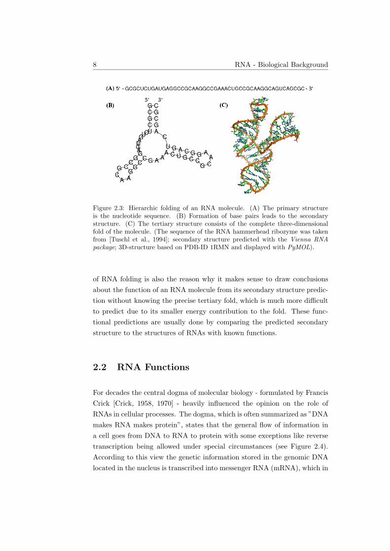
DNA usually consists of two complementary strands which can form basepairs from end to end, thus creating its famous double helix structure. Be-cause of its single-strandedness RNA is able to form intramolecular basepairs. Multiple successive base pairs form helical stems interspersed withunpaired loop regions. The resulting structure is referred to as the RNAsecondary structure. Figure 2.3b shows an example of an RNA secondarystructure. The properties of secondary structures will be covered in moredetail in chapter 3.
The embedding of an RNA molecule with its secondary structure in three-dimensional space is called the tertiary structure. This three-dimensionalfold is often the result of stabilizing non-standard base pairs, triple base pairsand backbone-loop interactions. These interactions are usually weaker thanthe interactions responsible for the canonical base pairs of the secondarystructure and as a consequence the secondary structure contributes the big-ger part of the stabilizing energy of the whole RNA fold. RNA folding cantherefore be seen as a hierarchical process with the base pairs forming first,before the full three-dimensional arrangement of the RNA molecule in itstertiary structure takes place [Tinoco et al., 1999]. This hierarchical nature
8 RNA - Biological Background
Figure 2.3: Hierarchic folding of an RNA molecule. (A) The primary structureis the nucleotide sequence. (B) Formation of base pairs leads to the secondarystructure. (C) The tertiary structure consists of the complete three-dimensionalfold of the molecule. (The sequence of the RNA hammerhead ribozyme was takenfrom [Tuschl et al., 1994]; secondary structure predicted with the Vienna RNApackage; 3D-structure based on PDB-ID 1RMN and displayed with PyMOL).
of RNA folding is also the reason why it makes sense to draw conclusionsabout the function of an RNA molecule from its secondary structure predic-tion without knowing the precise tertiary fold, which is much more difficultto predict due to its smaller energy contribution to the fold. These func-tional predictions are usually done by comparing the predicted secondarystructure to the structures of RNAs with known functions.
2.2 RNA Functions
For decades the central dogma of molecular biology - formulated by FrancisCrick [Crick, 1958, 1970] - heavily influenced the opinion on the role ofRNAs in cellular processes. The dogma, which is often summarized as ”DNAmakes RNA makes protein”, states that the general flow of information ina cell goes from DNA to RNA to protein with some exceptions like reversetranscription being allowed under special circumstances (see Figure 2.4).According to this view the genetic information stored in the genomic DNAlocated in the nucleus is transcribed into messenger RNA (mRNA), which in
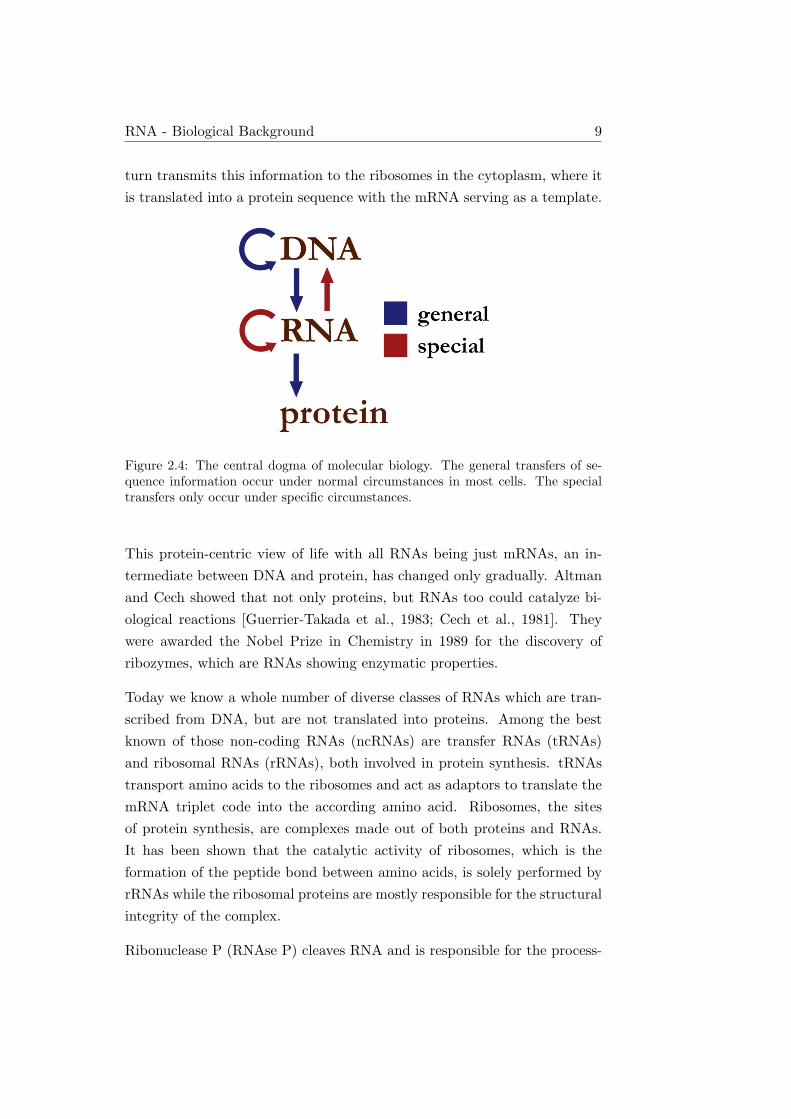
RNA - Biological Background 9
turn transmits this information to the ribosomes in the cytoplasm, where itis translated into a protein sequence with the mRNA serving as a template.
Figure 2.4: The central dogma of molecular biology. The general transfers of se-quence information occur under normal circumstances in most cells. The specialtransfers only occur under specific circumstances.
This protein-centric view of life with all RNAs being just mRNAs, an in-termediate between DNA and protein, has changed only gradually. Altmanand Cech showed that not only proteins, but RNAs too could catalyze bi-ological reactions [Guerrier-Takada et al., 1983; Cech et al., 1981]. Theywere awarded the Nobel Prize in Chemistry in 1989 for the discovery ofribozymes, which are RNAs showing enzymatic properties.
Today we know a whole number of diverse classes of RNAs which are tran-scribed from DNA, but are not translated into proteins. Among the bestknown of those non-coding RNAs (ncRNAs) are transfer RNAs (tRNAs)and ribosomal RNAs (rRNAs), both involved in protein synthesis. tRNAstransport amino acids to the ribosomes and act as adaptors to translate themRNA triplet code into the according amino acid. Ribosomes, the sitesof protein synthesis, are complexes made out of both proteins and RNAs.It has been shown that the catalytic activity of ribosomes, which is theformation of the peptide bond between amino acids, is solely performed byrRNAs while the ribosomal proteins are mostly responsible for the structuralintegrity of the complex.
Ribonuclease P (RNAse P) cleaves RNA and is responsible for the process-

10 RNA - Biological Background
ing of the 5’-leader sequence of precursor tRNAs to form mature tRNAs[Guerrier-Takada et al., 1983]. Small nuclear RNAs (snRNAs), which areassociated together with proteins in complexes called small nuclear ribonu-cleoproteins (snRNPs) are involved in mRNA intron splicing, regulation oftranscription and telomer maintenance [Valadkhan, 2005]. Small nucleolarRNAs (snoRNAs) are responsible for targeting the site for chemical mod-ifications (methylation or pseudouridylation) of nucleotides of other RNAs[Bachellerie et al., 2002]. Transfer-messenger RNA (tmRNA) is a bacterialRNA with tRNA- and mRNA-like properties. It rescues ribosomes thathave stalled in the middle of protein synthesis by recycling the stalled ribo-some, adding a proteolysis-inducing tag to the unfinished polypeptide, andfacilitating the degradation of the aberrant messenger RNA [Keiler et al.,1996].
MicroRNAs (miRNAs) were first discovered in 1993 [Lee et al., 1993]. To-gether with small interfering RNAs (siRNAs) [Hamilton and Baulcombe,1999; Elbashir et al., 2001] they play a crucial role in the RNA interference(RNAi) pathway, which most commonly results in post-transcriptional genesilencing, but in some cases RNAi can be activating as well. Today RNAi isa valuable research tool, both in vitro and in vivo because this pathway en-ables researchers to suppress specific genes of interest in a simple and cheapway by introducing synthetic short double-stranded RNAs into cells. The2006 Nobel Prize in Physiology or Medicine was awarded to Andrew Fireand Craig Mello for their work on RNAi in the nematode worm C. elegans[Fire et al., 1998]. These examples highlight the versatility of RNA (seeFigure 2.5) and show that RNAs can catalyze chemical reactions of varioustypes including phosphoryl group transfers, isomerization of C-C bonds andhydrolytic reactions.
RNAs are the only known biological macromolecules with the ability toact as both carrier of genetic information as well as catalytically activesubstance, thereby combining genotype and phenotype in one molecule. Thisproperty has lead to the formulation of the RNA world hypothesis [Gilbert,1986; Joyce, 1989, 1991], which proposes some form of Darwinian evolutionbased on RNA alone, prior to our current world of life based on DNA ascarrier of information and protein as catalytically active substance.

RNA - Biological Background 11
Figure 2.5: Summary of the biological processes RNA molecules are involved in.(Image reproduced from [Gruber, 2007])
Another piece of evidence for the importance of RNA comes from the hugeamount of sequence data which has become available only in recent years.Protein coding genes amount to only about 1.5% of the human genome andthe estimated number of human protein-coding genes is 23000, which is alot lower than expected [Stein, 2004]. This number does not appropriatelyreflect the increased complexity of humans compared to much simpler organ-isms such as C. elegans with about 20000 genes. Combined with the knowl-edge that a big fraction of the human genome is being transcribed [Birneyet al., 2007; Johnson et al., 2005], while many transcripts lack protein-codingpotential, this results in the assumption that functional RNAs comprise asignificant part of the human genome. John Mattick states that the com-plexity of higher organisms cannot solely be achieved by proteins, but thatthere has to be an additional layer of regulatory ncRNAs [Mattick, 2003].
All these findings have contributed to a switch away from a protein dom-inated view of biological life and to an increased effort in scientific RNAresearch over the last decades. Our knowledge of RNAs and especially func-

12 RNA - Biological Background
tional RNAs is still very limited and new discoveries will lead to a betterunderstanding of the fundamental processes of cellular life. This makesthe analysis and prediction of RNA structures and functions an important,challenging and interesting task.
RNA Secondary Structure 13
Chapter 3
RNA Secondary Structure
Solving the full structure problem for RNA is difficult, because the numberof degrees of freedom of the RNA chain is very high. However, there areseveral reasons why the secondary structure can be used as a valid substitutefor the full three-dimensional fold:
• The secondary structure with its base pairing and base stacking ener-gies is responsible for the major part of the free energy of folding.
• RNA folding is a hierarchical process causing the secondary structureto be the starting point for the tertiary folding.
• RNA secondary structure provides distance constraints for the forma-tion of the full tertiary fold.
• RNA secondary structures are evolutionary conserved and can be usedto successfully predict RNA function.
RNA secondary structures are discrete, easy to visualize and compare, andcan be handled efficiently by computational methods. All these factors con-tribute to the fact that RNA secondary structure prediction is an importantand popular task in bioinformatics.

14 RNA Secondary Structure
3.1 Definitions
An RNA sequence is defined as a string s over the nucleotide alphabet{A,C,G,U}. n denotes the length of s and (i, j) with i < j denotes basepairing between si and sj , the nucleotides at positions i and j. A secondarystructure is defined as a set S of base pairs with the allowed base pairs being(A,U), (G,C), (G,U) and their reversals. For any two base pairs (i, j) ∈ Sand (k, l) ∈ S with i < k, the following must hold [Waterman and Smith,1978]:
1. j − i > 3.
2. i = k if and only if j = l.
The first condition imposes a minimal hairpin loop size of three nucleotidesand the second condition states that each base can be part of at most onebase pair and forbids e.g. triple base pair interactions. A secondary structureS is called pseudoknot-free if for any two base pairs (i, j) ∈ S and (k, l) ∈ Sthe condition
3. Either i < j < k < l or i < k < l < j.
also holds. Otherwise S contains at least one pseudoknot. In a pseudoknot-free secondary structure all base pairs either precede each other or are prop-erly nested, while pseudoknots consist of overlapping base pairs.
A base k is called immediately interior to the base pair (i, j) if i < k < j
and if there is no base pair (p, q) such that i < p < k < q < j. A basepair (p, q) is called immediately interior to a base pair (i, j) if p and q areimmediately interior to (i, j) [Zuker and Sankoff, 1984].
All bases immediately interior to the same base pair (i, j) form the loopenclosed by the exterior pair (i, j). The external loop is defined as the set ofbases which are not immediately interior to any base pair, i.e. all nucleotidesnot enclosed by any base pair.
Loops are characterized by their size u, the number of unpaired bases in theloop, and by their degree k. k − 1 is the number of base pairs in the loop

RNA Secondary Structure 15
(not counting the enclosing base pair), therefore k is the number of basepairs delimiting the loop (including the enclosing base pair).
The different loop types, which are depicted in Figure 3.1, are the buildingblocks for the loop-based RNA energy model described in section 4.1. Loopsof degree 1 have no immediately interior base pair and are called hairpinloops. Loops with a degree of 2 and a size of 0 are stacked (base) pairs.Loops with k = 2 and u > 0 are called interior loops. Bulge loops are aspecial case of interior loops where the immediately interior base pair (p, q)lies directly next to the enclosing base pair (i, j), i.e. p = i+ 1 or q = j − 1.Loops with a degree greater than 2 are called multiloops.
Figure 3.1: Loop types in RNA secondary structures. The bases being part of thesame loop are colored in red, the respective enclosing base pairs in blue.
NcRNAs often have similar secondary structures without having very similarprimary sequences. With the help of multiple sequence alignments and sta-

16 RNA Secondary Structure
tistical covariance models the RNA secondary structures of known ncRNAshave been categorized into families which are based on evolution from a com-mon ancestor. The known information about these ncRNA families is storedin the Rfam database, which is among other things very useful for comparingpotential new functional RNAs with known ncRNAs [Griffiths-Jones et al.,2005].
3.2 Representations
Instead of as a list of base pairs, which is not intuitive to the human mind,RNA secondary structures are commonly displayed in one of the followingways:
3.2.1 Squiggle Plot
The most common representation of an RNA secondary structure is theso-called squiggle plot. The nucleotide labels are placed along a curvedline representing the sugar-phosphate backbone and base pairs are visual-ized by (usually) short straight lines. Figure 3.2 shows an example. Onlypseudoknot-free structures are guaranteed to result in a planar graph, i.e. agraph that can be drawn without any crossing lines.
3.2.2 Dot-Bracket Notation
The computer science community commonly uses the dot-bracket notation,where the RNA sequence is aligned next to a row of symbols: a dot representsan unpaired nucleotide and each base pair is symbolized by a pair of openingand closing brackets of the same type. For pseudoknot-free structures onlyone type of brackets is needed, pseudoknotted structures require more thanone type. The following lines show an example of an RNA sequence and itssecondary structure in dot-bracket notation:
Human Gln-tRNA
UAGGACGUGGUGUAGUAGGUAGCAUGGAGAAUGUUGAAUUCUCAGGGGUAGGUUCAAUUCCUAUAGUUCUAG
((((((...((((........)))).((((((.....))))))....(((((.......))))).)))))).

RNA Secondary Structure 17
Figure 3.2: Squiggle plot of a human tRNA. (Image generated with Pseudoviewer3[Byun and Han, 2009])
3.2.3 Arc Plot
Here the RNA backbone is drawn as a straight line and the nucleotides ofeach base pair are connected by an arc (see Figure 3.3). For pseudoknot-freesecondary structures all arcs can be drawn on the same side of the backboneline without intersecting each other.
Figure 3.3: Arc plot of a human tRNA. (Image generated with jViz.Rna 2.0 [Wieseet al., 2005])

18 RNA Secondary Structure
3.2.4 Circular Graph
In a circular RNA secondary structure graph the backbone is represented bya circle. The base pairs are symbolized by arcs in the interior of the circleexactly like in an arc plot (see Figure 3.4). Only pseudoknot-free secondarystructures can be drawn in this way without any crossing of arcs. In graphtheory, the formal equivalent of circular RNA graphs are called outerplanargraphs. Outerplanar graphs can be drawn in the plane without crossing ofany edges in such a way that all of the vertices belong to the unboundedface of the drawing. Every outerplanar graph is planar, but not every planargraph is outerplanar.
Figure 3.4: Circular graph of a human tRNA. (Image generated with jViz.Rna 2.0[Wiese et al., 2005])
3.2.5 Dot Plot
Dot plots can convey more information about the RNA sequence than just asingle secondary structure. In these two-dimensional graphs the nucleotidesequence is written along both x- and y-axis and the whole graph is dividedinto two parts by a diagonal from the top-left to the bottom-right corner(see Figure 3.5). In the lower left half of the plot, a dot at the intersectionof column i and row j represents a base pair (i, j). Typically, the secondary

RNA Secondary Structure 19
structure with the minimum free energy is displayed in the lower left part.In the upper right half, more than one structure is visualized. The dotsizes in this part of the plot are proportional to the probabilities of thecorresponding base pairs being formed. This allows the visualization of aweighted set of secondary structures such as the Boltzmann ensemble.
Figure 3.5: Dot plot of human tRNA. The single structure with minimum freeenergy is displayed in the lower left half, and the base pair probabilities in theupper right half. (Image generated with the Vienna RNA package)

20 RNA Secondary Structure
3.2.6 Mountain Plot
A mountain plot is another two-dimensional graph to display an RNA sec-ondary structure. The nucleotide index number is plotted against the x-axisand the number of enclosing base pairs against the y-axis (a nucleotide kis enclosed by a base pair (i, j) if i < k < j). The resulting plot usuallylooks similar to a mountain range (see Figure 3.6). Peaks correspond tohairpin loops, showing the unpaired bases enclosed by slopes representingthe stem. Plateaus within a sloped region represent bulge loops or, if theyare paired with another plateau on the other side of the mountain, interiorloops. Mountain plots are not suited to display pseudoknotted secondarystructures because pseudoknots cause mountain plots to be ambiguous.
3.3 RNA Pseudoknots
3.3.1 Pseudoknot Types
An RNA pseudoknot is a secondary structure element where unpaired baseswithin a loop pair with complementary bases in a single-stranded regionoutside the loop, therefore violating the condition of perfectly nested basepairing which only holds for pseudoknot-free secondary structures. Formally,an RNA secondary structure S is said to contain a pseudoknot if and onlyif there exist base pairs (i, j) ∈ S and (k, l) ∈ S, such that i < k < j < l.
The simplest type of pseudoknot is called H-type pseudoknot. In thesestructures the bases in a hairpin loop interact with bases outside of thestem enclosing the hairpin. This generates a second stem and loop withthe two stems stacking on top of each other, forming a quasi-continuoushelix consisting of one continuous and one discontinuous strand (see Figure3.7). Another typical and simple pseudoknot structure, the kissing hairpin,is produced by the unpaired bases in a hairpin loop interacting with theunpaired bases in another hairpin loop (see Figure 3.8).
H-type pseudoknots, kissing hairpins and most other observed pseudoknotsare planar pseudoknots, i.e. they can be drawn on a plane (as a squiggle orarc plot) without any crossing of arcs. All bi-secondary structures, which

RNA Secondary Structure 21H
eigh
t
mfepfcentroid
02
46
810
12
0 10 20 30 40 50 60 70
0.0
0.5
1.0
1.5
Position
Ent
ropy
Figure 3.6: Mountain plot for the MFE structure (red), the thermodynamic en-semble of RNA structures (green), and the centroid structure (blue) of a humantRNA. The lower part of the figure displays the positional entropy of the sequence.(Image generated with the RNAfold web server [Gruber et al., 2008])

22 RNA Secondary Structure
Figure 3.7: H-type pseudoknot architecture.(A) Linear arrangement of the elements forming the pseudoknot. Base pairing in-dicated by dashed lines.(B) Bases within the initial hairpin pair with bases outside of the loop.(C) Schematic view of the final H-type pseudoknot fold.(D) 3-dimensional fold of the SAM-II riboswitch, an H-type pseudoknot. The stack-ing helices are shown in black and gray, the connecting loops in yellow and greenand the bound SAM in red.(Images (A)-(C) reproduced from [Staple and Butcher, 2005], Image (D) reproducedfrom [Brierley et al., 2008])

RNA Secondary Structure 23
are defined as the superposition of two disjoint pseudoknot-free secondarystructures (see Figure 3.9), are planar. The converse is not true, since thereare planar structures which are not bi-secondary. An example for sucha planar, not bi-secondary structure would be a helix crossing scheme ofABCA’B’C’ with X-X’ denoting one or more consecutive base pairs. Forcircular RNAs, bi-secondary structures and planar structures are equivalent[Witwer et al., 2004].
Even more complex non-planar folds, which cannot be drawn in a planewithout crossing of arcs, are possible as well. A helix crossing scheme suchas ABCDA’C’B’D’ results in the formation of a non-planar pseudoknot. Sofar, pseudoknots of this type are quite rare. An example is the ribosomebinding site of the E. coli α operon (see Figure 3.10).
As an extension to bi-secondary structures, k-partite structures are theunion of k pseudoknot-free sub-structures. These structures can be intu-itively visualized by imagining a book. The spine of the book representsthe RNA sequence, which is shared by all sub-structures. Each of the k
pages of the book then contains the arcs of one of the k pseudoknot-freedisjoint sub-structures. Rigorous definitions and the mathematical proper-ties of bi-secondary and k-partite RNA secondary structures can be foundin [Haslinger and Stadler, 1999].
Figure 3.8: Kissing hairpin type pseudoknot. The dotted lines indicate the pseu-doknot interaction.

24 RNA Secondary Structure
Figure 3.9: A bi-secondary structure is formed by the union of two disjointpseudoknot-free sub-structures. (Image reproduced from [Witwer et al., 2004])
Figure 3.10: The non-planar pseudoknot of the ribosome binding site of the E. coliα operon (Image reproduced from [Schlax et al., 2001])
3.3.2 Examples of Pseudoknots
It is known that pseudoknots play an important role in many RNA mediatedprocesses and often the pseudoknots are not only structurally relevant, butare directly responsible for the RNA molecule’s function.
A lot of catalytically active RNAs have been discovered to contain pseu-doknots. Human telomerase is a riboprotein complex. The 5′ end of thetelomerase RNA forms a highly conserved pseudoknot, which is required forthe activity of the complex [Staple and Butcher, 2005]. Mutations withinthis H-type pseudoknot have been shown to be connected to the diseases au-tosomal dyskeratosis congenita [Marciniak et al., 2000] and aplastic anemia[Vulliamy et al., 2002]. Most of the ribosomal RNAs contain pseudoknotsas well, although in this case they do not seem to sit at the catalyticallyactive site and are assumed to be of mainly structural importance [Cannoneet al., 2002]. Another ribozyme is Ribonuclease P, which is responsible forcleaving of a precursor sequence on tRNA molecules. Its catalytically active

RNA Secondary Structure 25
subunit contains a pseudoknot which is required for the RNase P’s function[Brown, 1996; Mann et al., 2003]. The versatile tmRNAs, which are involvedin translation by rescuing stalled ribosomes and facilitating the degradationof aberrant messenger RNAs, contain multiple pseudoknots. Although thesepseudoknots are evolutionarily conserved, they seem to be not directly in-volved in the tmRNA’s function [Andersen et al., 2006; Nameki et al., 2000].Other pseudoknots were found in the mRNA of prion proteins [Barette et al.,2001].
In eukaryotic life, introns have to be removed from pre-mRNA to get maturemRNA. This is usually done by the spliceosome riboprotein. Some introns,however, are self-cleaving, catalyzing their removal from the pre-mRNA ontheir own [Staple and Butcher, 2005; Cech, 1988]. In one group of suchintrons, the group I self-splicing introns, the catalytic core consists of apseudoknot [Adams et al., 2004]. Self-cleaving RNA pseudoknots can alsobe found in various viral genomes: the pseudoknotted self-cleaving Hepatitisdelta virus ribozyme is essential for the virus’ replication. It is responsible forcutting the replicated multi-genome RNA strand into genome-length unitsrequired for virus packaging. It is the fastest-known self-cleaving ribozymewith a reaction rate of one per second [Brierley et al., 2007]. Pseudoknotscan also be involved in other viral processes, such as the regulation of viralprotein synthesis [Brierley et al., 2008].
Not only catalytically active RNAs contain pseudoknots. Pseudoknots arealso involved in ribosomal frameshifting, again commonly found in viruses.Ribosomes normally translate mRNAs without shifting the translationalreading frame, but specific pseudoknots together with ”slippery” nucleotidesequences can cause the ribosome to change its reading frame during trans-lation [Giedroc and Cornish, 2009; Staple and Butcher, 2005]. Viruses typ-ically have small genomes with very densely packed information becausethere is no room for bigger genomes in the virus envelope. A lot of virusesare therefore employing a −1 frameshifting mechanism which allows twoproteins to be encoded within one genomic region. Because this frameshift-ing is necessary for the survival of all retroviruses, the pseudoknots involvedin this mechanism are attractive targets for drug development. Other ex-amples of viruses with ribosomal frameshift inducing pseudoknots are thesevere respiratory syndrome (SARS) coronavirus and other coronaviruses,

26 RNA Secondary Structure
the mouse mammary tumor virus, the beet western yellow virus and the peaenation mosaic virus.
RNA pseudoknots have been discovered in nearly every organism and havebeen found to be of functional relevance for ribozymes, self-splicing introns,ribonucleoprotein complexes, viral genomes and other biological systems.The determination of their structure is therefore of great importance.

RNA Energy Models 27
Chapter 4
RNA Energy Models
In order to predict RNA secondary structures a method of comparing andevaluating different structures is needed. RNA energy models condense thewhole secondary structure information into a single number representingthe structure’s free energy. Energy models therefore serve as scoring func-tions for RNA folding algorithms. The simplest scoring method employed inthe first secondary structure prediction algorithm was to just maximize thenumber of base pairs [Nussinov and Jacobson, 1980]. A structure with morebase pairs than another was assumed to be energetically favorable. Thisapproach has been replaced by the loop-based energy model, which evalu-ates RNA structures by decomposing them into loops and assigning energyvalues to these loops (RNA loops have been described in section 3.1).
4.1 The Loop-based RNA Energy Model
The loop-based energy model scores different secondary structures accordingto their free energy. This idea in its principles has first been proposedabout 30 years ago [Waterman and Smith, 1978; Waterman, 1978; Zukerand Stiegler, 1981]. Today’s standard model [Mathews et al., 1999; Turnerand Mathews, 2009] has been refined several times since then.
Any RNA secondary structure S can be unambiguously decomposed intoloops as shown in Figure 4.1. S consists of all base pair enclosed loops Li,j

28 RNA Energy Models
Figure 4.1: RNA Loop Decomposition. The right hand side shows the loop decom-position of the secondary structure depicted on the left hand side. The enclosingbase pairs of each loop are indicated by dashed lines. (Image adapted from [Flamm,1998])
and the external loop L0 which contains all nucleotides not enclosed by anybase pair.
S = L0
⋃ ⋃(i,j)∈S
Li,j
The energy E of an RNA structure is then assumed to be the sum of theenergy contributions of all loops of the structure:
E(S) = E(L0) +∑
(i,j)∈S
E(Li,j)
Since absolute energy values are impossible to determine, energy differencesbetween unfolded and folded states in solution are considered. Base pairsare formed by the creation of hydrogen bonds between bases. These hydro-gen bonds themselves do not contribute a lot to the free energy of RNAs insolution, since unfolded RNA molecules with no base pairs still form hydro-gen bonds with the surrounding water molecules instead. But base pairingalso causes adjacent base pairs to stack on top of each other creating stemsof stacked base pairs. Base pair stacking is an energetically very favorable

RNA Energy Models 29
interaction. Single bases next to stacking base pairs, i.e. at the end of astem, can stack onto the stem as well. These dangling end interactions areenergetically favorable too. On the other hand, base pairing causes the for-mation of loops, and in loops the free movement of the nucleotide chain isrestricted by the fixed end points, which are the bases forming the pair.This leads to an energetically unfavorable destabilizing entropic effect of allloops.
The concrete energy parameters for the different structure elements havebeen derived empirically by RNA oligomer folding experiments. The valuesfor stacked bases and small hairpin, internal and bulge loops have beentabulated explicitly. Longer loops are assigned an estimated logarithmicallyincreasing penalty as derived from polymer folding theory. For reasons ofcomputational efficiency, multiloops are usually scored with an affine energymodel with a large penalty for the initiation of a multiloop and a smallerpenalty for each additional stem added.
4.2 Energy Models for RNA Pseudoknots
Because pseudoknotted RNA secondary structures contain overlapping basepairs, loop decomposition does not work for them, at least not in the wayas described above. Therefore the pseudoknotted parts of secondary struc-tures cannot be evaluated with the standard loop-based energy model. Inaddition, there is also only very limited experimental data on pseudoknotenergies available, making the estimation of pseudoknot energies even moredifficult.
For pseudoknot-free structures, steric considerations are of no concern, be-cause all secondary structures that can be decomposed into loops are ster-ically possible. This is not true for pseudoknotted structures and checkingwhether a suggested pseudoknotted fold is sterically possible is no trivialtask. Even for the simple H-type pseudoknots there are restrictions con-cerning the lengths of the different loops depending among other factorson whether those loops are crossing the minor or the major grove of theRNA. The first pseudoknot energy model dealing with these problems waspresented by Gultyaev [Gultyaev et al., 1999].

30 RNA Energy Models
Consequently, a good energy model for pseudoknots has to be substantiallymore complex than the current model for pseudoknot-free RNA structures.In the absence of any measured parameters for pseudoknots, most pseu-doknot prediction algorithms resort to estimating the energy parameters byusing simplified energy models. A common approach is to treat pseudoknotssimilar to how multiloops are treated in the standard loop decompositionmodel. This means that the energy associated with a pseudoknot is de-scribed by the following linear equation:
Epk = β1 + β2Bp + β3Up (4.1)
β1 is a penalty for introducing a pseudoknot, which depends on whether thepseudoknot is embedded in the exterior loop, in a multiloop, or in anotherpseudoknot. Bp is the number of base pairs that border the interior of thepseudoknot, β2 is the penalty for each such base pair, Up is the number ofunpaired bases inside the pseudoknot and β3 is the penalty for each unpairedbase.
4.3 The Cao-Chen Energy Model for H-type Pseu-
doknots
Song Cao and Shi-Jie Chen recently presented a more advanced pseudoknotenergy model based on applying polymer physics to the evaluation of pseu-doknot loops [Cao and Chen, 2006, 2009]. Loop free energies are made up ofenthalpic and entropic contributions, which are depending on factors suchas temperature and ionic strength. In Cao and Chen’s model the enthalpiccontributions are captured by base pairing and stacking energies and theirmain effort was to estimate the loop entropies of pseudoknot loops. Unfor-tunately, their model is only applicable to H-type pseudoknots in which thetwo helices are directly stacked on top of each other and to the more generalcase in which the two helices are connected by a loop of up to 6 nucleotides.This restriction unfortunately still severely limits the range of pseudoknotsthat can be evaluated with their model.
For a given pseudoknot defined by the lengths of its two stems and three

RNA Energy Models 31
loops Cao and Chen employed a three-vector virtual-bond-based RNA con-formational model to enumerate all possible loop conformations on a grid,while accounting for excluded volume effects. This information was used tocompute the loop entropies and following that their free energies (see Fig-ure 4.2). These pre-computed results are stored in tables as loop entropyparameters and can be easily looked up during the evaluation of an actualpseudoknot.
Figure 4.2: (A) A schematic view of a pseudoknot covered by the Cao-Chen model.It is made up of two stems and three loops.(B) The three vector virtual bond model involves the bonds Pi − C4, C4 − Pi+1
and C4 −N1 (pyrimidine) or C4 −N9 (purine). (Image reproduced from [Cao andChen, 2009])
The entropy values of different pseudoknot loops have not been measuredexperimentally yet and have often been ignored or treated in a simplifiedway by previous models [Ren et al., 2005; Dirks and Pierce, 2003]. Usingthe values provided by Cao and Chen allows more accurate calculations ofthe free energies of H-type and some other pseudoknots. There are still alot of other pseudoknot types where this model is not applicable and a moregeneral energy model for pseudoknotted RNA secondary structures wouldbe highly desirable.

32 RNA Energy Models

RNA Secondary Structure Prediction Algorithms 33
Chapter 5
RNA Secondary Structure
Prediction Algorithms
5.1 Maximizing the Number of Base Pairs
The goal of the first algorithm for RNA secondary structure prediction wasto find the structure with the maximum number of base pairs. This algo-rithm was published by Ruth Nussinov [Nussinov and Jacobson, 1980] basedon the idea by Michael Waterman [Waterman and Smith, 1978; Waterman,1978] to use a dynamic programming approach. The basic principle of dy-namic programming is to decompose the overall problem into a number ofsimpler and smaller subproblems for which optimal solutions can be found.
Figure 5.1: Decomposition of RNA structures in the Nussinov algorithm. (Imagereproduced from [Gruber, 2007])
We start with an RNA sequence s of length n. M(i, j) denotes the maxi-mum number of base pairs of the subsequence si, . . . , sj . The main idea isthat M(i, j) can be calculated recursively in the following way: sj , the lastnucleotide of the subsequence, is either unpaired or paired to some base sk.In the former case M(i, j) equals M(i, j − 1). In the latter case si, . . . , sj is

34 RNA Secondary Structure Prediction Algorithms
divided into the intervals si, . . . , sk−1 and sk+1, . . . , sj−1 for which the max-imum number of base pairs is already known. A graphical representationof this decomposition is shown in Figure 5.1. It produces to the followingrecursion:
M(i, j) = max
M(i, j − 1)
maxi≤k<j(k,j)∈S
(M(i, k − 1) +M(k + 1, j − 1) + 1)
The matrix M(i, j) is filled proceeding from shorter to longer subsequences.The value stored in M(1, n) contains the maximum number of base pairsfor the whole sequence. The corresponding structure is then deduced viabacktracking. This means going backwards through the calculated matrixreconstructing the path and therefore the list of base pairs for M(1, n).
Although the matrix can be stored as a triangular matrix the algorithm stillrequires O(n2) memory space. The runtime scales with O(n3) because thealgorithm iterates over i, j and k.
This non-thermodynamic base pair maximization model is too simple toogive realistic RNA secondary structure predictions, but it serves as a startingpoint for more sophisticated algorithms. The basic principle of dynamicprogramming with backtracking stays the same, but base pair maximizationis replaced by thermodynamic considerations.
5.2 Minimizing the Free Energy
Thermodynamic energy models are based on the loop decomposition de-scribed in section 4.1. Each loop is assigned an energy value and the energyof the whole structure is the sum of the energies of all loops making upthe structure. The goal of the folding algorithm is to find the secondarystructure with the minimum free energy:
Fmin = minS∈S
(F (S)) (5.1)
F (S) denotes the free energy of a structure S ∈ S, with S being the set ofall possible secondary structures S.

RNA Secondary Structure Prediction Algorithms 35
MFE folding was first described by Michael Zuker and Patrick Stiegler [Zukerand Stiegler, 1981]. They make a major simplification by regarding multi-loops as bifurcation loops, which means that they do not assign an energyto the multiloop itself, they just add up the energies of its constituent loops.
Today’s models explicitly take multiloops into account. It makes no sense todetermine multiloop energy values experimentally and then use tabulatedvalues in the energy model because due to a combinatorial explosion thenumber of possible multiloops is simply too big. The models therefore oftenuse a linear approach for the energy of a multiloop M:
M = a+ b · k + c · u
a is the cost of initiating a multiloop, b is the penalty for each helix pro-truding from the loop, k is the degree of the loop, c is the penalty for eachunpaired base in the loop, and the loop size u is the number of unpairedbases.
The standard energy model, as implemented for example in the Vienna RNApackage [Hofacker et al., 1994], is based on the model by Zuker and Stiegler.In addition to scoring multiloops with the described linear approach, thismodel also decomposes multiloops unambiguously. This ensures that everystructure is encountered exactly once, which is important for calculating thepartition function as described in the next section.
The array Fi,j stores the minimum free energy of all possible structures onthe subsequence si, . . . , sj . The base si is either unpaired or paired with abase sk (see Figure 5.2), leading to the following recursion for Fi,j :
Figure 5.2: Recursion for Fi,j . (Image adapted from [Lorenz, 2007])
Fi,j = min
Fi+1,j
mini<k≤j
Ci,k + Fk+1,j

36 RNA Secondary Structure Prediction Algorithms
Ci,j stores the minimum energy of the subsequence si, . . . , sj provided thatsi and sj form a base pair. Every base pair encloses either a hairpin loop, aninterior loop, or a multiloop (see Figure 5.3). The recursion for Ci,j thereforelooks like this:
Figure 5.3: Recursion for Ci,j . (Image adapted from [Lorenz, 2007])
Ci,j = min
H(i, j)
mini<k<l<j
I(i, j; k, l) + Ck,l
mini+1<u<j−1
Mi+1,u +M1u+1,j−1 + a
The energy values H(i, j) for hairpin loops and I(i, j; k, l) for interior loopsare tabulated. Multiloops are decomposed into a left part M , which containsat least one base pair, and a right part M1 containing exactly one base pair.a is the cost of initiating a multiloop.
In Mi,j , the final base sj is either unpaired, leading to a penalty of c forthe enclosing multiloop, or it pairs with a base su, causing a penalty b fora base pair within a multiloop. si, . . . , su−1 is then either unpaired (u − iunpaired bases within a multiloop) or contains at least one base pair (andtherefore has a minimum energy of Mi,u−1)(see Figure 5.4). This results inthe following recursion for M :
Figure 5.4: Recursion for Mi,j . (Image adapted from [Lorenz, 2007])

RNA Secondary Structure Prediction Algorithms 37
Mi,j = min
Mi,j−1 + c
mini≤u<j
Cu,j + b+ c(u− i)
mini<u<j
Cu,j + b+Mi,u−1
b is the penalty for a base pair within a multiloop and c the penalty for anunpaired base within a multiloop. The recursion for M1
i,j is simple, since siis paired and there is no further base pair. So sj is either unpaired or it ispaired with si (see Figure 5.5):
Figure 5.5: Recursion for M1i,j . (Image adapted from [Lorenz, 2007])
M1i,j = min
M1i,j−1 + c
Ci,j + b
All matrices are filled gradually, starting with the smallest feasible subse-quences, which are pentanucleotides due to the minimum loop size for ahairpin loop which has to enclose at least three bases. Once the matricesare filled, the MFE can be found in F1,n and the corresponding MFE struc-ture is determined via backtracking. The algorithm requires O(n2) memory.When dealing with interior loops, the algorithm has to evaluate I(i, j; k, l)for all combinations of i, j, k and l. This would result in a time complexity ofO(n4). To reduce this time complexity and because very large interior loopsare biologically irrelevant and energetically unfavorable, the maximum loopsize of interior loops is usually set to some constant, resulting in an overalltime complexity of O(n3).
In addition, modern RNA secondary structure prediction algorithms usuallytake dangling ends into account. The bases immediately next to the end ofhelices can stack onto the helix and contribute favorably to the energy ofthe structure. Still missing from most modern approaches, however, is theinclusion of pseudoknots. While more and more pseudoknotted structures

38 RNA Secondary Structure Prediction Algorithms
have been experimentally discovered, they are still being ignored by mostRNA secondary structure prediction algorithms and software packages.
5.3 The Partition Function
MFE calculations only return information about a single secondary struc-ture, the one with minimum free energy. But this is not the only secondarystructure occurring in nature, in non-equilibrium states it might not evenbe the most common structure. RNAs are synthesized in an unfolded stateand only later fold into a secondary structure. The multidimensional fold-ing space contains many local minima which can be occupied by the RNAmolecule. In order to answer questions about the space of all possible sec-ondary structures, the likelihood of particular structures, or the frequencyof structures with specific features, one has to look at the partition functionZ. The equilibrium partition function contains information about the wholeset S of possible secondary structures and is defined as
Z =∑S∈S
e−F (S)
RT (5.2)
with F (S) denoting the free energy of a structure S ∈ S. T is the absolutetemperature and R the gas constant (a transformation of the Boltzmannconstant if dealing with energies per mol). After introducing β = 1
RT forsimplicity, the relative frequency P (S) of a specific secondary structure S inequilibrium is then given by
P (S) =e−βF (S)
Z(5.3)
Similarly, the probability of various features, such as the frequency of a spe-cific base pair, can be calculated by adding the probabilities of all structurescontaining that feature.
The number of possible secondary structures and therefore the number ofsummands in the partition function grows exponentially with increasing se-quence length [Waterman, 1978]. Therefore it seems to be impossible tocalculate Z in polynomial time, but McCaskill introduced a dynamic pro-

RNA Secondary Structure Prediction Algorithms 39
gramming algorithm in [McCaskill, 1990] which achieves this computation.McCaskill’s partition function algorithm is very similar to the MFE algo-rithm described previously and is implemented in the folding routines of theVienna RNA package. The minima in the MFE algorithm are replaced bysums in the partition function algorithm, because every possible secondarystructure and not just the one with minimum free energy contributes tothe partition function. The additivity of free energies causes in a similarfashion the multiplicativity of the Boltzmann-weighted contributions to thepartition function. Using the same decomposition of secondary structuresas in the MFE approach described in section 5.2 is only possible becausethat decomposition is unambiguous and does not count any structures morethan once. This property is not needed for MFE calculations, as only thestructure with minimum energy is relevant, but it is necessary for the parti-tion function because every single structure contributes to Z. Overall, thisresults in the following recursion for the partition function:
Qi,j = Qi+1,j +∑i<k≤j
QBi,k ·Qk+1,j
QBi,j = e−β·H(i,j)
+∑
i<k<l<j
e−β·I(i,j;k,l) ·QBk,l
+∑
i+1<u<j−1
QMi+1,u ·QM1
u+1,j−1 · e−β·a
QMi,j = QMi,j−1 · e−β·c
+∑i≤u<j
e−β·(u−i)·c ·QBu,j · e−β·b
+∑
i<u<j−1
QMi,u ·QBu+1,j · e−β·b
QM1
i,j = QM1
i,j−1 · e−β·c +QBi,j · e−β·b (5.4)
The matrix Qi,j is the analogon to Fi,j in the MFE algorithm, storing thepartition function of the subsequence [i, j]. Similarly QBi,j corresponds to Ci,jand stores the partition function of the subsequence [i, j] with (i, j) forming abase pair. QM and QM
1are equivalent to M and M1 and are responsible for
the calculation of multiloops. By summation over all Boltzmann-weightedenergies, starting with the smallest subsequences, the algorithm generatesthe partition function Z = Q1,n of the whole sequence.

40 RNA Secondary Structure Prediction Algorithms
5.4 Structure Prediction Including Pseudoknots
The commonly used MFE-based RNA secondary structure prediction al-gorithms rely on the condition that the structure can be unambiguouslydecomposed into independent loops, i.e. that all secondary structure mo-tifs are non-crossing and self-contained, which allows the application of thedynamic programming approach. Despite their importance, pseudoknotsare usually excluded in this approach because they violate the restrictionto non-crossing structure elements. While secondary structure predictionof pseudoknot-free structures with the MFE approach requires O(n3) timeand O(n2) space [Zuker and Stiegler, 1981; Lyngsø et al., 1999], allowing forarbitrary pseudoknots under the loop-based energy model has been shownto be NP-complete [Akutsu, 2000; Lyngsø and Pedersen, 2000].
Interestingly, if instead of using the loop-based energy model, one only wantsto maximize the number of base pairs, the problem can be solved: Akutsuproposed a dynamic programming algorithm for base pair maximization thatrequiresO(n4) time andO(n3) space [Akutsu, 2000]. Another approach usedfor the prediction of RNA pseudoknots and originating from graph theoryis maximum weighted matching with a time complexity of O(n3) [Tabaskaet al., 1998; Ruan et al., 2004]. Maximum weighted matching was also usedby Witwer in the hxmatch algorithm, which is very useful for predictingpseudoknots in multiple RNA sequences with known covariances [Witweret al., 2004].
Instead of simplifying the energy model, one can also keep the well-provencombination of dynamic programming and the loop-based energy modeland restrict the types of possible pseudoknots. Rivas and Eddy designeda secondary structure prediction algorithm which requires O(n6) time andO(n4) space [Rivas and Eddy, 1999]. Other algorithms, further restrictingthe range of predictable pseudoknots, reduced the runtime to O(n5) usingO(n4) or O(n3) space [Dirks and Pierce, 2003; Lyngsø and Pedersen, 2000].
A modern example for a dynamic programming algorithm for the predic-tion of pseudoknots is pknotsRG, developed by Jens Reeder and RobertGiegerich, which computes the MFE structure for canonical simple recursivepseudoknots in O(n4) time and O(n2) space [Reeder and Giegerich, 2004].

RNA Secondary Structure Prediction Algorithms 41
Reeder and Giegerich define simple pseudoknots as H-type pseudoknots. Ifthe three loops of this simple pseudoknot are allowed to fold internally, eveninto further pseudoknots, the resulting structure is called a simple recursivepseudoknot. Every simple recursive pseudoknot has a canonical representa-tive which can be handled by pknotsRG. A simple recursive pseudoknot iscanonical if it fulfills the following conditions:
• Both strands in a helix must have the same length and must not con-tain any bulges.
• The helices making up the pseudoknot must both have the maximumpossible length.
• If the two maximal helices would overlap, their boundary is fixed atan arbitrary point between them.
Simple recursive pseudoknots are defined by the eight end points of the foursequence intervals making up the two stems of the pseudoknot. The can-onization rules reduce these eight independent variables to four (see Figure5.6), allowing a dynamic programming recursion to find the MFE structureof an RNA sequence possibly containing canonical simple recursive pseudo-knots with a runtime of O(n4).
Figure 5.6: pknotsRG defines pseudoknots by their eight boundaries i, j, k, l, r,s, t and v. The canonization rules requiring bulge-free helices of maximum lengthreduce these eight variables to four (i, j, k and l).
Whenever possible, the energy model of pknotsRG uses the standard pa-rameters for pseudoknot-free secondary structures. For pseudoknots, thestabilizing effect of nearest neighbor stacking energies of the pseudoknot he-lices and of dangling bases at the helix ends are being accounted for. The

42 RNA Secondary Structure Prediction Algorithms
coaxial stacking of helices such as in H-type pseudoknots has also been in-corporated into the energy model. Apart from that, pseudoknots are treatedsimilar to multiloops in the standard energy model: they are assigned aninitiation penalty of 9 kcal/mol. This value is relatively high in order to pre-vent the prediction of too many false positive pseudoknots. In addition, eachunpaired nucleotide within a pseudoknot loop is penalized with 0.1 kcal/mol.
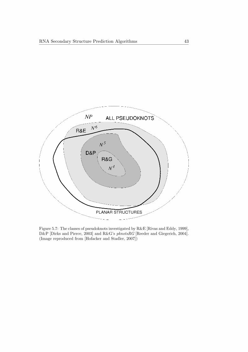
So far, pknotsRG is the fastest dynamic programming algorithm for theprediction of RNA pseudoknots. But it also restricts the type of pseudo-knots, which it is able to predict, further than other dynamic programmingalgorithms (see Figure 5.7). While pknotsRG can handle more than onepseudoknot per sequence and even recursive pseudoknots, the biologicallyquite common cases of kissing hairpins or pseudoknots with helices contain-ing bulges or interior loops are not covered by pknotsRG. Nevertheless, withits low computational complexity of O(n4), it is the most useful dynamicprogramming pseudoknot prediction algorithm in many cases.
An alternative to dynamic programming are heuristic approaches. Heuristicmethods can handle a wider class of pseudoknots and more complex energymodels. However, they are not guaranteed to find the MFE structure. In thecontext of RNA secondary structure prediction including pseudoknots ap-proaches using genetic algorithms [Gultyaev et al., 1995], stochastic context-free grammars [Cai et al., 2003], kinetic folding simulations [Xayaphoum-mine et al., 2003] and iterative stem adding procedures [Ruan et al., 2004]have all been employed.
A recent representative of the stem adding approach is HotKnots, which firstgenerates a tree of energetically favorable structures by partially folding theinput sequence with i and j paired, for all i and j with j−1 > 3 [Andronescuet al., 2010; Ren et al., 2005]. New base pairs are added using a dynamicprogramming algorithm leaving the already existing partial structures un-changed. For the calculation of the energy of folded structures HotKnotsuses the model of Dirks and Pierce [Dirks and Pierce, 2003], which is basedon the standard energy model for pseudoknot-free secondary structures. Itsadditional parameters concerning pseudoknots include three different penal-ties for initiating a pseudoknot, depending on whether the pseudoknot isnot enclosed by another loop, nested within a multiloop, or nested within
RNA Secondary Structure Prediction Algorithms 43
Figure 5.7: The classes of pseudoknots investigated by R&E [Rivas and Eddy, 1999],D&P [Dirks and Pierce, 2003] and R&G’s pknotsRG [Reeder and Giegerich, 2004].(Image reproduced from [Hofacker and Stadler, 2007])

44 RNA Secondary Structure Prediction Algorithms
another pseudoknot. Similar to the treatment of multiloops in the stan-dard energy model, there are additional penalties for each unpaired baseand each stem within a pseudoknot. The actual energy parameter valuesused in the latest version of HotKnots are not those published by Dirks andPierce. Instead, improved values obtained by applying constraint generationand Boltzmann likelihood parameter estimation methods to a large datasetof RNA structures are used.
Pseudoknot detection algorithms are following another heuristic approach,in which pseudoknot candidates are generated and analyzed before foldingthe remaining sequence with the standard MFE approach. An example isDotKnot [Sperschneider and Datta, 2010], which uses the dot plot generatedby the partition function of the Vienna RNA package as a starting point topick stems for the construction of pseudoknots. First, two crossing stemsare selected to form core H-type pseudoknots, which serve as the buildingblocks for more complex pseudoknots. Within the loops of these core H-type pseudoknots recursive secondary structure elements are allowed to formindependently of each other, before the whole recursive H-type pseudoknotcandidate is assembled and verified.
DotKnot uses three different pseudoknot energy models depending on thetype of pseudoknot it encounters. In each case, the stabilizing effect of thepseudoknot helices are calculated via the standard loop decomposition en-ergy model. Only the destabilizing entropic effect of the pseudoknot loopsis then calculated via one of three methods: for pseudoknots with heliceswithout bulge or interior loops and with a loop length between the two pseu-doknot helices of only 0 or 1 nucleotide, the original energy model by Caoand Chen [Cao and Chen, 2006] is used. If the loop length is between 2 and6 nucleotides, Cao and Chen’s extended energy model [Cao and Chen, 2009]is used. Finally, for pseudoknots with a longer loop connecting the two pseu-doknot helices or for pseudoknots with bulge or interior loops within theirhelices, a simple heuristic energy model is used. This heuristic model penal-izes the initiation of a pseudoknot loop with 7 kcal/mol and each unpairednucleotide within a pseudoknot loop with 0.1 kcal/mol.
The three algorithms described in this section, the dynamic programmingalgorithm pknotsRG, and the heuristic algorithms HotKnots and DotKnot,

RNA Secondary Structure Prediction Algorithms 45
will be used as benchmarks to compare the results of PKplex against insection 7.1. pknotsRG was chosen because it is the leading dynamic pro-gramming solution to the problem of pseudoknot prediction. And althoughdynamic programming and heuristic algorithms are not fully comparable,HotKnots and DotKnot are included in the analysis as well to show thestrengths and weaknesses of the different general approaches.

46 RNA Secondary Structure Prediction Algorithms

PKplex 47
Chapter 6
PKplex
In this chapter I am describing PKplex, a new dynamic programming RNAsecondary structure prediction algorithm which takes pseudoknots into ac-count. In the PKplex model, the thermodynamics of an RNA pseudoknotessentially consist of two components: the energy necessary to make theresidues of a potential pseudoknot accessible, i.e. unpaired, which is calcu-lated with the algorithm used in RNAplfold [Bernhart et al., 2006], and theenergy gained from the base pairing of the nucleotides involved in the pseu-doknot interaction. A dynamic programming routine based on the RNAplex[Tafer and Hofacker, 2008] algorithm combines these two energy values tocompute the optimal pseudoknot for a given RNA sequence.
In the next chapter I am taking a look at the strengths and limitations ofPKplex, present the results of evaluating the algorithm on a broad set ofknown RNA structures, both with and without pseudoknots, and comparePKplex with other published RNA pseudoknot prediction algorithms.
6.1 Algorithm
The PKplex algorithm operates within the framework of the Vienna RNApackage [Hofacker et al., 1994]. It uses the standard RNA energy modeldescribed e.g. by Mathews and Turner [Mathews et al., 1999; Turner andMathews, 2009] and is based on the classic RNA-folding algorithm by Zuker

48 PKplex
and Stiegler [Zuker and Stiegler, 1981]. The recursions for the equilibriumpartition function are based on those suggested by McCaskill [McCaskill,1990].
Figure 6.1: Two different secondary structures for the same RNA sequence, onewithout pseudoknots (a), and the other with a single big pseudoknot (b). Thedotted lines in (a) indicate the area of the pseudoknot interaction shown in (b).(Image generated with Pseudoviewer3 [Byun and Han, 2009])
Even though there are plenty of RNAs which contain pseudoknots, the frac-tion of nucleotides which are part of a pseudoknot is generally pretty low.That is because the pseudoknot usually comprises only a small part of thewhole sequence. Since we already know how to predict pseudoknot-freestructures decently, the approach used in PKplex starts with a pseudoknot-free secondary structure and then potentially adds just a single pseudoknot.The actual kinetic RNA folding process in living cells often follows the samesequential order: the pseudoknot-free secondary structure is built first, andonly then the sequence forms its pseudoknotted base pairs.
Other pseudoknot-prediction algorithms often have to avoid predicting toomany false positive pseudoknots. PKplex somewhat bypasses this problemby predicting at most one pseudoknot per sequence. However, if we areinterested in multiple pseudoknots on a single sequence, it is possible toextend PKplex in such a way as to run multiple times iteratively on thesame sequence and therefore generating structures containing more than

PKplex 49
one pseudoknot.
PKplex constructs pseudoknots by starting with a pseudoknot-free secondarystructure and then picking two non-overlapping intervals and letting thebases within those two intervals form pairs between each other - indepen-dent of the base pairing of the remainder of the sequence. If those twointervals do not lie within the same loop of the pseudoknot-free structure,this results in the formation of a pseudoknot (see Figure 6.1). This approachcauses all secondary structures predicted by PKplex to contain at most onepseudoknot. Because the frequency of pseudoknots is low, multiple pseudo-knots on a single sequence are very rare, and prediction accuracy does notsuffer much by this simplification. In addition, during the construction ofthe pseudoknot, base pairing is only allowed between the two sequence in-tervals, but not within one of the intervals. The resulting pseudoknot stemscan therefore contain bulge and interior loops, but multiloops are not pos-sible. Since most known pseudoknot stems are quite short, this restrictionshould not impair the prediction quality by much either.
The secondary structures covered by PKplex can be decomposed into apseudoknot-free structure and additional base pairs responsible for form-ing the pseudoknot, i.e. one of the stems of the pseudoknot. The free energy∆G of an RNA structure is then calculated as the sum of ∆Gpkfree and∆Gpk, the energy of the pseudoknot-free structure and the energy of thepseudoknot itself.
∆G = ∆Gpkfree + ∆Gpk
In the PKplex model, ∆Gpk consists of ∆Gint, the energy gained throughthe base pair interactions and cpk, the cost of initiating a pseudoknot, whichis constant.
∆Gpk = ∆Gint + cpk
∆Gpkfree is the energy of the pseudoknot-free part of the secondary struc-ture. The two intervals involved in the pseudoknot interaction cannot takepart in the pseudoknot-free structure and therefore have to be accessibleand unpaired. PKplex models ∆Gpkfree as
∆Gpkfree = ∆GMFE + ∆Gu[k, i][j, l]

50 PKplex
∆Gpkfree ≈ ∆GMFE + ∆Gu[k, i] + ∆Gu[j, l]
with ∆GMFE being the the pseudoknot-free MFE and ∆Gu[k, i] and ∆Gu[j, l]being the energies needed to render the two pseudoknot intervals [k, i] and[j, l] unpaired and accessible. The approximation of PKplex assumes thatthe energy of rendering two different non-overlapping intervals accessible isthe sum of rendering each interval accessible individually, i.e. that these twoprocesses are independent of each other.
In order to construct the optimum pseudoknot PKplex therefore searchesfor the interacting intervals [k, i] and [j, l], which minimize the expression∆Gu[k, i] + ∆Gu[j, l] + ∆Gint.
6.1.1 Calculation of Accessibility
∆Gu[a, b], the energy necessary to render the sequence interval [a, b] single-stranded and accessible can be calculated from Pu[a, b], the probability that[a, b] is unpaired via
∆Gu[a, b] = −RT ln(Pu[a, b]) (6.1)
Pu[a, b] is calculated for all subsequences by RNAplfold [Bernhart et al.,2006; Bompfunewerer et al., 2008], which is part of the Vienna RNA Pack-age, following the approach first used in RNAup [Muckstein et al., 2006;Muckstein et al., 2008].
The equilibrium partition function Z is defined as
Z =∑S
e−βF (S) (6.2)
with F (S) denoting the free energy of a secondary structure S and β denot-ing the inverse of the temperature times Boltzmann’s constant. Accordingto equation 5.3 we can use the partition function to calculate the probabilityof a sequence interval being unpaired via
Pu[a, b] =1Z
∑S∈Su
[i,j]
e−βF (S) (6.3)

PKplex 51
with Su[i,j] denoting the set of secondary structures with the interval [i, j]being unpaired. [i, j] is either part of the external loop of the secondarystructure or part of a loop enclosed by a base pair (p, q). Pu[i, j] can thereforebe expressed as
Pu[i, j] =Z(1, i− 1)Z(j + 1, n) +
∑p<i
∑j<q Z(p, q)Zpq[i, j]
Z(1, n)(6.4)
The first term handles the case of [i, j] being part of the external loop bymultiplying the partition functions of all sub-structures upstream and down-stream of [i, j]. The second term handles the case of [i, j] being part of aloop enclosed by the base pair (p, q). Z(p, q) denotes the partition functionoutside the base pair (p, q) and Zpq[i, j] the partition function inside (p, q)given that the interval [i, j] is unpaired. The restricted partition functionsZ(p, q) and Zpq[i, j] are then further decomposed according to the type ofloop containing the unpaired interval [i, j].
Depending of the details of the implementation one or more additional ma-trices compared to the partition function algorithm of McCaskill have to bestored. In RNAplfold the CPU requirements have been reduced to O(n3)and for very long sequences a sliding window technique which only looks atsequence intervals of fixed length instead of at the whole sequence can beused [Bernhart, 2009]. The implementation of the PKplex algorithm directlycalls RNAplfold to calculate Pu[i, j] which is then transformed into ∆Gu[a, b]via equation 6.1.
6.1.2 Calculation of the Interaction Energy
∆Gpk is calculated by a dynamic programming recursion based on the oneused in RNAplex [Tafer and Hofacker, 2008] to evaluate RNA-RNA inter-actions. Every potential pseudoknot can be described as an interactionbetween two intervals [k, i] and [j, l] with both (i, j) and (k, l) forming basepairs. PKplex uses the table Ci,j,k,l to store the best energy of an interactionbetween those two intervals. Ci,j,k,l is filled via the following recursion (seeFigure 6.2):

52 PKplex
Figure 6.2: Decomposition of a pseudoknot spanning the intervals [k, i] and [j, l].(i, j), (k, l) and (p, q) each form a base pair. The minimum energy of an interactionbetween [p, i] and [j, q] has been already been calculated and is stored in the tableCi,j,p,q. L(k, l, p, q), the energy of the loop enclosed by (k, l) and (p, q) can becalculated directly.
Ci,j,k,l = mink<p≤min(i,k+v)
max(j,l−v)≤q<l
(Ci,j,p,q + L(k, l, p, q)) (6.5)
with L(k, l, p, q) being the energy of a loop enclosed by the base pairs (k, l)and (p, q), and v being the maximum size of an interior/bulge loop. Thepseudocode for this recursion is shown in Figure 6.3.
for(i=n...1)for(j=i+4...n)for(k=i-1...1)
for(l=j+1...n)for(p=k+1...min(i,k+v))
for(q=l-1...max(j,l-v))E = LoopEnergy(p,q;k,l)C(i,j,k,l) = min(C(i,j,k,l), C(i,j,p,q)+E)
Figure 6.3: Pseudocode for the calculation of the interaction energy.
Ci,j,k,l is initialized by setting Ci,j,i,j to the pseudoknot initialization costconstant cpk. After the matrix Ci,j,k,l is filled, ∆Gu[i, j] and ∆Gu[k, l], theenergies of rendering the involved intervals unpaired and accessible, whichhave been previously calculated by RNAplfold and stored for later use, areadded - resulting in ∆G(i, j, k, l), the free energy change for the completepseudoknot interaction. If the minimum of ∆G(i, j, k, l) is greater than0, no energetically favorable pseudoknot has been found and the optimum

PKplex 53
pseudoknot-free structure according to the Vienna RNA package’s RNAfoldis returned. Otherwise backtracking is employed to get the structure ofthe pseudoknot. The non-pseudoknot part of the structure is generatedby RNAfold, using constraints to force the pseudoknot intervals [k, i] and[j, l] to be not involved in the formation of the remainder of the secondarystructure.
6.2 Time Complexity and Implementation
The time complexity of calculating the accessibilities ∆Gu[a, b] is O(n3).The recursion for ∆Gpk uses 6 loops for the 6 variables i, j, k, l, p andq. A naıve implementation would therefore require O(n6) computationaltime. Since the pseudoknot helices of known pseudoknots are generally ofvery limited length (the longest continuous pseudoknotted helix in the fulldataset is only 9 base pairs long), PKplex uses a parameter w to limit themaximum length of the intervals [k, i] and [j, l] involved in the pseudoknotformation. The CPU time required for the ∆Gpk recursion then scales withO(n2w4), because i and j run over the whole sequence, whereas the rangesof values for k, l, p and q are limited by w. Adding up both components ofthe algorithm, the runtime for PKplex as a whole scales with O(n3 +n2w4).
Instead of storing the whole four-dimensional table Ci,j,k,l, it is sufficient toonly store a single two-dimensional table Ck,l for every combination of i andj which can be discarded as soon as the calculations for the next combinationof i and j are started. The memory consumption of the recursion for ∆Gpkis therefore O(w2).
The pseudocode of Figure 6.3 shows that the loop energy L(k, l, p, q) iscalculated in every step of the innermost loop. Although the loop energyfunction is constant in its time complexity, the amount of times this functionis called still makes this a time consuming step. L(k, l, p, q) is called withexactly the same parameter values more than once for different combinationsof i and j, therefore unnecessarily repeating some calculations.
I managed to reduce the overall runtime of PKplex by reordering the loops insuch a way that L(k, l, p, q) is called less often, because it is not part of the in-

54 PKplex
nermost loop anymore. This reordering requires the addition of a dimensionto the storing table and keeping track of Cj,k,l. This increases the memoryrequirement of the ∆Gpk recursion to O(nw2), which does not cause anyproblems since calculating the accessibilities already requires O(n2) mem-ory space. This loop rearrangement reduces the runtime due to fewer callsof the loop energy function. The time complexity of the whole algorithmstays unchanged though, since the loop energy function is of constant time.The pseudocode for the recursion after implementing the described changesis shown in Figure 6.4.
for(i=n...1)for(k=i-1...i-w)for(l=i+5...n)
for(p=k+1...min(i,k+v))for(q=l-1...max(i+4,l-v))
E = LoopEnergy(p,q;k,l)for(j=max(i+4,l-w)...q)C(j,k,l) = min(C(j,k,l), C(j,p,q)+E)
Figure 6.4: Pseudocode for the calculation of the interaction energy with rearrangedloop order to reduce the runtime.
With a time complexity of O(n3 + n2w4) PKplex should theoretically com-pare favorably with other dynamic programming algorithms for pseudoknotprediction. Both computation time and the quality of its results are analyzedin the following chapter.

Results and Discussion 55
Chapter 7
Results and Discussion
7.1 Results
7.1.1 Accuracy Measures
To evaluate the quality of a predicted secondary structure, the predictionis compared to the experimentally determined true structure (called refer-ence structure). The following statistical measures are used to evaluate theprediction quality:
Sensitivity =correctly predicted base pairsreference structure base pairs
=true positives
true positives + false negatives
(7.1)
Selectivity = Positive prediction value (PPV)
=correctly predicted base pairs
predicted base pairs
=true positives
true positives + false positives
(7.2)
F-measure =2× Sensitivity× PPV
Sensitivity + PPV(7.3)

56 Results and Discussion
The F-measure is the harmonic mean of sensitivity and selectivity. It is closeto the arithmetic mean of sensitivity and PPV when those two numbers areclose to each other, but smaller than their arithmetic mean when they arefurther apart.
7.1.2 Results of PKplex for Selected Sample Sequences
The 35-nucleotide sequence HIVRT32 is an inhibitor of the human immu-nodeficiency virus type 1 reverse transcriptase (HIV-1-RT) and contains asimple H-type pseudoknot [Tuerk et al., 1992]. PKplex is able to perfectlypredict its secondary structure, while the pseudoknot-free prediction withRNAfold only returns one of the two stems of the pseudoknot:
HIVRT32
Sequence: UCAAGUAUUCCGAAGCUCAACGGGAAAAUGAGCUA
Reference: .......[[[[[.((((((.]]]]]...)))))).
PKplex: .......[[[[[.((((((.]]]]]...)))))).
RNAfold: .............((((((.........)))))).
Figure 7.1 shows squiggle plots of the reference structure/PKplex predictionand the RNAfold prediction and compares the two different structures inan arc plot. For HIVRT32 PKplex simply adds a stem to the pseudoknot-free MFE structure. This addition does not change the structure of theremainder of the sequence at all since the bases involved in the additionalpseudoknotted stem are already accessible in the MFE structure.
Figure 7.1: The secondary structure of HIVRT32 as predicted by RNAfold (left)and its true secondary structure as predicted by PKplex (middle). The arc plot(right) shows the base pairs contained in both structures in black and the basepairs contained only in the PKplex prediction in green. (Squiggle plots generatedwith Pseudoviewer3 [Byun and Han, 2009], arc plot generated with the help ofhxmatch [Witwer et al., 2004])

Results and Discussion 57
The sequence MMTV contains a short frameshifting pseudoknot of the mousemammary tumor virus [Theimer and Giedroc, 2000]. PKplex again managesto predict the pseudoknot, but in this case it adds a single false positive basepair to one of the pseudoknot stems. Contrary to the first example though,RNAfold predicts a completely different structure for MMTV. The PKplexalgorithm therefore first renders the site of the pseudoknot stem accessibleand unpaired, then adds the pseudoknotted stem and finally lets RNAfoldfold the remainder of the sequence, which results in a totally different foldoverall. The following lines show the different structures for MMTV in dot-bracket notation:
MMTV
Sequence: GGGGCAGUCCCCUAGCCCCACUCAAAAGGGGGAU
Reference: [[[[[..((((((.]]]]]........)))))).
PKplex: [[[[[.(((((((.]]]]]........)))))))
RNAfold: ((((....))))...((((.((....))))))..
Figure 7.2 displays squiggle plots of the different secondary structures forMMTV and compares the structures in an arc plot.
Figure 7.2: The secondary structure of MMTV as predicted by RNAfold (left) andby PKplex (middle). The single false positive base pair compared to the referencestructure is marked with a green box. The arc plot (right) shows the base pairsof the RNAfold prediction in red and the base pairs of the PKplex prediction ingreen (true positives) and blue (false positives). (Squiggle plots generated withPseudoviewer3 [Byun and Han, 2009], arc plot generated with the help of hxmatch[Witwer et al., 2004])
PKplex does not improve the prediction results compared to RNAfold for allsequences though. An example is the pseudoknot of the human telomeraseRNA [Chen et al., 2000]. The following lines show the dot-bracket notationof reference structure, PKplex prediction and RNAfold prediction:
telo.human
GGGUUGCGGAGGGUGGGCCUGGGAGGGGUGGUGGCCAUUUUUUGUCUAACCCUAACUGAGAAGGGCGUAGGCGCCG
UGCUUUUGCUCCCCGCGCGCUGUUUUUCUCGCUGACUUUCAGCGGGCGGAAAAGCCUCGGCCUGCCGCCUUCCACC
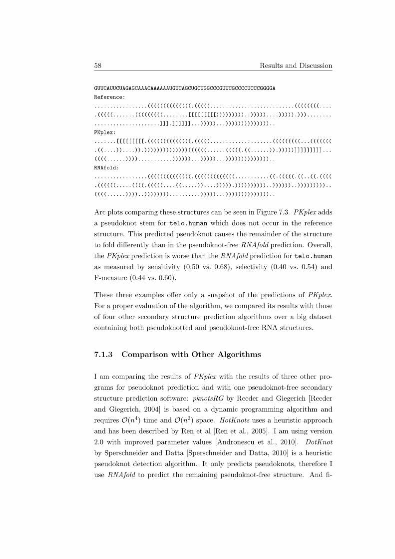
58 Results and Discussion
GUUCAUUCUAGAGCAAACAAAAAAUGUCAGCUGCUGGCCCGUUCGCCCCUCCCGGGGA
Reference:
.................((((((((((((((.(((((...........................((((((((....
.(((((.......(((((((((........[[[[[[[[[)))))))))..)))))....))))).)))........
.....................]]].]]]]]]...)))))...))))))))))))))..
PKplex:
.......[[[[[[[[[.((((((((((((((.(((((....................(((((((((...(((((((
.((....))....)).))))))))))))))((((((......(((((.((......)).)))))]]]]]]]]]...
((((......))))...........))))))...)))))...))))))))))))))..
RNAfold:
.................((((((((((((((.(((((((((((((...........((.(((((.((..((.((((
.((((((.....((((.(((((....((.....))....))))).))))))))))..))))))..)))))))))..
((((......))))..))))))))..........)))))...))))))))))))))..
Arc plots comparing these structures can be seen in Figure 7.3. PKplex addsa pseudoknot stem for telo.human which does not occur in the referencestructure. This predicted pseudoknot causes the remainder of the structureto fold differently than in the pseudoknot-free RNAfold prediction. Overall,the PKplex prediction is worse than the RNAfold prediction for telo.humanas measured by sensitivity (0.50 vs. 0.68), selectivity (0.40 vs. 0.54) andF-measure (0.44 vs. 0.60).
These three examples offer only a snapshot of the predictions of PKplex.For a proper evaluation of the algorithm, we compared its results with thoseof four other secondary structure prediction algorithms over a big datasetcontaining both pseudoknotted and pseudoknot-free RNA structures.
7.1.3 Comparison with Other Algorithms
I am comparing the results of PKplex with the results of three other pro-grams for pseudoknot prediction and with one pseudoknot-free secondarystructure prediction software: pknotsRG by Reeder and Giegerich [Reederand Giegerich, 2004] is based on a dynamic programming algorithm andrequires O(n4) time and O(n2) space. HotKnots uses a heuristic approachand has been described by Ren et al [Ren et al., 2005]. I am using version2.0 with improved parameter values [Andronescu et al., 2010]. DotKnotby Sperschneider and Datta [Sperschneider and Datta, 2010] is a heuristicpseudoknot detection algorithm. It only predicts pseudoknots, therefore Iuse RNAfold to predict the remaining pseudoknot-free structure. And fi-

Results and Discussion 59
Figure 7.3: Arc plots comparing the reference structure for telo.human to the pre-dictions of RNAfold (top) and PKplex (bottom). In both plots base pairs containedin both structures (true positives) are black, base pairs contained only in the ref-erence structure (false negatives) are green, and base pairs contained only in thestructure prediction (false positives) are red. (Images generated with the help ofhxmatch [Witwer et al., 2004])

60 Results and Discussion
nally, the Vienna RNA package’s RNAfold [Hofacker et al., 1994], whichonly predicts pseudoknot-free structures, is included in the analysis as well.
7.1.4 Data Sets
I evaluated the different pseudoknot prediction approaches by applying themto RNA sequences with known secondary structures and comparing the pre-dictions with the reference structures. The full data set consists of 2253sequences, both pseudoknotted and pseudoknot-free, and is the union of theS-Train and S-Test data sets used in [Andronescu et al., 2010]. The sequence-structure pairs were originally taken from the RNA STRAND v2.0 database[Andronescu et al., 2008] and from Pseudobase [van Batenburg et al., 2001].About 80% of the sequences are pseudoknot-free, other properties are shownin Table 7.1. To evaluate the prediction quality on different types of struc-tures the data set was split into four subsets: short pseudoknotted sequences(length <100 nucleotides [nt]), short pseudoknot-free sequences, long pseu-doknotted sequences and long pseudoknot-free sequences.
Data set Description Number of Percentage Average Standardsequences (%) length deviation
Complete All sequences 2253 100.0 74.1 40.8Short PK <100nt, pseudoknotted 327 14.5 46.7 19.6Short PKfree <100nt, pseudoknot-free 1358 60.3 57.4 23.7Long PK ≥100nt, pseudoknotted 98 4.3 148.2 41.4Long PKfree ≥100nt, pseudoknot-free 470 20.9 125.2 23.9
Table 7.1: Properties of the data sets used for testing the pseudoknot predictionalgorithms.
7.1.5 Prediction Accuracy
Table 7.2 shows the results of applying each of the five different secondarystructure prediction algorithms on the test dataset and its subsets. Theparameter values for PKplex were chosen as follows: cpk, the pseudoknotinitialization cost, was set to 8.1 kcal/mol, which results in a good balancebetween the quality and the quantity of the pseudoknots returned by PKplex.The maximum length w of the interacting intervals was set to 12 and themaximum size v of internal and bulge loops was set to 10. Larger values didnot improve the quality of results and only increased the runtime of PKplex.

Results and Discussion 61
Dataset PKplex pknotsRG DotKnot HotKnots RNAfold
Complete Sensitivity 0.748 0.735 0.694 0.791 0.699PPV 0.690 0.696 0.656 0.762 0.684F-measure 0.718 0.715 0.675 0.776 0.691Containing PK (%) 25.5 22.4 52.2 17.1 0Runtime (s) 192 140 2100 33290 20.9
ShPK Sensitivity 0.712 0.779 0.797 0.744 0.501PPV 0.741 0.779 0.801 0.766 0.659F-measure 0.726 0.779 0.799 0.755 0.569Containing PK (%) 55.1 71.9 79.8 64.2 0Runtime (s) 10.8 6.0 186 367 2.1
ShPKfree Sensitivity 0.774 0.743 0.701 0.814 0.752PPV 0.700 0.695 0.651 0.776 0.705F-measure 0.735 0.718 0.675 0.794 0.728Containing PK (%) 12.4 7.2 42.6 7.7 0Runtime (s) 64.7 32.0 796 2354 12.4
LoPK Sensitivity 0.640 0.594 0.606 0.597 0.496PPV 0.601 0.556 0.597 0.599 0.510F-measure 0.620 0.558 0.601 0.598 0.503Containing PK (%) 80.6 60.2 92.9 53.1 0Runtime (s) 28.3 33.7 502 16671 2.1
LoPKfree Sensitivity 0.722 0.719 0.619 0.798 0.727PPV 0.642 0.668 0.582 0.755 0.678F-measure 0.679 0.692 0.600 0.776 0.701Containing PK (%) 31.5 24.0 51.9 4.3 0Runtime (s) 91.6 69.9 677 13983 7.7
Table 7.2: Results of applying the five different prediction algorithms on the datasets.
For the complete dataset the quality of PKplex’ results as judged by theF-measure is about the same as the quality of the results derived withthe only other dynamic programming approach, pknotsRG. Looking at theheuristic approaches, HotKnots returns better results and DotKnot returnsworse results than PKplex. But the computation time is a lot longer for theheuristic approaches than for the dynamic programming approaches. Inter-estingly, even the pseudoknot-free secondary structure prediction algorithmof RNAfold delivers better results than DotKnot and is not far behind theother algorithms.
When analyzing the predictions for the different subsets of sequences it turnsout that RNAfold performs worse than all pseudoknot-prediction algorithmson the data sets that contain pseudoknotted sequences, whereas the oppositeis not the case: the secondary structure prediction algorithms that includepseudoknots do not necessarily perform worse than RNAfold on data setsthat do not contain any pseudoknotted sequences. For ShPKfree, the subsetcontaining short pseudoknot-free sequences, PKplex returns marginally bet-ter results than RNAfold. This means that for the sequences where PKplex

62 Results and Discussion
wrongly predicts a pseudoknot, the quality of these predictions is on averagestill better than the prediction quality of RNAfold for these sequences.
PKplex performs equally well over all four subsets. The F-measure for thedata sets containing longer sequences is lower than that for the shorter se-quences. That is a trend that is true for all methods and a consequence of thefact that secondary structure prediction difficulty increases with sequencelength.
7.1.6 Computational Performance
The O(n3) algorithm of RNAfold takes only about 21 s to fold all 2253sequences of the complete data set (on an Intel Core 2 Quad Q6600 with2.4 GHz and 4 GB RAM). The dynamic programming methods pknotsRGand PKplex take 140 s and 192 s respectively, while both heuristic methodscompare unfavorably in this regard with runtimes of 2100 s for DotKnot and33290 s for HotKnots (Table 7.2).
Figure 7.4 shows the runtime of the different secondary structure predictionalgorithms when applied to 20 random RNA sequences of various lengths.PKplex compares favorably, especially for longer sequences, where it is thefastest of all tested pseudoknot prediction algorithms. For even longer se-quences, such as the 1542 bp 16 s E. coli rRNA, the differences in runtimeare substantial. While PKplex’ runtime is 98 s, DotKnot already requires1404 s and pknotsRG 2811 s.
To summarize this analysis, PKplex is the fastest of the tested pseudoknotprediction algorithms. For shorter sequences the other dynamic program-ming algorithm pknotsRG is a little bit faster than PKplex, but for longersequences, where differences in runtime are a lot more important, PKplexis faster due to its lower time complexity of O(n3 + n2w4) versus O(n4) forpknotsRG. This low time complexity makes PKplex the fastest of all knowndynamic programming pseudoknot prediction algorithms. The heuristic ap-proaches cannot compete with either of the two tested dynamic program-ming algorithms regarding computation time and HotKnots in particular isimpractical for longer RNA sequences.

Results and Discussion 63
Figure 7.4: Comparison of the runtime of the secondary structure prediction algo-rithms for folding 20 random RNA sequences of different lengths.

64 Results and Discussion
The bandwidth of differences in the quality of the results of the tested meth-ods is a lot smaller than the differences in runtime. The F-measure of the re-sults of calculating the secondary structures of all 2253 sequences in the dataset varies between 0.67 and 0.78 for the different methods. HotKnots, whichis the slowest algorithm by far, returns the best results. It is followed by thefaster dynamic programming approaches PKplex and pknotsRG. RNAfoldand DotKnot occupy the lower end of that spectrum. As a consequence I rec-ommend using HotKnots for pseudoknot prediction of short RNA sequences.For longer sequences or bigger datasets where HotKnots becomes impracticaldue to its long runtime, or if one wants to use a dynamic programming ap-proach instead of a heuristic approach, PKplex is the recommended methodfor pseudoknot prediction.
7.2 Discussion
The general idea behind the PKplex algorithm - calculating the cost of ren-dering two sequence intervals accessible and adding the gain of letting thosetwo intervals form base pairs among each other - is very simple, but it alsoimplies some assumptions and restrictions of the algorithm which I want tomention in this section:
To calculate ∆Gu[a, b], the energy necessary to make a sequence interval ac-cessible, PKplex employs the algorithm used in RNAplfold. This algorithmtakes an approach based on the partition function, whereas the other partsof PKplex are based on MFE calculations. During the development processI also tried an MFE based approach to calculate ∆Gu[ab], but I found thatthis did not make a difference on the quality of the results. Because theimplementation based on the partition function is faster, there is no reasonto use an MFE-based approach instead. PKplex therefore returns the en-ergetically most favorable pseudoknot for the ensemble of pseudoknot-freestructures, not the most favorable pseudoknot for the pseudoknot-free MFEstructure.
In the PKplex algorithm, pseudoknots are constructed by forming basepairs between two continuous sequence intervals in addition to the regularpseudoknot-free secondary structure. This results in H-type pseudoknots,

Results and Discussion 65
kissing hairpins and other pseudoknots with only a single helix violatingthe condition of perfectly nested structures. The pseudoknotted structuresreturned by PKplex are therefore all bi-secondary structures. Non-planarpseudoknots or k-partite structures with k > 2 cannot be predicted byPKplex. For the same reason PKplex cannot predict multiple pseudoknotson a single sequence either. Because a high percentage of the known pseu-doknotted sequences are planar and contain only a single pseudoknot, thisis not a severe restriction. It should also be possible to adapt the PKplexalgorithm to run multiple times on the same sequence, enabling it to predictmore complex multiple stem pseudoknots.
Figure 7.5: The runtime requirements and classes of pseudoknots covered by R&E[Rivas and Eddy, 1999], D&P [Dirks and Pierce, 2003], R&G’s pknotsRG [Reederand Giegerich, 2004] and PKplex. (Image adapted from [Hofacker and Stadler,2007])
pknotsRG has other restrictions regarding the pseudoknots it is able to pre-dict: on the one hand, it cannot handle kissing hairpins or bulge or internalloops within pseudoknot helices, but on the other hand it is able to generatestructures with more than one pseudoknot and even recursive pseudoknots.

66 Results and Discussion
Other dynamic programming algorithms such as the one suggested in [Ri-vas and Eddy, 1999] can handle more general pseudoknots, but at the costof a severely higher runtime making these algorithms quite impractical touse. A comparison of the covered pseudoknot classes and runtime require-ments of different dynamic programming algorithms for pseudoknot predic-tion is shown in Figure 7.5. In contrast to dynamic programming algorithms,heuristic algorithms for pseudoknot prediction are not by their very natureforced to reduce the search space, i.e. restrict the types of pseudoknots theycan handle. But this advantage comes at the cost of not being guaranteedto find the optimal solution with minimum energy within their search space.
Figure 7.6: The formation of a pseudoknot (dotted lines) changes three loops inthe left sequence (hairpin loop L1, L2 created by the pseudoknot and the externalloop L3) and four loops in the right sequence (hairpin loops L1 and L2, L3 createdby the pseudoknot and the external loop L4).
Whenever two unpaired sequence intervals form a pseudoknot, at least threesecondary structure elements are directly involved (see Figure 7.6): the twoloops enclosing the pseudoknot stems and the new loop created by the pseu-doknot formation. The pseudoknot energy model of PKplex relies on theadditivity of secondary structure elements and therefore assumes that theenergies of those loops are not changed by the formation of the pseudo-knot. As a compensation the pseudoknot initialization cost cpk is intro-duced, which is a constant and does not depend on the actual compositionof the loops changed by the pseudoknot.
In an effort to increase the prediction quality of PKplex, I analyzed the

Results and Discussion 67
loops containing the bases involved in the pseudoknot formation. I foundthat PKplex significantly overestimates the amount of pseudoknot stemsforming within multiloops. This lead to the creation of an energy model inwhich the pseudoknot initiation cost cpk depends on the loop type enclosingthe pseudoknot stems, with an increased penalty for multiloops. But whilethis reduced the amount of pseudoknot stems within multiloops in the pre-dictions of PKplex, the overall prediction accuracy did not increase and Ireturned to the original simpler energy model.
The pseudoknot energy models of pknotsRG, HotKnots and DotKnot aredescribed in section 5.4. While not as simple as the single constant usedin PKplex, these models are not very complex either and mostly resemblethe linear multiloop valuation in the standard RNA models: in addition tothe pseudoknot initiation constant, they penalize longer loops by adding anenergy penalty for every unpaired base and/or for every base pair within thepseudoknot loop. These models are not necessarily better than the simpleconstant used in PKplex, and in fact all these energy models can be consid-ered makeshift and in need of improvement, because they do not properlyaccount for the pseudoknot loop entropies. The problem is not that a morecomplex pseudoknot loop energy model would increase the computationalcosts too much, the heuristic algorithms at least could easily handle such amodel. The problem rather is that the pseudoknot loop entropies are largelyunknown and no proper model for them exists, apart from the one by Caoand Chen described in section 4.3 [Cao and Chen, 2009], but unfortunatelytheir model is only applicable to a subset of the pseudoknots predicted byeach of the four algorithms.
An advantage of the PKplex algorithm, which is caused by the design ofthe dynamic programming routine to calculate the energy of an interactionbetween two intervals, is the possibility to calculate suboptimal pseudoknot-ted secondary structures at no additional cost. These structures can thenbe further analyzed by either manual inspection or some automated filteringto potentially further improve the quality of the results.
The pseudoknot energy model by Cao and Chen was used as such a filterfor improving the quality of the prediction results. The suboptimal struc-tures predicted by PKplex were evaluated with the Cao and Chen energy

68 Results and Discussion
model and the prediction with the best energy according to this evaluationwas picked. But because the Cao and Chen energy model can only han-dle H-type pseudoknots, a lot of the candidate suboptimal structures couldnot be evaluated with it. This resulted in complicated rules for picking thebest structure out of the suboptimal candidates depending on whether theCao and Chen model could handle all, some, one or none of the candidatestructures. Overall, this strategy did not improve the quality of the resultssignificantly, most likely due to the problems with the limited applicabilityof the energy model. Other modifications that failed to improve the predic-tion quality include disallowing bulge and interior loops for the pseudoknotstems or restricting the loop lengths of the loops involved in the pseudoknotformation.

Conclusion and Outlook 69
Chapter 8
Conclusion and Outlook
RNAs are a very important class of molecules for the living world. Theunofficial creation of the research area now known as molecular biology wasthe discovery of the structure of DNA, the carrier of genetic information in1953. For the first couple of decades following this discovery, RNA was onlyseen as occupying the less interesting role of passively carrying informationfrom DNA to proteins. Almost all research was focused on the latter twomolecule classes and RNAs were neglected. Only after the discovery thatthere existed RNAs which could exhibit catalytic activity themselves, RNAgained the attention of the scientific community that it deserves.
More and more functional RNAs are still being discovered today. The func-tion of an RNA molecule strongly depends on its structure, and while a greatnumber of RNA sequences are known and easily available in various sequencedata bases, determining RNA structures requires a lot of experimental work.Therefore, predicting the structure of an RNA from its sequence has the po-tential to accelerate the scientific progress and save a lot of experimentalwork as well as money. Theoretically predicting the structure of RNA se-quences cannot replace classic wet lab work, but it can filter the huge amountof potential functional RNAs to the most promising candidates, which canthen be analyzed experimentally.
Most RNA secondary structure prediction algorithms are based on a dy-namic programming approach, breaking apart the structure prediction prob-

70 Conclusion and Outlook
lem into a number of smaller and simpler subproblems which can eventuallybe solved easily, and then constructing the solution to the bigger problemout of the solutions to the smaller problems. This approach depends onthe loop decomposition of RNA structures requiring RNA structures to beperfectly nested. No bases enclosed by a base pair are allowed to bind withbases outside of the enclosing base pair. This restriction is only true forpseudoknot-free secondary structures, but more and more experimentallydetermined RNA structures contain pseudoknots, which violate this restric-tion.
In the effort to include pseudoknots into structure prediction algorithms ithas been found that predicting pseudoknotted RNA structures without anyrestrictions is an NP-complete problem. Only after simplifying the energymodel or by restricting the allowed classes of pseudoknots are current dy-namic programming algorithms able to predict RNA pseudoknots. Someheuristic algorithms have also been employed for the prediction of pseudo-knots, but in contrast to dynamic programming algorithms they are notguaranteed to find the optimum solution.
In this diploma thesis I introduced PKplex, a dynamic programming basedapproach within the MFE framework to predict RNA secondary structureswith pseudoknots. The simple idea is to render two sequence intervals un-paired and accessible and let them interact with each other while the restof the sequence is folded by a conventional pseudoknot-free folding algo-rithm. PKplex achieves a time complexity of O(n3 + n2w4) and a spacecomplexity of O(nw2). Testing this approach on a large set of pseudoknot-ted and pseudoknot-free sequences shows that PKplex generates results thatare qualitatively as good as those of other pseudoknot prediction algorithmswhile being computationally less expensive. The PKplex algorithm has beenimplemented in C as an extension to the Vienna RNA package.
Especially for longer pseudoknotted sequences the prediction quality of allavailable algorithms is not satisfying yet. The consensus is that this is lessdue to the computational effort required to handle longer sequences, butdue to the fact that there is no general RNA energy model available yetwhich takes pseudoknots into account. Pseudoknots contain loops that areno hairpin-, interior-, bulge- or multiloop and that do not allow a simple

Conclusion and Outlook 71
loop decomposition like in the case of pseudoknot-free structures. Cao andChen have created an energy model for H-type pseudoknots with energiesfor the pseudoknotted loops occurring in these structures but the energiesof more general pseudoknotted loops are still unknown. An energy modelwhich is able to handle a wider range of pseudoknots would potentially allowthe creation of new algorithms for the prediction of RNA pseudoknots withgreater accuracy.
Availability
The source code of PKplex and the Vienna RNA package are available onthe web server of the Institute for Theoretical Chemistry of the Universityof Vienna at http://www.tbi.univie.ac.at/~wolfgang/PKplex/ and athttp://www.tbi.univie.ac.at/~ivo/RNA/.

72 Conclusion and Outlook

Bibliography 73
Bibliography
P.L. Adams, M.R. Stahley, M.L. Gill, A.B. Kosek, J. Wang, and S.A. Stro-bel. Crystal structure of a group I intron splicing intermediate. RNA, 10(12):1867, 2004.
T. Akutsu. Dynamic programming algorithms for RNA secondary structureprediction with pseudoknots. Discrete Applied Mathematics, 104:45–62,2000.
E.S. Andersen, M.A. Rosenblad, N. Larsen, J.C. Westergaard, J. Burks,I.K. Wower, J. Wower, J. Gorodkin, T. Samuelsson, and C. Zwieb. ThermRDB and SRPDB resources. Nucleic Acids Research, 34:163–168, 2006.
M. Andronescu, V. Bereg, H.H. Hoos, and A. Condon. RNA STRAND:the RNA secondary structure and statistical analysis database. BMCBioinformatics, 9(1):340, 2008.
M.S. Andronescu, C. Pop, and A.E. Condon. Improved free energy parame-ters for RNA pseudoknotted secondary structure prediction. RNA, 16(1):26, 2010.
J.P. Bachellerie, J. Cavaille, and A. Huttenhofer. The expanding snoRNAworld. Biochimie, 84(8):775–790, 2002.
I. Barette, G. Poisson, P. Gendron, and F. Major. Pseudoknots in prionprotein mRNAs confirmed by comparative sequence analysis and patternsearching. Nucleic Acids Research, 29(3):753–758, 2001.
S. Bernhart. Personal communication, 2009.
S.H. Bernhart, I.L. Hofacker, and P.F. Stadler. Local RNA base pairingprobabilities in large sequences. Bioinformatics, 22(5):614, 2006.

74 Bibliography
E. Birney, J. Stamatoyannopoulos, A. Dutta, and R. Guigo. Identificationand analysis of functional elements in 1% of the human genome by theENCODE pilot project. Nature, 447(7146):799–816, 2007.
A.F. Bompfunewerer, R. Backofen, S.H. Bernhart, J. Hertel, I.L. Hofacker,P.F. Stadler, and S. Will. Variations on RNA folding and alignment:lessons from Benasque. Journal of Mathematical Biology, 56(1):129–144,2008.
I. Brierley, S. Pennell, and R.J.C. Gilbert. Viral RNA pseudoknots: versatilemotifs in gene expression and replication. Nature Reviews Microbiology,5(8):598–610, 2007.
I. Brierley, R. Gilbert, and S. Pennell. RNA pseudoknots and the regulationof protein synthesis. Biochemical Society Transactions, 36:684–689, 2008.
P. Brion and E. Westhof. Hierarchy and dynamics of RNA folding. Annualreview of biophysics and biomolecular structure, 26(1):113–137, 1997.
J.W. Brown. The ribonuclease P database. Nucleic Acids Research, 24(1):236, 1996.
Y. Byun and K. Han. PseudoViewer3: generating planar drawings of large-scale RNA structures with pseudoknots. Bioinformatics, 25(11):1435,2009.
L. Cai, R.L. Malmberg, and Y. Wu. Stochastic modeling of RNA pseudo-knotted structures: a grammatical approach. Bioinformatics, 19(Suppl1):i66–i73, 2003.
J.J. Cannone, S. Subramanian, M.N. Schnare, J.R. Collett, L.M. D’Souza,Y. Du, B. Feng, N. Lin, L.V. Madabusi, K.M. Muller, et al. The Compar-ative RNA Web(CRW) Site: an online database of comparative sequenceand structure information for ribosomal, intron, and other RNAs. BMCBioinformatics, 3(1):2, 2002.
S. Cao and S.J. Chen. Predicting RNA pseudoknot folding thermodynamics.Nucleic Acids Research, 34(9):2634, 2006.
S. Cao and S.J. Chen. Predicting structures and stabilities for H-type pseu-doknots with interhelix loops. RNA, 15(4):696, 2009.

Bibliography 75
T. Cech. Conserved sequences and structures of group I introns: buildingan active site for RNA catalysis - A review. Gene, 73:259–271, 1988.
T.R. Cech, A.J. Zaug, and P.J. Grabowski. In vitro splicing of the ribosomalRNA precursor of Tetrahymena: involvement of a guanosine nucleotide inthe excision of the intervening sequence. Cell, 27(3):487–496, 1981.
J.L. Chen, M.A. Blasco, and C.W. Greider. Secondary structure of verte-brate telomerase RNA. Cell, 100(5):503–514, 2000.
F. Crick. Central dogma of molecular biology. Nature, 227(5258):561–563,1970.
F.H.C. Crick. The biological replication of macromolecules. In Symp. Soc.Exp. Biol, 1958.
R.M. Dirks and N.A. Pierce. A partition function algorithm for nucleic acidsecondary structure including pseudoknots. Journal of ComputationalChemistry, 24(13):1664–1677, 2003.
S.M. Elbashir, J. Harborth, W. Lendeckel, A. Yalcin, K. Weber, andT. Tuschl. Duplexes of 21-nucleotide RNAs mediate RNA interferencein cultured mammalian cells. Nature, 411(6836):494–498, 2001.
A. Fire, S.Q. Xu, M.K. Montgomery, S.A. Kostas, S.E. Driver, and C.C.Mello. Potent and specific genetic interference by double-stranded RNAin Caenorhabditis elegans. Nature, 391(6669):806–811, 1998.
C. Flamm. Kinetic Folding of RNA. PhD thesis, Universitat Wien, 1998.
D.P. Giedroc and P.V. Cornish. Frameshifting RNA pseudoknots: structureand mechanism. Virus research, 139(2):193–208, 2009.
W. Gilbert. Origin of life: the RNA world. Nature, 319(6055), 1986.
S. Griffiths-Jones, S. Moxon, M. Marshall, A. Khanna, S.R. Eddy, andA. Bateman. Rfam: annotating non-coding RNAs in complete genomes.Nucleic Acids Research, 33(Database Issue):D121, 2005.
A. Gruber. Strategies for measuring evolutionary conservation of RNA sec-ondary structures. Master’s thesis, Universitat Wien, 2007.

76 Bibliography
A.R. Gruber, R. Lorenz, S.H. Bernhart, R. Neubock, and I.L. Hofacker. TheVienna RNA websuite. Nucleic Acids Research, 36(Web Server issue):W70, 2008.
C. Guerrier-Takada, K. Gardiner, T. Marsh, N. Pace, and S. Altman. TheRNA moiety of ribonuclease P is the catalytic subunit of the enzyme.Cell, 35(3):849–857, 1983.
A.P. Gultyaev, F.H.D. van Batenburg, and C.W.A. Pleij. The computersimulation of RNA folding pathways using a genetic algorithm. Journalof Molecular Biology, 250(1):37–51, 1995.
A.P. Gultyaev, F.H. van Batenburg, and C.W. Pleij. An approximation ofloop free energy values of RNA H-pseudoknots. RNA, 5(5):609, 1999.
A.J. Hamilton and D.C. Baulcombe. A species of small antisense RNA inposttranscriptional gene silencing in plants. Science, 286(5441):950, 1999.
C. Haslinger and P.F. Stadler. RNA structures with pseudo-knots: Graph-theoretical, combinatorial, and statistical properties. Bulletin of Mathe-matical Biology, 61(3):437–467, 1999.
I.L. Hofacker and P.F. Stadler. RNA secondary structures. In T. Lengauer,editor, Bioinformatics: From Genomes to Therapies, pages 438–489.Wiley-VCH, 2007.
I.L. Hofacker, W. Fontana, P.F. Stadler, L.S. Bonhoeffer, M. Tacker, andP. Schuster. Fast folding and comparison of RNA secondary structures.Monatshefte fur Chemie/Chemical Monthly, 125(2):167–188, 1994.
J.M. Johnson, S. Edwards, D. Shoemaker, and E.E. Schadt. Dark matter inthe genome: evidence of widespread transcription detected by microarraytiling experiments. TRENDS in Genetics, 21(2):93–102, 2005.
G.F. Joyce. RNA evolution and the origins of life. Nature, 338(6212):217–224, 1989.
G.F. Joyce. The rise and fall of the RNA world. The New Biologist, 3(4):399, 1991.

Bibliography 77
K.C. Keiler, P.R.H. Waller, and R.T. Sauer. Role of a peptide taggingsystem in degradation of proteins synthesized from damaged messengerRNA. Science, 271(5251):990, 1996.
R.C. Lee, R.L. Feinbaum, and V. Ambros. The C. elegans heterochronicgene lin-4 encodes small RNAs with antisense complementarity to lin-14.Cell, 75(5):843–854, 1993.
R. Lorenz. Secondary structure prediction for circular RNAs. Master’sthesis, Universitat Leipzig, 2007.
R.B. Lyngsø and C.N.S. Pedersen. RNA pseudoknot prediction in energy-based models. Journal of Computational Biology, 7(3-4):409–427, 2000.
R.B. Lyngsø, M. Zuker, and C.N. Pedersen. Fast evaluation of internal loopsin RNA secondary structure prediction. Bioinformatics, 15(6):440, 1999.
H. Mann, Y. Ben-Asouli, A. Schein, S. Moussa, and N. Jarrous. EukaryoticRNase P:: Role of RNA and Protein Subunits of a Primordial CatalyticRibonucleoprotein in RNA-Based Catalysis. Molecular cell, 12(4):925–935, 2003.
R.A. Marciniak, F.B. Johnson, and L. Guarente. Dyskeratosis congenita,telomeres and human ageing. Trends in Genetics, 16(5):193–195, 2000.
D.H. Mathews, J. Sabina, M. Zuker, and D.H. Turner. Expanded sequencedependence of thermodynamic parameters improves prediction of RNAsecondary structure. Journal of Molecular Biology, 288(5):911–940, 1999.
J.S. Mattick. Challenging the dogma: the hidden layer of non-protein-codingRNAs in complex organisms. Bioessays, 25(10):930–939, 2003.
J.S. McCaskill. The equilibrium partition function and base pair bindingprobabilities for RNA secondary structure. Biopolymers, 29(6-7):1105–1119, 1990.
U. Muckstein, H. Tafer, J. Hackermuller, S.H. Bernhart, P.F. Stadler, andI.L. Hofacker. Thermodynamics of RNA-RNA binding. Bioinformatics,22(10):1177, 2006.
78 Bibliography
U. Muckstein, H. Tafer, S.H. Bernhart, M. Hernandez-Rosales, J. Vogel,P.F. Stadler, and I.L. Hofacker. Translational control by RNA-RNA in-teraction: Improved computation of RNA-RNA binding thermodynamics.Bioinformatics Research and Development, pages 114–127, 2008.
N. Nameki, T. Tadaki, H. Himeno, and A. Muto. Three of four pseudoknotsin tmRNA are interchangeable and are substitutable with single-strandedRNAs. FEBS letters, 470(3):345–349, 2000.
R. Nussinov and A.B. Jacobson. Fast algorithm for predicting the secondarystructure of single-stranded RNA. Proceedings of the National Academyof Sciences of the United States of America, 77(11):6309, 1980.
J. Reeder and R. Giegerich. Design, implementation and evaluation of apractical pseudoknot folding algorithm based on thermodynamics. BMCBioinformatics, 5(1):104, 2004.
J. Ren, B. Rastegari, A. Condon, and H.H. Hoos. HotKnots: heuristicprediction of RNA secondary structures including pseudoknots. RNA, 11(10):1494, 2005.
E. Rivas and S.R. Eddy. A dynamic programming algorithm for RNA struc-ture prediction including pseudoknots. Journal of Molecular Biology, 285(5):2053–2068, 1999.
J. Ruan, G.D. Stormo, and W. Zhang. An iterated loop matching approachto the prediction of RNA secondary structures with pseudoknots. Bioin-formatics, 20(1):58, 2004.
P.J. Schlax, K.A. Xavier, T.C. Gluick, and D.E. Draper. TranslationalRepression of the Escherichia coli α Operon mRNA. Journal of BiologicalChemistry, 276(42):38494, 2001.
J. Sperschneider and A. Datta. DotKnot: pseudoknot prediction using theprobability dot plot under a refined energy model. Nucleic Acids Research,38(7):e103, 2010.
D.W. Staple and S.E. Butcher. Pseudoknots: RNA structures with diversefunctions. PLoS biology, 3(6), 2005.

Bibliography 79
L.D. Stein. Human genome: end of the beginning. Nature, 431(7011):915–916, 2004.
J.E. Tabaska, R.B. Cary, H.N. Gabow, and G.D. Stormo. An RNA foldingmethod capable of identifying pseudoknots and base triples. Bioinformat-ics, 14(8):691, 1998.
H. Tafer and I.L. Hofacker. RNAplex: a fast tool for RNA-RNA interactionsearch. Bioinformatics, 24(22):2657, 2008.
C.A. Theimer and D.P. Giedroc. Contribution of the intercalated adeno-sine at the helical junction to the stability of the gag-pro frameshiftingpseudoknot from mouse mammary tumor virus. RNA, 6(3):409, 2000.
I. Tinoco et al. How RNA folds. Journal of Molecular Biology, 293(2):271–281, 1999.
C. Tuerk, S. MacDougal, and L. Gold. RNA pseudoknots that inhibit humanimmunodeficiency virus type 1 reverse transcriptase. Proceedings of theNational Academy of Sciences of the United States of America, 89(15):6988, 1992.
D.H. Turner and D.H. Mathews. NNDB: the nearest neighbor parameterdatabase for predicting stability of nucleic acid secondary structure. Nu-cleic Acids Research, 2009.
T. Tuschl, C. Gohlke, T.M. Jovin, E. Westhof, and F. Eckstein. A three-dimensional model for the hammerhead ribozyme based on fluorescencemeasurements. Science, 266(5186):785, 1994.
S. Valadkhan. snRNAs as the catalysts of pre-mRNA splicing. CurrentOpinion in Chemical Biology, 9(6):603–608, 2005.
F.H.D. van Batenburg, A.P. Gultyaev, and C.W.A. Pleij. PseudoBase:structural information on RNA pseudoknots. Nucleic Acids Research,29(1):194, 2001.
T. Vulliamy, A. Marrone, I. Dokal, and P.J. Mason. Association betweenaplastic anaemia and mutations in telomerase RNA. The Lancet, 359(9324):2168–2170, 2002.

80 Bibliography
M.S. Waterman. Secondary structure of single-stranded nucleic acids. Adv.Math. Suppl. Studies, 1:167–212, 1978.
M.S. Waterman and T.F. Smith. RNA secondary structure: A completemathematical analysis. Mathematical Biosciences, 42(3-4):257–266, 1978.
J. D. Watson and F. H. Crick. Molecular structure of nucleic acids; a struc-ture for deoxyribose nucleic acid. Nature, 171:737–738, Apr 1953.
K.C. Wiese, E. Glen, and A. Vasudevan. JViz.Rna - A Java tool for RNAsecondary structure visualization. IEEE Transactions on Nanobioscience,4(3), 2005.
C. Witwer, I.L. Hofacker, and P.F. Stadler. Prediction of consensus RNAsecondary structures including pseudoknots. IEEE/ACM Transactions onComputational Biology and Bioinformatics, 1(2):66–77, 2004.
A. Xayaphoummine, T. Bucher, F. Thalmann, and H. Isambert. Predictionand statistics of pseudoknots in RNA structures using exactly clusteredstochastic simulations. Proceedings of the National Academy of Sciencesof the United States of America, 100(26):15310, 2003.
M. Zuker. Calculating nucleic acid secondary structure. Current Opinionin Structural Biology, 10(3):303–310, 2000.
M. Zuker and D. Sankoff. RNA secondary structures and their prediction.Bulletin of Mathematical Biology, 46(4):591–621, 1984.
M. Zuker and P. Stiegler. Optimal computer folding of large RNA sequencesusing thermodynamics and auxiliary information. Nucleic Acids Research,9(1):133, 1981.

Mag. Wolfgang Beyer
Personal
Information Address:
Phone:
Email:
Date of Birth:
Nationality:
Hasnerstraße 105/26, A-1160 Vienna, Austria
+43 (0) 650-5858999
Feb 10th, 1980 in Vienna, Austria
Austrian
Education Since 2003 Spring 2007 1999 - 2004 Spring 2002 1998 – 1999 1990 – 1998
University of Vienna Molecular Biology specializing in Bioinformatics, Structural Biology and Genetics Universidade Nova de Lisboa (Portugal), Exchange Semester Research internship topic: Modeling Metabolic Pathways Vienna University of Economics and Business Administration Degree in Business Administration, graduation with distinction Diploma thesis: “The Quadrature Method in Comparison with Other Methods for Valuing Barrier Options” Graduation average: 1.5 Majors: Finance, Controlling University of Technology Sydney (Australia) Exchange Semester, Bachelor of Business course Coursework includes: International Financial Management, Derivative Securities Military Service Secondary School, Gymnasium und Realgymnasium Wien 8 Graduation average: 1.0
Experience Oct 2004 - Feb 2005 Oct 2002 – Dec 2002
Altea Trading Company LLC Proprietary Futures Trader Merrill Lynch (Frankfurt) Internship at the European Equity Sales department
Language &
Computer
Skills
First language German, fluent English, basic French and Portuguese C, C++, HTML, Latex, Visual Basic, Bloomberg
Awards &
Activities
Academic Scholarship for 2001/2002 Member of the Austrian team at the International Mathematical Olympiad 1998 in Taipeh, Taiwan Youth Work (Scout leader since 1998)
Vienna, October 22nd, 2010