directions for use/package insertglaukos® corporation istent inject ® trabecular micro-bypass...
TRANSCRIPT

GLAUKOS® CORPORATION ISTENT inject ® TRABECULAR MICRO-BYPASS SYSTEM PMA P170043
DIRECTIONS FOR USE/PACKAGE INSERT Glaukos Corporation iStent inject Trabecular Micro-Bypass System
DIRECTIONS FOR USE TABLE OF CONTENTS
1. DEVICE DESCRIPTION 10. STORAGE REQUIREMENTS 2. INDICATIONS FOR USE 11. EXPIRATION DATE 3. CONTRAINDICATIONS 12. RETURN GOODS POLICY 4. WARNINGS 13. CLINICAL TRIAL RESULTS 5. PRECAUTIONS 14. POST-APPROVAL STUDY RESULTS 6. ADVERSE REACTIONS 15. LABELING 7. INSTRUCTIONS FOR USE 16. MRI SAFETY INFORMATION 8. ADVERSE EVENT REPORTING 17. CAUTION 9. HOW SUPPLIED
1. DEVICE DESCRIPTION
The iStent inject Trabecular Micro-Bypass System Model G2-M-IS contains two preloaded intraocular stents that are manufactured from titanium (Ti6Al4V ELI) and are coated with stearalkonium heparin (note: the heparin is from a porcine source). The stent has a single piece design, is 230 µm in diameter, 360 µm in height, and the central inlet and outlet lumen has a diameter of 80 µm (Figure 1). The head of the stent has four side outlets that each have a diameter of 50 µm.
Figure 1. iStent inject Stent Dimensions
360 µm
230 µm dia.
Head – resides in Schlemm’s Canal
Thorax – resides in the Trabecular Meshwork)
Flange – resides in the Anterior Chamber
GLAUKOS® CORPORATION ISTENT inject ® TRABECULAR MICRO-BYPASS SYSTEM PMA P170043
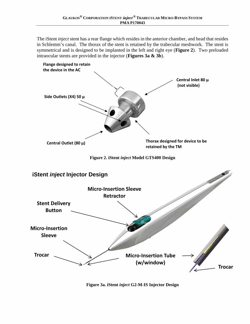
The iStent inject stent has a rear flange which resides in the anterior chamber, and head that resides in Schlemm’s canal. The thorax of the stent is retained by the trabecular meshwork. The stent is symmetrical and is designed to be implanted in the left and right eye (Figure 2). Two preloaded intraocular stents are provided in the injector (Figures 3a & 3b).
Figure 2. iStent inject Model GTS400 Design
Figure 3a. iStent inject G2-M-IS Injector Design
Side Outlets (X4) 50 µ
Central Outlet (80 µ) Thorax designed for device to be retained by the TM
Flange designed to retain the device in the AC
Central Inlet 80 µ (not visible)
Micro-Insertion Tube (w/window)
Micro-Insertion Sleeve
Micro-Insertion Sleeve Retractor
Stent Delivery Button
Trocar
iStent inject Injector Design
Trocar
GLAUKOS® CORPORATION ISTENT inject ® TRABECULAR MICRO-BYPASS SYSTEM PMA P170043
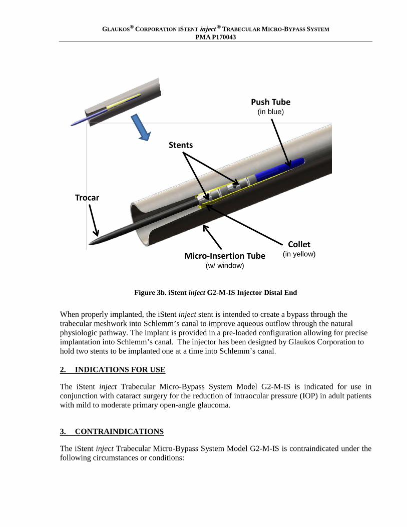
Figure 3b. iStent inject G2-M-IS Injector Distal End
When properly implanted, the iStent inject stent is intended to create a bypass through the trabecular meshwork into Schlemm’s canal to improve aqueous outflow through the natural physiologic pathway. The implant is provided in a pre-loaded configuration allowing for precise implantation into Schlemm’s canal. The injector has been designed by Glaukos Corporation to hold two stents to be implanted one at a time into Schlemm’s canal. 2. INDICATIONS FOR USE
The iStent inject Trabecular Micro-Bypass System Model G2-M-IS is indicated for use in conjunction with cataract surgery for the reduction of intraocular pressure (IOP) in adult patients with mild to moderate primary open-angle glaucoma.
3. CONTRAINDICATIONS
The iStent inject Trabecular Micro-Bypass System Model G2-M-IS is contraindicated under the following circumstances or conditions:
Push Tube (in blue)
Stents
Trocar
Collet (in yellow) Micro-Insertion Tube
(w/ window)
GLAUKOS® CORPORATION ISTENT inject ® TRABECULAR MICRO-BYPASS SYSTEM PMA P170043
• In eyes with angle closure glaucoma. • In eyes with traumatic, malignant, uveitic, or neovascular glaucoma or discernible congenital
anomalies of the anterior chamber (AC) angle • In patients with retrobulbar tumor, thyroid eye disease, Sturge-Weber Syndrome or any other
type of condition that may cause elevated episcleral venous pressure
4. WARNINGS
1. The following conditions may prohibit sufficient visualization of the angle required for safe and successful stent implantation: corneal haze, corneal opacity, or any other conditions that may inhibit the gonioscopic view in the intended implant location.
2. The surgeon should perform a slit lamp gonioscopy examination prior to taking a patient to surgery to exclude congenital anomalies of the angle, including peripheral anterior synechiae (PAS), rubeosis, and any other angle abnormalities that could lead to improper placement of the stent and pose a hazard.
3. Patients with peripheral iridotomies are at risk of stent dislocation to the posterior chamber and related sequelae.
4. The iStent inject is intended for implantation in conjunction with cataract surgery, which may impact corneal health. Therefore, caution is indicated in eyes with evidence of corneal compromise (e.g., corneal guttae or low endothelial cell density) or with risk factors for corneal compromise following cataract surgery (e.g., advanced age, severe nuclear sclerosis).
5. Non-clinical testing has demonstrated that the iStent inject is MR Conditional. Please see the “MRI SAFETY INFORMATION” section at the end of this document on conditions for safe scanning.
5. PRECAUTIONS
1. The surgeon should inform the patient that the stent is MR Conditional (as noted on their Patient ID card), and if the patient needs to undergo an MRI, they should let their doctor know they have an iStent inject stent implanted in their eye.
2. After the surgery, the surgeon should give the patient the Patient ID card (enclosed in the iStent inject packaging) with the appropriate information filled in, and should advise the patient to keep the card in a safe place, e.g., his or her wallet, for future reference. The surgeon should advise the patient that this Patient ID card contains important information related to the iStent inject and that the card should be shown to their current and future health care providers.
3. The surgeon should monitor the patient postoperatively for proper maintenance of intraocular pressure. If intraocular pressure is not adequately maintained after surgery, the surgeon should consider an appropriate additional therapy to reduce intraocular pressure.
4. The safety and effectiveness of the iStent inject system has not been established as an alternative to the primary treatment of glaucoma with medications. The effectiveness of this

GLAUKOS® CORPORATION ISTENT inject ® TRABECULAR MICRO-BYPASS SYSTEM PMA P170043
device has been demonstrated only in patients with mild to moderate open-angle glaucoma who are undergoing concurrent cataract surgery for visually significant cataract.
5. The safety and effectiveness of the iStent inject system has not been established in patients with the following circumstances or conditions which were not studied in the pivotal trial:
• In children • In eyes with significant prior trauma • In eyes with abnormal anterior segment • In eyes with chronic inflammation • In glaucoma associated with vascular disorders • In pseudophakic patients with glaucoma • In uveitic glaucoma • In eyes with prior incisional glaucoma surgery or cilioablative procedures • In eyes with prior laser trabeculoplasty (LT) with selective LT within 90 days prior to
screening or prior argon laser trabeculoplasty (ALT) at any time • In patients with medicated intraocular pressure greater than 24 mmHg • In patients with unmedicated IOP less than 21 mmHg nor greater than 36 mmHg after
“washout” of medications • For implantation of more or less than two stents • After complications during cataract surgery, including but not limited to, severe corneal
burn, vitreous removal/vitrectomy required, corneal injuries, or complications requiring the placement of an anterior chamber IOL
• When implantation has been without concomitant cataract surgery with IOL implantation for visually significant cataract
• In patients with pseudoexfoliative glaucoma or pigmentary glaucoma, or in patients with other secondary open-angle glaucomas.
6. The stent is comprised of implant grade titanium (Ti6-Al-4V-ELI) with a stearalkonium
heparin coating. The total amount of heparin is estimated to be less than 0.9 microgram per stent, or approximately 0.01 to 0.02 units.
6. ADVERSE REACTIONS
Refer to the Pivotal Clinical Trial Results section for the adverse events that occurred in the pivotal clinical trial. Additional adverse events that may be reasonably associated with the use of the device include but are not limited to the following: anterior chamber shallowing, severe, prolonged, or persistent intraocular inflammation, aqueous misdirection, choroidal effusion, choroidal hemorrhage, corneal decompensation, corneal injury, corneal opacification, cyclodialysis cleft, damage to trabecular meshwork, hyphema, hypopyon, hypotony, hypotony maculopathy, IOL dislocation, iridodialysis, loss of vitreous, perforation of sclera, posterior capsular bag rupture, proliferative vitreoretinopathy, pupillary block, pupillary membrane formation, retinal detachment, retinal dialysis, retinal flap tears, secondary surgical intervention, including but not limited to glaucoma surgery, premature stent release, stent dislocation, stent not

GLAUKOS® CORPORATION ISTENT inject ® TRABECULAR MICRO-BYPASS SYSTEM PMA P170043
retrievable, stent not visible with gonioscopy, over implanted stents that are not visible with gonioscopy, stent malfunction, and vitreous hemorrhage.

GLAUKOS® CORPORATION ISTENT inject ® TRABECULAR MICRO-BYPASS SYSTEM PMA P170043
7. INSTRUCTIONS FOR USE
Cataract Surgery 1. Cataract surgery with IOL implantation should be performed first followed by implantation of
the iStent inject. 2. The stent implantations are designed for nasal placement; therefore, it is suggested that surgery
is performed from the temporal side of the head. 3. An intracameral miotic can be injected to deepen the angle after cataract surgery prior to
placement of the iStent inject stent. 4. To mitigate difficulty with patient movement or non-compliance, consider using a peri-bulbar
or retro-bulbar block. Stent Implantation a. Prepare for gonioscopy by turning the patient head away by approximately 35° and the scope
toward surgeon by approximately 35° (70° total). b. Inspect angle with a gonioprism to ensure that a good view is available at the nasal implant
location. c. Place the gonioprism on the cornea and position the patient and surgical microscope as needed
to visualize the trabecular meshwork, through the gonioprism, on the nasal side of the eye. Focus on the landmarks in the angle of the eye (Figures 4a & 4b). Look up from the iris root to find the scleral spur (white line). Then look for Schwalbe’s line (white line) down from the cornea. The trabecular meshwork (typically a red/brown line) is between the scleral spur and Schwalbe’s line. Schlemm’s canal is behind the trabecular meshwork.

GLAUKOS® CORPORATION ISTENT inject ® TRABECULAR MICRO-BYPASS SYSTEM PMA P170043
Figure 4a. iStent inject Implant Site
Figure 4b. iStent inject Implant Site
d. After visualization of the trabecular meshwork, the Tyvek® tray lid containing the iStent inject system should be opened and presented to the user. The device should be handled in the sterile

GLAUKOS® CORPORATION ISTENT inject ® TRABECULAR MICRO-BYPASS SYSTEM PMA P170043
field. Caution: Do not use the device if the Tyvek lid has been opened or if the packaging appears damaged. In such cases, the sterility of the device may be compromised.
e. Hold the injector as shown in Figure 5 with your index finger comfortably on the micro insertion sleeve retractor and within reach of the stent delivery button.
Figure 5. Hand position on injector
f. Injection of two stents: a. Inject cohesive viscoelastic into the anterior chamber to assist with chamber maintenance. b. Remove the Tube Protector prior to entering the eye. c. Place injector through the same temporal corneal incision used to perform cataract surgery.
Guide the injector across the anterior chamber, just beyond the pupillary margin, and then slide back the micro-insertion sleeve retractor (teal colored) to expose the micro insertion tube and trocar.
GLAUKOS® CORPORATION ISTENT inject ® TRABECULAR MICRO-BYPASS SYSTEM PMA P170043
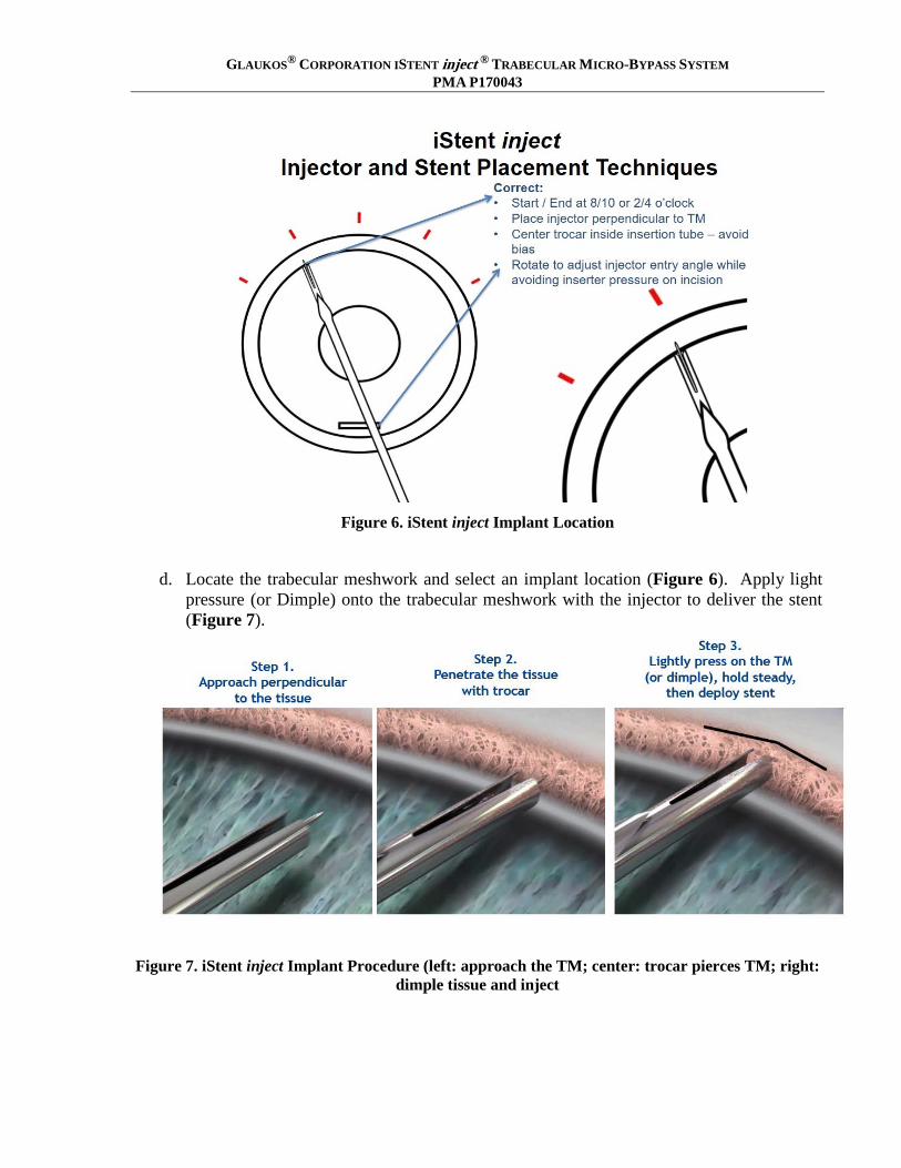
Figure 6. iStent inject Implant Location
d. Locate the trabecular meshwork and select an implant location (Figure 6). Apply light pressure (or Dimple) onto the trabecular meshwork with the injector to deliver the stent (Figure 7).
Figure 7. iStent inject Implant Procedure (left: approach the TM; center: trocar pierces TM; right: dimple tissue and inject

GLAUKOS® CORPORATION ISTENT inject ® TRABECULAR MICRO-BYPASS SYSTEM PMA P170043
e. Center the trocar inside the micro-insertion tube, relax hand and squeeze the stent delivery button with your index finger. A single audible click will indicate that the first stent has been delivered from the injector through the trabecular meshwork and into Schlemm’s Canal. Look through the micro-insertion tube window during stent implantation to verify the stent is securely in place within the tissue before withdrawing injector back.
f. Important: Hold the stent delivery button down and carefully withdraw the injector from the stent prior to releasing your finger from the stent delivery button.
g. Upon release of the stent delivery button, a second audible click will indicate that the next stent is in position and ready to deliver.
h. Carefully move the injector at least two clock hours away from the first stent implant. Approach the trabecular meshwork and repeat steps 5c-5f.
i. After successful implantation of the second stent, carefully withdraw the injector from the implant site, release the stent delivery button and remove the injector from the eye.
j. Confirm proper placement of the two implanted stents, ensuring that each stent flange is visible in the anterior chamber (shown below in Figure 8).
k. Note: minimal blood reflux is a normal physiological response to placement of the stents, although this does not occur in all cases.
Figure 8. iStent inject Implant Sites

GLAUKOS® CORPORATION ISTENT inject ® TRABECULAR MICRO-BYPASS SYSTEM PMA P170043
Important Notes: l. If the first stent is under implanted and remains on the trocar, then use an alternative “flush
technique” procedure to re-attempt stent implantation in the nearest available trabecular meshwork tissue (within 1 clock hour away); see Figure 9.
m. If the first stent is under implanted and does not remain on trocar, this stent can be ‘rethreaded’ onto the trocar by placing the trocar through the central inlet (Figure 9). Use the alternative “flush technique” to implant the stent.
Figure 9. iStent inject rethreading of stent (left) and flush technique (right)
n. Re-loading can be considered if the surgeon prematurely releases a stent prior to engaging the trocar with the trabecular meshwork.
o. If there is only one stent remaining in the injector, it’s important to use the standard “dimple technique” to implant the stent after it’s been rethread onto the trocar.
p. There are a total of four positions available on the injector to implant the two stents. After the stent delivery button has been depressed for the fourth time, the injector will no longer function.
q. In the event that the first injector does not deliver two stents successfully, confirm that the number of stents implanted is less than two (2) before utilizing a second injector. Perform the following steps:
• Inspect the micro-insertion tube under the surgical microscope and verify that at least
one stent remains within the injector; or, verify that at least one stent has been retrieved from the eye.
• To prevent implantation of more than two stents, do not attempt delivery of additional stents with a second injector above the number verified still within the first injector or retrieved from the eye.
r. At the end of the procedure, the following should be performed:
• Irrigate the anterior chamber with balanced salt solution (BSS) through the corneal wound manually, or with automated irrigation/aspiration to remove viscoelastic and refluxed blood. Repeat as needed until all viscoelastic has been removed.

GLAUKOS® CORPORATION ISTENT inject ® TRABECULAR MICRO-BYPASS SYSTEM PMA P170043
• Inflate the anterior chamber with saline solution as needed to achieve physiologic pressure.
• Ensure that the corneal incision is sealed, and place 10-0 nylon suture if needed. Postoperative Instructions 1. Patients should be managed postoperatively for IOP increases that may occur in the early
postoperative period as a possible sequelae following cataract surgery in patients with glaucoma. Additionally, monitor the patient postoperatively and consider an appropriate treatment regimen to reduce intraocular pressure if need be.
2. Gonioscopy should be performed to assess the iStent inject position postoperatively. 3. Ultrasound biomicroscopy (UBM) is a useful adjunctive diagnostic aid in case of poor
visualization of stents via gonioscopy. 4. Variations in gonioscopic visualization and limitations of UBM may prevent localization of a
stent. However, in the absence of clinical sequelae, device adjustment or removal is not recommended.
5. It is highly recommended that Glaukos be contacted prior to post-operative device removal. Postoperative Retrieval of an Implanted Stent If the surgeon determines that an instrument is required to recapture a stent after the procedure, micro forceps of the surgeon’s choice can be used by the surgeon as follows: 1. Prep the patient as one would for stent implantation surgery. 2. Re-open the eye at the preferred location in order to reach the stent. A clear corneal incision
measuring approximately 1.5 mm in length is recommended. 3. Use cohesive viscoelastic to inflate the anterior chamber to create access to the stent’s
location, move the stent away from a delicate structure if loose, and/or protect intraocular tissues.
4. Use a gonioscope if needed to visualize the location of the stent in the anterior chamber. 5. Insert a micro forceps device through the corneal incision and grasp the stent in a convenient
and secure manner before removing the stent from the anterior chamber. 6. Irrigate the anterior chamber with balanced salt solution (BSS) through the corneal wound to
remove all viscoelastic. Press down on the posterior edge of the incision as needed to facilitate complete removal of viscoelastic. Repeat as needed until all viscoelastic has been removed.
7. Inflate the anterior chamber with saline solution as needed to achieve normal physiologic pressure.
8. Ensure that the corneal incision is sealed.
8. ADVERSE EVENT REPORTING
Adverse events and/or potentially sight-threatening complications that may reasonably be regarded as device related must be reported to Glaukos Corporation at:

GLAUKOS® CORPORATION ISTENT inject ® TRABECULAR MICRO-BYPASS SYSTEM PMA P170043
U.S. Toll Free Phone Number: 1-800-GLAUKOS (452-8567) Alternate Phone Number: 949-367-9600 Fax Number: 949-297-4540
9. HOW SUPPLIED
The iStent inject Trabecular Micro-Bypass System is supplied as follows. Two stents are preloaded within the single-use injector system, and the system is provided sterile and non-pyrogenic in a Tyvek tray. Each stent system is individually serialized, and the serial number is provided on the tray lid and unit carton. The device has been sterilized by gamma radiation. 10. STORAGE REQUIREMENTS
The device should be stored at room temperature in the range of 15-30° C. 11. EXPIRATION DATE
The expiration date on the device package (Tyvek tray lid) is the sterility expiration date. In addition, there is a sterility expiration date that is clearly indicated on the outside of the unit carton. Sterility is assured if the tray seal is not punctured or damaged before the expiration date. This device should not be used past the indicated sterility expiration date. 12. RETURN GOODS POLICY
Please contact Glaukos Corporation.

GLAUKOS® CORPORATION ISTENT inject ® TRABECULAR MICRO-BYPASS SYSTEM PMA P170043
13. PIVOTAL CLINICAL TRIAL RESULTS
The safety and effectiveness of the iStent inject System was assessed through a clinical trial, known as the iStent inject Pivotal Trial (Protocol GC-008) under Investigational Device Exemption (IDE) G1003261. The aim of the iStent inject Pivotal Trial was to establish a reasonable assurance of safety and effectiveness of the iStent inject for use in conjunction with cataract surgery for the reduction of intraocular pressure (IOP) in adult patients with mild to moderate primary open-angle glaucoma (OAG). Data from this clinical study were the primary basis for the PMA approval decision. Key safety and effectiveness information derived from the pivotal study are summarized below. A. Study Design The iStent inject Pivotal Trial (Protocol GC-008) was a prospective, randomized, comparative, multicenter investigation conducted in the United States, in which a total of 505 eyes from 40 sites were randomized in a 3:1 fashion to undergo either implantation of the iStent inject after uncomplicated cataract surgery (iStent inject group) or to undergo cataract surgery without implantation of the iStent inject (Control group). A total of 387 eyes were randomized to the iStent inject group and 118 eyes were randomized to the Control group. The study was initiated in September 2011 under IDE G100326. At the time of the database lock for this report, all available eyes had reached the time point at which the safety and effectiveness endpoints are evaluated, i.e., 24 months postoperative. The database for this PMA was locked on November 13, 2017. The subjects and Medical Monitor were masked to treatment assignments. Each IOP measurement was to be performed using Goldmann applanation by two observers, one of whom was masked to the treatment group assignment. There were two (2) hypotheses for the primary effectiveness endpoint defined as ≥ 20% reduction in medication-free diurnal IOP at Month 24. The first hypothesis was that a larger proportion of eyes who received the iStent inject would meet the primary effectiveness endpoint than those who received cataract surgery alone. The second hypothesis was that the 24-month IOP response rate of the iStent inject group would be better than 50%. This hypothesis was to be tested if the observed Cataract surgery-only response rate was greater than 35%. The sample size calculation was based on the hypothesis testing for effectiveness, and evaluation for safety. For effectiveness, the sample size was estimated to be at least 376 eyes (282 iStent inject and 94 control) for the first set of hypotheses, and 274 iStent inject eyes for the second set of hypotheses. For safety, a sample size of 300 iStent inject eyes at 24 months is sufficient to detect safety events occurring at a rate of 1% or greater. With allowance for up to 10% losses per year to follow-up at two years, at least 370 iStent inject eyes and 123 control eyes were to be
1 The iStent inject implants were implanted using an injector that is slightly different from the commercially available injector. Minor changes were made to some of the IDE injector components and to the manufacturing process to improve manufacturability and to accommodate production scale-up. Validation testing was performed to demonstrate that injector functionality was not altered. Clinical testing is not available for the modified injector.
GLAUKOS® CORPORATION ISTENT inject ® TRABECULAR MICRO-BYPASS SYSTEM PMA P170043
randomized. Therefore, the sample size was set at 500 randomized eyes (375 iStent inject and 125 control). The study included a medical monitor, data safety monitoring board (DSMB), and specular microscopy reading center.
1. Clinical Inclusion and Exclusion Criteria
Enrollment in the iStent inject Pivotal Trial was limited to subjects who met the following key preoperative inclusion criteria:
• Male or female, 45 years of age or older • Diagnosis of mild to moderate primary open-angle glaucoma in the designated study eye • At the Screening visit, a medicated mean (or median) IOP ≤ 24 mmHg on a regimen of 1
– 3 medications • At the Baseline visit, following medication washout, an unmedicated mean diurnal IOP >
21 mmHg and ≤ 36 mmHg, which also had to be ≥ 3.0 mmHg higher than the medicated IOP measured at the Screening Visit, in the study eye.
• Gonioscopy confirming normal open angle in the designated study eye as defined by Shaffer grade ≥ 3, and absence of peripheral anterior synechia (PAS), rubeosis or other angle abnormalities that could impair proper placement of stent
• Clinically significant age-related cataract eligible for phacoemulsification and BCVA 20/40 or worse with medium Brightness Acuity Meter (BAT)
• Ability to provide an adequate, interpretable visual field • Corneal endothelial cell criteria based on images taken prior to Operative visit as follows:
• minimum endothelial cell density as shown in Table 1 below • maximum coefficient of variation (CV) = 0.45
Table 1. Minimum Endothelial Cell Density at Screening
Age at time of enrollment Minimum endothelial cell density
45 years 2200 cells/mm2 46 to 55 years 2000 cells/mm2 56 to 65 years 1800 cells/mm2 > 65 years 1600 cells/mm2
• Subjects able and willing to provide written informed consent and to attend scheduled
follow-up exams for two years postoperatively (and up to five years postoperatively as part of a post-approval study)
Enrollment in the iStent inject Pivotal Trial was limited to subjects who did not undergo complications of cataract surgery such as posterior capsular rupture, vitreous loss or complications associated with posterior chamber IOL implantation.

GLAUKOS® CORPORATION ISTENT inject ® TRABECULAR MICRO-BYPASS SYSTEM PMA P170043
Subjects were not permitted to enroll in the study if they met any of the following key exclusion criteria related to glaucoma or IOP:
• pigmentary or pseudoexfoliative glaucoma • traumatic, uveitic, neovascular, or angle-closure glaucoma; or glaucoma associated with
vascular disorders • functionally significant visual field loss • prior incisional glaucoma surgery • prior SLT within 90 days prior to screening • prior ALT • prior iridectomy or laser iridotomy • visual field (mean deviation) worse than -12 db • ineligible for ocular hypotensive medication washout period as determined by the
investigator: a) visual field status would be placed at risk by washout period or b) unmedicated IOP after washout would be expected to exceed 36 mmHg
• clinically significant corneal dystrophy, active inflammation or surgery that may interfere with IOP measurement reliability
• elevated episcleral venous pressure such as associated with active thyroid orbitopathy or cavernous sinus fistula
• use of systemic medications that could cause an increase in IOP

GLAUKOS® CORPORATION ISTENT inject ® TRABECULAR MICRO-BYPASS SYSTEM PMA P170043
2. Follow-up Schedule
All subjects were scheduled to return for follow-up examinations at defined intervals through 24 months. Table 2 shows the schedule of events and procedures at each protocol-required visit.
Table 2. Schedule of Events and Procedures
Procedure
Scre
enin
g
Bas
elin
e
Ope
rativ
e
6 H
r
Day
1
Wee
k 1
Mon
th 1
Mon
th 3
Mon
th 6
Mon
th 1
11
Mon
th 1
2
Mon
th 1
8
Mon
th 2
31
Mon
th 2
4
Informed Consent X
Ocular Medical History X X
Ocular Medication Assessment X X X X X X X X X X X X
Medical History/ Demographics X X
Medication Assessment X X X X X X X X X X X X
Manifest Refraction X X X X X X X X X X
Best Corrected VA (Snellen) with BAT X
Best Spectacle Corrected VA (ETDRS) X X X X X X X X X
Pinhole VA X X
Slit Lamp Exam X X X X X X X X X X X
Specular Microscopy X X X X X X
IOP via Applanation Tonometry X X X X X X X X X
Diurnal IOP via Applanation Tonometry X X X X
Gonioscopy (all subjects) X X2 X2 X X X X X X X X
Ultrasound Biomicroscopic (UBM) Imaging X3 X3 X3 X3 X3 X3
Dilated Fundus Exam X X X X X X X
Clinical Assessment of Nerve Abnormality X X X X X X X
Optic Nerve Head Imaging4 X X X X X
Vertical C/D Ratio X X X X X
Visual Field X X X X X
Pachymetry X X X X X
Randomization X
Surgical Data X
Adverse Event Assessment X X X X X X X X X X X X X
Subjective Assessment X X X X X X X X X X X
VFQ-25 Questionnaire X X X X X
OSDI Questionnaire X X X X X
PHQ-9 Questionnaire X X X X X
1. ne-month washout visit –subjects on ocular hypotensive medication(s) at Month 11 visit or at Month 23 visit were washed out of medications in study eye for one month. 2. Gonioscopy was performed unless other changes (e.g., corneal edema) made it too difficult to do so. 3. UBM was performed if stent visualization was not possible with gonioscopy or if elevated IOP > 30 mmHg at one month or l later. 4. Optic nerve head imaging was performed at screening and Months 6, 12, 18, and 24 unless certain conditions (e.g., small pupil, dry eye) made it too difficult to do so.
GLAUKOS® CORPORATION ISTENT inject ® TRABECULAR MICRO-BYPASS SYSTEM PMA M160031/M004
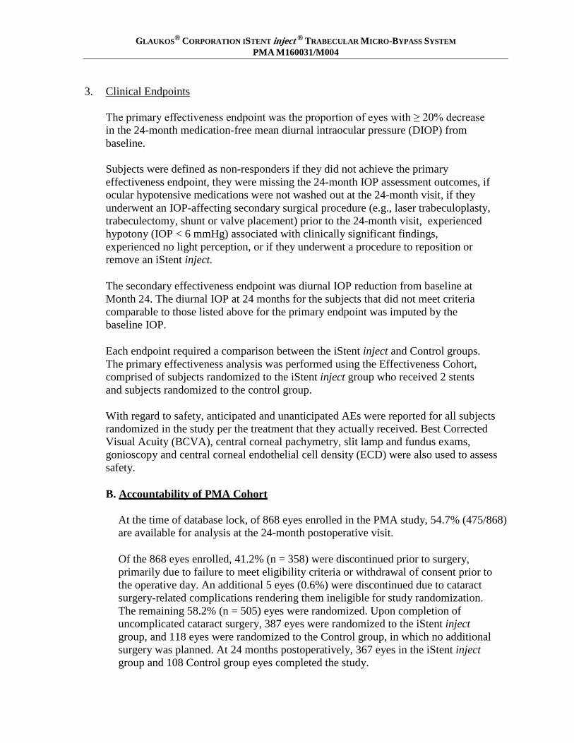
3. Clinical Endpoints The primary effectiveness endpoint was the proportion of eyes with ≥ 20% decrease in the 24-month medication-free mean diurnal intraocular pressure (DIOP) from baseline. Subjects were defined as non-responders if they did not achieve the primary effectiveness endpoint, they were missing the 24-month IOP assessment outcomes, if ocular hypotensive medications were not washed out at the 24-month visit, if they underwent an IOP-affecting secondary surgical procedure (e.g., laser trabeculoplasty, trabeculectomy, shunt or valve placement) prior to the 24-month visit, experienced hypotony (IOP < 6 mmHg) associated with clinically significant findings, experienced no light perception, or if they underwent a procedure to reposition or remove an iStent inject. The secondary effectiveness endpoint was diurnal IOP reduction from baseline at Month 24. The diurnal IOP at 24 months for the subjects that did not meet criteria comparable to those listed above for the primary endpoint was imputed by the baseline IOP. Each endpoint required a comparison between the iStent inject and Control groups. The primary effectiveness analysis was performed using the Effectiveness Cohort, comprised of subjects randomized to the iStent inject group who received 2 stents and subjects randomized to the control group. With regard to safety, anticipated and unanticipated AEs were reported for all subjects randomized in the study per the treatment that they actually received. Best Corrected Visual Acuity (BCVA), central corneal pachymetry, slit lamp and fundus exams, gonioscopy and central corneal endothelial cell density (ECD) were also used to assess safety.
B. Accountability of PMA Cohort
At the time of database lock, of 868 eyes enrolled in the PMA study, 54.7% (475/868) are available for analysis at the 24-month postoperative visit. Of the 868 eyes enrolled, 41.2% (n = 358) were discontinued prior to surgery, primarily due to failure to meet eligibility criteria or withdrawal of consent prior to the operative day. An additional 5 eyes (0.6%) were discontinued due to cataract surgery-related complications rendering them ineligible for study randomization. The remaining 58.2% (n = 505) eyes were randomized. Upon completion of uncomplicated cataract surgery, 387 eyes were randomized to the iStent inject group, and 118 eyes were randomized to the Control group, in which no additional surgery was planned. At 24 months postoperatively, 367 eyes in the iStent inject
group and 108 Control group eyes completed the study.

GLAUKOS® CORPORATION ISTENT inject ® TRABECULAR MICRO-BYPASS SYSTEM PMA M160031/M004
The outcomes provided were analyzed according to three (3) separate population cohorts:
• The Intent to Treat (ITT) population was defined as all randomized
eyes. Eyes were grouped according to their randomization assignment (as randomized).
• The Effectiveness Cohort was used for the effectiveness analyses. The Effectiveness Cohort included 380 eyes randomized to the iStent inject group who were implanted with 2 stents and 118 subjects randomized to the control group.
• The Safety population was defined as all randomized eyes. All subjects in the Safety population were analyzed according to the treatment they actually received (i.e., 386 subjects who received iStent inject in conjunction with cataract surgery and 119 eyes that underwent cataract surgery only).
C. Study Population Demographics and Baseline Parameters
The demographics and preoperative characteristics of the study population were as follows:
GLAUKOS® CORPORATION ISTENT inject ® TRABECULAR MICRO-BYPASS SYSTEM PMA M160031/M004
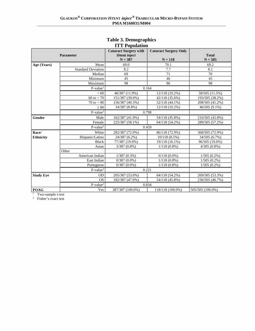
Table 3. Demographics ITT Population
Parameter
Cataract Surgery with iStent inject
Cataract Surgery Only Total
N = 387 N = 118 N = 505 Age (Years) Mean 69.0 70.1 69.2 Standard Deviation 8.2 7.7 8.1 Median 69 71 70 Minimum 45 46 45 Maximum 98 86 98 P-value1 0.164 < 60 46/387 (11.9%) 12/118 (10.2%) 58/505 (11.5%) 60 to < 70 151/387 (39.0%) 42/118 (35.6%) 193/505 (38.2%) 70 to < 80 156/387 (40.3%) 52/118 (44.1%) 208/505 (41.2%) ≥ 80 34/387 (8.8%) 12/118 (10.2%) 46/505 (9.1%) P-value2 0.798 Gender Male 162/387 (41.9%) 54/118 (45.8%) 216/505 (42.8%) Female 225/387 (58.1%) 64/118 (54.2%) 289/505 (57.2%) P-value2 0.459 Race/ White 282/387 (72.9%) 86/118 (72.9%) 368/505 (72.9%) Ethnicity Hispanic/Latino 24/387 (6.2%) 10/118 (8.5%) 34/505 (6.7%) Black 77/387 (19.9%) 19/118 (16.1%) 96/505 (19.0%) Asian 3/387 (0.8%) 1/118 (0.8%) 4/505 (0.8%) Other American Indian 1/387 (0.3%) 0/118 (0.0%) 1/505 (0.2%) East Indian 0/387 (0.0%) 1/118 (0.8%) 1/505 (0.2%) Portuguese 0/387 (0.0%) 1/118 (0.8%) 1/505 (0.2%) P-value2 0.221 Study Eye OD 205/387 (53.0%) 64/118 (54.2%) 269/505 (53.3%) OS 182/387 (47.0%) 54/118 (45.8%) 236/505 (46.7%) P-value2 0.834 POAG Yes 387/387 (100.0%) 118/118 (100.0%) 505/505 (100.0%) 1 Two-sample t-test 2 Fisher’s exact test

GLAUKOS® CORPORATION ISTENT inject ® TRABECULAR MICRO-BYPASS SYSTEM PMA M160031/M004
Table 4. Preoperative Characteristics ITT Population
Parameter
Cataract Surgery with iStent inject
Cataract Surgery Only Total
N = 387 N = 118 N = 505 Number of 1 224/387 (57.9%) 71/118 (60.2%) 295/505 (58.4%) Ocular Hypotensive 2 98/387 (25.3%) 30/118 (25.4%) 128/505 (25.3%) Medications at 3 63/387 (16.3%) 17/118 (14.4%) 80/505 (15.8%) Screening 4 2/387 (0.5%) 0/118 (0.0%) 2/505 (0.4%) P-value2 0.943 Visual Field Mean -3.392 -3.357 -3.384 Mean Deviation (MD) Standard Deviation 3.285 3.143 3.249 at Screening (dB) Median -2.79 -3.07 -2.89 Minimum -12.58 -11.67 -12.58 Maximum 3.12 2.04 3.12 P-value1 0.915 Corneal Thickness at Mean 546.49 546.06 546.39 Screening (µm) Standard Deviation 36.16 35.74 36.03 Median 545.0 548.5 546.0 Minimum 455.0 448.0 448.0 Maximum 620.0 620.0 620.0 P-value1 0.909 Medicated IOP at Mean 17.54 17.54 17.54 Screening (mmHg) Standard Deviation 2.99 2.78 2.94 Median 17.5 18.0 17.5 Minimum 9.0 11.0 9.0 Maximum 26.0 24.0 26.0 P-value1 0.997 Unmedicated IOP at Mean 24.83 24.50 24.75 Baseline (mmHg) Standard Deviation 3.34 3.08 3.28 Median 24.0 23.4 23.8 Minimum 20.8 20.7 20.7 Maximum 35.8 34.3 35.8 P-value1 0.328 BSCVA at Baseline Mean (Snellen) 0.234 (20/34) 0.232 (20/34) 0.234 (20/34) LogMAR Standard Deviation 0.168 0.161 0.166 Median (Snellen) 0.22 (20/33) 0.20 (20/32) 0.22 (20/33) Minimum (Snellen) -0.10 (20/16) -0.08 (20/17) -0.10 (20/16) Maximum (Snellen) 1.00 (20/200) 1.00 (20/200) 1.00 (20/200) P-value1 0.901 Shaffer Angle Grade III (25 - 35) 142/387 (36.7%) 40/118 (33.9%) 182/505 (36.0%) at Screening IV (> 35) 245/387 (63.3%) 78/118 (66.1%) 323/505 (64.0%) P-value2 0.661 Oral medications count as 1 medication. Combination medications count as 2 medications. Two subjects in the Cataract surgery with iStent inject group took Diamox at Screening. 1 Two-sample t-test 2 Fisher’s exact test
GLAUKOS® CORPORATION ISTENT inject ® TRABECULAR MICRO-BYPASS SYSTEM PMA M160031/M004
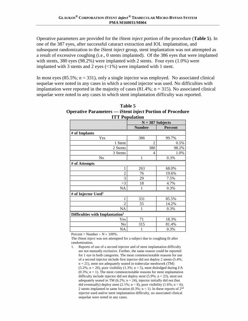
Operative parameters are provided for the iStent inject portion of the procedure (Table 5). In one of the 387 eyes, after successful cataract extraction and IOL implantation, and subsequent randomization to the iStent inject group, stent implantation was not attempted as a result of excessive coughing (i.e., 0 stents implanted). Of the 386 eyes that were implanted with stents, 380 eyes (98.2%) were implanted with 2 stents. Four eyes (1.0%) were implanted with 3 stents and 2 eyes (<1%) were implanted with 1 stent. In most eyes (85.5%; n = 331), only a single injector was employed. No associated clinical sequelae were noted in any cases in which a second injector was used. No difficulties with implantation were reported in the majority of cases (81.4%; n = 315). No associated clinical sequelae were noted in any cases in which stent implantation difficulty was reported.
Table 5
Operative Parameters — iStent inject Portion of Procedure ITT Population
N = 387 Subjects Number Percent
# of Implants Yes 386 99.7%
1 Stent 2 0.5% 2 Stents 380 98.2% 3 Stents 4 1.0%
No 1 0.3% # of Attempts
1 263 68.0% 2 76 19.6% 3 29 7.5%
>3 18 4.7% NA 1 0.3%
# of Injector Used1
1 331 85.5% 2 55 14.2%
NA 1 0.3% Difficulties with Implantation1
Yes 71 18.3% No 315 81.4% NA 1 0.3%
Percent = Number ÷ N × 100%. The iStent inject was not attempted for a subject due to coughing fit after randomization. 1. Reports of use of a second injector and of stent implantation difficulty
are not mutually exclusive. Further, the same reason could be reported for 1 eye in both categories. The most common/notable reasons for use of a second injector include first injector did not deploy 2 stents (5.4%; n = 21), stent not adequately seated in trabecular meshwork (TM) (5.2%; n = 20), poor visibility (1.3%; n = 5), stent dislodged during I/A (0.3%; n = 1). The most common/notable reasons for stent implantation difficulty include injector did not deploy stent (5.9%; n = 23), stent not adequately seated in TM (6.2%; n = 24), injector initially did not (but did eventually) deploy stent (2.1%; n = 8), poor visibility (1.6%; n = 6); 2 stents implanted in same location (0.3%; n = 1). In these reports of 2nd injector used and/or stent implantation difficulty, no associated clinical sequelae were noted in any cases.
GLAUKOS® CORPORATION ISTENT inject ® TRABECULAR MICRO-BYPASS SYSTEM PMA M160031/M004
D. Safety and Effectiveness Results
1. Safety Results
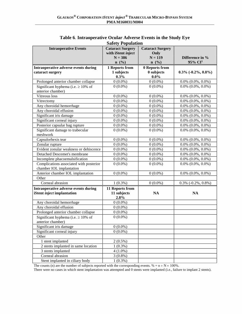
All safety analyses were performed on the Safety population. Findings are summarized for events occurring during the intraoperative period through the 24-month post-operative visit. The key safety outcomes for this study are presented below in Tables 6 to 8. Best Spectacle Corrected Visual Acuity (BSCVA) Most eyes in both groups achieved BSCVA of 20/40 or better at Month 24, with a slightly higher proportion of eyes achieving BSCVA of 20/40 or better in the iStent inject arm (98.9%) than in the control group (98.2%). Adverse Effects that Occurred in the PMA Clinical Study Intraoperative AEs A summary of intraoperative AEs is shown in Table 6. Because final study eligibility and randomization to treatment was determined post-cataract surgery, no subjects experiencing a predetermined cataract-surgery related AE such as posterior capsular rupture, vitreous loss or complications associated with posterior chamber IOL implantation were randomized. One eye experienced a corneal abrasion during cataract surgery and was subsequently randomized to the iStent inject group because this was not a clinically significant operative complication.
One of the 387 subjects randomized to iStent inject implantation experienced a coughing fit that resulted in increased positive pressure requiring a corneal suture. Therefore, no attempts to implant stents was made, and this subject was included in the control group of the Safety population. In the 386 iStent inject subjects implanted, 11 intraoperative AEs were reported during stent implantation (2.8%). Among these cases, there were 4 cases of 3 stents being implanted (1.0%) and two cases of only 1 stent being implanted (0.5%).
GLAUKOS® CORPORATION ISTENT inject ® TRABECULAR MICRO-BYPASS SYSTEM PMA M160031/M004
Table 6. Intraoperative Ocular Adverse Events in the Study Eye Safety Population
Intraoperative Events Cataract Surgery with iStent inject
Cataract Surgery Only
N = 386 n (%)
N = 119 n (%)
Difference in % 95% CI1
Intraoperative adverse events during cataract surgery
1 Reports from 1 subjects
0.3%
0 Reports from 0 subjects
0.0%
0.3% (-0.2%, 0.8%)
Prolonged anterior chamber collapse 0 (0.0%) 0 (0.0%) 0.0% (0.0%, 0.0%) Significant hyphema (i.e. ≥ 10% of
anterior chamber) 0 (0.0%) 0 (0.0%) 0.0% (0.0%, 0.0%)
Vitreous loss 0 (0.0%) 0 (0.0%) 0.0% (0.0%, 0.0%) Vitrectomy 0 (0.0%) 0 (0.0%) 0.0% (0.0%, 0.0%) Any choroidal hemorrhage 0 (0.0%) 0 (0.0%) 0.0% (0.0%, 0.0%) Any choroidal effusion 0 (0.0%) 0 (0.0%) 0.0% (0.0%, 0.0%) Significant iris damage 0 (0.0%) 0 (0.0%) 0.0% (0.0%, 0.0%) Significant corneal injury 0 (0.0%) 0 (0.0%) 0.0% (0.0%, 0.0%) Posterior capsular bag rupture 0 (0.0%) 0 (0.0%) 0.0% (0.0%, 0.0%) Significant damage to trabecular
meshwork 0 (0.0%) 0 (0.0%) 0.0% (0.0%, 0.0%)
Capsulorhexis tear 0 (0.0%) 0 (0.0%) 0.0% (0.0%, 0.0%) Zonular rupture 0 (0.0%) 0 (0.0%) 0.0% (0.0%, 0.0%) Evident zonular weakness or dehiscence 0 (0.0%) 0 (0.0%) 0.0% (0.0%, 0.0%) Detached Descemet's membrane 0 (0.0%) 0 (0.0%) 0.0% (0.0%, 0.0%) Incomplete phacoemulsification 0 (0.0%) 0 (0.0%) 0.0% (0.0%, 0.0%) Complications associated with posterior
chamber IOL implantation 0 (0.0%) 0 (0.0%) 0.0% (0.0%, 0.0%)
Anterior chamber IOL implantation 0 (0.0%) 0 (0.0%) 0.0% (0.0%, 0.0%) Other Corneal abrasion 1 (0.3%) 0 (0.0%) 0.3% (-0.2%, 0.8%) Intraoperative adverse events during iStent inject implantation
11 Reports from 11 subjects
2.8%
NA
NA
Any choroidal hemorrhage 0 (0.0%) Any choroidal effusion 0 (0.0%) Prolonged anterior chamber collapse 0 (0.0%) Significant hyphema (i.e. ≥ 10% of
anterior chamber) 0 (0.0%)
Significant iris damage 0 (0.0%) Significant corneal injury 0 (0.0%) Other 1 stent implanted 2 (0.5%) 2 stents implanted in same location 1 (0.3%) 3 stents implanted 4 (1.0%) Corneal abrasion 3 (0.8%) Stent implanted in ciliary body 1 (0.3%) The counts (n) are the number of subjects reported with the corresponding events. % = n ÷ N × 100%. There were no cases in which stent implantation was attempted and 0 stents were implanted (i.e., failure to implant 2 stents).

GLAUKOS® CORPORATION ISTENT inject ® TRABECULAR MICRO-BYPASS SYSTEM PMA M160031/M004
Postoperative AEs
There were no unanticipated adverse events. There were no reports of flat AC with lens cornea touch, shallow AC with iridocorneal apposition, shallow AC with peripheral iridocorneal apposition, wound dehiscence, endophthalmitis, corneal decompensation, choroidal hemorrhage or effusion,, aqueous misdirection, cyclodialysis, hypotony at one month postoperative or later, hypotony maculopathy, atrophy/phthisis, cup-to-disc (CD) ratio increase of ≥ 0.3, loss of light perception or stent dislocation. Moreover, no cases of pupillary block or hypopyon were reported during the study. A lower proportion of subjects in the iStent inject group experienced postoperative ocular AEs than in the Control group (54.1% of subjects [n = 209] in the iStent inject group and 62.2% of subjects [n = 74] in the Control group). A list of the more common AEs (occurring at a rate of 2% or greater) and the associated rates are provided in Table 7. Anterior segment inflammation, which was generally mild, was reported in 5.7% of iStent inject subjects and 4.2% of Control subjects.
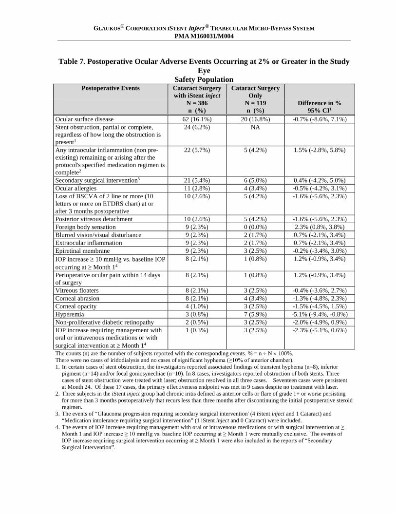
GLAUKOS® CORPORATION ISTENT inject ® TRABECULAR MICRO-BYPASS SYSTEM PMA M160031/M004
Table 7. Postoperative Ocular Adverse Events Occurring at 2% or Greater in the Study Eye
Safety Population Postoperative Events Cataract Surgery
with iStent inject Cataract Surgery
Only
N = 386 n (%)
N = 119 n (%)
Difference in % 95% CI1
Ocular surface disease 62 (16.1%) 20 (16.8%) -0.7% (-8.6%, 7.1%) Stent obstruction, partial or complete, regardless of how long the obstruction is present1
24 (6.2%) NA
Any intraocular inflammation (non pre-existing) remaining or arising after the protocol's specified medication regimen is complete2
22 (5.7%) 5 (4.2%) 1.5% (-2.8%, 5.8%)
Secondary surgical intervention3 21 (5.4%) 6 (5.0%) 0.4% (-4.2%, 5.0%) Ocular allergies 11 (2.8%) 4 (3.4%) -0.5% (-4.2%, 3.1%) Loss of BSCVA of 2 line or more (10 letters or more on ETDRS chart) at or after 3 months postoperative
10 (2.6%) 5 (4.2%) -1.6% (-5.6%, 2.3%)
Posterior vitreous detachment 10 (2.6%) 5 (4.2%) -1.6% (-5.6%, 2.3%) Foreign body sensation 9 (2.3%) 0 (0.0%) 2.3% (0.8%, 3.8%) Blurred vision/visual disturbance 9 (2.3%) 2 (1.7%) 0.7% (-2.1%, 3.4%) Extraocular inflammation 9 (2.3%) 2 (1.7%) 0.7% (-2.1%, 3.4%) Epiretinal membrane 9 (2.3%) 3 (2.5%) -0.2% (-3.4%, 3.0%) IOP increase ≥ 10 mmHg vs. baseline IOP occurring at ≥ Month 14
8 (2.1%) 1 (0.8%) 1.2% (-0.9%, 3.4%)
Perioperative ocular pain within 14 days of surgery
8 (2.1%) 1 (0.8%) 1.2% (-0.9%, 3.4%)
Vitreous floaters 8 (2.1%) 3 (2.5%) -0.4% (-3.6%, 2.7%) Corneal abrasion 8 (2.1%) 4 (3.4%) -1.3% (-4.8%, 2.3%) Corneal opacity 4 (1.0%) 3 (2.5%) -1.5% (-4.5%, 1.5%) Hyperemia 3 (0.8%) 7 (5.9%) -5.1% (-9.4%, -0.8%) Non-proliferative diabetic retinopathy 2 (0.5%) 3 (2.5%) -2.0% (-4.9%, 0.9%) IOP increase requiring management with oral or intravenous medications or with surgical intervention at ≥ Month 14
1 (0.3%) 3 (2.5%) -2.3% (-5.1%, 0.6%)
The counts (n) are the number of subjects reported with the corresponding events. % = n ÷ N × 100%. There were no cases of iridodialysis and no cases of significant hyphema (≥10% of anterior chamber). 1. In certain cases of stent obstruction, the investigators reported associated findings of transient hyphema (n=8), inferior
pigment (n=14) and/or focal goniosynechiae (n=10). In 8 cases, investigators reported obstruction of both stents. Three cases of stent obstruction were treated with laser; obstruction resolved in all three cases. Seventeen cases were persistent at Month 24. Of these 17 cases, the primary effectiveness endpoint was met in 9 cases despite no treatment with laser.
2. Three subjects in the iStent inject group had chronic iritis defined as anterior cells or flare of grade 1+ or worse persisting for more than 3 months postoperatively that recurs less than three months after discontinuing the initial postoperative steroid regimen.
3. The events of “Glaucoma progression requiring secondary surgical intervention' (4 iStent inject and 1 Cataract) and “Medication intolerance requiring surgical intervention” (1 iStent inject and 0 Cataract) were included.
4. The events of IOP increase requiring management with oral or intravenous medications or with surgical intervention at ≥ Month 1 and IOP increase ≥ 10 mmHg vs. baseline IOP occurring at ≥ Month 1 were mutually exclusive. The events of IOP increase requiring surgical intervention occurring at ≥ Month 1 were also included in the reports of “Secondary Surgical Intervention”.

GLAUKOS® CORPORATION ISTENT inject ® TRABECULAR MICRO-BYPASS SYSTEM PMA M160031/M004
In addition to the AEs reported in Table 7, events that occurred at a rate of < 2% in both groups included age-related macular degeneration, chalazion, conjunctivitis, corneal guttata, cystoid macular edema, diplopia, disc hemorrhage, ectropion, glaucoma progression requiring surgical intervention, lattice degeneration, nerve fiber layer loss, ocular irritation, optic nerve thinning/cupping, visual field loss ≥ 2.5 dB and vitreous hemorrhage. AEs that occurred at < 2% in the iStent inject group included one case (0.3%) each of blepharospasm, branch retinal vein occlusion, corneal edema ≥ 30 days, corneal striae, eyelash loss, iris atrophy, iris strand, medication intolerance requiring surgical intervention, ptosis, residual cortex, retinal detachment, retinal tear, and worsening glaucoma; 2 cases (0.5%) each of anterior basement membrane dystrophy, extraocular papilloma, ocular pain, punctal stenosis, retinal drusen, retinal hemorrhage and retinal pigment epithelial changes; 3 cases (0.8%) each of peripapillary atrophy, retinal flap tears, retinal hole and notching; 4 cases (1.0%) of deep stents2 and transient mild ocular discomfort; 5 cases (1.3%) of subconjunctival hemorrhage and 7 cases (1.8%) of goniosynechiae. AEs that occurred at < 2% in the control group included 1 case (0.8%) each of anterior scleritis, central retinal artery occlusion, corneal ulcer, flashes, iris neovascularization and IOL dislocation; and 2 cases (1.7%) of extraocular trauma.
The study investigators determined for each intraoperative and postoperative ocular AE reported whether an event was considered serious. The proportion of eyes with serious AEs (SAEs) was 0.8% (n=3) in the iStent inject group and 2.5% (n=3) in the control group. iStent inject SAEs comprised 1 case each of mild partial stent obstruction that did not require intervention, retinal tear requiring laser retinopexy, and glaucoma progression requiring ExPress shunt implantation. SAEs reported for the control group consisted of 1 case each of blurred vision/visual disturbance; epiretinal membrane requiring vitrectomy with membrane peel, and central retinal artery occlusion and neovascularization requiring pan-retinal photocoagulation. A total of 56 AEs reported for 48 iStent inject eyes (12.4%) were determined to be device related including all cases of stent obstruction, deep stents, 3 stents implanted, 1 stent implanted, 2 stents implanted in the same location, and stent implanted in the ciliary body, which accounted for 36 of the 56 device-related AEs. Other AEs determined to be device-related included 8 cases (2.1%) of intraocular inflammation, 7 cases (1.8%) of goniosynechiae, 3 cases (0.8%) of intraoperative corneal abrasion, and 1 case (0.3%) each of iris strand and ocular irritation.
2. In each of the four eyes with “deep stents,” there was a single stent per eye that was unable to be visualized by either
gonioscopy or UBM at the last 3 visits, despite being visualized intraoperatively and/or at an earlier postoperative exam. . Among these cases, there were no associated clinical sequelae or secondary surgeries to modify device positioning, none experienced an endothelial cell loss >30% at 24 months or posterior segment sequelae, and three of the four eyes met the primary effectiveness endpoint.
GLAUKOS® CORPORATION ISTENT inject ® TRABECULAR MICRO-BYPASS SYSTEM PMA M160031/M004
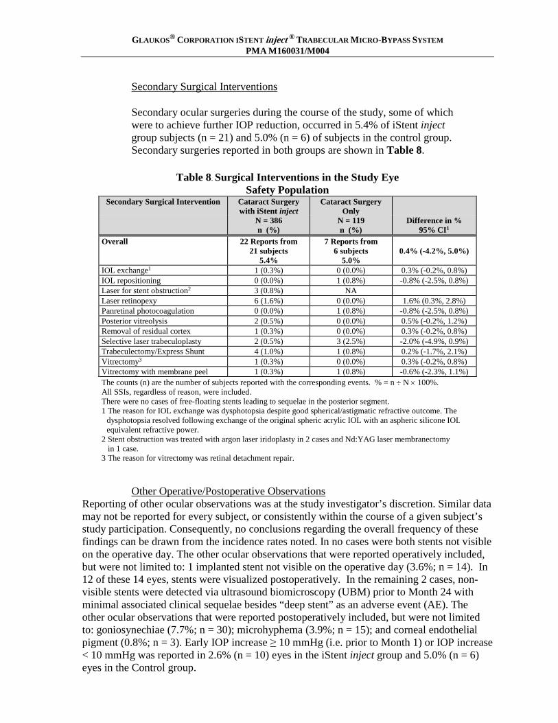
Secondary Surgical Interventions Secondary ocular surgeries during the course of the study, some of which were to achieve further IOP reduction, occurred in 5.4% of iStent inject
group subjects (n = 21) and 5.0% (n = 6) of subjects in the control group. Secondary surgeries reported in both groups are shown in Table 8.
Table 8. Surgical Interventions in the Study Eye
Safety Population Secondary Surgical Intervention Cataract Surgery
with iStent inject Cataract Surgery
Only
N = 386 n (%)
N = 119 n (%)
Difference in % 95% CI1
Overall 22 Reports from 21 subjects
5.4%
7 Reports from 6 subjects
5.0%
0.4% (-4.2%, 5.0%)
IOL exchange1 1 (0.3%) 0 (0.0%) 0.3% (-0.2%, 0.8%) IOL repositioning 0 (0.0%) 1 (0.8%) -0.8% (-2.5%, 0.8%) Laser for stent obstruction2 3 (0.8%) NA Laser retinopexy 6 (1.6%) 0 (0.0%) 1.6% (0.3%, 2.8%) Panretinal photocoagulation 0 (0.0%) 1 (0.8%) -0.8% (-2.5%, 0.8%) Posterior vitreolysis 2 (0.5%) 0 (0.0%) 0.5% (-0.2%, 1.2%) Removal of residual cortex 1 (0.3%) 0 (0.0%) 0.3% (-0.2%, 0.8%) Selective laser trabeculoplasty 2 (0.5%) 3 (2.5%) -2.0% (-4.9%, 0.9%) Trabeculectomy/Express Shunt 4 (1.0%) 1 (0.8%) 0.2% (-1.7%, 2.1%) Vitrectomy3 1 (0.3%) 0 (0.0%) 0.3% (-0.2%, 0.8%) Vitrectomy with membrane peel 1 (0.3%) 1 (0.8%) -0.6% (-2.3%, 1.1%) The counts (n) are the number of subjects reported with the corresponding events. % = n ÷ N × 100%. All SSIs, regardless of reason, were included. There were no cases of free-floating stents leading to sequelae in the posterior segment. 1 The reason for IOL exchange was dysphotopsia despite good spherical/astigmatic refractive outcome. The
dysphotopsia resolved following exchange of the original spheric acrylic IOL with an aspheric silicone IOL equivalent refractive power.
2 Stent obstruction was treated with argon laser iridoplasty in 2 cases and Nd:YAG laser membranectomy in 1 case. 3 The reason for vitrectomy was retinal detachment repair.
Other Operative/Postoperative Observations Reporting of other ocular observations was at the study investigator’s discretion. Similar data may not be reported for every subject, or consistently within the course of a given subject’s study participation. Consequently, no conclusions regarding the overall frequency of these findings can be drawn from the incidence rates noted. In no cases were both stents not visible on the operative day. The other ocular observations that were reported operatively included, but were not limited to: 1 implanted stent not visible on the operative day (3.6%; n = 14). In 12 of these 14 eyes, stents were visualized postoperatively. In the remaining 2 cases, non-visible stents were detected via ultrasound biomicroscopy (UBM) prior to Month 24 with minimal associated clinical sequelae besides “deep stent” as an adverse event (AE). The other ocular observations that were reported postoperatively included, but were not limited to: goniosynechiae (7.7%; n = 30); microhyphema (3.9%; n = 15); and corneal endothelial pigment (0.8%; n = 3). Early IOP increase ≥ 10 mmHg (i.e. prior to Month 1) or IOP increase < 10 mmHg was reported in 2.6% (n = 10) eyes in the iStent inject group and 5.0% (n = 6) eyes in the Control group.

GLAUKOS® CORPORATION ISTENT inject ® TRABECULAR MICRO-BYPASS SYSTEM PMA M160031/M004
Corneal Endothelial Cell Density There was little difference in endothelial cell loss (ECL) between the iStent inject and Control groups. Results were consistent with previous reports of cataract surgery-related ECL. The mean percent change in ECD from baseline to 24 months was -13.1% (SD 12.4; 95% CI -14.4%, -11.8%) for the iStent inject group and -12.3% (SD 12.7%; 95% CI -14.8%, -9.8%) for the control group. A similar proportion of eyes in each group (10.4% in the iStent inject group and 9.5% in the control group) experienced ECL > 30% at 24 months postoperatively.
2. Effectiveness Results
Results from the primary and secondary endpoints are shown in Table 9. The primary effectiveness endpoint was met, with 75.8% (288/380) in the iStent inject group and 61.9% (73/118) in the Control group achieving a clinically significant (≥ 20%) reduction in medication-free diurnal IOP from baseline at 24 months. This difference between groups was statistically significant (p=0.003). The secondary endpoint, a clinically significant mean change in medication-free diurnal IOP from baseline at 24-month postoperative examination, was met. The mean reduction in medication-free mean diurnal IOP from baseline to 24 months was 7.0 mmHg (SD 4.0) in the iStent inject group compared to 5.4 mmHg (SD 3.7) in the control group (p <0.001).
Table 9. Primary and Secondary Effectiveness Results
Cataract Surgery with iStent inject
N = 380
Cataract Surgery Only N = 118
Difference (iStent inject vs. control)
Effectiveness Endpoint P-value for
(Evaluated at 24 Months Postoperatively)
difference
Proportion of subjects with medication-free DIOP reduction ≥ 20% from baseline
75.8% 61.9%
13.9% 0.0032
Medication-free mean DIOP (mmHg) change from baseline1
-7.0 -5.4 -1.6 < 0.0013
Subjects without Month 24 medication-free diurnal IOP, or with IOP-related SSIs, loss of light perception or hypotony (IOP < 6 mmHg) associated with clinically significant findings prior to 24 months were treated as non-responders. iStent inject subjects with stent reposition or removal prior to 24 months were treated as non-responders. 1. The 24-month diurnal IOP values were subtracted from baseline diurnal IOP in all subjects, except for the non-responders described
above. For the non-responders described above, the baseline diurnal IOP values were used for the 24-month diurnal IOP values (i.e., a change of 0 mmHg was used).
2. One-sided Fisher’s exact test with a significance level of 0.025. 3. One-sided two-sample t-test with a significance level of 0.025.

GLAUKOS® CORPORATION ISTENT inject ® TRABECULAR MICRO-BYPASS SYSTEM PMA M160031/M004
Additional detail regarding the reasons patients did not achieve the primary endpoint (IOP non-responders) is shown in Table 10.
Table 10. Non-Responder Categories at 24 Months
Effectiveness Cohort Cataract Surgery
with iStent inject Cataract Surgery
Only N = 380 N = 118 n/N (%) n/N (%)
Total Non-Responders 92 (24.2%) 45 (38.1%) Non-Responders: 24-month unmedicated diurnal IOP reduction from baseline < 20%
56 (14.7%) 26 (22.0%)
Non-Responders for reasons other than IOP reduction1
36 (9.5%) 19 (16.1%)
Secondary glaucoma surgery2 5 (1.3%) 3 (2.5%) Other IOP-affecting secondary surgery3 0 (0.0%) 0 (0.0%) Stent reposition or removal 0 (0.0%) 0 (0.0%) Loss of light perception 0 (0.0%) 0 (0.0%) Clinically significant hypotony 0 (0.0%) 0 (0.0%) Did not complete medication washout –
Safety concerns 12 (3.2%) 4 (3.4%)
Did not complete medication washout – Instructions not provided/followed4
0 (0.0%) 2 (1.7%)
Missing 24-month diurnal IOP data4 19 (5.0%) 10 (8.5%) Death 4 (1.1%) 6 (5.1%) Investigator’s decision 1 (0.3%) 0 (0.0%) Lost contact 8 (2.1%) 2 (1.7%) Subject’s decision 6 (1.6%) 2 (1.7%) n = number of eyes with the corresponding responses. % = n ÷ N × 100%. 1 Subjects were included in the primary category of ''Non-Responders for reasons other than IOP reduction”. 2 Secondary glaucoma surgeries include trabeculectomy, and laser trabeculoplasty. 3 Other IOP-affecting secondary surgeries. 4 The outcomes of these subjects were imputed for the 24-month analysis. There were 2 subjects on oral medication at 23 months and both subjects underwent washout. Hence, although any subjects on oral medication at 24 months would have been considered non-responders due to the potential to confound the endpoint analysis, there were no subjects in this category.
3. Summary of Supplemental Clinical Information
A. For the pivotal trial of the iStent inject, the Ocular Surface Disease Index (OSDI©) was self-administered by study subjects. The OSDI questionnaire contains 12 questions involving ocular symptoms, vision-related function and environmental triggers experienced by the subject during the past week, and is assessed on a scale of 0 to 100 with higher scores representing greater disability. Table 11 summarizes the change in OSDI subscales and overall score from baseline. The mean improvements at 24 months from baseline were slightly higher in the iStent inject group compared to the control group involving ocular symptoms (-16.41 vs. -10.69) and vision-related function (-22.60 vs. -18.56) and similar involving environmental triggers (-7.41 vs. -7.70). The mean

GLAUKOS® CORPORATION ISTENT inject ® TRABECULAR MICRO-BYPASS SYSTEM PMA M160031/M004
improvement in OSDI overall score at 24 months was also higher in the iStent inject group compared to the control group (-16.25 vs. -12.38). The questionnaire used to collect these data has not been validated, and therefore the true rates of these symptoms may differ from those presented in the Table 11.
Table 11
Change in OSDI Questionnaire Sub-Scale Score from Baseline Safety Population
Cataract Surgery with iStent inject Cataract Surgery Only Total Number of Subjects = 386 Total Number of Subjects = 119 1M 6M 12M 24M 1M 6M 12M 24M
Statistics n (%) n (%) n (%) n (%) n (%) n (%) n (%) n (%) Ocular Symptoms (Q1, Q2, Q3) N 382 376 367 361 117 118 115 109 Mean -11.87 -15.04 -16.93 -16.41 -6.41 -10.55 -11.53 -10.69 SD 22.39 21.23 19.96 21.13 20.53 18.45 17.16 17.74 Median -10.0 -15.0 -15.0 -15.0 -5.0 -10.0 -10.0 -10.0 Min -100 -100 -90.0 -100 -55.0 -60.0 -75.0 -65.0 Max 75.0 50.0 33.8 60.0 80.0 40.0 35.0 35.0 Not Reported 2 1 3 5 2 0 1 0 Vision-Related Function (Q4, Q5, Q6, Q7, Q8, Q9) N 379 374 363 359 117 118 115 109 Mean -16.07 -21.46 -22.82 -22.60 -14.08 -17.32 -20.92 -18.56 SD 29.80 27.93 28.22 27.30 29.94 27.49 27.66 28.92 Median -12.5 -18.8 -18.8 -18.8 -6.3 -12.5 -16.7 -12.5 Min -93.8 -100 -100 -100 -100 -100 -100 -100 Max 100.0 77.1 62.5 62.5 87.5 75.0 37.5 68.8 Not Reported 5 3 7 7 2 0 1 0 Environmental Triggers (Q10, Q11, Q12) N 370 367 358 353 114 116 113 106 Mean -5.20 -7.27 -7.83 -7.41 -4.61 -7.26 -7.82 -7.70 SD 21.52 20.70 21.65 22.61 21.95 21.61 21.60 20.66 Median 0.0 0.0 0.0 0.0 0.0 0.0 0.0 0.0 Min -83.3 -100 -100 -100 -75.0 -100 -100 -75.0 Max 100.0 58.3 75.0 66.7 66.7 41.7 33.3 75.0 Not Reported 14 10 12 13 5 2 3 3 Overall Composite Score N 382 376 367 361 117 118 115 109 Mean -11.87 -15.44 -16.66 -16.25 -8.48 -11.91 -13.60 -12.38 SD 20.29 19.39 19.38 19.73 20.02 18.01 17.18 18.38 Median -10.4 -12.5 -13.3 -12.5 -6.2 -10.4 -10.7 -10.4 Min -93.8 -93.8 -95.8 -100 -60.4 -66.7 -64.6 -62.5 Max 72.9 37.5 31.3 45.8 70.8 37.5 17.6 56.3 Not Reported 2 1 3 5 2 0 1 0 Each sub-scale is a summarization of some specific questions to the ODSI.
GLAUKOS® CORPORATION ISTENT inject ® TRABECULAR MICRO-BYPASS SYSTEM PMA M160031/M004
B. In the iStent inject pivotal trial, at 24 months, the proportion of subjects with medication-free diurnal IOP ≤ 18 mmHg was 63.2% in the treatment group and 50.0% in the control group (difference 13.2%; 95% CI 2.9%, 23.4%).3
C. In the iStent inject pivotal trial, mean observed unmedicated IOP was higher at baseline and lower at 24 months in the iStent inject group. IOP at baseline was 24.8 (SD 3.4) mmHg in the iStent inject group and 24.5 (SD 3.1) mmHg in the control group. Unmedicated IOP at 24 months was 17.1 mmHg (SD 3.6) at 24 months in the iStent inject group and 17.8 mmHg (SD 3.5) in the control group.4
D. Of the subjects who were responders (e.g., 24-month unmedicated mean DIOP was reduced by ≥20% as compared with baseline in the absence of IOP-affecting surgery during the study), 84% of subjects in the iStent inject group (243/288) and 67% of subjects in the Control Group (49/73) were not using ocular hypotensive medication at 23 months.
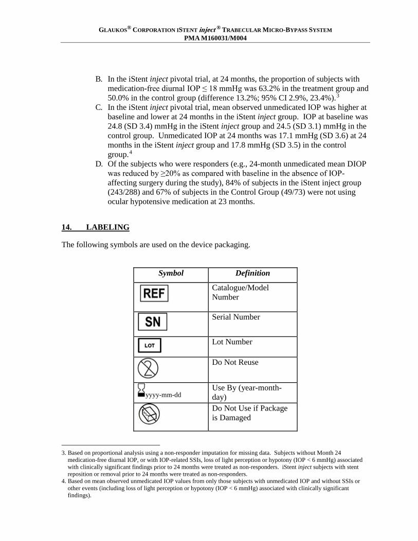
14. LABELING
The following symbols are used on the device packaging.
Symbol Definition
Catalogue/Model Number
Serial Number
Lot Number
Do Not Reuse
yyyy-mm-dd Use By (year-month-day)
Do Not Use if Package is Damaged
3. Based on proportional analysis using a non-responder imputation for missing data. Subjects without Month 24
medication-free diurnal IOP, or with IOP-related SSIs, loss of light perception or hypotony (IOP < 6 mmHg) associated with clinically significant findings prior to 24 months were treated as non-responders. iStent inject subjects with stent reposition or removal prior to 24 months were treated as non-responders.
4. Based on mean observed unmedicated IOP values from only those subjects with unmedicated IOP and without SSIs or other events (including loss of light perception or hypotony (IOP < 6 mmHg) associated with clinically significant findings).

GLAUKOS® CORPORATION ISTENT inject ® TRABECULAR MICRO-BYPASS SYSTEM PMA M160031/M004
Consult Instructions For Use
Manufacturer’s Address
Sterilized by Gamma Irradiation
For prescription use only
Room temperature storage requirement
MR Conditional
15. MRI SAFETY INFORMATION
Non-clinical testing has demonstrated that the iStent inject Trabecular Micro-Bypass System Model G2-M-IS is MR Conditional. A patient with this device can be safely scanned in an MR system meeting the following conditions:
∙ Static magnetic field of 3 T or less ∙ Maximum spatial gradient magnetic field of 4,000 gauss/cm (40 T/m) ∙ Maximum MR system reported, whole body averaged specific absorption rate (SAR) of 4 W/kg
Under the scan conditions defined above, the iStent inject Trabecular Micro-Bypass System Model G2-M-IS is not expected to produce a clinically significant temperature rise after 15 minutes of continuous scanning.
In non-clinical testing, the image artifact caused by the device extends less than 15 mm from the device when imaged with a gradient echo pulse sequence and a 3.0 T MRI system.
16. CAUTION
GLAUKOS® CORPORATION ISTENT inject ® TRABECULAR MICRO-BYPASS SYSTEM PMA M160031/M004
Federal law restricts this device to sale by, or on the order of, a physician.
Physician training by certified Glaukos personnel is required prior to use of this device. Training consists of three main parts:
• Didactic session • Simulated implantation of iStent inject • Supervised iStent inject implantation of clinical cases until implantation proficiency
is demonstrated
Manufacturer: Glaukos Corporation 229 Avenida Fabricante San Clemente, CA 92672 Tel: 949.367.9600, Fax: 949.367.9984 www.glaukos.com Toll-Free: 1-800-GLAUKOS (452-8567) Glaukos® and iStent inject® are registered trademarks of Glaukos Corporation.
45-0176 Rev. 1 draft 6-15-18