disclaimer - eshre
TRANSCRIPT

1

2
Disclaimer The European Society of Human Reproduction and Embryology (hereinafter referred to as 'ESHRE') developed the current good practice recommendations document, to provide clinical recommendations to improve the quality of healthcare delivery within the European field of human reproduction and embryology. This guideline represents the views of ESHRE, which were achieved after careful consideration of the scientific evidence available at the time of preparation. In the absence of scientific evidence on certain aspects, a consensus between the relevant ESHRE stakeholders has been obtained. The aim of clinical practice guidelines and good practice recommendations is to aid healthcare professionals in everyday clinical decisions about appropriate and effective care of their patients. However, adherence to these clinical practice guidelines does not guarantee a successful or specific outcome, nor does it establish a standard of care. Clinical practice guidelines or good practice recommendations do not override the healthcare professional's clinical judgment in diagnosis and treatment of particular patients. Ultimately, healthcare professionals must make their own clinical decisions on a case-by-case basis, using their clinical judgment, knowledge, and expertise, and taking into account the condition, circumstances, and wishes of the individual patient, in consultation with that patient and/or the guardian or carer. ESHRE makes no warranty, express or implied, regarding the clinical practice guidelines or good practice recommendations and specifically excludes any warranties of merchantability and fitness for a particular use or purpose. ESHRE shall not be liable for direct, indirect, special, incidental, or consequential damages related to the use of the information contained herein. While ESHRE makes every effort to compile accurate information and to keep it up-to-date, it cannot, however, guarantee the correctness, completeness, and accuracy of the good practice recommendations in every respect. In any event, these good practice recommendations do not necessarily represent the views of all clinicians that are member of ESHRE. The information provided in this document does not constitute business, medical or other professional advice, and is subject to change.
3
Recommendations for good practice in Ultrasound: oocyte pick-up.
Content
Authors .................................................................................................................................................... 4
Introduction ............................................................................................................................................ 5
Methodology ........................................................................................................................................... 6
Recommendations .................................................................................................................................. 9
Definition: ........................................................................................................................................... 9
Prior to OPU .............................................................................................................................. 11
Equipment and consumables .................................................................................................... 12
OPU preparation (see also Box B: Before OPU-Checklist) ......................................................... 14
OPU procedure .......................................................................................................................... 19
Post-procedure care (see also BOX D: After OPU-Checklist) ..................................................... 23
Associated pathologies and cautions during OPU ..................................................................... 24
Complications and risks ............................................................................................................. 25
Future developments ................................................................................................................ 32
Training and competence .......................................................................................................... 33
Quality assurance and performance ..................................................................................... 34
Concluding remarks / discussion ........................................................................................... 35
References ............................................................................................................................................. 36

4
The ESHRE Working group on Ultrasound in ART
Arianna D’Angelo (chair) Costas Panayotidis Nazar Amso Roberto Marci Roberto Matorras Mircea Onofriescu Ahmet Turp Frank Vandekerckhove Zdravka Veleva Nathalie Vermeulen Veljko Vlaisavljevic Conflict of interest The authors declare that they have no conflict of interest. Acknowledgement The working group would like to thank Dr Jouni Ahonen, Dr Orhan Binici and Dr Tahsin Zatman for their helpful expert input on the sedation section. The WG would also like to thank all contributors to the stakeholder review. The meetings of the WG and methodological support were funded by the European Society of Human Reproduction and Embryology (ESHRE).
Submitte
d for
publi
catio
n in H
ROpen
5
Introduction The World Health Organisation (WHO) calculated that 48.5 million (45.0 million to 52.6 million) of couples are affected by infertility worldwide (Mascarenhas et al., 2012). During fertility treatment women need an ultrasound approach for both diagnostic and therapeutic procedures, which can be performed with either a transvaginal or a transabdominal approach (Lutz and Buscarini, 2013). Results of the images obtained during these scans are vital for the patient’s care, as they might impact on which treatment protocol the patient will follow and on the treatment outcome. This is why the operator’s findings and approach play an essential role in the treatment of the clinical problem. During the early days of IVF, oocyte pick-up (OPU) was systematically performed by laparoscopy. This required a surgical procedure, general anaesthesia and hospital admission. After the first reports on transvaginal oocyte retrieval (TVOR) in the early 1980s (Dellenbach et al., 1984, Gleicher et al., 1983, Schulman et al., 1985), OPU is almost always performed transvaginally. The advantages of transvaginal OPU, in comparison with the transabdominal or laparoscopic approach, include:
• better visualization and shorter distance of ovary from the transducer;
• high recovery rate of good quality oocytes with minimal discomfort for patients;
• the use of local anaesthesia with sedation instead of general anaesthesia;
• decreased risk of intestinal trauma;
• it can be easily learned, especially by operators trained in US;
• decreased costs for patients;
• and quick post-interventional recovery.
However, in some patients, transabdominal ultrasound facilitated access when the ovaries were transposed or enlarged above the pelvic brim. Transabdominal-guided oocyte retrieval continues to be used at some centres for rare patients who have ovaries inaccessible by vaginal ultrasound. Nowadays, transvaginal OPU is a widely performed procedure, with a low complication rate (European IVF monitoring Consortium for the European Society of Human Reproduction and Embryology et al., 2017). In this paper, recommendations for the different steps of transvaginal OPU will be described. Laparoscopic OPU, transabdominal OPU and OPU for in vitro maturation (IVM) are outside the scope of this document. The recommendations for good practice in this paper were based on evidence (where available) and experts’ opinions on ultrasound practice in assisted reproductive technology (ART). This paper is aimed to guide clinicians, especially in countries where there are no national guidelines, and it could have a significant impact on patients’ care and safety worldwide.
Submitte
d for
publi
catio
n in H
ROpen
6
Methodology The current recommendations were written by a working group (WG) of experts on ultrasound, according to the methodology described in the manual for development of recommendations for good practice (Vermeulen et al., 2018). The current document’s first draft was based on the results of the doctoral thesis of one of the authors (C.P.) (Panayotidis, 2017). C.P. conducted a systematic review of the literature and a Delphi survey of 15 experts reporting their opinions on current practice in ultrasound-guided oocyte retrieval. The Delphi method survey included 53 questions completed in three rounds and resulted in 32 standards of practice. In addition to the results of the dissertation, a new literature search was conducted. Databases (PUBMED/Medline and the Cochrane Library) were searched from inception to 17 July 2018. Search terms focussed on ultrasound, oocyte retrieval, Doppler, sedation, anaesthesia, infection, antibiotics, hydrosalpinx and flushing, and included extended key words and MESH terms. References were divided according to topic, and full texts were assessed (see Supplementary Figure 1, available as Supplementary data at ESHRE online). Where possible, references of papers providing indirect evidence or referrals to other guidelines were added, based on expert opinion. The first draft of the paper, based on the dissertation, was presented and discussed by the WG during a teleconference in November 2017, after which WG members submitted their written comments and suggestions for improvement. A full day consensus meeting was organized to discuss the paper further until consensus. The results of the literature search were included where relevant. The document was published on the ESHRE website for 4 weeks (between 25 March and 23 April 2019) and stakeholders were invited to submit their comments. After addressing all comments from the stakeholder review (report available on www.eshre.eu/guidelines), the document was finalized and approved by the ESHRE Executive Committee. The paper outlines recommendations for good practice in OPU, with addition of further basic information, checklists and troubleshooting in boxes A-F. For ease of use of the recommendations, a list of abbreviations is provided in table 1.

Submitte
d for
publi
catio
n in H
ROpen
7
Figure 1
2.
Scre
enin
g 3
. El
igib
ility
Records identified through database searching
(n = 1015)
Additional records identified through other sources
(n =35)
Records after duplicates removed (n = 3)
Records screened (n = 1047)
Records excluded (n = 857)
Full-text articles assessed for eligibility
(n = 190)
Full-text articles excluded, with reasons
(n = 82)
Studies included in qualitative synthesis
(n = 108)
Studies included in quantitative synthesis
(meta-analysis) (n = )
1. I
den
tifi
cati
on
4.
Incl
ud
ed

Submitte
d for
publi
catio
n in H
ROpen
8
Table 1: List of abbreviations
3D Three-dimensional
AIUM American Institute of Ultrasound in Medicine
ART Assisted reproductive technologies
CRP C-reactive protein
CT Computed tomography
ECG Electrocardiogram
FBC Full blood count
fx For example
GnRH Gonadotrophin-releasing hormone
Hb Haemoglobin level
hCG Human chorionic gonadotrophin
HIV Human immunodeficiency virus
IV Intravenous
IVF In vitro fertilisation
IVM In vitro maturation
LH Luteinizing hormone
OHSS Ovarian hyperstimulation syndrome
OPU Oocyte pick-up
PACS Picture archiving and communication system
PCOS Polycystic ovary syndrome
PCSA Patient controlled sedation/analgesia
PID Pelvic inflammatory disease
RCOG Royal College of Obstetricians and Gynaecologists
TVOR Transvaginal oocyte retrieval
TV-US Transvaginal ultrasound
US Ultrasound
VA Verbal anaesthesia
WG Working group
WHO World Health Organisation
Submitte
d for
publi
catio
n in H
ROpen
9
Recommendations Definition:
Oocyte Pick-up (OPU) is an ultrasound-guided technique in which oocytes are aspirated using a needle connected to a suction pump. There are different terms and abbreviations used in clinical practice and research to describe the collection of oocytes in ART, including (transvaginal) oocyte retrieval, egg retrieval, oocyte collection, and follicle aspiration. The WG suggests for further use the term “Oocyte Pick-Up” or OPU, to increase consistency, facilitate literature searches, and further assess best clinical practice in performing OPU. The current paper outlines recommendations for good practice in OPU, and is subdivided into the following sections:
1. Prior to OPU
2. Equipment and consumables
3. OPU preparation
4. OPU procedure
5. Post-procedure care
6. Associated pathologies and cautions during OPU
7. Complications and risks
8. Future developments
9. Training and competence
10. Quality assurance and performance
Some general aspects of the OPU technique are outlined in Box A.
Submitte
d for
publi
catio
n in H
ROpen
10
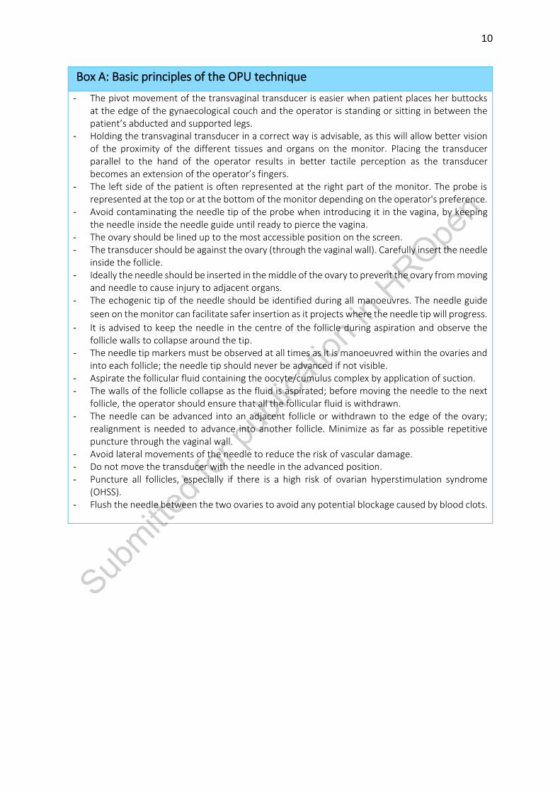
Box A: Basic principles of the OPU technique
- The pivot movement of the transvaginal transducer is easier when patient places her buttocks at the edge of the gynaecological couch and the operator is standing or sitting in between the patient’s abducted and supported legs.
- Holding the transvaginal transducer in a correct way is advisable, as this will allow better vision of the proximity of the different tissues and organs on the monitor. Placing the transducer parallel to the hand of the operator results in better tactile perception as the transducer becomes an extension of the operator’s fingers.
- The left side of the patient is often represented at the right part of the monitor. The probe is represented at the top or at the bottom of the monitor depending on the operator's preference.
- Avoid contaminating the needle tip of the probe when introducing it in the vagina, by keeping the needle inside the needle guide until ready to pierce the vagina.
- The ovary should be lined up to the most accessible position on the screen. - The transducer should be against the ovary (through the vaginal wall). Carefully insert the needle
inside the follicle. - Ideally the needle should be inserted in the middle of the ovary to prevent the ovary from moving
and needle to cause injury to adjacent organs. - The echogenic tip of the needle should be identified during all manoeuvres. The needle guide
seen on the monitor can facilitate safer insertion as it projects where the needle tip will progress.
- It is advised to keep the needle in the centre of the follicle during aspiration and observe the follicle walls to collapse around the tip.
- The needle tip markers must be observed at all times as it is manoeuvred within the ovaries and into each follicle; the needle tip should never be advanced if not visible.
- Aspirate the follicular fluid containing the oocyte/cumulus complex by application of suction. - The walls of the follicle collapse as the fluid is aspirated; before moving the needle to the next
follicle, the operator should ensure that all the follicular fluid is withdrawn. - The needle can be advanced into an adjacent follicle or withdrawn to the edge of the ovary;
realignment is needed to advance into another follicle. Minimize as far as possible repetitive puncture through the vaginal wall.
- Avoid lateral movements of the needle to reduce the risk of vascular damage. - Do not move the transducer with the needle in the advanced position. - Puncture all follicles, especially if there is a high risk of ovarian hyperstimulation syndrome
(OHSS). - Flush the needle between the two ovaries to avoid any potential blockage caused by blood clots.

Submitte
d for
publi
catio
n in H
ROpen
11
Prior to OPU
Pelvic Ultrasound - An ultrasound (US) evaluation should be performed before starting an ART treatment (i) to decide
the ovarian stimulation protocol, (ii) to determine whether there is any anatomical abnormality or
a malposition of the ovaries (Grimbizis et al., 2016) and (iii) to assess ovarian placement and
ovarian/follicular accessibility after previous surgery (gynaecological surgery for myomas,
endometriomas, adhesions). A basic diagnostic US examination also allows for the detection of
recent lesions, such as endometrial abnormalities or ovarian cysts, in a timely manner. In addition,
such transvaginal diagnostic US is of value to visualize not only the ovaries but also the uterus and
to check for potential difficulties during oocyte retrieval. The accessibility of the ovaries and follicles
and any potential complications or difficulties of the OPU should be clearly documented in the
patient case notes, for the team to be prepared and for the patient to be counselled accordingly.
- Pre-OPU 3D Ultrasound and Doppler investigation are considered helpful for the operator to
become familiar with the anatomy of the patient, and to prevent (vascular) complications.
- The time frame to perform the US is at the discretion of the clinician. The American Institute of
Ultrasound in Medicine (AIUM) guidelines suggest a comprehensive sonographic evaluation of the
pelvis within 4-6 months from the start of ovarian stimulation (American Institute of Ultrasound in
Medicine, 2017). This time frame should be shortened in cases of significant conditions
(endometriosis, surgery, specific symptoms). The WG recommends a baseline US closer to the OPU
to highlight any difficulties or reconfirm previous findings, for example shortly before starting the
ovarian stimulation with gonadotrophins.
Vaginal infection screening - Screening for vaginal infection (by taking a vaginal sample for bacteriological examination) should
be performed during diagnostic work-up and can be required based on local guidelines and
regulations. However, the incidence of vaginal infections after OPU is low, and several aspects of
vaginal screening and treatment remain unclear in asymptomatic patients without a history of
pelvic infections, including the relevance of the screening tests, the implications of treatment
before OPU, and the impact on future pregnancy (Amso, 1995, Matorras et al., 2018).
- In women with symptoms of infection, it is recommended to perform specific testing for cervical
and vaginal infections and take appropriate actions.
Patient medical history - As OPU is performed under sedation or anaesthesia, a full blood count (FBC) and any additional test
can be ordered, depending on local regulations regarding pre-operative management. There is no
evidence suggesting value for FBC or any additional test before OPU with regard to preventing
complications.
- Taking accurate patient history before OPU is essential to highlight potential comorbidities and take
actions to prevent any possible associated complications. Patients should at least be asked about
the use of medications - more specifically the use of blood thinning agents (aspirin and others) -,
relevant previous surgeries, and any relevant disease or deficit of coagulation factors.
Information provision and informed consent - Recent or confirmation of (written) informed consent for treatment should be obtained according
to local regulations.
- Verbal and written information should be provided to patients, according to local templates,
explaining the procedure, the risks and their incidence. Counselling should be provided regarding
additional risks associated with specific diagnostic or incidental findings.

Submitte
d for
publi
catio
n in H
ROpen
12
Equipment and consumables
During the OPU, the operator should be equipped as required by European standards and local
regulations.
The following equipment for OPU should be available on a sterile operation table: sterile small gauzes,
and a disposable or reusable speculum for cervical examination and to visualize any bleeding site.
Furthermore, a test tube warmer and heating block should be available (at 37°C), and culture medium
for flushing should be prepared and ready at 37°C. Additional equipment and consumables that might
be used during OPU should also be available in the procedure room, such as ovary clamps, sponge
holder, vaginal surgery equipment, including (absorbable) sutures. Resuscitation equipment, reversal
anaesthetic drugs, a prepared kit for anaphylactic shock treatment and oxygen should also be available
in (the near proximity of) the procedure room. All equipment, materials, and consumables used should
be compliant with European standards.
Ultrasound system and transducer - The ultrasound system should be fit for OPU with a high frequency transvaginal ultrasound
transducer, which offers the best quality in real-time imaging of the field of view.
- The ultrasound system should have the ability to (i) adjust the field of view depth and zoom, (ii)
adjust of the focal zone to the region of interest (except where image processing techniques have
been dispensed with this feature), (iii) image gain adjustment controls, (iv) adjust the acoustic
power, colour and power Doppler capabilities, (v) display the mechanical and thermal indices on
screen, (vi) display the needle guide super-imposed on the field of view and, (vii) print or save
images/cine loops in the system’s hard drive or a central picture archiving and communication
system (PACS).
- The software of the system should be up to date, the system should be calibrated regularly, and it
should be serviced according to the manufacturer’s instructions and any local institutional
requirements. To ensure safety, the system should be timely replaced, as recommended by the
manufacturer.
- Some manufacturers have introduced software that automatically counts and calculates the mean
diameter and volume of follicles. Caution should be taken in correlating these new parameters with
oocyte maturity in comparison with conventional mean follicular diameter.
- The transvaginal transducer, or probe, should have a frequency range of 5-8 MHz and an abdominal
transducer with a frequency range of 2-6 MHz, or their contemporaneous equivalents at the time
of purchase.
- An appropriate protocol for transducer disinfection should be established, in accordance with
manufacturer’s instructions. OPU operators and assistants should be familiar with the disinfection
technique and keep detailed documentation of the disinfection procedure.
- The transducer should be designed for easy application of a specific sterile cover, incorporating a
good quality sonographic gel on the tip of the transducer.
- An appropriate transducer cover should be used, powder-free and compatible with the ultrasound
device, as this may affect the quality of the image. A latex-free cover should be used in case of latex
allergy.
- The use of lubrication (on the outside of the cover) does not offer any improvement for the quality
of the image and should be avoided as there is a hypothetical gametotoxic and embryotoxic effect.
When needed, sterile water or culture media can act as lubricant and conductor of ultrasound
waves.
Submitte
d for
publi
catio
n in H
ROpen
13
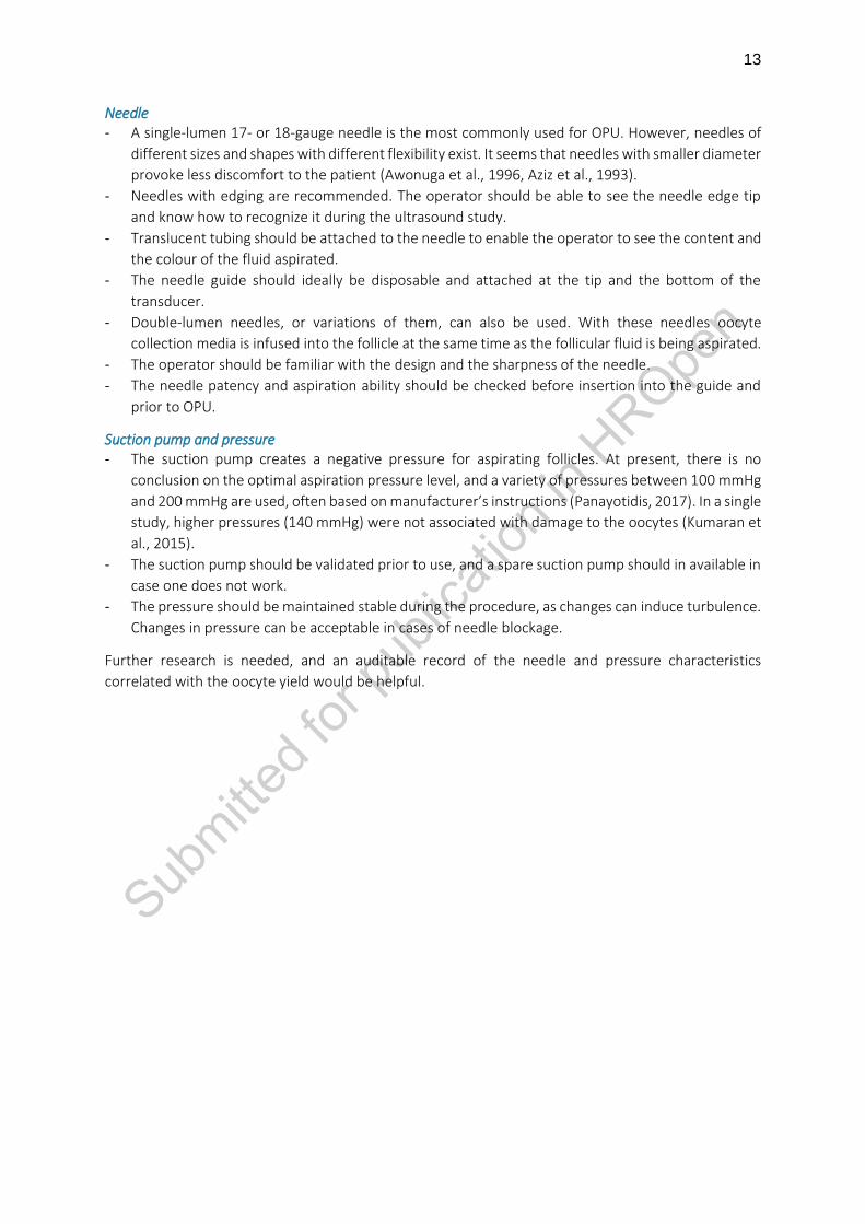
Needle - A single-lumen 17- or 18-gauge needle is the most commonly used for OPU. However, needles of
different sizes and shapes with different flexibility exist. It seems that needles with smaller diameter
provoke less discomfort to the patient (Awonuga et al., 1996, Aziz et al., 1993).
- Needles with edging are recommended. The operator should be able to see the needle edge tip
and know how to recognize it during the ultrasound study.
- Translucent tubing should be attached to the needle to enable the operator to see the content and
the colour of the fluid aspirated.
- The needle guide should ideally be disposable and attached at the tip and the bottom of the
transducer.
- Double-lumen needles, or variations of them, can also be used. With these needles oocyte
collection media is infused into the follicle at the same time as the follicular fluid is being aspirated.
- The operator should be familiar with the design and the sharpness of the needle.
- The needle patency and aspiration ability should be checked before insertion into the guide and
prior to OPU.
Suction pump and pressure - The suction pump creates a negative pressure for aspirating follicles. At present, there is no
conclusion on the optimal aspiration pressure level, and a variety of pressures between 100 mmHg
and 200 mmHg are used, often based on manufacturer’s instructions (Panayotidis, 2017). In a single
study, higher pressures (140 mmHg) were not associated with damage to the oocytes (Kumaran et
al., 2015).
- The suction pump should be validated prior to use, and a spare suction pump should in available in
case one does not work.
- The pressure should be maintained stable during the procedure, as changes can induce turbulence.
Changes in pressure can be acceptable in cases of needle blockage.
Further research is needed, and an auditable record of the needle and pressure characteristics
correlated with the oocyte yield would be helpful.

Submitte
d for
publi
catio
n in H
ROpen
14
OPU preparation (see also Box B: Before OPU-Checklist)
The team performing the OPU should consist at least of the operator and one assistant or nurse. It is
recommended that at least one person in the room is trained in advanced life support (Hinkelbein et
al., 2018).
The identity of the patient should be checked, and the WHO surgical safety checklist (time out) applied
(World Alliance for Patient Safety, 2008).
Ovarian stimulation It is important that OPU is performed according to a precise timing. Most authors recommend a 36 h
interval between medical triggering and OPU, but intervals between 34 h and 38 h have been applied
(Weiss et al., 2014).
- Immediately before OPU, patients should be specifically asked about the timing of human chorionic
gonadotrophin (hCG) or gonadotrophin-releasing hormone (GnRH) agonist injection. In case it is
not clear whether the patient has had hCG administered, an hCG immunology urine test or serum
beta-hCG test should be performed. Serum beta-hCG levels below 23 mUI/ml suggest inadequate
hCG administration (Matorras et al., 2012).The WG does not suggest routine hCG testing for all
patients before OPU.
- In agonist trigger cycles, baseline serum level of luteinizing hormone (LH) should be measured on
the day of the trigger. If no oocytes are found during OPU, LH levels can be checked and compared
with baseline LH levels. If LH levels are below 0,5 mIU/mL, the trigger should be repeated with
recombinant hCG instead of GnRH agonists (Meyer et al., 2015).
- In patients undergoing OPU under general anaesthesia and/or undergoing a natural cycle where
there was the development of only a few follicles and/or when there are concerns of premature
ovulation, transvaginal ultrasound should be performed before starting the procedure.
Patient position and preparation for the procedure - The patient must fast, 6 h for food and 2 h for clear fluids (see also section sedation)
- A peripheral intravenous line should be established, with Ringer’s lactate solution or physiological
saline solution at the minimum speed required in order to keep the vein open. Correction of blood
glucose levels and any other infusions should be performed according to local protocols.
- The patient must empty her bladder immediately prior to OPU. An empty bladder improves the
image quality during transvaginal examination as it decreases the posterior enhancement
(ultrasound artefact), whereas a full bladder can distort the anatomy of the uterus and ovary and
may increase the risk of injuries.
- Patient positioning during OPU needs to be comfortable for both patient and operator.
- Gynaecological positioning in the semi-lithotomy or lithotomy position can facilitate the OPU
manoeuvre. The position of the patient may need to be adapted to patient mobility.
- The operator can be seated or standing during the OPU procedure.
Speculum examination - Speculum vaginal and cervical examination should be performed before OPU to check normal
anatomy and exclude any new leucorrhoea, polyp, or other conditions that could interfere with the
procedure.
Disinfection - Cleansing of the vagina/cervix should be done prior to OPU to minimize bacterial vaginal/cervical
contamination. Vaginal cleansing is commonly done with (warmed) normal saline (Ludwig et al.,
2006, Tobler et al., 2014). Other vaginal preparations are used (e.g. 0.5% chlorohexidine solution,
Submitte
d for
publi
catio
n in H
ROpen
15
povidone iodine or culture medium), but there is no evidence on safety or superiority to normal
saline. Furthermore, these agents may act on the cell membrane and not be safe for oocytes
(Mangram et al., 1999). Povidone–iodine preparations have been shown to be toxic to murine
oocytes/embryos (Hershlag et al., 2003)
- Further studies are needed on the safety of antiseptic methods, and how different agents may
influence reproductive outcomes and post-OPU complications (Funabiki et al., 2014, Tsai et al.,
2005).
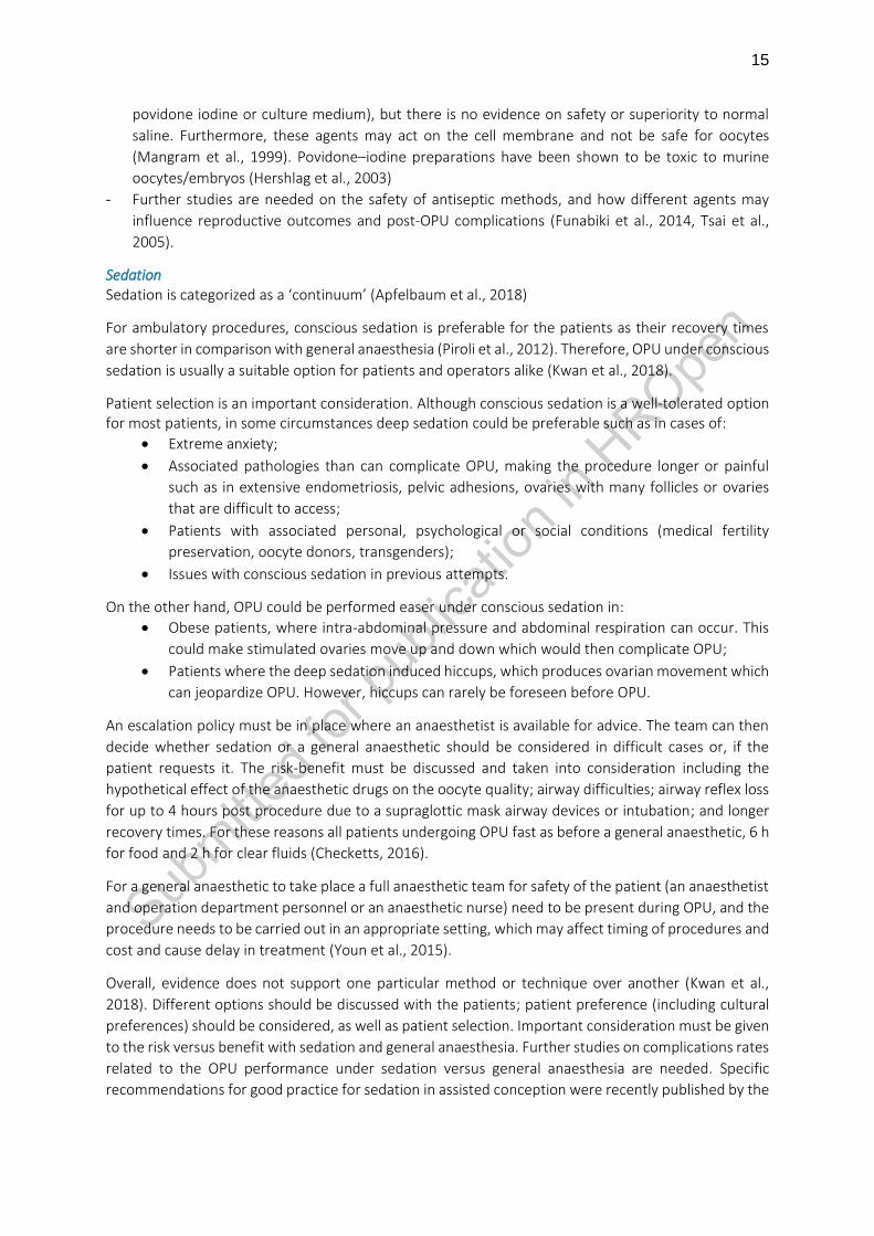
Sedation Sedation is categorized as a ‘continuum’ (Apfelbaum et al., 2018)
For ambulatory procedures, conscious sedation is preferable for the patients as their recovery times
are shorter in comparison with general anaesthesia (Piroli et al., 2012). Therefore, OPU under conscious
sedation is usually a suitable option for patients and operators alike (Kwan et al., 2018).
Patient selection is an important consideration. Although conscious sedation is a well-tolerated option for most patients, in some circumstances deep sedation could be preferable such as in cases of:
• Extreme anxiety;
• Associated pathologies than can complicate OPU, making the procedure longer or painful
such as in extensive endometriosis, pelvic adhesions, ovaries with many follicles or ovaries
that are difficult to access;
• Patients with associated personal, psychological or social conditions (medical fertility
preservation, oocyte donors, transgenders);
• Issues with conscious sedation in previous attempts.
On the other hand, OPU could be performed easer under conscious sedation in:
• Obese patients, where intra-abdominal pressure and abdominal respiration can occur. This
could make stimulated ovaries move up and down which would then complicate OPU;
• Patients where the deep sedation induced hiccups, which produces ovarian movement which
can jeopardize OPU. However, hiccups can rarely be foreseen before OPU.
An escalation policy must be in place where an anaesthetist is available for advice. The team can then
decide whether sedation or a general anaesthetic should be considered in difficult cases or, if the
patient requests it. The risk-benefit must be discussed and taken into consideration including the
hypothetical effect of the anaesthetic drugs on the oocyte quality; airway difficulties; airway reflex loss
for up to 4 hours post procedure due to a supraglottic mask airway devices or intubation; and longer
recovery times. For these reasons all patients undergoing OPU fast as before a general anaesthetic, 6 h
for food and 2 h for clear fluids (Checketts, 2016).
For a general anaesthetic to take place a full anaesthetic team for safety of the patient (an anaesthetist
and operation department personnel or an anaesthetic nurse) need to be present during OPU, and the
procedure needs to be carried out in an appropriate setting, which may affect timing of procedures and
cost and cause delay in treatment (Youn et al., 2015).
Overall, evidence does not support one particular method or technique over another (Kwan et al.,
2018). Different options should be discussed with the patients; patient preference (including cultural
preferences) should be considered, as well as patient selection. Important consideration must be given
to the risk versus benefit with sedation and general anaesthesia. Further studies on complications rates
related to the OPU performance under sedation versus general anaesthesia are needed. Specific
recommendations for good practice for sedation in assisted conception were recently published by the

Submitte
d for
publi
catio
n in H
ROpen
16
British Fertility Society (Acharya et al., 2019). Further information on different types of sedation is
provided in table 2.
Monitoring Non-invasive blood pressure and pulse oximetry must be used when drugs have been administered
intravenously; electrocardiogram (ECG) and CO2 monitoring are developmental standards for
conscious sedation but are minimal monitoring standards for deep sedation and general anaesthesia
(Checketts, 2016).
Post-operative analgesia Most studies have reported very little difference between analgesia pre-procedure analgesia
immediately after starting. The most favourable post-operative pain management is that of a
multimodal peri-operative approach, using intra-operative analgesia with opiates and local anaesthetic
block with oral/IV/per rectal medication (Vadivelu et al., 2014). Post-operative analgesia can consist of
the following:
• Oral Paracetamol 1 g + Codeine 60 mg is shown to be more effective than paracetamol alone
(Zhang and Li Wan Po, 1996).
• NSAIDs can be given (if no contraindications) about 1-1.5 h prior to the procedure.
• Per rectal diclofenac (100 mg) can be given post procedure, followed by IV Paracetamol (15
mg/Kg) in the recovery. Whether given orally or rectally, this has the same post-operative
analgesic effect, although the rectal dose has a faster onset of action.
Sperm collection It is not exceptional that the semen sample cannot be obtained on the day of OPU. Sperm collection at
home or storing a previous obtained sperm sample as back-up is advisable. If a back-up sample is not
available, oocyte cryopreservation, or further options for sperm retrieval (administration of sildenafil,
or surgical sperm retrieval) can be considered. In any case, patients should be counselled on the
possibility of such complications and informed consent should be obtained before starting the OPU
procedure.
Antibiotic prophylaxis Patients with history of endometriosis, pelvic inflammatory disease (PID), pelvic adhesions, dermoids,
or previous pelvic surgery can be considered at high risk for pelvic infection. In these patients,
administration of antibiotics is recommended shortly before or during OPU (according to local
protocols).
There is no evidence for the use of antibiotic prophylaxis in low risk patients, and this can be decided
according to local protocols and regulations, taking into account generic antibiotic resistance (Aslam et
al., 2018) and the lack of studies on the effect on uterine environment.
Further evidence (studies or observational/audit data) should be collected on infection rates and their
association with antibiotic administration.
Change to transmyometrial or laparoscopic oocyte retrieval In exceptional cases, transmyometrial or laparoscopic oocyte retrieval may be required, usually due to
abnormal ovarian placement or tubal adhesions. For transmyometrial OPU, there were no significant
differences in oocyte recovery rates, implantation rates and pregnancy rates compared to transvaginal
OPU (Davis and Ginsburg, 2004, Roman-Rodriguez et al., 2015). An anaesthesiologist may be present
during the procedure, depending on local protocol.
Submitte
d for
publi
catio
n in H
ROpen
17
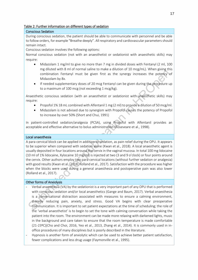
Table 2: Further information on different types of sedation
Conscious Sedation
During conscious sedation, the patient should be able to communicate with personnel and be able to follow orders, for example "Breathe deeply”. All respiratory and cardiovascular parameters should remain intact. Conscious sedation involves the following options:
Normal conscious sedation (not with an anaesthetist or sedationist with anaesthetic skills) may
require:
• Midazolam 1 mg/ml to give no more than 7 mg in divided doses with Fentanyl (2 ml, 100
mg diluted with 8 ml of normal saline to make a dilution of 10 mcg/mL). When giving this
combination Fentanyl must be given first as the synergy increases the potency of
Midazolam by 8x.
• If needed supplementary doses of 20 mcg Fentanyl can be given during the procedure up
to a maximum of 100 mcg (not exceeding 1 mcg/kg).
Anaesthetic conscious sedation (with an anaesthetist or sedationist with anaesthetic skills) may require:
• Propofol 1% 18 mL combined with Alfentanil 1 mg (2 ml) to provide a dilution of 50 mcg/ml.
• Midazolam is not advised due to synergism with Propofol causes the potency of Propofol
to increase by over 50% (Short and Chui, 1991)
In patient-controlled sedation/analgesia (PCSA), using Propofol with Alfentanil provides an acceptable and effective alternative to bolus administration (Roseveare et al., 1998).
Local anaesthesia
A para-cervical block can be applied in addition to sedation, as pain relief during the OPU. It appears to be superior when compared with sedation alone (Kwan et al., 2018). A local anaesthetic agent is usually deposited in four locations around the cervix in the vaginal mucosa. In total 100 mg lidocaine (10 ml of 1% lidocaine, Xylocaine 10 mg/ml) is injected at two (3 and 9 o’clock) or four points around the cervix. Other authors employ two para-cervical locations (without further sedation or analgesia) with good results (Kwan et al., 2018, Rolland et al., 2017). Satisfaction with the procedure was higher when the blocks were used during a general anaesthesia and postoperative pain was also lower (Rolland et al., 2017).
Other forms of Anxiolysis
- Verbal anaesthesia (VA) by the sedationist is a very important part of any OPU that is performed
with conscious sedation and/or local anaesthetics (Gange and Baum, 2017). Verbal anaesthesia
is a conversational distraction associated with measures to ensure a calming environment,
thereby reducing pain, anxiety, and stress. Good VA begins with clear preoperative
communication. It is important to set patient expectations at the time of scheduling; the role of
the ‘verbal anaesthetist’ is to begin to set the tone with calming conversation while taking the
patient into the room. The environment can be made more relaxing with darkened lights, music
in the background and care taken to ensure that the room temperature is made comfortable
(21-23ºC)(Cho and Choi, 2016, Yeo et al., 2013, Zhang et al., 2014). It is commonly used in in-
office procedures of many disciplines but is poorly described in the literature.
- Hypnosis is another form of anxiolytic which can be used to achieve better patient satisfaction,
fewer complications and less drug usage (Faymonville et al., 1995).
Submitte
d for
publi
catio
n in H
ROpen
18
BOX B. Before OPU-Checklist
✓ It is important that before starting OPU, the entire system is tested by aspirating some
culture medium (Null Aspiration).
✓ Make sure that the suction pump is turned on and that the suction pedal is functioning (many
aspiration pumps have a light flashed, and some have audible signals, when the pump is
activated).
✓ Check the setting of the suction pressure taking into account the maximum pressure limit of
the suction pump.
✓ Check the needle connection with the tubing system and ensure that they are tightly
connected with no air leakage
✓ Ensure that the suction tubing system is new, or undamaged (when the system is re-used)
✓ Exclude any cracks in the aspiration test tube.
✓ Ensure that the collection tubing is not kinked or damaged.
✓ Check the needle and the connection of the needle with the tubing system.
✓ Ensure that the needle guide is appropriately placed on the ultrasound probe with no gaps
and that the protective probe sheath is not on the way of the needle projection.
✓ Ensure that the needle guide is well applied on the probe and not loose.
Submitte
d for
publi
catio
n in H
ROpen
19
OPU procedure
Setting and image optimization - The surgical theatre/procedure room for OPU should be of reasonable size and in semi-darkness,
as this allows a better visualization of the ultrasound images and the hypothetical adverse effect of
the light into the oocytes is precluded. The preferred temperature is about 22-23°C, and if
necessary, a warming blanket and socks can be used by the patient. Hypothermia has been
associated with increased perception of pain.
- During OPU, the ultrasound field and anatomical orientation are set from the top or from the lower
part of the monitor. The representation of the transducer from the lower part of the monitor may
help the controlled manipulation of the transducer. The initial structures are seen exactly at the
beginning of the transducer as seen in the monitor and the tactile sensation during the scanning
and needle manipulation is more realistically represented. This point is very important as the
anatomy is better visualized, which is of crucial importance for safety of the procedure. The
laterality is again a parameter that can be set depending on the operator's preference.
- Before starting the surgical procedure, the pelvis should be systematically scanned to assess the
anatomy and check for incidental findings. Special attention should be focused on identification of
big iliac vessels to avoid wrong interpretation as a follicle. The needle guide track should be on the
screen. It is relevant to first have a panoramic view of the ovary and then have a closer look. A
larger view field is preferable during the OPU—magnify until the whole ovary occupies 75% of the
field (depending on the dynamics of the ovary)—to ensure visualization of the intrapelvic part of
the needle during the OPU.
- In case of doubt, Doppler study is advised for recognition of the vascular structures [positioned in
the line between transducer (vaginal wall) and ovary], and to reduce the risk of haemorrhagic
complications (Risquez and Confino, 2010). Using Doppler imaging during the OPU procedure may
be seen as an additional complex study to perform switching from bi-dimensional to Doppler image
and it is not recommended to do this during the OPU. However, Doppler study can be useful to
detect vascular areas in case of doubt in 2D-ultrasound imaging before OPU. It could differentiate
from the hypo-echogenic areas that look alike as superficial follicles versus iliac or para-ovarian
vessels (position, content, fluid movement). Further research is needed to answer whether this
modality of imaging needs to be applied routinely before starting OPU in order to further decrease
the risks of an accidental vascular trauma.
- Information regarding the perifollicular Doppler vascularization, before OPU, could be used for
academic purposes (for instance oocyte quality) (Bhal et al., 1999).
- Adaptation of the ultrasound frequency with other image adjustments should be considered in real-
time to improve the clarity of the image and facilitate the accurate visualization of the needle. The
ultrasound frequency used for OPU varies between 5 and 7 MHz to obtain sufficient resolution and
depth penetration. Additional filters and image adjustment set-ups can be activated to improve the
image.
- Recording the OPU procedure can be a useful audit tool for quality control of ultrasound image,
learning and teaching as well as to explore factors related to complications. Video recording can be
used in prevention of future complications and improvement of OPU techniques.
Submitte
d for
publi
catio
n in H
ROpen
20
Technique - The transvaginal ultrasonographic transducer must be gently applied well into the vaginal wall in
order to position the ovary just adjacent to vaginal fornices. There should be no space between
vaginal transducer and ovarian cortex, thus avoiding any bowel loop within the trajectory of the
needle. To stabilize the ovary in one place, external abdominal pressure or suprapubic pressure can
be applied (with the help of an assistant) towards the vaginal fornix of the patient by the site (right
or left) where OPU is performed. This can move the ovarian follicles closer to the vaginal wall,
thereby avoiding multiple ovarian punctures. Push the puncture needle through the needle guide
to the vaginal top and gently puncture the vaginal wall until just below the ovary, then puncture
the nearest follicle in one movement.
- The operator needs to be familiar with the tactile resistance when the follicle /ovary is penetrated
with the needle, and able to manipulate the transducer which produces the ultrasound image. A
fingertip handle on the distal end of the needle can facilitate the puncture with good clinical touch.
In case of resistance or hard-to-reach follicle(s), one should pull back the needle in one movement.
- Techniques for tracking the needle (edging) should be used. The edging of the needle should always
be visible.
- Follicle curetting involves gently and rapidly rotating the needle in a clockwise and counter-
clockwise fashion inside the follicle after complete aspiration of the follicular fluid (Yao and Schust,
2002). It has been suggested (in a single retrospective study) that follicle curetting during OPU could
increase the number of recovered oocytes as well as the number of mature oocytes, without
damaging the oocyte, and prevent the adhesion of granulosa cell layer to the needle lumen which
could block it during aspiration (Dahl et al., 2009). More studies are needed to confirm the clinical
value and safety of this technique
- Vacuum suction should be used just before follicle penetration. The pressure of the pump suction
must be calibrated to 100-220 mmHg (according to manufacturer’s instructions) just before
starting, and it should be kept constant during the procedure. The pedal for the pump can be
controlled by the operator or an assistant. Collapse of the follicle should be visualized when
aspirating in order not to lose oocytes. If the collapse of the follicle cannot be seen, the oocyte can
remain in the follicular cavity.
- The needle should be gently withdrawn without negative suction pressure to avoid sudden forward
flow of follicular fluid towards the collection tube (Horne et al., 1996). The movement of the hand
holding the transducer should be minimal when the needle is in the ovary. Lateral movements
cause more pain and can result in increased intra-ovarian bleeding.
- Small follicles (<10 mm) can be left unpunctured (to avoid collection of immature eggs), unless
there is a high risk of OHSS.
- Experts prefer to start the OPU from the ovary nearest to the vaginal probe rather than the ovary
with the largest follicles or complex appearances, because in hyperstimulated ovaries sometimes
the length of needle cannot reach the length of ovary, and the procedure can be dangerous if near
vascular structures. The laterality, whether to prefer the right versus the left ovary, is based on
operator’s preferences rather anatomical considerations.
- One should use both planes, longitudinal and transverse, while performing the OPU in order to be
sure about the anatomy and boundaries of the ovarian cortex.
- It is preferred to maintain the needle within the ovary—avoiding repetitive punctures or ovarian
penetrations—during the OPU as this could reduce the risk of complications, mainly ovarian surface
bleeding. Multiple punctures should be avoided as much as possible.
- It is recommended to access as many follicles as is safely possible through the same ovarian cortex
puncture.
- Manipulation of the OPU needle should be gentle and steady, avoiding abrupt movements.
Submitte
d for
publi
catio
n in H
ROpen
21
Flushing - Follicular flushing has been proposed to increase the number of retrieved oocytes. Closed flushing
(i.e. every follicle is rinsed three to four times, and tubes are passed on to the laboratory when all
follicles are punctured) has been recommended for patients with >6 follicles, and open flushing (i.e.
with direct communication between the laboratory staff and the operator; the follicle is rinsed until
an oocyte is detected in the laboratory, or until no cell material is detected) for those with ≤6
follicles. However, studies on flushing performed failed to show any benefit (Georgiou et al., 2018).
The results of this technique should be regularly audited. Flushing needs to be performed with a
double-lumen needle to reduce damage to the oocyte.
- Oocyte recovery (see also BOX C: Troubleshooting during OPU)
- The follicular fluid should be collected in preheated test tubes held in a heating block calibrated at
37°C.
- Embryology laboratory staff should inform the medical doctor of oocytes and granulosa cells during
OPU procedure in order to differentiate empty follicle syndrome and wrong timing of hCG injection.
- In case of suspected premature ovulation, peritoneal fluid can be aspirated at the end of OPU in
search of additional oocytes that could have been ovulated or fallen into the peritoneal cavity
during the procedure.
End of procedure - At the end of the procedure, the ovary should be checked to see whether all follicles were
punctured and to detect any internal bleeding.
- Speculum examination should be performed to check for vaginal bleeding.
- If abdominal bleeding is suspected, transabdominal US should be performed before moving the
patient.
- Post-OPU vaginal compression with a swab may enhance haemostasis and stop potential vaginal
bleeding, which may otherwise be a disturbing finding for the couple after OPU. With prolonged
bleeding, packing can be applied (No, 2016). A pad can also be used after vaginal compression for
monitoring of vaginal bleeding. If necessary, a haemostatic suture is placed.
- Post-OPU analgesia (paracetamol, ibuprofen) should be considered, especially in cases where more
than 10 oocytes are retrieved and in patients with endometriosis.
- The proportion of OPUs without obtaining an oocyte is usually 1-2% (Ben-Shlomo et al., 1991,
Matorras et al., 2012, Traina et al., 1993). This lack of oocyte recovery is much more common in
women with few adequately sized follicles (Zreik et al., 2000). It has been reported that in 5-20% of
dominant adequately sized follicles, no oocytes are retrieved (Coskun et al., 2011, Coskun et al.,
2010, Nargund et al., 2001). Furthermore, poor responders may have an increased rate of impaired
folliculogenesis and oocyte may have lower quality (Matorras et al., 2014). (see box B)
• hCG determination is mandatory for patients with no oocyte retrieval in hCG triggered cycles,
to assess the correct hCG administration. No oocyte retrieval with high hCG levels could
indicate an ectopic pregnancy (Bringer-Deutsch et al., 2010)
• In cases were OPU failed to recover oocytes and the administration of the medication was
adequate, performing a (rescue) intrauterine insemination is associated with very low
pregnancy rates (< 7%) and should not be done (Matorras et al., 2014).
Submitte
d for
publi
catio
n in H
ROpen
22
Box C. Troubleshooting during OPU
What to do when the needle tip is not visualized? ✓ Withdraw the needle and ensure that the needle director/gauge is in place allowing the
needle to move in the same sector as the ultrasound beam
What to do when suction fails? ✓ Rotate the needle within the follicle to ensure that it is not blocked by follicular wall tissue.
✓ If there is still no suction, remove the needle and perform a “retrograde flush” to clear any
blockage.
✓ Before re-inserting the needle, re-check by aspirating some culture medium.
✓ In case of a double-lumen needle, the needle can be flushed without taking it out of the
ovary
What to do when no oocytes are retrieved? When no oocytes are discovered, or if the fluid collected is very clear and devoid of (granulosa and cumulus) cells after aspiration of the first follicles, suspicion may be raised that the patient has not had her trigger. In case of human chorionic gonadotrophin (hCG) trigger
✓ Before follicles from the second ovary are aspirated, a urinary pregnancy test or beta-hCG
serum test can be performed. Alternatively, follicular fluid can be tested with a urinary
pregnancy test strip (Enien et al., 1995).
In case of agonist trigger (triptorelin). ✓ An ovulation test can be performed.
✓ The serum LH levels can be checked.
- If the test shows that the patient did not receive the trigger, a new dose may be administered
and the OPU repeated after 36 h. If the time interval since OPU was too short, OPU can be
delayed.
- If premature ovulation is suspected, peritoneal fluid can be aspirated to recover some oocytes.
Submitte
d for
publi
catio
n in H
ROpen
23
Post-procedure care (see also BOX D: After OPU-Checklist)
- After the procedure, patients should remain in bed at the centre for recovery (about 2 hours, or
less if the procedure was performed with local anaesthetics only). General status, abdominal
distension, blood pressure and heart rate should be monitored by a nurse.
- In cases of significant pain or abdominal distension, a blood analysis and/or an ultrasound scan,
should be performed before discharge to check for potential intra-abdominal bleeding.
- Patients should be able to eat, drink, and pass urine before discharge. Furthermore, it is important
to check awareness, orientation, and respiratory rate. A written information leaflet about post-care
procedures, complications, and a 24 h emergency number should be provided.
- In patients with hematoma, bleeding or infection after the OPU, antibiotic coverage is
recommended.
- Procedures should be in place for when severe complications occur, including hospital admission
arrangements, specialist responsibility and continued care.
Box D: After OPU checklist
✓ Monitor general status and be alert for complications.
✓ Provide patients with a leaflet with written information and a 24 h emergency number.
Submitte
d for
publi
catio
n in H
ROpen
24
Associated pathologies and cautions during OPU
- Standard management of hydrosalpinx should be removal or clipping before OPU (Song et al.,
2017). When a hydrosalpinx is only discovered during OPU, the first option should be oocyte or
embryo cryopreservation. The benefit of aspiration on the day of OPU needs further study (Fouda
et al., 2015, Hammadieh et al., 2008, Zhou et al., 2016).
- Patients with potential infectious risk (HIV, hepatitis) should be managed in an isolated circuit or in
specialized centres to avoid cross-contamination.
- In women with endometriosis, OPU can be challenging and it may affect the individual operator’s
or centre’s performance rate (Kasapoglu et al., 2018). Endometriomas should not be aspirated.
During OPU, the puncture of endometriomas should be avoided to prevent contamination of the
follicular aspirate and reduce the risk of intra-abdominal infection. However, piercing the
endometrioma is often the only way to avoid losing an important number of oocytes. Dermoid cysts
should not be punctured during OPU, since this could increase the risk of PID and peritonitis. In
patients with an endometrioma or teratoma, the risk of PID is increased, even if they have not been
punctured (Benaglia et al., 2008, Kasapoglu et al., 2018, Moini et al., 2005, Villette et al., 2016).
These patients should be counselled preoperatively and consent obtained appropriately.
- If an endometrioma or a hemorrhagic follicle is inadvertently punctured, the needle should be
immediately withdrawn and flushed with media, and the collecting tube should be changed.
- In patients with borderline ovarian tumours, it is unclear whether ART procedures are associated
with an increased risk of recurrence (Denschlag et al., 2010).
- Increased risk for bleeding has been suggested in lean women, and in women with polycystic ovary
syndrome (PCOS) (Liberty et al., 2010, Zhen et al., 2010).
Submitte
d for
publi
catio
n in H
ROpen
25
Complications and risks
ESHRE IVF Monitoring (EIM) data collection reports data on complications from OPU. In the latest data
available, including 776 556 cycles, complications from OPU were reported in 1328 cycles (0.17%),
including 919 bleeding (0.11% of cycles), 108 infections (0.013%), and 301 (0.038%) other
complications. There were three maternal deaths reported as complications of ART, but none were
related to the OPU procedure (De Geyter et al., 2018).
Large observational studies in oocyte donors undergoing OPU have reported low incidences of
complications (0.42% in (Bodri et al., 2008), 0.7% in (Maxwell et al., 2008)). The most common reported
complications were OHSS (although a complication of ovarian stimulation rather than OPU) and intra-
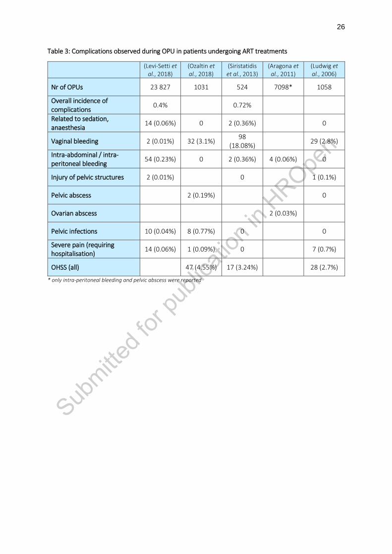
abdominal bleeding. Large observational studies have also been conducted (Table 3). Overall, the
incidence and severity of the reported complications are low (Aragona et al., 2011, Ludwig et al., 2006,
Ozaltin et al., 2018, Siristatidis et al., 2013).
Associated conditions may increase the risk of complications, but very little information is available,
and over-and under-reporting have been suggested (Villette et al., 2016). Recommendations regarding
associated conditions during OPU have been addressed above.
Apart from data collection and observational studies (Aragona et al., 2011, Ludwig et al., 2006, Ozaltin
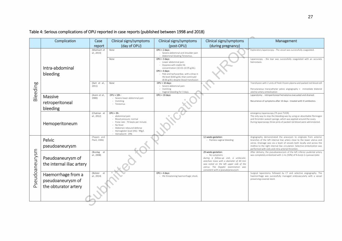
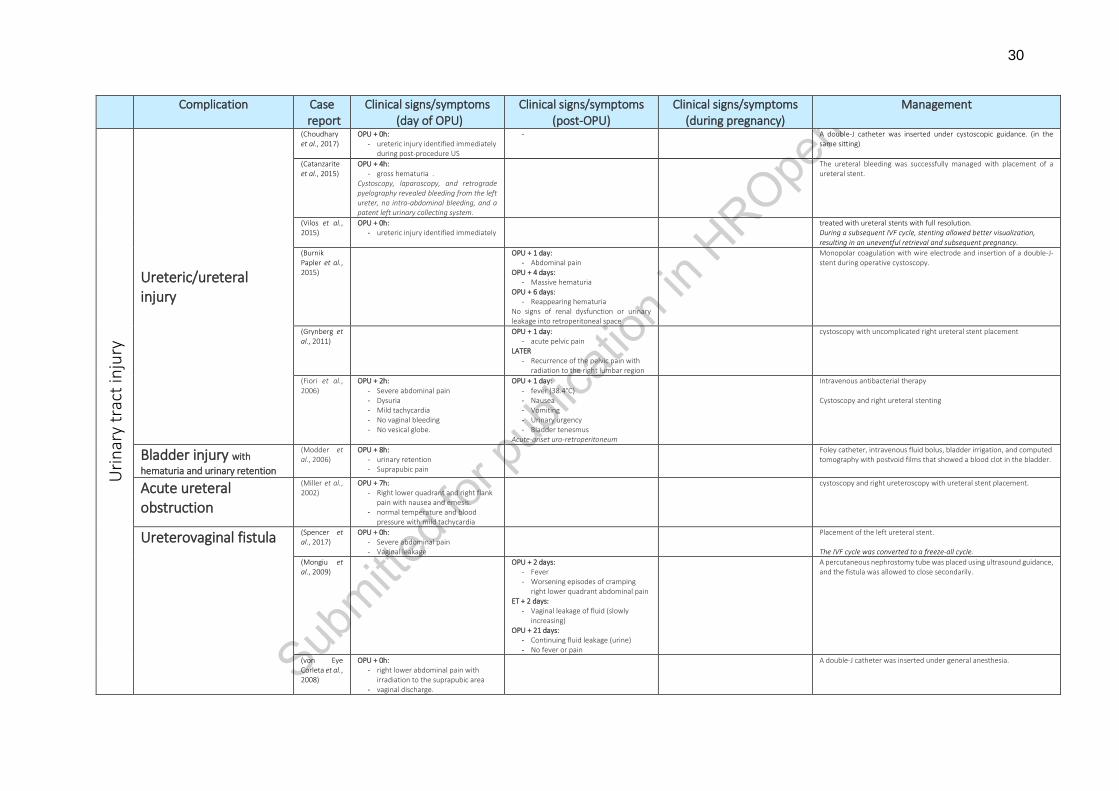
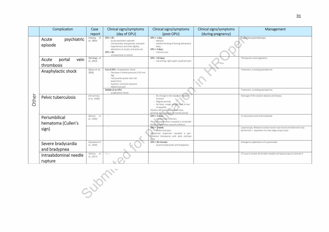
et al., 2018, Siristatidis et al., 2013), most reports on serious complications during and after OPU have
been published in case reports (Table 4). Reported complications include bleeding, infection, urinary
tract injury, and pseudoaneurysm.
• Infection: An infection can originate from the vaginal puncture during the OPU procedure
where there is a contamination from vaginal bacteria into the intra-peritoneal space (Kelada
and Ghani, 2007). The presence of pre-existent latent pelvic infection or pelvic endometriosis
or teratoma may be another contributing factor. In some difficult cases, puncture of
hydrosalpinx or an accidental puncture of an attached bowel loop during the procedure may
occur, which may lead to severe septicaemia (Amso, 1995).
• Bleeding: The quantity of blood loss following OPU is clinically unremarkable in most women.
In a prospective study of 150 consecutive OPUs, the estimated median blood loss was 72 ml
(interquartile range -8-162 ml) (Ragni et al., 2009). None of the recruited women was found
to have signs of haemoperitoneum (Ragni et al., 2009). Haemoperitoneum after OPU has
been defined as a haemoglobin reduction >2g/day, an increase in the pelvic free fluid >200
ml or a calculated blood loss >500 ml.
- After para-cervical block, a transient leg paresis may develop, which usually disappears after 2-4 h.
- The IVF centre should have proactively established policies with respect to how to provide patient
resuscitation and access to surgical theatre in case of internal bleeding or other organ injury in a
haemodynamically unstable patient (safety standard).
- OPU video recording may help to identify reasons for complications and how to improve the OPU
technique with respect to ultrasound settings for clear imaging and types of manoeuvres during
oocyte aspiration.
All severe complications should be registered according to local requirements. It is recommended that
more details on severe complications are gathered from the EIM data collection.
Submitte
d for
publi
catio
n in H
ROpen
26
Table 3: Complications observed during OPU in patients undergoing ART treatments
(Levi-Setti et al., 2018)
(Ozaltin et al., 2018)
(Siristatidis et al., 2013)
(Aragona et al., 2011)
(Ludwig et al., 2006)
Nr of OPUs 23 827 1031 524 7098* 1058
Overall incidence of complications
0.4% 0.72%
Related to sedation, anaesthesia
14 (0.06%) 0 2 (0.36%) 0
Vaginal bleeding 2 (0.01%) 32 (3.1%) 98
(18.08%) 29 (2.8%)
Intra-abdominal / intra-peritoneal bleeding
54 (0.23%) 0 2 (0.36%) 4 (0.06%) 0
Injury of pelvic structures 2 (0.01%) 0 1 (0.1%)
Pelvic abscess 2 (0.19%) 0
Ovarian abscess 2 (0.03%)
Pelvic infections 10 (0.04%) 8 (0.77%) 0 0
Severe pain (requiring hospitalisation)
14 (0.06%) 1 (0.09%) 0 7 (0.7%)
OHSS (all) 47 (4.55%) 17 (3.24%) 28 (2.7%)
* only intra-peritoneal bleeding and pelvic abscess were reported
Submitte
d for
publi
catio
n in H
ROpen
27
Table 4: Serious complications of OPU reported in case reports (published between 1998 and 2018)
Complication Case report
Clinical signs/symptoms (day of OPU)
Clinical signs/symptoms (post-OPU)
Clinical signs/symptoms (during pregnancy)
Management
Ble
edin
g
Intra-abdominal bleeding
(Mashiach et al., 2013)
None OPU + 2 days: - Severe abdominal and shoulder pain - Abdominal bloating Tenesmus.
Exploratory laparoscopy - The vessel was successfully coagulated.
None OPU + 3 days: - Lower abdominal pain - Dyspnea with stable Hb
concentration (10.43–10.95 g/dL). OPU + 4 days:
- Pale and tachycardiac, with a drop in Hb level (8.84 g/dL) that continued (8.66 g/dL) despite blood transfusion
Laparoscopy - the tear was successfully coagulated with an accurate hemostasis.
(Kart et al., 2011)
None OPU + 10 days: - Severe abdominal pain - Vomiting - Vaginal bleeding for 3 days.
Transfusion with 2 units of fresh-frozen plasma and packed red blood cell Percutaneous transcatheter pelvic angiography + immediate bilateral uterine artery embolization
Massive retroperitoneal bleeding
(Azem et al., 2000)
OPU + 10h : - Severe lower abdominal pain - Vomiting - Tenesmus
OPU + 10 days:
Laparotomy - retroperitoneal hematoma evacuated and drained. Recurrence of symptoms after 10 days - treated with IV antibiotics.
Hemoperitoneum
(Chatrian et al., 2012)
OPU + 3h: - abdominal pain - Blood pressure: normal - Pulse rate : 70 beats per minute. - No fever - Abdomen rebound defense - Hemoglobin level (Hb): 99g/L - Hematocrit: 29%
emergency laparoscopy (7h post TVOR) The only way to stop the bleeding was by using an absorbable fibrinogen and thrombin sealant sponge, which was applied around the ovary. During laparoscopy three pints of packed red blood were administered.
Pse
ud
oan
eury
sm
Pelvic pseudoaneurysm
(Pappin and Plant, 2006)
12 weeks gestation: - Painless vaginal bleeding
Angiography demonstrated the aneurysm to originate from anterior branches of the left internal iliac artery close to the lower uterus and cervix. Drainage was via a leash of vessels both locally and across the midline to the right internal iliac circulation. Selective embolization was performed with coils and intra-arterial thrombin.
Pseudoaneurysm of the internal iliac artery
(Bozdag et al., 2008)
29 weeks gestation: - No symptoms
during a follow-up visit, a unilocular, anechoic mass with a diameter of 40 mm was noted on the left upper side of the uterus. The Doppler examination was consistent with a (pseudo)aneurysm.
After delivery; the pseudoaneurysm of the left inferior pudental artery was completely embolized with 1 mL (50%) of N-butyl-2-cyanoacrylate
Haemorrhage from a pseudoaneurysm of the obturator artery
(Bolster et al., 2014)
OPU + 4 days: - life threatening haemorrhagic shock.
Surgical laparotomy followed by CT and selective angiography. The haemorrhage was successfully managed endovascularly with a vessel preserving covered stent.

Submitte
d for
publi
catio
n in H
ROpen
28
Complication Case
reports Clinical signs/symptoms
(day of OPU) Clinical signs/symptoms
(post-OPU) Clinical signs/symptoms
(during pregnancy) Management
Infe
ctio
n
Pelvic abscess
(den Boon et al., 1999)
end of 2nd trimester: Rupture of bilateral ovarian abscesses
emergency laparotomy was necessary because of an acute abdomen. Severe maternal and neonatal morbidity, preterm birth and neonatal death.
(Patounakis et al., 2012)
gestation of 11 weeks + 2 days: - left lower quadrant abdominal pain.
Serial pelvic ultrasounds showed growth of the mass from 13.2 to 15 cm over 3 days and a viable twin pregnancy (Streptococcus anginosus)
Left salpingo-oophorectomy for resection of the mass. Complete spontaneous pregnancy loss by vaginal delivery of both fetuses on post- operative day 1
(Asemota et al., 2013)
OPU + 6 days: Actinomycosis pelvic abscess - urinary retention - pelvic pain - Fever
6 days of intravenous antibiotics CT-guided drainage of the pelvic abscesses
Tubo-ovarian abscess
(Han et al., 2015)
gestation of 31 weeks and 2 days - Lower abdominal pain for 8 hours
Emergent exploratory laparotomy and cesarean section to terminate gestation. + IV antibiotics
(Kim et al., 2013)
7th week of gestation: - Intermittent right lower abdominal
pain. US: 1 fetus appropriate for gestational age and growth of the mass (10.6 x 7.4 cm) 14 weeks:
- Right abdominal pain
Laparoscopy. The abscess was encapsulated within the ovary and there was no pus within the pelvis. IV cefotiam (1 g every 12 hours for 10 days) and metronidazole (500 mg every 8 hours for 5 days) Spontaneous delivery at 37 weeks and 3 days of gestation without any complications.
(Romero et al., 2013)
OPU + 1month: - 8 cm pelvic abscess
surgical drainage
OPU + 2 months: - 9 cm pelvic abscess
IV antibiotics (did not resolve) + surgical drainage
OPU + 3weeks: - 9 cm pelvic abscess
IV antibiotic treatment (favorably response) + surgical drainage and right adnexectomy.
(Yalcinkaya et al., 2011)
early pelvic infection Broad spectrum antibiotics TV-US-guided drainage was performed, posterior colpotomy and T-drain replacement into the cul de sac. (OPU + 9days) Pregnancy follow-up uncomplicated
Van Hoecke, 2013 #890;
bacteremia due to Actinomyces urogenitalis. Bacteremia was secondary to a tubo-ovarian abscess
(Kelada and Ghani, 2007)
OPU + 16 days : - Left iliac fossa pain for 5 days. - Diarrhea - Vomiting 3 times - Fresh vaginal bleeding.
Bilateral ovarian abscesses (staphylococci)
Laparotomy, a large amount of pus was drained on incising the capsule of each ovary. The peritoneal cavity was washed with normal saline. Two drains were placed through the abdominal wall in the pouch of Douglas, IV Gentamicin and Clindamycin were continued postoperatively.
(Sharpe et al., 2006)
30 weeks gestation: - Low-grade fever
broad-spectrum antibiotics Abscess was drained percutaneously after cesarean delivery of twins
(Matsunaga et al., 2003)
16 weeks gestation: - Fever - Lower abdominal pain
20 weeks gestation: readmitted - Fever - Lower abdominal pain - Small amount of bloody discharge
Treatment with IV antibiotics Left salpingo-oophorectomy after delivery Delivered at 22 weeks of gestation.
(Varras et al., 2003)
- abdominal pain, fever and leukocytosis

Submitte
d for
publi
catio
n in H
ROpen
29
Complication Case report
Clinical signs/symptoms (day of OPU)
Clinical signs/symptoms (post-OPU)
Clinical signs/symptoms (during pregnancy)
Management In
fect
ion
Pelvic infection (gram positive cocci arranged in chains similar to group A b-haemolytic streptococci.)
(El-Toukhy and Hanna,
2006) OPU + 1 day:
- Tiredness - Nausea - Lower - abdominal pain. - Tachycardic, normotensive and
afebrile. - Mild abdominal distension and
tenderness - Cervical motion tenderness
IV hydration with physiological saline solution and human albumin 4.5% infusion for suspected OHSS. IV antibiotics
Spondylodiscitis
(Debusscher et al., 2005) OPU + 1 day:
- Increasing pelvic and sacroiliac pain OPU + 2 days:
- Unbearable pain - Tenderness in lumbosacral area
without neurological implications - CRP of 14.2mg/dl
Later - Chills and fever
IV antibiotics Surgery; the lumbosacral joint was carefully débrided and filled up with a tricortical iliac crest graft. Oral antibiotics continued for 8 months
Infectious spondylitis (Staphylococcus aureus)
(Kim et al., 2015) OPU + 14 weeks:
- Lower back pain over the past 3 weeks.
Lumbar spine magnetic resonance imaging showed infectious spondylitis
Intravenous cefazolin was continued for 6 weeks Delivery healthy baby
Pyometra (vancomycin-resistant enterococci)
(Nikkhah-Abyaneh et al., 2010)
OPU + 4 weeks: - High fever, chills - No gynecologic symptoms.
OPU + 6 weeks: - Unrelenting fever - Abdominal pain
antibiotics After recurrence of symptoms: hysterectomy, (showing autolyzed endometrium, subserosal and intramural abscess)
Vertebral osteomyelitis
(Almog et al., 2000)
OPU + 0h: - Low back pain
OPU + 1 week: - Fever
OPU + 2 weeks: - elevated erythrocyte sedimentation
rate
Treated with antibiotics
Submitte
d for
publi
catio
n in H
ROpen
30
Complication Case report
Clinical signs/symptoms (day of OPU)
Clinical signs/symptoms (post-OPU)
Clinical signs/symptoms (during pregnancy)
Management U
rin
ary
trac
t in
jury
Ureteric/ureteral injury
(Choudhary et al., 2017)
OPU + 0h: - ureteric injury identified immediately
during post-procedure US
- A double-J catheter was inserted under cystoscopic guidance. (in the same sitting)
(Catanzarite et al., 2015)
OPU + 4h: - gross hematuria .
Cystoscopy, laparoscopy, and retrograde pyelography revealed bleeding from the left ureter, no intra-abdominal bleeding, and a patent left urinary collecting system.
The ureteral bleeding was successfully managed with placement of a ureteral stent.
(Vilos et al., 2015)
OPU + 0h: - ureteric injury identified immediately
treated with ureteral stents with full resolution. During a subsequent IVF cycle, stenting allowed better visualization, resulting in an uneventful retrieval and subsequent pregnancy.
(Burnik Papler et al., 2015)
OPU + 1 day: - Abdominal pain
OPU + 4 days: - Massive hematuria
OPU + 6 days: - Reappearing hematuria
No signs of renal dysfunction or urinary leakage into retroperitoneal space
Monopolar coagulation with wire electrode and insertion of a double-J-stent during operative cystoscopy.
(Grynberg et al., 2011)
OPU + 1 day: - acute pelvic pain
LATER - Recurrence of the pelvic pain with
radiation to the right lumbar region
cystoscopy with uncomplicated right ureteral stent placement
(Fiori et al., 2006)
OPU + 2h: - Severe abdominal pain - Dysuria - Mild tachycardia - No vaginal bleeding - No vesical globe.
OPU + 1 day: - fever (38.4°C) - Nausea - Vomiting - Urinary urgency - Bladder tenesmus
Acute-onset uro-retroperitoneum
Intravenous antibacterial therapy Cystoscopy and right ureteral stenting
Bladder injury with
hematuria and urinary retention
(Modder et al., 2006)
OPU + 8h: - urinary retention - Suprapubic pain
Foley catheter, intravenous fluid bolus, bladder irrigation, and computed tomography with postvoid films that showed a blood clot in the bladder.
Acute ureteral obstruction
(Miller et al., 2002)
OPU + 7h: - Right lower quadrant and right flank
pain with nausea and emesis. - normal temperature and blood
pressure with mild tachycardia
cystoscopy and right ureteroscopy with ureteral stent placement.
Ureterovaginal fistula (Spencer et al., 2017)
OPU + 0h: - Severe abdominal pain - Vaginal leakage
Placement of the left ureteral stent. The IVF cycle was converted to a freeze-all cycle.
(Mongiu et al., 2009)
OPU + 2 days: - Fever - Worsening episodes of cramping
right lower quadrant abdominal pain ET + 2 days:
- Vaginal leakage of fluid (slowly increasing)
OPU + 21 days: - Continuing fluid leakage (urine) - No fever or pain
A percutaneous nephrostomy tube was placed using ultrasound guidance, and the fistula was allowed to close secondarily.
(von Eye Corleta et al., 2008)
OPU + 0h: - right lower abdominal pain with
irradiation to the suprapubic area - vaginal discharge.
A double-J catheter was inserted under general anesthesia.
Submitte
d for
publi
catio
n in H
ROpen
31
Complication Case report
Clinical signs/symptoms (day of OPU)
Clinical signs/symptoms (post-OPU)
Clinical signs/symptoms (during pregnancy)
Management
Oth
er
Acute psychiatric episode
(Hwang et al., 2002)
OPU + 0h: - acute psychiatric episode
(Tachycardia, tachypnoea, transient hypertension and limb rigidity, alterations to stupor and posture)
OPU + 9h: - unresponsive to stimuli
OPU + 1 day: - Aphasia - wishful thinking of having delivered a
baby OPU + 3 days:
- memory loss
Supportive psychotherapy.
Acute portal vein thrombosis
(Mmbaga et al., 2012)
OPU + XX days: - worsening, right upper quadrant pain
Therapeutic anticoagulation
Anaphylactic shock (Iikura et al., 2002)
End of OPU : Anaphylactic shock - Decrease in blood pressure (<50 mm
Hg) - Tachycardia (pulse rate 150
beats/min), - Systemic urticarial reactions - Abdominal pain
Treatment, including epinephrine
Middle of an OPU: - anaphylactic shock
Treatment, including epinephrine
Pelvic tuberculosis (Annamraju et al., 2008)
- No change in her bowel or bladder function
- Regular periods - No fever, cough, weight loss, or loss
of appetite Painless left lower abdominal mass, growing slowly during a 3-month period.
Drainage of the ovarian abscess and biopsy.
Periumbilical hematoma (Cullen’s sign)
(Bentov et al., 2006)
- OPU + 3 days: - urinary tract infection.
Physical examination revealed a nontender bluish discoloration around umbilicus
IV cefuroxime and metronidazole
- OPU + 1 week: - Abdominal pain.
Abdominal inspection revealed a peri-umbilical hematoma with dark red-blue color
Laparoscopy: Bilateral ovarian torsion was found and detorsion was performed + aspiration of a few large corpus lutei.
Severe bradycardia and bradypnea
(Ayestaran et al., 2000)
OPU + 85 minutes: - severe bradycardia and bradypnea
Emergency application of a pacemaker
Intraabdominal needle rupture
(Sõritsa et al., 2017)
Nonz CT-scan to locate the broken needle and laparoscopy to remove it.

Submitte
d for
publi
catio
n in H
ROpen
32
Future developments
- Doppler studies can be useful to detect vascular areas in case of doubt in 2D ultrasound imaging.
Doppler could differentiate from hypo-echogenic areas that look alike as superficial follicles versus
iliac or para-ovarian vessels (position, content, fluid movement). Further research is needed to
answer whether this modality of imaging needs to be applied routinely during OPU.
- Artificial intelligence, based on US features, patient profile and biochemical metrics information,
can be used as a predictor of how much the follicle grows in the next few days. Artificial intelligence
here can be very useful when predicting the growth of poor responders’ follicles and this can be a
direction for future research. Another application could be to classify the follicles during the process
of growth and use this information in OPU and embryo selection for transfer.
Submitte
d for
publi
catio
n in H
ROpen
33
Training and competence
OPU should be performed by doctors competently trained in reproductive medicine. In some European
countries, fertility specialist or nurses can be trained to perform OPU. This depends on local regulations
and on clinical practice.
There are currently no generally accepted minimal requirements for OPU training; the Royal College of
Obstetricians and Gynaecologists (RCOG) subspecialty curriculum does not contain any specific
minimum number of OPUs to be performed (RCOG, 2015), nor does the recent AIUM Practice
Parameter for Ultrasound Examinations in Reproductive Medicine and Infertility (American Institute of
Ultrasound in Medicine, 2017).
- For safety reasons, and wherever feasible, the simulator could be the initial part of a structured
training for novices who want to perform OPU, enabling them to acquire basic skills and to reach a
predefined level of performance in a safe and controlled environment, before applying the
procedure to real patients (Soave et al., 2019).
- Adequate training in OPU includes basic training in IVF ultrasound, and at least 30 OPU procedures
should be done under supervision to reach minimum criteria for competency (but this can vary
depending on the type of training, background and progress of trainees) and at least 50 OPUs
should be performed independently before the acquirement of the qualification
- Proficiency has been defined as retrieving ≥80% of the oocytes compared with a senior operator
performing OPU in the contralateral ovary (Dessolle et al., 2014). (see also Box E: Criteria for
assessing proficiency/competency on the technical aspects of OPU).
- Maintaining skills is essential, aiming for at least 50 OPUs per year. If this cannot be achieved,
additional simulator training can be helpful.
- To reach an expert level in OPU, at least 250 OPUs should be performed, based on a large
retrospective analysis showing an association of the expertise of the operator with the risk of
complications, and significant fewer complications if the operator has performed at least 250 OPUs
(Levi-Setti et al., 2018).
Box E: Assessing competency
Criteria for assessing proficiency/competency on the technical aspects of OPU (based on (Dessolle et al., 2014, Panayotidis, 2017)) include:
✓ the number of oocytes collected
✓ the number of ovarian punctures
✓ the ratio of number of oocytes retrieved to the number of follicles aspirated
✓ any complications from the procedure, in the short or long term
✓ the duration of the OPU procedure
Submitte
d for
publi
catio
n in H
ROpen
34
Quality assurance and performance
Good practice suggests keeping clear and readable documentation regarding the OPU description with
images and results (fx. how many oocytes obtained, difficulties during the procedure, information for
future audit or research, information about the equipment). Forms should be available for admission
and discharge, and for reporting on the procedure.
Quality assurance in OPU performance should analyse key clinical performance indicators and it should
be undertaken at least once per year in the IVF centre. A list of key performance indicators for ART,
including ultrasound-guided OPU, will be published shortly.
The entire OPU procedure should be audited at least once a year. This can be done by external auditors,
quality managers or members of the same team observing each other performing the procedure
(Human Fertilisation and Embryology Authority, 2017).
Submitte
d for
publi
catio
n in H
ROpen
35
Concluding remarks / discussion
This paper provides good practice recommendations for ultrasound-guided OPU. A literature search for
evidence of the key aspects of the procedure revealed that there was a scarcity of studies on the actual
procedural OPU technique. Selected papers (n = 190) relevant to the topic were analysed by the WG.
The WG members considered the following key points in the papers: (i) whether ultrasound practice
standards were explained; (ii) to what extent the OPU technique was described; and (iii) whether
complications or incidents and how to prevent such events were reported. In the end, only 108 papers
could be used to support the recommendations in this document.
Most evidence focussed on comparing different equipment (needles) and on complications and risks,
including the risk of infection. For these topics, the recommendations were largely based on the results
of the studies. Evidence for the other aspects was limited, and these recommendations were based
mainly on expert opinion, considering whatever (indirect) evidence was available.
One of the major research gaps was training and competence. Training and OPU proficiency appeared
to be less specific and new more objective ways of evaluating performance should be used in future.
Newer technologies, such as simulation training, could help to improve and standardize future training
(Box F).
The current recommendations were aimed to support clinics in assessing their OPU procedures and to
update them to the highest standards of patient care. In addition, this paper has outlined a list of
research priorities, which if conducted in future might support confirmation or changes to the current
recommendations.
Box F: Recommendations for future research in OPU
Further research is recommended on the following aspects of OPU: ✓ Value of FBC or any additional test (microbiology) before OPU for prevention of complications
✓ Value of antibiotic prophylaxis in low-risk patients
✓ Value of flushing to increase number of eggs retrieved
✓ Effect of different aspiration pressures on oocyte yield
✓ Number of OPUs necessary for proficiency and for maintaining skills
✓ Complications rates related to the OPU performance under sedation versus general
anaesthesia.
Submitte
d for
publi
catio
n in H
ROpen
36
References Acharya U, Elkington N, Manning L, Thorp-Jones D, Tavener G. Recommendations for good practice for sedation in assisted conception. Hum Fertil (Camb) 2019: 1-9. Almog B, Rimon E, Yovel I, Bar-Am A, Amit A, Azem F. Vertebral osteomyelitis: a rare complication of transvaginal ultrasound-guided oocyte retrieval. Fertil Steril 2000;73: 1250-1252. American Institute of Ultrasound in Medicine. AIUM Practice Parameter for Ultrasound Examinations in Reproductive Medicine and Infertility. 2017. Amso NN. Potential health hazards of assisted reproduction. Problems facing the clinician. Hum Reprod 1995;10: 1628-1630. Annamraju H, Ganapathy R, Webb B. Pelvic tuberculosis reactivated by in vitro fertilization egg collection? Fertil Steril 2008;90: 2003.e2001-2003. Apfelbaum J, Gross J, Connis R, Arnold D, Coté C, Dutton R, Tung A. Practice guidelines for moderate procedural sedation and analgesia 2018: A report by the American Society of Anesthesiologists Task Force on Moderate Procedural Sedation and Analgesia, the American Association of Oral and Maxillofacial Surgeons, American College of Radiology, American Dental Association, American Society of Dentist Anesthesiologists, and Society of Interventional Radiology. Anesthesiology 2018;128: 437-479. Aragona C, Mohamed MA, Espinola MS, Linari A, Pecorini F, Micara G, Sbracia M. Clinical complications after transvaginal oocyte retrieval in 7,098 IVF cycles. Fertil Steril 2011;95: 293-294. Asemota OA, Girda E, Duenas O, Neal-Perry G, Pollack SE. Actinomycosis pelvic abscess after in vitro fertilization. Fertil Steril 2013;100: 408-411. Aslam B, Wang W, Arshad MI, Khurshid M, Muzammil S, Rasool MH, Nisar MA, Alvi RF, Aslam MA, Qamar MU et al. Antibiotic resistance: a rundown of a global crisis. Infect Drug Resist 2018;11: 1645-1658. Awonuga A, Waterstone J, Oyesanya O, Curson R, Nargund G, Parsons J. A prospective randomized study comparing needles of different diameters for transvaginal ultrasound-directed follicle aspiration. Fertil Steril 1996;65: 109-113. Ayestaran C, Matorras R, Gomez S, Arce D, Rodriguez-Escudero F. Severe bradycardia and bradypnea following vaginal oocyte retrieval: a possible toxic effect of paracervical mepivacaine. Eur J Obstet Gynecol Reprod Biol 2000;91: 71-73. Azem F, Wolf Y, Botchan A, Amit A, Lessing JB, Kluger Y. Massive retroperitoneal bleeding: a complication of transvaginal ultrasonography-guided oocyte retrieval for in vitro fertilization-embryo transfer. Fertil Steril 2000;74: 405-406.
Aziz N, Biljan MM, Taylor CT, Manasse PR, Kingsland CR. Effect of aspirating needle calibre on outcome of in-vitro fertilization. Hum Reprod 1993;8: 1098-1000. Ben-Shlomo I, Schiff E, Levran D, Ben-Rafael Z, Mashiach S, Dor J. Failure of oocyte retrieval during in vitro fertilization: a sporadic event rather than a syndrome. Fertil Steril 1991;55: 324-327. Benaglia L, Somigliana E, Iemmello R, Colpi E, Nicolosi AE, Ragni G. Endometrioma and oocyte retrieval-induced pelvic abscess: a clinical concern or an exceptional complication? Fertil Steril 2008;89: 1263-1266. Bentov Y, Levitas E, Silberstein T, Potashnik G. Cullen's sign following ultrasound-guided transvaginal oocyte retrieval. Fertil Steril 2006;85: 227. Bhal PS, Pugh ND, Chui DK, Gregory L, Walker SM, Shaw RW. The use of transvaginal power Doppler ultrasonography to evaluate the relationship between perifollicular vascularity and outcome in in-vitro fertilization treatment cycles. Hum Reprod 1999;14: 939-945. Bodri D, Guillen JJ, Polo A, Trullenque M, Esteve C, Coll O. Complications related to ovarian stimulation and oocyte retrieval in 4052 oocyte donor cycles. Reprod Biomed Online 2008;17: 237-243. Bolster F, Mocanu E, Geoghegan T, Lawler L. Transvaginal oocyte retrieval complicated by life-threatening obturator artery haemorrhage and managed by a vessel-preserving technique. Ulster Med J 2014;83: 146-148. Bozdag G, Basaran A, Cil B, Esinler I, Yarali H. An oocyte pick-up procedure complicated with pseudoaneurysm of the internal iliac artery. Fertil Steril 2008;90: 2004.e2011-2003. Bringer-Deutsch S, Mayenga JM, Grefenstette I, Grzegorczyk V, Kulski O, Belaisch-Allart J. [In Vitro Fertilization: beware of oocyte retrieval without oocyte!]. Gynecol Obstet Fertil 2010;38: 690-692. Burnik Papler T, Vrtacnik Bokal E, Salamun V, Galic D, Smrkolj T, Jancar N. Ureteral Injury with Delayed Massive Hematuria after Transvaginal Ultrasound-Guided Oocyte Retrieval. Case Rep Obstet Gynecol 2015;2015: 760805. Catanzarite T, Bernardi LA, Confino E, Kenton K. Ureteral Trauma During Transvaginal Ultrasound-Guided Oocyte Retrieval: A Case Report. Female Pelvic Med Reconstr Surg 2015;21: e44-45. Chatrian A, Vidal C, Equy V, Hoffmann P, Sergent F. Hemoperitoneum presenting with the use of a topical hemostatic agent in oocyte retrieval: a case report. J Med Case Rep 2012;6: 395.
Submitte
d for
publi
catio
n in H
ROpen
37
Checketts MR. AAGBI recommendations for standards of monitoring during anaesthesia and recovery 2015. Anaesthesia 2016;71: 470-471. Cho SW, Choi HJ. Effect of Music on Reducing Anxiety for Patients Undergoing Transrectal Ultrasound-Guided Prostate Biopsies: Randomized Prospective Trial. Urol J 2016;13: 2612-2614. Choudhary RA, Bhise NM, Mehendale AV, Ganla KN. Ureteric Injury During Transvaginal Oocyte Retrieval (TVOR) and Review of Literature. J Hum Reprod Sci 2017;10: 61-64. Coskun D, Gunaydin B, Tas A, Inan G, Celebi H, Kaya K. A comparison of three different target-controlled remifentanil infusion rates during target-controlled propofol infusion for oocyte retrieval. Clinics (Sao Paulo) 2011;66: 811-815. Coskun S, Madan S, Bukhari I, Al-Hassan S, Al-Rejjal R, Awartani K. Poor prognosis in cycles following "genuine" empty follicle syndrome. Eur J Obstet Gynecol Reprod Biol 2010;150: 157-159. Dahl SK, Cannon S, Aubuchon M, Williams DB, Robins JC, Thomas MA. Follicle curetting at the time of oocyte retrieval increases the oocyte yield. J Assist Reprod Genet 2009;26: 335-339. Davis LB, Ginsburg ES. Transmyometrial oocyte retrieval and pregnancy rates. Fertil Steril 2004;81: 320-322. De Geyter C, Calhaz-Jorge C, Kupka MS, Wyns C, Mocanu E, Motrenko T, Scaravelli G, Smeenk J, Vidakovic S, Goossens V et al. ART in Europe, 2014: results generated from European registries by ESHRE: The European IVF-monitoring Consortium (EIM) for the European Society of Human Reproduction and Embryology (ESHRE). Hum Reprod 2018;33: 1586-1601. Debusscher F, Troussel S, Van Innis F, Holemans X. Spondylodiscitis after transvaginal oocyte retrieval for in vitro fertilisation. Acta Orthop Belg 2005;71: 249-251. Dellenbach P, Nisand I, Moreau L, Feger B, Plumere C, Gerlinger P, Brun B, Rumpler Y. Transvaginal, sonographically controlled ovarian follicle puncture for egg retrieval. Lancet 1984;1: 1467. den Boon J, Kimmel CE, Nagel HT, van Roosmalen J. Pelvic abscess in the second half of pregnancy after oocyte retrieval for in-vitro fertilization: case report. Hum Reprod 1999;14: 2402-2403. Denschlag D, von Wolff M, Amant F, Kesic V, Reed N, Schneider A, Rodolakis A. Clinical recommendation on fertility preservation in borderline ovarian neoplasm: ovarian stimulation and oocyte retrieval after conservative surgery. Gynecol Obstet Invest 2010;70: 160-165. Dessolle L, Leperlier F, Biau DJ, Freour T, Barriere P. Proficiency in oocyte retrieval assessed by the learning curve cumulative summation test. Reprod Biomed Online 2014;29: 187-192. El-Toukhy T, Hanna L. Pelvic infection after oocyte retrieval: a preventable complication or an inevitable risk? J Obstet Gynaecol 2006;26: 701-703. Enien WM, el Sahwy S, Harris CP, Seif MW, Elstein M. Human chorionic gonadotrophin and steroid concentrations in follicular fluid: the relationship to oocyte maturity and fertilization rates in stimulated and natural in-vitro fertilization cycles. Hum Reprod 1995;10: 2840-2844. European IVF monitoring Consortium for the European Society of Human Reproduction and Embryology, Calhaz-Jorge C, De Geyter C, Kupka MS, de Mouzon J, Erb K, Mocanu E, Motrenko T, Scaravelli G, Wyns C et al. Assisted reproductive technology in Europe, 2013: results generated from European registers by ESHRE†. Human Reproduction 2017;32: 1957-1973. Faymonville ME, Fissette J, Mambourg PH, Roediger L, Joris J, Lamy M. Hypnosis as adjunct therapy in conscious sedation for plastic surgery. Reg Anesth 1995;20: 145-151. Fiori O, Cornet D, Darai E, Antoine JM, Bazot M. Uro-retroperitoneum after ultrasound-guided transvaginal follicle puncture in an oocyte donor: a case report. Hum Reprod 2006;21: 2969-2971. Fouda UM, Sayed AM, Abdelmoty HI, Elsetohy KA. Ultrasound guided aspiration of hydrosalpinx fluid versus salpingectomy in the management of patients with ultrasound visible hydrosalpinx undergoing IVF-ET: a randomized controlled trial. BMC Womens Health 2015;15: 21. Funabiki M, Taguchi S, Hayashi T, Tada Y, Kitaya K, Iwaki Y, Karita M, Nakamura Y. Vaginal preparation with povidone iodine disinfection and saline douching as a safe and effective method in prevention of oocyte pickup-associated pelvic inflammation without spoiling the reproductive outcome: evidence from a large cohort study. Clin Exp Obstet Gynecol 2014;41: 689-690. Gange SN, Baum NH. Verbal anesthesia: How it’s used in urologic procedures. In Gonzalez Christopher M. (ed) Urology times. 2017. Georgiou EX, Melo P, Brown J, Granne IE. Follicular flushing during oocyte retrieval in assisted reproductive techniques. Cochrane Database Syst Rev 2018;4: CD004634. Gleicher N, Friberg J, Fullan N, Giglia RV, Mayden K, Kesky T, Siegel I. EGG retrieval for in vitro fertilisation by sonographically controlled vaginal culdocentesis. Lancet 1983;2: 508-509.
Submitte
d for
publi
catio
n in H
ROpen
38
Grimbizis GF, Di Spiezio Sardo A, Saravelos SH, Gordts S, Exacoustos C, Van Schoubroeck D, Bermejo C, Amso NN, Nargund G, Timmerman D et al. The Thessaloniki ESHRE/ESGE consensus on diagnosis of female genital anomalies. Hum Reprod 2016;31: 2-7. Simultaneous publication in Gynecol Surg. 2016;2013:2011-2016. . Grynberg M, Berwanger AL, Toledano M, Frydman R, Deffieux X, Fanchin R. Ureteral injury after transvaginal ultrasound-guided oocyte retrieval: a complication of in vitro fertilization-embryo transfer that may lurk undetected in women presenting with severe ovarian hyperstimulation syndrome. Fertil Steril 2011;96: 869-871. Hammadieh N, Coomarasamy A, Ola B, Papaioannou S, Afnan M, Sharif K. Ultrasound-guided hydrosalpinx aspiration during oocyte collection improves pregnancy outcome in IVF: a randomized controlled trial. Hum Reprod 2008;23: 1113-1117. Han C, Wang C, Liu XJ, Geng N, Wang YM, Fan AP, Yuan BB, Xue FX. In vitro fertilization complicated by rupture of tubo-ovarian abscess during pregnancy. Taiwan J Obstet Gynecol 2015;54: 612-616. Hershlag A, Feng HL, Scholl GS. Betadine (povidone-iodine) is toxic to murine embryogenesis. Fertil Steril 2003;79: 1249-1250. Hinkelbein J, Lamperti M, Akeson J, Santos J, Costa J, De Robertis E, Longrois D, Novak-Jankovic V, Petrini F, Struys M et al. European Society of Anaesthesiology and European Board of Anaesthesiology guidelines for procedural sedation and analgesia in adults. Eur J Anaesthesiol 2018;35: 6-24. Horne R, Bishop CJ, Reeves G, Wood C, Kovacs GT. Aspiration of oocytes for in-vitro fertilization. Hum Reprod Update 1996;2: 77-85. Human Fertilisation and Embryology Authority. Code of practice. 2017. Hwang JL, Kuo MC, Hsieh BC, Chang CH, Jou LC, Chen WH, Wu GJ. An acute psychiatric episode following transvaginal oocyte retrieval. Hum Reprod 2002;17: 1124-1126. Iikura M, Yamaguchi M, Hirai K, Suenaga A, Fujiwara T, Fujii T, Taketani Y, Yamamoto K. Case report: streptomycin-induced anaphylactic shock during oocyte retrieval procedures for in vitro fertilization. J Allergy Clin Immunol 2002;109: 571-572. Kart C, Guven S, Aran T, Dinc H. Life-threatening intraabdominal bleeding after oocyte retrieval successfully managed with angiographic embolization. Fertil Steril 2011;96: e99-e102. Kasapoglu I, Turk P, Dayan A, Uncu G. Does the presence of endometriosis cause a challenge for transvaginal oocyte retrieval? A comparison between patients with and without endometriosis. J Turk Ger Gynecol Assoc 2018;19: 151-157. Kelada E, Ghani R. Bilateral ovarian abscesses following transvaginal oocyte retrieval for IVF: a case report and review of literature. J Assist Reprod Genet 2007;24: 143-145. Kim HH, Yun NR, Kim DM, Kim SA. Successful Delivery Following Staphylococcus aureus Bacteremia after In Vitro Fertilization and Embryo Transfer. Chonnam Med J 2015;51: 47-49. Kim JW, Lee WS, Yoon TK, Han JE. Term delivery following tuboovarian abscess after in vitro fertilization and embryo transfer. Am J Obstet Gynecol 2013;208: e3-6. Kumaran A, Narayan PK, Pai PJ, Ramachandran A, Mathews B, Adiga SK. Oocyte retrieval at 140-mmHg negative aspiration pressure: A promising alternative to flushing and aspiration in assisted reproduction in women with low ovarian reserve. J Hum Reprod Sci 2015;8: 98-102. Kwan I, Wang R, Pearce E, Bhattacharya S. Pain relief for women undergoing oocyte retrieval for assisted reproduction. Cochrane Database Syst Rev 2018;5: CD004829. Levi-Setti PE, Cirillo F, Scolaro V, Morenghi E, Heilbron F, Girardello D, Zannoni E, Patrizio P. Appraisal of clinical complications after 23,827 oocyte retrievals in a large assisted reproductive technology program. Fertil Steril 2018;109: 1038-1043 e1031. Liberty G, Hyman JH, Eldar-Geva T, Latinsky B, Gal M, Margalioth EJ. Ovarian hemorrhage after transvaginal ultrasonographically guided oocyte aspiration: a potentially catastrophic and not so rare complication among lean patients with polycystic ovary syndrome. Fertil Steril 2010;93: 874-879. Ludwig AK, Glawatz M, Griesinger G, Diedrich K, Ludwig M. Perioperative and post-operative complications of transvaginal ultrasound-guided oocyte retrieval: prospective study of >1000 oocyte retrievals. Hum Reprod 2006;21: 3235-3240. Lutz H, Buscarini E. Manual of diagnostic ultrasound, 2013. World Health Organization. Mangram AJ, Horan TC, Pearson ML, Silver LC, Jarvis WR, Committee HICPA. Guideline for prevention of surgical site infection, 1999. Infection Control & Hospital Epidemiology 1999;20: 247-280. Mascarenhas MN, Flaxman SR, Boerma T, Vanderpoel S, Stevens GA. National, regional, and global trends in infertility prevalence since 1990: a systematic analysis of 277 health surveys. PLoS Med 2012;9: e1001356. Mashiach R, Stockheim D, Zolti M, Orvieto R. Delayed intra-abdominal bleeding following trans-vaginal ultrasonography guided oocyte retrieval for in vitro fertilization in patients at risk for thrombo-embolic events under anticoagulant therapy. F1000Res 2013;2: 189.
Submitte
d for
publi
catio
n in H
ROpen
39
Matorras R, Aparicio V, Corcostegui B, Prieto B, Mendoza R, Ramon O, Gomez-Picado O, Exposito A. Failure of intrauterine insemination as rescue treatment in low responders with adequate HCG timing with no oocytes retrieved. Reprod Biomed Online 2014;29: 634-639. Matorras R, Meabe A, Mendoza R, Prieto B, Ramon O, Mugica J, Aspichueta F, Exposito A. Human chorionic gonadotropin (hCG) plasma levels at oocyte retrieval and IVF outcomes. J Assist Reprod Genet 2012;29: 1067-1071. Matorras R, Rubio K, Iglesias M, Vara I, Exposito A. Risk of pelvic inflammatory disease after intrauterine insemination: a systematic review. Reprod Biomed Online 2018;36: 164-171. Matsunaga Y, Fukushima K, Nozaki M, Nakanami N, Kawano Y, Shigematsu T, Satoh S, Nakano H. A case of pregnancy complicated by the development of a tubo-ovarian abscess following in vitro fertilization and embryo transfer. Am J Perinatol 2003;20: 277-282. Maxwell KN, Cholst IN, Rosenwaks Z. The incidence of both serious and minor complications in young women undergoing oocyte donation. Fertil Steril 2008;90: 2165-2171. Meyer L, Murphy LA, Gumer A, Reichman DE, Rosenwaks Z, Cholst IN. Risk factors for a suboptimal response to gonadotropin-releasing hormone agonist trigger during in vitro fertilization cycles. Fertil Steril 2015;104: 637-642. Miller PB, Price T, Nichols JE, Jr., Hill L. Acute ureteral obstruction following transvaginal oocyte retrieval for IVF. Hum Reprod 2002;17: 137-138. Mmbaga N, Torrealday S, McCarthy S, Rackow BW. Acute portal vein thrombosis complicating in vitro fertilization. Fertil Steril 2012;98: 1470-1473. Modder J, Kettel LM, Sakamoto K. Hematuria and clot retention after transvaginal oocyte aspiration: a case report. Fertil Steril 2006;86: 720.e721-722. Moini A, Riazi K, Amid V, Ashrafi M, Tehraninejad E, Madani T, Owj M. Endometriosis may contribute to oocyte retrieval-induced pelvic inflammatory disease: report of eight cases. J Assist Reprod Genet 2005;22: 307-309. Mongiu AK, Helfand BT, Kielb SJ. Ureterovaginal fistula formation after oocyte retrieval. Urology 2009;73: 444.e441-443. Nargund G, Waterstone J, Bland J, Philips Z, Parsons J, Campbell S. Cumulative conception and live birth rates in natural (unstimulated) IVF cycles. Hum Reprod 2001;16: 259-262. Nikkhah-Abyaneh Z, Khulpateea N, Aslam MF. Pyometra after ovum retrieval for in vitro fertilization resulting in hysterectomy. Fertil Steril 2010;93: 268.e261-262. No G-tG. Prevention and Management of Postpartum Haemorrhage. Bjog 2016;124: e106-e149. Ozaltin S, Kumbasar S, Savan K. Evaluation of complications developing during and after transvaginal ultrasound - guided oocyte retrieval. Ginekol Pol 2018;89: 1-6. Panayotidis C. Dissertation: Interventional Ultrasound : Standardisation of Oocyte Retrieval in Assisted Reproduction Treatments. Postgraduate Medical School University of Cardiff. 2017. Cardiff University, UK - available on line at https://www.researchgate.net/publication/327142563_Interventional_Ultrasound_Standardisation_of_Oocyte_Retrieval_in_Assisted_Reproduction_Treatments/related. Pappin C, Plant G. A pelvic pseudoaneurysm (a rare complication of oocyte retrieval for IVF) treated by arterial embolization. Hum Fertil (Camb) 2006;9: 153-155. Patounakis G, Krauss K, Nicholas SS, Baxter JK, Rosenblum NG, Berghella V. Development of pelvic abscess during pregnancy following transvaginal oocyte retrieval and in vitro fertilization. Eur J Obstet Gynecol Reprod Biol 2012;164: 116-117. Piroli A, Marci R, Marinangeli F, Paladini A, Di Emidio G, Giovanni Artini P, Caserta D, Tatone C. Comparison of different anaesthetic methodologies for sedation during in vitro fertilization procedures: effects on patient physiology and oocyte competence. Gynecol Endocrinol 2012;28: 796-799. Ragni G, Scarduelli C, Calanna G, Santi G, Benaglia L, Somigliana E. Blood loss during transvaginal oocyte retrieval. Gynecol Obstet Invest 2009;67: 32-35. RCOG. Reproductive medicine curriculum and logbook. 2015, https://www.rcog.org.uk/en/careers-training/specialty-training-curriculum/subspecialty-training/reproductive-medicine-subspecialty-training/#post . Last accessed 31 May 2019. Risquez F, Confino E. Can Doppler ultrasound-guided oocyte retrieval improve IVF safety? Reprod Biomed Online 2010;21: 444-445. Rolland L, Perrin J, Villes V, Pellegrin V, Boubli L, Courbiere B. IVF oocyte retrieval: prospective evaluation of the type of anesthesia on live birth rate, pain, and patient satisfaction. J Assist Reprod Genet 2017;34: 1523-1528.

Submitte
d for
publi
catio
n in H
ROpen
40
Roman-Rodriguez CF, Weissbrot E, Hsu CD, Wong A, Siefert C, Sung L. Comparing transabdominal and transvaginal ultrasound-guided follicular aspiration: A risk assessment formula. Taiwan J Obstet Gynecol 2015;54: 693-699. Romero B, Aibar L, Martinez Navarro L, Fontes J, Calderon MA, Mozas J. Pelvic abscess after oocyte retrieval in women with endometriosis: A case series. Iran J Reprod Med 2013;11: 677-680. Roseveare C, Seavell C, Patel P, Criswell J, Kimble J, Jones C, Shepherd H. Patient-controlled sedation and analgesia, using propofol and alfentanil, during colonoscopy: a prospective randomized controlled trial. Endoscopy 1998;30: 768-773. Schulman JD, Dorfmann A, Jones S, Joyce B, Hanser J. Outpatient in vitro fertilization using transvaginal oocyte retrieval and local anesthesia. N Engl J Med 1985;312: 1639. Sharpe K, Karovitch AJ, Claman P, Suh KN. Transvaginal oocyte retrieval for in vitro fertilization complicated by ovarian abscess during pregnancy. Fertil Steril 2006;86: 219.e211-213. Short TG, Chui PT. Propofol and midazolam act synergistically in combination. Br J Anaesth 1991;67: 539-545. Siristatidis CS, Vrachnis N, Creatsa M, Maheshwari A, Bhattacharya S. In vitro maturation in subfertile women with polycystic ovarian syndrome undergoing assisted reproduction Cochrane Database of Systematic Reviews. 2013. John Wiley & Sons, Ltd. Soave I, D'Angelo A, Piva I, Marci R. A Pilot Study on Oocyte Retrieval Simulator: A New Tool for Training? J Med Syst 2019;43: 202. Song XM, Jiang H, Zhang WX, Zhou Y, Ni F, Wang XM. Ultrasound sclerotherapy pretreatment could obtain a similar effect to surgical intervention on improving the outcomes of in vitro fertilization for patients with hydrosalpinx. J Obstet Gynaecol Res 2017;43: 122-127. Sõritsa D, Sõritsa A, Peters M, Salumets A. A CASE REPORT OF BROKEN NEEDLE DURING COMPLICATED OOCYTE RETRIEVAL 7th International IVI Congress | Reproductive Medicine & Beyond. 2017. May 11th to 13th 2017, Bilbao, Spain. Spencer ES, Hoff HS, Steiner AZ, Coward RM. Immediate ureterovaginal fistula following oocyte retrieval: A case and systematic review of the literature. Urol Ann 2017;9: 125-130. Tobler KJ, Zhao Y, Weissman A, Majumdar A, Leong M, Shoham Z. Worldwide survey of IVF practices: trigger, retrieval and embryo transfer techniques. Arch Gynecol Obstet 2014;290: 561-568. Traina V, Boyer P, Piccinni O, Costantino G, D'Amato G, D'Addario V, Cho YS. Failed oocyte retrieval in in-vitro fertilization with documented positive serum beta-human chorionic gonadotrophin (HCG) concentration on day HCG+1. Hum Reprod 1993;8: 1854-1855. Tsai YC, Lin MY, Chen SH, Chung MT, Loo TC, Huang KF, Lin LY. Vaginal disinfection with povidone iodine immediately before oocyte retrieval is effective in preventing pelvic abscess formation without compromising the outcome of IVF-ET. J Assist Reprod Genet 2005;22: 173-175. Vadivelu N, Mitra S, Schermer E, Kodumudi V, Kaye AD, Urman RD. Preventive analgesia for postoperative pain control: a broader concept. Local Reg Anesth 2014;7: 17-22. Varras M, Polyzos D, Tsikini A, Antypa E, Apessou D, Tsouroulas M. Ruptured tubo-ovarian abscess as a complication of IVF treatment: clinical, ultrasonographic and histopathologic findings. A case report. Clin Exp Obstet Gynecol 2003;30: 164-168. Vermeulen N, Le Clef N, D'Angelo A, Veleva Z, Tilleman K. Manual for the development of recommendations for good practice 2018. Villette C, Bourret A, Santulli P, Gayet V, Chapron C, de Ziegler D. Risks of tubo-ovarian abscess in cases of endometrioma and assisted reproductive technologies are both under- and overreported. Fertil Steril 2016;106: 410-415. Vilos AG, Feyles V, Vilos GA, Oraif A, Abdul-Jabbar H, Power N. Ureteric injury during transvaginal ultrasound guided oocyte retrieval. J Obstet Gynaecol Can 2015;37: 52-55. von Eye Corleta H, Moretto M, D'Avila AM, Berger M. Immediate ureterovaginal fistula secondary to oocyte retrieval--a case report. Fertil Steril 2008;90: 2006.e2001-2003. Weiss A, Neril R, Geslevich J, Lavee M, Beck-Fruchter R, Golan J, Shalev E. Lag time from ovulation trigger to oocyte aspiration and oocyte maturity in assisted reproductive technology cycles: a retrospective study. Fertil Steril 2014;102: 419-423. World Alliance for Patient Safety. WHO surgical safety checklist. 2008. Yalcinkaya TM, Erman-Akar M, Jennell J. Term delivery following transvaginal drainage of bilateral ovarian abscesses after oocyte retrieval: a case report. J Reprod Med 2011;56: 87-90. Yao M, Schust D. Infertility In: Berek JS, editor. Novak’s Gynecology. 2002. Philadelphia (PA): Lippincott Company.
Submitte
d for
publi
catio
n in H
ROpen
41
Yeo JK, Cho DY, Oh MM, Park SS, Park MG. Listening to music during cystoscopy decreases anxiety, pain, and dissatisfaction in patients: a pilot randomized controlled trial. J Endourol 2013;27: 459-462. Youn AM, Ko YK, Kim YH. Anesthesia and sedation outside of the operating room. Korean J Anesthesiol 2015;68: 323-331. Zhang WY, Li Wan Po A. Analgesic efficacy of paracetamol and its combination with codeine and caffeine in surgical pain--a meta-analysis. J Clin Pharm Ther 1996;21: 261-282. Zhang ZS, Wang XL, Xu CL, Zhang C, Cao Z, Xu WD, Wei RC, Sun YH. Music reduces panic: an initial study of listening to preferred music improves male patient discomfort and anxiety during flexible cystoscopy. J Endourol 2014;28: 739-744. Zhen X, Qiao J, Ma C, Fan Y, Liu P. Intraperitoneal bleeding following transvaginal oocyte retrieval. Int J Gynaecol Obstet 2010;108: 31-34. Zhou Y, Jiang H, Zhang WX, Ni F, Wang XM, Song XM. Ultrasound-guided aspiration of hydrosalpinx occurring during controlled ovarian hyperstimulation could improve clinical outcome of in vitro fertilization-embryo transfer. J Obstet Gynaecol Res 2016;42: 960-965. Zreik TG, Garcia-Velasco JA, Vergara TM, Arici A, Olive D, Jones EE. Empty follicle syndrome: evidence for recurrence. Hum Reprod 2000;15: 999-1002.