discovery of the first macrolide antibiotic binding protein in ... · antibiotics. one of these is...
TRANSCRIPT

LETTER
Discovery of the first macrolide antibioticbinding protein in Mycobacteriumtuberculosis: a new antibiotic resistance drugtarget
Dear Editor,
The prevalence of multidrug-resistant Mycobacteriumtuberculosis (M. tuberculosis) is an increasing problemworldwide (Zumla et al., 2013; Dong et al., 2015). Accordingto a 2014 World Health Organization (WHO) report, 480,000individuals world-wide developed multidrug-resistant tuber-culosis (MDR-TB) and more than 100 countries have casesof extensively drug-resistant tuberculosis (XDR-TB). Com-pared with drug-susceptible TB, MDR-TB and XDR-TBrequire prolonged therapeutic treatment with a combinationof a number of second-line drugs (Chen et al., 2013). Forpatients where TB remains persistent despite prolongedtherapy with second-line TB drugs, the add-on agentsincluding bedaquiline and delamanid are recommended forsalvage therapy (Günther, 2014; WHO, 2014; Shim and Jo,2013).
The mechanisms of antibiotic resistance developed bybacteria are highly diverse (Alekshun and Levy, 2007),including protection of the antibiotic target by site-of-actionmutations or by chemical modification of the target/target site(e.g., methylation in the ribosomal 23S rRNA by ErmMTmethyl-transferase) (Buriankova et al., 2003). The directmodification or inactivation of antibiotics by specific enzymes(e.g., acetyltransferases, phosphotransferases) can beanother resistance mechanism. Alternatively, reduced per-meability or increased efflux (Black et al., 2014) of the drugscan also occur, thus preventing the drug from gaining accessto the target. M. tuberculosis has adopted several of thesestrategies to become resistant to the most widely usedantibiotics. One of these is the utilization of the ATP-bindingcassette (ABC) transporters, which are drug efflux pumpsthat lower the concentration of antibiotics within the bac-terium. Indeed, ABC transporters have been divided intothree classes based on phylogenetic analysis (Dassa andBouige, 2001): (i) the classical exporters (ii) importers whichare composed of hydrophobic transmembrane domains andhydrophilic nucleotide-binding domains, and (iii) the type-IIABC proteins that lack transmembrane domains (Nunez-
Samudio and Chesneau, 2013), some of which mediateantibiotic resistance (Kerr et al., 2005; Sharkey et al., 2016).
Bioinformatics analysis shows that M. tuberculosis pos-sesses as many as 87 ABC containing proteins. Amongstthese, Rv3197 is predicted to be a non-canonical ABC pro-tein with an N-terminal extension (1–117), an ABC1 motif(118–232) and an aminoglycoside phosphotransferase(APH) motif (256–312) but lacking any transmembraneregions. The sequence characterization suggested Rv3197might be an antibiotic transporter. In our preliminary study,we evaluated the effect of Rv3197 in antibiotic susceptibilityby measuring the MICs of several antibiotics including iso-niazid, rifampicin, erythromycin, streptomycin, ampicillin,chloromycetin and tetracycline by overexpression of Rv3197in Mycobacterium smegmatis (M. smegmatis), which iscommonly used in lab as a model bacteria for M. tubercu-losis. Our results indicated that Rv3197 affected the sus-ceptibility of erythromycin (Table S1).
Since a biosafety level 3 lab is needed to direct study ofM. tuberculosis and the amino acid sequence of Rv3197 inM. tuberculosis is equivalent to Mb3320 in M. bovis BCG, avaccine strain with low pathogenicity, we chose M. bovisBCG as a model for studying the function of Rv3197.Quantitative real-time PCR (qRT-PCR) analysis showed thatthe transcription level of rv3197 increases by 3.6 fold whenM. bovis BCG is treated with 6 mg/L erythromycin (Fig. 1A),showing that expression of rv3197 is erythromycin-inducible.To verify the effect of Rv3197 on erythromycin susceptibility,we first made an Mb3320-deletion mutant strain, denotedΔrv3197-BCG. This mutant strain grew at a similar rate to thewild-type strain in 7H9 medium (Fig. S1A), demonstratingthat, under the conditions where no antibiotic is added,Rv3197 has no effect on cell growth. However, upon expo-sure to 2 mg/L of erythromycin, cell growth was reduced inΔrv3197-BCG compared to wild-type BCG. This effect waspartially reversed in a complemented pMV361-rv3197/Δrv3197-BCG strain (Fig. 1B). In the presence of 3 mg/L oferythromycin, M. smegmatis, which contains the rv3197
© The Author(s) 2018. This article is an open access publication
Protein Cell 2018, 9(11):971–975https://doi.org/10.1007/s13238-017-0502-7 Protein&Cell
Protein
&Cell
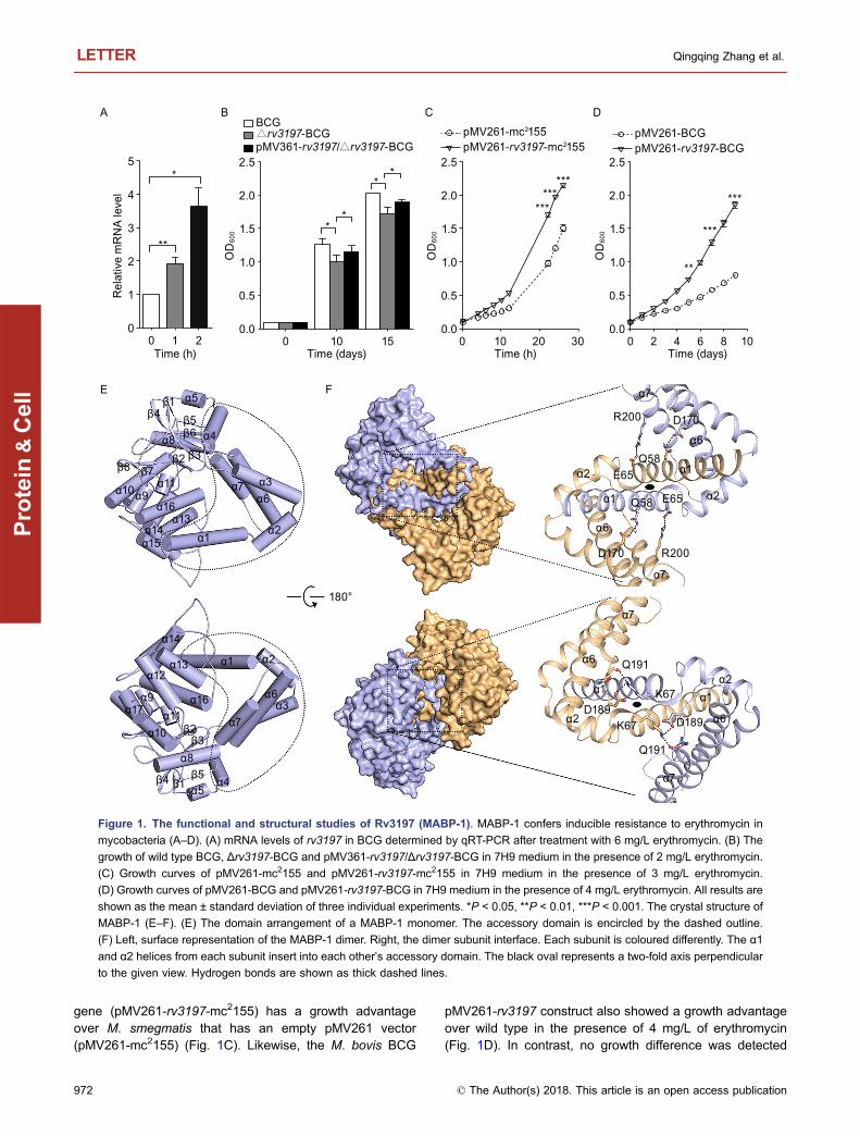
gene (pMV261-rv3197-mc2155) has a growth advantageover M. smegmatis that has an empty pMV261 vector(pMV261-mc2155) (Fig. 1C). Likewise, the M. bovis BCG
pMV261-rv3197 construct also showed a growth advantageover wild type in the presence of 4 mg/L of erythromycin(Fig. 1D). In contrast, no growth difference was detected
A
E F
B DCBCG△rv3197-BCGpMV361-rv3197/△rv3197-BCG
pMV261-mc2155pMV261-rv3197-mc2155
*
**
0
1
2
3
4
5
0 1Time (h) Time (days)
Rel
ativ
e m
RN
A le
vel
OD
600
20.0
β1
β2
α11
α11
α12
α17
β8 β7β3
β4 β5β6
α5
α4α8
α1
α1
R200
R200D170
D170
Q58
Q58 E65
E65
Q191
Q191
D189D189
K67
K67
α1
α2
α2
α2α3α6
α6
α6
180°
α7
α7
α7
α1α1
α2
α2
α6
α6
α7
α7
α16
α14α15
α9α10
α13
β1
β2β3
β4 β5α5
α4
α8
α1 α2
α3α6
α7
α16
α14
α9
α10
α13
0.5
1.0
1.5
2.0
2.5
0 10 15Time (h)
OD
600
0.0
0.5
1.0
1.5
2.0
2.5
0 10 3020Time (days)
OD
600
0.0
0.5
1.0
1.5
2.0
2.5
0 2 864 10
**
**
****** ***
***
**
***
pMV261-BCGpMV261-rv3197-BCG
Figure 1. The functional and structural studies of Rv3197 (MABP-1). MABP-1 confers inducible resistance to erythromycin in
mycobacteria (A–D). (A) mRNA levels of rv3197 in BCG determined by qRT-PCR after treatment with 6 mg/L erythromycin. (B) The
growth of wild type BCG, Δrv3197-BCG and pMV361-rv3197/Δrv3197-BCG in 7H9 medium in the presence of 2 mg/L erythromycin.
(C) Growth curves of pMV261-mc2155 and pMV261-rv3197-mc2155 in 7H9 medium in the presence of 3 mg/L erythromycin.
(D) Growth curves of pMV261-BCG and pMV261-rv3197-BCG in 7H9 medium in the presence of 4 mg/L erythromycin. All results are
shown as the mean ± standard deviation of three individual experiments. *P < 0.05, **P < 0.01, ***P < 0.001. The crystal structure of
MABP-1 (E–F). (E) The domain arrangement of a MABP-1 monomer. The accessory domain is encircled by the dashed outline.
(F) Left, surface representation of the MABP-1 dimer. Right, the dimer subunit interface. Each subunit is coloured differently. The α1
and α2 helices from each subunit insert into each other’s accessory domain. The black oval represents a two-fold axis perpendicular
to the given view. Hydrogen bonds are shown as thick dashed lines.
LETTER Qingqing Zhang et al.
972 © The Author(s) 2018. This article is an open access publication
Protein
&Cell
between pMV261-rv3197-mc2155 and pMV261-mc2155 andbetween pMV261-rv3197-BCG and pMV261-BCG in theabsence of erythromycin (Fig. S1B and S1C). These datademonstrate that Rv3197 is a factor responsible for ery-thromycin resistance in both M. bovis and M. smegmatis.Based on sequence similarity, this protein is also expected toperform a similar role in other Mycobacteria (e.g., My-cobacterium tuberculosis, Mycobacterium kansasii, andMycobacterium avium). In consideration of its antibioticresistance phenotype, we henceforth refer to this protein asmacrolide antibiotic binding protein-1 (MABP-1), signifying itas the first characterization of a macrolide antibiotic bindingprotein in Mycobacterium.
The crystal structure of apo-MABP-1 was determined at2.2 Å by single wavelength anomalous dispersion (SAD)(Table S2). MABP-1 is a homodimer with each subunitcontaining a bilobal kinase domain (Fig. S2) and an acces-sory domain (Figs. 1E and S2). According to a DALI searchthe kinase domain strongly resembles that of an ancientUbIB protein kinase (ADCK3) (Table S3). The accessorydomain consists of six α-helices: α1–α4 helices (residues 1–120) and α6–α7 helices (residues 161–206). These regionsare insertions that are not found in the canonical kinasestructures. Crucially, the α1 and α2 helices in each subunitinsert into the accessory domain of its adjoining subunit,resulting in an N-terminal domain-swapped dimer (Fig. 1F).The accessible surface area that is buried upon dimerizationis extensive, at 2021 Å2 per subunit.
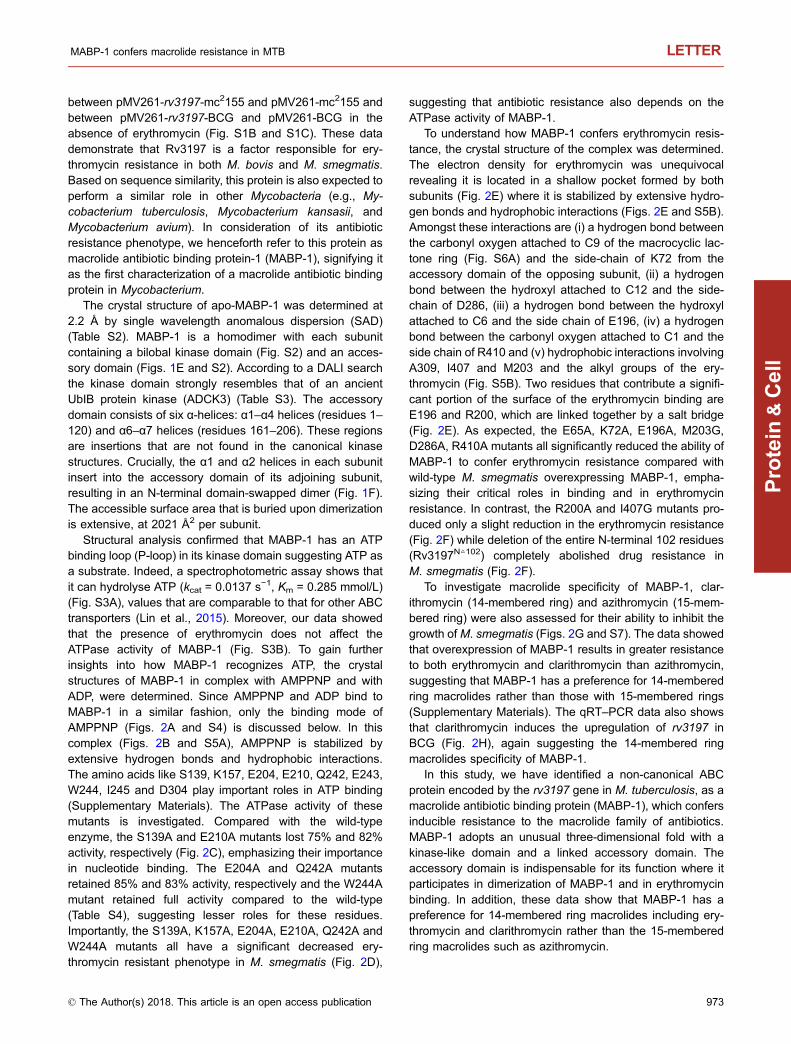
Structural analysis confirmed that MABP-1 has an ATPbinding loop (P-loop) in its kinase domain suggesting ATP asa substrate. Indeed, a spectrophotometric assay shows thatit can hydrolyse ATP (kcat = 0.0137 s−1, Km = 0.285 mmol/L)(Fig. S3A), values that are comparable to that for other ABCtransporters (Lin et al., 2015). Moreover, our data showedthat the presence of erythromycin does not affect theATPase activity of MABP-1 (Fig. S3B). To gain furtherinsights into how MABP-1 recognizes ATP, the crystalstructures of MABP-1 in complex with AMPPNP and withADP, were determined. Since AMPPNP and ADP bind toMABP-1 in a similar fashion, only the binding mode ofAMPPNP (Figs. 2A and S4) is discussed below. In thiscomplex (Figs. 2B and S5A), AMPPNP is stabilized byextensive hydrogen bonds and hydrophobic interactions.The amino acids like S139, K157, E204, E210, Q242, E243,W244, I245 and D304 play important roles in ATP binding(Supplementary Materials). The ATPase activity of thesemutants is investigated. Compared with the wild-typeenzyme, the S139A and E210A mutants lost 75% and 82%activity, respectively (Fig. 2C), emphasizing their importancein nucleotide binding. The E204A and Q242A mutantsretained 85% and 83% activity, respectively and the W244Amutant retained full activity compared to the wild-type(Table S4), suggesting lesser roles for these residues.Importantly, the S139A, K157A, E204A, E210A, Q242A andW244A mutants all have a significant decreased ery-thromycin resistant phenotype in M. smegmatis (Fig. 2D),
suggesting that antibiotic resistance also depends on theATPase activity of MABP-1.
To understand how MABP-1 confers erythromycin resis-tance, the crystal structure of the complex was determined.The electron density for erythromycin was unequivocalrevealing it is located in a shallow pocket formed by bothsubunits (Fig. 2E) where it is stabilized by extensive hydro-gen bonds and hydrophobic interactions (Figs. 2E and S5B).Amongst these interactions are (i) a hydrogen bond betweenthe carbonyl oxygen attached to C9 of the macrocyclic lac-tone ring (Fig. S6A) and the side-chain of K72 from theaccessory domain of the opposing subunit, (ii) a hydrogenbond between the hydroxyl attached to C12 and the side-chain of D286, (iii) a hydrogen bond between the hydroxylattached to C6 and the side chain of E196, (iv) a hydrogenbond between the carbonyl oxygen attached to C1 and theside chain of R410 and (v) hydrophobic interactions involvingA309, I407 and M203 and the alkyl groups of the ery-thromycin (Fig. S5B). Two residues that contribute a signifi-cant portion of the surface of the erythromycin binding areE196 and R200, which are linked together by a salt bridge(Fig. 2E). As expected, the E65A, K72A, E196A, M203G,D286A, R410A mutants all significantly reduced the ability ofMABP-1 to confer erythromycin resistance compared withwild-type M. smegmatis overexpressing MABP-1, empha-sizing their critical roles in binding and in erythromycinresistance. In contrast, the R200A and I407G mutants pro-duced only a slight reduction in the erythromycin resistance(Fig. 2F) while deletion of the entire N-terminal 102 residues(Rv3197N△102) completely abolished drug resistance inM. smegmatis (Fig. 2F).
To investigate macrolide specificity of MABP-1, clar-ithromycin (14-membered ring) and azithromycin (15-mem-bered ring) were also assessed for their ability to inhibit thegrowth ofM. smegmatis (Figs. 2G and S7). The data showedthat overexpression of MABP-1 results in greater resistanceto both erythromycin and clarithromycin than azithromycin,suggesting that MABP-1 has a preference for 14-memberedring macrolides rather than those with 15-membered rings(Supplementary Materials). The qRT–PCR data also showsthat clarithromycin induces the upregulation of rv3197 inBCG (Fig. 2H), again suggesting the 14-membered ringmacrolides specificity of MABP-1.
In this study, we have identified a non-canonical ABCprotein encoded by the rv3197 gene in M. tuberculosis, as amacrolide antibiotic binding protein (MABP-1), which confersinducible resistance to the macrolide family of antibiotics.MABP-1 adopts an unusual three-dimensional fold with akinase-like domain and a linked accessory domain. Theaccessory domain is indispensable for its function where itparticipates in dimerization of MABP-1 and in erythromycinbinding. In addition, these data show that MABP-1 has apreference for 14-membered ring macrolides including ery-thromycin and clarithromycin rather than the 15-memberedring macrolides such as azithromycin.
MABP-1 confers macrolide resistance in MTB LETTER
© The Author(s) 2018. This article is an open access publication 973
Protein
&Cell

Here we speculate that MABP-1 could dissociate ery-thromycin from the ribosome as has previously been sug-gested (Sharkey et al., 2016) in an ATP-dependent manner,reducing the concentration of erythromycin in the cytoplasm
which is able to inhibit ribosomal activity. Another possibilityis that MABP-1 is involved in modifying the structure of theantibiotic (Blair et al., 2015). The alternative hypothesis isthat it cooperates with other efflux pumps to expel the drug
E204
AMPPNP
S139
E210
D304
K157
Q242E243
I245
W244
A B
90°
90°D286
K72
E65
R200E196
M203
I407
A309
R410
HE
F G
C
D
0
1
2
3
0 1 2R
elat
ive
mR
NA
leve
lTime (h)
**
**
0
4020
6080
100120
Native
S139A
E204AE21
0A
Q242A
W24
4A
E204A
K157A
S139A
pMV26
1
261-r
v319
7E21
0A
Q242A
W24
4A
E204A
K157A
S139A
pMV26
1
261-rv
3197
E210A
Q242A
W24
4AE19
6AK72
AE65
A
pMV26
1
261-rv
3197
R200A
M203GD28
6AI407
GR41
0A
N△
102 tru
ncati
onE19
6AK72
AE65
A
pMV26
1
261-rv
3197
pMV26
1
261-rv
3197
pMV26
1
261-rv
3197
R200A
M203GD28
6AI407
GR41
0A
N△
102 tru
ncati
on
Rel
ativ
e AT
Pas
e ac
tivity
(%)
0
1
2
3
***
0
1
2
3
*** ******
***
**
***
************
******
********
OD
600
-Erythromycin+Erythromycin
0
1
2
3
OD
600
OD
600
-Erythromycin+Erythromycin -Clarithromycin
+Clarithromycin
Figure 2. MABP-1 is an ATPase and the accessory domain of MABP-1 is vital for erythromycin binding and dimerization.
(A) Structure of the MABP-1⋅AMPNP complex. MABP-1 is shown as a surface representation with AMPPNP as a stick model. Each
subunit is coloured light orange and light blue. (B) The nucleotide-binding site in MABP-1. Hydrogen bonds are shown as dashed
lines. (C) Relative ATPase activity of MABP-1 mutants in the nucleotide binding site (mean ± SD of three individual experiments).
(D) The growth of mc2155 wild type and mutant strains in the presence (+) or absence (−) of 6.25 mg/L erythromycin (mean ± SD of
three individual experiments, *P < 0.05, **P < 0.01, ***P < 0.001). (E) Left, surface representation of the erythromycin binding site.
Erythromycin has green carbon atoms. Right, the Fo − Fc omit map for erythromycin, contoured at 3.0 σ. Residues that interact with
erythromycin are shown as stick models. Hydrogen bonds are shown as dashed lines. (F) The growth of mc2155 wild type and mutant
strains in the presence (+) or absence (−) of 6.25 mg/L erythromycin. (G) The growth of pMV261-mc2155 and pMV261-rv3197-
mc2155 in 7H9 medium with (+) or without (−) 0.75 mg/L clarithromycin. (H) Quantification of mRNA levels of rv3197 in BCG by qRT-
PCR at the indicated time points after treatment with 1.5 mg/L clarithromycin (mean ± SD of three individual experiments, *P < 0.05,
**P < 0.01, ***P < 0.001).
LETTER Qingqing Zhang et al.
974 © The Author(s) 2018. This article is an open access publication
Protein
&Cell
(Nunez-Samudio and Chesneau, 2013). The structuralcomparison between MABP-1⋅AMPPNP and MABP-1⋅ery-thrmoycin may provide some clues to this hypothesis(Fig. S8 and Supplementary Materials). Clarification of theresistance mechanism of MABP-1 will require furtherinvestigation.
FOOTNOTES
We thank the staff at the Shanghai Synchrotron Radiation Facility
(China) and Photon Factory (Japan) for their assistance in data
collection. This work was supported by grants from the State Key
Development Program for Basic Research of the Ministry of Science
and Technology of China (973 Project Grant Nos. 2014CB542800),
and the National Natural Science Foundation of China (Grant Nos.
81330036 and 81520108019). The atomic coordinates and structure
factors have been deposited in the Protein Data Bank, www.pdb.org
(PDB ID codes 5YJZ, 5YK0, 5YK1 and 5YK2).
Z.R. designed research. Q.Z., H.L., D.J., H.T. and B.Z. performed
research. Q.Z., X.L., C.Y., H.Y., K.M. and Z.R. analyzed data. Q.Z.,
L.W.G., H.Y. and Z.R. wrote the paper. The authors declare no
conflict of interest. This article does not contain any studies with
human or animal subjects performed by the any of the authors.
Qingqing Zhang1,7, Huijuan Liu1, Xiang Liu2,Dunquan Jiang1, Bingjie Zhang3, Hongliang Tian4,Cheng Yang2, Luke W. Guddat5, Haitao Yang4, Kaixia Mi3,Zihe Rao1,2,6&
1 College of Life Sciences, Nankai University, Tianjin 300071, China2 College of Pharmacy, Nankai University, Tianjin 300071, China3 CAS Key Laboratory of Pathogenic Microbiology and Immunology,
Institute of Microbiology, Chinese Academy of Sciences, Beijing
100101, China4 School of Life Sciences, Tianjin University, Tianjin 300072, China5 School of Chemistry and Molecular Biosciences, The University of
Queensland, Brisbane, QLD 4072, Australia6 Laboratory of Structural Biology, School of Medicine, Tsinghua
University, Beijing 100084, China7 Tianjin International Joint Academy of Biotechnology & Medicine,
Tianjin 300457, China
& Correspondence: [email protected] (Z. Rao)
OPEN ACCESS
This article is distributed under the terms of the Creative Commons
Attribution 4.0 International License (http://creativecommons.org/
licenses/by/4.0/), which permits unrestricted use, distribution, and
reproduction in any medium, provided you give appropriate credit to
the original author(s) and the source, provide a link to the Creative
Commons license, and indicate if changes were made.
REFERENCES
Alekshun, M.N., and Levy, S.B. (2007). Molecular mechanisms of
antibacterial multidrug resistance. Cell 128, 1037–1050.Alimuddin Zumla, Andrew George, Virendra Sharma, Nick Herbert,
and Masham, B. (2013). WHO’s 2013 global report on tubercu-
losis: successes, threats, and opportunities. Lancet 382, 1765–1767.
Black, P.A., Warren, R.M., Louw, G.E., van Helden, P.D., Victor, T.C.,
and Kana, B.D. (2014). Energy metabolism and drug efflux in
Mycobacterium tuberculosis. Antimicrob Agents Chemother 58,
2491–2503.Blair, J.M., Webber, M.A., Baylay, A.J., Ogbolu, D.O., and Piddock,
L.J. (2015). Molecular mechanisms of antibiotic resistance. Nat
Rev Microbiol 13, 42–51.Buriankova, K., Doucet-Populaire, F., Dorson, O., Gondran, A.,
Ghnassia, J.C., Weiser, J., and Pernodet, J.L. (2003). Molecular
Basis of Intrinsic Macrolide Resistance in the Mycobacterium
tuberculosis Complex. Antimicrob Agents Chemother 48, 143–150.
Chen, S., Huai, P., Wang, X., Zhong, J., Wang, X., Wang, K., Wang,
L., Jiang, S., Li, J., Peng, Y., et al. (2013). Risk factors for
multidrug resistance among previously treated patients with
tuberculosis in eastern China: a case-control study. Int J Infect
Dis 17, e1116–e1120.Dassa, E., and Bouige, P. (2001). The ABC of ABCs: a phylogenetic
and functional classification of ABC systems in living organisms.
Res Microbiol 152, 211–229.Dong, Y., Qiu, X., Shaw, N., Xu, Y., Sun, Y., Li, X., Li, J., and Rao, Z.
(2015). Molecular basis for the inhibition of beta-hydroxyacyl-
ACP dehydratase HadAB complex from Mycobacterium tuber-
culosis by flavonoid inhibitors. Protein Cell 6, 504–517.Günther, G. (2014). Multidrug-resistant and extensively drug-resis-
tant tuberculosis: a review of current concepts and future
challenges. Clin Med 14, 279–285.Kerr, I. D., Reynolds, E.D., and Cove, J.H. (2005). ABC proteins and
antibiotic drug resistance: is it all about transport? Biochem Soc
33, 1000–1002.Lin, D.Y., Huang, S., and Chen, J. (2015). Crystal structures of a
polypeptide processing and secretion transporter. Nature 523,
425–430.Nunez-Samudio, V., and Chesneau, O. (2013). Functional interplay
between the ATP binding cassette Msr(D) protein and the
membrane facilitator superfamily Mef(E) transporter for macrolide
resistance in Escherichia coli. Res Microbiol 164, 226–235.Shim, T.S., and Jo, K.-W. (2013). Medical treatment of pulmonary
multidrug-resistant tuberculosis. Infect Chemother 45, 367–374.Sharkey, L.K., Edwards, T.A., and O’Neill, A.J. (2016). ABC-F
proteins mediate antibiotic resistance through ribosomal protec-
tion. MBio 7, e01975.
WHO (2014). Global tuberculosis report 2014 (Geneva, World
Health Organization).
Electronic supplementary material The online version of thisarticle (https://doi.org/10.1007/s13238-017-0502-7) contains sup-
plementary material, which is available to authorized users.
MABP-1 confers macrolide resistance in MTB LETTER
© The Author(s) 2018. This article is an open access publication 975
Protein
&Cell