diseases? a systematic review and meta-analysis is oral
TRANSCRIPT
Page 1/19
Is oral lichen planus a risk factor for peri-implantdiseases? A systematic review and meta-analysisXiaoqin Xiong
Guangzhou Medical UniversityTing Yu ( [email protected] )
https://orcid.org/0000-0002-8381-010XTiantian Xu
Guangzhou Medical UniversityXinhong Wang
Guangzhou Medical UniversityWenguang Qin
Guangzhou Medical UniversityLuo Gang
Guangzhou Medical University
Research article
Keywords: dental implants; peri-implant diseases; oral lichen planus; systematic review; meta-analysis.
Posted Date: March 4th, 2020
DOI: https://doi.org/10.21203/rs.3.rs-15960/v1
License: This work is licensed under a Creative Commons Attribution 4.0 International License. Read Full License
Version of Record: A version of this preprint was published at BMC Oral Health on May 20th, 2020. Seethe published version at https://doi.org/10.1186/s12903-020-01134-2.

Page 2/19
AbstractBackground : To evaluate whether oral lichen planus (OLP) is a risk factor for peri-implant diseases(PIDs) with a systematic review and meta-analysis. Methods : Six electronic databases including Medline,Web of Science, etc. were searched. Included studies are observational human studies written in English.Population of interest were those with/without OLP who received dental implant treatment. Follow-uptime after implantation is from one month to 20 years. The quality of the included literature regarding riskof bias and methodology was assessed with Newcastle-Ottawa Scale or the Agency for HealthcareResearch and Quality. The data involving exposure (OLP), primary outcomes (implants having PIDs) andsecondary outcomes (probing depth/PD, bleeding on probing/BOP and bone loss/BL) and potentialconfounders were extracted. Heterogeneity was assessed by I² tests. Dichotomous data were expressedas risk ratio (RR) and 95% con�dence interval (CI) which were calculated with a �xed effect model.Results : Of 139 literatures, two studies were enrolled and evaluated as high quality, which totallycontained 68 participants receiving 222 (OLP vs. non-OLP, 112 vs. 110) implants with 12 to 120-monthfollow-up time. Proportions of implants with PIDs between OLP and non-OLP groups were as follows:19.6% (22/112) vs. 22.7% (25/110) for PIM; 17.0% (19/112) vs. 10.9% (12/110) for PI. Meta-analysisfound no recognizable difference in number of implants with PIDs (PI: RR = 1.49, 95% CI 0.77-2.90, P =0.24; PIM:RR = 0.88, 95% CI 0.53 -1.46, P = 0.61; PIDs: RR = 1.08, 95% CI 0.75 -1.55, P = 0.68) or BOP (RR= 0.90, 95% CI: 0.70-1.15, P = 0.40) between OLP and non-OLP groups. Conclusions : Available literatureregarding the effects of OLP on PIDs remains very limited. Existing evidence seems not support OLP as asuspected risk factor for PIDs. Large-scale prospective trials are required to test the �ndings. Keywords :dental implants; peri-implant diseases; oral lichen planus; systematic review; meta-analysis.
BackgroundDental implant treatment is a well-established method for restoring lost teeth. Despite a high rate ofsurvival (>94%) for more than 5 years [1, 2], a very high proportion (8%-44%) of dental implants developperi-implant diseases (PIDs), including peri-implant mucositis (PIM) and peri-implantitis (PI) [3], whichcan lead to implant failure or loss. PIDs are pathological in�ammatory conditions, of which PIM involvesonly peri-implant mucosal in�ammation, while PI causes both peri-implant mucosa and alveolar bonedamage [4], accompanied by bleeding on probing, suppuration, increased probing depth , and progressivemarginal bone loss[5]. PID-associated risk factors include poor oral hygiene [6], smoking [7], a history ofperiodontitis [4, 8] and systemic diseases, such as diabetes [9].
Oral lichen planus (OLP), a chronic systemic autoimmune disease, affects one to two percent of thegeneral population, especially middle-aged and elderly females [10]. Typically, OLP manifests as plaque-like or white reticular lesions, an erythematous area or ulceration [11, 12], and it can affect the oralmucosa, tongue or gingiva [13]. Recently, OLP has been suspected as a potential risk factor for PIDs.Some scholars have speculated that OLP may directly alter the nature of the mucosal barrier arounddental implants, promoting bacterial invasion [14]. Furthermore, OLP patients have been found to havepoor oral hygiene[15] and quality of life [16]. These concerns have previously made OLP a
Page 3/19
contraindication to implant treatment[14]. However, some studies, in which 66 short implants wereimplanted in 23 OLP patients, with a survival rate of 98.5%, did not observe any difference in the successrate between OLP patients and normal controls [17, 18]. Notably, most of the studies regardingimplantation in OLP populations were observational studies (including case reports, retrospective studies,etc.) with small sample sizes. Hence, whether dental implantation in OLP patients is safe remainsinconclusive. The present systematic review aimed to analyze the existing literature to determine whetherOLP is a risk factor for PIDs.
MethodsFocused question
Compared with non-OLP populations, are there changes in the rates of probing depth, bone loss orbleeding on probing around implants in OLP patients? Are there different rates of PIM and PI prevalencein OLP patients compared with non-OLP populations? This manuscript has been prepared based on thePreferred Reporting Items for Systematic Review and Meta-analyses (PRISMA) guidelines for reportingsystematic reviews.
Population and exposure
The population of interest was patients with and without OLP who received dental implant treatment. Thestudies included participants aged ≥18 years with no periodontitis or stable periodontitis after treatment.Their OLP status was diagnosed before implant treatment. It was preferred that OLP patients had adiagnosis of the OLP subtype or concomitant symptoms, such as desquamative gingivitis which could beconsidered separate exposure factors. Detailed records of treatments, including medications, wereincluded for all OLP patients. It was assumed that implant treatment was performed during the remissionstage of OLP. The primary outcomes were PIDs, and the secondary outcomes were probing depth,bleeding on probing, and bone loss. Implant failure was also considered if its infective origin was clearlystated.
Disease determinants, risk factors, and etiologic agents
The potential association of OLP with PID determinants, including sex, age, educational background,socioeconomic position, genetics, lifestyle, nutrition, health behaviors, and microbiological factors, wasrecorded when available. Whenever possible, the quality of the determinant/exposure measurementswere assessed.
There are some general risk indicators/factors for PI and PIM, including a history of periodontitis[19],systemic diseases, genetic traits, and smoking. Local risk indicators/factors for PI and PIM include oralhygiene/poor plaque control, poor compliance with supportive implant therapy, presence of excesscement, implant materials and surface characteristics, design of implant-supported prostheses anddimensions of keratinized mucosa [4, 8].

Page 4/19
Study follow-up duration
All studies with a mean duration or follow-up interval of at least one month were included because theearliest time for PID diagnosis was approximately one month after loading restorations [4, 20]. If thefollow-up time was reported as a range, the low value of the range was more than one month.
Types of studies
With the goal of identifying studies for inclusion, certain methods were designed based on acceptabilitycriteria in this study. Eligible studies, including observational or longitudinal studies or prospectiveresearch, were those with a follow-up time ≥1 month, with primary outcomes of PIDs. The secondaryoutcomes were probing depth, bleeding on probing and bone loss.
Inclusion criteria
1. Observational human studies, such as case-control, cross-sectional, retrospective or prospectivecohort studies;
2. Studies in which OLP and PIDs were the main exposure factor and outcome, respectively;
3. Studies in which both OLP and PIDs were the de�nitive diagnoses, and the diagnosis of OLP wasmade according to clinical and pathological evidence;
4. Studies with a follow-up time of ≥1 month after implant placement; and
5. Studies published in the English language.
Exclusion criteria
1. Studies lacking control groups (i.e., a non-OLP group);
2. Narrative reviews, comments, letters, case reports or series, and conference abstracts; and
3. Studies for which the full-text articles or original data could not be obtained.
Search strategy
The search was performed in June 2019. All available articles published online before this time in sixelectronic databases, including Medline, Web of Science, the Cochrane Library, John Wiley, Scopus andSpringer, were retrieved. Manual searching in dental journals, especially those focusing on implantology,was also performed. The search strategy was established with adequate discussion among all thecoauthors (Table 1).
Study selection
Two researchers independently screened the headlines and abstracts of the studies identi�ed in thesearches. Afterward, the full texts of all the publications that met the inclusion criteria were obtained;however, it was not possible to make a decision about inclusion due to a lack of information in the titlesand abstracts. Any research that was considered relevant by one of the reviewers was included in the
Page 5/19
following screening stage. Subsequently, the articles were independently assessed in duplicate by thereviewers. Any disparity about research eligibility was settled by discussion among the two reviewers, andif no consensus could be reached, the decision was resolved through arbitration by a third reviewer. Allarticles that did not ful�ll the eligibility criteria were excluded.
For studies with missing data or those for which a clear decision on study eligibility could not be madeafter assessment of the full text, the corresponding authors were e-mailed to ask for the informationneeded for a �nal decision. Messages via social media or academic websites were also sent to thecorresponding authors in the absence of response by e-mail.
Data extraction and management
A detailed form designed speci�cally for data extraction was used, and all the relevant data from thecollected articles were independently extracted by two independently. Any disagreements were settledthrough evaluation by the third examiner or group consensus.
The following research information was extracted (Table 2):
1. Type of research;
2. Sample framework (e.g., community, university);
3. Sample size, sex and age of participants;
4. Diagnostic criteria for PI, PIM and OLP;
5. Follow-up time; and
�. Confounding factors relative to exposure factors.
Outcomes
1. probing depth change;
2. bone loss level change;
3. bleeding on probing level change; and
4. Number of implants with/without PI and PIM;
Quality assessment
The Newcastle Ottawa Scale (NOS) [21] and the Agency for Healthcare Research and Quality (AHRQ) [22]standards were used to assess the quality of cohort and cross-sectional studies, respectively. The cohortstudy degree of bias was categorized with a star-based quality assessment system that considers threecategories as follows: the selection of the study groups (four stars), the comparability of the groups (twostars), and the ascertainment of either the exposure or outcome of interest (three stars). An 11-itemchecklist recommended by the AHRQ was used to assess the methodological quality of the cross-sectional studies. Article quality was assessed as follows: low quality (0 to 3); moderate quality (4 to 7);and high quality (8 to 11).

Page 6/19
Data synthesis
Statistical heterogeneity in risk in patients suffering from PIDs in the collected studies was evaluatedusing both risk ratios (RRs) and I² measures. The use of I² values was based on the Cochrane Handbook:0 < I²< 40% represents a lack of heterogeneity; 30% < I²< 60% represents moderate heterogeneity; 50% < I²<90% signals substantial heterogeneity; and 75% < I²< 100% shows considerable heterogeneity.
When suitable, meta-analysis was used to assess risk in patients suffering from PIDs based on implantand patient characteristics. A random effects approach was used if there was mild to moderate statisticalheterogeneity; otherwise, a �xed-effect approach was chosen. Considering that only observational studieswere included in the present systematic review, the primary outcome variable was the RRs of PI and PIM.The RRs were calculated with 95% con�dence intervals (CIs) (Review Manager, v5.2, The Nordic CochraneCenter, The Cochrane Collaboration, Copenhagen, Denmark) to quantify the association between OLP andthe risk of PI and PIM. Forest plots were generated showing the RRs and 95% CIs of the involved studies.If there were more than 10 studies included in the meta-analysis, publication bias was evaluatedqualitatively by a funnel plot [23].
ResultsSearch
A total of 139 studies were identi�ed after electronic and manual searches (Fig 1). After removingduplicates, 83 articles were retained. Then, 58 articles were excluded after evaluating the titles andabstracts, and the remaining 25 articles were selected for full-text evaluation. Of the 25 articles, 13articles involved both OLP and implant treatment. Then, 11 articles were excluded for various reasons,such as being case reports, reviews, etc., and the remaining 2 articles were retained for �nal review. Thescreening outcomes were similar regardless of whether the literature was published in English [24].
Study characteristics
Location and sample characteristics
The two included studies included one prospective [25] study and one cross-sectional study [16] (Table2). Both studies were conducted in the same country (Spain) but by different institutions. The two studiescontained 34 OLP patients and 34 non-OLP controls aged between 29 and 79 years. The 68 participantsreceived 224 implants (OLP vs. non-OLP, 112 vs. 110) with a follow-up time of 12 to 120 months.
At the implant level, 19.6% (22/112) and 22.7% (25/110) developed PIM in the OLP and non-OLP groups,while 17.0% (19/112) and 10.9% (12/110) developed PI in the two groups, respectively. Additionally,bleeding on probing was present at the sites of 42.6% (48/112) and 50.0% (55/110) of implants in thetwo groups, respectively.

Page 7/19
The ratios of PIDs in the two groups could not be calculated at the individual level due to missing data.The outcomes regarding probing depth and bone loss could not be combined because they were reportedin different forms, and only one study’s authors provided the original data for these parameters by onlinecontact.
Risk of bias and methodological quality
Quality evaluation according to the NOS showed that the prospective study was rated as having 7 starsand was classi�ed as high quality (Table 3). Based on the AHRQ criterion, the �nal score of the cross-sectional study was 8, and it was classi�ed as having high quality (Table 4).
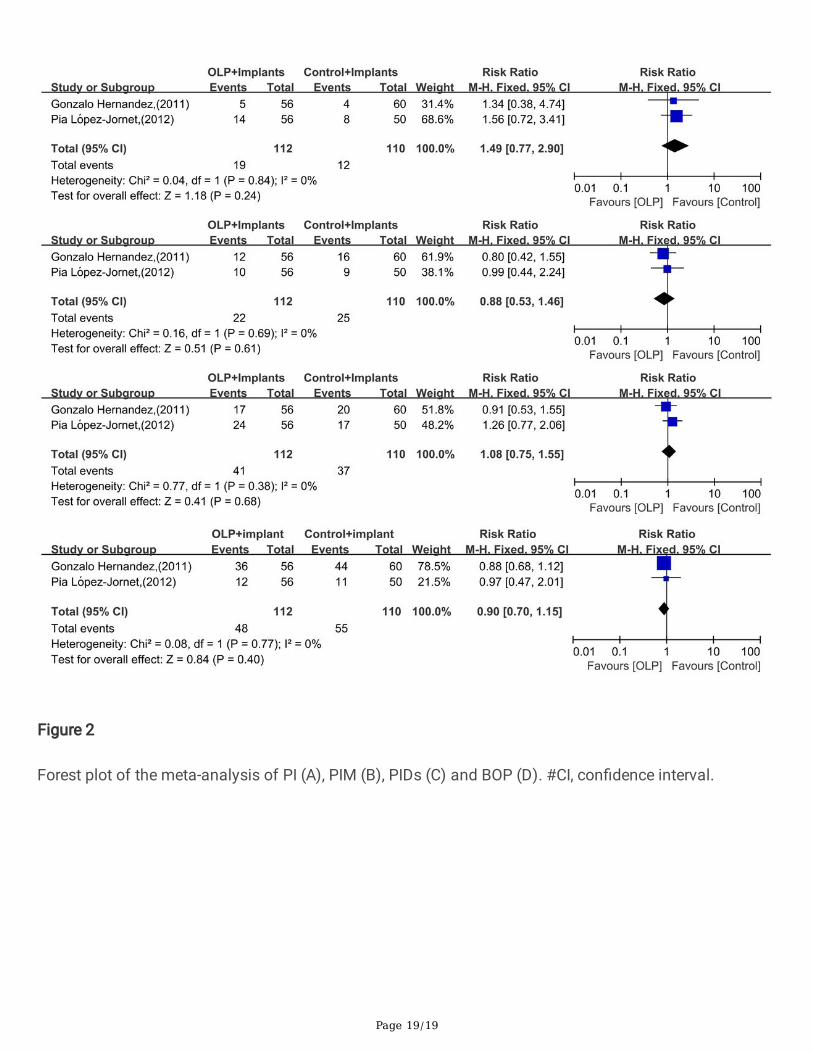
Risk of PIDs in the OLP versus non-OLP groups
Due to the small sample size and good homogeneity (χ²= 0.04, P = 0.84; I²= 0) of the included literature,the �xed-effect model was employed to calculate the pooled RR in this meta-analysis. The model showedno signi�cant difference in the number of implants suffering from PI (RR = 1.49, 95% CI: 0.77-2.90, P =0.24, Fig 2A) or PIM (RR = 0.88, 95% CI: 0.53 -1.46, P = 0.61, Fig 2B) between the OLP and non-OLPgroups at the implant level. There was a similar result when combining PI and PIM into PID (RR = 1.08,95% CI: 0.75 -1.55, P = 0.68, Fig 2C). This was also the case for the presence of bleeding on probingbetween the two groups (RR = 0.90, 95% CI: 0.70-1.15, P = 0.40, Fig 2D).
DiscussionKey �ndings
The present systematic review assessed for the �rst time the current existing evidence on the relationshipbetween OLP and PIDs and attempted to determine whether OLP is a potential risk factor for PIDs. Of 139studies, two, which contained 68 participants receiving 222 (OLP vs. non-OLP, 112 vs. 110) implants, witha 12- to 120-month follow-up time, were included and evaluated as having high quality. The proportionsof implants with PIDs between the OLP and non-OLP groups were as follows: 19.6% (22/112) vs. 22.7%(25/110) for PIM and 17.0% (19/112) vs. 10.9% (12/110) for PI. Existing evidence seems to suggest thatOLP is not a suspected risk factor for PIDs during the 10-year follow-up period.
Overall completeness and applicability of the evidence
The diagnostic criteria for OLP were clearly stated in the two enrolled studies, while those for PIDs wereabsent in the cross-sectional study. Moreover, in the two enrolled studies, PIDs were evaluated at only theimplant level and not at the individual level, while OLP was diagnosed at the individual level. Hence, theevidence was insu�cient to determine whether OLP is a risk factor for PIDs at the individual level.
Concerning secondary outcomes of PIDs, both studies had complete bleeding on probing data. After datacombination, OLP was found to not be a risk factor for bleeding on probing at the implant level. For thesecondary outcomes for PIDs, i.e., probing depth and bone loss, the data were complete but reported in
Page 8/19
different forms in the two studies. The prospective study set cutoff values for probing depth and boneloss to determine the percentages of involved implants, while the cross-sectional study directly presentedthe medians and ranges of the two parameters. Despite repeated contact with the authors, we obtainedonly the raw data of the prospective study. Hence, an attempt to combine the data of the two studies interms of probing depth or bone loss could not be achieved. Ultimately, both studies found no signi�cantdifference in probing depth or bone loss between the OLP and non-OLP groups.
When the participant was used as the unit of analysis, the intragroup analysis of the OLP group failed todemonstrate a statistically signi�cant relationship between the existence of desquamative gingivitis andPIM or desquamative gingivitis and PI, but 20 out of 25 implant patients with PIM were among those withdesquamative gingivitis in the OLP group; hence, PIM with DG in the OLP group occurred more frequentlythan non-desquamative gingivitis in the OLP group in this subgroup of implant patients in the prospectivestudy [25]. Since the cross-sectional study presented insu�cient information, although the correspondingauthors were contacted in various ways and only the author of the prospective study provided the originaldata of the parameters of interest, no subgroup analysis was performed. Therefore, subgroupcombinations in the meta-analysis were not possible.
Overall quality, strength, and consistency of the evidence
The present systematic review included only two observational studies that met the inclusion criteriamainly because related research on implantation in OLP populations is scarce. However, the durations ofthe two enrolled studies were very similar (2003 to 2009 vs. 2005 to 2010), and the study populationswere from the same country, which may be bene�cial for data synthesis. Nevertheless, the results mightbe more applicable to speci�c ethnicities or countries and may not be easily generalized for peopleglobally. The average age of the OLP patients in the two studies was quite different (64.5 vs. 53.7). Thisdistinction may introduce uncertainty regarding the applicable age after pooling the data. Additionally, themean follow-up time of the two studies was similar but did not exceed 5 years; therefore, it is unclearwhether OLP is a risk factor for PIDs over an observation period longer than 5 years. The two studiescontained detailed records of important confounders, such as smoking, but only the prospective studycontrolled for confounders. The response rate was not reported in either study; thus, the number of lostsamples was unknown. In the prospective study, two implants in the non-OLP group were lost forunknown reasons, and the study did not state whether it was a PI-associated consequence. Even if thatwas the case, there was no difference in the results of the PI (RR=1.31, 95% CI: 0.70-2.45, P = 0.40)analysis between the two cases.
Case de�nitions for PIDs vary considerably in previous studies[8], and PID data are especially challengingto interpret. Only the prospective study, which reported clear de�nitions of PIM and PI, was evaluatedaccording to the de�ned criteria [25, 26]. The cross-sectional study did not elaborate on the diagnosticcriteria for PIDs; the two studies had different de�nitions of diseases, and there may have been some biasin the data. The diagnostic criteria for PI in the study were not uniform, and limited studies were includedin the meta-analysis, so no meta-analysis could be conducted to prove the effect of the differences on the
Page 9/19
meta-analysis outcomes. Clinical indicators such as bleeding on probing and bone loss are relativelyeasy to standardize, although no effect of OLP on these indicators has been observed.
The results of the statistical analysis show that the homogeneity between the results was good. However,the limited number of enrolled studies made it unfeasible to draw a funnel plot to assess the variation inthe studies. Although the sample size was small, the strength of the study evidence was high as analyzedby quality-control assessment, and the overall estimate of the meta-analysis represents the best availableevidence.
Implications for practice and policy
Although there was no clear association between OLP and the occurrence of PIDs, implant-related issuesin OLP patients cannot be easily ignored, and several issues are worth noting. First, in a clinical study [15],the gingival index of the OLP group was calculated, and the mean value of the periodontal index and rateof bleeding on probing were higher than those in the control group, revealing that the periodontalcondition in the OLP group was poor compared with that in the control group. Poor oral hygiene is a well-known risk factor for PIDs; therefore, OLP may have a certain impact on PIDs. Second, some scholarsbelieve that the capacity of the epithelium to adhere to the titanium surface of the implants in OLPpatients may be affected [14, 27] because the adhesion of epithelial cells decreases, affecting theepithelial barrier around the implant surface [28]. Third, desquamative gingivitis is a special type of OLPthat causes gingival damage. An increased frequency of PIM was reported in cases of desquamativegingivitis compared with non-desquamative gingivitis OLP patients [25]. Another study demonstrated thatthe presence of desquamative gingivitis was associated with relatively deep periodontal pockets [29]. Thestudy inferred that desquamative gingivitis lesions might circuitously enhance the long-term risk forperiodontal diseases via plaque accumulation by impeding proper oral hygiene around natural teeth andimplants. It is important to clarify the constructive association between desquamative gingivitis and PIDsin the future. Moreover, OLP is a T cell‐mediated chronic in�ammatory autoimmune disease[30]. PIDs inhumans cause leukocytic in�ltration through barrier epithelial migration and an increase in the proportionof T and B cells [31]. In this context, the two conditions might aggravate each other via in�ammatorypathways. Additionally, dysbiosis might be a potential link between OLP and PIDs, given that bothconditions have been related to microbial alterations [32, 33]. Finally, systemic corticosteroid treatment inOLP patients was frequently associated with decreased bone mineral density, especially during the �rst 6months of corticosteroid therapy [34]. The osteoporotic effects of corticosteroids might cause morealveolar bone loss around dental implants during infection [35].
In the systematic review, the available evidence did not support OLP as a risk factor for PIDs. However,this does not mean that implant restoration can be performed indiscriminately in OLP patients. Forpatients with acute/erosive forms of OLP or desquamative gingivitis, immune system disorder and poororal hygiene may be potential risk factors for PIDs. OLP should not be considered a contraindication forimplant treatment in patients who do not have evident symptoms or mucosal non-erosive congestion andhave good oral hygiene. Furthermore, the mental state of OLP patients might be improved after implant
Page 10/19
treatment, which in turn is better for OLP control, considering that those without implant treatment havereported a poor quality of life [16]. Additionally, for OLP patients receiving glucocorticoid treatment,assessment of alveolar bone mineral density in the area to be implanted should be considered. Finally, itis worth noting the effect of implants in the mouth on the recovery of OLP patients, although there wereno signi�cant differences in OLP signs and symptoms between the implant group (14 patients) andnonimplant group (15 patients) during the 12-24 month follow-up period [14]. For OLP patients who haveundergone implant restoration, OLP conditions may need to be monitored frequently, and implantmaintenance planning may need to be personalized.
Implications for further research
This review systematically analyzed the existing evidence on OLP and PIDs for the �rst time. Large-scaleprospective trials are required to validate the �ndings.
The results suggest that OLP is not a potential risk factor for PIDs during the 1 to 10-year follow-upperiod. However, given the relatively small amount of evidence available, the �nal answer to this questiondepends on large, well-designed prospective and randomized clinical trials in the future. At present, theproportion of people undergoing implant treatment and the prevalence of OLP worldwide are not high,which may cause di�culty in researching the correlation between the two diseases; therefore, multicentercooperative clinical research is needed. Additionally, OLP has various clinical classi�cations, and theprospective study included in this systematic review showed that patients with OLP with desquamativegingivitis had a higher frequency of PIM. This suggests that in future studies, a subgroup analysis of OLPmay be helpful in exploring the association between OLP and PIDs. Moreover, interventional studies onthe effects of OLP treatment on peri-implant status are still lacking; these studies can further elucidatethe association between OLP and PIDs. Molecular biology techniques can also help to explore themicroscopic effects of OLP on peri-implant pathophysiological processes at the molecular level.
ConclusionsAvailable literature regarding the effects of OLP on PIDs remains very limited. Existing evidence seemsnot support OLP as a suspected risk factor for PIDs. Large-scale prospective trials are required to test the�ndings.
Abbreviations
Page 11/19
OLP oral lichen planus
PIDs peri-implant diseases
RR risk ratio
CI con�dence interval
PIM peri-implant mucositis
PI peri-implantitis
PRISMA Preferred Reporting Items for Systematic Review and Meta-analyses
NOS Newcastle-Ottawa Scale
AHRQ Agency for Healthcare Research and Quality
DeclarationsAcknowledgements
The authors thank Liting Yan and Yinshen Yang for their help with meta-analysis work.
Funding
This study was supported by the National Natural Science Foundation of China, Beijing, China, Grants81700985 and 81271159. The role of funding agencies in research: data collection, analysis, andmanuscripts.
Availability of data and materials
The datasets used and/or analyzed during the current study are available from the corresponding authoron reasonable request.
Authors’ contributions
XX and TY formulated inclusion and exclusion criteria, literature search and screening. XX and TYanalyzed and interpreted the data and drafted the manuscript. XW, WQ and LG designed and supervisedthe study and edited and gave �nal approval for the manuscript to be published. All authors read andapproved the �nal manuscript.
Ethics and consent to participate
Not Applicable
Consent for publication
Not applicable.

Page 12/19
Competing interests
The authors declare that they have no competing interests.
Author details
Xiaoqin Xiong 1†, MD, Dental specialist in Periodontology and Oral Medicine; Tiantian Xu 1† ,MD, Dentalspecialist in Periodontology; Xinhong Wang 1, DMD, Associate professor, Dental specialist in OralMedicine; Wenguang Qin 1 ,MD, Dental specialist in Periodontology; Gang Luo 1*, DMD, Professor; TingYu 1*,DMD, Associate professor, Dental specialist in Periodontology
1 Department of Periodontology and Oral Medicine, Stomatology Hospital of Guangzhou MedicalUniversity, Guangzhou, China.
† Xiaoqin Xiong and Tiantian Xu are �rst authors.
* Correspondence: Ting Yu, Key Laboratory of Oral Medicine, Guangzhou Institute of Oral Disease,Department of Periodontology and Oral Medicine, Stomatology Hospital of Guangzhou MedicalUniversity, NO.195 Dongfeng West Road, Guangzhou 510140, China, Tel.: +86-020-81299940. E-mail:[email protected]
References1. BE P, U B, NP L, M Z: Comparison of survival and complication rates of tooth-supported �xed dental
prostheses (FDPs) and implant-supported FDPs and single crowns (SCs). Clinical oral implantsresearch 2007, null(unde�ned):97-113.
2. BE P, NA V, M S, M Z, S L, I S: A systematic review of the survival and complication rates of zirconia-ceramic and metal-ceramic single crowns. Clinical oral implants research 2018, null(unde�ned):199-214.
3. S R, S O, D Z, K L, P E: Prevalence of periimplant disease in partially edentulous patients: a practice-based cross-sectional study. Clinical oral implants research 2011, 22(8):826-833.
4. Heitz-May�eld LJA, Salvi GE: Peri-implant mucositis. Journal of Periodontology 2018, 89:S257-S266.
5. T B, G A, MG A, G A-O, J B, PM C, S C, D C, J D, E F et al: Peri-implant diseases and conditions:Consensus report of workgroup 4 of the 2017 World Workshop on the Classi�cation of Periodontaland Peri-Implant Diseases and Conditions. Journal of clinical periodontology 2018,null(unde�ned):S286-S291.
�. BCV G, SCL M, PMC D, ALB P, KC L, PDS C: Frequency of peri-implant diseases and associatedfactors. Clinical oral implants research 2017, 28(10):1211-1217.
7. MS B, A M, Z A, SS A: Peri-implant clinical and radiographic status and whole salivary cotinine levelsamong cigarette and waterpipe smokers and never-smokers. Journal of oral science 2018, 60(2):247-252.

Page 13/19
�. F S, J D, A M, HL W: Peri-implantitis. Journal of periodontology 2018, null(unde�ned):S267-S290.
9. ZH A-S, SMA G, K S, F V, Z A: Peri-implant conditions and levels of advanced glycation end productsamong patients with different glycemic control. Clinical implant dentistry and related research 2018,20(3):345-351.
10. M C, R T: Oral lichen planus: a review. Minerva stomatologica 2009, 58(10):519-537.
11. YS C, A G, Z K, J F, S M: Diagnosis of oral lichen planus: a position paper of the American Academyof Oral and Maxillofacial Pathology. Oral surgery, oral medicine, oral pathology and oral radiology2016, 122(3):332-354.
12. PB S, NW S, LJ W, ZZ Z, XJ Z, A K, GJ S, M B: The pathogenesis of oral lichen planus. Critical reviewsin oral biology and medicine : an o�cial publication of the American Association of Oral Biologists2002, 13(4):350-365.
13. M B, N G, A TZ, M V, M M: Oral lichen planus: clinical features, etiology, treatment and management;a review of literature. Journal of dental research, dental clinics, dental prospects 2010, 4(1):3-9.
14. R C, M E, A W, A S: Oral lichen planus and dental implants--a retrospective study. Clinical implantdentistry and related research 2013, 15(2):234-242.
15. NP R, P K, SM M, DD D, AA K, R R, S V, MH D, NR P, S A: Relation Between Periodontal Status and Pre-Cancerous Condition (Oral Lichen Planus): A Pilot Study. Advances in clinical and experimentalmedicine : o�cial organ Wroclaw Medical University 2016, 25(4):763-766.
1�. P L-J, F C-A, M S-S: Dental implants in patients with oral lichen planus: a cross-sectional study.Clinical implant dentistry and related research 2014, 16(1):107-115.
17. M P, M DB, R C, L M, A L, R S, FR G: Implant rehabilitation in patients with oral lichen planus: anoverview. Clinical oral investigations 2012, 16(5):1347-1352.
1�. E A, L P, V E-A, RS F, MH A: Short dental implants in patients with oral lichen planus: a long-termfollow-up. The British journal of oral & maxillofacial surgery 2018, 56(3):216-220.
19. S R, A A, H H, GR P: Factors related to peri-implantitis - a retrospective study. Clinical oral implantsresearch 2014, 25(4):522-529.
20. GE S, M A, S E, A S, NP L, CA R: Reversibility of experimental peri-implant mucositis compared withexperimental gingivitis in humans. Clinical oral implants research 2012, 23(2):182-190.
21. Wells GA: The Newcastle-Otawa Scale (NOS) for assessing the quality of non-randomzed studies inmetal-analyses. 2009.
22. X Z, Y Z, JS K, C Z, S L, F S, Y N, L D: The methodological quality assessment tools for preclinical andclinical studies, systematic review and meta-analysis, and clinical practice guideline: a systematicreview. Journal of evidence-based medicine 2015, 8(1):2-10.
23. Sterne JA: Recommendations for examining and interpreting funnel plot asymmetry in meta-analysisof randomised contrlolled trials. BMJ 2011.
24. Fu L, A L, Li CY, Zhou YM: Research progress of dental implant in patients with oral lichen planus.Zhonghua kou qiang yi xue za zhi = Zhonghua kouqiang yixue zazhi = Chinese journal of
Page 14/19
stomatology 2019, 54(2):135-137.
25. G H, RM L-P, L A, J T, JC dV: Implant treatment in patients with oral lichen planus: a prospective-controlled study. Clinical oral implants research 2012, 23(6):726-732.
2�. AM R-J, C L, H R, S R: Nine- to fourteen-year follow-up of implant treatment. Part II: presence of peri-implant lesions. Journal of clinical periodontology 2006, 33(4):290-295.
27. ME C-M, J A-A, D P-O, M P-D, JV B: Dental implants in patients with oral mucosal alterations: Anupdate. Medicina oral, patologia oral y cirugia bucal 2011, 16(6):e787-793.
2�. C S-M, G B, M J, C E, GJ S, PB S, NW S, UI D: Distribution of interleukin-2, -4, -10, tumour necrosisfactor-alpha and transforming growth factor-beta mRNAs in oral lichen planus. Archives of oralbiology 1999, 44(6):499-507.
29. L LR, R G, G P, G F, D C, L LM, G C: Effect of desquamative gingivitis on periodontal status: a pilotstudy. Oral diseases 2010, 16(1):102-107.
30. MS A, N C, M M: Oral lichen planus: a literature review and update. Archives of dermatologicalresearch 2016, 308(8):539-551.
31. F G, T B: Immunohistochemical characteristics of in�ammatory lesions at implants. Journal ofclinical periodontology 2003, 30(1):14-18.
32. GR P, S R: Cluster of bacteria associated with peri-implantitis. Clinical implant dentistry and relatedresearch 2014, 16(6):783-793.
33. K W, W L, Q T, Y G, J H, Y Z, Y G, JD VN, Y Q, J L et al: Preliminary analysis of salivary microbiomeand their potential roles in oral lichen planus. Scienti�c reports 2016, 6(unde�ned):22943.
34. MA G-M, C S: Vesiculo-erosive oral mucosal disease--management with topical corticosteroids: (2)Protocols, monitoring of effects and adverse reactions, and the future. Journal of dental research2005, 84(4):302-308.
35. AC V, M B: Limited bone loss due to corticosteroids; a systematic review of prospective studies inrheumatoid arthritis and other diseases. The Journal of rheumatology 1997, 24(8):1495-1503.
TablesTable 1. Electronic databases used and search strategies.

Page 15/19
Database Search strategy Items found
Medline #1 ([MeSH Terms] OR [Title/Abstract])
AND #2 ([MeSH Terms] OR [Title/Abstract])
32
Web of Science TS=#1
AND TS=#2
60
Cochrane Library #1:ti, ab, kw AND #2:ti, ab, kw 5
John Wiley "#1" anywhere AND "#2" in Abstract 3
Springer #1 AND #2 28
Scopus TITLE-ABS-KEY (#1) AND TITLE-ABS-KEY (#2) 3
Manual search #1 AND #2 8 (JP: 1;CIDRR: 3; COIR: 1; JCP: 1; COI: 1; JD: 1)
#1: peri-implantitis OR peri-implant disease OR peri-implant mucositis OR dental implant OR implant; #2: OLP OR (oral lichen
planus)
IP: Journal of Periodontology; CIDRR: Clinical Implant Dentistry and Related Research; COIR: Clinical Oral Implants Research;
JCP: Journal of Clinical Periodontology; COI: Clinical Oral Investigations; JD: Journal of Dentistry
Table 2. General description of the included studies.
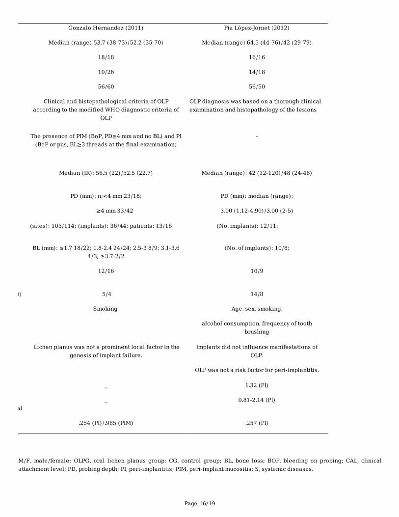
Page 16/19
Gonzalo Hernandez (2011) Pia López-Jornet (2012)
Median (range) 53.7 (38-73)/52.2 (35-70) Median (range) 64.5 (44-76)/42 (29-79)
18/18 16/16
10/26 14/18
56/60 56/50
Clinical and histopathological criteria of OLPaccording to the modified WHO diagnostic criteria of
OLP
OLP diagnosis was based on a thorough clinicalexamination and histopathology of the lesions
The presence of PIM (BoP, PD≥4 mm and no BL) and PI(BoP or pus, BL≥3 threads at the final examination)
-
Median (IR): 56.5 (22)/52.5 (22.7) Median (range): 42 (12-120)/48 (24-48)
PD (mm): n:<4 mm 23/18;
≥4 mm 33/42
PD (mm): median (range):
3.00 (1.12-4.90)/3.00 (2-5)
(sites): 105/114; (implants): 36/44; patients: 13/16 (No. implants): 12/11;
BL (mm): ≤1.7 18/22; 1.8-2.4 24/24; 2.5-3 8/9; 3.1-3.64/3; ≥3.7:2/2
(No. of implants): 10/8;
12/16 10/9
s) 5/4 14/8
Smoking Age, sex, smoking,
alcohol consumption, frequency of toothbrushing
Lichen planus was not a prominent local factor in thegenesis of implant failure.
Implants did not influence manifestations ofOLP.
OLP was not a risk factor for peri-implantitis.
_ 1.32 (PI)
al_ 0.81-2.14 (PI)
.254 (PI)/.985 (PIM) .257 (PI)
M/F, male/female; OLPG, oral lichen planus group; CG, control group; BL, bone loss; BOP, bleeding on probing; CAL, clinicalattachment level; PD, probing depth; PI, peri-implantitis; PIM, peri-implant mucositis; S, systemic diseases.

Page 17/19
Study
Selection
(max 4 asterisks)
Comparability
(max 2 asterisks)
Exposure
(max 3 asterisks)
Score Quality
Gonzalo Hernandez (2011) *** ** ** 7 Low risk
Table 3. Quality assessment of the prospective study.
Table 4. Quality
assessment of
the cross-
sectional study.
Items Yes No Unclear
the information (survey, record review)1
clusion and exclusion criteria for exposed and unexposed subjects (cases and controls) or refer to previousations
1
e time period used for identifying patients1
e whether or not subjects were consecutive if population-based1
5) Indicate if evaluators of study subjects were blinded to other aspects of the status of the participants 1
6) Describe any assessments undertaken for quality assurance purposes (e.g., test/retest of primary
outcome measurements)
1
7) Explain any patient exclusions from analysis 0
be how confounding was assessed and/or controlled1
cable, explain how missing data were handled in the analysis 0
arize patient response rates and completeness of data collection 0
what follow-up, if any, was expected and the percentage of patients for which incomplete data or follow-up
obtained
1

Page 18/19
Figures
Figure 1
Study screening process.
Page 19/19
Figure 2
Forest plot of the meta-analysis of PI (A), PIM (B), PIDs (C) and BOP (D). #CI, con�dence interval.