diseases acquired from animals - safe work … · several diseases acquired from animals do ... of...
TRANSCRIPT

National Occupational Health and Safety Commission
DISEASES ACQUIRED
FROM ANIMALS
December 1989
Australian Government Publishing Service Canberra
WAP 89/028 GS 006-1989

© Commonwealth of Australia 1989 ISBN 0 644 09192 4 This work is copyright. Apart from any use as permitted under the Copyright Act 1968, no part may be reproduced by any process without written permission from the Director Publishing and Marketing, AGPS. Inquiries should be directed to the Manager, AGPS Press, Australian Government Publishing Service, GPO Box 84, Canberra ACT 2601. The National Commission's address is: National Occupational Health and Safety Commission Level 30, St Martins Tower 31 Market Street Sydney NSW 2000 GPO Box 58 Sydney NSW 2001 Tel: (02) 265 7555 Facsimile: (02) 265 7538 Telex: 177243 Printed in Australia by Alken Press, Smithfield.
iii
Foreword_______________________________________ The National Occupational Health and Safety Commission, Worksafe Australia, is a tripartite body established by the Commonwealth Government to develop, facilitate and implement a national approach to occupational health and safety. The National Commission comprises representatives of the peak employee and employer bodies - the Australian Council of Trade Unions (ACTU) and Confederation of Australian Industry (CAI) - as well as the Commonwealth, State and Territory governments. Since its establishment, the National Commission has produced occupational health guides. Be fore the National Commission was established, a series of similar guides was published by the National Health and Medical Research Council. This Guide has been reviewed and endorsed by a working group of the National Commission as part of the co-ordinated effort by the Commonwealth, State and Territory governments and employee and employer organisations to make Australian workplaces safe and healthy. Although this Guide has been endorsed by the National Commission, it is an advisory document only. It is produced and distributed in the interests of providing useful information on occupational health and safety for employers, employees and others. This document does not replace statutory requirements under relevant State and Territory legislation. This Guide is aimed primarily at workers and managers but should also be useful to occupational health and safety personnel and others. It may be used in conjunction with appropriate training and consultation, in line with good management practice.


v
Contents________________________________________ Foreword iii Introduction 1 Identification 2 Occupational Exposures: Prevention and Management 6 Further Reading 25


Diseases acquired from animals 1
Introduction This Guide summarises several important aspects of some diseases acquired by people from contact with animals or animal products. Such diseases are often termed zoonoses, and the Commonwealth Department of Community Services and Health publication, Synopsis of Zoonoses in Australia, should be referred to for more specific information on particular diseases. People are often exposed to a variety of living agents which normally cause disease in animals and may also affect human health. The virus rabies is a notable example. If an infected (rabid) dog bites a person, virus particles will pass to the bloodstream and multiply in the body tissues. Rabies is often fatal. It has not been reported in Australia, although it is relatively common in Europe, America and Asia. Several diseases acquired from animals do cause human death and illness in this country. A variety of disease agents (viruses, bacteria, fungi and parasites) and animals harbouring the diseases (cattle, pigs, sheep, birds, dogs, cats and other domestic, native or exotic species) are involved. This Guide summarises the important aspects of the recognition, control and prevention of those diseases acquired by people from animals with particular emphasis on occupational exposures. This Guide is particularly relevant to those engaged in:
• animal production and marketing, including farmers and their families, abattoir workers (including maintenance and cleaning personnel) and food processors;
• health work, including veterinarians and veterinary nurses, stock inspectors, meat inspectors, animal impounding officers, health surveyors, medical personnel and sewage treatment workers;
• biological research, including private and governmental laboratory technicians, animal handlers and scientific staff, pharmaceutical researchers, and scientists/technical workers engaged in field studies;
• livestock handling, including stock and station agents, horse trainers and handlers;
• the pet industry, including pet store proprietors, cat and dog kennel operators and bird breeders; and
• wildlife park, circus and zoological operations.
Diseases acquired from animals 2
Identification Due to the peculiar nature of these diseases and their modes of transmission, diagnosis may often be difficult. People who work in the above-mentioned areas should alert medical personnel to the nature of their occupations. Several of these diseases require notification, by a medical practitioner, to the appropriate health authority. It is important to note that animals or people can be carriers of a disease. In this situation the disease organisms are not necessarily having a noticeable effect on the infected person or animal. When considering the variety of diseases which people can contract from animals or animal products, it is essential to have some knowledge of:
• the biological agent causing the disease, for example, is it a virus, bacterium, fungus or parasite;
• the animal reservoir or host involved: this defines the pattern of transmission of the disease when people are not infected, for example, when a ringworm fungus is simply spread from dog to dog; and
• the mode of transmission of the disease from the animal reservoir to people. A disease may be spread by inhalation of infective dust or droplets, by ingestion of contaminated food or water, or by direct contact with skin or mucous membranes. Some diseases, particularly those caused by viruses, bacteria and fungi, have a relatively simple, direct mode of transmission. For example, people can become infected with Campylobacter and Salmonella bacteria, which can cause diarrhoea, by direct contact with infected animals, resulting in the ingestion of organisms. Other organisms, notably the parasites which can infect both animals and people, go through a rather complex series of changes in their life cycle. This may involve an intermediate host, at this stage the parasite is immature, and a definitive host, the organism matures and reproduces in this host. The following diagram, which outlines the life cycle of the hydatid tapeworm, Echinococcus granulosus, illustrates the complexity of the mode of transmission of some zoonotic diseases.

Diseases acquired from animals 3
This tapeworm grows to maturity in dogs (1) which pass worm eggs from their intestines when they are infected. If a grazing animal eats material contaminated with eggs (2), cysts form in the internal organs. If these organs are fed to a dog, the adult worm will develop in that dog's intestine, and the disease cycle will continue (3). Human infection is not necessary for the cycle to continue. However, cysts can form in people if they ingest eggs, and severe disease may result. The hydatid cycle will be further discussed later in this guide. Note that it displays an important feature, common to several diseases acquired from animals, that is, the organism causing the disease is transmitted as a tiny, microscopic form which grows to a much larger form, in this case a worm. Over 200 diseases have been classified as zoonoses throughout the world. Of these, approximately fifty have been reported in Australia. They are caused by a variety of viruses, bacteria, fungi and parasites, and some examples are given in the following table.

Diseases acquired from animals 4
Disease and causative organism
Animal(s) involved and most common mode of transmission to people
Characteristic effect of human infection
Occupational groups at risk include
MURRAY VALLEY ENCEPHALITIS an arbovirus involving an arthropod (insect) vector.
Birds, native and domestic animals are implicated. Spread by mosquito bites.
Headache, fever drowsiness, neck stiffness, loss of appetite, giddiness. Coma death or brain damage may occur.
Farmers, outdoor workers.
Q FEVER Coxiella burnetii, a bacteria-like organism.
Cattle, sheep, goats. Inhalation of air or dust.
Chills, fever, sweating, headache, loss of appetite, muscle soreness. Inflammation of lungs liver and heart may occur.
Abattoir workers, livestock handlers, veterinarians, biological laboratory workers.
PSITTACOSIS (ORNITHOSIS) Chlamydia psittaci, a bacteria-like organism.
Parrots, parakeets, pigeons, poultry. Inhalation of contaminated air.
Loss of appetite, chills, fever, head-ache, sensitivity to light, irritation to throat.
Bird breeders, pet store personnel, veterinarians.
ACUTE GASTROENTERITIS caused by Campylobacter bacteria.
Dogs, chickens, pigs, sheep. Ingestion of contaminated food or water.
Diarrhoea, abdominal pain, fever, nausea and vomiting.
Personnel involved in food processing and food handling.
LEPTOSPIROSIS Leptospira interrogans bacteria.
Pigs, cattle and rats. Penetration of the skin by the organisms, or via the mouth or nose.
Headache, fever, sensations of chill, muscle pain, soreness stiffness in the neck. Kidneys may be affected and vomiting, coughing, or exhaustion may occur.
Abattoir workers, pig and dairy farmers, sewage treatment workers, veterinarians.
RINGWORM of the scalp or body, Microsporum canis and other fungus species.
Mainly dogs and cats. Contact with infected animals or articles.
Single or multiple rings of inflamed skin around a scaly central region.
Animal handlers.
HYDATID DISEASE Echinococcus granulosus, a tapeworm.
Dogs harbour adult worm. Sheep and wallabies develop cysts from ingesting eggs. People also develop cysts if eggs are ingested.
People develop cysts in lungs, brain or kidneys. Nausea after meals, coughing, jaundice, or abdominal pain may occur. Anaphylactic shock may follow rupture of a cyst. This can be fatal.
Sheep farmers and their families.

Diseases acquired from animals 5
The diseases outlined in the table above illustrate the range of animals from which diseases can be acquired, and the disease outcomes which may result. Many other diseases could be discussed, and the publication, Synopsis of Zoonoses in Australia, should be consulted for further information. This Guide provides information on several animal diseases which are currently of importance in Australia. The reader should note, when travel overseas is contemplated, that many additional zoonotic diseases exist in overseas countries. Several of these are potentially debilitating or may result in death if not treated. Officers of the Commonwealth Department of Community Services and Health should be approached for information regarding the prevention of disease when visiting other countries. In this guide the following diseases will be discussed:
• Q Fever;
• leptospirosis;
• brucellosis;
• hydatidosis;
• campylobacter and salmonella infections;
• psittacosis;
• arboviruses;
• erysipeloid;
• orf;
• ringworm; and
• anthrax.
Diseases acquired from animals 6
Occupational Exposures: Prevention and Management It is important to note that many of the human diseases acquired from animals do not necessarily have a noticeable effect on the animal involved. For example, Tetanus bacteria are normal residents in the gut of healthy horses, but after being passed out of the animal they can produce an infection if they gain entry to a deep wound. Subsequently, the toxin they produce may result in a severe, often fatal, disease in people. This disease is preventable by immunisation. Similarly, for several diseases, animals may acquire a high degree of immunity to the organisms involved. They may be able to pass on the infection to other animals, and people, without themselves suffering any ill effects. All animals should be handled with particular caution, with due regard to the risk of contracting a disease, especially if they appear to be sick. A summary of the major aspects of the effects and prevention of some zoonotic diseases readily identifiable with occupational exposures follows. Q Fever The nature and mode of transmission of this disease remained undiscovered for many years. It is caused by a rickettsial (bacteria- like) organism, Coxiella burnetii, which lives in a variety of domestic and wild animals, especially cattle, sheep, goats, bandicoots, kangaroos and wallabies. Ticks can transport the organisms from animal to animal. As the organisms which cause this disease are most plentiful in the uterus and udder of pregnant cattle and sheep, the following occupational groups are especially at risk of contracting Q Fever:
• animal handlers, including farmers;
• veterinarians and veterinary nurses;
• abattoir workers;
• meat inspectors; and
• biological researchers working with pregnant animals.

Diseases acquired from animals 7
Distribution and extent of the disease Q Fever exists throughout mainland Australia. Queensland usually has the highest number of reported Q Fever cases per year.
The disease in animals Animals do not normally display any ill effects from infection with Coxiella burnetii. However, the organisms may multiply extensively in a female animal during the latter stages of pregnancy, and the placental tissues and birth fluids may contain large numbers of the organism. Q Fever can be spread to other animals, and people, by the inhalation of infective material from these placental tissues and fluids. Human infection can also occur through the ingestion of unpasteurised milk and by contact with infected animals, their waste products, or contaminated straw, wool, hair and hides.
The disease in people If a person is infected, and infection with only small numbers of the organisms can result in human disease, the health effects will abruptly occur some sixteen to eighteen days later. Muscle pains, severe headache and high fever are the usual signs and symptoms manifested. There may be a progression to pneumonia, and Q Fever is often misdiagnosed as influenza or pneumonia. The fever usually disappears after five to fifteen days.
Prevention and management of Q Fever Antibiotics such as tetracycline are the preferred treatment and recovery from the infection is usually rapid. However, patients who have diseased heart valves prior to contracting Q Fever may develop severe inflammation and damage to these valves. The liver is commonly affected.

Diseases acquired from animals 8
To minimise inhalation, ingestion or contact with Coxiella burnetii organisms, the following preventive measures should be undertaken:
• People working in the at-risk groups should know of the sources of infection and the modes of transmission of the disease.
• Strict hygienic practices must be followed when pregnant animals, hides, wool, straw, or other contaminated material is handled. This involves the prevention of inhalation of contaminated dust or fluid droplets, adequate disinfection and disposal of material, and prompt treatment of cuts and abrasions.
• Placental and other birth material should be burnt or buried. Contaminated litter should be burnt.
• Milk should be pasteurised or boiled. A vaccine has been developed in Australia to immunise high-risk groups against Q Fever. Preliminary trials with the vaccine are promising, but it is not yet available for general use. It is most important for anyone with flu- like symptoms who is likely to have had contact with infected animals to seek medical advice, stating clearly that contact with the Q Fever organisms has possibly occurred. Leptospirosis Leptospirosis is a widespread disease of cattle, pigs, sheep and a variety of native animals, especially rats. People usually acquire the disease from the urine of animals infected with Leptospira bacteria. A variety of closely-related leptospiral organisms can infect people. Refer to Synopsis of Zoonoses in Australia for details. The human disease can be quite debilitating. Abattoir workers, dairy farmers, piggery workers, sugar cane farmers and veterinarians are most at risk of contracting leptospirosis. Drainage workers may also be exposed to leptospiral organisms, via contact with contaminated soil.
The disease in animals Infection in cattle typically causes fever and loss of appetite. Cows exhibit a sharp drop in milk production and may suffer from inflamed udders. Milk may be yellowish or blood-stained. Abortions and blood passed in urine are common outcomes of infection. The disease is more severe, and often fatal, in calves.

Diseases acquired from animals 9
Following infection pigs usually suffer a mild or inapparent form of leptospirosis. Abortion, or the birth of weak, stunted piglets may occur. Pigs are an important source of infection to other domestic animals, especially cattle, as they spread large numbers of the Leptospira bacteria to the environment, via urine, for a long time following infection.
The disease in people Human infection varies considerably in severity and may occur as:
• Milder (and more common) forms, characterised by rapid onset of severe headache, chills, muscle pains, sensitivity to light and stiffness of the neck. Symptoms usually subside within ten days, but relapses may occur.
• The serious form, characterised by fever, vomiting, muscle pain, headache and, frequently, jaundice (yellowing of the skin and eyes due to liver malfunction). The kidneys may be affected, and internal bleeding may occur. This form of leptospirosis may be fatal.
It is most important for anyone with these symptoms who is likely to have had contact with leptospiral organisms to seek medical attention.
Prevention and management of leptospirosis Because such a wide variety of domestic and native animals can be involved in the transmission of leptospirosis, it is a difficult disease to control. The following measures should be adopted to reduce the incidence of infection in animals and people:
• All workers who are involved in handling potentially infective animals should be fully aware of the modes of transmission of leptospirosis. People must avoid contact with

Diseases acquired from animals 10
potentially contaminated water or wear protective boots or gloves when work necessitates contact with such water.
• Rats and mice should be eradicated whenever possible. The firing of sugar cane prior to harvest assists in controlling rat and mouse populations.
• The provision of adequate drainage in farm areas and the hygienic disposal of effluent, particularly from piggeries. Pigs are a common source of acute infection in calves. For this reason, pigs and cattle should be raised separately. Pigs should not be permitted access to cattle areas.
• Vaccination of animals. Although this is commonly practised, surveys indicate that only a small proportion of farmers give the full two doses of vaccine to their cattle or pigs. It is most important to follow the correct vaccination schedule, and to vaccinate all animals which may become infected with leptospirosis. Veterinarians should be consulted to ensure that the appropriate vaccine is employed for the particular animal and geographical location.
• Streptomycin injections have been shown to eliminate Leptospira bacteria from pigs and cattle.
Brucellosis Brucellosis, also known as Undulant Fever, is caused by any one of a group of six Brucella bacteria. In Australia, the disease is transmitted to people from infected cattle or pigs. Brucellosis should be considered in the diagnosis for anyone who has a history of contact with domestic cattle and is experiencing a fever of unknown origin. Farmworkers, veterinarians and abattoir workers are particularly at risk.
The disease in animals The disease in beef and dairy cattle causes inflammation of the genital tract and frequently results in abortion. When an infected cow gives birth or aborts, the Brucella abortus bacteria are spread to the environment in large numbers. They may also be passed in the cow's milk for long periods, possibly years. Discharges from the vagina of a cow may be infective for more than three weeks. Cattle usually acquire the disease by eating contaminated fodder or pasture. Pigs infected with Brucella suis may develop internal abscesses, arthritis and bone degeneration. Diseased sows often give birth to small, weak piglets.

Diseases acquired from animals 11
The disease in people Brucellosis in people is characterised by a fluctuating fever, headache, weakness, sweating, muscular pains and depression. These signs occur two to four weeks after infection has occurred. Infection may be acquired by:
• inhalation of infective droplets or dust;
• ingestion of unpasteurised milk; and
• direct contact of animal discharges with mucous membranes or skin, especially if the skin is broken.
Following this acute phase, which usually subsides within a few weeks, a patient may suffer relapses and be chronically ill for several years. Recovery is usually prompt if the appropriate antibiotic treatment is given, although relapses may still occur. People working with pigs, on farms or in abattoirs, may become infected with Brucella suis, which readily infects people and is believed to be limited to Queensland. Feral pigs are a potential source of infection.
Prevention and management of brucellosis As with most of the diseases discussed in this Guide, the key to prevention is elimination of the organisms from animals. A national program for eradicating brucellosis from cattle commenced in 1970. Tasmania and Western Australia were declared free of the disease in 1986. Australia-wide eradication is anticipated by 1989. The following control measures should be adopted to complement this eradication program:
• All personnel handling potentially infected animals should be tho roughly informed regarding the danger of contracting the disease from carcasses, milk, birth products or discharges.
• Individuals with uncovered wounds should not be permitted to work in a meat handling area. Cuts and abrasions should be treated immediately. Eating and smoking should be banned in meat handling areas. Splashes of animal material on clothing, equipment or skin surfaces should be removed as soon as possible.
• Trucks taking cattle and pigs to abattoirs should be regularly disinfected.

Diseases acquired from animals 12
• Animals that have reacted to diagnostic tests for brucellosis should be segregated, prior to slaughter, in an area that can readily be disinfected.
• Reactive animals should be carefully slaughtered at the end of the day's operations. All slaughtering, washing and cleaning procedures should minimise the opportunity for spillage or splash.
• The dressing techniques, particularly the handling of the uterus and udder, should minimise the dissemination of potentially infective material.
• Protective clothing and equipment, such as face and eye protection, gloves and impervious boots should be available and used where necessary. Particular care should be taken when handling udders, uteri, bladders, brisket saws and knives in slaughtering positions where there is a risk of spillage.
• As frequently as possible, all utensils, instruments, machinery, chutes, floors and other areas of potential contamination should be cleaned by agents approved by the relevant meat inspection authority and then cleansed with water at temperatures above 82°C.
• Occupationally exposed groups should have access to specialised medical services to facilitate the early and correct diagnosis of brucellosis.
Hydatid disease In Australia, the organism which causes this disease is the small tapeworm Echinococcus granulosus. This parasite grows to maturity in dogs or dingoes, which spread infective eggs to the environment in their faeces. Grazing or foraging animals, such as sheep, cattle, pigs, wallabies and kangaroos, become intermediate hosts to the parasite when they ingest Echinococcus eggs. Fluid-filled cysts form in the internal organs of these intermediate hosts. If a dog or dingo is allowed to feed on diseased internal organs (offal), tiny larval worms are liberated from the cysts and grow to maturity in the dog, perpetuating the cycle. People can also develop cysts if they ingest Echinococcus eggs. This can readily occur when infected dogs are patted or contaminated surfaces are handled. The earlier diagram summarises these aspects of the spread of hydatid disease. Sheep farmers and their families are especially at risk of contracting hydatidosis.
Distribution and extent of the disease While hydatid disease continues to be reported throughout Australia, few cases are reported in Tasmania due to the success of a control program in that State. It is most common in South East
Diseases acquired from animals 13
New South Wales, the shires surrounding the Australian Capital Territory and the districts surrounding Perth.
The disease in animals Dogs and dingoes are not usually affected by the adult tapeworms. However, the cysts in the grazing/foraging animals may damage the liver, lungs, brain, kidneys, heart, spleen or bones. Two separate life-cycle patterns exist in Australia for the hydatid parasite:
• a domestic dog/sheep cycle in sheep grazing areas; and
• a dingo and feral dog/wallaby and kangaroo cycle. Recent surveys have shown that kangaroos and feral pigs have been involved in passing the disease to hunting dogs such as bull-terriers.
The disease in people People may also develop cysts in a variety of organs, with the liver and lungs the most common sites. When infective eggs are ingested, the tiny larvae may form cysts. These cysts will normally continue to enlarge with time and may pose a risk to health because:
• They may interfere with the normal functioning of the organ in which they have lodged.
• They may rupture, either spontaneously or following an accident such as a fall. Following rupture of a cyst, the fluid liberated may cause anaphylactic shock or larvae released may form other cysts.
Due to the time required for the development of cysts, several years may pass following ingestion of Echinococcus eggs before signs or symptoms of the disease occur. By this time the person's occupation may be quite different to that which resulted in the actual exposure to the organisms. The presence of cysts may not be recognised until they rupture or are evident on an x-ray. The majority of cases come from sheep grazing areas. The main form of treatment, where indicated, is surgery.
Prevention and management of hydatid disease Hydatid control programs are now operating in many parts of Australia. It is essential that the following control measures are adopted for the disease to be minimised:
• All people who come in contact with potentially infected dogs or contaminated areas should be educated regarding the mode of transmission of hydatid disease and the need for strict personal hygiene.
Diseases acquired from animals 14
• Dogs should be kept away from animal carcasses and offal should be buried in deep pits. Hunting dogs should not be permitted to feed on wallabies, kangaroos or pigs.
• Kangaroo meat sold as pet food should be free of cysts.
• Potentially infected dogs should be tested for the presence of Echinococcus granulosus and subsequently treated with the appropriate oral medication.
• Wild and stray dogs should be destroyed. The number of working dogs in sheep areas should be kept to a minimum.
For further information, contact the relevant public health authority. Campylocbacter and Salmonella infections Campylobacter and Salmonella organisms are natural inhabitants of the gut in cattle, sheep, pigs and birds. In people, these bacteria can cause gastro-intestinal disturbances, including diarrhoea.
The diseases in animals Several related subspecies of Campylobacter infect animals, resulting in abortion and infertility in cattle, abortion in sheep, liver disease in birds and gut disturbances in dogs, cats, cattle and turkeys. The infection is passed from animal to animal through contamination of their food sources. A significant proportion of animals may act as carriers of the infection, that is, they may pass on the infection without exhibiting symptoms of disease.
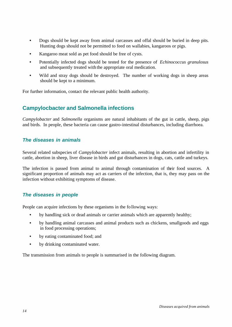
The diseases in people People can acquire infections by these organisms in the fo llowing ways:
• by handling sick or dead animals or carrier animals which are apparently healthy;
• by handling animal carcasses and animal products such as chickens, smallgoods and eggs in food processing operations;
• by eating contaminated food; and
• by drinking contaminated water. The transmission from animals to people is summarised in the following diagram.
Diseases acquired from animals 15
Features of human infection range from mild gastro- intestinal discomfort to severe inflammation of the intestines with pain and profuse watery diarrhoea.
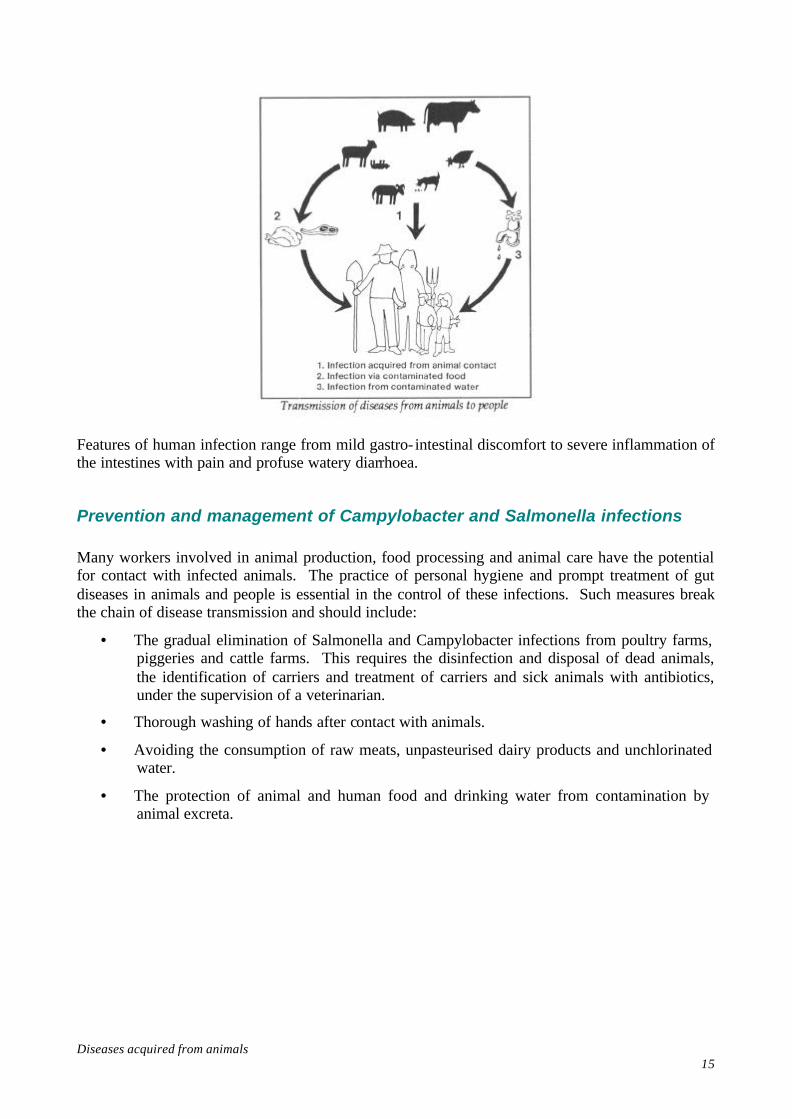
Prevention and management of Campylobacter and Salmonella infections Many workers involved in animal production, food processing and animal care have the potential for contact with infected animals. The practice of personal hygiene and prompt treatment of gut diseases in animals and people is essential in the control of these infections. Such measures break the chain of disease transmission and should include:
• The gradual elimination of Salmonella and Campylobacter infections from poultry farms, piggeries and cattle farms. This requires the disinfection and disposal of dead animals, the identification of carriers and treatment of carriers and sick animals with antibiotics, under the supervision of a veterinarian.
• Thorough washing of hands after contact with animals.
• Avoiding the consumption of raw meats, unpasteurised dairy products and unchlorinated water.
• The protection of animal and human food and drinking water from contamination by animal excreta.
Diseases acquired from animals 16
• The hygienic disposal of all human waste.
• The diagnosis and treatment, and the notification of all human cases of these diseases to State or Territory health authorities.
• The education of caterers and food handlers regarding food hygiene practices.
• In poultry processing using negative pressure hoods for defeathering machines, washing carcasses in superchlorinated water and rapid transfer to refrigeration chambers.
• Handling procedures during slaughtering which ensure that surface contamination of the carcasses by faecal material, intestinal contents, hide or fleece is kept to a minimum.
• Rendering works constructed to provide a barrier between the raw and processed material.
• Refrigeration of foods (below 5°C).
• Hygienic production and cold storage of eggs, hygienic preparation of egg products and effective heat treatment of the final product before distribution.
Psittacosis Several species of birds which are associated with human activity may suffer from respiratory disorders caused by the organism Chlamydia psittaci.

Diseases acquired from animals 17
These birds include those in the fowl family (chickens and turkeys), pigeons, lorikeets, budgerigars and cockatoos. Caged birds are most likely to carry the disease, due to the potential for transmission from one bird to another in those situations. Human infection can occur in those occupational settings where people contact infected birds. Bird breeders, pet store operators and veterinarians are therefore especially at risk of contracting psittacosis.
The disease in birds The chlamydial organisms typically go through a latent, hidden, phase in birds. Fowl are virtually unaffected by the infection, but other birds may display the following symptoms:
• difficulty in breathing;
• diarrhoea and loss of appetite;
• fever and emaciation; and
• conjunctivitis. The infective organisms are passed out in the faeces of birds while they are sick. If the birds recover, they may still contaminate the environment for some time. The infection will often become apparent in birds when they are stressed by poor nutrition, crowding, lengthy transportation or dirty conditions.
The disease in people Human infection is acquired by inhalation of Chlamydiae in a contaminated work environment. This could occur in pet shops, chicken farms, chicken processing areas, wildlife refuges, pigeon coops and biological research establishments. People infected with Chlamydia psittaci typically become sick one or two weeks after inhaling contaminated air. Combinations of the following symptoms may occur:
• fever or chills;
• headache or sore throat;
• loss of appetite;
• bronchitis;

Diseases acquired from animals 18
• sensitivity to bright light; and
• pneumonia. In severe infections, gastro- intestinal disturbances, nausea, vomiting, constipation or diarrhoea may occur, and the liver and spleen may be affected. Relapses may occur if the antibiotic treatment, tetracycline drugs, is not continued for two weeks after the symptoms subside.
Prevention and management of psittacosis The following measures are available to minimise the transmission of chlamydial organisms:
• water containing antibiotics can be used to prevent the infection in aviary birds;
• medication can be administered to new birds added to disease-free aviary flocks;
• sick birds should be quarantined during antibiotic treatment, or alternatively, those birds suspected of being infected should be destroyed and their bodies disinfected in two per cent phenol or some other suitable disinfectant; and
• pet shop facilities, coops and aviaries should be thoroughly cleaned and aired if an outbreak occurs.

Diseases acquired from animals 19
Arboviruses The arboviruses (arthropod-borne viruses) cause a large group of diseases transmitted to people by ticks, mosquitoes and other arthropod organisms. In Australia, Murray Valley Encephalitis and Ross River Virus are the more important arboviruses.
Murray Valley Encephalitis Evidence of contact with the virus that causes this disease has been found in people, horses, dogs, foxes, marsupials, birds and chickens. The disease has been reported in all Australian states and Papua New Guinea. However, outbreaks of the disease occur particularly in the more populated areas of southern Australia, in association with increases in mosquito numbers.
The disease in animals Chickens and aquatic fowl are believed to act as reservoirs of the disease, with mosquitoes responsible for transmitting the virus from bird to bird and from birds to people. The birds do not display any noticeable signs of infection.
The disease in people A person infected with Murray Valley Encephalitis virus by the bite of a mosquito harbouring the virus may develop mild or severe inflammation of the brain and spinal cord, or may show no signs or symptoms of disease. In severe infections high fever, headache, disorientation and tremors may occur. Children may suffer convulsions and paralysis. Less severe cases display the symptoms of mild meningitis. Children and elderly people are particularly susceptible to severe disease. Because of the potential involvement of birds in the disease cycle, anyone working in those areas where the virus is found, and where large populations of birds and mosquitoes exist, is at risk of infection. Defence force personnel carrying out field exercises, farmers, biologists and other field workers represent occupational groups which are likely to be exposed to the virus.
Ross River Virus This virus, also transmitted by mosquitoes, causes a form of arthritis, swelling and pain in joints, often accompanied by a mild rash. The wrists, fingers, knees, ankles and toes are particularly

Diseases acquired from animals 20
affected. Major outbreaks have occurred in Northern Queensland, the Northern Territory and Central New South Wales. The arthritic condition may persist for two days to eight months, after which recovery is complete and immunity is established.
Prevention and management of arboviral diseases The destruction of adult mosquitoes and larvae, the elimination of mosquito breeding sites, the screening of sleeping areas and the avoidance of exposure to mosquitoes during their biting times are the main control methods for these arboviruses. In addition, small numbers of chickens may be placed at strategic locations and routinely checked for viral infection. Potentially exposed workers should be informed of the features of the spread of these diseases. Erysipeloid The bacterium Erysipelothrix rhusiopathiae causes erysipelas in pigs, lambs and turkeys. These organisms are often found in the soil on farms, and may also be associated with fish. Infection with Erysipelothrix rhusiopathiae in people can cause the disease erysipeloid. Kitchen workers, meat workers, poultry workers, fishermen and fish dealers are at risk of contracting erysipeloid.
The disease in animals Infected pigs usually develop diamond-shaped skin patches, with a high fever, exhaustion, loss of appetite, inflammation of the eyelids, vomiting and arthritis (inflamed joints). Pregnant sows may give birth to dead or weak piglets.

Diseases acquired from animals 21
Lambs may develop arthritis and become lame. Infected turkeys and ducks often have a high fever accompanied by arthritis.
The disease in people People infected with these bacteria typically develop a skin eruption on the hands or fingers. Swollen, slightly elevated patches appear; they are often associated with a burning, tingling sensation and may be intensely itchy. Mild arthritis of the finger joints occasionally occurs. In addition to these skin and joint disorders, fever, headache and heart or nervous system complications sometimes result. The skin eruptions usually heal in two to six weeks and penicillin treatment speeds the recovery. However, relapses may occur and, rarely, skin infection may persist for a long time.
Prevention and management of erysipeloid People are commonly infected via cuts and abrasions to the skin. The following measures should therefore be adopted to minimise the spread of erysipeloid:
• prompt washing and dressing of cuts and abrasions on the hands;
• regular cleaning and disinfection, preferably with steam, of work benches and equipment;
Diseases acquired from animals 22
• thorough decontamination of hands and arms if a fevered pig carcass or any other potentially infective material has been handled; and
• notification to the appropriate public health authority if several cases of erysipeloid occur in the same workplace.
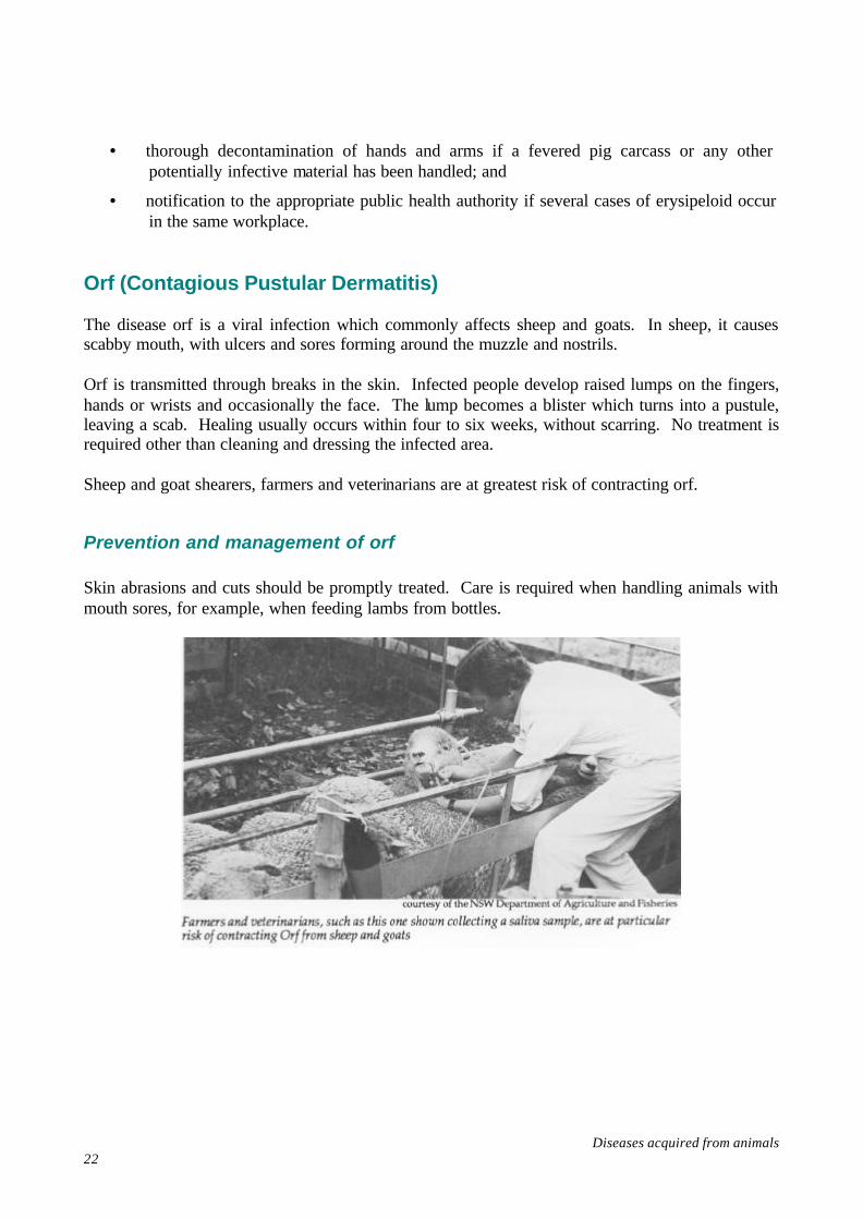
Orf (Contagious Pustular Dermatitis) The disease orf is a viral infection which commonly affects sheep and goats. In sheep, it causes scabby mouth, with ulcers and sores forming around the muzzle and nostrils. Orf is transmitted through breaks in the skin. Infected people develop raised lumps on the fingers, hands or wrists and occasionally the face. The lump becomes a blister which turns into a pustule, leaving a scab. Healing usually occurs within four to six weeks, without scarring. No treatment is required other than cleaning and dressing the infected area. Sheep and goat shearers, farmers and veterinarians are at greatest risk of contracting orf.
Prevention and management of orf Skin abrasions and cuts should be promptly treated. Care is required when handling animals with mouth sores, for example, when feeding lambs from bottles.

Diseases acquired from animals 23
Ringworm - fungal infections Several species of fungi cause the skin condition known as ringworm in cats, dogs, horses, cattle, pigs, mice, kangaroos and people. These fungi grow on the surface of skin, producing characteristic ring-shaped lesions. Ringworm infections acquired from animals usually involve direct contact with infected dogs or cats, or contact with contaminated material. The infected animals may not show obvious signs of infection.
Prevention and management of ringworm An infected person can spread the infection to animals or to other people. Personal hygiene is therefore an essential element in preventing the spread of ringworm. An orally-administered drug will kill the fungus in animals and people. The clipping of hair, where appropriate, and the application of a topical fungicidal preparation are also advisable. Anthrax This disease, also known as Malignant Pustule and Woolsorter's Disease, is caused by the bacterium Bacillus anthracis. Although it is now an uncommon disease in Australia, cases could still occur and it therefore warrants consideration in this Guide.
The disease in animals Infected animals, typically cattle, sheep, horses and goats, develop septicemia (blood poisoning) which often kills the animal. Spores of the bacteria, which are extremely resistant, can contaminate hair, hides, wool, the soil and products such as feeds and fertilisers prepared from infected animals.
The disease in people Human disease occurs in two main forms:
• Cutaneous Anthrax or Malignant Pustule. This is the commonest form and can occur as a result of anthrax spores entering cuts or abrasions on the arms or face. The initial lesion is a small flared red lump which later develops a central black depression. The surrounding area is inflamed and swollen.

Diseases acquired from animals 24
• Pulmonary Anthrax or Woolsorter's Disease. As the name implies, the disease can occur as a result of inhaling anthrax spores into the lung in the process of sorting infected wool. Coughing of blood occurs and the disease has a high fatality rate.
Diagnosis of cutaneous anthrax is based on the characteristic appearance of a pustule and isolation of the type of anthrax organism from it. An abattoir worker with a rash or pustules of any sort should report to a doctor for diagnosis and treatment.
Diseases acquired from animals 25
Further Reading Acha, P.N. and Szyfres, B., Zoonoses and Communicable Diseases Common to Man and Animals, Pan American Health Organization, World Health Organization, Washington, 1980. American Public Health Association, Control of Communicable Diseases in Man, 14th Edition, Benenson, A.S., ed., American Public Health Association, Washington, 1985. Department of Community Services and Health, Communicable Diseases Intelligence Bulletin, (various). Faine, S., ‘Leptospirosis - still here’, Medical Journal of Australia, vol.144, no.7, pp.561, 1986. Miller, C.D., Songer, J.R., Sullivan, J.F., ‘A twenty-five year review of laboratory-acquired human infections at the National Animal Disease Center’, American Industrial Hygiene Association Journal, vol.48, no.3, pp.271-75, 1987. Standards Australia, AS 2243-1985 Safety in Laboratories Part 3 - Microbiology, Sydney. Steele, T.W., ‘Campylobacter infection: A changing scene’, Medical Journal of Australia, vol. 145, pp.491-92. Stevenson, W.J. and Hughes, K.L., Synopsis of Zoonoses in Australia, 2nd Edition, Australian Government Publishing Service, Canberra, 1987.