diseases of the parathyroid glands hyperparathyroidism hypoparathyroidism
TRANSCRIPT

DISEASES OF THE PARATHYROID GLANDS
HYPERPARATHYROIDISMHYPOPARATHYROIDISM

Thyroid/Parathyroid glands
1=normal thyroid gland2 and 3=parathyroid gland4=enlarged thyroid gland

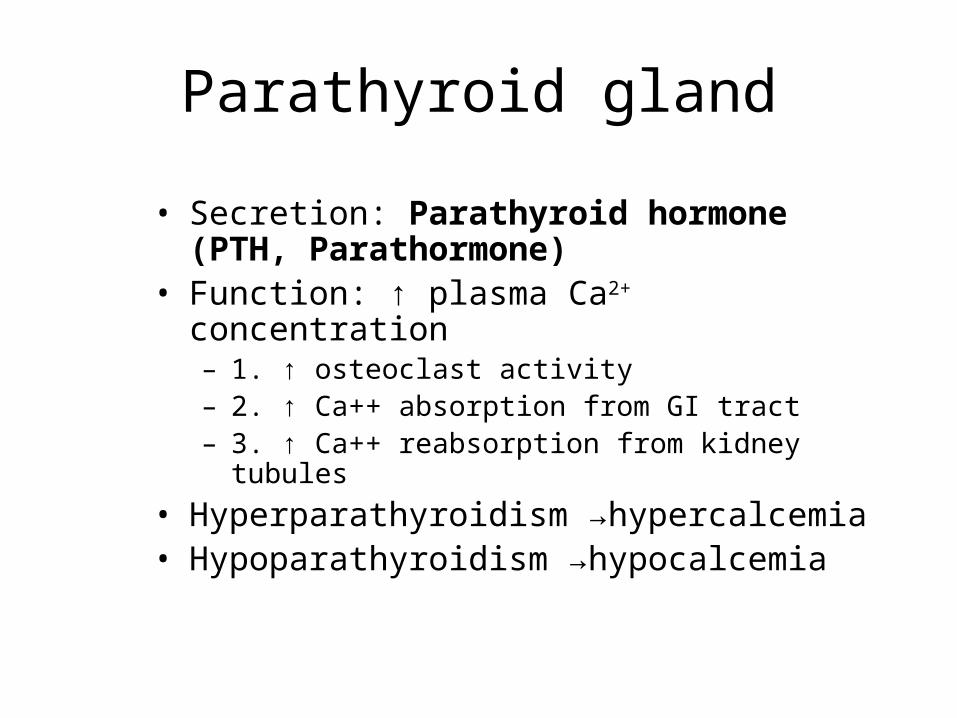
Parathyroid gland
• Secretion: Parathyroid hormone (PTH, Parathormone)
• Function: ↑ plasma Ca2+ concentration– 1. ↑ osteoclast activity– 2. ↑ Ca++ absorption from GI tract– 3. ↑ Ca++ reabsorption from kidney tubules
• Hyperparathyroidism →hypercalcemia• Hypoparathyroidism →hypocalcemia
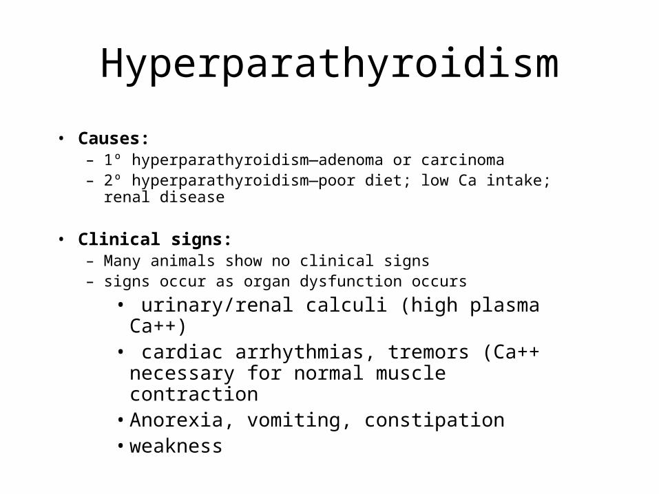
Hyperparathyroidism
• Causes:– 1º hyperparathyroidism—adenoma or carcinoma– 2º hyperparathyroidism—poor diet; low Ca intake; renal disease
• Clinical signs:– Many animals show no clinical signs– signs occur as organ dysfunction occurs
• urinary/renal calculi (high plasma Ca++)• cardiac arrhythmias, tremors (Ca++ necessary
for normal muscle contraction• Anorexia, vomiting, constipation• weakness

Hyperparathyroidism
Dx:• Routine chemistry panel
– ↑ blood Calcium (normal: ~8-10 mg/dl))– +/- ↓ blood Phosphorus (normal: ~2-6 mg/dl)
• PTH assay– normal PTH: dogs ~20 pg/ml, cats ~17 pg/ml– In a normal animal: if blood Ca++ is high, PTH is low (neg feedback)– 1º Hyperparathyroidism: Ca++ high, PTH elevated
• Ultrasound of neck – enlarged glands, abdomen - uroliths

Hyperparathyroidism
Tx:1. Surgical removal of diseased parathyroid (generally 4 lobes are imbedded in thyroid gland)
Other options:2. Ultrasound-guided chemical (ethanol) ablation
3. Ultrasound-guided heat (laser) ablation
Post-Op Care:1. Hospitalize for 1 wk; ↓PTH may predispose animal to hypocalcemia2. Calcium therapy (oral tabs, liquid)3. Vit D supplements (promotes Ca intestinal absorption)
Hyperparathyroidism
Client Info1. Most hyperparathyroid animals show no
signs when first diagnosed2. Run yearly chem panels on all normal, older
animals

• Hyperparathyroidism clinical case

Hypercalcemia: Other causes
• Causes– Neoplasia (lymphoma, perianal gland
tumors)– Renal failure– Hypoadenocorticism– Vitamin D rodenticide– Drugs or artifacts (ex lipemia)
• Clinical signs vary with cause– PU/PD, anorexia, lethargy, vomiting,
weakness, stupor/coma (severe), uroliths
Hypercalcemia
• Tests– Elevated serum calcium levels– Low to low-normal phosphorus concentrations

Hypercalcemia
• Treatment– Fluids: 0.9% NaCl
• No Ca2+ containing fluids
– Diuretics (furosemide)– Steroids
• Complications– Irreversible renal failure– Soft tissue calcifications

HypocalcemiaCauses:1. Parathyroid disease
a. Inadvertent removal of parathyroid during thyroidectomy (most common causeb. 1º Hypoparathyroidism (uncommon in animals)
2. Chronic renal failure—a. may cause ↑ serum P, which can result in ↓ serum Ca (Ca:P inverse relation)b. Vit D normally activated in kidneyc. Protein-losing nephropathy results in loss of albumin-bound Ca
3. Puerperal Tetany (Eclampsia)—late gestation thru post-partum perioda. Improper prenatal nutritionb. Heavy lactationc. Inappropriate Ca++ supplementation
http://www.thepetcenter.com/gen/eclampsia.html#The_video

Hypocalcemia
Clinical Signs:1. Restlessness, muscle tremors, tonic-clonic contractions,
seizures 2. Tachycardia with excitement; bradycardia in severe cases
(Ca++ is necessary for proper muscle contractions)3. Hyperthermia4. Stiffness, ataxic

Hypocalcemia
Dx:Total serum <6.5 mg/dl
Tx:1. IV infusion of 10% Ca gluconate solution (monitor
HR and rhythm during infusion)2. Diazepam (IV) to control seizures3. Oral supplements of Ca (tabs, caps, syrup)4. Improve nutrition

Hypocalcemia
Client info:1. Well-balanced diet; increase volume as
pregnancy progresses2. Signs in pregnant animal is emergency; call
vet immediately3. May recur with subsequent pregnancies4. Early weaning is recommended

DISEASES OF THE PANCREAS
DIABETES MELLITUSINSULINOMA
EXOCRINE PANCREATIC INSUFFICIENCY

Review of pancreas functions
• Long flat organ near duodenum and stomach
• Exocrine function (the majority of the pancreas):– Digestive enzymes
• Endocrine function – islets of Langerhans– Alpha cells => glucagon– Beta cells => insulin– Delta cells => somatostatin

Pancreas

Pancreas: beta cells

Review
• Insulin – Moves glucose into cells to be used for energy– Decreases blood glucose
• Glucagon– Raises blood glucose
• Stimulates liver to release glucose• Stimulates gluconeogenesis
– Other hormones from other glands perform similar functions (hyperglycemic effect)
• Growth hormone• Glucocorticoids

Insulin/Glucagon Balance

Endocrine Pancreas
• Hyperglycemia– Definition: Excessively high blood glucose
levels• Normal in dogs: 60-120 mg/dl• Normal in cats: 70 -150 mg/dl

Diabetes Mellitus
• Definition: Disorder of carbohydrate, fat and protein metabolism caused by an absolute or relative insulin deficiency
• Type I – Insulin Dependent DM – very low or absent insulin secretory ability
• Type II – Non insulin dependent DM (insulin insensitivity) – inadequate or delayed insulin secretion relative to the needs of the patient

Diabetes mellitusIncidence:
Dogs: ~100% Type I (Insulin dependent)Cats: ~ 50% Type I and 50% Type II
-non-insulin dependent catscan sometimes be managed withdiet and drug therapy
Causes: Chronic pancreatitisImmune-mediated disease -beta cell destruction
Predisposing/risk factors:Cushing’s DiseaseAcromegalyObesityGenetic predispositionDrugs (steroids)

Diabetes mellitus
• Age/sex: – Dogs: 4-14 yrs, females 2x more likely to
be affected– Cats: all ages, but 75% are 8-13yrs,
neutered males most affected
• Breeds: Poodles, Schnauzers, Keeshonds, Cairn Terriers, Dachshunds, Cockers, Beagles

DIABETES MELLITUS
• Pathophysiology– Insulin deficiency => impaired ability to use glucose from
carbohydrates, fats and proteins– Impaired glucose utilization + gluconeogenesis => hyperglycemia– Clinical signs develop when:
• Exceeds capacity of renal tubular cells to reabsorb• Dogs – BG > 180-220 mg/dl• Cats - BG > 200-280 mg/dl
– Glycosuria develops• Osmotic diuresis• Polyuria/polydipsia• UTI• Suppress immune system

DIABETES MELLITUS
• SYSTEMS AFFECTED:– Endocrine/metabolic: electrolyte depletion
and metabolic acidosis– Hepatic: liver failure 2° to hepatic lipidosis
(mobilization of free fatty acids to liver leads to hepatic lipidosis and ketogenesis)
– Ophthalmic: cataracts (dogs) from glaucoma
– Renal/urologic: UTI, osmotic diuresis– Nervous: peripheral neuropathy in cats– Musculoskeletal: Compensatory weight loss

Diabetes Mellitus
• Clinical Signs:– Polyuria– Polydipsia– Polyphagia– Weight loss– Dehydration– Cataract formation-dogs– Plantigrade stance-cats

Plantigrade postureDiabetic neuropathy
Diabetes in Cats:Plantigrade posture

Diabetes: Cataracts
Increase in sugar (sorbitol) in lens causes an influxof water, which breaks down the lens fibers

Diabetic Ketoacidosis
2 metabolic crises: ↑ lipolysis in adipose tissue → fatty acids →ketone bodies →ketoacidosis →coma (insulin normally inhibits lipolysis)↑ hepatic gluconeogenesis (in spite of high plasma glucose levels)
(insulin normally inhibits gluconeogenesis)

Diabetic Ketoacidosis
• Definition: True medical emergency secondary to absolute or relative insulin deficiency causing hyperglycemia, ketonemia, metabolic acidosis, dehydration and electrolyte depletion
• DM causes increased lipolysis => ketone production and acidosis

Diabetic Ketoacidosis
• Diagnosed with ketones in urine or ketones in blood – Can use urine dip stick with serum.
• Clinical Signs– All of the DM signs – Depression– Weakness– Tachypnea– Vomiting– Odor of acetone on breath

Diabetic Ketoacidosis
• IV fluids to rehydrate 0.9% NaCl – K (potassium) supplement
• Regular insulin to slowly decrease BG • Monitor BG q 2-3 hrs• When BG close to normal and patient
stable switch to longer acting insulin

DIABETES MELLITUS
• DIAGNOSIS:– CBC: normal– Biochemistry panel:
• Glucose > 200 mg/dl (dogs), >250 (cats)– UA
• Glycosuria!!!! (causes UTI)• Ketonuria• USG – low
– Electrolytes may be low due to osmotic diuresis– Blood gases (if ketoacidotic)– Fructosamine levels – mean glucose level for last 2-3 weeks
(dogs)• Ideal to test for regulation checks

DM Rx: INSULIN AND DIET!!!
Table 1. Traditional insulin outline.
Duration/onset category
Insulin types Concentration
Rapid acting Regular (Humulin R) U-100 (100 units/ml)
Intermediate acting NPH (Humulin N) U-100
Lente (Vetsulin® by Intervet) NO LONGERAVAILABLE*
U-40 (40 units/ml)
Long acting PZI (Idexx) U-40
Ultralente NO LONGERAVAILABLE*
U-100
Glargine insulin analog U-100

Diabetes: Insulin therapy
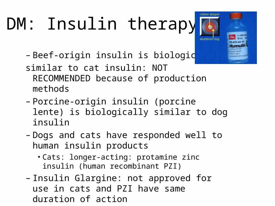
DM: Insulin therapy
– Beef-origin insulin is biologically similar to cat insulin: NOT RECOMMENDED
because of production methods– Porcine-origin insulin (porcine lente) is
biologically similar to dog insulin– Dogs and cats have responded well to
human insulin products• Cats: longer-acting: protamine zinc insulin
(human recombinant PZI)
– Insulin Glargine: not approved for use in cats and PZI have same duration of action

DM: Insulin therapy
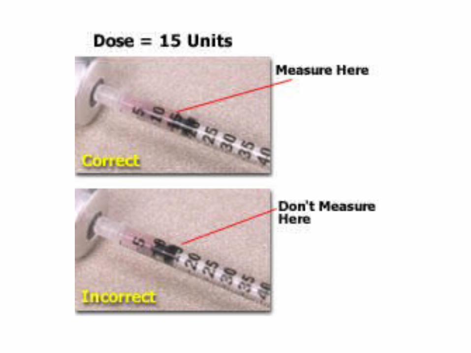
• INSULIN ADMINISTRATION:– ALWAYS USE THE
APPROPRIATE INSULIN SYRINGE! (U-40 vs. U-100)
• Insulin is given in units (insulin syringes are labeled in units, not mL)
• 30 units, 50 units, 100 units

DM: dietary management
• DIET– DOGS: high fiber, complex carbohydrate diets
• Slows digestion, reduces the post-prandial glucose spike, promotes weight loss, reduces risk of pancreatitis
• Hill’s R/D or W/D– CATS: high protein, low carbohydrate diets
• Cats use protein as their primary source of energy – blood glucose is maintained primarily through liver metabolism of fats and proteins
• Purina DM, Hill’s M/D• Often a diet change in cats can dramatically reduce or
eliminate the need for insulin– This is particularly true for type II
Diabetes Mellitus
• Oral hypoglycemics:o Sulfonylureas – Glipizide: cats
o Direct stimulation of insulin secretion from the pancreaso Adverse side effects, although uncommon, include vomiting,
loss of appetite, and liver damage o Alpha-Glucosidase Inhibitors – Acarbose
o Delays digestion of complex carbohydrates and delays absorption of glucose from the intestinal tract.
• Insulin is more effective than oral hypoglycemics
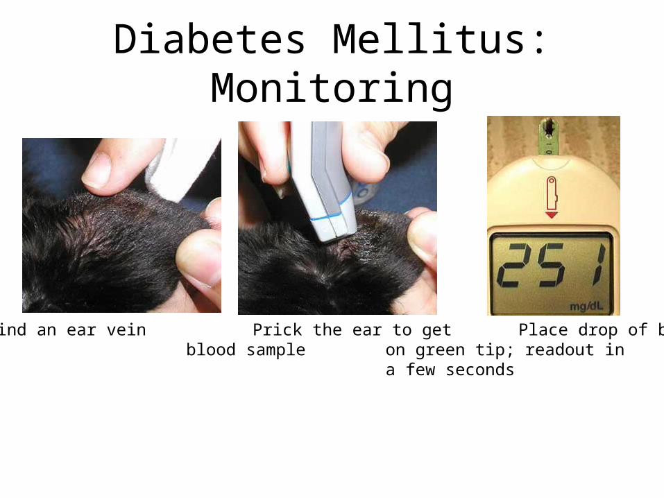
Diabetes Mellitus: Monitoring
Find an ear vein Prick the ear to get Place drop of blood blood sample on green tip; readout in
a few seconds

Diabetes Rx: Urine glucose

Diabetes monitoring: Urine glucose

DM: monitoring

DM• Client Education
– Lifelong insulin replacement therapy– Insulin administered by injection– Refrigerate insulin, mix gently (no bubbles), single use syringes– Cataracts common, permanent– Consistent diet and exercise– Recheck BG or curve regularly or fructosamine levels– Progressive– If animal does not eat- NO INSULIN

• Diabetes Mellitus clinical case

Endocrine Pancreas
• Hypoglycemia– Definition: Low blood glucose levels– Causes
• Neonatal and juvenile• Septicemia• Neoplasia• Starvation• Iatrogenic – insulin overdose• Portosystemic shunt• Many others

Insulin Shock
Causes:1. Insulin overdose (misread syringe)2. Too much exercise3. AnorexiaSigns:Weakness, incoordination, seizures, coma

Insulin Shock
Prevention1. Consistent diet (type and amount)/consistent
exercise (less insulin with exercise)2. Monitor urine/blood glucose at same time each
day3. Feed 1/3 with insulin; the rest 8-10 h later (at
insulin peak)4. Have sugar supply handy

Insulinoma• CAUSE: tumor of beta cells, secreting an excess of insulin• SIGNS: prolonged hypoglycemia→weakness, ataxia, muscle
fasciculations, posterior paresis, brain damage, seizures, coma, death,

Insulinoma: Dx
• Chem Panel– ↓blood glucose– Simultaneous glucose and insulin tests
Low glucose, High insulin => insulinoma
• Observations– Symptoms occur after fasting or exercise– when symptomatic, blood glucose<50 mg/dl– symptoms corrected with sugar administration

Insulinoma: RxSurgical Rx: removal of tumorMedical Rx: Acute, at home: administer glucose (Karo); keep animal quiet, seek vet careAcute, in Hosp adm. glucose (50% Dextrose)Chronic care feed 3-6 small meals/day (high protein, low fat)
limited exerciseglucocorticooid therapy (antagonizes insulin effect at cellular level)Diazoxide (↓insulin secretion, tissue use of glucose, ↑blood glucose)Octreotide (Sandostatin) injections—inhibits synthesis and release of insulin by both normal and neoplastic beta cells

Insulinoma: Client info• 1. Usually, by the time insulinoma is diagnosed, metastasis has
occurred so prognosis is poor• 2. With proper medical therapy, survival may be 12-24 mo• 3. Always limit exercise and excitement• 4. Feed multiple, small meals throughout day; keep sugar source close
during exercise• 5. Karo syrup on mm provides for rapid absorption of glucose into
blood stream• 6. Avoid placing hand into dog’s mouth during seizure to avoid being
bitten