disentangling the many layers of eukaryotic transcriptional
TRANSCRIPT

GE46CH03-Mann ARI 6 October 2012 15:17
Disentangling the ManyLayers of EukaryoticTranscriptional RegulationKatherine M. Lelli,1 Matthew Slattery,2
and Richard S. Mann3
1Department of Genetics and Development, College of Physicians and Surgeons,Columbia University, New York, NY 100322Institute for Genomics and Systems Biology, University of Chicago, Chicago, Illinois 606373Department of Biochemistry and Molecular Biophysics, College of Physicians andSurgeons, Columbia University, New York, NY 10032; email: [email protected]
Annu. Rev. Genet. 2012. 46:43–68
First published online as a Review in Advance onAugust 28, 2012
The Annual Review of Genetics is online atgenet.annualreviews.org
This article’s doi:10.1146/annurev-genet-110711-155437
Copyright c© 2012 by Annual Reviews.All rights reserved
0066-4197/12/1201-0043$20.00
Keywords
gene regulation, cis-regulatory module, cooperativity, DNAaccessibility, chromosomal interactions
Abstract
Regulation of gene expression in eukaryotes is an extremely complexprocess. In this review, we break down several critical steps, emphasiz-ing new data and techniques that have expanded current gene regulatorymodels. We begin at the level of DNA sequence where cis-regulatorymodules (CRMs) provide important regulatory information in the formof transcription factor (TF) binding sites. In this respect, CRMs func-tion as instructional platforms for the assembly of gene regulatory com-plexes. We discuss multiple mechanisms controlling complex assembly,including cooperative DNA binding, combinatorial codes, and CRMarchitecture. The second section of this review places CRM assemblyin the context of nucleosomes and condensed chromatin. We discusshow DNA accessibility and histone modifications contribute to TFfunction. Lastly, new advances in chromosomal mapping techniqueshave provided increased understanding of intra- and interchromosomalinteractions. We discuss how these topological maps influence generegulatory models.
43
Ann
u. R
ev. G
enet
. 201
2.46
:43-
68. D
ownl
oade
d fr
om w
ww
.ann
ualr
evie
ws.
org
by U
nive
rsity
of
Illin
ois
- C
hica
go o
n 03
/15/
13. F
or p
erso
nal u
se o
nly.

GE46CH03-Mann ARI 6 October 2012 15:17
TF: transcriptionfactor
cis-regulatorymodule (CRM):collection oftranscription factorbinding sites thatcoordinate to regulategene expression; alsocalled enhancers
INTRODUCTION
Gene regulation is fundamental for everybiological process. Reflective of its importanceto cell survival and function, the regulatorymechanisms controlling gene expression areexquisitely sophisticated. Unraveling thiscomplexity has broad implications, from un-derstanding animal development to preventingand treating clinical pathologies. Innovativetechnologies and creative experimental designare rapidly expanding the current models ofgene expression. From new insights into DNA-binding specificity to the contribution of nu-clear architecture, this review aims to integraterecent discoveries into a more comprehensivepicture of eukaryotic gene regulation.
Gene Regulation StrippedDown to the DNA
The first gene regulatory model was pioneeredby Francois Jacob and Jacques Manod inthe early 1960s. They postulated that geneproducts are able to feed back and regulatethe expression of genes, a concept that laid thefoundation for contemporary gene expressionmodels. Currently, the most basic modeldictates that regulatory proteins called tran-scription factors (TFs) act in trans to promoteor inhibit expression from a locus by bindingspecific DNA sequences in cis-regulatorymodules (CRMs) or enhancers (66). TFs arecharacterized by the sequence and structureof their DNA-binding domains. Throughoutevolution, gene duplication events have ex-panded the number of TFs, resulting in groupsor families of highly related TFs. As a conse-quence, evolutionarily related TFs often sharesimilar DNA-binding domains and similar invitro DNA-binding specificities. In some cases,related TFs display functional redundancyin vivo. However, there are many instancesin which individual TFs with highly similarDNA-binding properties carry out distinctfunctions. Given that related TFs have bothoverlapping and unique functions, they musthave the capacity to regulate both common
and specific gene targets. Although it is easyto understand how TFs with similar bindingproperties recognize the same binding sites andregulate some of the same target genes, it isless obvious how binding to different CRMs isrestricted to specific TFs (130). In the first sec-tion of this review, we focus on recent studiesthat provide new insights into how specificityis achieved at the level of DNA recognition.Throughout the review, many of the examplesare taken from Drosophila melanogaster becauseof the many whole genome, genetic, andbiochemical studies of it that have been carriedout over the past several years.
CRMs usually harbor multiple TF-bindingsites. In some cases, binding sites are stably oc-cupied only by TFs that bind cooperatively.However, the mechanisms by which cooper-ative DNA binding increases TF specificityvaries according to TF family and CRM (25, 51,119). Focusing on recent examples with signif-icant structural and functional data, we discussdifferent ways cooperative binding is achieved.Simply relying on the combinatorial bindingand activity of multiple factors is another wayCRMs coordinate gene expression, allowing in-tegration of cell type and environmental inputs(Figure 1).
Dressing Up Gene Regulationwith Chromatin
The biophysical realities of DNA packagedwithin a nucleus are in stark contrast tothe naked DNA researchers typically thinkabout when discussing protein-DNA binding.Although the stripped-down view is importantfor understanding the biophysical propertiesthat govern TF-DNA interactions, it ignores allof the complexities of the nuclear environment.DNA in eukaryotic genomes is compacted intochromatin; the basic unit of chromatin, thenucleosome, consists of 147 base pairs of DNAwrapped around a histone octamer containingtwo copies of each of the core histones H2A,H2B, H3, and H4 (88, 98). DNA associatedwith histones is less accessible to TFs andRNA polymerase than is naked DNA, making
44 Lelli · Slattery · Mann
Ann
u. R
ev. G
enet
. 201
2.46
:43-
68. D
ownl
oade
d fr
om w
ww
.ann
ualr
evie
ws.
org
by U
nive
rsity
of
Illin
ois
- C
hica
go o
n 03
/15/
13. F
or p
erso
nal u
se o
nly.
GE46CH03-Mann ARI 6 October 2012 15:17
Nuclearlamin
NPC
Cellsignal
TF
NPC
P
TF
PPh
TF
TF TF TF TFTSS
Me
Me
Me
Me
Cellsignal
P
TF
TF TF
P
TF
TF
P
K
Cytoplasm
PcG body
ChromosomeChromosometerritoryterritory
Chromosometerritory
cis regulatory moduleNucleosomes
Chromatin
ChromosomeChromosometerritoryterritory
Chromosometerritory
ChromosomeChromosometerritoryterritory
Chromosometerritory
TranscriptionTranscriptionfactoryfactory
Transcriptionfactory
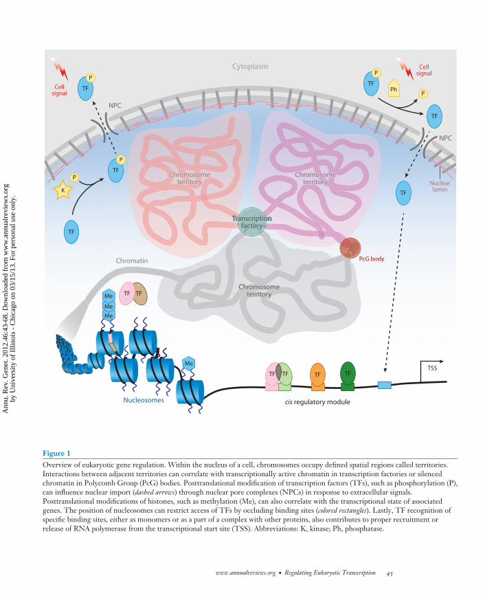
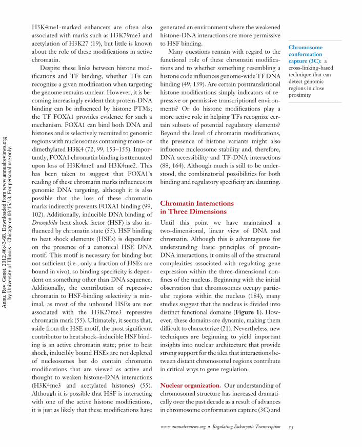
Figure 1Overview of eukaryotic gene regulation. Within the nucleus of a cell, chromosomes occupy defined spatial regions called territories.Interactions between adjacent territories can correlate with transcriptionally active chromatin in transcription factories or silencedchromatin in Polycomb Group (PcG) bodies. Posttranslational modification of transcription factors (TFs), such as phosphorylation (P),can influence nuclear import (dashed arrows) through nuclear pore complexes (NPCs) in response to extracellular signals.Posttranslational modifications of histones, such as methylation (Me), can also correlate with the transcriptional state of associatedgenes. The position of nucleosomes can restrict access of TFs by occluding binding sites (colored rectangles). Lastly, TF recognition ofspecific binding sites, either as monomers or as a part of a complex with other proteins, also contributes to proper recruitment orrelease of RNA polymerase from the transcriptional start site (TSS). Abbreviations: K, kinase; Ph, phosphatase.
www.annualreviews.org • Regulating Eukaryotic Transcription 45
Ann
u. R
ev. G
enet
. 201
2.46
:43-
68. D
ownl
oade
d fr
om w
ww
.ann
ualr
evie
ws.
org
by U
nive
rsity
of
Illin
ois
- C
hica
go o
n 03
/15/
13. F
or p
erso
nal u
se o
nly.
GE46CH03-Mann ARI 6 October 2012 15:17
PTM:posttranslationalmodification
Cooperative DNAbinding: occurs whenthe binding affinity oftwo or more factors ismore than the sum ofthe individual affinities
Latent specificity:novel DNArecognition propertiesthat are revealed as aconsequence ofprotein-proteininteractions
chromatin transcriptionally more repressedcompared with naked DNA. Additionally,chromatin structure is not homogenousalong the entire genome and can adapt morecomplex local structures and higher-levelthree-dimensional arrangements. In the As-sembling cis-Regulatory Module Complexessection below, we discuss how chromatin struc-ture contributes to gene regulation (Figure 1).
Historically, chromatin has been describedas existing in two distinct flavors: euchromatinand heterochromatin. Heterochromatin iscondensed, transcriptionally inactive, and as-sociated with repressive histone modifications,whereas euchromatin is relatively accessibleand associated with actively transcribed genesand active histone modifications (42, 69).This low-resolution view is still accurate, butrecent genomic studies have resulted in a muchhigher-resolution and more nuanced view ofchromatin states. We discuss how nucleosomeoccupancy as well as histone modifications andvariants correlate with gene regulation. Finally,with the help of several technological advances,our understanding of higher-order chromatinstructure and the three-dimensional organiza-tion of chromosomes in nuclei has increaseddramatically over the past decade. We discusshow nuclear architecture and chromosomalconformation have also been implicated ineukaryotic gene regulation (Figure 1).
ASSEMBLING CIS-REGULATORYMODULE COMPLEXES
In prokaryotes, single TFs are able to regulategene expression. However, this type of generegulation is usually insufficient for eukaryoticgene regulation. Instead, eukaryotes relyon combinatorial transcriptional inputs intoCRMs to regulate gene expression in space andtime (66). The specific recruitment of manyindividual factors refines expression on thebasis of cellular context, timing of expression,and extracellular signals. For example, thesame binding sites in the same CRM of theDrosophila nidogen gene have been shownto bind different forkhead domain TFs in
different tissues, with distinct regulatory out-puts (183). Alternatively, multiple homeobox(Hox) TFs, which have highly similar DNA-binding specificity as monomers, can target thesame gene via distinct CRMs in different tissues(39). It also appears that TFs can bind non-canonical motifs in certain contexts, althoughthe mechanism by which these motifs are distin-guished from canonical motifs remains unclear(4, 20). Regulation can be further refined byposttranslational modifications (PTMs) ofTFs, which can affect subcellular localization,DNA binding, and protein-protein interactions(Figure 1) (14, 16, 24, 27, 169). Some re-searchers propose a PTM code in whichmultisite PTM events provide an importantregulatory mechanism for different signalingpathways to affect TF function and influencegene expression (14). In this section, we focuson two additional mechanisms, cooperativeDNA binding and combinatorial codes thatregulate the assembly and activities of CRMcomplexes.
Cooperativity
One mechanism cells use to increase the DNA-binding specificity of TFs is cooperative DNAbinding. With an emphasis on structural datapaired with in vitro DNA-binding assays, wedistinguish three types of cooperative complexformation (Figure 2). The first type, whichwe refer to here as classical cooperativity,relies on direct protein-protein interactionsbetween TFs and their cofactors to increaseDNA-binding affinity (Figure 2). A variationon classical cooperativity, termed latent speci-ficity, is when protein-protein interactions leadnot only to increased DNA-binding affinity butalso to a change in DNA-binding specificity(Figure 2) (161). We refer to a second formof cooperativity as enhanceosome or modularcooperativity (Figure 2). The distinguishingfeature here is that, unlike classical cooper-ativity, which is typically defined for homo-and heterodimers of TFs, enhanceosomecooperativity is observed for large complexesof proteins and, at least in some cases, appears
46 Lelli · Slattery · Mann
Ann
u. R
ev. G
enet
. 201
2.46
:43-
68. D
ownl
oade
d fr
om w
ww
.ann
ualr
evie
ws.
org
by U
nive
rsity
of
Illin
ois
- C
hica
go o
n 03
/15/
13. F
or p
erso
nal u
se o
nly.

GE46CH03-Mann ARI 6 October 2012 15:17
Local cooperativity Flexible grammar
b Billboard or TF collective
Modular cooperativity Strict grammar
Enhanceosome cooperativity
a Enhanceosome
c DNA allostery
Gene X
e Collaborative competitiond Classical cooperativity
Latent specificity
Gene Y
A' B'
Multiple partners
Gene X
A + B
Gene Y
A + C
A
B
C
Gene X
A + B
Gene Y
+
+
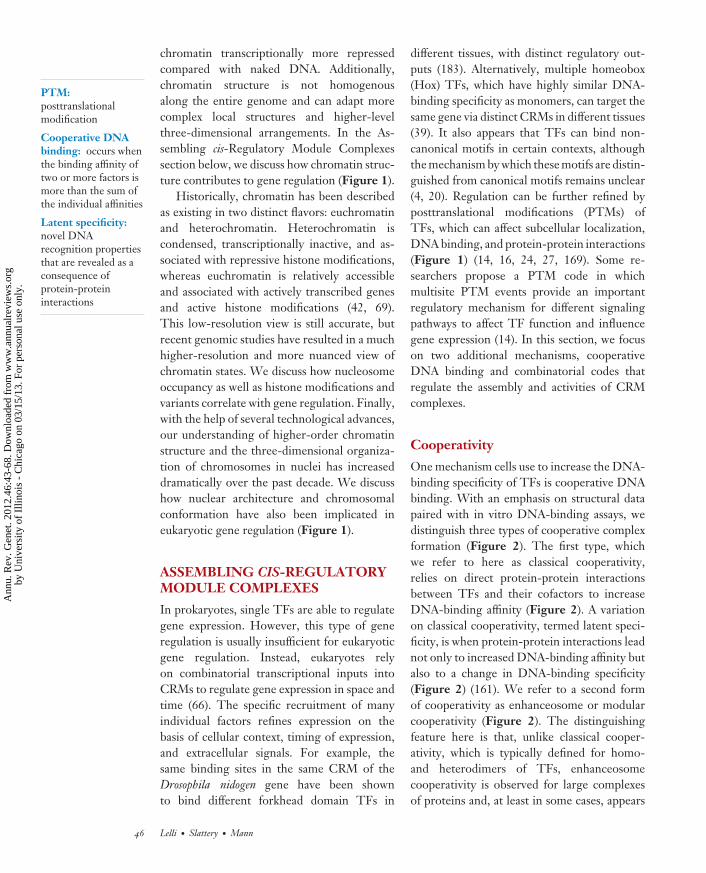
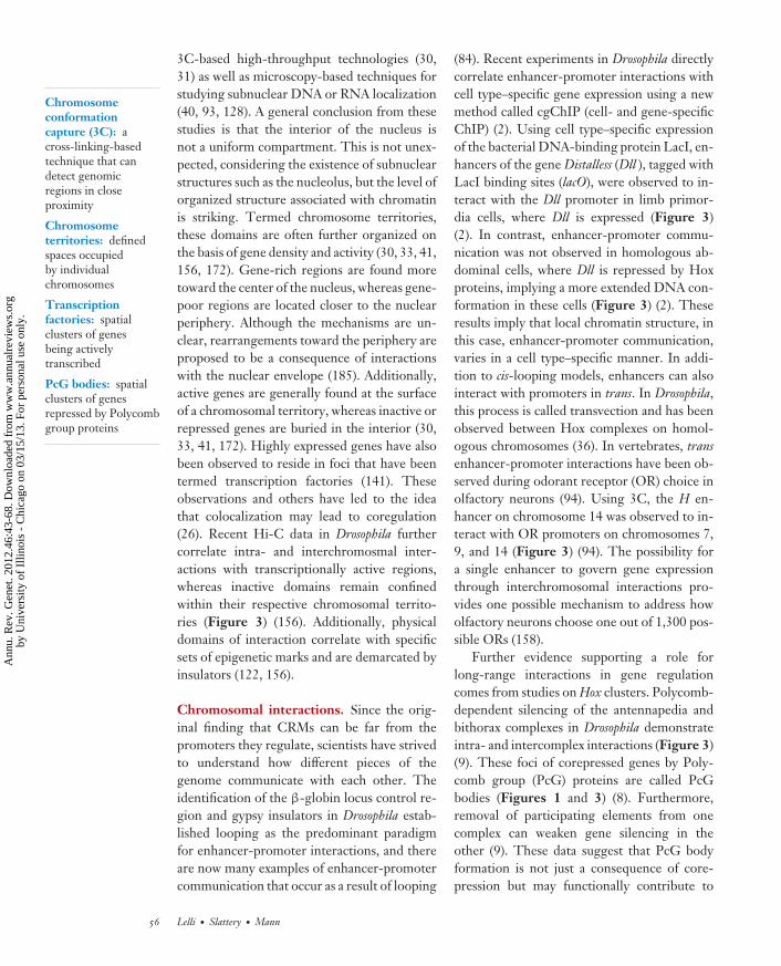
Figure 2Cis-regulatory module (CRM) assembly and cooperative DNA-binding models. This image depicts three different models of CRMassembly and related cooperativity mechanisms. (a) The enhanceosome model requires strict modular cooperativity between alltranscription factors (TFs) (129, 131). (b) In contrast, the flexibility of the Billboard and TF Collective models permits differentcooperativity mechanisms to control CRM assembly. (c) In the case of DNA allostery, interactions between the DNA sequence and theTF can facilitate conformational changes in the TF that result in the recruitment of different regulatory complexes (rounded rectangles)in a sequence-specific manner (108). (e) Classical cooperativity uses protein-protein interactions between TFs to facilitate cooperativebinding. These types of cooperative interactions help to increase TF DNA-binding specificity by restricting recruitment to dimericsites (A+B). In the case of latent specificity, direct protein-protein interactions alter binding specificities so that TFs recognize novelcomposite sites (A′B′) (161). ( f ) Lastly, collaborative competition between TFs and nucleosomes can lead to cooperative binding whenthe binding of one TF provides access for another TF to bind a neighboring site (114, 120, 167).
www.annualreviews.org • Regulating Eukaryotic Transcription 47
Ann
u. R
ev. G
enet
. 201
2.46
:43-
68. D
ownl
oade
d fr
om w
ww
.ann
ualr
evie
ws.
org
by U
nive
rsity
of
Illin
ois
- C
hica
go o
n 03
/15/
13. F
or p
erso
nal u
se o
nly.

GE46CH03-Mann ARI 6 October 2012 15:17
TALE: three aminoacid loop extension
SELEX: systematicevolution of ligandsby exponentialenrichment
to not depend on protein-protein interactions(131). A third form of cooperativity is termedcollaborative competition (Figure 2) (138). Inthis case, cooperative binding occurs only ona chromatin template because multiple TFsare more effective than are individual TFsat competing with nucleosomes for bindingto target sequences (113). As the first twoforms of cooperativity are measured on nakedDNA, we discuss them here, whereas the thirdform, which is apparent only in the context ofchromatin, is discussed in a later section.
Protein-protein interactions reveal latentspecificities. Interactions between TFs notonly increase DNA-binding affinity to theircognate binding sites but may also result ina modification of their DNA recognitionproperties (Figure 2). This phenomenon hasrecently been described for the Hox familyof TFs, which provides a classic example ofthe TF specificity paradox. Characterized bya highly conserved DNA-binding domaincalled the homeodomain, all Hox proteinsrecognize similar AT-rich sequences in vitro(15, 127) but confer phenotypically distinctidentities in vivo (65). One way they achievefunctional specificity is through cooperativeDNA binding with cofactors. In Drosophila, thebest-characterized Hox cofactors are Extraden-ticle (Exd) and Homothorax (Hth), which haveorthologs called Pbx and Meis, respectively, invertebrates (87, 116). Exd/Pbx and Hth/Meisare members of the three amino acid loopextension (TALE) class of homeodomain-containing proteins (103, 116). Functional andstructural studies have identified a conservedprotein-protein interaction between Exd andHox proteins that facilitates cooperative DNAbinding (105, 112). Tryptophan-containingmotifs (W motifs), most commonly YPWM,found N terminal to the Hox homeodomain,directly interact with the TALE motif in theExd homeodomain (105, 112). Mutation ofthese W motifs can dramatically affect bothcooperative DNA binding in vitro and in vivofunction of some Hox proteins (87, 105, 110,148). For example, for two Drosophila Hox
proteins, Sex combs reduced (Scr) and De-formed (Dfd), mutation of the W motif is suffi-cient to abolish cooperative complex formationon a known specific binding site in vitro as wellas several Exd-dependent functions measuredin vivo (74, 87). In addition, minimal Hoxproteins that contain only the homeodomainsand W motifs retain many wild-type functionswhen assessed in vivo, suggesting that they aresufficient for many in vivo activities (134, 135).
In addition to increasing affinity, the bindingof Exd with Hox proteins modifies their speci-ficity, a phenomenon referred to as latent speci-ficity (161). Using systematic evolution of lig-ands by exponential enrichment (SELEX)-seq,in which traditional SELEX (38, 170) is pairedwith deep sequencing, cooperative bindingwith Exd was shown to elicit changes in HoxDNA-binding preferences that are distinctfrom their monomeric DNA-binding prefer-ences (161). Importantly, for at least one Hoxprotein (Ubx), the types of latent heterodimerspecificities revealed by SELEX-seq were over-represented in DNA sequences bound by thisfactor in vivo, which suggests that the in vitro–measured specificities are biologically relevant(161). In another recent example, DNA-binding measurements from protein-bindingmicroarrays for the Saccharomyces cerevisiaeTFs Met4, Met28, and Cbf1 demonstratethat cooperative complex formation increasesDNA-binding specificity (159). In this case,Cbf1, together with its non-DNA-binding co-factors Met28 and Met4, recognize additionalDNA sequences that are adjacent to the tra-ditional Cbf1 binding site (159). Importantly,these additional DNA sequences are essentialfor gene regulation in vivo. Together, thesedata suggest that interactions between TFsand their cofactors have the potential to revealspecificities that are not available in the absenceof cofactors. Moreover, latent specificities canapparently be induced by both DNA-binding(e.g., Hox-Exd) and non-DNA-binding (e.g.,Cbf1-Met4-Met28) cofactors.
The E twenty-six (ETS) family of TFsmay provide an additional example oflatent specificity, referred to in this case as
48 Lelli · Slattery · Mann
Ann
u. R
ev. G
enet
. 201
2.46
:43-
68. D
ownl
oade
d fr
om w
ww
.ann
ualr
evie
ws.
org
by U
nive
rsity
of
Illin
ois
- C
hica
go o
n 03
/15/
13. F
or p
erso
nal u
se o
nly.
GE46CH03-Mann ARI 6 October 2012 15:17
acquired specificity (173). ETS proteins arecharacterized by a highly conserved wingedhelix-loop-helix DNA-binding domain andrecognize 5′-GGA(A/T)-3′ sequence motifs.Similar to Hox proteins, ETS factors usedirect interactions with cofactors to increaseDNA-binding specificity (63). CooperativeDNA binding between PAX5 and ETS1 iscritical for activation of mlb-1 during B cell de-velopment (46). Structural studies demonstratethat direct interactions between the paireddomain of PAX5 and the ETS domain ofETS1 can rearrange protein-DNA contacts toincrease DNA binding (50): A PAX5-inducedrotation of a particular tyrosine residue in therecognition helix of ETS1 promotes bindingto a low-affinity site (50). A similar mechanismhas been proposed for other ETS/cofactorcomplexes. All members of the ternary complexfactor subfamily of ETS factors, which includesELK1 and SAP1, interact with serum responsefactor (SRF) to activate immediate-early genes(63). In the case of ELK1/SRF complexes,studies of related SAP1s/SRFs suggest thatprotein-protein interactions may induce con-formational changes in an analogous tyrosineresidue in the recognition helix of ELK1 toincrease DNA-binding affinity (115). Alteringthe structure of the ETS domain may prove tobe a general mechanism for increasing speci-ficity, especially considering it mediates manyof the functional interactions between ETSfactors and several cofactors (173). An analo-gous cofactor-induced change in conformationwas also proposed to underlie Hox-Exd latentspecificity. In this case, X-ray crystal structuresdemonstrated that Exd positions a normallyunstructured region of the Hox protein Scr sothat it can interact with DNA, specifically, anarrow region of the minor groove (73, 105).The recognition of minor groove structureor, more generally, DNA shape is widespreadamong TFs, suggesting that it may be a com-mon mode of DNA recognition (144, 145).
The similarities between ETS complexesand Hox complexes extend to another phe-nomenon called autoinhibition in which DNAbinding of a TF is inhibited by domains in
the TF itself. Mutual relief of autoinhibitionhas been shown to mediate cooperative com-plex formation between ETS1 and RUNX1(63) as well as complex formation between Hoxand Exd proteins, in which the Hox W mo-tif apparently interferes with monomeric DNAbinding (23). Therefore, protein-protein inter-actions can alter DNA-binding specificity andincrease affinity by both rearranging protein-DNA contacts and suppressing autoinhibition.
It is noteworthy that some of the DNA-interacting residues in the Scr-Exd complexand ETS complexes are outside the tradition-ally defined DNA-binding domain. Althoughcrystal structures are not yet available, theDrosophila Hox protein Dfd requires residuesthat are in an analogous position to Scr’sminor groove–interacting residues to bind andregulate some of its specific targets in vivo(74). These observations blur the traditionaldefinition of a DNA-binding domain, in thatthey show that additional motifs can contributedirectly to DNA binding when TFs interactwith cofactors. Although it is currently notclear which residues in the Cbf1-Met28-Met4complex contact its expanded binding site(159), it is plausible that, analogous to Hox-Exd, residues not normally considered partof the DNA-binding domain make some ofthese contacts. Additional nuclear magneticresonance or X-ray crystal structures would bevery helpful for resolving these questions.
Additional complexity and, perhaps, DNA-binding specificity also comes from the factthat some Hox proteins have additional waysto interact with Exd beyond their W motifs.For example, some Hox proteins, namely Ubxand AbdA, have multiple W and non-W motifsthat are used to bind DNA cooperatively withExd/Hth (22, 87, 109–111, 126, 148). Context-dependent interactions between motifs withinthe same Hox protein have also been proposedto contribute to functional diversity (110,148). Unfortunately, no structural informationis currently available to show how theseadditional motifs interact with each other,Exd, or Hth. In the case of AbdA, where upto four potential sequence motifs contribute to
www.annualreviews.org • Regulating Eukaryotic Transcription 49
Ann
u. R
ev. G
enet
. 201
2.46
:43-
68. D
ownl
oade
d fr
om w
ww
.ann
ualr
evie
ws.
org
by U
nive
rsity
of
Illin
ois
- C
hica
go o
n 03
/15/
13. F
or p
erso
nal u
se o
nly.
GE46CH03-Mann ARI 6 October 2012 15:17
cooperative complex formation with Exd, somemotifs were differentially required dependingon the readout examined (87, 110, 126).Studies using vertebrate Hox proteins havealso demonstrated differential requirementsfor W motifs (32, 45, 82, 142, 150, 157, 174,178). For example, targeted mutagenesis of themurine Hoxa1 W motif parallels the Hoxa1loss-of-function phenotype during hindbrainpatterning (142). However, other functions,such as neural crest cell migration and cranialnerve development, are not as severely affectedas in the Hoxa1 loss-of-function animals,suggesting that the W motif is required ina context-dependent manner, analogous tosome of the Drosophila Hox proteins (142).These data suggest that, depending on themode of Exd interaction, different target sitesmay be recognized, and the three-dimensionalstructure of the bound complex may vary. Thepresence of additional interaction modes alsocontributes to the phenomenon of posteriorprevalence, also known as phenotypic suppres-sion, in which more posterior Hox proteinsdominate in a posttranslational manner overmore anterior Hox proteins (34, 35, 52, 53,104). A recent study comparing the abilitiesof Scr and AbdA to bind and regulate a sharedtarget site found that the quality and quantity ofAbdA’s Exd interaction modes both increasedcooperative DNA-binding affinity in vitro andcontributed to AbdA’s ability to outcompeteScr for target gene regulation in vivo (126).
Promiscuous cooperativity with multiplecofactors. As described above, some TFs,such as Hox proteins, increase their specificityby interacting with a small number of cofactorsand using a variety of mechanisms to reveallatent specificities. Another strategy used byother TFs to expand their regulatory repertoireis to interact with a large number of proteinpartners, which allows TFs to gain cell- andtissue-specific control of gene expressiondepending on the cell-type availability ofcofactors (Figure 2). For example, ETS factorsutilize a variety of partners to bind DNA(63). With more than 40 different regulatory
partners, the Sox [Sex-determining region Y(SRY)-related-high mobility group (HMG)-box] family of proteins provides anotherexample of how multiple cofactors can con-tribute to specificity (16, 83). Classified by theirHMG box DNA-binding domain, Sox familyproteins control a variety of developmentalprocesses and are key regulators of pluripotency(16, 83). During melanocyte development theSOX10/PAX3 pair activates expression ofthe TF MITF, which subsequently functionsas another SOX10 partner to promote pro-gression of melanocyte differentiation (16,83). Additionally, recent data suggest thatMEF2C can also function as a SOX10 partnerto promote maintenance of the melanocytefate (1). An analogous regulatory mechanism isobserved during SOX10 regulation of Schwanncell development (16, 83).
Interactions with different partners can alsoaffect DNA-binding specificity, perhaps usingmechanisms that are analogous to the latentspecificity model described above. Recently,two studies have demonstrated that singleamino acid substitutions in SOX2 and SOX17can disrupt or promote, respectively, cooper-ative binding with OCT4 in vitro. The abilityto form cooperative complexes with OCT4correlated with cell-reprogramming potential(68, 123). Because most Sox proteins recognizesimilar binding sites, restricting interactions toonly a specific set of cofactors is important forregulating proper CRM binding.
Enhanceosome cooperativity. The CRMresponsible for viral-inducible expression ofinterferon-β (IFN-β) is among the most-studied human transcriptional regulatoryelements. Eight proteins cooperatively bindthis 55–base pair (bp) enhancer in a structuretermed the enhanceosome: one ATF2/c-Jundimer, four interferon response factors (ini-tially IRF-3, which is replaced with IRF-7after IFN-β induction), and one NFκB dimer(p50/RELA) (131, 165). Activated by threedifferent pathways, the specific expression ofIFN-β is ultimately regulated by the coinci-dental activation and cooperative binding of all
50 Lelli · Slattery · Mann
Ann
u. R
ev. G
enet
. 201
2.46
:43-
68. D
ownl
oade
d fr
om w
ww
.ann
ualr
evie
ws.
org
by U
nive
rsity
of
Illin
ois
- C
hica
go o
n 03
/15/
13. F
or p
erso
nal u
se o
nly.

GE46CH03-Mann ARI 6 October 2012 15:17
of these factors. Each factor is unable to indi-vidually activate IFN-β expression, and loss ofany single protein abolishes IFN-β activation(165). Despite the binding sites within this55-bp element being tightly packed, severalcrystal structures that capture subsets of theenhanceosome display a paucity of protein-protein interactions between pairs of dimers(131–133). Additionally, recent moleculardynamics simulations suggest that the DNA-bound complexes display an unusually highlevel of flexibility (129). From these observa-tions, it seems unlikely that direct interactionsbetween TFs are mediating the observed co-operative DNA binding. Instead, these studiessuggest that sequence-dependent structuralchanges in the DNA may facilitate binding ofTFs to overlapping sites (129, 131, 133). Useof a combination of higher- and lower-affinitysites can regulate the order in which complexesassemble (129); in this way, binding of onefactor could facilitate the cooperative bindingof another factor to an overlapping low-affinitysite through complementary structural changesin shared nucleotides. Further, the architec-tural factor HMGA1a and other secondaryfactors may also enhance cooperative DNAbinding on the IFN-β enhancer (131). There-fore, the IFN-β enhanceosome representsa type of modular cooperativity with strictrequirements for binding-site arrangementand overlap (Figure 2).
Binding-site allostery. Lastly, we wish toemphasize that the binding site itself can be anactive player in TF function and activity. Stud-ies on the glucocorticoid receptor (GR) haveshown that DNA can function as an allostericregulator of TF activity (108). Structuralstudies of the GR demonstrate that a regioncalled the lever arm within the DNA-bindingdomain adopts different conformations ac-cording to the DNA sequence bound (108).These structural changes parallel functionalvariations in cognate regulatory complexes forthe different binding sites (108). Therefore,different conformations of the DNA-bindingdomain induced by the DNA-binding site
can affect the transcriptional activity of GRin a site-specific manner (108). As with latentspecificity, we speculate that a variety ofinfluences on the three-dimensional structureof TF complexes, in part influenced by theDNA-binding site, can affect TF functions.Therefore, understanding the sequence andshape of CRMs and how they impact the struc-ture of bound factors is critical to decoding theregulatory logic driving gene expression.
cis-Regulatory Module Architecture
A common feature of all CRMs is that theyprovide a scaffold for a combinatorial logic codein which the assembly of multiple factors pro-vides cell type– and environment-dependentgene regulation (66). In the above section,we focused on several types of DNA-bindingcooperativity used by TFs to bind CRMs. Withthe exception of enhanceosome cooperativity,in which all factors must bind to an inflexibleCRM, the other types of cooperativity de-scribed above allow for much greater CRMflexibility: A single CRM can potentiallyintegrate a large variety of inputs, some ofwhich may be cooperative. For example, theHox-targeted CRM from reaper integrates notonly the Hox protein Deformed (Dfd) but alsoat least eight additional TFs (162). In contrast,a Dfd autoregulatory target requires multipleDfd-Exd heterodimer inputs plus additional, asyet unidentified, inputs (74). The ability to in-terchange CRM inputs provides cells with theflexibility to maintain a tight regulatory controlin a wide variety of distinct contexts. Given thisrequirement, how are CRMs organized, andcan generalizations be gleaned from the data?
Three different models have been proposedto describe CRM architecture: the enhanceo-some, the billboard, and the TF collective(Figure 2) (3, 75). As described above, theenhanceosome model posits that cooperativebinding of a group of TFs using a strict ar-rangement or grammar of binding sites in theDNA is necessary for CRM activity (Figure 2).Evidence for this model comes primarily fromthe IFN-β enhanceosome, described above,
www.annualreviews.org • Regulating Eukaryotic Transcription 51
Ann
u. R
ev. G
enet
. 201
2.46
:43-
68. D
ownl
oade
d fr
om w
ww
.ann
ualr
evie
ws.
org
by U
nive
rsity
of
Illin
ois
- C
hica
go o
n 03
/15/
13. F
or p
erso
nal u
se o
nly.

GE46CH03-Mann ARI 6 October 2012 15:17
ChIP: chromatinimmunoprecipitation
as well as from the tumor necrosis factor-αenhanceosome (13, 131). Given the small num-ber of identified enhanceosome-like CRMs inwhich the arrangement of the binding sites isinflexible, they may be more the exception thanthe rule. Enhanceosomes may be limited toregulatory events that must be controlled withexquisite precision, as they can be activatedonly when all factors are present. In addition,once formed, enhanceosomes are unusuallystable, allowing them to activate transcriptionuntil subsequent mechanisms disassemblethem (165).
A second model for CRM architecture is thebillboard model (Figure 2) (3). At the oppositeend of the spectrum from inflexible enhanceo-some CRMs, billboard CRMs are hypothesizedto be very flexible; each binding site is critical,but their relative orientation and spacing do notcontribute to CRM function (3, 85). Accord-ing to this view, individual TF inputs might actindependently to recruit different componentsof the basal transcription machinery, adaptercomplexes, or chromatin-modifying complexesto promote transcription (3).
A third view of CRM architecture refersto CRMs as TF collectives (Figure 2) (75).According to this view, TFs are cooperativelyrecruited to CRMs but without a precise motifgrammar (75). Using whole-embryo chro-matin immunoprecipitation (ChIP)-chip andChIP-seq at different stages during Drosophilaembryogenesis, binding events for a set of fivefactors involved in cardiac gene regulationwere analyzed (75). Interestingly, the authorsfound that a large number of CRMs includedall five factors and that some combinationswere very rare (75). For example, when one wasmissing [Tinman (Tin)], the other four factorswere rarely found together (75). Althoughsuch observations could represent cooperativebinding or cooperative recruitment of thesefive factors, they could also represent anevolutionary selection for functional CRMsthat contain all five inputs and selection againstCRMs that have only four of the five inputs.Another potential explanation for these resultsis that Tin functions as a so-called pioneer
factor that is required to initiate binding tothese CRMs in a chromatin environment. Addi-tional biochemical experiments are required todetermine if cobinding of these factors is coop-erative rather than a consequence of selection.
Another feature of the TF-collective modelis that binding sites need not necessarily bepresent for every TF present at the CRM(75). Accordingly, some factors are indirectlybound at CRMs because of protein-proteininteractions alone (Figure 2). Although thisis certainly plausible, it is also possible thatlow-affinity binding sites or latent specificitymechanisms make it difficult for current com-putational methods to recognize all of the es-sential binding sites in CRMs. Consistent withthis idea, low-affinity binding sites are criticalfor the accurate activities of some CRMs (136).
Detailed in vivo structure-function analy-sis of the sparkling CRM of Drosophila Pax2 ineye development provides another informativeview of CRM logic (163). In this case, the au-thors asked how binding sites for the knownTFs rearrange relative to each other duringthe course of evolution (163). Several inter-esting conclusions come from this work. Forone, sparkling CRMs from different Drosophilaspecies can have a very different arrangement ofbinding sites but still function to drive accurateexpression in D. melanogaster, similar to conclu-sions obtained from evolutionary comparisonsof the stripe 2 CRM from even-skipped (96, 97).In addition, despite rapid evolutionary turnoverof binding sites at the sparkling CRM, the au-thors were able to recognize that the spacing be-tween pairs of some binding sites was conserved(163). This conserved CRM grammar suggeststhat there may be interactions, either director mediated by additional factors, between thebound TFs. The idea that cooperative or inter-acting subelements could be among a set of oth-erwise independent inputs was also part of theoriginal billboard model (3) and is consistentwith the important role of TF DNA-bindingspecificity discussed in the previous section.
Regardless of whether biologists refer toCRMs as a billboard, a TF collective, or someother nom du jour, the emerging view from
52 Lelli · Slattery · Mann
Ann
u. R
ev. G
enet
. 201
2.46
:43-
68. D
ownl
oade
d fr
om w
ww
.ann
ualr
evie
ws.
org
by U
nive
rsity
of
Illin
ois
- C
hica
go o
n 03
/15/
13. F
or p
erso
nal u
se o
nly.

GE46CH03-Mann ARI 6 October 2012 15:17
NDR:nucleosome-depletedregion
numerous studies is that many, perhaps themajority of, enhancer elements are not en-hanceosome in nature but instead flexibly inte-grate multiple TF inputs in a surprisingly largenumber of arrangements. Consistent with a keyrole for multiple TF inputs, a recent compar-ative study found that in vivo binding sites forthe mesodermal TF Twist (Twi) are highly con-served across several Drosophila species, and lossof Twi binding in one species is often associatednot with loss of a Twi motif but with loss of acofactor binding site (59). Some of these inputsmay be singly bound TFs, others may be coop-eratively bound pairs of TFs, and yet others mayinteract indirectly via a third factor (Figure 2).Such flexibility in CRM architecture makes thede novo identification of CRMs, based solelyon DNA sequence, a big challenge for biologistsbecause apparently the same set of inputs can beencoded in the DNA sequence in many ways.
CHROMATIN STRUCTURE
In the previous section, we discussed how TFsrecognize and bind target sites in the context ofnaked DNA. However, to fit within the minis-cule confines of the nucleus, DNA is wrappedaround histones and condensed into chromatin.In addition to limiting TF access, histone-DNAcomplexes are subject to many PTMs that af-fect gene expression. Furthermore, chromatinis not uniformly distributed throughout the nu-cleus, so distant regions of the genome, basedon linear DNA sequence, may actually be inclose proximity. In this section, we describehow modifications of histone-DNA complexesand chromatin architecture contribute to generegulation. It is not possible to catalog all ofthe many modifications known to occur on hi-stones; for this, the reader is referred to manyrecent reviews covering this topic (7, 12, 86,139, 181). Instead, we focus on the small subsetof modifications that are correlated with CRMsor CRM activity.
DNA Accessibility
DNA accessibility is increasingly recognizedas an important variable in gene regulation
(79, 80, 91, 166). Although it has been difficultto mechanistically test the causal relation-ship between chromatin structure and genetranscription, many genome-wide studieshave demonstrated significant correlations.Through modeling of TF binding and DNase1sensitivity data, two recent studies revealed thatchromatin accessibility has a significant impacton the genome-wide binding patterns of anumber of developmental regulatory factorsexpressed in the Drosophila embryo (79, 91).Within regions of open chromatin, TF bindingis primarily determined by sequence specificity(79). A similar correlation between accessibilityand TF binding has been observed in mam-malian cells (70) and yeast (182). Additionally,several genome-wide nucleosome-mappingstudies reveal trends suggesting that nu-cleosome positioning may influence geneexpression (5). First, promoter regions tend tocontain nucleosome-depleted regions (NDRs)(5). Second, nucleosomes around transcriptionstart sites are often well organized (5). The for-mation of NDRs is predicted to reveal bindingsites and facilitate TF binding at regulatorysequences (106). Many mechanisms have beenproposed to regulate nucleosome positioningand NDR formation (5, 67, 81, 152). In additionto the controversial role of the primary DNAsequence (70, 76–78), ATP-dependent chro-matin remodelers, such as the Swi/Snf complex,and binding by specific TFs have also been im-plicated in NDR formation (6, 168, 176, 179).The formation of NDRs falls under the moregeneral heading of establishing gene- and celltype–specific chromatin architectures, whichcan include the positioning of nucleosomes atregulatory elements and promoters (47, 95).
Similar to nucleosome-depleting factors,so-called pioneer factors have been proposedto prime chromatin environments to initiatesubsequent TF binding. Unlike many TFs,pioneer factors such as the FOXA and GATAfactors, PU.1, and AP1 have the ability tobind their target motifs in a closed chromatinenvironment (17, 61, 102, 180). Upon bindingtheir target DNA, the pioneer factors candrive local chromatin remodeling and create
www.annualreviews.org • Regulating Eukaryotic Transcription 53
Ann
u. R
ev. G
enet
. 201
2.46
:43-
68. D
ownl
oade
d fr
om w
ww
.ann
ualr
evie
ws.
org
by U
nive
rsity
of
Illin
ois
- C
hica
go o
n 03
/15/
13. F
or p
erso
nal u
se o
nly.

GE46CH03-Mann ARI 6 October 2012 15:17
accessible enhancers for additional TF binding(102, 180). This type of synergy has previouslybeen termed nucleosome-mediated coopera-tivity or collaborative competition (Figure 2)(113, 114, 138). Thermodynamic and invitro studies have demonstrated that TFs cancompete with nucleosomes for DNA bindingand, by unwrapping or evicting the overlappingnucleosome, induce the cooperative binding ofanother TF (114, 120, 167). Because protein-protein interactions are not required and therelative arrangement and orientation of bind-ing sites are flexible, collaborative competitionmay be one explanation for why many CRMshave multiple flexible inputs without a strictgrammar (e.g., the billboard and TF collectivemodels; see above) (3, 75). Additionally, math-ematical simulations suggest that by mediatingchromatin reorganization, pioneer factors canincrease the steady-state binding of other TFs,a process called assisted loading (175).
The Drosophila zinc-finger protein Zelda(Zld) may represent another type of pioneer fac-tor. Zld is bound to its target motifs throughoutthe Drosophila genome during the maternal-to-zygotic transition, the point when zygotictranscription commences in the developingembryo (57, 92, 124, 149). Most Zld-targetedregions remain bound by Zld and highlyaccessible later in embryonic development andare also targeted by numerous developmentalTFs at these later stages (57, 124). Because Zldis highly associated with the relatively opengenome before zygotic transcription begins, ithas been proposed that rather than reorganiz-ing or opening chromatin, Zld binding preventsnucleosome occupancy and maintains DNA ac-cessibility in certain regions of the genome (57).
Histone Modifications
Recent studies have greatly expanded the tra-ditional view that chromatin exists in twostates, heterochromatin and euchromatin. Theemerging view is that there are many varietiesof active and inactive chromatin present in agiven cell type (44, 146). Moreover, active chro-matin territories are not simply permissive for
DNA binding. Two recent genome-wide stud-ies making use of different techniques (171)in different cell types have provided higher-resolution views that reveal intricate patternsof histone modifications and DNA accessibil-ity at enhancers and promoters (44, 80, 146).Despite experimental differences, and the factthat different chromatin factors were studied,the results of the two studies had a number ofsimilarities. Of note, both studies found thatTFs were more likely to bind their DNA mo-tifs in the enhancer chromatin state, a subre-gion of active chromatin that is characterizedin part by monomethylation of histone H3 onlysine 4 (H3K4me1). Methylation patterns onH3K4 appear to be closely linked to regulatoryenhancers, promoters, and active transcrip-tion. Monomethylated H3K4 is associated withenhancers, dimethylated H3K4 (H3K4me2)is associated with both enhancers and pro-moters or transcription start sites (TSSs) ofactively transcribed genes, and trimethylatedH3K4 (H3K4me3) is associated only with thepromoters/TSSs of actively transcribed genes(11, 60, 140). The presence of H3K4me1 isstrongly enriched on developmental enhancers,but this chromatin modification does not cor-relate with enhancer activity (19). A similarfinding—enhancer marking in the absence oftranscriptional activation—has been reportedfor the H3K4me2 chromatin mark (58).
The list of chromatin modifications with thepotential to affect TF DNA recognition extendswell beyond methylation of histone H3. En-hancers marked with H3K4me1 are susceptibleto both transcriptional repression and activa-tion, and these repressed or activated statesusually coincide with additional chromatinmodifications. At one end of the spectrum,gene silencing and a repressive regulatory stateare associated with trimethylation of H3 onlysine 27 (H3K27me3). H3K27me3-modifiednucleosomes are generated by the Polycombrepressive complex 2 (PRC2) and recognizedby another Polycomb complex (PRC1) (160).PRC1 silences gene expression, but the mech-anism by which this occurs is unclear (107,160). At the other end of the spectrum, active,
54 Lelli · Slattery · Mann
Ann
u. R
ev. G
enet
. 201
2.46
:43-
68. D
ownl
oade
d fr
om w
ww
.ann
ualr
evie
ws.
org
by U
nive
rsity
of
Illin
ois
- C
hica
go o
n 03
/15/
13. F
or p
erso
nal u
se o
nly.
GE46CH03-Mann ARI 6 October 2012 15:17
Chromosomeconformationcapture (3C): across-linking-basedtechnique that candetect genomicregions in closeproximity
H3K4me1-marked enhancers are often alsoassociated with marks such as H3K79me3 andacetylation of H3K27 (19), but little is knownabout the role of these modifications in activechromatin.
Despite these links between histone mod-ifications and TF binding, whether TFs canrecognize a given modification when targetingthe genome remains unclear. However, it is be-coming increasingly evident that protein-DNAbinding can be influenced by histone PTMs;the TF FOXA1 provides evidence for such amechanism. FOXA1 can bind both DNA andhistones and is selectively recruited to genomicregions with nucleosomes containing mono- ordimethylated H3K4 (72, 99, 153–155). Impor-tantly, FOXA1 chromatin binding is attenuatedupon loss of H3K4me1 and H3K4me2. Thishas been taken to suggest that FOXA1’sreading of these chromatin marks influences itsgenomic DNA targeting, although it is alsopossible that the loss of these chromatinmarks indirectly prevents FOXA1 binding (99,102). Additionally, inducible DNA binding ofDrosophila heat shock factor (HSF) is also in-fluenced by chromatin state (55). HSF bindingto heat shock elements (HSEs) is dependenton the presence of a canonical HSE DNAmotif. This motif is necessary for binding butnot sufficient (i.e., only a fraction of HSEs arebound in vivo), so binding specificity is depen-dent on something other than DNA sequence.Additionally, the contribution of repressivechromatin to HSF-binding selectivity is min-imal, as most of the unbound HSEs are notassociated with the H3K27me3 repressivechromatin mark (55). Ultimately, it seems that,aside from the HSE motif, the most significantcontributor to heat shock–inducible HSF bind-ing is an active chromatin state; prior to heatshock, inducibly bound HSEs are not depletedof nucleosomes but do contain chromatinmodifications that are viewed as active andthought to weaken histone-DNA interactions(H3K4me3 and acetylated histones) (55).Although it is possible that HSF is interactingwith one of the active histone modifications,it is just as likely that these modifications have
generated an environment where the weakenedhistone-DNA interactions are more permissiveto HSF binding.
Many questions remain with regard to thefunctional role of these chromatin modifica-tions and to whether something resembling ahistone code influences genome-wide TF DNAbinding (49, 139). Are certain posttranslationalhistone modifications simply indicators of re-pressive or permissive transcriptional environ-ments? Or do histone modifications play amore active role in helping TFs recognize cer-tain subsets of potential regulatory elements?Beyond the level of chromatin modifications,the presence of histone variants might alsoinfluence nucleosome stability and, therefore,DNA accessibility and TF-DNA interactions(88, 164). Although much is still to be under-stood, the combinatorial possibilities for bothbinding and regulatory specificity are daunting.
Chromatin Interactionsin Three Dimensions
Until this point we have maintained atwo-dimensional, linear view of DNA andchromatin. Although this is advantageous forunderstanding basic principles of protein-DNA interactions, it omits all of the structuralcomplexities associated with regulating geneexpression within the three-dimensional con-fines of the nucleus. Beginning with the initialobservation that chromosomes occupy partic-ular regions within the nucleus (184), manystudies suggest that the nucleus is divided intodistinct functional domains (Figure 1). How-ever, these domains are dynamic, making themdifficult to characterize (21). Nevertheless, newtechniques are beginning to yield importantinsights into nuclear architecture that providestrong support for the idea that interactions be-tween distant chromosomal regions contributein critical ways to gene regulation.
Nuclear organization. Our understanding ofchromosomal structure has increased dramati-cally over the past decade as a result of advancesin chromosome conformation capture (3C) and
www.annualreviews.org • Regulating Eukaryotic Transcription 55
Ann
u. R
ev. G
enet
. 201
2.46
:43-
68. D
ownl
oade
d fr
om w
ww
.ann
ualr
evie
ws.
org
by U
nive
rsity
of
Illin
ois
- C
hica
go o
n 03
/15/
13. F
or p
erso
nal u
se o
nly.
GE46CH03-Mann ARI 6 October 2012 15:17
Chromosomeconformationcapture (3C): across-linking-basedtechnique that candetect genomicregions in closeproximity
Chromosometerritories: definedspaces occupiedby individualchromosomes
Transcriptionfactories: spatialclusters of genesbeing activelytranscribed
PcG bodies: spatialclusters of genesrepressed by Polycombgroup proteins
3C-based high-throughput technologies (30,31) as well as microscopy-based techniques forstudying subnuclear DNA or RNA localization(40, 93, 128). A general conclusion from thesestudies is that the interior of the nucleus isnot a uniform compartment. This is not unex-pected, considering the existence of subnuclearstructures such as the nucleolus, but the level oforganized structure associated with chromatinis striking. Termed chromosome territories,these domains are often further organized onthe basis of gene density and activity (30, 33, 41,156, 172). Gene-rich regions are found moretoward the center of the nucleus, whereas gene-poor regions are located closer to the nuclearperiphery. Although the mechanisms are un-clear, rearrangements toward the periphery areproposed to be a consequence of interactionswith the nuclear envelope (185). Additionally,active genes are generally found at the surfaceof a chromosomal territory, whereas inactive orrepressed genes are buried in the interior (30,33, 41, 172). Highly expressed genes have alsobeen observed to reside in foci that have beentermed transcription factories (141). Theseobservations and others have led to the ideathat colocalization may lead to coregulation(26). Recent Hi-C data in Drosophila furthercorrelate intra- and interchromosmal inter-actions with transcriptionally active regions,whereas inactive domains remain confinedwithin their respective chromosomal territo-ries (Figure 3) (156). Additionally, physicaldomains of interaction correlate with specificsets of epigenetic marks and are demarcated byinsulators (122, 156).
Chromosomal interactions. Since the orig-inal finding that CRMs can be far from thepromoters they regulate, scientists have strivedto understand how different pieces of thegenome communicate with each other. Theidentification of the β-globin locus control re-gion and gypsy insulators in Drosophila estab-lished looping as the predominant paradigmfor enhancer-promoter interactions, and thereare now many examples of enhancer-promotercommunication that occur as a result of looping
(84). Recent experiments in Drosophila directlycorrelate enhancer-promoter interactions withcell type–specific gene expression using a newmethod called cgChIP (cell- and gene-specificChIP) (2). Using cell type–specific expressionof the bacterial DNA-binding protein LacI, en-hancers of the gene Distalless (Dll ), tagged withLacI binding sites (lacO), were observed to in-teract with the Dll promoter in limb primor-dia cells, where Dll is expressed (Figure 3)(2). In contrast, enhancer-promoter commu-nication was not observed in homologous ab-dominal cells, where Dll is repressed by Hoxproteins, implying a more extended DNA con-formation in these cells (Figure 3) (2). Theseresults imply that local chromatin structure, inthis case, enhancer-promoter communication,varies in a cell type–specific manner. In addi-tion to cis-looping models, enhancers can alsointeract with promoters in trans. In Drosophila,this process is called transvection and has beenobserved between Hox complexes on homol-ogous chromosomes (36). In vertebrates, transenhancer-promoter interactions have been ob-served during odorant receptor (OR) choice inolfactory neurons (94). Using 3C, the H en-hancer on chromosome 14 was observed to in-teract with OR promoters on chromosomes 7,9, and 14 (Figure 3) (94). The possibility fora single enhancer to govern gene expressionthrough interchromosomal interactions pro-vides one possible mechanism to address howolfactory neurons choose one out of 1,300 pos-sible ORs (158).
Further evidence supporting a role forlong-range interactions in gene regulationcomes from studies on Hox clusters. Polycomb-dependent silencing of the antennapedia andbithorax complexes in Drosophila demonstrateintra- and intercomplex interactions (Figure 3)(9). These foci of corepressed genes by Poly-comb group (PcG) proteins are called PcGbodies (Figures 1 and 3) (8). Furthermore,removal of participating elements from onecomplex can weaken gene silencing in theother (9). These data suggest that PcG bodyformation is not just a consequence of core-pression but may functionally contribute to
56 Lelli · Slattery · Mann
Ann
u. R
ev. G
enet
. 201
2.46
:43-
68. D
ownl
oade
d fr
om w
ww
.ann
ualr
evie
ws.
org
by U
nive
rsity
of
Illin
ois
- C
hica
go o
n 03
/15/
13. F
or p
erso
nal u
se o
nly.
GE46CH03-Mann ARI 6 October 2012 15:17
a Single locus
b Gene complex
c Interchromosomal
AbdomenRepressed
Distalless
ThoraxActive
Distalless
ANT-CBX-C
NK-C
ANT-CBX-C
NK-C
ANT-CBX-C
NK-C
HoxA, C & D
Forebrain Anterior trunk Posterior trunk
HoxA, C & D HoxA, C & D
Transcription factoryErythroid cells
Trans-regulationOlfaction
Chromosome 14
Chromosome 7
H MOR10
M50
Klf1-regulated genes
Inactive ActiveInactive ActiveInactive
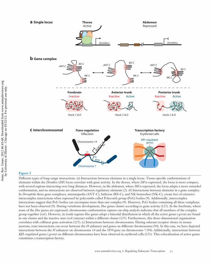
Figure 3Different types of long-range interactions. (a) Interactions between elements in a single locus. Tissue-specific conformations ofelements within the Distalless (Dll ) locus correlate with gene activity. In the thorax, where Dll is expressed, the locus is more compact,with several regions interacting over long distances. However, in the abdomen, where Dll is repressed, the locus adapts a more extendedconformation, and no interactions are observed between regulatory elements (2). (b) Interactions between elements in a gene complex.In Drosophila three gene complexes, antennapedia (ANT-C), bithorax (BX-C), and NK homeobox (NK-C), create foci of extensiveintracomplex interactions when repressed by polycombs called Polycomb group (PcG) bodies (9). Additionally, intercomplexinteractions suggest that PcG bodies can encompass more than one complex (9). However, PcG bodies containing all three complexeshave not been observed (9). During vertebrate development, Hox genes cluster according to gene activity (125). In the forebrain, wherenone of the Hox genes are expressed, chromosome conformation capture-on-chip analysis indicates that all members of the complexgroup together (red ). However, in trunk regions Hox genes adopt a bimodal distribution in which all the active genes ( green) are foundin one cluster and the inactive ones (red ) interact within a different cluster (125). Furthermore, this three-dimensional organizationcorrelates with collinear gene activation (125). (c) Interactions between chromosomes. During odorant receptor choice in mouseneurons, trans interactions can occur between the H enhancer and genes on different chromosomes (94). In this case, we have depictedinteractions between the H enhancer on chromosome 14 and the M50 gene on chromosome 7 (94). Additionally, interactions betweenKlf1-regulated genes ( green) on different chromosomes have been observed in erythroid cells (151). This colocalization of active genesconstitutes a transcription factory.
www.annualreviews.org • Regulating Eukaryotic Transcription 57
Ann
u. R
ev. G
enet
. 201
2.46
:43-
68. D
ownl
oade
d fr
om w
ww
.ann
ualr
evie
ws.
org
by U
nive
rsity
of
Illin
ois
- C
hica
go o
n 03
/15/
13. F
or p
erso
nal u
se o
nly.

GE46CH03-Mann ARI 6 October 2012 15:17
4C: chromosomeconformationcapture-on-chip
gene regulation. Spatial clustering of Hoxgenes is conserved between vertebrates andinvertebrates (43, 117, 125). Recent 3C andchromosome conformation capture-on-chip(4C) analysis of the mammalian HoxD genecluster in developing limb buds found thatfunctional regulatory regions dispersed withina gene desert upstream of the coding regioninteracted with the active Hoxd promoter (117)(Figure 3). These three-dimensional inter-actions were proposed to form a regulatoryarchipelago that through regulation of Hoxdgene expression could function to modulatedigit morphology; this concept of partiallyredundant or shadow enhancers workingtogether to regulate a single gene has beendescribed for multiple Drosophila genes (2, 10,37, 48, 64, 137). At the Hoxd regulatory locus,alterations in chromatin interactions corre-lated with collinear activation of the Hox genesduring development (125). In tissues where theHox clusters are silent, genes were observedto reside in a single three-dimensional domainmarked by H3K27me3 (Figure 3) (125). How-ever, once gene expression began, a bimodalorganization was observed (Figure 3) (125). Insamples from either anterior or posterior por-tions of the embryo, genes known to be activelytranscribed in those regions occupied one do-main that correlated with H3K4me3, whereasgenes known to be silent occupied a separatedomain that correlated with H3K27me3(125).
Although these Hox complex studiesprovide strong correlations, there is somecontroversy as to the relationship betweenthree-dimensional chromatin structure andgene regulation. In erythroid cells, specificintra- and interchromosomal interactions be-tween coregulated genes were dependent on asingle TF, Klf1 (Figure 3) (151). Additionally,transgenes carrying Klf1-regulated genes re-locate to transcription factories when insertedinto other genomic locations (151). Theseresults suggest that active, coregulated genescan preferentially organize into transcriptionalinteractomes (151). However, other studiesusing glucocorticoid-inducible gene expression
in cell lines did not observe significant chromo-somal rearrangements upon activation (56). In-stead, GR activates genes within preexisting locithat are enriched for DNase1-hypersensitivesites (56). These studies highlight the func-tional complexity that can be elucidated using3C and related strategies. However, they alsocaution against making broad interpretationsregarding the role of nuclear architecture ingene regulation, as observations can be highlyspecific to a particular gene or group of genes.Furthermore, interactions, or lack thereof, canbe highly cell-type specific, as was recentlyshown in a chromosome conformation–basedstudy of RNA polymerase II transcription (89)and by cgChIP in Drosophila (2). New advancesin microscopy may help to sort out data fromcross-linking-based studies by visualizinginteractions in situ (71).
Regulatory factories and the hierarchicallystructured nature of chromatin in general sug-gest a regulatory environment in the nucleusin which the local concentration of TFs andaccessory factors can vary significantly fromregion to region. Protein concentration is animportant determinant of TF-DNA interactionand has been incorporated into recent modelsof genome-wide DNA binding (18, 79, 177),so the potential for regional variation in TFconcentrations throughout the nucleus mustbe taken into account. Such foci might also leadto the identification of indirect protein-DNAinteractions when using a cross-linking-basedprotocol such as ChIP; whether this is the casewill become evident as more genome-wide 3Cand cell type–specific data become available.Thus, although the study of chromosomalconformation is a relatively nascent field whencompared with most of TF biology, thesestudies have the potential to impact both ourinterpretation of genome-wide ChIP data andour protein concentration–centric models ofgenome-wide TF binding.
CONCLUDING REMARKS
Much has been made of the finding that manyTFs bind thousands of genomic regions in vivo,
58 Lelli · Slattery · Mann
Ann
u. R
ev. G
enet
. 201
2.46
:43-
68. D
ownl
oade
d fr
om w
ww
.ann
ualr
evie
ws.
org
by U
nive
rsity
of
Illin
ois
- C
hica
go o
n 03
/15/
13. F
or p
erso
nal u
se o
nly.
GE46CH03-Mann ARI 6 October 2012 15:17
perhaps because these numbers easily exceedthe number of expected target genes for mostsequence-specific TFs (90, 100, 101). However,the number of binding events is still lower thanthe predicted number of sites throughout thegenome on the basis of DNA sequence aloneand close to what is expected when account-ing for both DNA sequence and chromatin ac-cessibility (177). TFs are most typically viewedas components of discrete regulatory networks,with separate target and nontarget genes foreach TF (28, 29, 121). However, it has beenproposed, on the basis of genome-wide bind-ing data, that TF regulatory networks shouldinstead be viewed as continuous networks (18).The continuous network model of TF functionposits that, because of the high nuclear con-centration of most expressed TFs, specificitymediated by protein-protein interactions is un-necessary, and essentially all genes are targetedby all TFs. According to this view, biologicalfunction is determined by quantitative differ-ences in TF binding at accessible DNA ratherthan binary on/off TF binding (18). Aspects ofthis model are supported by data from a surveyof Drosophila TFs expressed early in Drosophilaembryogenesis (79, 91). However, the over-expression of many TFs, for example, by theGal4-UAS method in Drosophila, generally donot lead to aberrant phenotypes; instead, higherthan normal levels of TFs in cells typically resultin wild-type readouts, which suggests that otherfactors besides concentration must be limitingfor TF function. Moreover, there are many ex-amples in which small differences in TF DNA-binding domains are important for their specificin vivo functions, arguing that DNA-bindingspecificity is critical. In addition, the continu-ous network model assumes that there are nocell type–specific differences in binding and thatthe signal generated by ChIP is indicative of di-rect DNA binding. But this may not always bethe case, as there is ample evidence for ChIPsignals resulting from indirect DNA bindingvia protein-protein interactions or interactions
between regulatory elements (2, 54, 62, 118).On the basis of the current data, we suggest thatchromatin accessibility is critical for limitingwhich TF-binding sites and which CRMs areavailable in specific cell types, but within acces-sible regions, DNA-binding specificities of TFsand TF complexes are essential for determin-ing which binding sites are productively boundwithin these accessible regions.
The era of TF genomics has clearly changedour view of how TFs target the genome, butmany of the methods routinely used are lowresolution or are blind to cell type–specific dif-ferences. New, higher-resolution technologieswill undoubtedly lead to refinement and re-structuring of these models. For example, anew variation on ChIP, termed ChIP-exo, cangenerate genome-wide TF–DNA binding pro-files down to single-base resolution (143). Notonly does this high-resolution method providea more precise view of a TF’s DNA-bindingmotifs, but it also eliminates a significant num-ber of false positive binding events generated bytraditional ChIP-chip or ChIP-seq (143). Thismethod has the potential to refine the mod-els of genome-wide TF binding that have beengenerated over the past five years.
Beyond advances in ChIP, increasinglysophisticated methods for monitoring nuclearorganization will begin to generate a compre-hensive picture of nuclear and chromosomalstructure in vivo. This will require a com-bination of both 3C-based techniques andsuper-resolution microscopy techniques, suchas stochastic optical reconstruction microscopy(147). Combining these approaches withhigh-resolution ChIP data will be essential forunderstanding TF-DNA interactions in thecontext of chromatin looping and transcriptionfactories. Ultimately, integrating these multiplelayers of data, in combination with the studiesdescribed in this review, will allow TF biologiststo generate and test models regarding directversus indirect, specific versus nonspecific, andfunctional versus nonfunctional binding.
www.annualreviews.org • Regulating Eukaryotic Transcription 59
Ann
u. R
ev. G
enet
. 201
2.46
:43-
68. D
ownl
oade
d fr
om w
ww
.ann
ualr
evie
ws.
org
by U
nive
rsity
of
Illin
ois
- C
hica
go o
n 03
/15/
13. F
or p
erso
nal u
se o
nly.

GE46CH03-Mann ARI 6 October 2012 15:17
SUMMARY POINTS
1. Multiple mechanisms that provide site-specific TFs with increased specificity contributeto cooperative DNA binding.
2. For most CRMs, architecture is flexible, allowing the modular integration of multipleTF inputs, some of which may depend on cooperativity.
3. Multiple chromatin states have been described on the basis of histone PTMs and tran-scriptional activity.
4. Genetic material is structurally organized within nuclei in a manner that is relevant togene regulation.
FUTURE ISSUES
1. Structural studies are needed to address how different protein-protein and protein-DNAinteractions affect CRM assembly and function at a molecular level.
2. Additional rigorous studies of CRM architecture would help build better models toidentify CRMs and predict their functions.
3. Is there a causal relationship between histone modifications and transcriptionalregulation?
4. High-resolution, cell type–specific studies of chromatin and nuclear architecture will beimportant for fully understanding gene regulation.
DISCLOSURE STATEMENT
The authors are not aware of any affiliations, memberships, funding, or financial holdings thatmight be perceived as affecting the objectivity of this review.
ACKNOWLEDGMENTS
We thank members of the Mann laboratory for helpful discussions. R.S.M. was supported by NIHgrant GM54510 and K.L. by the 5T32DK07328 training grant.
LITERATURE CITED
1. Agarwal P, Verzi MP, Nguyen T, Hu J, Ehlers ML, et al. 2011. The MADS box transcription factorMEF2C regulates melanocyte development and is a direct transcriptional target and partner of SOX10.Development 138:2555–65
2. Agelopoulos M, McKay DJ, Mann RS. 2012. Developmental regulation of chromatin conformation byHox proteins in Drosophila. Cell Rep. 1(4):350–59
3. Arnosti DN, Kulkarni MM. 2005. Transcriptional enhancers: intelligent enhanceosomes or flexiblebillboards? J. Cell Biochem. 94:890–98
4. Badis G, Berger MF, Philippakis AA, Talukder S, Gehrke AR, et al. 2009. Diversity and complexity inDNA recognition by transcription factors. Science 324:1720–23
5. Bai L, Morozov AV. 2010. Gene regulation by nucleosome positioning. Trends Genet. 26:476–83
60 Lelli · Slattery · Mann
Ann
u. R
ev. G
enet
. 201
2.46
:43-
68. D
ownl
oade
d fr
om w
ww
.ann
ualr
evie
ws.
org
by U
nive
rsity
of
Illin
ois
- C
hica
go o
n 03
/15/
13. F
or p
erso
nal u
se o
nly.
GE46CH03-Mann ARI 6 October 2012 15:17
6. Bai L, Ondracka A, Cross FR. 2011. Multiple sequence-specific factors generate the nucleosome-depletedregion on CLN2 promoter. Mol. Cell 42:465–76
7. Bannister AJ, Kouzarides T. 2011. Regulation of chromatin by histone modifications. Cell Res. 21:381–958. Bantignies F, Cavalli G. 2011. Polycomb group proteins: repression in 3D. Trends Genet. 27:454–649. Bantignies F, Roure V, Comet I, Leblanc B, Schuettengruber B, et al. 2011. Polycomb-dependent
regulatory contacts between distant Hox loci in Drosophila. Cell 144:214–2610. Barolo S. 2012. Shadow enhancers: frequently asked questions about distributed cis-regulatory informa-
tion and enhancer redundancy. BioEssays 34:135–4111. Barski A, Cuddapah S, Cui K, Roh TY, Schones DE, et al. 2007. High-resolution profiling of histone
methylations in the human genome. Cell 129:823–3712. Barth TK, Imhof A. 2010. Fast signals and slow marks: the dynamics of histone modifications. Trends
Biochem. Sci. 35:618–2613. Barthel R, Tsytsykova AV, Barczak AK, Tsai EY, Dascher CC, et al. 2003. Regulation of tumor necrosis
factor alpha gene expression by mycobacteria involves the assembly of a unique enhanceosome dependenton the coactivator proteins CBP/p300. Mol. Cell. Biol. 23:526–33
14. Benayoun BA, Veitia RA. 2009. A post-translational modification code for transcription factors: sortingthrough a sea of signals. Trends Cell Biol. 19:189–97
15. Berger MF, Badis G, Gehrke AR, Talukder S, Philippakis AA, et al. 2008. Variation in homeodomainDNA binding revealed by high-resolution analysis of sequence preferences. Cell 133:1266–76
16. Bernard P, Harley VR. 2010. Acquisition of SOX transcription factor specificity through protein-proteininteraction, modulation of Wnt signalling and post-translational modification. Int. J. Biochem. Cell Biol.42:400–10
17. Biddie SC, John S, Sabo PJ, Thurman RE, Johnson TA, et al. 2011. Transcription factor AP1 potentiateschromatin accessibility and glucocorticoid receptor binding. Mol. Cell 43:145–55
18. Biggin MD. 2011. Animal transcription networks as highly connected, quantitative continua. Dev. Cell21:611–26
19. Bonn S, Zinzen RP, Girardot C, Gustafson EH, Perez-Gonzalez A, et al. 2012. Tissue-specific analysisof chromatin state identifies temporal signatures of enhancer activity during embryonic development.Nat. Genet. 44:148–56
20. Busser BW, Shokri L, Jaeger SA, Gisselbrecht SS, Singhania A, et al. 2012. Molecular mechanism un-derlying the regulatory specificity of a Drosophila homeodomain protein that specifies myoblast identity.Development 139:1164–74
21. Cavalli G. 2007. Chromosome kissing. Curr. Opin. Genet. Dev. 17:443–5022. Chan SK, Mann RS. 1993. The segment identity functions of Ultrabithorax are contained within its
homeodomain and carboxy-terminal sequences. Genes Dev. 7:796–81123. Chan SK, Popperl H, Krumlauf R, Mann RS. 1996. An extradenticle-induced conformational change
in a HOX protein overcomes an inhibitory function of the conserved hexapeptide motif. EMBO J.15:2476–87
24. Charlot C, Dubois-Pot H, Serchov T, Tourrette Y, Wasylyk B. 2010. A review of post-translationalmodifications and subcellular localization of Ets transcription factors: possible connection with cancerand involvement in the hypoxic response. Methods Mol. Biol. 647:3–30
25. Courey AJ. 2001. Cooperativity in transcriptional control. Curr. Biol. 11:R250–5226. Dai Z, Dai X. 2012. Nuclear colocalization of transcription factor target genes strengthens coregulation
in yeast. Nucleic Acids Res. 40:27–3627. Daitoku H, Sakamaki J, Fukamizu A. 2011. Regulation of FoxO transcription factors by acetylation and
protein-protein interactions. Biochim. Biophys. Acta 1813:1954–6028. Davidson EH. 2010. Emerging properties of animal gene regulatory networks. Nature 468:911–2029. Davidson EH, Levine MS. 2008. Properties of developmental gene regulatory networks. Proc. Natl. Acad.
Sci. USA 105:20063–6630. de Wit E, de Laat W. 2012. A decade of 3C technologies: insights into nuclear organization. Genes Dev.
26:11–2431. Dekker J, Rippe K, Dekker M, Kleckner N. 2002. Capturing chromosome conformation. Science
295:1306–11
www.annualreviews.org • Regulating Eukaryotic Transcription 61
Ann
u. R
ev. G
enet
. 201
2.46
:43-
68. D
ownl
oade
d fr
om w
ww
.ann
ualr
evie
ws.
org
by U
nive
rsity
of
Illin
ois
- C
hica
go o
n 03
/15/
13. F
or p
erso
nal u
se o
nly.
GE46CH03-Mann ARI 6 October 2012 15:17
32. Delval S, Taminiau A, Lamy J, Lallemand C, Gilles C, et al. 2011. The Pbx interaction motif of Hoxa1is essential for its oncogenic activity. PLoS ONE 6:e25247
33. Dostie J, Bickmore WA. 2012. Chromosome organization in the nucleus: charting new territory acrossthe Hi-Cs. Curr. Opin. Genet. Dev. 22(2):125–31
34. Duboule D. 1991. Patterning in the vertebrate limb. Curr. Opin. Genet. Dev. 1:211–1635. Duboule D, Morata G. 1994. Colinearity and functional hierarchy among genes of the homeotic com-
plexes. Trends Genet. 10:358–6436. Duncan IW. 2002. Transvection effects in Drosophila. Annu. Rev. Genet. 36:521–5637. Dunipace L, Ozdemir A, Stathopoulos A. 2011. Complex interactions between cis-regulatory modules
in native conformation are critical for Drosophila snail expression. Development 138:4075–8438. Ellington AD, Szostak JW. 1990. In vitro selection of RNA molecules that bind specific ligands. Nature
346:818–2239. Enriquez J, Boukhatmi H, Dubois L, Philippakis AA, Bulyk ML, et al. 2010. Multi-step control of muscle
diversity by Hox proteins in the Drosophila embryo. Development 137:457–6640. Eskiw CH, Fraser P. 2011. Ultrastructural study of transcription factories in mouse erythroblasts. J. Cell
Sci. 124:3676–8341. Ethier SD, Miura H, Dostie J. 2011. Discovering genome regulation with 3C and 3C-related technolo-
gies. Biochim. Biophys. Acta 1819:401–1042. Felsenfeld G, Groudine M. 2003. Controlling the double helix. Nature 421:448–5343. Ferraiuolo MA, Rousseau M, Miyamoto C, Shenker S, Wang XQ, et al. 2010. The three-dimensional
architecture of Hox cluster silencing. Nucleic Acids Res. 38:7472–8444. Filion GJ, van Bemmel JG, Braunschweig U, Talhout W, Kind J, et al. 2010. Systematic protein location
mapping reveals five principal chromatin types in Drosophila cells. Cell 143:212–2445. Fischback NA, Rozenfeld S, Shen W, Fong S, Chrobak D, et al. 2005. HOXB6 overexpression in
murine bone marrow immortalizes a myelomonocytic precursor in vitro and causes hematopoietic stemcell expansion and acute myeloid leukemia in vivo. Blood 105:1456–66
46. Fitzsimmons D, Lutz R, Wheat W, Chamberlin HM, Hagman J. 2001. Highly conserved amino acidsin Pax and Ets proteins are required for DNA binding and ternary complex assembly. Nucleic Acids Res.29:4154–65
47. Floer M, Wang X, Prabhu V, Berrozpe G, Narayan S, et al. 2010. A RSC/nucleosome complex determineschromatin architecture and facilitates activator binding. Cell 141:407–18
48. Frankel N, Davis GK, Vargas D, Wang S, Payre F, Stern DL. 2010. Phenotypic robustness conferredby apparently redundant transcriptional enhancers. Nature 466:490–93
49. Gardner KE, Allis CD, Strahl BD. 2011. Operating on chromatin, a colorful language where contextmatters. J. Mol. Biol. 409:36–46
50. Garvie CW, Hagman J, Wolberger C. 2001. Structural studies of Ets-1/Pax5 complex formation onDNA. Mol. Cell 8:1267–76
51. Georges AB, Benayoun BA, Caburet S, Veitia RA. 2010. Generic binding sites, generic DNA-bindingdomains: Where does specific promoter recognition come from? FASEB J. 24:346–56
52. Gonzalez-Reyes A, Morata G. 1990. The developmental effect of overexpressing a Ubx product inDrosophila embryos is dependent on its interactions with other homeotic products. Cell 61:515–22
53. Gonzalez-Reyes A, Urquia N, Gehring WJ, Struhl G, Morata G. 1990. Are cross-regulatory interactionsbetween homoeotic genes functionally significant? Nature 344:78–80
54. Gordan R, Hartemink AJ, Bulyk ML. 2009. Distinguishing direct versus indirect transcription factor–DNA interactions. Genome Res. 19:2090–100
55. Guertin MJ, Lis JT. 2010. Chromatin landscape dictates HSF binding to target DNA elements.PLoS Genet. 6:e1001114
56. Hakim O, Sung MH, Voss TC, Splinter E, John S, et al. 2011. Diverse gene reprogramming eventsoccur in the same spatial clusters of distal regulatory elements. Genome Res. 21:697–706
57. Harrison MM, Li XY, Kaplan T, Botchan MR, Eisen MB. 2011. Zelda binding in the early Drosophilamelanogaster embryo marks regions subsequently activated at the maternal-to-zygotic transition.PLoS Genet. 7:e1002266
62 Lelli · Slattery · Mann
Ann
u. R
ev. G
enet
. 201
2.46
:43-
68. D
ownl
oade
d fr
om w
ww
.ann
ualr
evie
ws.
org
by U
nive
rsity
of
Illin
ois
- C
hica
go o
n 03
/15/
13. F
or p
erso
nal u
se o
nly.

GE46CH03-Mann ARI 6 October 2012 15:17
58. He HH, Meyer CA, Shin H, Bailey ST, Wei G, et al. 2010. Nucleosome dynamics define transcriptionalenhancers. Nat. Genet. 42:343–47
59. He Q, Bardet AF, Patton B, Purvis J, Johnston J, et al. 2011. High conservation of transcription factorbinding and evidence for combinatorial regulation across six Drosophila species. Nat. Genet. 43:414–20
60. Heintzman ND, Stuart RK, Hon G, Fu Y, Ching CW, et al. 2007. Distinct and predictive chromatinsignatures of transcriptional promoters and enhancers in the human genome. Nat. Genet. 39:311–18
61. Heinz S, Benner C, Spann N, Bertolino E, Lin YC, et al. 2010. Simple combinations of lineage-determining transcription factors prime cis-regulatory elements required for macrophage and B cellidentities. Mol. Cell 38:576–89
62. Heldring N, Isaacs GD, Diehl AG, Sun M, Cheung E, et al. 2011. Multiple sequence-specific DNA-binding proteins mediate estrogen receptor signaling through a tethering pathway. Mol. Endocrinol.25:564–74
63. Hollenhorst PC, McIntosh LP, Graves BJ. 2011. Genomic and biochemical insights into the specificityof ETS transcription factors. Annu. Rev. Biochem. 80:437–71
64. Hong JW, Hendrix DA, Levine MS. 2008. Shadow enhancers as a source of evolutionary novelty. Science321:1314
65. Hughes CL, Kaufman TC. 2002. Hox genes and the evolution of the arthropod body plan. Evol. Dev. 4:459–99
66. Istrail S, Davidson EH. 2005. Logic functions of the genomic cis-regulatory code. Proc. Natl. Acad. Sci.USA 102:4954–59
67. Jansen A, van der Zande E, Meert W, Fink GR, Verstrepen KJ. 2012. Distal chromatin structure influ-ences local nucleosome positions and gene expression. Nucleic Acids Res. 40(9):3870–85
68. Jauch R, Aksoy I, Hutchins AP, Ng CK, Tian XF, et al. 2011. Conversion of Sox17 into a pluripotencyreprogramming factor by reengineering its association with Oct4 on DNA. Stem Cells 29:940–51
69. Jenuwein T, Allis CD. 2001. Translating the histone code. Science 293:1074–8070. John S, Sabo PJ, Thurman RE, Sung MH, Biddie SC, et al. 2011. Chromatin accessibility pre-determines
glucocorticoid receptor binding patterns. Nat. Genet. 43:264–6871. Jones SA, Shim SH, He J, Zhuang X. 2011. Fast, three-dimensional super-resolution imaging of live
cells. Nat. Methods 8:499–50872. Joseph R, Orlov YL, Huss M, Sun W, Kong SL, et al. 2010. Integrative model of genomic factors for
determining binding site selection by estrogen receptor-α. Mol. Syst. Biol. 6:45673. Joshi R, Passner JM, Rohs R, Jain R, Sosinsky A, et al. 2007. Functional specificity of a Hox protein
mediated by the recognition of minor groove structure. Cell 131:530–4374. Joshi R, Sun L, Mann R. 2010. Dissecting the functional specificities of two Hox proteins. Genes Dev.
24:1533–4575. Junion G, Spivakov M, Girardot C, Braun M, Gustafson EH, et al. 2012. A transcription factor collective
defines cardiac cell fate and reflects lineage history. Cell 148:473–8676. Kaplan N, Hughes TR, Lieb JD, Widom J, Segal E. 2010. Contribution of histone sequence preferences
to nucleosome organization: proposed definitions and methodology. Genome Biol. 11:14077. Kaplan N, Moore I, Fondufe-Mittendorf Y, Gossett AJ, Tillo D, et al. 2010. Nucleosome sequence
preferences influence in vivo nucleosome organization. Nat. Struct. Mol. Biol. 17(8):918–20; author reply,920–22
78. Kaplan N, Moore IK, Fondufe-Mittendorf Y, Gossett AJ, Tillo D, et al. 2009. The DNA-encodednucleosome organization of a eukaryotic genome. Nature 458:362–66
79. Kaplan T, Li XY, Sabo PJ, Thomas S, Stamatoyannopoulos JA, et al. 2011. Quantitative models of themechanisms that control genome-wide patterns of transcription factor binding during early Drosophiladevelopment. PLoS Genet. 7:e1001290
80. Kharchenko PV, Alekseyenko AA, Schwartz YB, Minoda A, Riddle NC, et al. 2011. Comprehensiveanalysis of the chromatin landscape in Drosophila melanogaster. Nature 471:480–85
81. Khoueiry P, Rothbacher U, Ohtsuka Y, Daian F, Frangulian E, et al. 2010. A cis-regulatory signature inascidians and flies, independent of transcription factor binding sites. Curr. Biol. 20:792–802
www.annualreviews.org • Regulating Eukaryotic Transcription 63
Ann
u. R
ev. G
enet
. 201
2.46
:43-
68. D
ownl
oade
d fr
om w
ww
.ann
ualr
evie
ws.
org
by U
nive
rsity
of
Illin
ois
- C
hica
go o
n 03
/15/
13. F
or p
erso
nal u
se o
nly.
GE46CH03-Mann ARI 6 October 2012 15:17
82. Knoepfler PS, Sykes DB, Pasillas M, Kamps MP. 2001. HoxB8 requires its Pbx-interaction motif to blockdifferentiation of primary myeloid progenitors and of most cell line models of myeloid differentiation.Oncogene 20:5440–48
83. Kondoh H, Kamachi Y. 2010. SOX-partner code for cell specification: regulatory target selection andunderlying molecular mechanisms. Int. J. Biochem. Cell Biol. 42:391–99
84. Krivega I, Dean A. 2011. Enhancer and promoter interactions: long distance calls. Curr. Opin. Genet.Dev. 22(2):79–85
85. Kulkarni MM, Arnosti DN. 2003. Information display by transcriptional enhancers. Development130:6569–75
86. Lee JS, Smith E, Shilatifard A. 2010. The language of histone crosstalk. Cell 142:682–8587. Lelli KM, Noro B, Mann RS. 2011. Variable motif utilization in homeotic selector (Hox)-cofactor
complex formation controls specificity. Proc. Natl. Acad. Sci. USA 108:21122–2788. Li G, Reinberg D. 2011. Chromatin higher-order structures and gene regulation. Curr. Opin. Genet.
Dev. 21:175–8689. Li G, Ruan X, Auerbach RK, Sandhu KS, Zheng M, et al. 2012. Extensive promoter-centered chromatin
interactions provide a topological basis for transcription regulation. Cell 148:84–9890. Li XY, MacArthur S, Bourgon R, Nix D, Pollard DA, et al. 2008. Transcription factors bind thousands
of active and inactive regions in the Drosophila blastoderm. PLoS Biol. 6:e2791. Li XY, Thomas S, Sabo PJ, Eisen MB, Stamatoyannopoulos JA, Biggin MD. 2011. The role of chromatin
accessibility in directing the widespread, overlapping patterns of Drosophila transcription factor binding.Genome Biol. 12:R34
92. Liang HL, Nien CY, Liu HY, Metzstein MM, Kirov N, Rushlow C. 2008. The zinc-finger protein Zeldais a key activator of the early zygotic genome in Drosophila. Nature 456:400–3
93. Lieberman-Aiden E, van Berkum NL, Williams L, Imakaev M, Ragoczy T, et al. 2009. Comprehensivemapping of long-range interactions reveals folding principles of the human genome. Science 326:289–93
94. Lomvardas S, Barnea G, Pisapia DJ, Mendelsohn M, Kirkland J, Axel R. 2006. Interchromosomal inter-actions and olfactory receptor choice. Cell 126:403–13
95. Lomvardas S, Thanos D. 2002. Modifying gene expression programs by altering core promoter chro-matin architecture. Cell 110:261–71
96. Ludwig MZ, Manu, Kittler R, White KP, Kreitman M. 2011. Consequences of eukaryotic enhancerarchitecture for gene expression dynamics, development, and fitness. PLoS Genet. 7:e1002364
97. Ludwig MZ, Palsson A, Alekseeva E, Bergman CM, Nathan J, Kreitman M. 2005. Functional evolutionof a cis-regulatory module. PLoS Biol. 3:e93
98. Luger K, Mader AW, Richmond RK, Sargent DF, Richmond TJ. 1997. Crystal structure of the nucle-osome core particle at 2.8 A resolution. Nature 389:251–60
99. Lupien M, Eeckhoute J, Meyer CA, Wang Q, Zhang Y, et al. 2008. FoxA1 translates epigenetic signaturesinto enhancer-driven lineage-specific transcription. Cell 132:958–70
100. MacArthur S, Li XY, Li J, Brown JB, Chu HC, et al. 2009. Developmental roles of 21 Drosophilatranscription factors are determined by quantitative differences in binding to an overlapping set ofthousands of genomic regions. Genome Biol. 10:R80
101. MacQuarrie KL, Fong AP, Morse RH, Tapscott SJ. 2011. Genome-wide transcription factor binding:beyond direct target regulation. Trends Genet. 27:141–48
102. Magnani L, Eeckhoute J, Lupien M. 2011. Pioneer factors: directing transcriptional regulators withinthe chromatin environment. Trends Genet. 27:465–74
103. Mann RS, Chan SK. 1996. Extra specificity from extradenticle: the partnership between HOX andPBX/EXD homeodomain proteins. Trends Genet. 12:258–62
104. Mann RS, Hogness DS. 1990. Functional dissection of Ultrabithorax proteins in D. melanogaster. Cell60:597–610
105. Mann RS, Lelli KM, Joshi R. 2009. Hox specificity: unique roles for cofactors and collaborators.Curr. Top. Dev. Biol. 88:63–101
106. Mao C, Brown CR, Griesenbeck J, Boeger H. 2011. Occlusion of regulatory sequences by promoternucleosomes in vivo. PLoS ONE 6:e17521
64 Lelli · Slattery · Mann
Ann
u. R
ev. G
enet
. 201
2.46
:43-
68. D
ownl
oade
d fr
om w
ww
.ann
ualr
evie
ws.
org
by U
nive
rsity
of
Illin
ois
- C
hica
go o
n 03
/15/
13. F
or p
erso
nal u
se o
nly.

GE46CH03-Mann ARI 6 October 2012 15:17
107. Margueron R, Reinberg D. 2011. The Polycomb complex PRC2 and its mark in life. Nature 469:343–49108. Meijsing SH, Pufall MA, So AY, Bates DL, Chen L, Yamamoto KR. 2009. DNA binding site sequence
directs glucocorticoid receptor structure and activity. Science 324:407–10109. Merabet S, Kambris Z, Capovilla M, Berenger H, Pradel J, Graba Y. 2003. The hexapeptide and linker
regions of the AbdA Hox protein regulate its activating and repressive functions. Dev. Cell 4:761–68110. Merabet S, Litim-Mecheri I, Karlsson D, Dixit R, Saadaoui M, et al. 2011. Insights into Hox protein
function from a large scale combinatorial analysis of protein domains. PLoS Genet. 7:e1002302111. Merabet S, Saadaoui M, Sambrani N, Hudry B, Pradel J, et al. 2007. A unique extradenticle recruitment
mode in the Drosophila Hox protein Ultrabithorax. Proc. Natl. Acad. Sci. USA 104:16946–51112. Merabet S, Sambrani N, Pradel J, Graba Y. 2010. Regulation of Hox activity: insights from protein
motifs. Adv. Exp. Med. Biol. 689:3–16113. Miller JA, Widom J. 2003. Collaborative competition mechanism for gene activation in vivo. Mol. Cell.
Biol. 23:1623–32114. Mirny LA. 2010. Nucleosome-mediated cooperativity between transcription factors. Proc. Natl. Acad.
Sci. USA 107:22534–39115. Mo Y, Ho W, Johnston K, Marmorstein R. 2001. Crystal structure of a ternary SAP-1/SRF/c-fos SRE
DNA complex. J. Mol. Biol. 314:495–506116. Moens CB, Selleri L. 2006. Hox cofactors in vertebrate development. Dev. Biol. 291:193–206117. Montavon T, Soshnikova N, Mascrez B, Joye E, Thevenet L, et al. 2011. A regulatory archipelago
controls Hox genes transcription in digits. Cell 147:1132–45118. Moorman C, Sun LV, Wang J, de Wit E, Talhout W, et al. 2006. Hotspots of transcription factor
colocalization in the genome of Drosophila melanogaster. Proc. Natl. Acad. Sci. USA 103:12027–32119. Moretti R, Ansari AZ. 2008. Expanding the specificity of DNA targeting by harnessing cooperative
assembly. Biochimie 90:1015–25120. Moyle-Heyrman G, Tims HS, Widom J. 2011. Structural constraints in collaborative competition of
transcription factors against the nucleosome. J. Mol. Biol. 412:634–46121. Negre N, Brown CD, Ma L, Bristow CA, Miller SW, et al. 2011. A cis-regulatory map of the Drosophila
genome. Nature 471:527–31122. Negre N, Brown CD, Shah PK, Kheradpour P, Morrison CA, et al. 2010. A comprehensive map of
insulator elements for the Drosophila genome. PLoS Genet. 6:e1000814123. Ng CK, Li NX, Chee S, Prabhakar S, Kolatkar PR, Jauch R. 2012. Deciphering the Sox-Oct partner
code by quantitative cooperativity measurements. Nucleic Acids Res. doi: 10.1093/nar/gks153124. Nien CY, Liang HL, Butcher S, Sun Y, Fu S, et al. 2011. Temporal coordination of gene networks by
Zelda in the early Drosophila embryo. PLoS Genet. 7:e1002339125. Noordermeer D, Leleu M, Splinter E, Rougemont J, De Laat W, Duboule D. 2011. The dynamic
architecture of Hox gene clusters. Science 334:222–25126. Noro B, Lelli K, Sun L, Mann RS. 2011. Competition for cofactor-dependent DNA binding underlies
Hox phenotypic suppression. Genes Dev. 25:2327–32127. Noyes MB, Christensen RG, Wakabayashi A, Stormo GD, Brodsky MH, Wolfe SA. 2008. Analy-
sis of homeodomain specificities allows the family-wide prediction of preferred recognition sites. Cell133:1277–89
128. Osborne CS, Chakalova L, Brown KE, Carter D, Horton A, et al. 2004. Active genes dynamicallycolocalize to shared sites of ongoing transcription. Nat. Genet. 36:1065–71
129. Pan Y, Nussinov R. 2011. The role of response elements organization in transcription factor selectivity:the IFN-β enhanceosome example. PLoS Comput. Biol. 7:e1002077
130. Pan Y, Tsai CJ, Ma B, Nussinov R. 2010. Mechanisms of transcription factor selectivity. Trends Genet.26:75–83
131. Panne D. 2008. The enhanceosome. Curr. Opin. Struct. Biol. 18:236–42132. Panne D, Maniatis T, Harrison SC. 2004. Crystal structure of ATF-2/c-Jun and IRF-3 bound to the
interferon-β enhancer. EMBO J. 23:4384–93133. Panne D, Maniatis T, Harrison SC. 2007. An atomic model of the interferon-β enhanceosome. Cell
129:1111–23
www.annualreviews.org • Regulating Eukaryotic Transcription 65
Ann
u. R
ev. G
enet
. 201
2.46
:43-
68. D
ownl
oade
d fr
om w
ww
.ann
ualr
evie
ws.
org
by U
nive
rsity
of
Illin
ois
- C
hica
go o
n 03
/15/
13. F
or p
erso
nal u
se o
nly.
GE46CH03-Mann ARI 6 October 2012 15:17
134. Papadopoulos DK, Resendez-Perez D, Cardenas-Chavez DL, Villanueva-Segura K, Canales-del-Castillo R, et al. 2011. Functional synthetic Antennapedia genes and the dual roles of YPWM motifand linker size in transcriptional activation and repression. Proc. Natl. Acad. Sci. USA 108:11959–64
135. Papadopoulos DK, Vukojevic V, Adachi Y, Terenius L, Rigler R, Gehring WJ. 2010. Function andspecificity of synthetic Hox transcription factors in vivo. Proc. Natl. Acad. Sci. USA 107:4087–92
136. Parker DS, White MA, Ramos AI, Cohen BA, Barolo S. 2011. The cis-regulatory logic of Hedgehoggradient responses: key roles for Gli binding affinity, competition, and cooperativity. Sci. Signal. 4:ra38
137. Perry MW, Boettiger AN, Bothma JP, Levine M. 2010. Shadow enhancers foster robustness of Drosophilagastrulation. Curr. Biol. 20:1562–67
138. Polach KJ, Widom J. 1996. A model for the cooperative binding of eukaryotic regulatory proteins tonucleosomal target sites. J. Mol. Biol. 258:800–12
139. Rando OJ. 2012. Combinatorial complexity in chromatin structure and function: revisiting the histonecode. Curr. Opin. Genet. Dev. 22:148–55
140. Rando OJ, Chang HY. 2009. Genome-wide views of chromatin structure. Annu. Rev. Biochem. 78:245–71141. Razin SV, Gavrilov AA, Pichugin A, Lipinski M, Iarovaia OV, Vassetzky YS. 2011. Transcription factories
in the context of the nuclear and genome organization. Nucleic Acids Res. 39:9085–92142. Remacle S, Abbas L, De Backer O, Pacico N, Gavalas A, et al. 2004. Loss of function but no gain of
function caused by amino acid substitutions in the hexapeptide Hoxa1 in vivo. Mol. Cell. Biol. 24:8567–75
143. Rhee HS, Pugh BF. 2011. Comprehensive genome-wide protein-DNA interactions detected at single-nucleotide resolution. Cell 147:1408–19
144. Rohs R, Jin X, West SM, Joshi R, Honig B, Mann RS. 2010. Origins of specificity in protein-DNArecognition. Annu. Rev. Biochem. 79:233–69
145. Rohs R, West SM, Sosinsky A, Liu P, Mann RS, Honig B. 2009. The role of DNA shape in protein-DNArecognition. Nature 461:1248–53
146. Roy S, Ernst J, Kharchenko PV, Kheradpour P, Negre N, et al. 2010. Identification of functional elementsand regulatory circuits by Drosophila modENCODE. Science 330:1787–97
147. Rust MJ, Bates M, Zhuang X. 2006. Sub-diffraction-limit imaging by stochastic optical reconstructionmicroscopy (STORM). Nat. Methods 3:793–95
148. Saadaoui M, Merabet S, Litim-Mecheri I, Arbeille E, Sambrani N, et al. 2011. Selection of distinct Hox-Extradenticle interaction modes fine-tunes Hox protein activity. Proc. Natl. Acad. Sci. USA 108:2276–81
149. Satija R, Bradley RK. 2012. The TAGteam motif facilitates binding of 21 sequence-specific transcriptionfactors in the Drosophila embryo. Genome Res. 22:656–65
150. Schnabel CA, Jacobs Y, Cleary ML. 2000. HoxA9-mediated immortalization of myeloid progenitorsrequires functional interactions with TALE cofactors Pbx and Meis. Oncogene 19:608–16
151. Schoenfelder S, Sexton T, Chakalova L, Cope NF, Horton A, et al. 2010. Preferential associationsbetween co-regulated genes reveal a transcriptional interactome in erythroid cells. Nat. Genet. 42:53–61
152. Segal E, Widom J. 2009. What controls nucleosome positions? Trends Genet. 25:335–43153. Sekiya T, Muthurajan UM, Luger K, Tulin AV, Zaret KS. 2009. Nucleosome-binding affinity as a
primary determinant of the nuclear mobility of the pioneer transcription factor FoxA. Genes Dev. 23:804–9
154. Sekiya T, Zaret KS. 2007. Repression by Groucho/TLE/Grg proteins: Genomic site recruitment gen-erates compacted chromatin in vitro and impairs activator binding in vivo. Mol. Cell 28:291–303
155. Serandour AA, Avner S, Percevault F, Demay F, Bizot M, et al. 2011. Epigenetic switch involved inactivation of pioneer factor FOXA1-dependent enhancers. Genome Res. 21:555–65
156. Sexton T, Yaffe E, Kenigsberg E, Bantignies F, Leblanc B, et al. 2012. Three-dimensional folding andfunctional organization principles of the Drosophila genome. Cell 148:458–72
157. Shen W, Chrobak D, Krishnan K, Lawrence HJ, Largman C. 2004. HOXB6 protein is bound toCREB-binding protein and represses globin expression in a DNA binding–dependent, PBX interaction–independent process. J. Biol. Chem. 279:39895–904
158. Shykind BM. 2005. Regulation of odorant receptors: one allele at a time. Hum. Mol. Genet. 14(Spec.No. 1):R33–39
66 Lelli · Slattery · Mann
Ann
u. R
ev. G
enet
. 201
2.46
:43-
68. D
ownl
oade
d fr
om w
ww
.ann
ualr
evie
ws.
org
by U
nive
rsity
of
Illin
ois
- C
hica
go o
n 03
/15/
13. F
or p
erso
nal u
se o
nly.

GE46CH03-Mann ARI 6 October 2012 15:17
159. Siggers T, Duyzend MH, Reddy J, Khan S, Bulyk ML. 2011. Non-DNA-binding cofactors enhanceDNA-binding specificity of a transcriptional regulatory complex. Mol. Syst. Biol. 7:555
160. Simon JA, Kingston RE. 2009. Mechanisms of Polycomb gene silencing: knowns and unknowns.Nat. Rev. Mol. Cell Biol. 10:697–708
161. Slattery M, Riley T, Liu P, Abe N, Gomez-Alcala P, et al. 2011. Cofactor binding evokes latent differencesin DNA binding specificity between Hox proteins. Cell 147:1270–82
162. Stobe P, Stein MA, Habring-Muller A, Bezdan D, Fuchs AL, et al. 2009. Multifactorial regulation of ahox target gene. PLoS Genet. 5:e1000412
163. Swanson CI, Evans NC, Barolo S. 2010. Structural rules and complex regulatory circuitry constrainexpression of a Notch- and EGFR-regulated eye enhancer. Dev. Cell 18:359–70
164. Talbert PB, Henikoff S. 2010. Histone variants: ancient wrap artists of the epigenome. Nat. Rev. Mol.Cell Biol. 11:264–75
165. Thanos D, Maniatis T. 1995. Virus induction of human IFN beta gene expression requires the assemblyof an enhanceosome. Cell 83:1091–100
166. Thomas S, Li XY, Sabo PJ, Sandstrom R, Thurman RE, et al. 2011. Dynamic reprogramming ofchromatin accessibility during Drosophila embryo development. Genome Biol. 12:R43
167. Tims HS, Gurunathan K, Levitus M, Widom J. 2011. Dynamics of nucleosome invasion by DNA bindingproteins. J. Mol. Biol. 411:430–48
168. Tolkunov D, Zawadzki KA, Singer C, Elfving N, Morozov AV, Broach JR. 2011. Chromatin remod-elers clear nucleosomes from intrinsically unfavorable sites to establish nucleosome-depleted regions atpromoters. Mol. Biol. Cell 22:2106–18
169. Tootle TL, Rebay I. 2005. Post-translational modifications influence transcription factor activity: a viewfrom the ETS superfamily. BioEssays 27:285–98
170. Tuerk C, Gold L. 1990. Systematic evolution of ligands by exponential enrichment: RNA ligands tobacteriophage T4 DNA polymerase. Science 249:505–10
171. van Steensel B, Henikoff S. 2000. Identification of in vivo DNA targets of chromatin proteins usingtethered dam methyltransferase. Nat. Biotechnol. 18:424–28
172. Vaquerizas JM, Akhtar A, Luscombe NM. 2011. Large-scale nuclear architecture and transcriptionalcontrol. Subcell. Biochem. 52:279–95
173. Verger A, Duterque-Coquillaud M. 2002. When Ets transcription factors meet their partners. BioEssays24:362–70
174. Vitobello A, Ferretti E, Lampe X, Vilain N, Ducret S, et al. 2011. Hox and Pbx factors control retinoicacid synthesis during hindbrain segmentation. Dev. Cell 20:469–82
175. Voss TC, Schiltz RL, Sung MH, Yen PM, Stamatoyannopoulos JA, et al. 2011. Dynamic exchange atregulatory elements during chromatin remodeling underlies assisted loading mechanism. Cell 146:544–54
176. Wang X, Bai L, Bryant GO, Ptashne M. 2011. Nucleosomes and the accessibility problem. Trends Genet.27:487–92
177. Wunderlich Z, Mirny LA. 2009. Different gene regulation strategies revealed by analysis of bindingmotifs. Trends Genet. 25:434–40
178. Yaron Y, McAdara JK, Lynch M, Hughes E, Gasson JC. 2001. Identification of novel functional regionsimportant for the activity of HOXB7 in mammalian cells. J. Immunol. 166:5058–67
179. You JS, Kelly TK, De Carvalho DD, Taberlay PC, Liang G, Jones PA. 2011. OCT4 establishes andmaintains nucleosome-depleted regions that provide additional layers of epigenetic regulation of itstarget genes. Proc. Natl. Acad. Sci. USA 108:14497–502
180. Zaret KS, Carroll JS. 2011. Pioneer transcription factors: establishing competence for gene expression.Genes Dev. 25:2227–41
181. Zhou VW, Goren A, Bernstein BE. 2011. Charting histone modifications and the functional organizationof mammalian genomes. Nat. Rev. Genet. 12:7–18
182. Zhou X, O’Shea EK. 2011. Integrated approaches reveal determinants of genome-wide binding andfunction of the transcription factor Pho4. Mol. Cell 42:826–36
www.annualreviews.org • Regulating Eukaryotic Transcription 67
Ann
u. R
ev. G
enet
. 201
2.46
:43-
68. D
ownl
oade
d fr
om w
ww
.ann
ualr
evie
ws.
org
by U
nive
rsity
of
Illin
ois
- C
hica
go o
n 03
/15/
13. F
or p
erso
nal u
se o
nly.

GE46CH03-Mann ARI 6 October 2012 15:17
183. Zhu X, Ahmad SM, Aboukhalil A, Busser BW, Kim Y, et al. 2012. Differential regulation of mesodermalgene expression by Drosophila cell type–specific Forkhead transcription factors. Development 139:1457–66
184. Zorn C, Cremer C, Cremer T, Zimmer J. 1979. Unscheduled DNA synthesis after partial UV irradiationof the cell nucleus: Distribution in interphase and metaphase. Exp. Cell Res. 124:111–19
185. Zuleger N, Robson MI, Schirmer EC. 2011. The nuclear envelope as a chromatin organizer. Nucleus2:339–49
68 Lelli · Slattery · Mann
Ann
u. R
ev. G
enet
. 201
2.46
:43-
68. D
ownl
oade
d fr
om w
ww
.ann
ualr
evie
ws.
org
by U
nive
rsity
of
Illin
ois
- C
hica
go o
n 03
/15/
13. F
or p
erso
nal u
se o
nly.
GE46-FrontMatter ARI 10 October 2012 7:42
Annual Review ofGenetics
Volume 46, 2012Contents
Noncoding Transcription at Enhancers: General Principles andFunctional ModelsGioacchino Natoli and Jean-Cristophe Andrau � � � � � � � � � � � � � � � � � � � � � � � � � � � � � � � � � � � � � � � � � � � � � � 1
Transposable Elements: An Abundant and Natural Sourceof Regulatory Sequences for Host GenesRita Rebollo, Mark T. Romanish, and Dixie L. Mager � � � � � � � � � � � � � � � � � � � � � � � � � � � � � � � � � � � �21
Disentangling the Many Layers of EukaryoticTranscriptional RegulationKatherine M. Lelli, Matthew Slattery, and Richard S. Mann � � � � � � � � � � � � � � � � � � � � � � � � � � � � �43
Biosynthesis and Function of Posttranscriptional Modificationsof Transfer RNAsBasma El Yacoubi, Marc Bailly, and Valerie de Crecy-Lagard � � � � � � � � � � � � � � � � � � � � � � � � � � � �69
Genetics of Reproduction and Regulation of Honeybee(Apis mellifera L.) Social BehaviorRobert E. Page Jr, Olav Rueppell, and Gro V. Amdam � � � � � � � � � � � � � � � � � � � � � � � � � � � � � � � � � � � �97
Chromosome Replication and Segregation in BacteriaRodrigo Reyes-Lamothe, Emilien Nicolas, and David J. Sherratt � � � � � � � � � � � � � � � � � � � � � � � 121
Genetics of AggressionRobert R.H. Anholt and Trudy F.C. Mackay � � � � � � � � � � � � � � � � � � � � � � � � � � � � � � � � � � � � � � � � � � � � � 145
The Unfolded Protein Response in Secretory Cell FunctionKristin A. Moore and Julie Hollien � � � � � � � � � � � � � � � � � � � � � � � � � � � � � � � � � � � � � � � � � � � � � � � � � � � � � � � � 165
Genetics of Climate Change AdaptationSteven J. Franks and Ary A. Hoffmann � � � � � � � � � � � � � � � � � � � � � � � � � � � � � � � � � � � � � � � � � � � � � � � � � � 185
Border Crossings: Colicins and TransportersKaren S. Jakes and William A. Cramer � � � � � � � � � � � � � � � � � � � � � � � � � � � � � � � � � � � � � � � � � � � � � � � � � � 209
The Biosynthetic Capacities of the Plastids and Integration BetweenCytoplasmic and Chloroplast ProcessesNorbert Rolland, Gilles Curien, Giovanni Finazzi, Marcel Kuntz, Eric Marechal,
Michel Matringe, Stephane Ravanel, and Daphne Seigneurin-Berny � � � � � � � � � � � � � � � � 233
v
Ann
u. R
ev. G
enet
. 201
2.46
:43-
68. D
ownl
oade
d fr
om w
ww
.ann
ualr
evie
ws.
org
by U
nive
rsity
of
Illin
ois
- C
hica
go o
n 03
/15/
13. F
or p
erso
nal u
se o
nly.

GE46-FrontMatter ARI 10 October 2012 7:42
Fusion and Fission: Interlinked Processes Criticalfor Mitochondrial HealthDavid C. Chan � � � � � � � � � � � � � � � � � � � � � � � � � � � � � � � � � � � � � � � � � � � � � � � � � � � � � � � � � � � � � � � � � � � � � � � � � � � � � � 265
Regeneration and Transdetermination in Drosophila Imaginal DiscsMelanie I. Worley, Linda Setiawan, and Iswar K. Hariharan � � � � � � � � � � � � � � � � � � � � � � � � � � 289
The CRISPRs, They Are A-Changin’: How Prokaryotes GenerateAdaptive ImmunityEdze R. Westra, Daan C. Swarts, Raymond H.J. Staals, Matthijs M. Jore,
Stan J.J. Brouns, and John van der Oost � � � � � � � � � � � � � � � � � � � � � � � � � � � � � � � � � � � � � � � � � � � � � � � 311
Evolutionary Implications of Horizontal Gene TransferMichael Syvanen � � � � � � � � � � � � � � � � � � � � � � � � � � � � � � � � � � � � � � � � � � � � � � � � � � � � � � � � � � � � � � � � � � � � � � � � � � � � 341
Plant Virus Metagenomics: Biodiversity and EcologyMarilyn J. Roossinck � � � � � � � � � � � � � � � � � � � � � � � � � � � � � � � � � � � � � � � � � � � � � � � � � � � � � � � � � � � � � � � � � � � � � � � � 359
Probing Mechanisms That Underlie Human NeurodegenerativeDisease in DrosophilaM. Jaiswal, H. Sandoval, K. Zhang, V. Bayat, and H.J. Bellen � � � � � � � � � � � � � � � � � � � � � � � � 371
Uncovering the Molecular and Cellular Mechanisms of HeartDevelopment Using the ZebrafishDavid Staudt and Didier Stainier � � � � � � � � � � � � � � � � � � � � � � � � � � � � � � � � � � � � � � � � � � � � � � � � � � � � � � � � � 397
5-Methylcytosine DNA Demethylation: More Than Losinga Methyl GroupDon-Marc Franchini, Kerstin-Maike Schmitz, and Svend K. Petersen-Mahrt � � � � � � � � 419
RNA as a Structural and Regulatory Component of the CentromereJonathan I. Gent and R. Kelly Dawe � � � � � � � � � � � � � � � � � � � � � � � � � � � � � � � � � � � � � � � � � � � � � � � � � � � � � � 443
Mutations Arising During Repair of Chromosome BreaksAnna Malkova and James E. Haber � � � � � � � � � � � � � � � � � � � � � � � � � � � � � � � � � � � � � � � � � � � � � � � � � � � � � � 455
Recessively Inherited Forms of Osteogenesis ImperfectaPeter H. Byers and Shawna M. Pyott � � � � � � � � � � � � � � � � � � � � � � � � � � � � � � � � � � � � � � � � � � � � � � � � � � � � � 475
Neural Regeneration in Caenorhabditis elegansRachid El Bejjani and Marc Hammarlund � � � � � � � � � � � � � � � � � � � � � � � � � � � � � � � � � � � � � � � � � � � � � � � 499
Genetics of Borrelia burgdorferiDustin Brisson, Dan Drecktrah, Christian H. Eggers,
and D. Scott Samuels � � � � � � � � � � � � � � � � � � � � � � � � � � � � � � � � � � � � � � � � � � � � � � � � � � � � � � � � � � � � � � � � � � � � � 515
Dosage Compensation of the Sex ChromosomesChristine M. Disteche � � � � � � � � � � � � � � � � � � � � � � � � � � � � � � � � � � � � � � � � � � � � � � � � � � � � � � � � � � � � � � � � � � � � � � 537
Memories from the Polycomb Group ProteinsChiara Lanzuolo and Valerio Orlando � � � � � � � � � � � � � � � � � � � � � � � � � � � � � � � � � � � � � � � � � � � � � � � � � � � � 561
vi Contents
Ann
u. R
ev. G
enet
. 201
2.46
:43-
68. D
ownl
oade
d fr
om w
ww
.ann
ualr
evie
ws.
org
by U
nive
rsity
of
Illin
ois
- C
hica
go o
n 03
/15/
13. F
or p
erso
nal u
se o
nly.

GE46-FrontMatter ARI 10 October 2012 7:42
Understanding the Relationship Between Brain Gene Expression andSocial Behavior: Lessons from the Honey BeeAmro Zayed and Gene E. Robinson � � � � � � � � � � � � � � � � � � � � � � � � � � � � � � � � � � � � � � � � � � � � � � � � � � � � � � � � 591
Identity by Descent Between Distant Relatives: Detection andApplicationsSharon R. Browning and Brian L. Browning � � � � � � � � � � � � � � � � � � � � � � � � � � � � � � � � � � � � � � � � � � � � � 617
Paleopopulation GeneticsJeffrey D. Wall and Montgomery Slatkin � � � � � � � � � � � � � � � � � � � � � � � � � � � � � � � � � � � � � � � � � � � � � � � � � 635
Active Transposition in GenomesCheng Ran Lisa Huang, Kathleen H. Burns, and Jef D. Boeke � � � � � � � � � � � � � � � � � � � � � � � � � 651
Rules of Engagement: Molecular Insights from Host-Virus Arms RacesMatthew D. Daugherty and Harmit S. Malik � � � � � � � � � � � � � � � � � � � � � � � � � � � � � � � � � � � � � � � � � � � 677
Brassinosteroid Signaling Network and Regulationof PhotomorphogenesisZhi-Yong Wang, Ming-Yi Bai, Eunkyoo Oh, and Jia-Ying Zhu � � � � � � � � � � � � � � � � � � � � � � � 701
Errata
An online log of corrections to Annual Review of Genetics articles may be found athttp://genet.annualreviews.org/errata.shtml
Contents vii
Ann
u. R
ev. G
enet
. 201
2.46
:43-
68. D
ownl
oade
d fr
om w
ww
.ann
ualr
evie
ws.
org
by U
nive
rsity
of
Illin
ois
- C
hica
go o
n 03
/15/
13. F
or p
erso
nal u
se o
nly.