disorders of the cerebellum and its connections · c hap t e r 19 • disorders of the cerebellum...
TRANSCRIPT
C HAP T E R 19
bull Disorders of the Cerebellum
and Its Connections Christopher M Fredericks PhD
bull Signs and Symptoms oj Cerebellar Damage bull Extracerebellar Causes oj Cerebellar Signs and Symptoms bull Localization oj Cerebellar DysJunction bull Specific Etiologies
The cerebellum which lies just dorsal to the pons and medulla consists of two highly convoluted lateral cerebellar hemispheres and a narrow medial portion the vermis It is connected to the brain by three pairs of dense fiber bundles called the peduncles Although the structure and function of the cerebellum have long been studied the precise role of the cerebellum in motor control remains to be fully elucidated
As discussed in Chapter 8 it is clear that the cerebellum receives a tremendous number of inputs from the spinal cord and from many regions of both the cortical and subcortical brain In this way the cerebellum receives extensive information from somesthetic vestibushylar visual and auditory sensory systems as well as from motor and nonmotor areas of the cerebral cortex Although afferent connections outnumber efferent projections by about 40 to 1 the cerebellum has extensive outgoing connections to many areas of the brainstem midbrain and cerebral cortex
It is evident that while the cerebellum does not serve to initiate most movement it does interact with areas of the brain that do 1
-3 In doing so the cerebellum promotes the
synchrony and accuracy of movement required for purposeful motor activity The cerebellar mOQlJlalion and coordination of muscular activity are important in skilled voluntary movement as well as in the movements of posture and equilibrium
The cerebellum is vulnerable to most of the nonspecific disease processes that affect other areas of the central nervous system as well as to certain diseases unique to the cerebellum (Table 19-1) When the cerebellum or its direct connections are damaged a characteristic constellation of symptoms and clinical signs arises At middotfirst glance the motor deficits produced by such damage are less than one might expect of a structure so centrally located in the neuraxis and so intimately involved in motor control Extensive damage to the cerebellum for example does not abolish movement and rarely even causes muscle weakness Somesthetic or other sensibilities are not disrupted nor is cognition Instead the most prominent effects of cerebellar destruction are a type of incoordination or clumsiness
445
446 Disorders of Central Motor Control
Table 19-1 Cerebellar Disorders Organized by Etiology
bull Inherited or idiopathic degenerations bull Nutritional disorders bull Neoplastic and paraneoplastic disorders bull Developmental disorders bull Disorders due to infection bull Vascular disorders bull Intoxications bull Physical or mechanical trauma bull Metabolic disorders bull Demyelinating or dysmyelinating disorders
of movement called ataxia and abnormal muscle tone Although cerebellar lesions may delay the initiation of movements and alter their form they do not prevent their execution This is very different from the motor deficits that result from damage to the motor cortex or to the systems descending from it in which the strength and speed of contraction are impaired and the ability to contract individual muscles may be lost altogether If you recall that the role of the cerebellum is not to initiate motor activity but to modulate and refine motor behaviors initiated elsewhere then the signs and symptoms of cerebellar damage are not surprising
Destruction of small portions of the cerebellar cortex rarely causes detectable abnorshymalities in motor function To cause serious and continuing dysfunction the cerebellar lesion must be extensive and usually involves one or more of the deep cerebellar nuclei in addition to the cerebellar cortex It is interesting that the neurologic signs produced even by extensive damage tend to gradually diminish with time assuming that the underlying disease process does not itself progress Such improvement is particularly evident following childhood damage In experimental animals even after as much as 50 of the cerebellar cortex has been removed if the deep nuclei are left intact motor function appears normal as long as the movements are performed slowly
Signs and Symptoms of Cerebellar Damage
Although the specific neurologic signs associated with cerebellar disease and injury are numerous245 the basic functional deficits producing these signs are relatively few (Table 19-2) Moreover these basic functional deficits are a logical consequence of the disruption of the motor functions known to be carried out by the cerebellum
Incoordination of Movementl 245
The cerebellum is responsible for the smoothly integrated coordination of movements It is needed for movements that require the concerted synergistic contraction of multiple muscle groups and it permits such movements to be carried out efficiently and accurately
The most conspicuous and most common result of cerebellar dysfunction is an incoordination or clumsiness of movement This incoordination is referred to by clinicians as ataxia a term derived from the Greek word meaning lack of order Patients with ataxia have difficulty regulating the force range direction velOcity and rhythm of muscle contractions and in maintaining the synergy that normally exists among the various muscles involved in motor activities Ataxia is a general term and may be manifested in any number of specific clinical signs depending on the extent and locus of involvement Limb movements gait speech and eye movements all may be affected
I
Disorders of the Cerebellum and Its Connnections 447
Table 19-2 Basic Characteristics of Cerebellar Signs and Symptoms
bull Lesions of the cerebellum produce errors in the planning and execution of movements rather than paralysis or involuntary movements
bull In general if symptoms predominate in the trunk and legs the lesion is near the midline if symptoms are more obvious in the arms the lesion is in the lateral hemispheres
bull If only one side of the cerebellum is affected the symptoms are unilateral and ipsilateral to the lesion bull The most severe disturbances are produced by lesions in the superior cerebellar peduncle and the
deep nuclei bull Many of the symptoms of cerebellar disease improve gradually with time if the underlying disease
process does not itself progress bull Almost all patients with cerebellar lesions have some type of gait disturbance bull Speech disturbances occur only with bilateral damage bull Signs and symptoms similar to those produced by cerebellar lesions can appear with disorders that
affect structures adjacent to the cerebellum or affect the afferent or efferent connections of the cerebellum
If the legs and trunk are affected difficulty in maintaining posture and coordinating leg movements will result in ataxia of gait Such patients are unsteady during ambulation and attempt to improve their stability by walking with a broad-based gait and lower center of gravity Their steps are uncertain and irregular and they may stagger or veer from side to side Patients with gait ataxia also have a decrease in the normal free-flowing arm swing that normally accompanies ambulation Walking heel-to-toe or running the heel of one foot down the shin of the other leg while seated or lying down is difficult and serves as tests for this deficit Problems with standing or walking are present in almost all patients with cerebellar damage regardless of the site of the damage and when severe may cause considerable disability
Ataxia of the arms (limb ataxia) creates its own specific clinical signs Difficulty in bringing a limb smoothly and accurately to a specific target in space is called dysmetria An
j
involved limb may either overshoot (hypermetria) or undershoot (hypometria) its target i Complex movements because of errors in the timing and sequencing of their component
~ parts may deteriorate into a series of successive simple movements rather than one smooth coordinated movement This is termed decomposition of movement and is most evident in
~
II
movements involVing multiple joints At the end of such movements when the patient is attempting to achieve the greatest precision a coarse tremor may develop called an intention tremor These tremors do not occur at rest nor during postural fixation but develop while precise intentional movements are undertaken Intention tremors probably reflect impaired
icoordination of agonists and antagoniSts as well as an attempt to correct for overshoot and undershoot ~
Dysmetria decomposition and tremor all can be demonstrated by simply asking the patient to point from one stationary target to another such as in bringing the tip of the finger of the extended upper extremity to the nose (Fig 19-1) As the movement is undertaken each joint of the shoulder elbow wrist and finger may flex independently ina puppetlike fashion and large errors in the direction and rahgeof mbvementoccur as the target is approached As the finger nears the nose the hand and finger exhibit a tremor Limb ataxia may also be manifested as an impairment of the ability to perform rapidly alternating movements such as rapid supination and pronation of the forearm This is termed Idysdiadochokinesia I
Persistent incoordination of axial muscles may lead to reversible abnormalities of stance and posture such as head or body tiltor to more permanent skeletal abnormalishyties such as scoliosis Truncal ataxia may result in swaying of the trunk staggering gait and
difficulty in sitting unsupported
J ~Bulbar muscles may also be affected leading to slurred speech (dysarthria) and
numerous disturbances of oculomotor activity including nystagmus
448
c
Disorders of Central Motor Control
8 bull A Go I
~_Normal
~ I
-------(1Normal
iI
~---------------~I----~Abnormal I
I I
10lil I Delay Finish
Normal
Abnormal Figure 19-1 Typical Defects in Cerebellar Diseases (A) A lesion in the right cerebellar hemisphere causes a delay in the initiation of movement The patient is told to flex both arms at the same time on a go signal The left arm is flexed later than the right as evident in the recordings of elbow position (8) A patient moving his arm from a raised position to touch the tip of his nose exhibits dysmetria (inaccuracy in range and direction) and unsmooth movement with increased tremor on approaching the nose (C) Dysdiadochokinesia an irregular pattern of alternating movements can be seen in the abnormal position trace (From Ghez l p 643 with permission)
Hypotonia124-7
Muscle tone refers to the ease with which a muscle may be lengthened by passive stretch The normal cerebellum contributes to the maintenance of muscle tone through facilitatory influences on skeletal muscle stretch reflexes7 Cerebellar output increases gamma input to muscle spindles making them more sensitive to stretch and thus increasing overall muscle tone Without this input tone diminishes
Hypotonia refers to a decreased resistance to passive stretch as might occur with passive limb movement Although not as common as ataxia hypotonia may result from cerebellar damage and lead to a number of distinct clinical signs Hypotonia is most evident shortly after acute cerebellar injury and tends to decrease with time In early and severe cases a distinct flabbiness of muscle can be palpated and the muscle accommodates greater stretch without discomfort Decreased muscle tone may result in a pendular limb with pendular deep tendon reflexes For example when the petallar reflex is elicited the leg will continue to swing back and forth in a pendular fashion Hypotonia is often associated with an inability to stop a rapidly moving limb (Le lack of check) resulting in an overshoot followed by excessive rebound in the opposite direction If such a patient is asked to pull upward strongly with his or her arm while the clinician first holds it back and then releases it the arm will fly back unchecked until it strikes the face instead of being automatically stopped
Although hypotonia is not as conspicuous as ataxia it can exacerbate the symptoms produced by ataxia Decreased tone in postural muscles for example contributes to gait disturbances and postural asymmetry Hypotonia in the muscles of speech promotes abnorshymalities in pitch and loudness and in oculomotor muscles results in difficulty in maintaining the gaze
Disorders of the Cerebellum and Its Connnections 449
Dysequilibrium and Vertigo1 245
The most primitive parts of the cerebellum (the floccolonodular lobes) have extensive connections with both the vestibular nuclei and the vestibular apparatus It is likely that even in the human the cerebellum plays a significant role in the maintenance of equilibrium and the coordination of head and eye movements
Lesions in these regions result in disturbances of equilibrium that are particularly evident during rapid changes in body position or in the direction of movement Patients may exhibit unsteadiness of gait or an inability to sit or stand without swaying or falling as well as abnormalities of head posture and eye movement (nystagmus) These deficits are specifically related to an inability to carry out motor activities against the force of gravity The principal defect is in equilibrium not ataxia or abnormal muscle tone Moreover cerebellar infarction and hemorrhage (stroke) have been shown to induce signs and symptoms such as vertigo nausea vomiting and nystagmus which mimic damage done to the vestibular labyrinth itself
Delays in the Initiation and Termination of Movement1245
Lateral portions of the cerebellar hemispheres and the associated dentate nuclei play important roles in the planning and programming of movement This is particularly so in multijoint movements and in those requiring fine dexterity in the distal extremities Lesions on either side of the dentate nuclei or the overlying cortex can interfere with this programming resulting in delays in both the initiation and the termination of movement Intentional movements such as grasping or pointing may be slowed in both the buildup and the relaxation of force Consequently the movement of an affected limb is delayed and slowed
Nonmotor Deficits
Although the principal physiologic importance of the cerebellum resides in its contributions to somatic motor control evidence is accumulating that the cerebellum is also involved in a variety of nonmotor functions (see Chapter 8)
If this involvement is functionally significant one would expect evidence of this involvement to appear among the sequelae of cerebellar damage In fact nonmotor deficits are now beginning to be discussed in the context of human cerebellar disease Studies conducted in both animals and humans provide evidence that the cerebellum plays a role in motor learning8
bull9 Experimental cerebellar lesions in animals and pathologic lesions in
humans seem to interfere with these learning processes 10-12 Evidence is also accumulating through the use of active imaging techniques that the cerebellum is engaged in such mental functions as shape and word recognition 13
14 Although an association between some developmental disorders of the cerebellum and retarded intellectual development has been reported for some time15 cognitive abnormalitiesare~ri()t~usuallyapparent in patients with cerebellar disease Recently subtle defects in verbaland nonverbal intelligence in memory and in other higher functions in cerebellar patients have been reported 16
-18 Although
anatomic connections exist between the cerebellum and the areas of the brain involved in the expression of emotion and although animal experiments suggest involvement of the cerebellum in various emotion-laden behaviors such as rage fear and aggression little is known of the role the cerebellum may play in mediating or influencing emotions in hushymans In this regard specific structural abnormalities in the cerebellum of patients with
~ autism19-
21 and certain psychological disorders have been revealed by computed tomogshyraphy (CT) and magnetic resonance imaging (MRI) scans as well as by pathologic study22-24 As clinical skills and neuroimaging techniques are refined and more attention is focused on nonmotor deficits these deficits will undoubtedly be found within the constelshylation of findings associated with cerebellar dysfunction
450 Disorders of Central Motor Control
Extracerebellar Causes of Cerebellar Signs and Symptoms
Many of the signs and symptoms associated with cerebellar damage can also be caused by lesions outside the cerebellum itself Ataxia for example can be caused or exacerbated by a variety of extracerebellar lesions Conditions that disrupt the spinocerebelshylar tracts can cause dysmetria and ataxia by depriving the cerebellum of proprioceptive input These kinds of defects underlie Friedreichs ataxia (discussed later in this chapter) and many of the cerebellar findings of multiplesderosis (see Chapter 22) By the same token disruption of somatosensory nerves in the peripheral nervous system can impair the proprioceptive sense enough to cause a sensory ataxia such as might be observed in alcoholic or other types of peripheral neuropathy (see Chapter 15) Disorders of the vestibular system by interfering with balance and equilibrium can mimic and exacerbate the gait problems associated with cerebellar damage
Localization of Cerebellar Dysfunction
As discussed in Chapter 8 attempts have been made to functionally compartmentalize the cerebellum into three basic regions using either phylogenetic or neuroanatomic criteria (Fig 19_2)1225 Although not totally congruent the archicerebellum paleocerebellum and neocerebellum of the phylogenetic scheme correspond fairly well to the vestibulocerebellum spinocerebellum and cerebrocerebellum which are defined by their primary afferent and efferent connections Attempts have been made over the years to organize the various signs and symptoms that arise from cerebellar disease into distinct syndromes which reflect the region of the cerebellum that is damaged4 52526 Accordingly three syndromes have been described which some consider to be useful models for localizing cerebellar dysfunction
Vestibulocerebellar Archicerebellar or Flocculonodular Lobe Syndrome
The flocculonodular lobe is phylogenetic ally the oldest division of the cerebellum and receives extensive input from the vestibular system This is why this portion of the cerebellum is often termed the archicerebellum or vestibulocerebellum The vestibulocer-
Dentate Nucleus -t-----t-
Intermediate --I Spinocerebellum Hemisphere
F1occuIUS-Vestibulocerebellum IUJUI~IO
Figun~ 19-2 Major divisions of the cerebellum
lateral Hemisphere CerebrocerebeUum
451 Disorders of the Cerebellum and Its Connnections
ebellum receives mossy fiber input chiefly from the vestibular nerve and nuclei and projects back to the vestibular nuclei which in tum project to the spinal cord (vestibulospinal tracts) and the oculomotor nuclei This system is important for equilibrium and for control of the axial muscles that are used to maintain balance in the face of gravity The vestibulocerebelshylum also controls eye movement and coordinates movements of the head and eyes Because of the close relationship between the vestibulocerebellum and the vestibular system damage to this region of the cerebellum causes clinical findings that mimic vestibular disease itself Such disorders cause disturbances of locomotion and equilibrium with prominent truncal and gait ataxia Patients with isolated flocculonodular lesions lose their ability to stand or walk without swaying or falling and tend to fall even when sitting with their eyes open It is interesting that when the effects of gravity are reduced by the patient lying in bed or being physically supported movements may be completely normal Abnormalities of posture and station (eg head tilt) and of eye movements also occur Tremor is not evident and muscle tone remains normal
The most common lesion involving the vestibulocerebellum is a special type of tumor a medulloblastoma which usually occurs in children
Spinocerebellar or Paleocerebellar Syndrome
Most of the vermal and paravermal(intermediate) regions of the cerebellum receive extensive somatosensory input from the spinal cord and are thus called the spinocerebelshylum The spinocerebellum also receives input from the auditory visual and vestibular systems The vermal and intermediate portions of the spinocerebellum project to different deep nuclei controlling different components of the descending motor pathways The vermis projects to the fastigial nucleus and from there influences cortical and brainstem components of the medial descending systems (axial and girdle muscles) The intermediate part of the cerebellar hemispheres projects to the interposed nucleus to control the lateral descending systems (distal muscles of extremities) The spinocerebellum receives a continuous flow of somatosensory information regarding the status of the musculoskeletal system as well as concurrent information from cortical areas about motor commands It uses this feedback to monitor and refine the execution of movement and to control muscle tone
Discrete lesions limited to the spinocerebellum such as those described in experimental animals seldom occur in humans Damage to the human spinocerebellum is most commonly seen in the context of a late degeneration and atrophy of the anterior lobes associated with chronic alcoholism and thiamine deficiency The cardinal feature of spinocerebellar disease is involvement of the legs resulting in abnormal gait and stance The gait is wide-based and ataxic with small hesitant steps The gait ataxia of spinocerebelshylar damage is different from that arising from vestibulocerebellar (flocculonodular) damage Spinocerebellar ataxia reflects a more general deficit in the control of the muscles of ambulation whereas vestibulocerebellar ataxia reflects a particular inability to control the leg muscles in the presence of the force of gravity In the case of spinocerebellar daIl1age the ataxia is not relieved when the patienLis freed fromihe effects ofgraVity by being physically supported or lying in bed as it would be with vestibulocerebellar damage
CerebrocerebellarNeocerebellar or Lateral Cerebellar Syndrome
The cerebrocerebellum which occupies the lateral zone of the cerebellar hemishyspheres is phylogenetically late in developing and is particularly well developed in primates This region receives most of its input from sensory motor and premotor areas of the cerebral cortex that project to the cerebrocerebellum via the pontine nuclei Most of the output of this area is to the dentate nucleus which in tum projects back to the cerebral cortex Through its extensive connections with the cerebral cortex the cerebrocerebellum
452 Disorders of Central Motor Control
is thought to function in the planning and initiation of voluntary movements It is necessary for achieving precision in rapid limb movements especially those involving fine dexterity of the distal extremities and movement at multiple joints Damage to the lateral hemispheres and dentate nuclei disturbs skilled coordinated movements and speech Errors in direction deviation from proper course dysmetria dysdiadochokinesia and intention tremor all may be present especially in movements of the upper extremities The gait may actually be normal reflecting the relative sparing of the axial muscles and lower limbs Intentional movements such as grasping or pointing may be delayed in their initiation and slowed in both the buildup and the relaxation of intended force Stretch reflexes and muscle tone are often diminished resulting in flabbiness lack of check and pendular deep tendon reflexes Muscle weakness and fatigability although not that common in cerebellar disorders are most prominent in cerebrocerebellar syndrome Dysarthric speech may occur with bilateral involvement and can be pronounced Oculomotor signs may also occur
When the damage is unilateral the ipsilateral limb is affected With limited damage it is sometimes possible to show impairment only of highly trained movements such as playing a musical instrument whereas all other movements appear normaL
Problems with Localization of Dysfunction
Although the divisions of the cerebellum that are based on phylogenetic criteria and comparative anatomic studies (eg the archicerebellum paleocerebellum and neocerebelshylum) correspond reasonably well to the divisions of the cerebellum defined by the locus of the termination of the major afferent projections (eg the vestibulocerebellum spinocershyebellum and cerebrocerebellum respectively) This congruence is not total Considerable overlap exists between the regions defined by the anatomic sites of afferent terminations Moreover the phYSiologic effects of activating afferent sources project far beyond the boundaries ascribed to these regions Accordingly some authors feel that it is misleading to define the clinical scenarios arising from cerebellar damage in terms of these phylogenetic or neuroanatomic regions
In addition it should be recognized that many symptoms of cerebellar dysfunction simply defy limitation to anyone division of the cerebellum A good example of this is disturbance of gait which is the most common def cit seen in cerebellar disease Gait may be disturbed as a consequence of the impairml1t of equilibrium encountered in disorders involving the flocculonodular lobes Gait imp ltrment may also result from anterior lobe disorders that adversely affect postural control F 1ally posterior lobe lesions can disturb gait through effects on muscle tone and volitional movement Accordingly gait disturbance is to be expected with practically all cerebellar lesions and by itself does little to localize the site of cerebellar damage In addition gait can be impaired by disorders of the spinal cord or peripheral nerves that disrupt the flow of proprioceptive information to the cerebellum as well as by damage to the vestibular system Lesions in certain cerebral and brainstem areas may likewise interrupt the flow of information to or from the cerebellum causing gait disturbance similar to that seen in disease of the cerebellum itself6
Specific Etiologies
Although cerebellar disorders as a whole are not very common a wide variety of factors both inherited and acquired can adversely affect cerebellar function (see Table ~ 19-1) As with any region of the central nervous system these conditions may be organized or classified using a number of different criteria such as prominent clinical features pathologic criteria or etiologic factors For the purposes of this discussion the major
Disorders of the Cerebellum and Its Connnections 453
cerebellar disorders are organized along the lines of what is known of their etiology or pathogenesis It should be noted that because of our incomplete understanding of the causes of many of these disorders this classification scheme is somewhat arbitrary Moreover disorders may logically fall into more than one category
Inherited or Idiopathic Degenerations527-33
For unknown reasons certain regions of the nervous system are particularly vulnerable to degenerative disease Among these are the cerebellum and its connections Many of these disorders are genetic or of unknown etiology These may be distinguished from other degenerative conditions in which underlying toxic metabolic infectious or neoplastic conditions have been identified These are discussed elsewhere in this chapter
The genetic and idiopathic degenerations constitute a large group of chronic disshyorders in which progressive ataxia disintegration of gait and dysarthria are the most prominent features~7-33 This is a complex group of disorders and numerous attempts have been made to make order of their diversity Classification schemes have been proposed based on various clinical pathologic biochemical and genetic criteria Unfortunately because of our limited understanding of etiologic factors the variability of clinical features and the poor correlation between clinical presentations and pathologic findIngs none of these schemes is entirely satisfactory It is often difficult to discern where one disorder ends and another begins A more reliable classification of these disorders ultimately depends on a better understanding of the genetics of these disorders and the specific biochemical defects to which they give rise
Nonetheless for our descriptive purposes these degenerative diseases may be arbitrarily divided into large clinicopathologic groupings The entire cerebellar system is vulnerable one way to organize these disorders is to divide them into those with a predilection for the cerebellum itself and those with a predilection for the pathways to whiehit is connected With respect to the latter both peripheral and spinal neurons may be affected Disorders that primarily involve the peripheral nerves such as the hereditary sensory motor neuropathies are discussed in Chapter 15 those with prominent involvement of the spinocerebellar tracts are discussed below In either case disruption of the flow of somatosensory (proprioceptive) information to the cerebellum can result in an incoordination of movement These anatomic distinctions are somewhat arbitrary and although involvement of one particular part of the cerebellar system may be predominant other regions may also be involved particularly with disease progression
Spinal Ataxias
In spinal ataxias the pathology involves primarily the spinocerebellar tracts whereas the cerebellum itself and the brainstem are relatively spared Associated degenerative changes in the peripheral nervous system mayor may not be evident
Friedreichs ataxia is one of the most common hereditary disorders of the nervous system It is also the most common of the early-onset hereditary spinal ataxias accounting for at least 50 of these disorders The symptoms begin to develop in children between 8 and 15 years of age with clumsiness of gait being the most common presenting symptom As the condition develops it is characterized by relentlessly progressive ataxia with increasshy
~ ing weakness loss of tendon reflexes and impaired proprioceptive sensation in the lower limbs The ability to walk is usually lost within 15 years of onset Ataxia which begins in the lower limbs later becomes evident in the arms and then the trunk Scoliosis is frequent and may be severe particularly if the onset is early (Fig 19-3) This deformity contributes to
454 Disorders of Central Motor Control
Figure 19-3 Friedreichs Ataxia Note the foot deformity (pes cavus) and kyphoscoliosis in these patients (From Dow Kramer and Robertshyson5 p 56 with permission)
eventual cardiopulmonary problems Foot deformities especially pes cavus are also common Ocular movements are almost always abnormal and many patients develop a cerebellar-type dysarthria Cardiomyopathy with abnormal electrocardiogram (ECG) findshyings is present in most patients with Friedreichs ataxia and death from heart failure often occurs late in the disease
Characteristic pathologic changes are observed in both the peripheral and central nervous systems particularly in the sensory systems3 In the peripheral nervous system there is degeneration of sensory fibers sensory ganglion cells and posterior roots In the central nervous system the most conspicuous lesions are in the spinal cord the posterior columns and spinocerebellar tracts (Fig 19_4)33 Although there may be some patchy loss of cerebellar Purkinje cells and mild degenerative changes in cerebellar nuclei the ataxia of movement is largely a result of the loss of proprioceptive sense
The condition most likely to be confused with Friedreichs ataxia is the peroneal atrophy syndrome in which distal wasting and weakness of the lower limbs (and to a lesser degree the upper limbs) are associated with areflexia29
-31 This clinical syndrome is
associated with type I hereditary sensory and motor neuropathy (see Chapter 15) and distal spinal muscular atrophies (see Chapter 16) In sporadic cases without skeletal deformity distinguishing between Friedreichs ataxia and multiple sclerosis may also be difficult
Disorders of the Cerebellum and Its Connnections 455
I 1
I 11
I
~ ~Ii Figure 19-4 Characteristic Appearance of Lower Cervical Cord in Friedreichs Ataxia In this transverse section of the cervical spinal cord in which myelin is darkly stained a loss of myelin is evident in the posterior columns (Large ) spinocerebellar tracts (Small ) and the crossed (Lateral) and uncrossed (Anterior) corticospinal tracts (Arrows) (From Oppenheimer DR Brain lesions in Friedreichs ataxia Can J Neurol Sci 6173 1979 with permission)
Cerebellar Ataxias
In the cerebellar ataxias the predominant pathologic changes occur in the cerebellum and its immediate connections rather than in the spinal cord tracts
Olivopontocerebellar Atrophr27-3335
In this category are a number of similar disorders characterized by a combined degeneration of the cerebellum pons and inferior olives In general these disorders are characterized by progressive ataxia with a later onset than Friedreichs ataxia (eg between the third and fifth decades of life) The gait is affected first with progressive ataxia of the trunk and limbs impairment of equilibrium slowness of voluntary movement and abnormal speech Although patients often have a pure cerebellar syndrome during the first few years of their illness pyramidal tract signs autonomic disturbances and parkinsoshynian features with mild dementia may develop later in the illness Autonomic disturbances may present as urinary incontinence or orthostatic hypotension Considerable clinical variability exists among cases of olivopontocerebellar atrophy Some patients present a picture of relatively pure cerebellar ataxia indistinguishable from that seen in patients with atrophy limited to the cerebellar cortex Others may have more prominent parkinsonian features and an early dementia
Pathologic changes are widespread giving rise to the diverse clinical findings associated ~ with this syndrome Gross shrinkage of the pons and medulla may be evident whereas
neuronal loss in the inferior olives cerebellar cortex and basal ganglia is revealed by microscopic examination Some degenerative changes may also be evident in the long motor tracts of the spinal cord and the anterior horn cells3
456 Disorders of Central Motor Control
Attempts have been made to define various subtypes of this degeneration based on the particulars of the mode of inheritance and the predominant clinical features however these explorations are beyond the scope of this discussion
Pure Cerebellar Degeneration27-33
In some instances a relative pure cerebellar syndrome arises reflecting pathologic changes restricted to just the cerebellum Unlike Friedreichs ataxia and other spinocerebelshylar ataxias there is little evidence of spinal cord involvement Also unlike olivopontocershyebellar degenenltion there is no prominent involvement of other regions of the brain or brainstem Although pure cerebellar degeneration can occasionally occur sporadically in most cases it is evidently inh~rited as anmiddot autosomal-dominant trait This disorder is less common than Friedreichs ataxia Its age of onset is later usually oc~urring in the fourth decade of life or beyond The patient first develops gait ataxia (with abnormal stance and instability of gait) progressing to dysarthria and finally to ataxia of the upper extremities and trunk This disorder is progressive but may be so gradual that incapacitation does not occur for decades and does not appreciably shorten the life span
Pathologic changes include a marked loss of neurons (especially Purkinje cells) from the cerebellar cortex most prominent on the superior surface of the vermis and adjacent parts of the cortex In advanced cases atrophy of the cerebellar cortex may be readily apparent with CT scanning The deep cerebellar nuclei are relatively normaL
Ataxia-Telangectasias 27-3336-38
Ataxia-telangectasia is the most common cerebellar ataxia of infancy and childhood This inherited disorder is unusual to the extent that the cerebellar deficits are accoInpanied by characteristic vascular lesions (telangectasia) and recurrent pulmonary infections The first motor symptom is usually truncal ataxia which is noted when the child first begins to walk resulting in an awkward unsteady gait When the child reaches 4 or 5 years of age the limbs become ataxic and dysarthria may be evident With progression of the disease extrapyramidal signs such as dystonia and choreoathetosis may develop Telangectasia is a vascular lesion formed by the dilatation of a small group of blood vessels which is often observed as a birthmark For reasons unknown these lesions develop in the skin or conjunctiva of the eye in this disorder
PatholOgiC changes are noted in many regions of the nervous system inclUding a severe loss of Purkinje cells in the cerebellum as well as atrophy of the posterior columns and spinocerebellar tracts of the spinal cord336-38 Degenerative changes may also be evident in anterior horn cells sensory and autonomic ganglia and peripheral nerves
Nutritional Disorders
Adequate nutrition is necessary for both the normal development and ongoing funcshytioning of the entire nervous system Nutritional disorders particularly certain vitamin defiCiencies can adversely affect both the peripheral and central nervous systems creating a wide range of neurologic manifestations3940 Depending on the deficiency such findings may include changes in mental status (eg coma mental retardation psychosis) seizures cershyebellar ataxias and peripheral motor and sensory disturbances The few conditions in which cerebellar signs and symptoms are most prominent will be discussed in the following text529303940
Disorders of the Cerebellum and Its Connnections 457
Vitamin Bl (Thiamine) Dejidency5293039-45
Of all the vitamin deficiencies thiamine deficiency is probably the most common in Western society and produces the most severe cerebellar deficits This deficiency is most often seen in association with chronic alcoholism but may also be seen in patients with abnonnal gastrointestinal activity
Chronic alcoholics frequently develop a condition termed the Wernicke-Korsakoff syndrome41
-45 Wernickes disease is characterized by oculomotor abnormalities altered
mental status and ataxia of stance and gait This disease is often associated with Korsakoffs psychosis a cognitive disorder in which short-term memory is impaired out of proportion to other intellectual functions
Prominent cerebellar dysfunction occurs in about one third of all alcoholics and is prominent among those with Wernickes disease41
bull44 Stance and gait are primarily affected
the legs being more affected than the trunk or arms The ataxia may be so severe in the acute stage of the disease that the patient cannot walk or stand without support Less severe degrees of the disease are characterized by a wide-based stance and slow tentative steps Speech disturbances and abnormal eye movements are relatively infrequent Pathologic changes in the cerebellum consist of degeneration throughout the cortex with a striking loss of Purkinje cells4o This is most pronounced in the anterior superior aspects of the cerebellum (Fig 19-5) Signs of peripheral neuropathy are found in most patients with Wernicke-Korsakoff syndrome but in most cases involvement is mild and does not account for the gait disturbance
Despite the well-known acute affects of alcohol directly on the cerebellum it is generally thought that the chronic cerebellar syndrome observed in alcoholics is caused by thiamine deficiency rather than toxicity of the alcohol itself29
bull30
41 Alcoholics with this
Figur-e 19-5 Alcohol-Induced Cerebellar- Atrophy In this midsaggital section atrophy is apparent in the anterior and superior aspects of the cerebellum and is most evident in the shrinkage of the vermal folds (Arrows) Inferior structures appear grossly normal (From Victor M Adams RD and Mancail EL A restricted form of cerebellar cortical degeneration occurring in alcoholic patients AMA Arch Neurol 1579 1959 with permission)
458 Disorders of Central Motor Control
condition are almost always malnourished That this is not due to alcohol toxicity itself is further suggested by the facts that the ataxia may develop during periods of abstinence that the symptoms can be relieved by administration of thiamine alone and that an identical cerebellar degeneration may occur in other (nonalcoholic) states of poor nutrition
A cerebellar cortical degeneration may also occur in malnourished alcoholics which is distinct from that associated with Wernicke-Korsakoff syndrome Truncal instability is the major symptom often withmiddot incoordination of leg movements The symptoms of this cerebellar degeneration may evolve over weeks or months and may eventually stabilize even with continued drinking and poor nutrition In Wernickes disease on the other hand the symptoms are more likely to appear abruptly
Alcoholics may also develop a sensorimotor polyneuropathy that stabilizes or improves with abstinence and an adequate diet Although this neuropathy is found in most patients with Wernicke-Korsakoff syndrome it more often occurs alone As discussed in Chapter 15 this polyneuropathy is characterized by degeneration of both axons and myelin
Vitamin B12 (Cobalamin) Dejiciency53940
Vitamin B12 deficiency which is due to an inability to absorb this vitamin from the gut rather than dietary deficiency produces a condition called pernicious anemia The spinal cord brain optic nerves and peripheral nerves all may be involved in pernicious anemia The spinal cord is affected first and most often and reveals a diffuse degeneration of the white matter Sensory disturbances muscle weakness and spastic ataxia are common Paresthesias and decreased vibratory and position sense reflect lesions in both spinal and peripheral sensory pathways Muscle weakness spasticity and abnormal tendon reflexes result from lesions in corticospinal tracts Ataxia of gait and limbs probably reflects degeneration of spinocerebellar tracts and thus impairments of sensory feedback to the cerebellum
Vitamin E Dejiciency5293039404647
Vitamin E a highly fat-soluble vitamin is essential for normal neurologic function Severe and prolonged vitamin E deficiency produces spinocerebellar degeneration in a number of inherited and acquiredmiddot disorders The most severe vitamin E deficiency state that occurs in humans is due to an inherited failure to synthesis apoprotein B which is necessary for the intestinal absorption of fat The result is extremely low levels of circulating lipids and fat-soluble vitamins Serum vitamin E may be undetectable from birth Patients with vitamin E deficiency may present in adolescence with progressive ataxia areflexia and proprioceptive middot1055 reflecting the degeneration of posterior column and spinocerebellar tracts in the spinal cord and a loss of large myelinated fibers in the peripheral nervous system Vitamin E deficiency and similar neurologic symptoms may also occur in patients with diseases affecting bile salt concentrations in the small intestine or disturbing the absorptive surface of the gut
Neoplastic and Paraneoplastic Disorders
Neoplastic disease whether located within or near the cerebellum or at some distant site can adversely affect cerebellar function
Paraneoplastic Cerebellar Degeneration5293048-S1
All areas of the nervous system are susceptible to the deleterious effects of systemic carcinoma In addition to effects on the cerebellum neoplasm may cause encephalopathy peripheral neuropathy myopathy and defects of neuromuscular transmission (eg LambertshyEaton myasthenic syndrome Chapter 14) A nonmetastatic paraneoplastic degeneration of
459 Disorders oj the Cerebellum and Its Connnections
the cerebellar cortex is the most common paraneoplastic syndrome that affects the central nervous system Symptoms may develop before or aftermiddot discovery of the tumor They usually begin with gait ataxia and over a few days or weeks progress to severe truncal and limb ataxia with dysarthria and often with abnormal ocular movements Vertigo is common and patients frequently complain of diplopia Symptoms may progress in severity for several weeks or months and then stabilize Unfortunately by this stage the patient may already be severely disabled Often superimposed upon the cerebellar deficits are manifesshytations of a more diffuse paraneoplastic encephalopathy including cognitive deterioration bulbar palsy and limb weakness
Pathologic examination usually reveals a severe loss of Purkinje cells throughout the cerebellum with or without evidence of inflammation3348495253 Some patients may have more widespread pathologic findings including degeneration of spinocerebellar tracts dorsal columns and corticospinal tracts Although the pathogenesis of paraneoplastic cerebellar degeneration is poorly understood theories proposed to explain these remote effects of malignancy focus on nutritional deficiency viral infections and autoimmune mechanisms Evidence such as clinical improvement with plasmapheresis and the presence of anti-Purkinje cell antibodies supports the notion of disturbed immune activity52-55 The neurologic status of these patients can improve markedly with treatment of the underlying neoplasm
Paraneoplastic cerebellar degeneration occurs most often in association with lung breast or ovarian cancer or Hodgkins disease Up to 50 of all patients over the age of 40 presenting with degenerative cerebellar disease may have an underlying neoplasm
Primary Tumors5 28295657
The cerebellum and adjacent structures may also constitute the site of primary tumor development Posterior fossa tumors represent about one third of all intracranial tumors in adults and about two thirds in children As with other regions of the central nervous system these tumors may arise from either glial cells (eg astrocytomas) or neural cells (eg medulloblastomas) No particular type predominates in adults but in children most are astrocytomas or medulloblastomas 56 Lesions limited to just the cerebellum are rare but are most often due to the presence of a discrete tumor Cerebellar signs may occur with tumors of the cerebellum itself or with those arising in the fourth ventricle or brainstem
As with any posterior fossa mass nonspecific signs and symptoms reflecting increased intracranial pressure or compression of the brainstem may also develop Headache nausea and vomiting may be accompanied to a variable extent by cranial nerve deficits pyramidal tract signs sensory disturbances and decreasing consciousness An expanding cerebellar mass may compress the medulla and portions of the cervical spine to the extent that infarction occurs and life-threatening abnormalities of cardiovascular and respiratory regulation ensue
Tumors of the cerebellopontine angle although they may be considered extracerebellar are not an uncommon neoplastic cause of cerebellar signs These tumors damage the inferior cerebellar peduncle and the usual resulting complaints are impaired balance ~t~xi~ v~rtigQ and speCific cranial nerve defiCits(ecg~ healing loss oculomotor disturbances and facial paralysis) The most common turoorsin this area are acoustic neuromas which develop in the vestibulocochlear nerve
Metastatic Disease5 IS29S7
In adults metastasis is the most common source of neoplasia in the posterior fossa (Fig 19-6) Common primary tumor sites include the lung (about 50) followed by the breast kidney and melanoma The effects on the cerebellum reflect the location and extent of involvement Focal neurologic deficits include limb or truncal ataxia or cranial nerve dysfunction More generalized symptoms such as headache nausea or vomiting may result from obstructive hydrocephalus and elevated intracranial pressure
460 Disorders of Central Motor Control
Figure 19-6 Cerebellar Metasshytases Two hemorrhagic metastases (Arrows) from carcinoma of the lung are visible in the cerebellar hemishyspheres (From Hirano A (ed) Color Atlas of Pathology of the Nervous System ed 2 19aku-Shoin Tokyo p 62 with permission)
Developmental Disorders5 293o58-62
Congenital structural anomalies of the cerebellum are not uncommon and probably reflect both genetic (familial) and teratogenic factors The cerebellum has the longest period of embryologic development of any major structure of the brain and is consequently vulnerable to teratogenic insults longer than most parts of the nervous system The developing cerebellum is susceptible to the toxic effects of many drugs chemicals viral infections radiation and ischemic-hypoxic insults
Malformation of the cerebellum may be focal confined to the cerebellum or associated with other brains tern or cerebral abnormalities Congenital hypoplasia or even the absence of some or most of the cerebellum may occur Because the vermis forms after the hemispheres it is more likely to be absent or underdeveloped than other parts of the cerebellum Although ataxia hypotonia tremor and abnormal eye movements may be present marked cerebellar hypoplasia has been shown by imaging studies and autopsy to be present in totally asymptomatic individuals (Fig 19-7) A number of other malformations of the cerebellum have been described The Dandy-Walker malformation consists of a ballooning of the posterior half of the fourth ventricle and hypoplasia of the cerebellar vermis 6061 Swelling of the brain due to excessive cerebrospinal fluid (hydrocephalus) almost always develops and accounts for many of the accompanying clinical manifestations Hypotonia cerebellar deficits pyramidal signs and seizures are present to varying degrees in about 25 of these cases The Chiari malformations (the most common developmental abnormality of the posterior fossa) encompass a group of anomalies of the brainstem and cerebellum in which there is a herniation of part of the cerebellum medulla and sometimes
Disorders of the Cerebellum and Its Connnections 461
I
middot1 ~
Figure 19-7 Congenital Aplasia of the Left Cerebellar Hemisphere The left hemisphere was almost totally lacking in this brain of an asymptomatic adult (From Dow Kramer and RobertsonS p 95 with permission)
the pons through the foramen magnum into the upper cervical spinal canal 62 By compressshying the cerebellum lower brainstem and cervical cord this herniation may compromise neural function The Chiari malformations are frequently associated with other malformashytions of the nervous system such as spina bifida and hydrocephalus
Perinatal hypoxia may produce severe cerebellar cortical atrophy but signs of cerebellar dysfunction are usually overshadowed by evidence of damage to the cerebral cortex and other areas of the brain
Disorders Due to Infection5 293o63
A variety of organisms can infect the central nervous system and in certain infectious disorders cerebellar signs and symptoms may be preeminent Both slow and conventional viruses may produce a cerebellar syndrome63 Creutzfeldt-Jakob disease for example is an encephalopathy resulting from~jnfecti()n with_a socalled slow virus It is now thought --shythat almost 50 of affected patients may have a cerebellafbr ataxiC form of this disease in which cerebellar deficits dominate-the clinical picture for the first several months Encephashylitis produced by a wide range of conventional viruses can also give rise to cerebellar findings Viral cerebellitis has been associated with polio mumps rubella chickenpox and herpesviruses The most common cerebellar syndrome attributed to viral infection is an acute cerebellar ataxia that occurs in young children Children may develop over hours or a few days severe truncal ataxia with less prominent limb involvement Recovery is usually complete although it can take up to 6 months Bacteria fungi and other parasites may also infect the cerebellum Cerebellitis may accompany bacterial meningitis or be secondary to a variety of systemiC bacterial infections Ataxic syndromes in association with meningitis or systemic bacterial infection are usually transient and resolve within weeks Cerebellar syndromes as a sole result of fungal infection are rare Amebas tapeworms and other parasites may create cerebellar cysts or masses
462 Disorders of Central Motor Control
Vascular Disorders5 293064-68
Ischemic dIsease and hemorrhage in the posterior fossa seldom give rise to cerebellar signs alone Cerebellat deficits are usually ~ccompanied by brainstem and cranial nerve findings including nausea vomiting vertigo and visual disturbances which may domiriate the clinical picture
Cerebellar hemorrhage is estimated to account for about 10 of all intracranial hemorrhages and a few percent of all strokes (Fig 19-8) Cerebellar hemorrhage typically manifests as an acute onset of headache repeated vomiting vertigo and dizziness and an inability to walk orstand64
bull65 Coma develops over hours or days in about 50 of these
patientsc In many cases cerebellar hemorrhage is not suspected until neuroimaging or autopsy the typical patient is hypertensive and older than 60 years of age and frequently has a prior history of transient neurologic symptoms
Although cerebellar infarction is more common than cerebellar hemorrhage it represhysents only about 1 of all slIOkes (see Chapter 21)66--68 Infarction in this region however has one of the highest mortality rates estimated to be 20 to 500h Diagnosis is often missed because of the wide rallge of clinical presentations Actually many patients have few cerebellar signs despite radiologic evidence ofcerebellar infarction Newimaging techniques have greatly increased the accuracy of diagnosis and suggest that its incidence is greater than heretofore suspected The cerebellum is supplied by distal branches of the posterior inferior cerebellar artery the anterior inferior cerebellat artery and the superior cerebellar artery all of which are supplied by the basilar artery Although cerebellar infarction usually involves multiple vessels occlusion of anyone of the three principal arteries supplying the
Figure 19-8 large hemorrhage (hematoma) of the cerebellum From Hirano A (ed) Color Atlas of Patholshyogy of the Nervous System ed 2 19aku-Shoin Tokyo 1988 p 69 with permission)
Disorders oj the Cerebellum and Its Connnections 463
cerebellum may give rise to specific signsand symptoms Many different clinical patterns may develop but unsteadiness of gait dizziness nausea and vomiting are common early symptoms Cerebellar infarction with edema formation can lead to sudden respiratory arrest due to increased intracranial pressure in the posterior fossa
Intoxications293o6970
Cerebellar dysfunction may occur in association with exposure to a wide variety of toxins including drugs solvents and heavy metals These toxins may adversely affect the cerebellum directly or as part of a more generalized encephalopathy
Practically all drugs given at high enough doses can cause neurologic signs and symptoms including those indicating cerebellar dysfunction6970 The drug-induced cerebelshylar syndrome is characterized by transient gait ataxia dysarthria and nystagmus Symptoms usually subside with discontinuation of the offending agent The most common form of this syndrome is that associated with anticonvulsant medications71 Certain cardiac agents antineoplastic agents and lithium may produce similar findings
Recreational or accidental exposure to a wide variety of volatile solvents may cause ataxia along with other neurologic problems including psychoses cognitive impairment and pyramidal signs69707273 As with drug toxicity these deficits are usually reversible unless exposure has been heavy and prolonged These volatile chemicals are ubiquitous in our society and are found in many products such as adhesives solvents aerosols and fire extinguishers Unfortunately they are increasingly a choice for recreational abuse with devastating neurologic consequences
Poisoning with heavy metals such as mercury manganese bismuth thallium and lead can also result in neurologic syndromes including prominent ataxia 29306970
Injury Due to Physical or Mechanical Trauma5
Direct mechanical trauma to the head particularly in the area of the occiput can produce cerebellar hemorrhage and tissue disruption (see Chapter 20)s In most physical trauma resulting in closed-head injury however cerebellar dysfunction is not particularly apparent clinically and is overshadowed by the sequelae of the rest of the central nervous system damage As some patients emerge from the acute phase of closed-head injury cerebellar deficits may become more prominent
The cerebellum has one of the highest rates of oxygen consumption in the nervous system and is particularly sensitive to oxygen deprivations Following severe brain hypoxia however signs of cerebellar dysfunction may be overshadowed by diffuse cerebral dysfunction The cerebellum is also particularly sensitive to thermal injurys Cerebellar dysfunction is known to occur following hyperthermia whether it is due to heat stroke or prolonged fever Radiation-induced injury to the cerebellum can result from both therapeutic and accidental exposure to ionizing radiation manifested as diffuse atrophy and various functional deficits
Metabolic Disorders29-
31
A number of inherited and acquired metabolic disorders are associated with cerebellar dysfunction Disorders of lipids the urea cycle pyruvate and lactate metabolism and some aminoacidurias are associated with cerebellar symptoms Some of these disorders manifest in infancy or early childhood others are not evident until later in life They vary markedly in their severity and the extent to which they are progressive Genetically determined metabolic disorders may give rise to either intermittent bouts of ataxia due to the accumulation of circulating neurotoxic substances such as ammonia or to persistent progressive ataxia9-31 These metabolic disorders often cause disordered function at
6 Disorders of Central Motor Control
multiple sites in the nervous system Accordingly affected patients may present in addition to cerebellar signs and symptoms additional symptoms such as vomiting headache involuntary movements seizures confusion and varying degrees of mental retardation
Acquired disturbances of liver function electrolyte balance (eg hyponatremia) and endocrine activity may also produce cerebellar findings For example hypothyroidism may be associated with an ataxic syndrome in both children and adults as well as an accomshypanying peripheral neuropathy described in Chapter 15
Demyelinating and Dysmyelinating Disorders 74-76
Many of the nerve fibers of both the peripheral and central nervous systems are myelishynated and depend on this myelin for normal impulse propagation Myelin is disturbed in a variety of disorders both acquired and inherited with resultant abnormalities in both the speed and the quality of impulse conduction (see Chapter 22) In some of these disorders normal myelin may be damaged or destroyed (demyelinating diseases) In others myelin is never properly formed (dysmyelinating diseases) Both the spinocerebellar pathways and the cerebellum contain abundant myelin and may be damaged by these types of disorders
The most common of the demyelinating diseases of the CNS is multiple sclerosis (see Chapter 22) which is characterized by multisystem demyelination and clinical features encompassing spasticity visual and oculomotor disturbances urinary dysfunction and cerebellar deficits74
-76 The classic signs of cerebellar dysfunction are common in multiple
sclerosis in a variety of combinations which may include dysarthria instability of head and trunk intention tremor and incoordination of voluntary movements and gait Cerebellar signs such as nystagmus and ataxia may appear early in the disease Although most patients with multiple sclerosis have clinical manifestations referable to damage to many areas of the nervous system in a few patients cerebellar deficits predominate throughout much of the course of the disease The cerebellar deficits may be severe and may make a Significant contribution to patient disability
Cerebellar dysfunction may result from the direct involvement of the cerebellum or may be due to involvement of spinocerebellar tracts Demyelinating lesions (plaques) may be found randomly distributed throughout the cerebellar hemispheres the peduncles in the vicinity of the dentate nuclei and in the spinocerebellar tracts
Certain dysmyelinating diseases are also associated with progressive cerebellar dysfunction Although cerebellar deficits are not a predominant component of the leukoshydystrophies pathologic examination often reveals areas of demyelination throughout the cerebellar system as well as in the cerebrum
RECOMMENDED READINGS
Adams RD and Victor M Principles of Neurology ed 5 Gilman S Cerebellum and Motor Dysfunction Chapter Chapter 36 Multiple Sclerosis and Allied Demyelishy 23 In Asbury AK McKhann GM and McDonald nating Diseases McGraw-Hill New York 1993 WI (eds) Diseases of the Nervous System Clinical
Brooks VB The Neural Basis of Motor Control Chapter Neurobiology ed 2 WB Saunders Philadelphia 13 The Cerebellum Oxford University Press New 1992 York 1986 Gilman S Bloedel JR and Lechtenberg R Disorders of
Conner KE and Rosenberg RN The Hereditary Ataxshy the Cerebellum FA Davis Philadelphia 1981 ias Chapter 45 In Rosenberg RN et al (eds) The Harding AE The Hereditary Ataxias and Related DisorshyMolecular and Genetic Basis of Neurological Disease ders Churchill Livingstone Edinburgh 1984 Butterworth-Heinemann Boston 1993 Harding AE Cerebellar and Spinocerebellar Disorders
Dow RS Kramer RE and Robertson LT Disorders of Chapter 77 In Bradley WG et al (eds) Neurology in the Cerebellum Chapter 37 In Joynt RJ (ed) Clinical Clinical Practice vol II Butterworth-Heinemann Neurology vol 3 JB Lippincott Philadelphia 1991 Boston 1990
Ghez C The Cerebellum Chapter 41 In Kandel ER Harding AE and Deufel T (eds) Inherited Ataxias Adv Schwartz JH and Jessell TM (eds) Principles of Neurol 611 1993 Neural Science ed 3 Appleton amp Lange Norwalk CT1991
~ I
465 Disorders of the Cerebellum and Its Connnections
Ito M The Cerebellum and Neural Control Raven Press New York 1984
King JS (ed) New Concepts in Cerebellar Neurobiolshyogy Alan R Liss New York 1988
Lechtenberg R (ed) Handbook of Cerebellar Diseases Marcel Dekker New York 1993
Matthews WB et al (eds) McAlpines Multiple Scleroshysis ed 2 Churchill LiVingstone Edinburgh 1991
Stumpf DA Cerebellar Disorders In Rosenberg RN (ed) Comprehensive Neurology Raven Press New York 1991
REFERENCES
1 Ghez C The Cerebellum Chapter 41 In Kandel ER Schwartz jH and jessen TM (eds) Principles of Neural Science ed 3 Appleton amp Lange Norshywalk CT 1991
2 Gilman S Cerebellum and Motor Dysfunction Chapter 23 In Asbury AK McKhann GM and McDonald WI (eds) Diseases of the Nervous tem Clinical Neurobiology ed 2 WB Saunders Philadelphia 1992
3 Ito M The Cerebellum and Neural Control Raven Press New York 1984
4 Lechtenberg R Signs and Symptoms of Cerebellar Disease Chapter 4 In Lechtenberg R (ed) Handshybook of Cerebellar Diseases Marcel Dekker New York 1993
5 Dow RS Kramer RE and Robertson LT Disorders of the Cerebellum Chapter 37 In joynt RJ (ed) Clinical Neurology vol 3 JB Lippincott Philadelshyphia 1991
6 Thompson PD and Day BL The Anatomy and PhYSiology of Cerebellar Disease Adv Neurol 6115 1993
7 Rothwell JC Control of Human Voluntary Moveshyment Chapter 9 The Cerebellum Aspen Publishshyers Rockville MD 1987
8 laLonde Rand Botez MI The Cerebellum and Learning Processes in Animals Brain Res Rev 15325 1990
9 Glickstein M and Yeo C The Cerebellum and Motor Leamingj Cogn Neurosci 2691990
10 Lye RH et al Effects of a Unilateral Cerebellar Lesion on the Acquisition of Eye-Blink Conditionshying in Man J Physiol (Lond) 40358p 1988
11 Topka H Deficit in Classical Conditioning in Pashytients with Cerebellar Degeneration Brain 116(Pt 4)961 1993
12 Sanes jN Dimitrov B and HaUett M Motor Learning in Patients with Cerebellar Dysfunction Brain 113(Pt 1)103 1990
13 Decety j et al The Cerebellum Participates in Mental Activity Tomographic Measurement of Reshygional Blood Flow 535313 1990
14 Petersen SE et al Position Emission Tomographic Studies of the Cortical Anatomy of Single-Word Processing Nature 331585 1988
15 Samet HB and Alcala H Human Cerebellar HyposhyplaSia Arch Neuro137300 1980
16 Fiez JA et al Impaired Non-Motor Learning and Error Detection Associated with Cerebellar Damshyage A Single Case Study Brain 115 (Pt 1)155 1992shy
17 Akshoomoff NA et al Contribution of the Cershyebellum to Neuropsychological Functioning Evishydence from a Case of Cerebellar Degenerative Disshyorder Neuropsychologia 30315 1992
18 Ackermann H et al Speech Deficits in Ischaemic Cerebellar Lesions j Neurol 239273 1992
19 Murakami Jw et al Reduced Cerebellar Hemishysphere Size and Its Relationship to Vermal Hypoplashysia in Autism Arch Neurol 46689 1989
20 Kemper TL and Banman ML The contribution of neuropathologic studies to the understanding of autism Neurol Clin 11175 1993
2l Holroyd S Reiss AL and Bryan RN Autistic Features in Jouberts Syndrome A Genetic Disorder with Agenesis of the Cerebellar Vermis Bioi Psyshychiatry 29287 1992
22 Snider SR Cerebellar Pathology in Schizophrenia Cause or Consequence Neurosci Biobehav Rev 6471982
23 Volkow ND et al Low Cerebellar Metabolism in Medicated Patients with Schizophrenia Am J Psyshychiatry 149686 1992
24 Sandyk R Kay SR and Merriam AE Atrophy of the Cerebellar Vermis Relevance to the SymptOms of Schizophrenia Int J Neurosci 57205 1981
25 Martin JH Neuroanatomy Text and Atlas Appleshyton amp Lange Norwalk CT 1989
26 Dichgans j and Diener HC Clinical Evidence for Functional Compartmentalization of the Cerebelshylum In Bloedel JR Dichgans J and Precht W (eds) Cerebellar Functions Springer-Verlag Berlin 1985
27 Chadwick D Cartlidge N and Bates D Medical Neurology Chapter 15 Inherited and Degenerative Disorders of the Central Nervous System Churchill Livingstone Edinburgh 1989
28 Conner KE and Rosenberg RN The Hereditary Ataxias Chapter 45 In Rosenberg RN et al (eds) The Molecular and Genetic Basis of Neurological Disease Butterworth-Heinemann Boston 1993
29 Harding AE Cerebellar and Spinocerebellar Disorshyders Chapter 77 In Bradley WG et al (eds) Neurology in Clinical Practice vol II ButterworthshyHeinemann Boston 1990
30 Harding AE Hereditary Ataxias and Related Disorshyders Chapter 88 In Asbury AK McKhann GM and McDonald WI (eds) Diseases of the Nervous System Clinical Neurobiology ed 2 WB SaundersPhiladelphia l9~J2 ~
31 Harding AE Clinical Features and Classification of Inherited Ataxias Adv Neurol 611 1993
32 Rosenberg RN and Grossman A Hereditary Ataxia Neurol Clin 725 1989
33 Oppenheimer DR and Esiri MM Diseases of the Basal Ganglia Cerebellum and Motor Neurons Chapter 15 In Adams jH and Duchen LW (eds) Greenfields Neuropathology ed 5 Oxford Univershysity Press New York 1992
34 Manyam BV Friedreichs Disease Chapter 33 In Lechtenberg R edHandbook of Cerebellar Disshyeases Marcel Dekker New York 1993
466 Disorders of Central Motor Control
35 Duvosin RC and Plaitakis A (eds) The Olivoponshytocerebellar Atrophies Adv Neurol 411 1984
36 Gatti RA Candidates for the Molecular Defect in Ataxia Telangiectasia Adv NeuroI61127 1993
37 Jeret JS and Lechtenberg R Ataxia-Telangiectasia Chapter 40 In Lechtenberg R (ed) Handbook of Cerebellar Diseases Marcel Dekker New York 1993
38 Taylor AMR et al Variant Forms of Ataxia Telanshygiectasia J Med Genet 24669 1987
39 So YT and Simon RP Deficiency Diseases of the Nervous System Chapter 62 In Bradley WG et al (eds) Neurology in Clinical Practice vol 2 Butterworth-Heinemann Boston 1990
40 Adams RD and Victor M Principles of Neurology ed 4 Chapter 39 Diseases of the Nervous System Due to Nutritional Deficiency McGraw-Hill Inforshymation Services New York 1989
41 Womer TM Effects of Alcohol Chapter 46 In Lechtenberg R (ed) Handbook of Cerebellar Disshyeases Marcel Dekker New York 1993
42 Butterworth RF Pathophysiology of Cerebellar Dysfunction in the Wernicke-Korsakoff Syndrome CanJ Neurol Sci 20(suppl 3)5123 1993
43 Neiman J et al Movement Disorders in Alcoholshyism A Review Neurology 40741 1991
44 Lindboe CF and Loberg EM The Frequency of Brain Lesions in Alcoholics Comparison Between the 5-year Periods 1975-1979 and 1983-1987 J Neurol Sci 88107 1988
45 Pratt OE et al Genesis of Alcoholic Brain Tissue Injury Alcoholism 25217 1990
46 Muller DPR Lloyd JK and Wolff OH Vitamin E and Neurological Function Lancet 1225 1983
47 Harding AE Vitamin E and the Nervous System CRC Crit Rev Neurobiol 389 1987
48 Dropcho EJ Paraneoplastic Cerebellar Disorders Chapter 15 In Lechtenberg R (ed) Handbook of Cerebellar Diseases Marcel Dekker New York 1993
49 Posner JB Paraneoplastic Syndromes Chapter 83 In Asbury AK McKhann GM and McDonald WI (eds) Diseases of the Nervous System Clinical Neurobiology ed 2 WB Saunders Philadelphia 1992
50 Waterhouse DM Natale RB and Cody RL Breast Cancer and Paraneoplastic Cerebellar Degenerashytion Cancer 781835 1991
5l Posner JB Paraneoplastic Cerebellar Degeneration CanJ Neurol Sci 20(suppI3)S1l7 1993
52 PosnerJB PathogeneSiS of Central Nervous System Paraneoplastic Syndromes Rev Neurol 148502 1992
53 Graus F and Rene R Clinical and PatholOgical Advances on Central Nervous System Paraneoplasshytic Syndromes Rev Neurol 148496 1992
54 Anderson NE Rosenblum MK and Posner JB Paraneoplastic Cerebellar Degeneration ClinicalshyImmunological Correlations Ann Neurol 24559 1988
55 Dropcho EJ Autoimmune Aspects of Paraneoplasshytic Cerebellar Degeneration Prog Neuro Endocrin Immunol 390 1990
56 Roberts RO et al Medulloblastoma A PopulationshyBased Study of 532 Cases J Neuropathol Exp Neurol 50134 1990
57 Lechtenberg R (ed) Handbook of Cerebellar Disshyeases Part Iv Neoplastic Disease Marcel Dekker New York 1993
58 Lechtenberg R (ed) Handbook of Cerebellar Disshyeases Part m Structural Disease Marcel Dekker New York 1993
59 Harding BN Malformations of the Nervous Sysshytem Chapter lO In Adams JH and Duchen LW (eds) Greenfieldgt Neuropathology ed 5 Oxford University Press New York 1992
60 Bordarier C and Aicardi J Dandy-Walker Synshydrome and Agenesis of the Cerebellar Vermis Diagshynostic Problems and Genetic Counselling Dev Med Child Neurol 32285 1990
61 Johanson CE The Dandy~Walker Syndrome In Myrianthopoulos N-C (ed) Handbook of Clinical Neurology vol 6 Malformations Elsevier New York 1987 pp 323-336
62 Banberger BC The Chiari II Malformation In Myrianthopoulos N-C (ed) Handbook of Clinical Neurology vol 6 Malformations ElseVier New York 1987 pp 403-42
63 Tateishi J Kita Moto T and Doh-ura K Slow Transmissible Diseases Affecting the Cerebellum In Lechtenberg R (ed) Handbook of Cerebellar Disshyeases Marcel Dekker New York 1993
64 Shafer SQ and Brust JCM Cerebellar Hemorrhage Chapter 17 In Lechtenberg R (00) Handbook of Cerebellar Diseases Marcel Dekker New York 1993
65 Dunne JW Chakera T and Kermode 5 Cerebellar Hemorrhage-Diagnosis and Treatment A Study of 75 Consecutive Cases QJ Med 64739 1987
66 Amarenco P Hauw JJ and Caplan LR Cerebellar Infarctions Chapter 16 In Leclitenberg R (ed) Handbook of Cerebellar Diseases Marcel Dekker New York 1993
67 Amarenco P Hauw 11 and Gautier JC Arterial Pathology in Cerebellar Infarction Stroke 211299 1990
68 Kase CS et al Cerebellar Infarction ClinicoshyAnatomic Correlations J Neurol 237 160 1990
69 Johnson LM Hubble JP and Koller WC Effect of Medications and Toxins on Cerebellar Function Chapter 45 In Lechtenberg R (ed) Handbook of Cerebellar Diseases Marcel Dekker New York 1993
70 Goetz CG and Cohen MM Neurotoxic Agents Chapter 20 In Joynt RJ (ed) Clinical Neurology vol 2 JB Lippincott Philadelphia 1992
71 McLain LW Martin]T and Allen JH Cerebellar Degeneration Due to Chronic Phenytoin Therapy Ann Neurol 718 1980
72 Lotin Y Chronic NeurolOgical Toxicity Associated with Exposure to Volatile Substances Hum Toxicol 8293 1989
73 Fomazzari L et al Cerebellar Cortical and Funcshytional Impairment in Toluene Abusers Acta Neurol Scand 67319 1983
74 Troiano R Multiple Sclerosis and Other Demyelishynating Diseases Chapter 43 In Lechtenberg R (ed) Handbook of Cerebellar Diseases Marcel Deshykker New York 1993
75 Matthews WB et al (eds) McAlpines Multiple Sclerosis 00 2 Churchill Livingstone Edinburgh 1991
76 Adams RDand Victor M Principles of Neurology ed 5 Chapter 36 Multiple Sclerosis and Allied Demyelinating Diseases McGraw-Hill New York 1993
I
Disorders of the Cerebellum and Its Connnections 447
Table 19-2 Basic Characteristics of Cerebellar Signs and Symptoms
bull Lesions of the cerebellum produce errors in the planning and execution of movements rather than paralysis or involuntary movements
bull In general if symptoms predominate in the trunk and legs the lesion is near the midline if symptoms are more obvious in the arms the lesion is in the lateral hemispheres
bull If only one side of the cerebellum is affected the symptoms are unilateral and ipsilateral to the lesion bull The most severe disturbances are produced by lesions in the superior cerebellar peduncle and the
deep nuclei bull Many of the symptoms of cerebellar disease improve gradually with time if the underlying disease
process does not itself progress bull Almost all patients with cerebellar lesions have some type of gait disturbance bull Speech disturbances occur only with bilateral damage bull Signs and symptoms similar to those produced by cerebellar lesions can appear with disorders that
affect structures adjacent to the cerebellum or affect the afferent or efferent connections of the cerebellum
If the legs and trunk are affected difficulty in maintaining posture and coordinating leg movements will result in ataxia of gait Such patients are unsteady during ambulation and attempt to improve their stability by walking with a broad-based gait and lower center of gravity Their steps are uncertain and irregular and they may stagger or veer from side to side Patients with gait ataxia also have a decrease in the normal free-flowing arm swing that normally accompanies ambulation Walking heel-to-toe or running the heel of one foot down the shin of the other leg while seated or lying down is difficult and serves as tests for this deficit Problems with standing or walking are present in almost all patients with cerebellar damage regardless of the site of the damage and when severe may cause considerable disability
Ataxia of the arms (limb ataxia) creates its own specific clinical signs Difficulty in bringing a limb smoothly and accurately to a specific target in space is called dysmetria An
j
involved limb may either overshoot (hypermetria) or undershoot (hypometria) its target i Complex movements because of errors in the timing and sequencing of their component
~ parts may deteriorate into a series of successive simple movements rather than one smooth coordinated movement This is termed decomposition of movement and is most evident in
~
II
movements involVing multiple joints At the end of such movements when the patient is attempting to achieve the greatest precision a coarse tremor may develop called an intention tremor These tremors do not occur at rest nor during postural fixation but develop while precise intentional movements are undertaken Intention tremors probably reflect impaired
icoordination of agonists and antagoniSts as well as an attempt to correct for overshoot and undershoot ~
Dysmetria decomposition and tremor all can be demonstrated by simply asking the patient to point from one stationary target to another such as in bringing the tip of the finger of the extended upper extremity to the nose (Fig 19-1) As the movement is undertaken each joint of the shoulder elbow wrist and finger may flex independently ina puppetlike fashion and large errors in the direction and rahgeof mbvementoccur as the target is approached As the finger nears the nose the hand and finger exhibit a tremor Limb ataxia may also be manifested as an impairment of the ability to perform rapidly alternating movements such as rapid supination and pronation of the forearm This is termed Idysdiadochokinesia I
Persistent incoordination of axial muscles may lead to reversible abnormalities of stance and posture such as head or body tiltor to more permanent skeletal abnormalishyties such as scoliosis Truncal ataxia may result in swaying of the trunk staggering gait and
difficulty in sitting unsupported
J ~Bulbar muscles may also be affected leading to slurred speech (dysarthria) and
numerous disturbances of oculomotor activity including nystagmus
448
c
Disorders of Central Motor Control
8 bull A Go I
~_Normal
~ I
-------(1Normal
iI
~---------------~I----~Abnormal I
I I
10lil I Delay Finish
Normal
Abnormal Figure 19-1 Typical Defects in Cerebellar Diseases (A) A lesion in the right cerebellar hemisphere causes a delay in the initiation of movement The patient is told to flex both arms at the same time on a go signal The left arm is flexed later than the right as evident in the recordings of elbow position (8) A patient moving his arm from a raised position to touch the tip of his nose exhibits dysmetria (inaccuracy in range and direction) and unsmooth movement with increased tremor on approaching the nose (C) Dysdiadochokinesia an irregular pattern of alternating movements can be seen in the abnormal position trace (From Ghez l p 643 with permission)
Hypotonia124-7
Muscle tone refers to the ease with which a muscle may be lengthened by passive stretch The normal cerebellum contributes to the maintenance of muscle tone through facilitatory influences on skeletal muscle stretch reflexes7 Cerebellar output increases gamma input to muscle spindles making them more sensitive to stretch and thus increasing overall muscle tone Without this input tone diminishes
Hypotonia refers to a decreased resistance to passive stretch as might occur with passive limb movement Although not as common as ataxia hypotonia may result from cerebellar damage and lead to a number of distinct clinical signs Hypotonia is most evident shortly after acute cerebellar injury and tends to decrease with time In early and severe cases a distinct flabbiness of muscle can be palpated and the muscle accommodates greater stretch without discomfort Decreased muscle tone may result in a pendular limb with pendular deep tendon reflexes For example when the petallar reflex is elicited the leg will continue to swing back and forth in a pendular fashion Hypotonia is often associated with an inability to stop a rapidly moving limb (Le lack of check) resulting in an overshoot followed by excessive rebound in the opposite direction If such a patient is asked to pull upward strongly with his or her arm while the clinician first holds it back and then releases it the arm will fly back unchecked until it strikes the face instead of being automatically stopped
Although hypotonia is not as conspicuous as ataxia it can exacerbate the symptoms produced by ataxia Decreased tone in postural muscles for example contributes to gait disturbances and postural asymmetry Hypotonia in the muscles of speech promotes abnorshymalities in pitch and loudness and in oculomotor muscles results in difficulty in maintaining the gaze
Disorders of the Cerebellum and Its Connnections 449
Dysequilibrium and Vertigo1 245
The most primitive parts of the cerebellum (the floccolonodular lobes) have extensive connections with both the vestibular nuclei and the vestibular apparatus It is likely that even in the human the cerebellum plays a significant role in the maintenance of equilibrium and the coordination of head and eye movements
Lesions in these regions result in disturbances of equilibrium that are particularly evident during rapid changes in body position or in the direction of movement Patients may exhibit unsteadiness of gait or an inability to sit or stand without swaying or falling as well as abnormalities of head posture and eye movement (nystagmus) These deficits are specifically related to an inability to carry out motor activities against the force of gravity The principal defect is in equilibrium not ataxia or abnormal muscle tone Moreover cerebellar infarction and hemorrhage (stroke) have been shown to induce signs and symptoms such as vertigo nausea vomiting and nystagmus which mimic damage done to the vestibular labyrinth itself
Delays in the Initiation and Termination of Movement1245
Lateral portions of the cerebellar hemispheres and the associated dentate nuclei play important roles in the planning and programming of movement This is particularly so in multijoint movements and in those requiring fine dexterity in the distal extremities Lesions on either side of the dentate nuclei or the overlying cortex can interfere with this programming resulting in delays in both the initiation and the termination of movement Intentional movements such as grasping or pointing may be slowed in both the buildup and the relaxation of force Consequently the movement of an affected limb is delayed and slowed
Nonmotor Deficits
Although the principal physiologic importance of the cerebellum resides in its contributions to somatic motor control evidence is accumulating that the cerebellum is also involved in a variety of nonmotor functions (see Chapter 8)
If this involvement is functionally significant one would expect evidence of this involvement to appear among the sequelae of cerebellar damage In fact nonmotor deficits are now beginning to be discussed in the context of human cerebellar disease Studies conducted in both animals and humans provide evidence that the cerebellum plays a role in motor learning8
bull9 Experimental cerebellar lesions in animals and pathologic lesions in
humans seem to interfere with these learning processes 10-12 Evidence is also accumulating through the use of active imaging techniques that the cerebellum is engaged in such mental functions as shape and word recognition 13
14 Although an association between some developmental disorders of the cerebellum and retarded intellectual development has been reported for some time15 cognitive abnormalitiesare~ri()t~usuallyapparent in patients with cerebellar disease Recently subtle defects in verbaland nonverbal intelligence in memory and in other higher functions in cerebellar patients have been reported 16
-18 Although
anatomic connections exist between the cerebellum and the areas of the brain involved in the expression of emotion and although animal experiments suggest involvement of the cerebellum in various emotion-laden behaviors such as rage fear and aggression little is known of the role the cerebellum may play in mediating or influencing emotions in hushymans In this regard specific structural abnormalities in the cerebellum of patients with
~ autism19-
21 and certain psychological disorders have been revealed by computed tomogshyraphy (CT) and magnetic resonance imaging (MRI) scans as well as by pathologic study22-24 As clinical skills and neuroimaging techniques are refined and more attention is focused on nonmotor deficits these deficits will undoubtedly be found within the constelshylation of findings associated with cerebellar dysfunction
450 Disorders of Central Motor Control
Extracerebellar Causes of Cerebellar Signs and Symptoms
Many of the signs and symptoms associated with cerebellar damage can also be caused by lesions outside the cerebellum itself Ataxia for example can be caused or exacerbated by a variety of extracerebellar lesions Conditions that disrupt the spinocerebelshylar tracts can cause dysmetria and ataxia by depriving the cerebellum of proprioceptive input These kinds of defects underlie Friedreichs ataxia (discussed later in this chapter) and many of the cerebellar findings of multiplesderosis (see Chapter 22) By the same token disruption of somatosensory nerves in the peripheral nervous system can impair the proprioceptive sense enough to cause a sensory ataxia such as might be observed in alcoholic or other types of peripheral neuropathy (see Chapter 15) Disorders of the vestibular system by interfering with balance and equilibrium can mimic and exacerbate the gait problems associated with cerebellar damage
Localization of Cerebellar Dysfunction
As discussed in Chapter 8 attempts have been made to functionally compartmentalize the cerebellum into three basic regions using either phylogenetic or neuroanatomic criteria (Fig 19_2)1225 Although not totally congruent the archicerebellum paleocerebellum and neocerebellum of the phylogenetic scheme correspond fairly well to the vestibulocerebellum spinocerebellum and cerebrocerebellum which are defined by their primary afferent and efferent connections Attempts have been made over the years to organize the various signs and symptoms that arise from cerebellar disease into distinct syndromes which reflect the region of the cerebellum that is damaged4 52526 Accordingly three syndromes have been described which some consider to be useful models for localizing cerebellar dysfunction
Vestibulocerebellar Archicerebellar or Flocculonodular Lobe Syndrome
The flocculonodular lobe is phylogenetic ally the oldest division of the cerebellum and receives extensive input from the vestibular system This is why this portion of the cerebellum is often termed the archicerebellum or vestibulocerebellum The vestibulocer-
Dentate Nucleus -t-----t-
Intermediate --I Spinocerebellum Hemisphere
F1occuIUS-Vestibulocerebellum IUJUI~IO
Figun~ 19-2 Major divisions of the cerebellum
lateral Hemisphere CerebrocerebeUum
451 Disorders of the Cerebellum and Its Connnections
ebellum receives mossy fiber input chiefly from the vestibular nerve and nuclei and projects back to the vestibular nuclei which in tum project to the spinal cord (vestibulospinal tracts) and the oculomotor nuclei This system is important for equilibrium and for control of the axial muscles that are used to maintain balance in the face of gravity The vestibulocerebelshylum also controls eye movement and coordinates movements of the head and eyes Because of the close relationship between the vestibulocerebellum and the vestibular system damage to this region of the cerebellum causes clinical findings that mimic vestibular disease itself Such disorders cause disturbances of locomotion and equilibrium with prominent truncal and gait ataxia Patients with isolated flocculonodular lesions lose their ability to stand or walk without swaying or falling and tend to fall even when sitting with their eyes open It is interesting that when the effects of gravity are reduced by the patient lying in bed or being physically supported movements may be completely normal Abnormalities of posture and station (eg head tilt) and of eye movements also occur Tremor is not evident and muscle tone remains normal
The most common lesion involving the vestibulocerebellum is a special type of tumor a medulloblastoma which usually occurs in children
Spinocerebellar or Paleocerebellar Syndrome
Most of the vermal and paravermal(intermediate) regions of the cerebellum receive extensive somatosensory input from the spinal cord and are thus called the spinocerebelshylum The spinocerebellum also receives input from the auditory visual and vestibular systems The vermal and intermediate portions of the spinocerebellum project to different deep nuclei controlling different components of the descending motor pathways The vermis projects to the fastigial nucleus and from there influences cortical and brainstem components of the medial descending systems (axial and girdle muscles) The intermediate part of the cerebellar hemispheres projects to the interposed nucleus to control the lateral descending systems (distal muscles of extremities) The spinocerebellum receives a continuous flow of somatosensory information regarding the status of the musculoskeletal system as well as concurrent information from cortical areas about motor commands It uses this feedback to monitor and refine the execution of movement and to control muscle tone
Discrete lesions limited to the spinocerebellum such as those described in experimental animals seldom occur in humans Damage to the human spinocerebellum is most commonly seen in the context of a late degeneration and atrophy of the anterior lobes associated with chronic alcoholism and thiamine deficiency The cardinal feature of spinocerebellar disease is involvement of the legs resulting in abnormal gait and stance The gait is wide-based and ataxic with small hesitant steps The gait ataxia of spinocerebelshylar damage is different from that arising from vestibulocerebellar (flocculonodular) damage Spinocerebellar ataxia reflects a more general deficit in the control of the muscles of ambulation whereas vestibulocerebellar ataxia reflects a particular inability to control the leg muscles in the presence of the force of gravity In the case of spinocerebellar daIl1age the ataxia is not relieved when the patienLis freed fromihe effects ofgraVity by being physically supported or lying in bed as it would be with vestibulocerebellar damage
CerebrocerebellarNeocerebellar or Lateral Cerebellar Syndrome
The cerebrocerebellum which occupies the lateral zone of the cerebellar hemishyspheres is phylogenetically late in developing and is particularly well developed in primates This region receives most of its input from sensory motor and premotor areas of the cerebral cortex that project to the cerebrocerebellum via the pontine nuclei Most of the output of this area is to the dentate nucleus which in tum projects back to the cerebral cortex Through its extensive connections with the cerebral cortex the cerebrocerebellum
452 Disorders of Central Motor Control
is thought to function in the planning and initiation of voluntary movements It is necessary for achieving precision in rapid limb movements especially those involving fine dexterity of the distal extremities and movement at multiple joints Damage to the lateral hemispheres and dentate nuclei disturbs skilled coordinated movements and speech Errors in direction deviation from proper course dysmetria dysdiadochokinesia and intention tremor all may be present especially in movements of the upper extremities The gait may actually be normal reflecting the relative sparing of the axial muscles and lower limbs Intentional movements such as grasping or pointing may be delayed in their initiation and slowed in both the buildup and the relaxation of intended force Stretch reflexes and muscle tone are often diminished resulting in flabbiness lack of check and pendular deep tendon reflexes Muscle weakness and fatigability although not that common in cerebellar disorders are most prominent in cerebrocerebellar syndrome Dysarthric speech may occur with bilateral involvement and can be pronounced Oculomotor signs may also occur
When the damage is unilateral the ipsilateral limb is affected With limited damage it is sometimes possible to show impairment only of highly trained movements such as playing a musical instrument whereas all other movements appear normaL
Problems with Localization of Dysfunction
Although the divisions of the cerebellum that are based on phylogenetic criteria and comparative anatomic studies (eg the archicerebellum paleocerebellum and neocerebelshylum) correspond reasonably well to the divisions of the cerebellum defined by the locus of the termination of the major afferent projections (eg the vestibulocerebellum spinocershyebellum and cerebrocerebellum respectively) This congruence is not total Considerable overlap exists between the regions defined by the anatomic sites of afferent terminations Moreover the phYSiologic effects of activating afferent sources project far beyond the boundaries ascribed to these regions Accordingly some authors feel that it is misleading to define the clinical scenarios arising from cerebellar damage in terms of these phylogenetic or neuroanatomic regions
In addition it should be recognized that many symptoms of cerebellar dysfunction simply defy limitation to anyone division of the cerebellum A good example of this is disturbance of gait which is the most common def cit seen in cerebellar disease Gait may be disturbed as a consequence of the impairml1t of equilibrium encountered in disorders involving the flocculonodular lobes Gait imp ltrment may also result from anterior lobe disorders that adversely affect postural control F 1ally posterior lobe lesions can disturb gait through effects on muscle tone and volitional movement Accordingly gait disturbance is to be expected with practically all cerebellar lesions and by itself does little to localize the site of cerebellar damage In addition gait can be impaired by disorders of the spinal cord or peripheral nerves that disrupt the flow of proprioceptive information to the cerebellum as well as by damage to the vestibular system Lesions in certain cerebral and brainstem areas may likewise interrupt the flow of information to or from the cerebellum causing gait disturbance similar to that seen in disease of the cerebellum itself6
Specific Etiologies
Although cerebellar disorders as a whole are not very common a wide variety of factors both inherited and acquired can adversely affect cerebellar function (see Table ~ 19-1) As with any region of the central nervous system these conditions may be organized or classified using a number of different criteria such as prominent clinical features pathologic criteria or etiologic factors For the purposes of this discussion the major
Disorders of the Cerebellum and Its Connnections 453
cerebellar disorders are organized along the lines of what is known of their etiology or pathogenesis It should be noted that because of our incomplete understanding of the causes of many of these disorders this classification scheme is somewhat arbitrary Moreover disorders may logically fall into more than one category
Inherited or Idiopathic Degenerations527-33
For unknown reasons certain regions of the nervous system are particularly vulnerable to degenerative disease Among these are the cerebellum and its connections Many of these disorders are genetic or of unknown etiology These may be distinguished from other degenerative conditions in which underlying toxic metabolic infectious or neoplastic conditions have been identified These are discussed elsewhere in this chapter
The genetic and idiopathic degenerations constitute a large group of chronic disshyorders in which progressive ataxia disintegration of gait and dysarthria are the most prominent features~7-33 This is a complex group of disorders and numerous attempts have been made to make order of their diversity Classification schemes have been proposed based on various clinical pathologic biochemical and genetic criteria Unfortunately because of our limited understanding of etiologic factors the variability of clinical features and the poor correlation between clinical presentations and pathologic findIngs none of these schemes is entirely satisfactory It is often difficult to discern where one disorder ends and another begins A more reliable classification of these disorders ultimately depends on a better understanding of the genetics of these disorders and the specific biochemical defects to which they give rise
Nonetheless for our descriptive purposes these degenerative diseases may be arbitrarily divided into large clinicopathologic groupings The entire cerebellar system is vulnerable one way to organize these disorders is to divide them into those with a predilection for the cerebellum itself and those with a predilection for the pathways to whiehit is connected With respect to the latter both peripheral and spinal neurons may be affected Disorders that primarily involve the peripheral nerves such as the hereditary sensory motor neuropathies are discussed in Chapter 15 those with prominent involvement of the spinocerebellar tracts are discussed below In either case disruption of the flow of somatosensory (proprioceptive) information to the cerebellum can result in an incoordination of movement These anatomic distinctions are somewhat arbitrary and although involvement of one particular part of the cerebellar system may be predominant other regions may also be involved particularly with disease progression
Spinal Ataxias
In spinal ataxias the pathology involves primarily the spinocerebellar tracts whereas the cerebellum itself and the brainstem are relatively spared Associated degenerative changes in the peripheral nervous system mayor may not be evident
Friedreichs ataxia is one of the most common hereditary disorders of the nervous system It is also the most common of the early-onset hereditary spinal ataxias accounting for at least 50 of these disorders The symptoms begin to develop in children between 8 and 15 years of age with clumsiness of gait being the most common presenting symptom As the condition develops it is characterized by relentlessly progressive ataxia with increasshy
~ ing weakness loss of tendon reflexes and impaired proprioceptive sensation in the lower limbs The ability to walk is usually lost within 15 years of onset Ataxia which begins in the lower limbs later becomes evident in the arms and then the trunk Scoliosis is frequent and may be severe particularly if the onset is early (Fig 19-3) This deformity contributes to
454 Disorders of Central Motor Control
Figure 19-3 Friedreichs Ataxia Note the foot deformity (pes cavus) and kyphoscoliosis in these patients (From Dow Kramer and Robertshyson5 p 56 with permission)
eventual cardiopulmonary problems Foot deformities especially pes cavus are also common Ocular movements are almost always abnormal and many patients develop a cerebellar-type dysarthria Cardiomyopathy with abnormal electrocardiogram (ECG) findshyings is present in most patients with Friedreichs ataxia and death from heart failure often occurs late in the disease
Characteristic pathologic changes are observed in both the peripheral and central nervous systems particularly in the sensory systems3 In the peripheral nervous system there is degeneration of sensory fibers sensory ganglion cells and posterior roots In the central nervous system the most conspicuous lesions are in the spinal cord the posterior columns and spinocerebellar tracts (Fig 19_4)33 Although there may be some patchy loss of cerebellar Purkinje cells and mild degenerative changes in cerebellar nuclei the ataxia of movement is largely a result of the loss of proprioceptive sense
The condition most likely to be confused with Friedreichs ataxia is the peroneal atrophy syndrome in which distal wasting and weakness of the lower limbs (and to a lesser degree the upper limbs) are associated with areflexia29
-31 This clinical syndrome is
associated with type I hereditary sensory and motor neuropathy (see Chapter 15) and distal spinal muscular atrophies (see Chapter 16) In sporadic cases without skeletal deformity distinguishing between Friedreichs ataxia and multiple sclerosis may also be difficult
Disorders of the Cerebellum and Its Connnections 455
I 1
I 11
I
~ ~Ii Figure 19-4 Characteristic Appearance of Lower Cervical Cord in Friedreichs Ataxia In this transverse section of the cervical spinal cord in which myelin is darkly stained a loss of myelin is evident in the posterior columns (Large ) spinocerebellar tracts (Small ) and the crossed (Lateral) and uncrossed (Anterior) corticospinal tracts (Arrows) (From Oppenheimer DR Brain lesions in Friedreichs ataxia Can J Neurol Sci 6173 1979 with permission)
Cerebellar Ataxias
In the cerebellar ataxias the predominant pathologic changes occur in the cerebellum and its immediate connections rather than in the spinal cord tracts
Olivopontocerebellar Atrophr27-3335
In this category are a number of similar disorders characterized by a combined degeneration of the cerebellum pons and inferior olives In general these disorders are characterized by progressive ataxia with a later onset than Friedreichs ataxia (eg between the third and fifth decades of life) The gait is affected first with progressive ataxia of the trunk and limbs impairment of equilibrium slowness of voluntary movement and abnormal speech Although patients often have a pure cerebellar syndrome during the first few years of their illness pyramidal tract signs autonomic disturbances and parkinsoshynian features with mild dementia may develop later in the illness Autonomic disturbances may present as urinary incontinence or orthostatic hypotension Considerable clinical variability exists among cases of olivopontocerebellar atrophy Some patients present a picture of relatively pure cerebellar ataxia indistinguishable from that seen in patients with atrophy limited to the cerebellar cortex Others may have more prominent parkinsonian features and an early dementia
Pathologic changes are widespread giving rise to the diverse clinical findings associated ~ with this syndrome Gross shrinkage of the pons and medulla may be evident whereas
neuronal loss in the inferior olives cerebellar cortex and basal ganglia is revealed by microscopic examination Some degenerative changes may also be evident in the long motor tracts of the spinal cord and the anterior horn cells3
456 Disorders of Central Motor Control
Attempts have been made to define various subtypes of this degeneration based on the particulars of the mode of inheritance and the predominant clinical features however these explorations are beyond the scope of this discussion
Pure Cerebellar Degeneration27-33
In some instances a relative pure cerebellar syndrome arises reflecting pathologic changes restricted to just the cerebellum Unlike Friedreichs ataxia and other spinocerebelshylar ataxias there is little evidence of spinal cord involvement Also unlike olivopontocershyebellar degenenltion there is no prominent involvement of other regions of the brain or brainstem Although pure cerebellar degeneration can occasionally occur sporadically in most cases it is evidently inh~rited as anmiddot autosomal-dominant trait This disorder is less common than Friedreichs ataxia Its age of onset is later usually oc~urring in the fourth decade of life or beyond The patient first develops gait ataxia (with abnormal stance and instability of gait) progressing to dysarthria and finally to ataxia of the upper extremities and trunk This disorder is progressive but may be so gradual that incapacitation does not occur for decades and does not appreciably shorten the life span
Pathologic changes include a marked loss of neurons (especially Purkinje cells) from the cerebellar cortex most prominent on the superior surface of the vermis and adjacent parts of the cortex In advanced cases atrophy of the cerebellar cortex may be readily apparent with CT scanning The deep cerebellar nuclei are relatively normaL
Ataxia-Telangectasias 27-3336-38
Ataxia-telangectasia is the most common cerebellar ataxia of infancy and childhood This inherited disorder is unusual to the extent that the cerebellar deficits are accoInpanied by characteristic vascular lesions (telangectasia) and recurrent pulmonary infections The first motor symptom is usually truncal ataxia which is noted when the child first begins to walk resulting in an awkward unsteady gait When the child reaches 4 or 5 years of age the limbs become ataxic and dysarthria may be evident With progression of the disease extrapyramidal signs such as dystonia and choreoathetosis may develop Telangectasia is a vascular lesion formed by the dilatation of a small group of blood vessels which is often observed as a birthmark For reasons unknown these lesions develop in the skin or conjunctiva of the eye in this disorder
PatholOgiC changes are noted in many regions of the nervous system inclUding a severe loss of Purkinje cells in the cerebellum as well as atrophy of the posterior columns and spinocerebellar tracts of the spinal cord336-38 Degenerative changes may also be evident in anterior horn cells sensory and autonomic ganglia and peripheral nerves
Nutritional Disorders
Adequate nutrition is necessary for both the normal development and ongoing funcshytioning of the entire nervous system Nutritional disorders particularly certain vitamin defiCiencies can adversely affect both the peripheral and central nervous systems creating a wide range of neurologic manifestations3940 Depending on the deficiency such findings may include changes in mental status (eg coma mental retardation psychosis) seizures cershyebellar ataxias and peripheral motor and sensory disturbances The few conditions in which cerebellar signs and symptoms are most prominent will be discussed in the following text529303940
Disorders of the Cerebellum and Its Connnections 457
Vitamin Bl (Thiamine) Dejidency5293039-45
Of all the vitamin deficiencies thiamine deficiency is probably the most common in Western society and produces the most severe cerebellar deficits This deficiency is most often seen in association with chronic alcoholism but may also be seen in patients with abnonnal gastrointestinal activity
Chronic alcoholics frequently develop a condition termed the Wernicke-Korsakoff syndrome41
-45 Wernickes disease is characterized by oculomotor abnormalities altered
mental status and ataxia of stance and gait This disease is often associated with Korsakoffs psychosis a cognitive disorder in which short-term memory is impaired out of proportion to other intellectual functions
Prominent cerebellar dysfunction occurs in about one third of all alcoholics and is prominent among those with Wernickes disease41
bull44 Stance and gait are primarily affected
the legs being more affected than the trunk or arms The ataxia may be so severe in the acute stage of the disease that the patient cannot walk or stand without support Less severe degrees of the disease are characterized by a wide-based stance and slow tentative steps Speech disturbances and abnormal eye movements are relatively infrequent Pathologic changes in the cerebellum consist of degeneration throughout the cortex with a striking loss of Purkinje cells4o This is most pronounced in the anterior superior aspects of the cerebellum (Fig 19-5) Signs of peripheral neuropathy are found in most patients with Wernicke-Korsakoff syndrome but in most cases involvement is mild and does not account for the gait disturbance
Despite the well-known acute affects of alcohol directly on the cerebellum it is generally thought that the chronic cerebellar syndrome observed in alcoholics is caused by thiamine deficiency rather than toxicity of the alcohol itself29
bull30
41 Alcoholics with this
Figur-e 19-5 Alcohol-Induced Cerebellar- Atrophy In this midsaggital section atrophy is apparent in the anterior and superior aspects of the cerebellum and is most evident in the shrinkage of the vermal folds (Arrows) Inferior structures appear grossly normal (From Victor M Adams RD and Mancail EL A restricted form of cerebellar cortical degeneration occurring in alcoholic patients AMA Arch Neurol 1579 1959 with permission)
458 Disorders of Central Motor Control
condition are almost always malnourished That this is not due to alcohol toxicity itself is further suggested by the facts that the ataxia may develop during periods of abstinence that the symptoms can be relieved by administration of thiamine alone and that an identical cerebellar degeneration may occur in other (nonalcoholic) states of poor nutrition
A cerebellar cortical degeneration may also occur in malnourished alcoholics which is distinct from that associated with Wernicke-Korsakoff syndrome Truncal instability is the major symptom often withmiddot incoordination of leg movements The symptoms of this cerebellar degeneration may evolve over weeks or months and may eventually stabilize even with continued drinking and poor nutrition In Wernickes disease on the other hand the symptoms are more likely to appear abruptly
Alcoholics may also develop a sensorimotor polyneuropathy that stabilizes or improves with abstinence and an adequate diet Although this neuropathy is found in most patients with Wernicke-Korsakoff syndrome it more often occurs alone As discussed in Chapter 15 this polyneuropathy is characterized by degeneration of both axons and myelin
Vitamin B12 (Cobalamin) Dejiciency53940
Vitamin B12 deficiency which is due to an inability to absorb this vitamin from the gut rather than dietary deficiency produces a condition called pernicious anemia The spinal cord brain optic nerves and peripheral nerves all may be involved in pernicious anemia The spinal cord is affected first and most often and reveals a diffuse degeneration of the white matter Sensory disturbances muscle weakness and spastic ataxia are common Paresthesias and decreased vibratory and position sense reflect lesions in both spinal and peripheral sensory pathways Muscle weakness spasticity and abnormal tendon reflexes result from lesions in corticospinal tracts Ataxia of gait and limbs probably reflects degeneration of spinocerebellar tracts and thus impairments of sensory feedback to the cerebellum
Vitamin E Dejiciency5293039404647
Vitamin E a highly fat-soluble vitamin is essential for normal neurologic function Severe and prolonged vitamin E deficiency produces spinocerebellar degeneration in a number of inherited and acquiredmiddot disorders The most severe vitamin E deficiency state that occurs in humans is due to an inherited failure to synthesis apoprotein B which is necessary for the intestinal absorption of fat The result is extremely low levels of circulating lipids and fat-soluble vitamins Serum vitamin E may be undetectable from birth Patients with vitamin E deficiency may present in adolescence with progressive ataxia areflexia and proprioceptive middot1055 reflecting the degeneration of posterior column and spinocerebellar tracts in the spinal cord and a loss of large myelinated fibers in the peripheral nervous system Vitamin E deficiency and similar neurologic symptoms may also occur in patients with diseases affecting bile salt concentrations in the small intestine or disturbing the absorptive surface of the gut
Neoplastic and Paraneoplastic Disorders
Neoplastic disease whether located within or near the cerebellum or at some distant site can adversely affect cerebellar function
Paraneoplastic Cerebellar Degeneration5293048-S1
All areas of the nervous system are susceptible to the deleterious effects of systemic carcinoma In addition to effects on the cerebellum neoplasm may cause encephalopathy peripheral neuropathy myopathy and defects of neuromuscular transmission (eg LambertshyEaton myasthenic syndrome Chapter 14) A nonmetastatic paraneoplastic degeneration of
459 Disorders oj the Cerebellum and Its Connnections
the cerebellar cortex is the most common paraneoplastic syndrome that affects the central nervous system Symptoms may develop before or aftermiddot discovery of the tumor They usually begin with gait ataxia and over a few days or weeks progress to severe truncal and limb ataxia with dysarthria and often with abnormal ocular movements Vertigo is common and patients frequently complain of diplopia Symptoms may progress in severity for several weeks or months and then stabilize Unfortunately by this stage the patient may already be severely disabled Often superimposed upon the cerebellar deficits are manifesshytations of a more diffuse paraneoplastic encephalopathy including cognitive deterioration bulbar palsy and limb weakness
Pathologic examination usually reveals a severe loss of Purkinje cells throughout the cerebellum with or without evidence of inflammation3348495253 Some patients may have more widespread pathologic findings including degeneration of spinocerebellar tracts dorsal columns and corticospinal tracts Although the pathogenesis of paraneoplastic cerebellar degeneration is poorly understood theories proposed to explain these remote effects of malignancy focus on nutritional deficiency viral infections and autoimmune mechanisms Evidence such as clinical improvement with plasmapheresis and the presence of anti-Purkinje cell antibodies supports the notion of disturbed immune activity52-55 The neurologic status of these patients can improve markedly with treatment of the underlying neoplasm
Paraneoplastic cerebellar degeneration occurs most often in association with lung breast or ovarian cancer or Hodgkins disease Up to 50 of all patients over the age of 40 presenting with degenerative cerebellar disease may have an underlying neoplasm
Primary Tumors5 28295657
The cerebellum and adjacent structures may also constitute the site of primary tumor development Posterior fossa tumors represent about one third of all intracranial tumors in adults and about two thirds in children As with other regions of the central nervous system these tumors may arise from either glial cells (eg astrocytomas) or neural cells (eg medulloblastomas) No particular type predominates in adults but in children most are astrocytomas or medulloblastomas 56 Lesions limited to just the cerebellum are rare but are most often due to the presence of a discrete tumor Cerebellar signs may occur with tumors of the cerebellum itself or with those arising in the fourth ventricle or brainstem
As with any posterior fossa mass nonspecific signs and symptoms reflecting increased intracranial pressure or compression of the brainstem may also develop Headache nausea and vomiting may be accompanied to a variable extent by cranial nerve deficits pyramidal tract signs sensory disturbances and decreasing consciousness An expanding cerebellar mass may compress the medulla and portions of the cervical spine to the extent that infarction occurs and life-threatening abnormalities of cardiovascular and respiratory regulation ensue
Tumors of the cerebellopontine angle although they may be considered extracerebellar are not an uncommon neoplastic cause of cerebellar signs These tumors damage the inferior cerebellar peduncle and the usual resulting complaints are impaired balance ~t~xi~ v~rtigQ and speCific cranial nerve defiCits(ecg~ healing loss oculomotor disturbances and facial paralysis) The most common turoorsin this area are acoustic neuromas which develop in the vestibulocochlear nerve
Metastatic Disease5 IS29S7
In adults metastasis is the most common source of neoplasia in the posterior fossa (Fig 19-6) Common primary tumor sites include the lung (about 50) followed by the breast kidney and melanoma The effects on the cerebellum reflect the location and extent of involvement Focal neurologic deficits include limb or truncal ataxia or cranial nerve dysfunction More generalized symptoms such as headache nausea or vomiting may result from obstructive hydrocephalus and elevated intracranial pressure
460 Disorders of Central Motor Control
Figure 19-6 Cerebellar Metasshytases Two hemorrhagic metastases (Arrows) from carcinoma of the lung are visible in the cerebellar hemishyspheres (From Hirano A (ed) Color Atlas of Pathology of the Nervous System ed 2 19aku-Shoin Tokyo p 62 with permission)
Developmental Disorders5 293o58-62
Congenital structural anomalies of the cerebellum are not uncommon and probably reflect both genetic (familial) and teratogenic factors The cerebellum has the longest period of embryologic development of any major structure of the brain and is consequently vulnerable to teratogenic insults longer than most parts of the nervous system The developing cerebellum is susceptible to the toxic effects of many drugs chemicals viral infections radiation and ischemic-hypoxic insults
Malformation of the cerebellum may be focal confined to the cerebellum or associated with other brains tern or cerebral abnormalities Congenital hypoplasia or even the absence of some or most of the cerebellum may occur Because the vermis forms after the hemispheres it is more likely to be absent or underdeveloped than other parts of the cerebellum Although ataxia hypotonia tremor and abnormal eye movements may be present marked cerebellar hypoplasia has been shown by imaging studies and autopsy to be present in totally asymptomatic individuals (Fig 19-7) A number of other malformations of the cerebellum have been described The Dandy-Walker malformation consists of a ballooning of the posterior half of the fourth ventricle and hypoplasia of the cerebellar vermis 6061 Swelling of the brain due to excessive cerebrospinal fluid (hydrocephalus) almost always develops and accounts for many of the accompanying clinical manifestations Hypotonia cerebellar deficits pyramidal signs and seizures are present to varying degrees in about 25 of these cases The Chiari malformations (the most common developmental abnormality of the posterior fossa) encompass a group of anomalies of the brainstem and cerebellum in which there is a herniation of part of the cerebellum medulla and sometimes
Disorders of the Cerebellum and Its Connnections 461
I
middot1 ~
Figure 19-7 Congenital Aplasia of the Left Cerebellar Hemisphere The left hemisphere was almost totally lacking in this brain of an asymptomatic adult (From Dow Kramer and RobertsonS p 95 with permission)
the pons through the foramen magnum into the upper cervical spinal canal 62 By compressshying the cerebellum lower brainstem and cervical cord this herniation may compromise neural function The Chiari malformations are frequently associated with other malformashytions of the nervous system such as spina bifida and hydrocephalus
Perinatal hypoxia may produce severe cerebellar cortical atrophy but signs of cerebellar dysfunction are usually overshadowed by evidence of damage to the cerebral cortex and other areas of the brain
Disorders Due to Infection5 293o63
A variety of organisms can infect the central nervous system and in certain infectious disorders cerebellar signs and symptoms may be preeminent Both slow and conventional viruses may produce a cerebellar syndrome63 Creutzfeldt-Jakob disease for example is an encephalopathy resulting from~jnfecti()n with_a socalled slow virus It is now thought --shythat almost 50 of affected patients may have a cerebellafbr ataxiC form of this disease in which cerebellar deficits dominate-the clinical picture for the first several months Encephashylitis produced by a wide range of conventional viruses can also give rise to cerebellar findings Viral cerebellitis has been associated with polio mumps rubella chickenpox and herpesviruses The most common cerebellar syndrome attributed to viral infection is an acute cerebellar ataxia that occurs in young children Children may develop over hours or a few days severe truncal ataxia with less prominent limb involvement Recovery is usually complete although it can take up to 6 months Bacteria fungi and other parasites may also infect the cerebellum Cerebellitis may accompany bacterial meningitis or be secondary to a variety of systemiC bacterial infections Ataxic syndromes in association with meningitis or systemic bacterial infection are usually transient and resolve within weeks Cerebellar syndromes as a sole result of fungal infection are rare Amebas tapeworms and other parasites may create cerebellar cysts or masses
462 Disorders of Central Motor Control
Vascular Disorders5 293064-68
Ischemic dIsease and hemorrhage in the posterior fossa seldom give rise to cerebellar signs alone Cerebellat deficits are usually ~ccompanied by brainstem and cranial nerve findings including nausea vomiting vertigo and visual disturbances which may domiriate the clinical picture
Cerebellar hemorrhage is estimated to account for about 10 of all intracranial hemorrhages and a few percent of all strokes (Fig 19-8) Cerebellar hemorrhage typically manifests as an acute onset of headache repeated vomiting vertigo and dizziness and an inability to walk orstand64
bull65 Coma develops over hours or days in about 50 of these
patientsc In many cases cerebellar hemorrhage is not suspected until neuroimaging or autopsy the typical patient is hypertensive and older than 60 years of age and frequently has a prior history of transient neurologic symptoms
Although cerebellar infarction is more common than cerebellar hemorrhage it represhysents only about 1 of all slIOkes (see Chapter 21)66--68 Infarction in this region however has one of the highest mortality rates estimated to be 20 to 500h Diagnosis is often missed because of the wide rallge of clinical presentations Actually many patients have few cerebellar signs despite radiologic evidence ofcerebellar infarction Newimaging techniques have greatly increased the accuracy of diagnosis and suggest that its incidence is greater than heretofore suspected The cerebellum is supplied by distal branches of the posterior inferior cerebellar artery the anterior inferior cerebellat artery and the superior cerebellar artery all of which are supplied by the basilar artery Although cerebellar infarction usually involves multiple vessels occlusion of anyone of the three principal arteries supplying the
Figure 19-8 large hemorrhage (hematoma) of the cerebellum From Hirano A (ed) Color Atlas of Patholshyogy of the Nervous System ed 2 19aku-Shoin Tokyo 1988 p 69 with permission)
Disorders oj the Cerebellum and Its Connnections 463
cerebellum may give rise to specific signsand symptoms Many different clinical patterns may develop but unsteadiness of gait dizziness nausea and vomiting are common early symptoms Cerebellar infarction with edema formation can lead to sudden respiratory arrest due to increased intracranial pressure in the posterior fossa
Intoxications293o6970
Cerebellar dysfunction may occur in association with exposure to a wide variety of toxins including drugs solvents and heavy metals These toxins may adversely affect the cerebellum directly or as part of a more generalized encephalopathy
Practically all drugs given at high enough doses can cause neurologic signs and symptoms including those indicating cerebellar dysfunction6970 The drug-induced cerebelshylar syndrome is characterized by transient gait ataxia dysarthria and nystagmus Symptoms usually subside with discontinuation of the offending agent The most common form of this syndrome is that associated with anticonvulsant medications71 Certain cardiac agents antineoplastic agents and lithium may produce similar findings
Recreational or accidental exposure to a wide variety of volatile solvents may cause ataxia along with other neurologic problems including psychoses cognitive impairment and pyramidal signs69707273 As with drug toxicity these deficits are usually reversible unless exposure has been heavy and prolonged These volatile chemicals are ubiquitous in our society and are found in many products such as adhesives solvents aerosols and fire extinguishers Unfortunately they are increasingly a choice for recreational abuse with devastating neurologic consequences
Poisoning with heavy metals such as mercury manganese bismuth thallium and lead can also result in neurologic syndromes including prominent ataxia 29306970
Injury Due to Physical or Mechanical Trauma5
Direct mechanical trauma to the head particularly in the area of the occiput can produce cerebellar hemorrhage and tissue disruption (see Chapter 20)s In most physical trauma resulting in closed-head injury however cerebellar dysfunction is not particularly apparent clinically and is overshadowed by the sequelae of the rest of the central nervous system damage As some patients emerge from the acute phase of closed-head injury cerebellar deficits may become more prominent
The cerebellum has one of the highest rates of oxygen consumption in the nervous system and is particularly sensitive to oxygen deprivations Following severe brain hypoxia however signs of cerebellar dysfunction may be overshadowed by diffuse cerebral dysfunction The cerebellum is also particularly sensitive to thermal injurys Cerebellar dysfunction is known to occur following hyperthermia whether it is due to heat stroke or prolonged fever Radiation-induced injury to the cerebellum can result from both therapeutic and accidental exposure to ionizing radiation manifested as diffuse atrophy and various functional deficits
Metabolic Disorders29-
31
A number of inherited and acquired metabolic disorders are associated with cerebellar dysfunction Disorders of lipids the urea cycle pyruvate and lactate metabolism and some aminoacidurias are associated with cerebellar symptoms Some of these disorders manifest in infancy or early childhood others are not evident until later in life They vary markedly in their severity and the extent to which they are progressive Genetically determined metabolic disorders may give rise to either intermittent bouts of ataxia due to the accumulation of circulating neurotoxic substances such as ammonia or to persistent progressive ataxia9-31 These metabolic disorders often cause disordered function at
6 Disorders of Central Motor Control
multiple sites in the nervous system Accordingly affected patients may present in addition to cerebellar signs and symptoms additional symptoms such as vomiting headache involuntary movements seizures confusion and varying degrees of mental retardation
Acquired disturbances of liver function electrolyte balance (eg hyponatremia) and endocrine activity may also produce cerebellar findings For example hypothyroidism may be associated with an ataxic syndrome in both children and adults as well as an accomshypanying peripheral neuropathy described in Chapter 15
Demyelinating and Dysmyelinating Disorders 74-76
Many of the nerve fibers of both the peripheral and central nervous systems are myelishynated and depend on this myelin for normal impulse propagation Myelin is disturbed in a variety of disorders both acquired and inherited with resultant abnormalities in both the speed and the quality of impulse conduction (see Chapter 22) In some of these disorders normal myelin may be damaged or destroyed (demyelinating diseases) In others myelin is never properly formed (dysmyelinating diseases) Both the spinocerebellar pathways and the cerebellum contain abundant myelin and may be damaged by these types of disorders
The most common of the demyelinating diseases of the CNS is multiple sclerosis (see Chapter 22) which is characterized by multisystem demyelination and clinical features encompassing spasticity visual and oculomotor disturbances urinary dysfunction and cerebellar deficits74
-76 The classic signs of cerebellar dysfunction are common in multiple
sclerosis in a variety of combinations which may include dysarthria instability of head and trunk intention tremor and incoordination of voluntary movements and gait Cerebellar signs such as nystagmus and ataxia may appear early in the disease Although most patients with multiple sclerosis have clinical manifestations referable to damage to many areas of the nervous system in a few patients cerebellar deficits predominate throughout much of the course of the disease The cerebellar deficits may be severe and may make a Significant contribution to patient disability
Cerebellar dysfunction may result from the direct involvement of the cerebellum or may be due to involvement of spinocerebellar tracts Demyelinating lesions (plaques) may be found randomly distributed throughout the cerebellar hemispheres the peduncles in the vicinity of the dentate nuclei and in the spinocerebellar tracts
Certain dysmyelinating diseases are also associated with progressive cerebellar dysfunction Although cerebellar deficits are not a predominant component of the leukoshydystrophies pathologic examination often reveals areas of demyelination throughout the cerebellar system as well as in the cerebrum
RECOMMENDED READINGS
Adams RD and Victor M Principles of Neurology ed 5 Gilman S Cerebellum and Motor Dysfunction Chapter Chapter 36 Multiple Sclerosis and Allied Demyelishy 23 In Asbury AK McKhann GM and McDonald nating Diseases McGraw-Hill New York 1993 WI (eds) Diseases of the Nervous System Clinical
Brooks VB The Neural Basis of Motor Control Chapter Neurobiology ed 2 WB Saunders Philadelphia 13 The Cerebellum Oxford University Press New 1992 York 1986 Gilman S Bloedel JR and Lechtenberg R Disorders of
Conner KE and Rosenberg RN The Hereditary Ataxshy the Cerebellum FA Davis Philadelphia 1981 ias Chapter 45 In Rosenberg RN et al (eds) The Harding AE The Hereditary Ataxias and Related DisorshyMolecular and Genetic Basis of Neurological Disease ders Churchill Livingstone Edinburgh 1984 Butterworth-Heinemann Boston 1993 Harding AE Cerebellar and Spinocerebellar Disorders
Dow RS Kramer RE and Robertson LT Disorders of Chapter 77 In Bradley WG et al (eds) Neurology in the Cerebellum Chapter 37 In Joynt RJ (ed) Clinical Clinical Practice vol II Butterworth-Heinemann Neurology vol 3 JB Lippincott Philadelphia 1991 Boston 1990
Ghez C The Cerebellum Chapter 41 In Kandel ER Harding AE and Deufel T (eds) Inherited Ataxias Adv Schwartz JH and Jessell TM (eds) Principles of Neurol 611 1993 Neural Science ed 3 Appleton amp Lange Norwalk CT1991
~ I
465 Disorders of the Cerebellum and Its Connnections
Ito M The Cerebellum and Neural Control Raven Press New York 1984
King JS (ed) New Concepts in Cerebellar Neurobiolshyogy Alan R Liss New York 1988
Lechtenberg R (ed) Handbook of Cerebellar Diseases Marcel Dekker New York 1993
Matthews WB et al (eds) McAlpines Multiple Scleroshysis ed 2 Churchill LiVingstone Edinburgh 1991
Stumpf DA Cerebellar Disorders In Rosenberg RN (ed) Comprehensive Neurology Raven Press New York 1991
REFERENCES
1 Ghez C The Cerebellum Chapter 41 In Kandel ER Schwartz jH and jessen TM (eds) Principles of Neural Science ed 3 Appleton amp Lange Norshywalk CT 1991
2 Gilman S Cerebellum and Motor Dysfunction Chapter 23 In Asbury AK McKhann GM and McDonald WI (eds) Diseases of the Nervous tem Clinical Neurobiology ed 2 WB Saunders Philadelphia 1992
3 Ito M The Cerebellum and Neural Control Raven Press New York 1984
4 Lechtenberg R Signs and Symptoms of Cerebellar Disease Chapter 4 In Lechtenberg R (ed) Handshybook of Cerebellar Diseases Marcel Dekker New York 1993
5 Dow RS Kramer RE and Robertson LT Disorders of the Cerebellum Chapter 37 In joynt RJ (ed) Clinical Neurology vol 3 JB Lippincott Philadelshyphia 1991
6 Thompson PD and Day BL The Anatomy and PhYSiology of Cerebellar Disease Adv Neurol 6115 1993
7 Rothwell JC Control of Human Voluntary Moveshyment Chapter 9 The Cerebellum Aspen Publishshyers Rockville MD 1987
8 laLonde Rand Botez MI The Cerebellum and Learning Processes in Animals Brain Res Rev 15325 1990
9 Glickstein M and Yeo C The Cerebellum and Motor Leamingj Cogn Neurosci 2691990
10 Lye RH et al Effects of a Unilateral Cerebellar Lesion on the Acquisition of Eye-Blink Conditionshying in Man J Physiol (Lond) 40358p 1988
11 Topka H Deficit in Classical Conditioning in Pashytients with Cerebellar Degeneration Brain 116(Pt 4)961 1993
12 Sanes jN Dimitrov B and HaUett M Motor Learning in Patients with Cerebellar Dysfunction Brain 113(Pt 1)103 1990
13 Decety j et al The Cerebellum Participates in Mental Activity Tomographic Measurement of Reshygional Blood Flow 535313 1990
14 Petersen SE et al Position Emission Tomographic Studies of the Cortical Anatomy of Single-Word Processing Nature 331585 1988
15 Samet HB and Alcala H Human Cerebellar HyposhyplaSia Arch Neuro137300 1980
16 Fiez JA et al Impaired Non-Motor Learning and Error Detection Associated with Cerebellar Damshyage A Single Case Study Brain 115 (Pt 1)155 1992shy
17 Akshoomoff NA et al Contribution of the Cershyebellum to Neuropsychological Functioning Evishydence from a Case of Cerebellar Degenerative Disshyorder Neuropsychologia 30315 1992
18 Ackermann H et al Speech Deficits in Ischaemic Cerebellar Lesions j Neurol 239273 1992
19 Murakami Jw et al Reduced Cerebellar Hemishysphere Size and Its Relationship to Vermal Hypoplashysia in Autism Arch Neurol 46689 1989
20 Kemper TL and Banman ML The contribution of neuropathologic studies to the understanding of autism Neurol Clin 11175 1993
2l Holroyd S Reiss AL and Bryan RN Autistic Features in Jouberts Syndrome A Genetic Disorder with Agenesis of the Cerebellar Vermis Bioi Psyshychiatry 29287 1992
22 Snider SR Cerebellar Pathology in Schizophrenia Cause or Consequence Neurosci Biobehav Rev 6471982
23 Volkow ND et al Low Cerebellar Metabolism in Medicated Patients with Schizophrenia Am J Psyshychiatry 149686 1992
24 Sandyk R Kay SR and Merriam AE Atrophy of the Cerebellar Vermis Relevance to the SymptOms of Schizophrenia Int J Neurosci 57205 1981
25 Martin JH Neuroanatomy Text and Atlas Appleshyton amp Lange Norwalk CT 1989
26 Dichgans j and Diener HC Clinical Evidence for Functional Compartmentalization of the Cerebelshylum In Bloedel JR Dichgans J and Precht W (eds) Cerebellar Functions Springer-Verlag Berlin 1985
27 Chadwick D Cartlidge N and Bates D Medical Neurology Chapter 15 Inherited and Degenerative Disorders of the Central Nervous System Churchill Livingstone Edinburgh 1989
28 Conner KE and Rosenberg RN The Hereditary Ataxias Chapter 45 In Rosenberg RN et al (eds) The Molecular and Genetic Basis of Neurological Disease Butterworth-Heinemann Boston 1993
29 Harding AE Cerebellar and Spinocerebellar Disorshyders Chapter 77 In Bradley WG et al (eds) Neurology in Clinical Practice vol II ButterworthshyHeinemann Boston 1990
30 Harding AE Hereditary Ataxias and Related Disorshyders Chapter 88 In Asbury AK McKhann GM and McDonald WI (eds) Diseases of the Nervous System Clinical Neurobiology ed 2 WB SaundersPhiladelphia l9~J2 ~
31 Harding AE Clinical Features and Classification of Inherited Ataxias Adv Neurol 611 1993
32 Rosenberg RN and Grossman A Hereditary Ataxia Neurol Clin 725 1989
33 Oppenheimer DR and Esiri MM Diseases of the Basal Ganglia Cerebellum and Motor Neurons Chapter 15 In Adams jH and Duchen LW (eds) Greenfields Neuropathology ed 5 Oxford Univershysity Press New York 1992
34 Manyam BV Friedreichs Disease Chapter 33 In Lechtenberg R edHandbook of Cerebellar Disshyeases Marcel Dekker New York 1993
466 Disorders of Central Motor Control
35 Duvosin RC and Plaitakis A (eds) The Olivoponshytocerebellar Atrophies Adv Neurol 411 1984
36 Gatti RA Candidates for the Molecular Defect in Ataxia Telangiectasia Adv NeuroI61127 1993
37 Jeret JS and Lechtenberg R Ataxia-Telangiectasia Chapter 40 In Lechtenberg R (ed) Handbook of Cerebellar Diseases Marcel Dekker New York 1993
38 Taylor AMR et al Variant Forms of Ataxia Telanshygiectasia J Med Genet 24669 1987
39 So YT and Simon RP Deficiency Diseases of the Nervous System Chapter 62 In Bradley WG et al (eds) Neurology in Clinical Practice vol 2 Butterworth-Heinemann Boston 1990
40 Adams RD and Victor M Principles of Neurology ed 4 Chapter 39 Diseases of the Nervous System Due to Nutritional Deficiency McGraw-Hill Inforshymation Services New York 1989
41 Womer TM Effects of Alcohol Chapter 46 In Lechtenberg R (ed) Handbook of Cerebellar Disshyeases Marcel Dekker New York 1993
42 Butterworth RF Pathophysiology of Cerebellar Dysfunction in the Wernicke-Korsakoff Syndrome CanJ Neurol Sci 20(suppl 3)5123 1993
43 Neiman J et al Movement Disorders in Alcoholshyism A Review Neurology 40741 1991
44 Lindboe CF and Loberg EM The Frequency of Brain Lesions in Alcoholics Comparison Between the 5-year Periods 1975-1979 and 1983-1987 J Neurol Sci 88107 1988
45 Pratt OE et al Genesis of Alcoholic Brain Tissue Injury Alcoholism 25217 1990
46 Muller DPR Lloyd JK and Wolff OH Vitamin E and Neurological Function Lancet 1225 1983
47 Harding AE Vitamin E and the Nervous System CRC Crit Rev Neurobiol 389 1987
48 Dropcho EJ Paraneoplastic Cerebellar Disorders Chapter 15 In Lechtenberg R (ed) Handbook of Cerebellar Diseases Marcel Dekker New York 1993
49 Posner JB Paraneoplastic Syndromes Chapter 83 In Asbury AK McKhann GM and McDonald WI (eds) Diseases of the Nervous System Clinical Neurobiology ed 2 WB Saunders Philadelphia 1992
50 Waterhouse DM Natale RB and Cody RL Breast Cancer and Paraneoplastic Cerebellar Degenerashytion Cancer 781835 1991
5l Posner JB Paraneoplastic Cerebellar Degeneration CanJ Neurol Sci 20(suppI3)S1l7 1993
52 PosnerJB PathogeneSiS of Central Nervous System Paraneoplastic Syndromes Rev Neurol 148502 1992
53 Graus F and Rene R Clinical and PatholOgical Advances on Central Nervous System Paraneoplasshytic Syndromes Rev Neurol 148496 1992
54 Anderson NE Rosenblum MK and Posner JB Paraneoplastic Cerebellar Degeneration ClinicalshyImmunological Correlations Ann Neurol 24559 1988
55 Dropcho EJ Autoimmune Aspects of Paraneoplasshytic Cerebellar Degeneration Prog Neuro Endocrin Immunol 390 1990
56 Roberts RO et al Medulloblastoma A PopulationshyBased Study of 532 Cases J Neuropathol Exp Neurol 50134 1990
57 Lechtenberg R (ed) Handbook of Cerebellar Disshyeases Part Iv Neoplastic Disease Marcel Dekker New York 1993
58 Lechtenberg R (ed) Handbook of Cerebellar Disshyeases Part m Structural Disease Marcel Dekker New York 1993
59 Harding BN Malformations of the Nervous Sysshytem Chapter lO In Adams JH and Duchen LW (eds) Greenfieldgt Neuropathology ed 5 Oxford University Press New York 1992
60 Bordarier C and Aicardi J Dandy-Walker Synshydrome and Agenesis of the Cerebellar Vermis Diagshynostic Problems and Genetic Counselling Dev Med Child Neurol 32285 1990
61 Johanson CE The Dandy~Walker Syndrome In Myrianthopoulos N-C (ed) Handbook of Clinical Neurology vol 6 Malformations Elsevier New York 1987 pp 323-336
62 Banberger BC The Chiari II Malformation In Myrianthopoulos N-C (ed) Handbook of Clinical Neurology vol 6 Malformations ElseVier New York 1987 pp 403-42
63 Tateishi J Kita Moto T and Doh-ura K Slow Transmissible Diseases Affecting the Cerebellum In Lechtenberg R (ed) Handbook of Cerebellar Disshyeases Marcel Dekker New York 1993
64 Shafer SQ and Brust JCM Cerebellar Hemorrhage Chapter 17 In Lechtenberg R (00) Handbook of Cerebellar Diseases Marcel Dekker New York 1993
65 Dunne JW Chakera T and Kermode 5 Cerebellar Hemorrhage-Diagnosis and Treatment A Study of 75 Consecutive Cases QJ Med 64739 1987
66 Amarenco P Hauw JJ and Caplan LR Cerebellar Infarctions Chapter 16 In Leclitenberg R (ed) Handbook of Cerebellar Diseases Marcel Dekker New York 1993
67 Amarenco P Hauw 11 and Gautier JC Arterial Pathology in Cerebellar Infarction Stroke 211299 1990
68 Kase CS et al Cerebellar Infarction ClinicoshyAnatomic Correlations J Neurol 237 160 1990
69 Johnson LM Hubble JP and Koller WC Effect of Medications and Toxins on Cerebellar Function Chapter 45 In Lechtenberg R (ed) Handbook of Cerebellar Diseases Marcel Dekker New York 1993
70 Goetz CG and Cohen MM Neurotoxic Agents Chapter 20 In Joynt RJ (ed) Clinical Neurology vol 2 JB Lippincott Philadelphia 1992
71 McLain LW Martin]T and Allen JH Cerebellar Degeneration Due to Chronic Phenytoin Therapy Ann Neurol 718 1980
72 Lotin Y Chronic NeurolOgical Toxicity Associated with Exposure to Volatile Substances Hum Toxicol 8293 1989
73 Fomazzari L et al Cerebellar Cortical and Funcshytional Impairment in Toluene Abusers Acta Neurol Scand 67319 1983
74 Troiano R Multiple Sclerosis and Other Demyelishynating Diseases Chapter 43 In Lechtenberg R (ed) Handbook of Cerebellar Diseases Marcel Deshykker New York 1993
75 Matthews WB et al (eds) McAlpines Multiple Sclerosis 00 2 Churchill Livingstone Edinburgh 1991
76 Adams RDand Victor M Principles of Neurology ed 5 Chapter 36 Multiple Sclerosis and Allied Demyelinating Diseases McGraw-Hill New York 1993
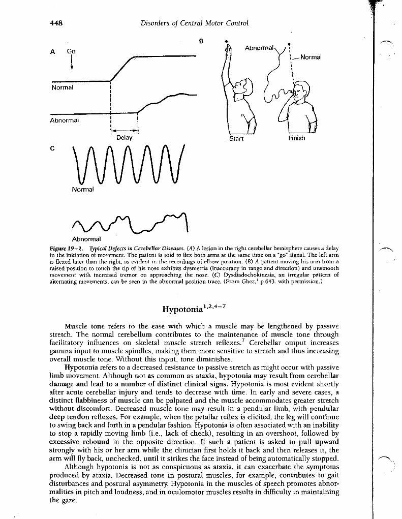
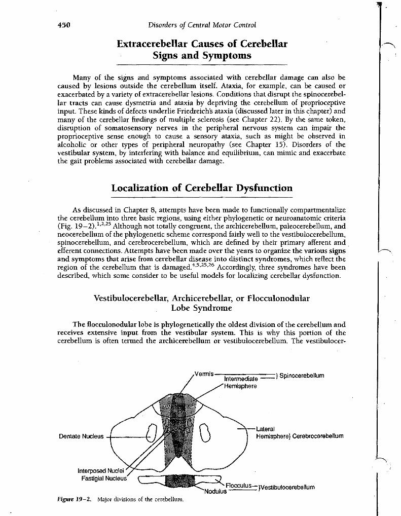
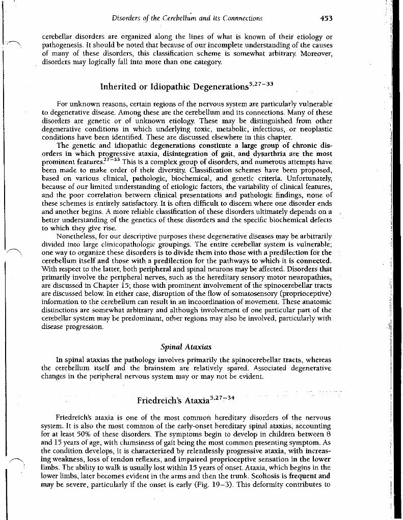
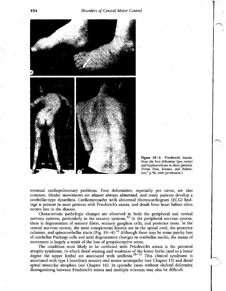
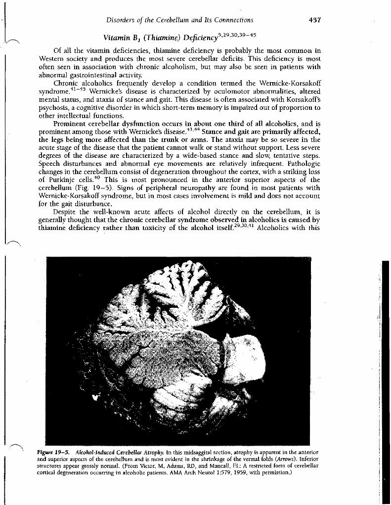
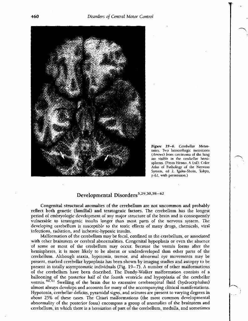
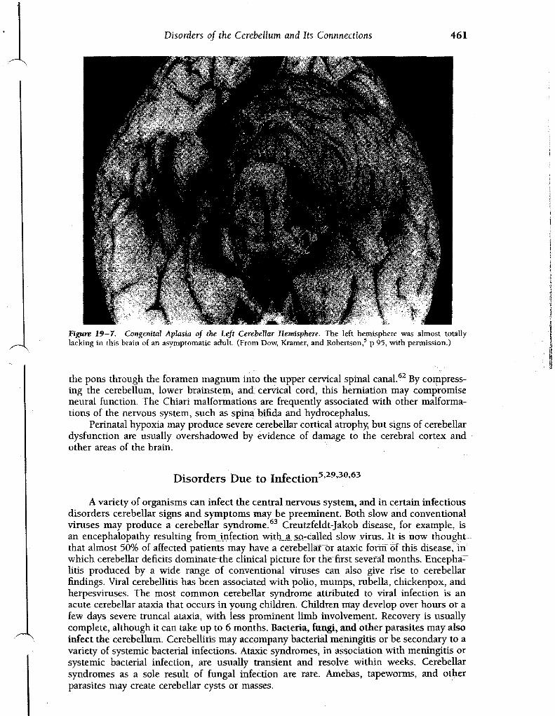
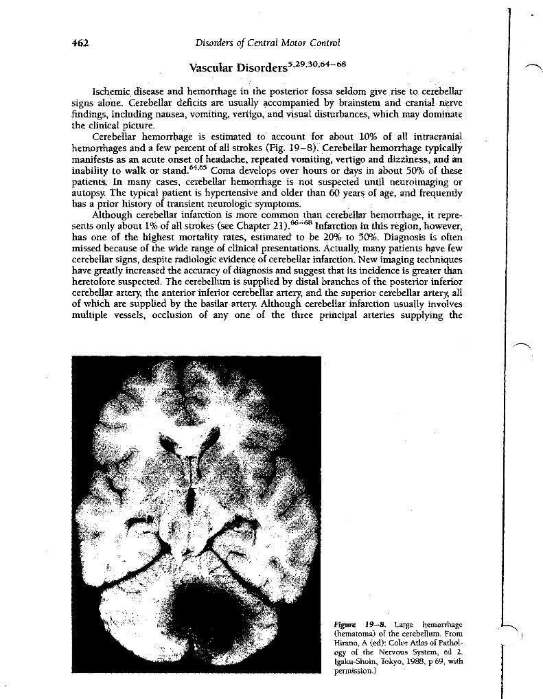