display of a pora peptide from neisseria meningitidis …iai.asm.org/content/65/11/4770.full.pdf ·...
TRANSCRIPT

INFECTION AND IMMUNITY,0019-9567/97/$04.0010
Nov. 1997, p. 4770–4777 Vol. 65, No. 11
Copyright © 1997, American Society for Microbiology
Display of a PorA Peptide from Neisseria meningitidis on theBacteriophage T4 Capsid Surface
JENNIFER JIANG, LARA ABU-SHILBAYEH, AND VENIGALLA B. RAO*
Department of Biology, The Catholic University of America, Washington, D.C. 20064
Received 30 January 1997/Returned for modification 29 May 1997/Accepted 20 August 1997
The exterior of bacteriophage T4 capsid is coated with two outer capsid proteins, Hoc (highly antigenic outercapsid protein; molecular mass, 40 kDa) and Soc (small outer capsid protein; molecular mass, 9 kDa), atsymmetrical positions on the icosahedron (160 copies of Hoc and 960 copies of Soc per capsid particle). Boththese proteins are nonessential for phage infectivity and viability and assemble onto the capsid surface aftercompletion of capsid assembly. We developed a phage display system which allowed in-frame fusions of foreignDNA at a unique cloning site in the 5* end of hoc or soc. A DNA fragment corresponding to the 36-amino-acidPorA peptide from Neisseria meningitidis was cloned into the display vectors to generate fusions at the Nterminus of Hoc or Soc. The PorA-Hoc and PorA-Soc fusion proteins retained the ability to bind to the capsidsurface, and the bound peptide was displayed in an accessible form as shown by its reactivity with specificmonoclonal antibodies in an enzyme-linked immunosorbent assay. By employing T4 genetic strategies, we showthat more than one subtype-specific PorA peptide can be displayed on the capsid surface and that the peptidecan also be displayed on a DNA-free empty capsid. Both the PorA-Hoc and PorA-Soc recombinant phages arehighly immunogenic in mice and elicit strong antipeptide antibody titers even with a weak adjuvant such asAlhydrogel or no adjuvant at all. The data suggest that the phage T4 hoc-soc system is an attractive system fordisplay of peptides on an icosahedral capsid surface and may emerge as a powerful system for construction ofthe next generation multicomponent vaccines.
Since the report by Smith (30), the ability to display a foreignpeptide on the surface of a viral capsid has emerged as apowerful strategy for a variety of biological investigations (21).The filamentous bacteriophages M13 and fd have been exten-sively used to display short random peptides (23) as fusionproducts of the minor capsid protein pIII (4, 22, 29) and, insome cases, as fusion products of the major capsid proteinpVIII (13). The use of phage l for peptide display has alsobeen reported recently. The outer capsid protein gpD (20, 31)and the tail protein gpV (5, 18) were used in these systems. Inall these phage display systems, the major focus has been toconstruct and screen either random short peptide libraries orcDNA expression libraries. Application of these systems forconstruction of recombinant vaccines has been limited (7, 19).Phage T4 offers some unique features that can be exploited fordisplay of peptides on the capsid surface and for their potentialuse as multicomponent vaccines. The phage T4 capsid is com-posed of three essential capsid proteins: the major capsid pro-tein gp23* (960 copies per phage particle) and the two minorcapsid proteins gp24 (vertex protein; 55 copies per particle)and gp20 (portal vertex protein; 12 copies per particle). Inaddition, the outer surface of the capsid is coated with twononessential outer capsid proteins Hoc (molecular mass, 40kDa) and Soc (molecular mass, 9 kDa) (1). These proteins arelocated at symmetrical positions on the icosahedral lattice (1,34). The important characteristics of these proteins, which arehighly suitable for phage display, are that (i) Hoc and Soc aredisplayed at a high copy number, a combined total of about1,120 copies per capsid particle (160 copies of Hoc and 960copies of Soc [8–10]); (ii) these proteins are nonessential forcapsid assembly and are added to the capsid surface after
completion of capsid assembly but prior to DNA packaging;and (iii) elimination by mutation of one or both proteins doesnot affect phage productivity, viability, or infectivity. Appar-ently, these proteins provide additional stability to T4 phageunder adverse conditions such as extreme pH or osmotic shock(3, 9).
The primary focus of this investigation is to address somebasic questions about the development of a phage T4 displaysystem which can then be used in a novel way for constructionof multicomponent recombinant vaccines. Therefore, unlikethe other display systems reported, in this study the followingquestions are analyzed from the context of using phage T4 asa recombinant vaccine. (i) Can foreign sequences be clonedinto hoc and soc and are the fusion products assembled on thecapsid surface? (ii) Would the assembled peptides be displayedin a form that is accessible for biological interactions? (iii) Canthe peptide be displayed on an empty capsid rather than onphage? (iv) Can more than one peptide be brought togetherfrom independent clones and displayed on the same capsidsurface? (v) Are the displayed peptides immunogenic and dothey elicit peptide-specific antibody responses? A 36-amino-acid peptide corresponding to the loop 4 of PorA from Neis-seria meningitidis is used as a model peptide to address thesequestions.
MATERIALS AND METHODS
Bacteria. Escherichia coli B40 (sup11) (14) from our laboratory collection was
used as a suppressor strain for all the phage amber mutants used in this study. E.coli P301 (sup minus) was used as the isogenic nonsuppressor strain. The non-expression strain E. coli BL21 was used for the initial transformation of recom-binant DNA constructs. The expression strains E. coli BL21(DE3) andBL21(DE3)pLYS-S (Novagen) were used for expression of DNA cloned in thepET vectors (32). N. meningitidis 8529 (subtype P1.3) and 99M (subtype P1.2)were kindly provided by W. Zollinger (Department of Bacterial Diseases, WalterReed Army Institute of Research, Washington, D.C.).
Phage. Wild-type T41D and hoc mutant phages were obtained from stocksprepared in this laboratory. The phage mutants 24amNG433, 24amN65, socmutant, and 17tsL2 were kindly provided by Lindsay Black (Department of
* Corresponding author. Mailing address: Department of Biology,103 McCort Ward Hall, The Catholic University of America, 620Michigan Ave. N.E., Washington, D.C. 20064. Phone: (202) 319-5271.Fax: (202) 319-5721. E-mail: [email protected].
4770
on August 4, 2018 by guest
http://iai.asm.org/
Dow
nloaded from

Biological Chemistry, University of Maryland at Baltimore Medical School,Baltimore).
Antibodies. Monoclonal antibodies against PorA subtypes P1.2 and P1.3 of N.meningitidis were kindly provided by W. Zollinger. Affinity-purified goat anti-mouse immunoglobulin G-alkaline phosphatase conjugate (Kirkegaard & Perry)was used as the second antibody in enzyme-linked immunosorbent assays(ELISA) and Western blotting. An affinity-purified anti-mouse immunoglobulinG–gold conjugate (beads, 15-nm diameter; EM Sciences) was used in the im-munogold experiments.
Plasmids. The phage T7 vector pET-9D (Kan) (Novagen), which has an NcoIsite and a BamHI site for cloning downstream from the T7 promoter, was usedfor construction of the Hoc and Soc display vectors.
Recombinant constructions. The primer sequences used for PCR amplifica-tion of T4 or Neisseria DNA fragments are shown below. The positions of primersequences on the T4 genome or porA can be obtained from previously publishedreports (15, 28). All the primers were designed with a 59 tag (indicated in italics)for efficient digestion of the adjacent restriction site (note the presence of eitherthe BamHI site, the NdeI site, or the KpnI site as shown in bold letters adjacentto the 59 tag sequence). The recombinant constructs were first transformed intoE. coli BL21. For expression of the cloned DNA, miniprep DNAs were preparedfrom the BL21 strain and were transformed into the expression strain E. coliBL21(DE3) or E. coli BL21(DE3)pLys-S (32).
Hoc display vector, pR.hoc. The 5-kb DNA between the T4 map units 107 to112 kb, consisting of three open reading frames (ORFs) including hoc, 38 kd, and24, was amplified by PCR with primers 59-CGGGGGATCCAGAGTAGCATGAGCTCCGATG-39 and 59-CGGGGGATCCATCATTCAAGTCATCAGTAG-39 (12, 15). The amplified DNA, after digestion with BamHI was cloned intothe BamHI-linearized and dephosphorylated pET-9D (32). The resulting con-struct and the orientation of the insert were established by digestion with ap-propriate restriction enzymes, marker rescue with 24amNG465, and expressionof Hoc (see Fig. 1).
Soc display vector, pR.soc. The 1.7-kb DNA containing soc (17) was amplifiedby PCR with primers 59-CATGCCATGGTACCTGGTGGTGGAGCTAGTACTCGCGGTTATGTTAAT-39 and 59-CGCGGATCCTTGCCTACTAATGGACCGTCAGGA-39. The former primer included a KpnI site (in bold letters) and asequence corresponding to the polyglycine spacer (underlined) immediately afterthe ATG initiation codon of soc ORF. Digestion of the amplified DNA with NcoIand BamHI gave rise to two fragments, a 0.45-kb fragment and a 1.25-kb frag-ment. The 0.45-kb fragment, which contained the soc ORF, was then cloned intothe dephosphorylated NcoI-BamHI fragment of pET9D.
pR.porA.hoc. pR.porA.hoc was constructed by cloning the PCR-amplified porAfragment into the NdeI site of pR.hoc display vector. PorA fragment was directlyamplified from a single colony of N. meningitidis 8529 (subtype P1.3) or 99M(P1.2) (both strains were kindly provided by W. Zollinger) with primers 59-GGAATTCCATATGTCACATCCGATCCGGGCTTGCC-39 and 59-GGAATTCCATATGCTCCGGCCCAAAACAGCAAGTCC-39 (28). The DNA was cleavedwith NdeI prior to cloning. This construction resulted in the addition of an extraTYA sequence at the N-terminal end of the PorA sequence. For expression ofPorA-Hoc, this construct was transformed into E. coli BL21(DE3)pLys-S.
pR.porA.soc. pR.porA.soc was constructed by cloning the PCR-amplified porAfragment into the KpnI site of the pR.soc display vector. Primers 59-CGGGGTACCTGCCCAAAACAGCAAGTCCGCCT-39 and 59-CGGGGTACCACATCCGATCCGGGCTTGCCGAC-39 were used to amplify the porA fragment from asingle colony of N. meningitidis 8529 (subtype P1.3). This construction resulted inthe addition of an extra V at the N-terminal end of the PorA sequence. Forexpression of PorA-Soc, this construct was transformed into E. coli BL21(DE3).
Immunoreactivity of the displayed peptide. For immunoreactivity analysis (seeTable 1), the recombinant phage carrying the displayed peptide were purified bydifferential centrifugation followed by CsCl step-gradient centrifugation. Theempty capsids were purified by differential centrifugation followed by DEAE-Sephacel column chromatography (25). Fractions 2 to 5 in Table 1 represent theDEAE column fractions of the empty capsid peak as determined by sodiumdodecyl sulfate-polyacrylamide gel electrophoresis (SDS-PAGE). The immuno-reactivity was determined by ELISA (6). Each well was coated with about 109
phage particles. The P1.3 monoclonal antibody was used as the first antibody.Immunogenicity of the displayed peptide. Male BALB/c mice (five per group)
were injected with the CsCl gradient-purified PorA-Hoc or PorA-Soc fusionphages (5 to 10 mg per injection) at 2 (experiment 2)- or 3 (experiment 1)-weekintervals (see Table 2). The fusion phages were generated by the second strategydescribed in Results. The T4.porA(P1.3).Soc phage preparation used in experi-ment 2 had roughly 10-fold-lower copy number of PorA-Soc compared to that inexperiment 1. Each injection (100 ml) contained equal volumes of phage (in 50mM Tris-Cl [pH 7.4]) and phosphate-buffered saline (no adjuvant), or completeFreund’s adjuvant (CFA) or Alhydrogel as an adjuvant. All the samples exceptthe ones with CFA were injected intramuscularly. The CFA samples were in-jected subcutaneously. Mice were bled through the tail vein on day 0 (preimmuneserum) and at 2- or 3-week intervals (immune serum). No booster injections weregiven. Only the data for the sera obtained on day 42 after the primary immuni-zation are shown. The peptide-specific antibody titers were determined byELISA (6) by using 1 mg of loop 4 synthetic peptide per well as the coatingantigen. The amino acid sequence of the synthetic peptide is identical to that of
the displayed peptide except that it has an additional GC at the N terminus anda GCC at the C terminus.
Standard techniques. Conditions for PCR amplification of porA fragmentfrom Neisseria or the hoc and soc DNA fragments from phage T4 have beendescribed previously (28, 35). Restriction enzyme digestions, dephosphoryla-tions, ligation reactions, and other recombinant DNA modifications were carriedout according to the reaction conditions recommended by the manufacturer.Transformations were done by electroporation with a Bio-Rad electroporator.Cloned DNA under the control of the phage T7 promoter was expressed by theaddition of isopropyl-b-D-thiogalactopyranoside (IPTG) (0.4 mM) to the log-phase cultures at 37°C and shaking of the cultures for the indicated time periods(32). SDS-PAGE was performed according to the method described by Laemmli(16). Western blotting was performed according to the method of Towbin et al.(33). ELISAs were done in triplicate according to the basic procedure describedby Engvall and Perlman (6). Immunogold electron microscopy (EM) was per-formed by the procedure described by Dunn (5).
RESULTS
Vector system. Two display vectors, a Hoc display vector anda Soc display vector, both of which allow in-frame fusion offoreign DNA to the 59 end of either hoc or soc, were con-structed (Fig. 1). The phage T7 vector system (32) was chosenfor insertion of foreign DNA because it allowed the confirma-tion of the expression of the right fusion protein prior to thephage display experiments (Fig. 2 and 3).
The Hoc and Soc display vectors were designed to test twoalternative strategies for expression and display of the fusionprotein. With the Hoc display vector, the fusion would be firsttransferred into the T4 genome, expressed under the control ofT4 promoter(s), and assembled onto the capsid during normalphage development. With the Soc-display vector, the fusionwould be first expressed in E. coli from the T7 promoter andlater assembled onto a soc mutant capsid during phage infec-tion.
Target peptide. We chose a 108-bp DNA fragment corre-sponding to the 36 amino acids (amino acids 163 to 198 of themature protein) of the class I porin, PorA, from N. meningitidis(subtype P1.3, which has been responsible for an epidemic ofmeningitis in Northern Chile [36]) as a target sequence forinsertion into Hoc and Soc. The reasons for this choice are asfollows. (i) Based on the recently developed structural modelfor PorA which traverses the outer membrane sixteen times,this sequence was predicted to correspond to loop 4, a loopthat is most extended into the external environment (28); (ii)there is strong evidence that this loop constitutes one of theprimary epitopes recognized by the human immune systemsince the bactericidal antibodies elicited in immune individualswere mapped to this epitope; and (iii) a battery of monoclonalantibodies specific to loop 4 from a number of subtypes of N.meningitidis are readily available (36).
Hoc display vector. The details of Hoc display vector(pR.hoc) construction are described in Materials and Methods.It includes, in addition to the hoc coding sequence, a 884-bpflanking sequence from the 59 end of hoc and a 3-kb flankingsequence from the 39 end of hoc. The 39 flanking sequence alsocontained the essential T4 gene 24. These flanking sequencesare included in the vector to facilitate the transfer of thehoc-porA fusion into T4 genome by homologous recombina-tion (see below).
Expression of Hoc from pR.hoc was tested by IPTG induc-tion and SDS-PAGE (Fig. 2A). A 40-kDa protein, the ex-pected size of Hoc, was expressed to a level of about 10% ofthe total cell protein (compare uninduced lane 1 with inducedlanes 2 and 3). This clone also expressed a 38-kDa protein(lanes 2 and 3); this is consistent with the reported presence ofan ORF of unknown function downstream from hoc (15).However, gp 24 was not expressed by this clone since the g24
VOL. 65, 1997 PEPTIDE DISPLAY ON BACTERIOPHAGE T4 CAPSID 4771
on August 4, 2018 by guest
http://iai.asm.org/
Dow
nloaded from

ORF is in the opposite orientation relative to the direction oftranscription from the T7 promoter (Fig. 1).
Construction of PorA-Hoc fusion phage. The 108-bp frag-ment corresponding to the loop 4 of PorA was amplified froma single colony of N. meningitidis and was cloned into theunique NdeI site of pR.hoc. The porA primers were designed insuch a way that insertion in the right orientation would fuse the36-amino-acid PorA peptide after the 36th amino acid of Hoc.Consistent with this, a clone containing the recombinant plas-mid (pR.porA.hoc) produced a new 45-kDa protein (with the
corresponding disappearance of the 40-kDa Hoc protein)upon induction with IPTG (Fig. 2B, compare lanes 4 and 5with lanes 2 and 3). That this protein was indeed the PorA-Hocfusion protein was further established by its strong reactivitywith a loop 4-specific monoclonal antibody by Western blottingfollowed by immunostaining (Fig. 2C, lane 7; lane 6 representsa control induced extract containing no insert) (this monoclo-nal antibody was previously mapped to a “linear” epitopeNGANNTI within the loop 4 sequence by Geysen pin analysis[28]).
FIG. 1. Schematic diagrams of display vectors pR.hoc, the Hoc display vector, and pR.soc, the Soc display vector. See Materials and Methods for details on theconstruction of these vectors. Only the relevant features of the vectors are shown. The arrows in parenthesis indicate the direction of transcription from the T7promoter. The plasmids are not drawn to scale.
FIG. 2. Expression of PorA-Hoc fusion protein and its display on T4 phage. (A to C) Expression of Hoc and PorA-Hoc in E. coli BL21(DE3)pLys-S as analyzedby SDS–10% PAGE. Lanes: 1, uninduced pR.hoc; 2, pR.hoc induced for 30 min; 3, pR.hoc induced for 60 min; 4, pR.porA.hoc induced for 30 min; 5, pR.porA.hocinduced for 60 min; 6 and 7, Western blot of pR.hoc induced for 60 min and pR.porA.hoc induced for 60 min respectively, followed by immunostaining with theP1.3-specific monoclonal antibody. (D) Expression of PorA-Hoc in phage T4. Plates: 8 and 9, T4.porA(P1.3).hoc plaques; 10 and 11, wild-type T4 plaques; 8 and 10,plaques grown on Luria-Bertani plates; 9 and 11, the same plaques from plates 8 and 10 after transfer to a nitrocellulose membrane followed by immunostaining withthe P1.3 monoclonal antibody. (E) Display of PorA-Hoc on phage T4. The phage preparations were purified by differential centrifugation followed by CsCl step gradientcentrifugation. Lanes: 12, hoc mutant phage; 13, wild-type phage; 14, T4.porA(P1.3).hoc phage; 15, T4.porA(P1.2).hoc phage. Hoc and PorA-Hoc band positions aremarked on the sides of the panels with thin and thick arrows (or lines), respectively.
4772 JIANG ET AL. INFECT. IMMUN.
on August 4, 2018 by guest
http://iai.asm.org/
Dow
nloaded from

The porA-hoc fusion was then transferred to phage T4 ge-nome by infecting E. coli containing the pR.porA.hoc plasmidwith 24amNG465 and selecting for wild-type recombinants onsup-minus E. coli P301. Since the wild-type phage would begenerated by a recombinational exchange near the gene 24sequence in pR.porA.hoc, a fraction of these recombinantswould also have exchanged the adjacent porA-hoc fusion se-quence. Direct PCR of random plaques showed that about30% of the plaques had recovered the porA-hoc fusion. TheporA-hoc fusion, although originally under the control of T7promoter, is now in the T4 genome and is controlled by thenative hoc promoter. One of the PorA-Hoc fusion plaques[T4.porA(P1.3).hoc] was purified, and its ability to express thePorA peptide was tested by immunostaining of the plaqueswith the monoclonal antibody. The data, as shown in Fig. 2D,showed that virtually all the plaques in the fusion phage stockreacted strongly with the monoclonal antibody (compare theimmunostained plaques in panel D9 with the unstainedplaques in panel D8). On the other hand, none of the controlwild-type plaques showed any reactivity (compare the immu-nostained filter in panel D11 with the unstained plaques inpanel D10).
Display of PorA peptide on the capsid surface. To testwhether the PorA-Hoc fusion protein is coated on the capsidsurface, the T4.porA(P1.3).hoc phage particles were purified byCsCl density gradient centrifugation. If the PorA peptide isdisplayed on the capsid surface in an accessible form, it shouldinteract with the monoclonal antibody. If so, phage particlescoated on the microtiter plates should react with the mono-clonal antibody in an ELISA. In a number of assays, theT4.porA(P1.3).hoc phage indeed showed strong reactivity withthe monoclonal antibody (Table 1), whereas no reactivity wasobserved with the control wild-type phage (Table 1). In an-other control, the fusion phage did not show any cross-reac-tivity with a monoclonal antibody that is specific to a differentPorA subtype P1.2 (data not shown).
The PorA-Hoc fusion protein displayed on the phage parti-cles appears to be about 6 kDa smaller than the expected45-kDa size (Fig. 2E, compare lanes 14 and 15 with lane 13).The same band appeared in both the T4.porA(P1.3).hoc andT4.porA(P1.2).hoc (see below) recombinant phages. However,the identity of this band as the PorA-Hoc is yet to be estab-lished. If it is indeed PorA-Hoc, it most likely represents acleaved form of the fusion protein. But the fact that the phageparticles showed strong immunoreactivity with the subtype-specific monoclonal antibody suggested that the displayed Hocfusion protein (or a portion of it) retained the P1.3 epitope. Itshould however be noted that the full-length PorA-Hoc wasdisplayed on the capsid by an alternative strategy in which thefusion protein was first expressed in E. coli and was thentransferred to hoc mutant capsids (data not shown; see belowfor details of this strategy).
Multicomponent phage display. An attractive feature of theT4 system is that the high copy number of Hoc and Soc wouldallow the transfer and display of more than one fusion proteinfrom independent constructions onto the same capsid by amixed infection strategy. The same can also be done to ma-nipulate the copy number of the displayed peptide by usingwild-type phage in the mixed infection. To test this, a secondPorA fusion phage [T4.porA(P1.2).hoc] displaying the loop 4peptide from subtype P1.2 (36) was constructed by using thescheme described above. Display of the P1.2 peptide on thecapsid surface was first confirmed by the reactivity of CsClgradient-purified T4.porA(P1.2).hoc phage particles with theP1.2 monoclonal antibody (Table 1) but not with the P1.3-specific monoclonal antibody (data not shown). To test the
mixed infection strategy, E. coli P301 was infected with a mix-ture of both T4.porA(P1.3).hoc and T4.porA(P1.2).hoc phagesat a multiplicity of 2.5 each, and the progeny phage werepurified by CsCl gradient centrifugation. The purified prepa-ration was then tested for the presence of both epitopes withspecific monoclonal antibodies. The data showed that theprogeny phage reacted with both monoclonal antibodies (Ta-ble 1).
Display on empty capsids. A unique feature of the T4 systemis that both Hoc and Soc assemble onto the capsid only aftercapsid expansion but prior to DNA packaging. In fact, phageT4 is the only double-stranded DNA phage which producesexpanded capsids in vivo (with assembled Hoc and Soc) whenDNA packaging is arrested (2, 25). For the use of T4 recom-binants as vaccines, it may be desirable to display epitopes onan empty capsid rather than on finished phage since the emptycapsids will be devoid of DNA and numerous tail proteins. Theabsence of DNA would be highly desirable from the perspec-tive of biosafety. The absence of tail proteins, on the otherhand, may be either desirable if the tail components interferein a negative way with the immune responses elicited towardsthe displayed epitope(s) or undesirable if the tail proteins offerfavorable adjuvant effects.
To test whether the peptide can be displayed on the emptycapsid, a packaging defective 17tsL2 mutant was first crossedinto T4.porA(P1.3).hoc followed by selection of a PorA-ex-pressing 17ts plaque [T4.17ts.porA(P1.3).hoc] by immuno-screening with the monoclonal antibody. Capsids were purifiedfrom cells infected with the T4.17ts.porA(P1.3)hoc phage atnonpermissive temperature by differential centrifugation andDEAE-Sephacel chromatography (25, 26). EM and SDS-PAGE showed that the purified capsids were empty and ex-panded and that the expected five major capsid proteins con-stituted the empty capsid (data not shown). ELISA of the peakcapsid fractions showed a corresponding peak of immunore-activity with the P1.3 monoclonal antibody (Table 1).
TABLE 1. Immunoreactivity of the displayed PorA peptide withthe subtype-specific monoclonal antibodiesa
T4 recombinant phageSubtype-specific
monoclonal antibodyused
A495
T4.porA(P1.3).hoc P1.3 0.79
T4.porA(P1.2).hoc P1.2 0.51
T4.porA(P1.3, P1.2).hoc P1.2 0.25
T4.porA(P1.3, P1.2).hoc P1.3 0.70
T4.17ts.porA(P1.3).hoc empty capsidsFraction 2 P1.3 0.03Fraction 3 P1.3 0.79Fraction 4 P1.3 0.86Fraction 5 P1.3 0.69
T4.porA(P1.3).Soc P1.3 0.91
a Immunoreactivity of the displayed PorA peptide was determined by ELISAwith the purified recombinant phage as the coating antigen. The data werederived from a number of independent experiments, each of which was per-formed with a positive control (outer membrane capsule displaying the nativePorA) and a negative control (wild-type phage T4). The negative control showedno reactivity with the monoclonal antibodies (the background absorbance was inthe range of 0.02 to 0.05). The values (except the DEAE-fractions) representmeans for triplicate assays. See Materials and Methods for more details.
VOL. 65, 1997 PEPTIDE DISPLAY ON BACTERIOPHAGE T4 CAPSID 4773
on August 4, 2018 by guest
http://iai.asm.org/
Dow
nloaded from
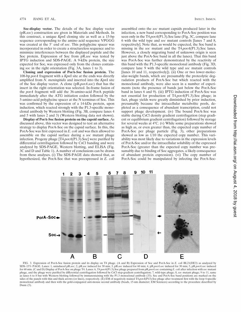
Soc-display vector. The details of the Soc display vector(pR.soc) construction are given in Materials and Methods. Inthis construct, a unique KpnI cloning site as well as a 15-bpsequence corresponding to the amino acid sequence VPGGGwas created at the 59 end of soc. This polyglycine spacer wasincorporated in order to create a structureless sequence and tominimize interferences between the displayed peptide and theSoc protein. Expression of Soc from pR.soc was tested byIPTG induction and SDS-PAGE. A 9-kDa protein, the sizeexpected for Soc, was expressed only from the clones contain-ing soc in the right orientation (Fig. 3A, lanes 1 to 3).
Cloning and expression of PorA-Soc fusion protein. The108-bp porA fragment with a KpnI site at the ends was directlyamplified from N. meningitidis and inserted into the KpnI siteof the Soc display vector. A clone (pR.porA.soc) that has theinsert in the right orientation was selected. In-frame fusion ofthe porA fragment will add the 36-amino-acid PorA peptideimmediately after the ATG initiation codon followed by the5-amino-acid polyglycine spacer at the N terminus of Soc. Thiswas confirmed by the expression of a 14-kDa protein, uponinduction, which reacted strongly with the P1.3-specific mono-clonal antibody by Western blotting (Fig. 3B, compare lanes 4and 5 with lanes 2 and 3) (Western blotting data not shown).
Display of PorA-Soc fusion protein on the capsid surface. Asdiscussed above, this vector was designed to test an alternativestrategy to display PorA-Soc on the capsid surface. In this, thePorA-Soc was first expressed in E. coli and was then allowed toassemble on the capsid surface during a soc mutant phageinfection. Progeny phage [T4.porA(P1.3).Soc] were purified bydifferential centrifugation followed by CsCl banding and wereanalyzed by SDS-PAGE, Western blotting, and ELISA (Fig.3C and D and Table 1). A number of conclusions can be drawnfrom these analyses. (i) The SDS-PAGE data showed that, ashypothesized, the PorA-Soc that was preexpressed in E. coli
assembled onto the soc mutant capsids produced later in theinfection, a new band corresponding to PorA-Soc position wasseen only in the T4.porA(P1.3).Soc lane (Fig. 3C, compare lane6 with the wild type and soc mutant controls [lanes 7 and 8,respectively]. Note that, as would be expected, the Soc band ismissing in the soc mutant and the T4.porA(P1.3).Soc lanes.However, a closely migrating band of unknown origin is seenjust above the PorA-Soc band in all the lanes). That this bandwas PorA-Soc was further demonstrated by the reactivity ofthis band with the P1.3-specific monoclonal antibody (Fig. 3D,compare lane 9 with the wild type and soc mutant controls[lanes 10 and 11, respectively]). (ii) One or two lower-molec-ular-weight bands, which are presumably the proteolytic deg-radation products of PorA-Soc but which reacted with themonoclonal antibody, were also seen in a number of experi-ments (note the presence of bands just below the PorA-Socband in lanes 6 and 9). (iii) IPTG induction of PorA-Soc wasnot essential for production of T4.porA(P1.3).Soc phage; infact, phage yields were greatly diminished by prior induction,presumably because the intracellular metabolite pools, de-pleted as a consequence of abundant transcription, could notsupport phage development. (iv) The bound PorA-Soc wasstable during CsCl density gradient centrifugation (step gradi-ent or equilibrium gradient centrifugation) followed by storagefor several weeks at 4°C. (v) While some preparations showedas high as, or even greater than, the expected copy number ofPorA-Soc per phage particle (Fig. 3), other preparationsshowed as low as 1/10 the expected copy number. This vari-ability was most likely due to variations in the expression levelsof PorA-Soc and/or the intracellular solubility of the expressedPorA-Soc (greater than the expected copy number was pre-sumably due to binding of Soc aggregates, a likely consequenceof abundant protein expression). (vi) The copy number ofPorA-Soc could be manipulated by infecting the PorA-Soc-
FIG. 3. Expression of PorA-Soc fusion protein and its display on T4 phage. (A and B) Expression of Soc and PorA-Soc in E. coli BL21(DE3) as analyzed bySDS–15% PAGE. Lanes: 1, uninduced pR.soc; 2, pR.soc induced for 30 min; 3, pR.soc induced for 60 min; 4, pR.porA.soc induced for 30 min; 5, pR.porA.soc inducedfor 60 min. (C and D) Display of PorA-Soc on phage T4. Lanes: 6, T4.porA(P1.3).Soc phage prepared from pR.porA.soc containing E. coli after infection with soc mutantphage, and the phage were purified by differential centrifugation followed by CsCl step-gradient centrifugation; 7, wild-type phage; 8, soc mutant phage; 9 to 11, sameas lanes 6 to 8 but with Western blotting followed by immunostaining with the P1.3 monoclonal antibody (33). Soc and PorA-Soc band positions are marked on thesides of the panels with thin and thick arrows (or lines), respectively. (E) EM of negatively stained T4.porA(P13).Soc phage after treatment first with the loop 4-specificmonoclonal antibody and then with the gold-conjugated anti-mouse second antibody (beads, 15-nm diameter; EM Sciences) according to the procedure described byDunn (5).
4774 JIANG ET AL. INFECT. IMMUN.
on August 4, 2018 by guest
http://iai.asm.org/
Dow
nloaded from
expressing cells with both the wild-type and soc mutant phages.Since in these cells, both Soc and PorA-Soc are expressed (Socfrom wild-type infection and PorA-Soc from the pR.porA.socplasmid), both forms are expected to occupy the numeroussites available on the capsid surface. Indeed, SDS-PAGE anal-ysis of the CsCl-purified phage from such a mixed infectionshowed the presence of both Soc and PorA-Soc bands (datanot shown). (vii) ELISA analyses suggested that the PorApeptide is displayed in an accessible form since a number ofindependent CsCl gradient-purified phage preparations re-acted strongly with the monoclonal antibody (Table 1). Immu-nogold-EM further showed cross-linking of phage particles inthe presence of the monoclonal antibody and specific associa-tion of gold particles with the capsids but not with the tails(Fig. 3E).
Immunogenicity of the displayed peptide. The immunoge-nicity of the displayed peptide was tested by injecting purifiedPorA-Hoc or PorA-Soc fusion phage particles into mice eitherwith no adjuvant or with CFA or Alhydrogel as an adjuvant.Induction of peptide-specific antibodies was analyzed byELISA with the synthetic PorA 1.3 peptide as the coatingantigen on microtiter plates. The data showed that high titersof peptide-specific antibodies were elicited with the recombi-nant fusion phages (Table 2), whereas no peptide-specific an-tibodies were detected either in the preimmune sera or in thesera of mice injected with the control soc mutant phage. Theantibody titers were lower in the second experiment apparentlybecause of the lower copy number of the PorA-Soc phage usedfor immunization (see Materials and Methods). It is particu-larly significant that Alhydrogel, a weak adjuvant approved forhuman use, also elicited high antibody titers. Furthermore, thePorA-Soc phage without any accompanying adjuvant also elic-ited comparable antibody titers. Finally, the PorA-Hoc fusionphage, which presumably have a lower copy number of thedisplayed peptide than the PorA-Soc phage, also elicited highantibody titers. This is consistent with the fact that Hoc isknown to be a highly immunogenic component of the T4 cap-sid surface (9).
DISCUSSION
Icosahedral phages that assemble in the cytoplasmic milieuwill have distinct advantages over the filamentous phages for
display of peptides (21). Most importantly, these phages, un-like the filamentous phages, will be able to display peptideswith various sizes and sequences in a much less restricted way.In phage T4, genetic and structural studies by Yanagida andcoworkers (8, 9, 34) established that a combined total of 1,120copies of Hoc and Soc are coated on the icosahedral capsidsurface. Unlike the proteins used in the phage M13 displaysystem, phage T4 Hoc and Soc are nonessential for phageinfectivity and productivity and are added onto the capsidsurface after the completion of capsid assembly. We reasoned,and showed in this study, that these distinctive features allowedthe display of peptides not only on finished phage but also onintermediate empty capsids.
This study answered a number of basic questions. First, thedata showed that fusions to the N terminus of either Hoc orSoc were tolerated and did not appear to disrupt capsid bind-ing functions. Additional constructs are being made by incor-porating features, such as the insertion of a longer spacer withmultiple cloning sites and the addition of flanking cysteines tofacilitate disulfide bridge formation, in order to display pep-tides in different structural contexts. Also, while this study wasbeing prepared for publication, we learned that fusions to theC terminus of Soc were also tolerated and displayed on thecapsid surface (27), suggesting that both termini of Soc can beused for peptide display.
Two basic modes of expression and display of the 36-amino-acid PorA peptide were tested. In one case, the fusion wastransferred to the T4 genome by recombination, and the fusionprotein was expressed in a native context during phage infec-tion. In the second case, the fusions were first expressed in E.coli and were then transferred to either soc mutant or hocmutant phage produced during infection. By the two criteriaused, (i) cosedimentation of the fusion protein with the phageparticles during CsCl gradient centrifugation and (ii) immuno-reactivity of purified phage particles with specific monoclonalantibodies, both strategies allowed stable display of the peptidein an accessible form. In fact, phage recovered after two CsClgradient centrifugations, or after storage for several weeks at4°C, did not show a significant loss of the fusion protein fromthe capsid surface.
Limited proteolysis was observed with both strategies. Pro-teolysis is a common problem in phage display systems sincethe fusion protein is abundantly expressed and it “sticks out”
TABLE 2. Induction of PorA-specific antibodies in mice upon immunization with PorA-Hoc or PorA-Soc fusion phagesa
Recombinant phage used forimmunization
A405 at the dilution indicated
Expt 1 Expt 2
Preimmune(1:50)
Immune(1:1,000)
Preimmune(1:50)
Immune(1:100)
Control soc mutant phage with CFA ND ,0 ,0
T4.porA(P1.3).Soc withNo adjuvant ND ,0 0.58 6 0.47Alhydrogel 0.02 0.59 6 0.30 ,0 0.69 6 0.52CFA 0.01 1.85 6 0.12 ,0 0.61 6 0.40
T4.porA(P1.3).hoc withAlhydrogel 0.01 0.39 6 0.13 NDCFA 0.02 1.34 6 0.33 ND
a Immunogenicity of the displayed PorA peptide was determined by injecting BALB/c mice in groups of five with the purified recombinant phage. The peptide-specificantibody titers were determined by using a synthetic PorA peptide as the coating antigen. Each serum was serially diluted with the cold blocking buffer, and appropriatedilutions were titrated in triplicate assays. The absorbance data represent the values after subtraction of the blank in which the first antibody was omitted from theELISA procedure. Each value represents an average for five serum samples from mice belonging to that group 6 standard deviation. No data were excluded incalculation of the values shown. See Materials and Methods for more details. ND, not done.
VOL. 65, 1997 PEPTIDE DISPLAY ON BACTERIOPHAGE T4 CAPSID 4775
on August 4, 2018 by guest
http://iai.asm.org/
Dow
nloaded from
on the capsid surface, making it an easy target for proteolysis.However, it should be noted that unlike the phage systemsdeveloped mainly for display of peptide libraries (21), limitedproteolysis will be more tolerated for vaccine developmentbecause the main objective here is to allow the host system toelicit antibodies against all the possible epitopes of the dis-played peptide. In the case of Hoc, it appears that the fusionprotein synthesized during the T4 infection is susceptible toproteolysis whereas the fully assembled Hoc (as in the case ofthe overexpressed protein) is not. Additional vectors are beingconstructed to clarify this phenomenon and possibly overcomeit. In the case of Soc, proteolysis was very limited and was nota significant issue. There is previous evidence suggesting that aportion of Soc is cleaved at the C terminus (even when ex-pressed from T4) to a form that is 2 kDa smaller (27). Ourresults are consistent with this finding since the truncatedPorA-Soc is immunoreactive with the peptide-specific mono-clonal antibody in the Western blotting experiments (Fig. 3D).This would also imply that the N-terminal fusion strategy forSoc that has been developed in this study is more desirablethan the C-terminal fusion strategy (27).
We have shown that T4 genetics imparts enormous flexibilityto the manipulation of this display system. For instance, twoPorA subtype peptides, the P1.2 and P1.3 peptides, can bebrought together presumably onto the same capsid surface bya mixed infection with both the P1.3 and P1.2 fusion phages.This strategy also allowed manipulation of the copy number ofPorA-Soc by a mixed infection with both the PorA-Soc andwild-type phages. While Maruyama et al. (18) described aconditional chain termination strategy to alter the copy num-ber of the fusion protein, the mixed infection strategy de-scribed here offers a simpler yet more flexible way to alter thecopy number or to display multiple peptides on the same cap-sid surface. We have also shown that the PorA peptide can bedisplayed on empty capsids rather than on finished phage par-ticles. Extension of this strategy should allow the display ofpeptides on either the giant empty capsids (26) (under thegenetic background of 23ptg mutations) or the abortive poly-heads (27) (under the genetic background of 20am mutations).
Immunogenicity experiments with mice established that thedisplayed PorA is immunogenic. This is evident by the elicita-tion of peptide-specific antibodies upon injection of highlypurified T4.porA.Soc and T4.porA.hoc phage particles. On theother hand, in earlier experiments, no detectable antibodieswere elicited upon injection of mice with the synthetic peptideeither alone or with Alhydrogel as an adjuvant (35a). Moreinterestingly, the results seem to suggest that the phage T4particle may have a favorable adjuvant effect on the immuno-genicity of displayed PorA-Soc. For example, we found that thePorA-Soc phage was immunogenic in the presence of a weakadjuvant such as Alum or no adjuvant at all as it was in thepresence of a strong adjuvant such as CFA. These responseswere not unique to PorA-Soc since similar high antibody re-sponses were also obtained against the other phage structuralproteins (data not shown). Similar observations were also re-ported recently with the potyvirus (a plant virus) display systemin which chimeric capsids carrying Plasmodium epitopes werefound to be highly immunogenic in the absence of an adjuvant(11).
In conclusion, we have shown that the loop 4 PorA peptidefrom N. meningitidis, a peptide that is known to have signifi-cance in vaccine development, can be displayed on the phageT4 capsid surface and that the displayed peptide is immuno-genic in mice. We believe that the distinctive features of the T4hoc-soc system would, upon further development, make it anattractive general system for construction of multicomponent
vaccines against infectious diseases. In cases where the vaccinetarget is known, the T4 system can be used to manipulate thenumber of peptides displayed on the capsid surface as well asthe sequence, size, structural context, and copy number, foroptimal elicitation of neutralizing antibody responses. Such aflexible customization of multicomponent vaccines has not yetbeen realized with any currently available systems, includingthe multiple antigenic peptides system (24) or other phagedisplay systems.
ACKNOWLEDGMENTS
We are grateful to Wendel Zollinger and Nancy Saunders from theDepartment of Bacterial Diseases, Walter Reed Army Institute ofResearch, Washington, D.C., for N. meningitidis strains, subtype-spe-cific monoclonal antibodies, a number of other reagents, many usefuldiscussions throughout the investigation, and for critically reading themanuscript. We thank Lindsay Black from the University of MarylandMedical School, Baltimore, for phage mutants and anti-Hoc antibodiesand Alasdair Steven from National Institutes of Health for the EMfacilities.
A grant-in-aid award by The Catholic University of America isgratefully acknowledged.
REFERENCES
1. Black, L. W., M. Showe, and A. C. Steven. 1994. Morphogenesis of the T4head, p. 218–253. In J. D. Karam (ed.), Bacteriophage T4. ASM Press,Washington, D.C.
2. Carrascosa, J. L. 1978. Head maturation pathway of bacteriophage T4 andT2. IV. In vitro transformation of T4 head-related particles produced bymutants in gene 17 to capsid-like structures. J. Virol. 26:420–428.
3. Childs, J. D. 1980. Effect of Hoc protein on electrophoretic mobility of intactbacteriophage T4D particles in polyacrylamide gel electrophoresis. J. Mol.Biol. 141:163–173.
4. Devlin, J. J., L. C. Panganiban, and P. E. Devlin. 1990. Random peptidelibraries: a source of specific protein binding molecules. Science 249:404–406.
5. Dunn, I. S. 1995. Assembly of functional bacteriophage lambda virionsincorporating C-terminal peptide or protein fusions with the major tailprotein. J. Mol. Biol. 248:497–506.
6. Engvall, E., and T. Perlman. 1971. Enzyme linked immunosorbent assay(ELISA): quantitation assay of immunoglobulin G. Immunochemistry 8:871–879.
7. Greenwood, J., A. E. Willis, and R. N. Perham. 1991. Multiple display offoreign peptides on a filamentous bacteriophage. Peptides from Plasmodiumfalciparum circumsporozoite protein as antigens. J. Mol. Biol. 220:821–827.
8. Ishii, T., and M. Yanagida. 1975. Molecular organization of the shell of theTeven bacteriophage head. J. Mol. Biol. 97:655–660.
9. Ishii, T., and M. Yanagida. 1977. The two dispensable structural proteins(Soc and Hoc) of the T4 phage capsid; their purification and properties,isolation and characterization of the defective mutants, and their bindingwith the defective heads in vitro. J. Mol. Biol. 109:487–514.
10. Ishii, T., Y. Yamaguchi, and M. Yanagida. 1978. Binding of the structuralprotein Soc to the head shell of bacteriophage T4. J. Mol. Biol. 120:533–544.
11. Jagdish, M. N., S. J. Edwards, M. B. Hayden, J. Grusovin, K. Vandenberg,P. Schoofs, R. C. Hamilton, D. D. Shukla, H. Kalnins, M. McNamara, J.Haynes, I. T. Nisbet, C. W. Ward, and D. Pye. 1996. Chimeric potyvirus-likeparticles as vaccine carriers. Intervirology 39:85–92.
12. Kaliman, A. V., M. A. Khasanova, V. M. Kryukov, V. I. Tanyashin, and A.Bayev. 1990. The nucleotide sequence of the region of bacteriophage T4inh(lip)-hoc gene. Nucleic Acid Res. 18:4277.
13. Kang, A. K., C. F. Barbas, K. D. Janda, S. J. Benkovic, and R. A. Lerner.1991. Linkage of recognition and replication functions by assembling com-binational antibody Fab libraries along phage surfaces. Proc. Natl. Acad. Sci.USA 88:4363–4366.
14. Kleina, L. G., J.-M. Masson, J. Normanly, J. Abelson, and J. H. Miller. 1990.Construction of Escherichia coli amber suppressor tRNA genes. II. Synthesisof additional tRNA genes and improvement of suppressor efficiency. J. Mol.Biol. 213:705–717.
15. Kutter, E., T. Stidham, B. Guttman, E. Kutter, D. Batts, S. Peterson, T.Djavakhishvili, F. Arisaka, V. Mesyanzhinov, W. Ruger, and G. Mosig. 1994.Genomic map of bacteriophage T4, p. 491–519. In J. D. Karam (ed.), Bac-teriophage T4. ASM Press, Washington, D.C.
16. Laemmli, U. K. 1970. Cleavage of structural proteins during the assembly ofthe head of bacteriophage T4. Nature 227:680–685.
17. Macdonald, P. M., E. Kutter, and G. Mosig. 1984. Regulation of a bacte-riophage late gene, soc, which maps in an early region. Genetics 106:17–27.
18. Maruyama, I. N., H. Maruyama, and S. Brenner. 1994. Lambda foo: a
4776 JIANG ET AL. INFECT. IMMUN.
on August 4, 2018 by guest
http://iai.asm.org/
Dow
nloaded from

lambda phage vector for the expression of foreign proteins. Proc. Natl. Acad.Sci. USA 91:8273–8277.
19. Meola, A., P. Delmastro, P. Monaci., A. Luzzago, A. Nicosia, F. Felici, R.Cortese, and G. Galfre. 1995. Derivation of vaccines from mimitopes. Im-munologic properties of human hepatitis B virus surface antigen mimitopesdisplayed on filamentous phage. J. Immunol. 154:3162–3172.
20. Mikawa, Y. G., I. N. Maruyama, and S. Brenner. 1996. Surface display ofproteins on bacteriophage lambda heads. J. Mol. Biol. 262:21–30.
21. O’Neil, K. T., and R. H. Hoess. 1995. Phage display: protein engineering bydirected evolution. Curr. Opin. Struct. Biol. 5:443–449.
22. Parmley, S. F., and G. P. Smith. 1988. Antibody-selectable filamentous fdphage vectors: affinity purification of target genes. Gene 73:305–318.
23. Perham, R. N., T. D. Terry, A. E. Willis, J. Greenwood, F. Marzo Versonese,and E. Eppella. 1995. Engineering a peptide epitope display system onfilamentous bacteriophage. FEMS Microbiol. Rev. 17:25–31.
24. Posnett, D. N., H. McGratte, and J. P. Tam. 1988. A novel method forproducing anti-peptide antibodies: production of site-specific antibodies tothe T cell antigen receptor beta chain. J. Biol. Chem. 263:1719–1726.
25. Rao, V. B., and L. W. Black. 1985. DNA packaging of bacteriophage T4proheads in vitro: evidence that prohead expansion is not coupled to DNApackaging. J. Mol. Biol. 185:565–578.
26. Rao, V. B., and G. Leffers. 1993. Purification and characterization of giantempty proheads from packaging-defective 23ptg mutants of bacteriophageT4. Virology 196:896–899.
27. Ren, Z. L., G. K. Lewis, P. T. Wingfield, E. G. Locke, A. C. Steven, and L. W.Black. 1996. Phage display of intact domains at high copy number: a system
based on SOC, the small outer capsid protein of bacteriophage T4. ProteinSci. 5:1–11.
28. Saunders, N. B., W. Zollinger, and V. B. Rao. 1993. A rapid and sensitivePCR strategy employed for amplification and sequencing of PorA from asingle colony-forming unit of Neisseria meningitidis. Gene 137:153–162.
29. Scott, S. K., and G. P. Smith. 1990. Searching for peptide ligands with anepitope library. Science 249:386–390.
30. Smith, G. P. 1985. Filamentous fusion phage: novel expression vectors thatdisplay cloned antigens on the virion surface. Science 228:315–317.
31. Sternberg, N., and R. H. Hoess. 1995. Display of peptides and proteins onthe surface of bacteriophage lambda. Proc. Natl. Acad. Sci. USA 92:1609–1613.
32. Studier, W., A. H. Rosenberg, J. J. Dunn, and J. W. Dubendorff. 1990. Useof T7 RNA polymerase to direct expression of cloned genes. Methods En-zymol. 185:61–89.
33. Towbin, H., T. Staehelin, and J. Gordon. 1979. Electrophoretic transfer ofproteins from polyacrylamide gels to nitrocellulose sheets: procedure andsome applications. Proc. Natl. Acad. Sci. USA 76:4350–4354.
34. Yanagida, M. 1977. Molecular organization of the shell of the T even bac-teriophage head. II. Arrangement of subunits in the head shells of giantphages. J. Mol. Biol. 109:515–537.
35. Yap, N., and V. B. Rao. 1996. Novel mutants in the 59-upstream region of theportal vertex gene 20 overcome a gp40-dependent prohead assembly block inbacteriophage T4. J. Mol. Biol. 263:539–550.
35a.Zollinger, W., D. Shoemaker, and B. Hansen. Unpublished data.36. Zollinger, W. D. 1991. Meningococcal meningitis, p. 113–126. In S. J. Cryz,
Jr. (ed.), Vaccines and immunotherapy. Pergamon Press, New York, N.Y.
Editor: J. R. McGhee
VOL. 65, 1997 PEPTIDE DISPLAY ON BACTERIOPHAGE T4 CAPSID 4777
on August 4, 2018 by guest
http://iai.asm.org/
Dow
nloaded from