dissecting nos electronics_biochem j 2013
TRANSCRIPT

Biochem. J. (2015) 467, 153–165 (Printed in Great Britain) doi:10.1042/BJ20141319 153
Dissecting structural and electronic effects in inducible nitric oxide synthaseLuciana Hannibal*†1, Richard C. Page‡§, Mohammad Mahfuzul Haque*, Karthik Bolisetty*, Zhihao Yu*, Saurav Misra‡ andDennis J. Stuehr*1
*Department of Pathobiology, Lerner Research Institute, Cleveland Clinic, Cleveland, OH 44195, U.S.A.†Lehrstuhl fur Bioanorganische Chemie, Department Chemie und Pharmazie, Universitat Erlangen-Nurnberg, Egerlandstraße 1, D-91058, Erlangen, Germany‡Department of Molecular Cardiology, Lerner Research Institute, Cleveland Clinic, Cleveland, OH 44195, U.S.A.§Department of Chemistry and Biochemistry, Miami University, 651 E. High Street, Oxford, OH 45056, U.S.A.
Nitric oxide synthases (NOSs) are haem-thiolate enzymes thatcatalyse the conversion of L-arginine (L-Arg) into NO andcitrulline. Inducible NOS (iNOS) is responsible for delivery ofNO in response to stressors during inflammation. The catalyticperformance of iNOS is proposed to rely mainly on the haemmidpoint potential and the ability of the substrate L-Arg toprovide a hydrogen bond for oxygen activation (O-O scission).We present a study of native iNOS compared with iNOS-mesohaem, and investigate the formation of a low-spin ferrichaem-aquo or -hydroxo species (P) in iNOS mutant W188Hsubstituted with mesohaem. iNOS-mesohaem and W188H-mesohaem were stable and dimeric, and presented substrate-binding affinities comparable to those of their native counterparts.Single turnover reactions catalysed by iNOSoxy with L-Arg (firstreaction step) or N-hydroxy-L-arginine (second reaction step)showed that mesohaem substitution triggered higher rates of
FeIIO2 conversion and altered other key kinetic parameters. Weelucidated the first crystal structure of a NOS substituted withmesohaem and found essentially identical features comparedwith the structure of iNOS carrying native haem. This facilitatedthe dissection of structural and electronic effects. Mesohaemsubstitution substantially reduced the build-up of species P inW188H iNOS during catalysis, thus increasing its proficiencytowards NO synthesis. The marked structural similarities ofiNOSoxy containing native haem or mesohaem indicate thatthe kinetic behaviour observed in mesohaem-substituted iNOS ismost heavily influenced by electronic effects rather than structuralalterations.
Key words: catalysis, haem, mesohaem, nitric oxide synthase,reaction mechanism, steady-state kinetics.
INTRODUCTION
Nitric oxide synthases (NOSs) are a group of homodimericenzymes (EC 1.14.13.39) that catalyse the conversion of L-arginine (L-Arg) and dioxygen into citrulline and nitric oxide.Each monomer comprises an N-terminal oxygenase domain(NOSoxy) incorporating binding sites for its substrate L-Arg,(6R)-5,6,7,8-tetrahydro-L-biopterin (H4B) and a Cys-co-ordinatedhaem, and a C-terminal reductase domain (NOSred) that hostssites for NADPH, FAD and FMN. A calmodulin (CaM)-bindingsequence bridges the NOSoxy and NOSred domains [1,2].
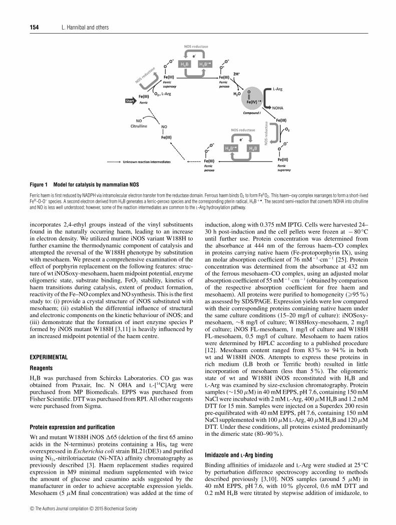
The sequence of reactions that leads to NO synthesis by NOSinvolves several steps of reduction and oxidation. Resting NOS isfirst reduced by electrons coming from the reductase domain.Ferrous haem then reacts with molecular oxygen to form atransient FeO2 species (Figure 1). This FeO2 intermediate isfurther reduced by the cofactor H4B to form a ferric-peroxocomplex. Scission of the O-O bond follows to form the highlyvalent compound I species, which ultimately oxidizes L-Arg toform N-hydroxy-L-arginine (NOHA). Conversion of NOHA intocitrulline and NO requires a second event of oxygen activation, inwhich H4B is proposed to serve as the electron donor (Figure 1).
All NOSs sequenced and crystallized to date possess aconserved tryptophan residue that forms stacking interactionswith the porphyrin ring and hydrogen bonds with the haem-thiolate bond. Replacement of this proximal tryptophan residueby histidine in murine inducible NOS (iNOS) (W188H) increasedthe midpoint potential of the haem group by + 88 mV and
reduced the rate of NO synthesis compared with wild-type(wt) iNOS. In addition, the W188H mutation stabilized aninert enzyme species, P, that formed downstream of the FeO2
species, which reacted slowly with L-Arg to form NOHA [3].Surprisingly, the W188F and W188A mutants of iNOS exhibitdefective haem binding, impeding further characterization [4].Substitution of Trp409 by phenylalanine in rat nueronal NOS(nNOS) reduced the haem midpoint potential of the protein, ledto a faster formation of the FeO2 species and to greater ratesof Fe(II)NO oxidation compared with the wt protein [5–7]. Aside-by-side characterization of Bacillus subtilis NOS and itsproximal tryptophan variants W66H and W66F revealed distinctthermodynamic and kinetic behaviours depending on the nature ofthe amino acid residue replacing the native tryptophan (histidineor phenylalanine) [8–10]. Thus, the proximal tryptophan residuemay play a role in controlling the enzyme’s reactivity by tuning theproperties and reactivity of its haem. Further, a study performedwith Staphylococcus aureus NOS revealed that NOSs display asteep dependence between the haem midpoint potential and therates of oxygen activation under single turnover conditions forthe hydroxylation of L-Arg to NOHA [8]. Lang et al. [8] showedthat mutations on the tryptophan residue proximal to the haem-thiolate bond altered the haem midpoint potential and the rates ofdisappearance of the FeO2 species in trends that correlated with thedonating or withdrawing electron density from the haem-thiolatebond that NOS enzymes display.
We have employed mesohaem-substituted iNOS to study theeffect of the haem midpoint potential in catalysis. Mesohaem
Abbreviations: H4B, (6R)-5,6,7,8-tetrahydro-L-biopterin; NOHA, N-hydroxy-L-arginine; NOS, nitric oxide synthase; iNOS, inducible nitric oxide synthase;nNOS, neuronal nitric oxide synthase; wt, wild-type.
1 Correspondence may be addressed to either of these authors (email [email protected] or [email protected]).The atomic co-ordinates for iNOSoxy in complex with mesohaem and H4B have been deposited in the PDB under code 4JS9.
c© The Authors Journal compilation c© 2015 Biochemical Society
Bio
chem
ical
Jo
urn
al
ww
w.b
ioch
emj.o
rg
154 L. Hannibal and others
Figure 1 Model for catalysis by mammalian NOS
Ferric haem is first reduced by NADPH via intramolecular electron transfer from the reductase domain. Ferrous haem binds O2 to form FeIIO2. This haem–oxy complex rearranges to form a short-livedFeIII-O-O– species. A second electron derived from H4B generates a ferric-peroxo species and the corresponding pterin radical, H4B+ • . The second semi-reaction that converts NOHA into citrullineand NO is less well understood; however, some of the reaction intermediates are common to the L-Arg hydroxylation pathway.
incorporates 2,4-ethyl groups instead of the vinyl substituentsfound in the naturally occurring haem, leading to an increasein electron density. We utilized murine iNOS variant W188H tofurther examine the thermodynamic component of catalysis andattempted the reversal of the W188H phenotype by substitutionwith mesohaem. We present a comprehensive examination of theeffect of porphyrin replacement on the following features: struc-ture of wt iNOSoxy-mesohaem, haem midpoint potential, enzymeoligomeric state, substrate binding, FeO2 stability, kinetics ofhaem transitions during catalysis, extent of product formation,reactivity of the Fe–NO complex and NO synthesis. This is the firststudy to: (i) provide a crystal structure of iNOS substituted withmesohaem; (ii) establish the differential influence of structuraland electronic components on the kinetic behaviour of iNOS; and(iii) demonstrate that the formation of inert enzyme species Pformed by iNOS mutant W188H [3,11] is heavily influenced byan increased midpoint potential of the haem centre.
EXPERIMENTAL
Reagents
H4B was purchased from Schircks Laboratories. CO gas wasobtained from Praxair, Inc. N OHA and L-[14C]Arg werepurchased from MP Biomedicals. EPPS was purchased fromFisher Scientific. DTT was purchased from RPI. All other reagentswere purchased from Sigma.
Protein expression and purification
Wt and mutant W188H iNOS �65 (deletion of the first 65 aminoacids in the N-terminus) proteins containing a His6 tag wereoverexpressed in Escherichia coli strain BL21(DE3) and purifiedusing Ni2+-nitrilotriacetate (Ni-NTA) affinity chromatography aspreviously described [3]. Haem replacement studies requiredexpression in M9 minimal medium supplemented with twicethe amount of glucose and casamino acids suggested by themanufacturer in order to achieve acceptable expression yields.Mesohaem (5 μM final concentration) was added at the time of
induction, along with 0.375 mM IPTG. Cells were harvested 24–30 h post-induction and the cell pellets were frozen at − 80 ◦Cuntil further use. Protein concentration was determined fromthe absorbance at 444 nm of the ferrous haem–CO complexin proteins carrying native haem (Fe-protoporphyrin IX), usingan molar absorption coefficient of 76 mM− 1·cm− 1 [25]. Proteinconcentration was determined from the absorbance at 432 nmof the ferrous mesohaem–CO complex, using an adjusted molarabsorption coefficient of 55 mM− 1·cm− 1 (obtained by comparisonof the respective absorption coefficient for free haem andmesohaem). All proteins were purified to homogeneity (�95%)as assessed by SDS/PAGE. Expression yields were low comparedwith their corresponding proteins containing native haem underthe same culture conditions (15–20 mg/l of culture): iNOSoxy-mesohaem, ∼8 mg/l of culture; W188Hoxy-mesohaem, 2 mg/lof culture; iNOS FL-mesohaem, 1 mg/l of culture and W188HFL-mesohaem, 0.5 mg/l of culture. Mesohaem to haem ratioswere determined by HPLC according to a published procedure[12]. Mesohaem content ranged from 83% to 94% in bothwt and W188H iNOS. Attempts to express these proteins inrich medium (LB broth or Terrific broth) resulted in littleincorporation of mesohaem (less than 5%). The oligomericstate of wt and W188H iNOS reconstituted with H4B andL-Arg was examined by size-exclusion chromatography. Proteinsamples (∼150 μM) in 40 mM EPPS, pH 7.6, containing 150 mMNaCl were incubated with 2 mM L-Arg, 400 μM H4B and 1.2 mMDTT for 15 min. Samples were injected on a Superdex 200 resinpre-equilibrated with 40 mM EPPS, pH 7.6, containing 150 mMNaCl supplemented with 100 μM L-Arg, 40 μM H4B and 120 μMDTT. Under these conditions, all proteins existed predominantlyin the dimeric state (80–90 %).
Imidazole and L-Arg binding
Binding affinities of imidazole and L-Arg were studied at 25 ◦Cby perturbation difference spectroscopy according to methodsdescribed previously [3,10]. NOS samples (around 5 μM) in40 mM EPPS, pH 7.6, with 10% glycerol, 0.6 mM DTT and0.2 mM H4B were titrated by stepwise addition of imidazole, to
c© The Authors Journal compilation c© 2015 Biochemical Society

Characterization of iNOS-mesohaem 155
a final concentration of 10 mM. The Kd of imidazole (Kdimid)was calculated by fitting the data to a simple saturation bindingequation. The Kd of L-Arg molar absorption was determinedunder the same conditions, in the presence of 10 mM imidazole.The data were fitted to a simple saturation binding equation,and Kd was calculated according to the following equation:appKd = Kd(1 + [imid]
Kdimid) , where appKd is the apparent Kd of the
enzyme for L-Arg in the presence of saturating concentrations ofimidazole.
Single turnover reactions
L-Arg hydroxylation and NOHA oxidation experiments werecarried out in an SF61-DX2 stopped-flow instrument (Hi-TechScientific) coupled to a diode array detector, as previouslydescribed [10,13]. An anaerobic solution of 20 μM ferrous NOS(obtained by titration of ferric NOS with a solution of dithionite),2 mM L-Arg (or 1 mM NOHA), 0.2 mM H4B and 1 mM DTTin 40 mM EPPS, pH 7.6, containing 10% glycerol and 150 mMNaCl was mixed at 10 ◦C with a solution containing air-saturatedbuffer, 2 mM L-Arg, 0.2 mM H4B and 1 mM DTT. Sequentialspectral data were fitted to a two-exponential, A → B → C,model using the Specfit/32 global analysis software, version3.0 (Spectrum Software Associates). Specfit is a multivariatedata analysis program for modelling and fitting 3D chemicalkinetics data by singular value decomposition (SVD). Rates arethe average for at least five reactions +− S.D.
Determination of reaction products by HPLC
End-point conversion of L-[14C]Arg to [14C]NOHA under singleturnover conditions were carried out according to a previouslypublished procedure [10]. L-[14C]Arg and [14C]NOHA wereextracted from the reaction mixtures and the conversion ofsubstrate to product was monitored by HPLC. The radioactivityof each fraction was counted using a Scintillation counter [10].
Midpoint potential determinations
Spectroelectrochemical titrations were conducted in a glove-box (Belle Technology) under an N2 atmosphere, as previouslydescribed [3,10]. NOS proteins were made air-free by gel filtrationin a Sephadex G-25 column (PD 10, GE Healthcare) pre-equilibrated with anaerobic buffer (100 mM phosphate buffer,pH 7.0 and 125 mM NaCl). Protein samples were diluted toa 3.5 ml final volume (final concentration ∼10 μM) and L-Arg (2 mM) and H4B (100 μM) were added. The followingelectron carrier dyes (0.5–1 μM) were utilized: phenosafranine(Em = − 252 mV), Benzyl Viologen (Em = − 358 mV),Methyl Viologen ( − 450 mV) and anthraquinone-2-sulfonate(Em = − 225 mV). The titration was performed at 15 ◦C bystepwise addition of a sodium dithionite solution. Oxidativetitrations were carried out under the same conditions usingpotassium ferricyanide to oxidize the haem centre. Reductiveand oxidative titrations yielded midpoint potential values within+−2 mV, suggesting that the process involves one-electron transferin either direction. Absorption spectra were collected with aCary 50 spectrophotometer equipped with a dip-probe detector,and the potentials were measured using a silver/silver chloridemicroelectrode saturated with 4 M KCl (Fisher Scientific).Midpoint potential values are expressed as midpoint potential+− error of the fit for a one-electron process.
Ferrous haem–NO complex oxidation (k ox)
Native and mesohaem-substituted iNOSoxy wt or W188Hoxy(∼5 μM) in 100 mM EPPS, pH 7.6, containing 150 mM NaCland 10% glycerol containing 2 mM L-Arg, 0.2 mM H4B and1 mM DTT were carefully titrated with dithionite under anaerobicconditions. The Fe(II)–NO complexes were generated bystepwise addition of anaerobic NO-saturated buffer. Both proteinsdisplayed stable Fe(II)–NO complexes at pH 7.6 (100 mM EPPS,pH 7.6, containing 150 mM NaCl and 10 % glycerol) and atpH 9.5 (100 mM CHES, pH 9.5, containing 150 mM NaCl and10% glycerol). Fe(II)–NO protein samples were then transferredto an anaerobic stopped-flow instrument using a gastight syringe,and the reactions were initiated by mixing with air-saturated bufferat 10 ◦C. Spectral data were fitted to a single exponential model,A→B, using Specfit global analysis software.
NO synthesis
NO synthesis by native and mesohaem-substituted full-lengthproteins was assessed by the oxyhaemoglobin NO capture assayusing an molar absorption coefficient of 38 mM− 1·cm− 1 formethaemoglobin minus oxyhaemoglobin as described elsewhere[12]. Total nitrite produced by these proteins in the presence ofNOHA was determined by the Griess assay [14] according topublished protocols [10].
Crystallization and data collection
Mesohaem-substituted iNOS �65 was expressed in minimalmedium and purified as described above. Crystals of murineiNOSoxy (residues 66–498) in complex with mesohaem andH4B were grown by hanging drop vapour diffusion at 293K in 1 μl drops (0.5 μl of protein with 0.5 μl of screen).Hanging drops consisted of a 1:1 ratio mixture of 20 mg/mliNOSoxy with 10 mM H4B and a reservoir solution. Crystals grewwithin 3–6 days in a reservoir solution composed of 700 mMammonium sulfate, 100 mM MES, pH 5.3, and 3.5% octyl-glucoside. Crystals were cryoprotected by brief transfer throughreservoir solution supplemented with 30% (w/v) ethyleneglycol and flash-frozen in liquid nitrogen. Diffraction datafor a crystal in space group P6122 was collected using aRigaku MicroMax-007HF generator and a Rigaku Saturn 044 +CCD detector. Data integration, reduction and scaling wereperformed with d*TREK [15] and processed to a cut-off of2.78 Å. The resolution cut-off was based on significant drops innon-averaged I/σ (<2.0) and significant increases in Rmerge
in shells at higher resolutions.
Structure solution, refinement, validation and deposition
An existing structure of iNOS (PDB code 3DWJ: chain A) [3]was used as a molecular replacement search model. Phases werecalculated by molecular replacement using the PHASER [16]component of PHENIX [17]. The best molecular replacementsolution was subjected to iterative rounds of model building inCOOT [18] and refinement in PHENIX. The refinement protocolused isotropic atomic displacement parameters for all atoms.Stereochemical analysis of the iNOSoxy structure was completedwith MolProbity [19]. All residues are within the allowed regionsof the Ramachandran plot and no poor rotamers or Cβ deviationswere found. MolProbity statistics are listed in SupplementaryTable S1. The atomic co-ordinates for iNOSoxy in complex withmesohaem and H4B in space group P6122 has been deposited inthe PDB (accession code 4JS9).
c© The Authors Journal compilation c© 2015 Biochemical Society

156 L. Hannibal and others
Steady-state kinetics of the reaction of full-length W188Hcontaining native haem
Steady-state reactions were carried out according to the conditionsdescribed by Abu-Soud et al. [20]. Briefly, full length W188H(∼2 μM) was mixed with 1 mM L-Arg and 4 μM H4B/DTTin 40 mM EPPS, pH 7.6 containing 125 mM NaCl and 10%glycerol. This solution was mixed with 140 μM NADPH in thesame buffer, and the reaction was monitored at different timepoints by rapid UV–visible scanning.
Simulations of iNOS distribution during steady-stateNO synthesis
The NO synthesis kinetics of wt iNOS were simulated using theglobal model described earlier [21] and implemented in Gepasisoftware version 3.30. For mutant W188H, the global modelwas adjusted to account for the formation and disappearanceof inert product P. The simulations assume constant values for[O2] = 180 μM and [NADPH] = 40 μM. Experimental kox valuesfor wt iNOS ferrous haem–NO complexes obtained under halfair-saturation conditions were multiplied by a factor of 1.5 (air-saturation) [22]. Likewise, oxygen-binding rates k2 and k6 (forwt enzymes) and k2 and k7 (for W188H mutant enzymes) weremultiplied by a factor of 2.0 (air-saturated condition) assumingthat this rate is proportional to [O2]. The selected concentrations ofNADPH and dioxygen were kept constant during each simulation,to avoid secondary effects due to their exhaustion.
RESULTS AND DISCUSSION
Expression of mesohaem-containing iNOS proteins
The oxygenase and full-length versions of wt iNOS andmutant W188H were expressed in E. coli grown in minimalmedium supplemented with 5 μM mesohaem. The expressionyield was very low, particularly for the full-length proteins(yield ∼0.5 mg of protein/l of culture). Despite this practicalhurdle, the purified proteins presented the expected spectralcharacteristics, suggesting the haem environment was suitablefor further characterization. Substitution of iNOS and W188Hwith Fe(III)-deuterohemin and Fe(III)-2,4-diacetyl-deuteroheminproved impossible under our experimental conditions. Gelfiltration analysis showed that both iNOSoxy and W188Hoxycontaining mesohaem exist predominantly as a dimer (resultsnot shown). Fractions of highly purified proteins were examinedfor their haem content (Reinheitszahl values, Rz, ASoret/A280).The haem content of both proteins substituted with mesohaemwas low compared with their corresponding native counterparts(Rz = 1.5–2.5). wt iNOSoxy-mesohaem had an Rz of 0.45,whereas W188Hoxy showed ∼2.5-fold decreased haem contentcompared with iNOSoxy-mesohaem (Rz = 0.20). This poorerhaem load has been observed in W188Hoxy carrying native haem,and the effect was exacerbated upon incorporation of the foreignmesohaem. The lower mesohaem content observed in W188Hoxyseems to be an effect of the tryptophan to histidine mutation.This could be related to the recently noted misalignment betweenthe imidazole ring of His188 and the porphyrin ring as opposedto the native arrangement of the indole group of Trp188 [11].Conceivably, this structural restriction may affect cellular haeminsertion during protein maturation. In fact, iNOSoxy mutantsW188A and W188F are isolated as apo-proteins (unpublishedwork, Zhiqiang Wang, Jesus Tejero and Dennis J. Stuehr).
Figure 2 X-ray crystal structure of iNOSoxy �65 with mesohaem (PDB code4JS9)
(A) The structures of native iNOSoxy�65 (PDB code 1NOD, grey) and iNOSoxy�65-mesohaem(magenta) are superimposed and with backbones shown as ribbons. Native haem (orange),mesohaem (cyan) and H4B groups from native iNOSoxy �65 (blue) and iNOSoxy�65-mesohaem (yellow) are shown as sticks. (B) A close-up of the iNOSoxy �65-mesohaemactive site highlights the spatial arrangement of haem and H4B groups (coloured as in A). (C)Electron density for ligands in the X-ray crystal structure of iNOSoxy �65 with mesohaem(PDB code 4JS9). The structure of iNOSoxy �65-mesohaem (magenta) is shown as ribbonswhile mesohaem (cyan) and H4B groups (yellow) are shown as sticks. Electron density (2F o-F c,drawn at 1σ ) is drawn as a mesh (grey). A proposed location for L-Arg (not included in thedeposited co-ordinates) places the backbone and guanidinum groups into two areas of weakelectron density situated above the haem iron and is consistent with the position of L-Arg innative iNOSoxy structure (PDB code 1NSI).
X-ray crystal structure of iNOSoxy-mesohaem
Crystals of iNOSoxy �65 substituted with mesohaem wereanalysed and compared with the reported crystal structures ofiNOSoxy �65 carrying native haem (PDB code 1NOD [2]), aswell as with the structure of iNOS mutant W188Hoxy (PDBcode 3DWJ [3]). X-ray crystallographic analysis revealed ahighly conserved iNOSoxy structure upon replacement withmesohaem. The structures of iNOSoxy-mesohaem and iNOSoxysuperimpose within 0.3 Å rmsd (Supplementary Table S1),suggesting an almost identical structure (Figure 2A). The haemcentre displays a similar hydrogen-bonding network comparedwith native iNOSoxy, which is suitable for catalysis. H4B is alsooriented within its pocket in the right configuration for electrontransfer (Figures 2B and 2C). Weak electron density was observedfor the guanidinum moiety and backbone atoms of the natural
c© The Authors Journal compilation c© 2015 Biochemical Society

Characterization of iNOS-mesohaem 157
Figure 3 Determination of the midpoint potentials of wt iNOSoxy andmutant W188Hoxy substituted with mesohaem
The fraction of oxidized protein at each redox potential upon titration with dithionite wasfitted to the Nernst equation. The calculated values for the midpoint potentials are (A)iNOSoxy-mesohaem − 295 +− 3 mV and (B) W188Hoxy-mesohaem − 239 +− 6 mV.
substrate L-Arg, although the density was deemed too weak forinclusion in the final co-ordinates. The electron density for L-Argis consistent with the position observed in wt iNOS, with theguanidinum moiety sitting in close proximity to the iron centre(Figure 2C).
Redox potentiometry
An anticipated consequence of replacing native Fe-protoporphyrin IX with Fe-mesoporphyrin IX is a loweringof the haem midpoint potential. Redox titrations in the presenceof L-Arg and H4B were performed for mesohaem-containingiNOSoxy and W188Hoxy by stepwise addition of dithionite(reduction) or potassium ferrycyanide (oxidation). Nernst plotsfor the reductive curves are shown in Figure 3 and the rawdata are provided in Supplementary Figure S1. The calculatedmidpoint potentials were − 295 +− 3 mV for iNOSoxy and− 239 +− 6 mV for W188Hoxy. Thus, substitution of nativehaem by mesohaem led to a significant decrease in the haem
Table 1 Spectral properties of iNOS wt and mutants W188H substitutedwith mesohaem
UV–visible absorption maxima in the presence of H4B and L-Arg or imidazole (Imid) of wtiNOSoxy and W188Hoxy substituted with mesohaem in EPPS (40 mM, pH 7.6) containing150 mM NaCl and 10 % glycerol.
Soret (nm) Visible (nm)
iNOS-mesohaem*Fe(III) 381 506, 536, 642Fe(III)-Imid 417 540Fe(II) 404 549Fe(II)-CO 431 545Fe(II)-NO 422 (broad) 559
W188H-mesohaemFe(III) 377 507, 536, 638Fe(III)-Imid 414 540Fe(II) 400 547Fe(II)-CO 428†, 413 546
Fe(II)-NO 420 (broad) 556
*The values for iNOS-mesohaem are in agreement with a previous study [24].†The species at 428 nm shifts slowly to an uncharacterized species with absorption maximum
at 413 nm.
midpoint potential compared with the corresponding proteins har-bouring native Fe-protoporphyrin IX (iNOSoxy = − 261 +− 2 mV;W188Hoxy = − 173 +− 2 mV [3]). As expected, substitutionof W188Hoxy with mesohaem did shift its midpoint potentialto a value that is closer to that of wt iNOSoxy, reversing theeffect of the mutation, thus enabling the dissection of electroniccompared with structural effects. This provided us with a uniqueopportunity to interrogate the role of the midpoint potential onthe formation of inert enzyme species P that forms downstreamof the FeO2 species.
Spectroscopic properties and substrate binding ofmesohaem-containing iNOS proteins
UV–visible data for wt iNOSoxy and W188Hoxy-containingmesohaem are given in Table 1. The UV–visible spectral featuresof iNOS proteins substituted with mesohaem are essentiallyidentical to those reported for nNOSoxy-mesohaem [12,23].Substitution with mesohaem led to a 12 nm blue-shift in the UV–visible spectra of the Fe(III), Fe(III)–imidazole, Fe(II), Fe(II)–CO and Fe(II)–NO complexes of iNOS proteins, consistentwith spectral changes observed in another variant of iNOS[24] and in other proteins substituted with this haem analogue[25–30]. In the presence of L-Arg and H4B both wt iNOSoxyand W188Hoxy substituted with mesohaem exist in the typicalFe(III) high-spin configuration, suggesting that the integrityof the haem electronic environment remained comparable tothat of their native counterparts (Figure 4). Reduction withdithionite and exposure to CO resulted in the formation of thecharacteristic Fe(II)–CO complex with a Soret band appearing at428–431 nm. UV–visible spectra of W188H proteins displayedthe previously observed 2–5 nm blue shift compared withwt iNOS [3], regardless of the haem centre used. Bindingdissociation constants, Kd, for imidazole and L-Arg in thepresence of H4B were measured spectrophotometrically. Both wtiNOSoxy and mutant W188Hoxy substituted with mesohaem dis-played competent binding for imidazole (Kd,iNOSoxy = 56 +− 9 μM;Kd,W188Hoxy = 39 +− 2 μM) and for L-Arg (Kd,iNOSoxy = 2.6 +− 0.4 μM;Kd,W188Hoxy = 5.6 +− 0.3 μM). The lower affinity of W188Hoxy-mesohaem for L-Arg compared with its wt counterpart isnot unprecedented [3]. However, substitution with mesohaem
c© The Authors Journal compilation c© 2015 Biochemical Society

158 L. Hannibal and others
Figure 4 UV–visible spectral properties of wt iNOSoxy and mutantW188Hoxy substituted with mesohaem, in the presence of H4B and L-Arg
Substitution with mesohaem led to a blue shift of ∼12 nm in the UV–visible spectra of both wtand W188Hoxy proteins. (A) wt iNOSoxy-mesohaem. (B) W188Hoxy-mesohaem. Absorptionmaxima are provided in Table 2.
produced a stronger association with the natural substratecompared with W188Hoxy possessing native haem (Kd ∼29 +− 3 μM) [3], thus partially reversing the effects of the structuralreplacement of Trp188 by a histidine residue.
Stopped-flow analysis of a single turnover L-Arg hydroxylationreaction in the presence of H4B
We next investigated the enzyme haem species that occurin iNOSoxy and W188Hoxy substituted with mesohaem. Ofparticular interest was to examine the formation (or absence) ofthe inert enzyme species P whose exact nature remains elusive[3,11]. L-Arg hydroxylation reactions of mesohaem-containingiNOSoxy could be best fitted to a two-exponential model A →B→C, with Fe(II), Fe(II)–O2 and Fe(III) as the only detectablespecies (Figure 5). We investigated whether reversal of themidpoint potential in W188Hoxy-mesohaem affects the formationand stability of species P, which forms downstream of the Fe–O2 species [3,11]. L-Arg hydroxylation reactions of mesohaem-containing W188Hoxy were fitted to a three-exponential modelA →B →C→D, with Fe(II), Fe(II)–O2, a presumptive species
P with absorption maximum at 411 nm and Fe(III) as the onlydetectable species (Figure 6). A summary of the measured rateconstants is given in Table 2. Fitting of the spectral data to a two-exponential model for reaction times 2 and 0.6 s confirmed theformation of an enzyme species with a marked shoulder at 411 nm(results not shown). For both mesohaem-containing proteins,disappearance of the Fe(II)–O2 species was faster compared withtheir native counterparts. The effect of the midpoint potentialon the stability of the FeIIO2 species has been studied in-depthin several enzyme systems [10,31–35]. The most widely usedapproach involves the introduction of mutations to modify thestrength of the haem-thiolate bond in both NOS and cytochromeP450 enzymes, which leads to a selective tuneable increase ordecrease in the haem midpoint potential. The FeIIO2 species isstabilized in proteins with higher haem midpoint potentials dueto a decrease in the driving force for its reduction to FeIIIO2
–.Likewise, a lower midpoint potential favours the ferric form ofthe protein, with the concomitant faster disappearance of the Fe–O2 species with respect to native haem-containing proteins. Thisgeneral relationship between the stability of the FeO2 species andthe haem midpoint potential has been well documented both inmammalian and bacterial NOSs and in related enzymes [5–7,10].A good illustration of this phenomenon is given by the haemtransition rates for the conversion of FeO2 to its downstreamspecies: k2 W188Hoxy-mesohaem = 10.5 s− 1 ≈ k2 iNOSoxynative haem = 12.5 s− 1; i.e. restoring the midpoint potentialof mutant W188H to resemble that of wt iNOSoxy regeneratedthe wt-like behaviour of the FeO2 species in W188Hoxy. Thissuggests that changes in haem electronics posed by the W188Hmutation can be at least partly reversed by substitution withmesohaem, making electron density the primary factor for thelonger-lived FeO2 species seen in W188Hoxy harbouring nativehaem (k2 = 2.03 s− 1 [3]). Reversal of the midpoint potentialin W188Hoxy did not result in the abolition of inert speciesP that forms downstream of the FeO2 species (Figure 6),arguing in favour of a structural effect of the mutation [11].However, a decrease in the midpoint potential in W188Hoxysignificantly diminished the stability of species P comparedwith native W188Hoxy (k3 W188Hoxy mesohaem = 0.198 s− 1;k3 W188Hoxy = 0.104 s− 1), highlighting the contribution ofelectronic effects.
Stopped-flow analysis of a single turnover NOHA oxidation reactionin the presence of H4B
The fact that mutant W188H displays altered behaviour in termsof NO synthesis compared with wt iNOS [3] prompted usto examine NOHA oxidation reactions under single turnoverconditions in proteins substituted with mesohaem. Reaction ofwt iNOSoxy-mesohaem with NOHA and H4B could be best fittedto a sequential model with three well-defined haem transitions(Table 2), similar to the reaction pattern observed for thisprotein carrying native haem. The observed reaction rates (k1,k2 and k3) suggested that the reaction is slower in iNOSoxy-mesohaem compared with the native haem-containing protein.A stable FeIII–NO complex was identified downstream of theFeO2 species, with a broad absorption maximum at 431 nm(Figure 7). Single turnover reactions of W188Hoxy carryingnative haem in the presence of NOHA and H4B do also occurvia the formation of three distinct species as observed with wtiNOSoxy, although formation and disappearance of the FeO2
species is substantially slower in the mutant (Table 5). Aremarkable effect of mesohaem substitution in mutant W188Hoxywas the change to an extremely short-lived FeO2 species and the
c© The Authors Journal compilation c© 2015 Biochemical Society

Characterization of iNOS-mesohaem 159
Figure 5 Single turnover reactions in iNOSoxy-mesohaem with L-Arg and H4B
Global analysis with Specfit 3.0 software identified three distinct species: Fe(II) (Soret maximum ∼402 nm), Fe-O2 (Soret maximum ∼415 nm) and Fe(III) (Soret maximum ∼378 nm). Haemtransitions occurred faster than in native iNOSoxy (Table 2), with a return to the resting state within 0.4 s. (A) Spectral changes observed during single turnover reactions. (B) Haem enzyme speciesidentified by global analysis. (C) Time courses for each haem enzyme species calculated by global analysis.
Table 2 Single turnover reactions
Observed rate constants (s− 1) for haem transitions during single turnover reactions in wt iNOSoxy and W188Hoxy substituted with mesohaem. Values for native and mesohaem-containing nNOSare provided for comparative purposes.
L-Arg/H4B NOHA/H4B Reference
iNOS Fe(II) k1−→FeO2k2−→Fe(III) Fe(II) k1−→FeO2
k2−→FeNOk3−→Fe(III)
iNOSoxy 52.7 +− 2.2; 12.5 +− 0.2* 47.3 +− 3.1; 36.7 +− 1.8; 2.30 +− 0.09 This workiNOSoxy-mesohaem 37.8 +− 2.3; 22.9 +− 0.8 35.6 +− 0.6; 18.1 +− 0.2; 1.70 +− 0.12 This work
Fe(II) k1−→FeO2k2−→P
k3−→Fe(III) Fe(II) k1−→FeO2k2−→FeNO
k3−→Fe(III)
Or Fe(II) k1−→FeNOk2−→Fe(III)
W188Hoxy 46.0 +− 1.1; 2.03 +− 0.04; 0.104 +− 0.003* 43.1 +− 2.1; 2.97 +− 0.27; 0.239 +− 0.010 This workW188Hoxy-mesohaem 45.3 +− 8.6; 10.5 +− 0.9; 0.198 +− 0.06 37.7 +− 1.2; 5.75 +− 0.08† This work
nNOSnNOSoxy 183 +− 11 s− 1; 17.5 +− 0.2 s− 1 133 +− 6 s− 1; 23.8 +− 0.4 s− 1; 5.1 +− 0.1 s− 1 [12]nNOSoxy-meso 168 +− 7 s− 1; 52.1 +− 0.7 s− 1 153 +− 13 s− 1; 117 +− 6 s− 1; 8.2 +− 0.1 s− 1 [12]
*Values taken from reference [3].†The FeO2 species could not be observed during single turnover reactions of W188Hoxy with NOHA and H4B.
c© The Authors Journal compilation c© 2015 Biochemical Society

160 L. Hannibal and others
Figure 6 Single turnover reactions in W188Hoxy-mesohaem with L-Arg and H4B
(A) Spectral changes observed for a 2 s reaction time. (B) Global analysis of the reaction kinetics identified an additional species (P + FeIII) in addition to those observed in the single turnoverreactions of wt iNOSoxy-mesohaem. Species P previously identified during hydroxylation of L-Arg to NOHA appeared downstream of the Fe–O2 complex, with an absorption maximum at 411 nm(shoulder). This inert enzyme species P has a shorter half-life (C and D) compared with the same reaction performed with native haem [3] and, as a result, its net build-up is diminished. The enzymereturns to the resting ferric state with an observed rate of 0.198 s− 1.
stabilization, for the first time, of a FeIII–NO complex (Figure 8).The observed rates of formation and disappearance of FeIII–NO iniNOSoxy-mesohaem were proportionally lower (∼1.5-fold) thanin the native enzyme. The FeIII–NO complexes identified duringsingle turnover reactions of iNOSoxy and W188Hoxy containingmesohaem are practically identical to the same species generatedby addition of NO to an anaerobic solution of the correspondingFeIII–NOS enzyme (insets to Figure 7A, and Figure 8B). Globalanalysis of the single turnover reactions of mutant W188Hoxy-mesohaem was compatible with a sequential two-exponentialmechanism, with the reaction being best described as: FeII →P + FeIII-NO →FeIII, where P defines an enzyme specieswith a Soret absorption maximum at ∼420 nm that formsin the reactions of W188H carrying native haem [3,11]. Weoriginally referred to this species as a ‘reaction intermediate’,but recent work [11] and the experimental findings presentedherein strongly suggest that species P is a non-native low-spinenzyme species that forms right after NO is made, and thusmasks or antagonizes the formation of the enzyme FeIII–NOproduct complex that otherwise transiently builds up in the wt
reaction. Previous studies provided strong evidence that species Pis a hydroxide-bound haem whose formation is accompanied byH4B•+ radical build-up, rather than it being a ferryl or a peroxo-hame species [11]. It was proposed that the haem-hydroxidespecies was stabilized by the W188H mutation, and that thehydroxide ligand prevents NO binding to the haem before itescapes the active site channel [11]. Our findings argue againsta pure structural effect of the W188H mutation, by showing thatthe stability of this presumptive hydroxide-bound haem species ischallenged by sole reversal of the midpoint potential in W188H.This in turns rescues the enzyme’s ability to synthesize NO byshifting oxygen activation toward the productive cycle of NOS.Nonetheless, our data support the proposal that species P couldbe indeed a hydroxide-bound haem derived from NOHA (productcomplex). Binding of substrate and pterin-free W188Hoxy to theNOHA analogues phenylguanidine and methoxyphenylguanidine(4 mM) led to perturbation of the UV–visible spectra, as expected.Binding of W188Hoxy to phenylguanidine produced a complexwith absorption maximum at 424 nm, suggestive of Fe–N co-ordination. Binding to methoxyphenylguanidine, which possesses
c© The Authors Journal compilation c© 2015 Biochemical Society

Characterization of iNOS-mesohaem 161
Figure 7 Single turnover reactions in iNOSoxy-mesohaem with NOHA and H4B
Global analysis of the spectral changes identified four species during the course of this reaction: Fe(II) (Soret maximum ∼402 nm), Fe–O2 (Soret maximum ∼415 nm), Fe(III)–NO (Soret maximum∼431 nm) and Fe(III) (Soret maximum ∼378 nm). (A) Species identified by global analysis. The inset to (A) shows authentic FeIII–NO complex generated by addition of NO to anaerobic FeIII
iNOSoxy-mesohaem. (B) Time courses for the haem enzyme species calculated by global analysis. (C) Overlay of the experimental and global analysis curves for the time course of Fe(III)–NOspecies during single turnover reactions with NOHA and H4B.
an available oxygen atom for axial ligation, resulted in a complexwith absorption maximum at 419 nm reminiscent of species P(Supplementary Figure S2). This finding is in line with bindingstudies carried out with NOHA [11] and points to the hydroxogroup as the axial ligand of species P.
Extent of L-Arg hydroxylation
An examination of [14C]NOHA formation from L-[14C]Arg undersingle turnover conditions at infinite time (10 min) showed thatthe yield of product formation for both mesohaem-substitutedproteins (iNOSoxy = 0.40 NOHA per haem; W188Hoxy = 0.48NOHA per haem) is in accord with previously reported datafor iNOSoxy (NOHA/haem 0.4–0.6 [36–39]) and W188Hoxy(0.37 NOHA/haem [3]) carrying native haem. Substitution ofmesohaem in W188H led to a slightly greater yield of L-Arghydroxylation. This correlates well with the formation of a stable
FeIII–NO complex during single turnover reactions with NOHAand the improved NO synthesis rate by this mutant (see nextsection).
NO synthesis
NO synthesis by full-length iNOS and W188H substituted withmesohaem was examined by the oxyhaemoglobin capture assay.The rate of NO synthesis by wt iNOS-mesohaem (34.9 +− 3 min− 1)was lower than that reported for the native enzyme (51–66 min− 1
[3,40]). In contrast, the NO synthesis rate of W188H-mesohaemwas 22 +− 5.4 min− 1, which is approximately 4-fold higher thanthe documented value for native W188H [3]. Although wt iNOShas a higher NO synthesis capacity than mutant W188H withmesohaem, the difference is less than in their counterparts withnative haem. Thus, replacement of native haem by mesohaemimproved the NO synthesis yield of iNOS mutant W188H.
c© The Authors Journal compilation c© 2015 Biochemical Society

162 L. Hannibal and others
Figure 8 Single turnover reactions in W188Hoxy-mesohaem with NOHA and H4B
(A) Spectral changes observed during a 0.6 s reaction time. (B) Global analysis of the spectral changes showed an extremely short-lived FeIIO2 species, with three major observable haem transitions:Fe(II) (Soret maximum ∼402 nm), Fe(III)–NO (Soret maximum ∼418–422 nm) and Fe(III) (Soret maximum ∼376–380 nm). Build-up of a FeIII–NO complex was observed for the first time inthe reactions of W188Hoxy, and it is thought to be a result of changes in the electronic environment of the haem. The inset to (B) shows authentic FeIII–NO complex generated by addition of NOto anaerobic FeIII W188Hoxy-mesohaem. (C) Time courses for each species calculated by global analysis. (D) Overlay of the experimental trace for the formation and disappearance of Fe(III)–NOcomplex and its calculated fit obtained by global analysis.
Oxidation rate of the ferrous–NO complex (k ox)
During NO synthesis, an FeIII–NO complex forms and can bereduced by NOS reductase at rates comparable to that of FeIII–NO dissociation [41,42]. Reaction of the ferrous–NO complexwith O2 resumes the catalytic cycle, ultimately leading to NOrelease. We investigated the oxidation of anaerobic FeII–NOcomplexes by O2 ([O2] ∼ 180 μM) by stopped-flow spectroscopyat pH 7.6 and pH 9.5 (Supplementary Figure S3). Oxidation ofFeII–NO by O2 occurred as a single step. Observed rate constantswere derived by fitting the data to a first-order exponentialmodel. FeII–NO complexes were stable at both pHs, with ratesin the following rank order: iNOS-mesohaem pH 7.6 = 8.89 +−0.43 s− 1 > iNOS-mesohaem pH 9.5 = 5.08 +− 0.09 s− 1 > W188H-mesohaem pH 7.6 = 4.68 +− 0.05 s− 1 > W188H-mesohaempH 9.5 = 2.89 +− 0.16 s− 1. At both pHs, the FeII–NO complexof W188Hoxy-mesohaem reacted more slowly with O2 comparedwith wt iNOSoxy-mesohaem, and more generally, the mesohaem-containing proteins displayed a faster conversion of FeII–NO
into FeIII compared with the native-haem containing versions(iNOSoxy kox = 3.11 s− 1 at pH 7.6) [43]. Overall, the FeII–NOoxidation rates by dioxygen in mutant W188H-mesohaem are ∼2-fold slower compared with wt iNOS-mesohaem at both pH 7.6and pH 9.5. This decreased kox rate is nonetheless relatively highcompared with the predicted haem reduction rate for W188H-mesohaem (0.3 s− 1), thus of predictably little effect on theenzyme’s steady-state behaviour and NO synthesis activity.
Haem reduction rates of iNOS and W188H containing native haem(k r)
The measured haem reduction rates for iNOS and W188Hunder our experimental conditions were kr = 0.5 +− 0.03 and1.79 +− 0.03 s− 1, respectively (Supplementary Figure S4). Thekr values reported for iNOS under similar conditions are 0.6–1.2s− 1 [20,42,43]. Poor expression yields of the corresponding full-length mesohaem-containing proteins prevented our measuring
c© The Authors Journal compilation c© 2015 Biochemical Society

Characterization of iNOS-mesohaem 163
Figure 9 Partition of haem-enzyme species during the reactions of wt and W188H iNOS simulated with Gepasi software version 3.3
The reactions of wt iNOS are characterized by FeIII as the predominant species, with a shorter-lived FeO2 species in iNOS containing mesohaem compared with its native counterpart. In contrast,mutant W188H features the formation and build-up of an inert species, P, which competes with both the productive NO–synthesis pathway and the futile cycle of iNOS. Incorporation of mesohaeminto W188H reduces the formation of and accelerates the decay of enzyme species P enabling a larger fraction of the oxygen-activated substrate to be channelled into the productive cycle of NOsynthesis.
their haem kr, therefore estimated values were calculated byextrapolation of kr values from a previous study performedwith nNOS, where kr for nNOS containing native or mesohaemwere: nNOS native = 6.6 s− 1, nNOS mesohaem = 1.1 s− 1 [12].Assuming a similar trend would apply to iNOS, we estimatedvalues of kr would be 0.1 and 0.3 s− 1 for iNOS-mesohaem
and W188H-mesohaem, respectively. While assumptions carry anintrinsic limitation, it is unlikely that haem reduction rates in iNOSwill deviate substantially from the trend observed in nNOS for twofundamental reasons: (i) the haem electronic environment andthe surface amino acid residues involved in reductase-oxygenaseinteractions necessary for electron transfer are highly conserved
c© The Authors Journal compilation c© 2015 Biochemical Society

164 L. Hannibal and others
in both enzymes [44], and (ii) X-ray structural analysis presentedherein indicates that substitution of native haem with mesohaemin iNOSoxy did not alter the bonding interactions around theporphyrin moiety where reduction by the reductase domain wouldoccur. We utilized these estimates of kr to model steady-statereactions that otherwise could not be studied experimentally.
Steady-state analysis of W188H carrying native haem
The enzyme species formed under steady-state conditions wereexamined in full-length W188H in the presence of L-Arg,H4B and NADPH. Spectra collected during 2 min (300 scans)(Supplementary Figure S5A) were subjected to global analysisusing Specfit software as described earlier. The reaction could bebest fitted to a three-exponential sequential model, with FeIII asthe initial and final species, and FeII and inert product P as theother two detectable haem species (Supplementary Figure S5B).This is the first study to demonstrate the formation of species Pin a NOS enzyme under steady-state conditions. The long-life(Supplementary Figure S5C) and spectral features of species P(Supplementary Figures S5B and S5D) are consistent with thoseobserved during single turnover reactions with L-Arg and NOHA,thus confirming that product P forms downstream of the FeIIO2
intermediate in both phases of oxygen activation in NOS catalysis.
Steady-state simulations of wt and W188H iNOS
To examine the predicted partition of enzyme species underour experimental conditions, the experimental kinetic parametersobtained for native and mesohaem-containing wt and W188HiNOS (Supplementary Tables S2 and S3) were submitted toGepasi software version 3.30 pre-loaded with our global model forNOS catalysis as described above. The results of these simulationsare presented in Figure 9. Substitution of mesohaem significantlyincreased the partition of the respective enzymes towards the ferricstate, FeIII, and reduced the build-up of the FeIIO2 intermediate.Replacement of native haem with mesohaem in W188H restoredthe relative partition of enzymes species to a pattern that resemblesthat of wt iNOS, i.e. it led to a substantial decrease in build-up ofspecies P, with the concomitant stabilization of ferric enzyme.While we observe FeIII–NO complex formation during singleturnover reactions with NOHA in W188H, this species is unlikelyto build up under steady-state conditions where product P is thepredominant species when L-Arg is used as the substrate andNADPH as the source of electrons (Supplementary Figure S5).Our global model for W188H was built to depict this experimentalfinding (Supplementary Figure S7). We next ran simulations of thesteady-state reactions for all four proteins utilized in the presentstudy (Supplementary Figures S6 and S7). We found that therelative distribution of each enzyme species is consistent withtheir different kinetic parameters and is sensitive to mesohaemreplacement both in wt and in W188H iNOS (Figure 9).
Conclusions
We employed two different strategies to investigate the kineticbehaviour of iNOS in depth: (i) mutation of the proximaltryptophan residue Trp188 to histidine, and (ii) replacementof native protohaem with the electron- rich mesohaem. Thekinetic behaviour of W188Hoxy-mesohaem with NOHA and H4Bdiffered from its native counterpart in two ways: (i) a FeIIO2
species could not be detected during single turnover reactions,and (ii) a stable FeIII–NO was observed, for the first time. Thereduced stability of the FeIIO2 species, also observed in the half-
reaction with L-Arg has been noted in the case of nNOS substitutedwith mesohaem [12]. Build-up of the FeIII–NO complex wasfaster in W188Hoxy-mesohaem compared with wt iNOSoxy-mesohaem, and the same was true for its transition to FeIII. This isfavourable for NO synthesis in W188H, by diminishing diversionof the available NO into the unproductive NO-bound form of theenzyme [7,41], but not sufficient to account for its greater NOsynthesis performance compared with its native counterpart. Asimilar relationship has been identified in nNOS mutant W409F[6,7], B. subtilis mutant W66F [10] and in nNOS harbouringFe-mesoporphyrin IX [12], all of which display reduced haemmidpoint potentials compared with their respective native orwt counterparts. While our results support the recent proposalthat structural changes are partly responsible for the build-upof species P in iNOS W188H [11], the remarkable effect ofmesohaem insertion in W188H leading to partial restorationof catalytic activity points to haem electronics as a key factorin controlling the formation and lifetime of species P. The presentstudy advanced our understanding of the nature of species Pby demonstrating that: (i) the haem midpoint potential is afundamental contributor to the formation of species P, (ii) speciesP forms during steady-state conditions, and (iii) decreasing thebuild-up of species P leads to an increase in NO synthesis byW188H. From a mechanistic standpoint, we conclude that speciesP is a hydroxo-haem ligated state that halts the reaction at a newpoint, competing with both the productive and futile cycles ofNOS.
AUTHOR CONTRIBUTION
Luciana Hannibal designed the study, performed the experiments and prepared the paper;Richard Page and Saurav Misra solved the crystal structure of iNOSoxy-mesohaem andwrote the corresponding Experimental and Results sections of the paper; MohammadMahfuzul Haque carried out the simulation studies under steady-state conditions; KarthikBolisetty purified proteins and performed experiments; Zhihao Yu purified proteins andobtained high-quality crystals of iNOSoxy-mesohaem; Dennis Stuehr designed the studyand prepared the paper. All authors performed a critical reading of the paper prior tosubmission and approved the final version of the paper.
ACKNOWLEDGEMENTS
We thank Deborah Durra for excellent technical support for the preparation of iNOSoxy-mesohaem and Dr D. Mansuy and Dr J.L. Boucher for the gift of NOHA analoguesphenylguanidine and methoxyphenylguanidine.
FUNDING
The present work was supported by the National Institutes of Health [grant numbersCA53914, GM51491, HL76491] (to D.J.S.), a National Institutes of Health PostdoctoralFellowship [grant number T32 HL007914 (to R.C.P.)] and the American Heart AssociationPostdoctoral Fellowship [grant number 11POST650034 (to L.H.)].
REFERENCES
1 Crane, B.R., Arvai, A.S., Gachhui, R., Wu, C., Ghosh, D.K., Getzoff, E.D., Stuehr, D.J. andTainer, J.A. (1997) The structure of nitric oxide synthase oxygenase domain and inhibitorcomplexes. Science 278, 425–431 CrossRef PubMed
2 Crane, B.R., Arvai, A.S., Ghosh, D.K., Wu, C., Getzoff, E.D., Stuehr, D.J. and Tainer, J.A.(1998) Structure of nitric oxide synthase oxygenase dimer with pterin and substrate.Science 279, 2121–2126 CrossRef PubMed
3 Tejero, J., Biswas, A., Wang, Z.Q., Page, R.C., Haque, M.M., Hemann, C., Zweier, J.L.,Misra, S. and Stuehr, D.J. (2008) Stabilization and characterization of a heme-oxyreaction intermediate in inducible nitric-oxide synthase. J. Biol. Chem. 283,33498–33507 CrossRef PubMed
c© The Authors Journal compilation c© 2015 Biochemical Society

Characterization of iNOS-mesohaem 165
4 Wilson, D.J. and Rafferty, S.P. (2001) A structural role for tryptophan 188 of induciblenitric oxide synthase. Biochem. Biophys. Res. Commun. 287, 126–129 PubMed
5 Adak, S., Crooks, C., Wang, Q., Crane, B.R., Tainer, J.A., Getzoff, E.D. and Stuehr, D.J.(1999) Tryptophan 409 controls the activity of neuronal nitric-oxide synthase byregulating nitric oxide feedback inhibition. J. Biol. Chem. 274, 26907–26911CrossRef PubMed
6 Adak, S. and Stuehr, D.J. (2001) A proximal tryptophan in NO synthase controls activityby a novel mechanism. J. Inorg. Biochem. 83, 301–308 CrossRef PubMed
7 Adak, S., Wang, Q. and Stuehr, D.J. (2000) Molecular basis for hyperactivity in tryptophan409 mutants of neuronal NO synthase. J. Biol. Chem. 275, 17434–17439CrossRef PubMed
8 Lang, J., Santolini, J. and Couture, M. (2011) The conserved Trp-Cys hydrogen bonddampens the “push effect” of the heme cysteinate proximal ligand during the first catalyticcycle of nitric oxide synthase. Biochemistry 50, 10069–10081CrossRef PubMed
9 Brunel, A., Wilson, A., Henry, L., Dorlet, P. and Santolini, J. (2011) The proximalhydrogen bond network modulates Bacillus subtilis nitric-oxide synthase electronic andstructural properties. J. Biol. Chem. 286, 11997–12005 CrossRef PubMed
10 Hannibal, L., Somasundaram, R., Tejero, J., Wilson, A. and Stuehr, D.J. (2011) Influenceof heme-thiolate in shaping the catalytic properties of a bacterial nitric-oxide synthase. J.Biol. Chem. 286, 39224–39235 CrossRef PubMed
11 Sabat, J., Egawa, T., Lu, C., Stuehr, D.J., Gerfen, G.J., Rousseau, D.L. and Yeh, S.R.(2013) Catalytic intermediates of inducible nitric-oxide synthase stabilized by the W188Hmutation. J. Biol. Chem. 288, 6095–6106 CrossRef PubMed
12 Tejero, J., Biswas, A., Haque, M.M., Wang, Z.Q., Hemann, C., Varnado, C.L., Novince, Z.,Hille, R., Goodwin, D.C. and Stuehr, D.J. (2010) Mesohaem substitution reveals howhaem electronic properties can influence the kinetic and catalytic parameters of neuronalNO synthase. Biochem. J. 433, 163–174 CrossRef PubMed
13 Wei, C.C., Wang, Z.Q. and Stuehr, D.J. (2002) Nitric oxide synthase: use of stopped-flowspectroscopy and rapid-quench methods in single-turnover conditions to examineformation and reactions of heme-O2 intermediate in early catalysis. Methods Enzymol.354, 320–338 CrossRef PubMed
14 Griess, P. (1879) Bemerkungen zu der abhandlung der H.H. Weselsky und Benedikt“Ueber einige azoverbindungen.”. Chem. Ber. 12, 426–428 CrossRef
15 Pflugrath, J.W. (1999) The finer things in X-ray diffraction data collection. ActaCrystallogr. D Biol. Crystallogr. 55, 1718–1725 CrossRef PubMed
16 McCoy, A.J., Grosse-Kunstleve, R.W., Adams, P.D., Winn, M.D., Storoni, L.C. and Read,R.J. (2007) Phaser crystallographic software. J. Appl. Crystallogr. 40, 658–674CrossRef PubMed
17 Adams, P.D., Afonine, P.V., Bunkoczi, G., Chen, V.B., Davis, I.W., Echols, N., Headd, J.J.,Hung, L.W., Kapral, G.J., Grosse-Kunstleve, R.W. et al. (2010) PHENIX: a comprehensivePython-based system for macromolecular structure solution. Acta Crystallogr. D Biol.Crystallogr. 66, 213–221 CrossRef PubMed
18 Emsley, P., Lohkamp, B., Scott, W.G. and Cowtan, K. (2010) Features and development ofCoot. Acta Crystallogr. D Biol. Crystallogr. 66, 486–501CrossRef PubMed
19 Davis, I.W., Leaver-Fay, A., Chen, V.B., Block, J.N., Kapral, G.J., Wang, X., Murray, L.W.,Arendall, W.B. 3rd, Snoeyink, J., Richardson, J.S. and Richardson, D.C. (2007)MolProbity: all-atom contacts and structure validation for proteins and nucleic acids.Nucleic Acids Res. 35, W375–W383 CrossRef PubMed
20 Abu-Soud, H.M., Ichimori, K., Nakazawa, H. and Stuehr, D.J. (2001) Regulation ofinducible nitric oxide synthase by self-generated NO. Biochemistry 40, 6876–6881CrossRef PubMed
21 Haque, M.M., Tejero, J., Bayachou, M., Wang, Z.Q., Fadlalla, M. and Stuehr, D.J. (2013)Thermodynamic characterization of five key kinetic parameters that define neuronal nitricoxide synthase catalysis. FEBS J. 280, 4439–4453 CrossRef PubMed
22 Tejero, J., Santolini, J. and Stuehr, D.J. (2009) Fast ferrous heme-NO oxidation in nitricoxide synthases. FEBS J. 276, 4505–4514 CrossRef PubMed
23 Bender, A.T., Kamada, Y., Kleaveland, P.A. and Osawa, Y. (2002) Assembly and activationof heme-deficient neuronal NO synthase with various porphyrins. J. Inorg. Biochem. 91,625–634 CrossRef PubMed
24 Woodward, J.J., Martin, N.I. and Marletta, M.A. (2007) An Escherichia coliexpression-based method for heme substitution. Nat. Methods 4, 43–45CrossRef PubMed
25 Stuehr, D.J. and Ikeda-Saito, M. (1992) Spectral characterization of brain andmacrophage nitric oxide synthases. Cytochrome P-450-like hemeproteins that contain aflavin semiquinone radical. J. Biol. Chem. 267, 20547–20550
26 Jeyarajah, S. and Kincaid, J.R. (1990) Resonance Raman studies of hemoglobinsreconstituted with mesoheme. Unperturbed iron-histidine stretching frequencies in afunctionally altered hemoglobin. Biochemistry 29, 5087–5094 PubMed
27 Kincaid, J.R., Zheng, Y., Al-Mustafa, J. and Czarnecki, K. (1996) Resonance Ramanspectra of native and mesoheme-reconstituted horseradish peroxidase and their catalyticintermediates. J. Biol. Chem. 271, 28805–28811 CrossRef PubMed
28 Seybert, D.W. and Moffat, K. (1977) Structure of hemoglobin reconstituted withmesoheme. J. Mol. Biol. 113, 419–430 CrossRef PubMed
29 Seybert, D.W., Moffat, K. and Gibson, Q.H. (1976) Ligand binding properties of horsehemoglobins containing deutero- and mesoheme. J. Biol. Chem. 251, 45–52 PubMed
30 Wojaczynski, J., Wojtowicz, H., Bielecki, M., Olczak, M., Smalley, J.W., Latos-Grazynski,L. and Olczak, T. (2011) Iron(III) mesoporphyrin IX and iron(III) deuteroporphyrin IX bindto the Porphyromonas gingivalis HmuY hemophore. Biochem. Biophys. Res. Commun.411, 299–304 CrossRef PubMed
31 Adak, S., Aulak, K.S. and Stuehr, D.J. (2001) Chimeras of nitric-oxide synthase types Iand III establish fundamental correlates between heme reduction, heme-NO complexformation, and catalytic activity. J. Biol. Chem. 276, 23246–23252 CrossRef PubMed
32 Ost, T.W., Miles, C.S., Munro, A.W., Murdoch, J., Reid, G.A. and Chapman, S.K. (2001)Phenylalanine 393 exerts thermodynamic control over the heme of flavocytochrome P450BM3. Biochemistry 40, 13421–13429 CrossRef PubMed
33 Ost, T.W., Munro, A.W., Mowat, C.G., Taylor, P.R., Pesseguiero, A., Fulco, A.J., Cho, A.K.,Cheesman, M.A., Walkinshaw, M.D. and Chapman, S.K. (2001) Structural andspectroscopic analysis of the F393H mutant of flavocytochrome P450 BM3. Biochemistry40, 13430–13438 CrossRef PubMed
34 Matsumura, H., Wakatabi, M., Omi, S., Ohtaki, A., Nakamura, N., Yohda, M. and Ohno, H.(2008) Modulation of redox potential and alteration in reactivity via the peroxide shuntpathway by mutation of cytochrome P450 around the proximal heme ligand. Biochemistry47, 4834–4842 CrossRef PubMed
35 Yoshioka, S., Takahashi, S., Ishimori, K. and Morishima, I. (2000) Roles of the axial pusheffect in cytochrome P450cam studied with the site-directed mutagenesis at the hemeproximal site. J. Inorg. Biochem. 81, 141–151 CrossRef PubMed
36 Wei, C.C., Wang, Z.Q., Wang, Q., Meade, A.L., Hemann, C., Hille, R. and Stuehr, D.J.(2001) Rapid kinetic studies link tetrahydrobiopterin radical formation to heme-dioxyreduction and arginine hydroxylation in inducible nitric-oxide synthase. J. Biol. Chem.276, 315–319 CrossRef PubMed
37 Boggs, S., Huang, L. and Stuehr, D.J. (2000) Formation and reactions of theheme-dioxygen intermediate in the first and second steps of nitric oxide synthesis asstudied by stopped-flow spectroscopy under single-turnover conditions. Biochemistry39, 2332–2339 CrossRef PubMed
38 Hurshman, A.R., Krebs, C., Edmondson, D.E., Huynh, B.H. and Marletta, M.A. (1999)Formation of a pterin radical in the reaction of the heme domain of inducible nitric oxidesynthase with oxygen. Biochemistry 38, 15689–15696 CrossRef PubMed
39 Bec, N., Gorren, A.C., Voelker, C., Mayer, B. and Lange, R. (1998) Reaction of neuronalnitric-oxide synthase with oxygen at low temperature. Evidence for reductive activation ofthe oxy-ferrous complex by tetrahydrobiopterin. J. Biol. Chem. 273, 13502–13508PubMed
40 Wang, Z.Q., Tejero, J., Wei, C.C., Haque, M.M., Santolini, J., Fadlalla, M., Biswas, A. andStuehr, D.J. (2012) Arg375 tunes tetrahydrobiopterin functions and modulates catalysisby inducible nitric oxide synthase. J. Inorg. Biochem. 108, 203–215 CrossRef PubMed
41 Santolini, J., Adak, S., Curran, C.M. and Stuehr, D.J. (2001) A kinetic simulation modelthat describes catalysis and regulation in nitric-oxide synthase. J. Biol. Chem. 276,1233–1243 CrossRef PubMed
42 Santolini, J., Meade, A.L. and Stuehr, D.J. (2001) Differences in three kinetic parametersunderpin the unique catalytic profiles of nitric-oxide synthases I, II, and III. J. Biol. Chem.276, 48887–48898 CrossRef PubMed
43 Wang, Z.Q., Wei, C.C. and Stuehr, D.J. (2010) How does a valine residue that modulatesheme-NO binding kinetics in inducible NO synthase regulate enzyme catalysis? J. Inorg.Biochem. 104, 349–356 CrossRef PubMed
44 Panda, K., Haque, M.M., Garcin-Hosfield, E.D., Durra, D., Getzoff, E.D. and Stuehr, D.J.(2006) Surface charge interactions of the FMN module govern catalysis by nitric-oxidesynthase. J. Biol. Chem. 281, 36819–36827 CrossRef PubMed
Received 29 October 2014/23 December 2014; accepted 22 January 2015Published as BJ Immediate Publication 22 January 2015, doi:10.1042/BJ20141319
c© The Authors Journal compilation c© 2015 Biochemical Society

Copyright of Biochemical Journal is the property of Portland Press Ltd. and its content maynot be copied or emailed to multiple sites or posted to a listserv without the copyright holder'sexpress written permission. However, users may print, download, or email articles forindividual use.