dissecting the roles of raf- and pi3k-signalling …the raf-mek-erk pathway is distinct among ras...
TRANSCRIPT

INTRODUCTIONCutaneous melanoma, an aggressive cancer arising from epidermalmelanocytes, is a disease with an unmet clinical need. Its incidencein Northern Europe, North America and Australia has risendramatically over the last few decades, faster than any other cancer(Cancer Research UK, 2006). In the majority of cases, the diseaseis still localised at diagnosis and can be cured by surgical excision.However, metastatic disease is largely refractory to all current formsof therapy and affected individuals have a median survival time ofonly 6 months. Approximately 20% of people diagnosed withmelanoma succumb to their disease (Cancer Research UK, 2006).Thus, there is an urgent need to understand the molecular basisof this disease in order to develop better treatments.
Melanoma progresses through a series of histological steps. Thefirst step is proliferation of cytologically normal, nestedmelanocytes and subsequent senescence, resulting in a benignnaevus. The second step is more deranged growth with scatteredcytological atypia, which may occur in a pre-existing benign naevusor, more typically, at a de novo site, generating a dysplastic naevus.At the third step, dysplastic melanocytes proliferate and invade theepidermis and papillary dermis, giving rise to a superficiallyspreading lesion with continuous atypia, commonly referred to asa radial-growth phase (RGP) melanoma, but also as melanoma insitu. At the fourth step, vertical-growth phase (VGP) melanoma,malignant cells acquire the ability to invade the dermis andsubcutaneous tissues, resulting in an expansile nodule. At the fifth
and final step, metastatic melanoma, malignant cells disseminatethrough the vasculature and lymphatic system to secondary sites.The transition from RGP to VGP is accompanied by a dramaticdeterioration in prognosis.
In common with other cancer progression models, the abovetransitions are assumed to reflect a stepwise transformation ofmelanocytes by the acquisition of mutations in select oncogenesand tumour suppressor genes, a number of which have now beenidentified (Haluska et al., 2006; Dahl and Guldberg, 2007).Activating BRAF mutations (particularly the valine for glutamatesubstitution at position 600) are believed to initiate the sequenceof melanocyte neoplasia, since both common naevi andmalignancies frequently (approximately 80% of cases) harbour suchmutations (Pollock et al., 2003). Clearly, BRAF mutation alone doesnot trigger malignant progression. Raf serine/threonine kinases thatact in the Mek-Erk signal transduction pathway are downstreameffectors of Ras oncoproteins. Activating mutations in NRAS(typically a lysine for glutamine substitution at position 61) occurin 15-25% of naevi and melanomas, but never occur together withBRAF mutations, suggesting that activation of NRAS can play anequivalent role to BRAF activation in initiating melanocyteneoplasia (Haluska et al., 2006; Dahl and Guldberg, 2007).
Class I phosphoinositide 3-kinases (PI3Ks) are also effectors ofRas signalling and are activated in a significant fraction ofmelanomas, largely through loss of function of the tumoursuppressor phosphatase and tensin homolog (PTEN) (Wu et al.,2003). PTEN is a phosphatase which removes the 3� phosphate fromthe secondary messenger molecule phosphatidylinositol (3,4,5)-trisphosphate [PIP3, also known as PtdIns(3,4,5)P3], which isgenerated by PI3K. Akt serine/threonine kinases that are stimulatedupon PIP3 binding are found constitutively activated in cells thatlack PTEN (Birck et al., 2000). Activated Akt promotes cell survival
RESEARCH ARTICLE
Disease Models & Mechanisms 399
Disease Models & Mechanisms 2, 399-411 (2009) doi:10.1242/dmm.001149Published by The Company of Biologists 2009
Dissecting the roles of Raf- and PI3K-signallingpathways in melanoma formation and progression in azebrafish modelChristina Michailidou1, Mary Jones1, Paul Walker1, Jivko Kamarashev2, Amanda Kelly1 and Adam F. L. Hurlstone1,*
SUMMARY
Deregulated Ras signalling is implicated in most human neoplasia, exemplified by melanoma. Whereas Raf activation occurs almost ubiquitouslyin benign and malignant melanocytic neoplasms, implying an involvement in tumour initiation, phosphoinositide 3-kinase (PI3K) activation occurspredominantly in malignant neoplasms, implying an involvement in malignant progression. Here, we dissect the contributions of these two pathwaysto tumourigenesis in vivo, by modulating their activities in zebrafish melanocytes. Misexpression of oncogenic Ras (V12RAS) in founder fish inducedfrequent melanoma, beginning at larval stages, with concomitant activation of Raf-Mek-Erk and PI3K-Akt signalling. Misexpression of effector-domainmutants of V12RAS, or of various downstream effectors, confirmed a selective role for the Raf-Mek-Erk pathway in initiating neoplasia, but highlightedthe requirement for additional Ras effector pathways for malignancy. The phenotype of animals with germ-line transmission of V12RAS resembledfamilial atypical mole and melanoma (FAMM) syndrome: melanocytes displayed hyperplasia, dysplasia, altered terminal differentiation andspontaneously progressed to invasive melanoma. Co-expressing a dominant-interfering form of PI3K abolished V12RAS-induced malignancy,demonstrating a direct role for PI3K signalling in the malignant progression of melanoma in vivo, and highlighting PI3K as a promising target formelanoma therapy.
1Molecular Cancer Studies Group, Faculty of Life Sciences, The University ofManchester, Oxford Road, Manchester M13 9PT, UK2Department of Dermatology, University Hospital Zürich, Gloriastrasse 31, 8091Zürich, Switzerland*Author for correspondence (e-mail: [email protected])
Dise
ase
Mod
els &
Mec
hani
sms
D
MM

by inhibiting apoptosis (Manning and Cantley, 2007), and increasedAkt activity, which can arise through either amplification of thegene encoding AKT3 or inactivation of PTEN, may occur in 40-60% of melanomas (Stahl et al., 2004). Loss of function of PTENis not observed in naevi (Tsao et al., 2004) and is observed mostlyas a late-stage event in melanoma (more than 60% of metastaticmelanomas have PTEN inactivation, in contrast to only one-thirdof primary melanomas) (Guldberg et al., 1997; Birck et al., 2000;Whiteman et al., 2002). Similarly, AKT3 activity levels also increasewith advancing melanoma stage (Stahl et al., 2004). These findingsimplicate PI3K activation in the malignant progression ofmelanoma. Intriguingly, NRAS and PTEN mutations appear to bemutually exclusive in melanoma. By contrast, BRAF and PTENmutations coincide frequently (Haluska et al., 2006). Thus, thehuman genetic data suggest that the biochemical functions of Rascan be partitioned in melanoma cells into a Raf-signalling arm anda PI3K-signalling arm, with discrete functions that synergise toinduce malignancy.
Studies investigating the susceptibility of hybrids of differentXiphophorus species to develop melanocytic neoplasms were thefirst to link genetic alterations with melanoma (Gordon, 1931). Fishand mammalian melanocytes are highly analogous; in both taxa,melanocyte precursors (melanoblasts), which are derived from asubset of neural crest cells that arise in the dorsum, migrate to theirfinal destination in the integument of the animal. The proteinsregulating melanoblast specification, proliferation, survival,migration and subsequent differentiation to melanocytes are highlyconserved in sequence and function (Bennett and Lamoreux,2003; Kelsh, 2004). Similarly, the biochemical changes observed inmelanoma cells in Xiphophorus reveal striking similarities withhuman melanoma, including activation of Ras, Erk and PI3Ksignalling modules (Meierjohann and Schartl, 2006). Notably,expression of oncogenic BRAFV600E in zebrafish melanocytes leadsto benign melanocyte neoplasia, the first demonstration in vivothat activation of Raf-Mek-Erk signalling is sufficient to initiatemelanocyte neoplasia. Moreover, misexpression of BRAFV600E in ap53-deficient background resulted in progression of zebrafishnaevi to melanoma (Patton et al., 2005).
In the present study, we have generated a novel melanoma modelby targeting expression of oncogenic Ras (HRASG12V) to zebrafishmelanocytes. Unlike BRAFV600E, oncogenic Ras alone was sufficientto induce malignancy, prompting us to explore which Ras effectorpathways are implicated in initiation and which are implicated inmalignant progression of melanocyte neoplasia. We establish thatthe Raf-Mek-Erk pathway is distinct among Ras effector pathwaysin being able to initiate melanocyte neoplasia and demonstrate, inthis autochthonous melanoma model, a requirement for PI3K inpromoting the malignant progression of melanocytes.
RESULTSZebrafish melanocytes expressing oncogenic Ras are malignantlytransformedThe pigment pattern of zebrafish larvae is largely complete by 60hours post-fertilisation (hpf), and comprises approximately 460post-mitotic melanocytes arrayed in four longitudinal stripes(control, Fig. 1A) (Kelsh et al., 1996). This pattern is graduallyreplaced by the adult pigment pattern during the larval-to-adultmetamorphosis [2-4 weeks post-fertilisation (wpf)], and new stripes
are added gradually as the animal grows (control, Fig. 1B) (Johnsonet al., 1995).
To generate a zebrafish model of melanoma, a promoterfragment from the zebrafish microphthalmia-associatedtranscription factor a (mitfa) gene, which has previously been
dmm.biologists.org400
In vivo roles of Raf and PI3K in melanomaRESEARCH ARTICLE
Fig. 1. Generation of a melanoma model. (A) Comparison of control (GFP)with V12RAS G0 larvae at 10 dpf. Left panel: the arrowheads indicate the wild-type pigment stripes (from top to bottom: dorsal, lateral, ventral, yolk sac).Right panel: V12RAS G0 larva with aberrant clonal expansion of bothmelanocytes (black pigment cells) and xanthophores (yellow pigment cells).Lesions are superficially spreading with irregular margins. There isaccompanying invasion of the ventral fin fold (arrowhead). (B) Comparison ofcontrol and V12RAS::GFP G0 animals at 4 wpf. Note the large superficiallyspreading lesion covering most of the trunk of the V12RAS::GFP G0 fish (rightpanel). Dashed lines show the plane of section in E,E’,F,F’. (C) Schematic of theRas transgene insert. (D) Simultaneous expression of GFP reveals the fullextent of malignant spread of melanoma cells in this 4 wpf V12RAS::GFP G0
animal. Note that melanin masks GFP expression. (D’) Magnification of the areaboxed in D reveals individual malignant melanocytes at the advancing edge ofthe tumour. (E,E’,F,F’) Transverse sections of the control and V12RAS::GFP G0
animals shown in B, stained with haematoxylin and eosin (H&E). In thesections of the control animal, note the melanocytes in their usual positionbeneath the dermis (arrowheads in E and E’). By contrast, the melanocytes inthe neoplastic regions in the V12RAS-injected animal have expanded, aremultilayered (arrowheads in F’) and have spread downward into the interstitialspaces between the muscle fascicles of the trunk (examples are circled) andenveloped the spinal cord; in addition, they have spread upward into theepidermis (arrow in F). Bars, 200 μm (A); 0.2 cm (B,D); 20 μm (D’); 100 μm(E,E’,F,F’).
Dise
ase
Mod
els &
Mec
hani
sms
D
MM
reported to direct exogenous protein expression to cells of themelanocyte lineage (Dorsky et al., 2000) and has been used tomisexpress BRAFV600E and generate zebrafish naevi (Patton et al.,2005), was coupled to a cDNA encoding human HRAS with anoncogenic valine for glycine substitution at position 12(HRASG12V, hereafter referred to as V12RAS). Whereas in humansNRAS is more frequently mutated in melanoma than HRAS,zebrafish Ras paralogues are equidistantly related to mammalianHRAS and NRAS (see supplementary material Fig. S1). Moreover,oncogenic HRAS and NRAS were shown to be equally effectivein inducing melanoma in a reconstituted human skin model(Chudnovsky et al., 2005). Furthermore, transgenic expression ofoncogenic HRAS (Chin et al., 1997; Broome Powell et al., 1999)or NRAS (Ackermann et al., 2005) in mouse melanocytes inducesmelanoma formation. To aid identification of transgenic cells andorganisms, the transgene plasmid used to induce melanoma inour model also contained a second expression cassette comprisinggreen fluorescent protein (GFP) cDNA coupled to the mitfapromoter fragment (Fig. 1C).
From 96 hpf onward, and in contrast to control animals injectedwith only a GFP-encoding plasmid, mosaic G0 animals injected withthe V12RAS plasmid developed one or more clones of ectopicmelanocytes that continued to expand radially, generatingsuperficial spreading lesions with irregular borders that werefrequently associated with scattered melanocytes [a representativelesion at 10 days post-fertilisation (dpf) is shown in Fig. 1A andanother at 4 wpf in Fig. 1B]. Ectopic patches of xanthophores werealso observed, consistent with Mitf being expressed in a subset ofxanthoblasts (xanthophore precursors) (Parichy et al., 2000). Withina few weeks, a fraction of these lesions progressed to give expansilenodules (arrowhead in Fig. 1D). Overall, we determined that, by12 weeks, 60% of transgenic animals possessed one or moresuperficial lesion, whereas 30% possessed nodules (n>200 injectedanimals); tumour nodules arose most frequently on the fins, butcould also arise on the flanks and, occasionally, the uvea (see Fig.4A). By examining malignant cells in living hosts by exploiting theirfluorescence, we consistently (n=12/12) observed an advancingfront of superficially located tumour cells that were GFP-positivebut unpigmented. This advancing front was continuous, albeit withan irregular margin, and we observed numerous pseudopodialextensions (Fig. 1D’). Fluorescence time-lapse videos of up to 8hours did not reveal significant translocation of tumour cells,although frequent retraction and extension of pseudopodia wereobserved (data not shown). These types of dynamic changes in cellshape are associated typically with a migratory and invasive cellularbehaviour (Yamaguchi et al., 2005).
Fish integument consists of a stratified squamous epidermisand an underlying dermis, itself comprising an outer loosernetwork of connective tissue (the stratum spongiosum) and aninner collagen-rich layer (the stratum compactum) (Ostrander,2000). In teleost (ray-finned) fish, the trunk epidermis is layeredover mineralised collagenous plates called elasmoid scales, whichoverlap in zebrafish. Squamation begins at around day 20 or whenfish reach a standard length of 8 mm (Sire et al., 1997). Thedisposition of pigment cells in the zebrafish integument has beendetailed previously (Hirata et al., 2003; Hirata et al., 2005). Themajority of melanocytes (typified by those cells comprising thehorizontal stripes of the trunk) reside in ribbons that are four to
six cells wide and one cell thick, sandwiched between sheets ofiridiphores (metallic-appearing pigment cells) and xanthophores.This sandwich of pigment cells is located between the stratumcompactum and the muscle layer, in a layer known as thehypodermis (Hirata et al., 2003; Hirata et al., 2005). Occasionalpigment cells are also dispersed in the stratum spongiosum ofthe dermis. This is particularly evident in the scales over thedorsum.
Histological examination of radially expanding lesions inducedby V12RAS revealed a malignant spread of atypical melanocytesbeneath the dermis, as well as into the loose connective tissuesurrounding the muscle fascicles of the trunk musculature and,occasionally, into the epidermis (Fig. 1E-F’). We classified theseneoplastic lesions as zebrafish RGP melanoma. This contrasts withthe presentation of benign melanocyte neoplasia induced byoncogenic BRAF, which has been reported previously in zebrafish(Patton et al., 2005), where hyplerplasia alone was observed,resulting in nests of melanocytes in their correct location and withnormal morphology. Moreover, histological examination of tumournodules and the surrounding structures was consistent with aclassification of VGP melanoma. V12RAS-expressing tumour cellswere found to have dispersed extensively (Fig. 2A,B), infiltratingbone, muscle and spinal chord (Fig. 2C,D), but not to have invadedthe peritoneum or to have formed visceral metastases (Fig. 2B).The lack of metastasis to visceral organs was common to allV12RAS-expressing animals that were examined histologically(n=22, three sections per animal, encompassing its breadth). Similarto human melanoma (Massi and LeBoit, 2004), zebrafish melanomacells within nodules most frequently displayed epithelioidmorphology, nuclear pleiomorphism with prominent nucleoli,frequent mitotic figures, and were unpigmented, although asignificant fraction of melanin-containing dendritic cells weretypically present, leading to an overall melanotic appearance (Fig.2E). Malignant cells were hyperproliferative as indicated by theinduction of proliferating cell nuclear antigen (PCNA) expression(Fig. 2F).
As further confirmation that the expansile nodules were largelycomprised of melanoma cells, Schmorl histochemical staining wasperformed to detect reducing substances such as melanin. Thisrevealed the presence of melanin throughout the malignant cells,even in the absence of visible pigment granules (Fig. 3A).Transmission electron microscopy (TEM) also confirmed theexistence of premelanosomes and mature melanosomes withincancer cells (Fig. 3B). Furthermore, using immunohistochemistry,fine granular tyrosinase expression could be detected in malignantcells, again even in the absence of visible pigment granules (Fig.3C). Since tyrosinase is required for the synthesis of melanin butnot pteridine or carotenoid pigment, its expression is consistentwith these cells belonging to the melanocyte lineage rather thanthe xanthophore lineage. Previously, it has been demonstrated that94% of human melanoma specimens displayed positiveimmunoreactivity for tyrosinase (Hofbauer et al., 1998). We didoccasionally (<5% of cases) also observe malignant neoplasmscomprising mainly pigmented cells with spindle morphology(supplementary material Fig. S2D,E), as well as nodules where thepredominant pigmented cell fraction contained yellow pigment(these neoplasms perhaps arise from cells committed to thexanthophore lineage) (data not shown).
Disease Models & Mechanisms 401
In vivo roles of Raf and PI3K in melanoma RESEARCH ARTICLED
iseas
e M
odel
s & M
echa
nism
s
DM
M

Only Raf-Mek-Erk activation is sufficient to initiate melanocyteneoplasiaZebrafish melanoma cells stained positively with anti-phosphoErkand anti-phosphoAkt polyclonal antibodies followingimmunohistochemistry (Fig. 3D and 3E, respectively). The anti-phosphoErk antibody detects Erk1 and Erk2 [p44 and p42 mitogen-activated protein kinase (Mapk)] when phosphorylated by Mek1
or Mek2 at threonine 202 and tyrosine 204 (these residues areconserved in zebrafish Erk1 and Erk2) (supplementary material Fig.S3A,B). Phosphorylation at these sites activates Erk (Payne et al.,1991). Thus, positive staining with this reagent indicated thatV12RAS could efficiently activate the zebrafish Raf-Mek-Erksignalling pathway. The anti-phosphoAkt antibody detects Akt1that is phosphorylated at serine 473, and Akt2 and Akt3 that arephosphorylated at the equivalent sites (these residues are conservedin zebrafish Akt) (supplementary material Fig. S3C).Phosphorylation at these sites (in combination withphosphorylation at threonine 308 of Akt1, or its equivalent in Akt2or Akt3) activates Akt (Alessi et al., 1996). Thus, again, positivestaining with this reagent indicated that V12RAS could efficientlyactivate zebrafish PI3K-Akt signalling. Immunoblotting andimmunocytochemistry performed on insulin-like growth factor 1(IGF-1)-treated zebrafish fibroblasts (AB9) also demonstrated thesuitability of the above-mentioned antibodies in detecting activatedzebrafish Erk and Akt (supplementary material Fig. S3D).
dmm.biologists.org402
In vivo roles of Raf and PI3K in melanomaRESEARCH ARTICLE
Fig. 2. Histological analysis of a representative expansile melanoticnodule. (A) H&E staining of a sagittal section showing malignant melanocytesinvading the anal fin and the surrounding trunk muscle (dashed oval) but notthe abdominal cavity. Inset: photograph of the fish before sectioning. Thedashed box depicts the region included in the section.(B) Immunofluorescence reveals a continuous track of V12RAS-expressing cellsspanning the region between the ventral fin and the dorsal side of the animal.(C-E) Magnifications of the region within the dashed oval in A, revealingextensive infiltration of the trunk muscle (C) and spinal column (D). Tumourcells were typically unpigmented with nuclear pleiomorphism and epithelioidmorphology, intermingled with some highly pigmented dendritic cells (mel)(E). The asterisk in D marks a nest of melanoma cells. (F) Malignant cellsexpress the proliferation marker PCNA: nuclei are stained blue (DAPI) andPCNA-expressing cells are stained red (Cy3). The inset shows the continuouslyproliferating intestinal crypts, stained positive for PCNA (control). Bars, 0.25 cm(A,B); 0.5 cm (inset in A); 50 μm (C,D), 20 μm (E,F,inset in F). Abbreviations: bv,blood vessel; int, intestine; mel, melanocyte; nr, nerve root; s.bl, swim bladder;ver, vertebra.
Fig. 3. V12RAS activates the Raf-Mek-Erk and PI3K-Akt pathways.(A) Schmorl cytochemical staining of melanoma cells. Blue-green granules ofinsoluble Prussian Blue reveal the presence of melanin. (B) Electronmicrograph of a section from a tumour nodule reveals highly electron-densemelanosomes and premelanosomes (see inset for higher magnification)within tumour cells. The boxed area indicates the area magnified in the inset.(C-F) Immunohistochemistry reveals melanoma cells that are positive (brownstain) for tyrosinase (C), phosphoErk (D) and phosphoAkt (E) in a V12RAS-induced melanoma. D and E indicate activation of the Raf-Mek-Erk and PI3K-Akt pathways, respectively. (F) Control section that was not incubated with theprimary antibody (negative control). Stainings are representative of sectionsfrom four animals. Bars, 20 μm (A); 10 μm (B); 500 nm (inset in B); 25 μm (C-F).Abbreviations: mel, melanocyte; mls, melanosomes; pmls, premelanosomes.
Dise
ase
Mod
els &
Mec
hani
sms
D
MM

To address which Ras effector pathways were sufficient forinitiating melanocyte neoplasia, we adopted two approaches: in thefirst, V12RAS effector-domain mutants were substituted forV12RAS; in the second, deregulated versions of Ras effectormolecules were substituted for V12RAS. A point mutation in theeffector domain of Ras can disrupt its interaction with a subset ofeffectors (Rodriguez-Viciana et al., 1997). In this study, we usedV12RAST35S that retains signalling through Raf but is compromisedin signalling through, among other effectors, PI3K; V12RASY40C
that retains signalling through the p110 catalytic subunit of PI3Kbut is compromised in signalling through, among other effectors,Raf; and V12RASE37G that is compromised in signalling throughboth Raf and PI3K but retains signalling through Ral guaninenucleotide dissociation stimulators (RalGDSs), which are anotherclass of Ras effectors. Again, transgene vectors were tagged with aGFP expression cassette that allowed us to confirm comparableuptake of the constructs (not shown). Expression of the Raseffector-domain dual cassettes was confirmed by transientexpression in cultured cells and by immunoblotting (supplementarymaterial Fig. S4A-C). Of these three effector-domain mutants,V12RAST35S induced melanocyte neoplasia (11% at 12 wpf, n=81)(Fig. 4B), whereas V12RASY40C (Fig. 4C) and V12RASE37G (notshown) did not (see Table 1 for neoplasia incidences per transgenicconstruct). All lesions were, however, benign naevi that took inexcess of 8 weeks to materialise and whose size was limited overthe life of the organism (progression to melanoma was not observedin >24 months). Histologically, lesions were indistinguishable fromthose induced by BRAFV600E (Patton et al., 2005), comprising onlyhyperplastic melanocytes (supplementary material Fig. S2B). Thefinding that lesions were obtained with V12RAST35S alone suggeststhat only Raf-Mek-Erk signalling is sufficient to initiate neoplasia.Furthermore, the absence of any lesions with V12RASY40C orV12RASE37G (which fail to activate Raf) is consistent with Raf-Mek-Erk signalling being necessary for melanocyte neoplasia. Finally,the finding that benign lesions only were obtained with V12RAST35S
with impaired PI3K signalling ability, whereas V12RAS with a wild-type effector domain induced malignancy, is consistent with a rolefor PI3K in the malignant progression of melanocytic neoplasms.
To further validate the selective role of Raf-Mek-Erk signallingin initiating melanocyte neoplasia, we also constructed transgenes
coupling the mitfa promoter fragment with cDNA encoding humanBRAFV600E (this oncogenic mutation introduces a phosphomimeticconformational change in the activation domain of BRAF, resultingin a large increase in basal kinase activity) (Wan et al., 2004);p110αCAAX (p110α is rendered constitutively active by membranetethering through fusion to an exogenous CAAX motif )(Rodriguez-Viciana et al., 1997); AktDD (Akt1, in which tworegulatory amino acids threonine 308 and serine 473 have beenreplaced with phosphomimetic aspartate residues, rendering thekinase constitutively active) (Govindarajan et al., 2007); RlfCAAX(a constitutively active membrane-tethered form of the RalGDS Rlf,also known as Rgl2, which also lacks its auto-inhibitory Ras-association domain) (Wolthuis et al., 1997); or N-terminallytruncated (C1199)Tiam1 (a markedly stabilised version of Tiam1)(Habets et al., 1994), a Rac guanine nucleotide exchange factor thatis also a Ras-interacting protein and downstream effector (Lambertet al., 2002). The proteins encoded by these cDNAs are highlyconserved in zebrafish (supplementary material Fig. S5A-E).Likewise, the zebrafish Ral and Rac isoforms, which interact withRlf and Tiam1, respectively, are >95% identical to their mammaliancounterparts (supplementary material Fig. S5F,G).
Successful cloning of all the above constructs was verified bytransient transfection and immunoblotting (supplementarymaterial Fig. S4D-F). Each of the above constructs was injectedinto zebrafish zygotes. GFP expression from an accompanyingexpression cassette confirmed equal uptake of all transgeneconstructs (not shown). Only BRAFV600E induced naevi (Fig. 4D),with an incidence (15%, n=87) and latency that were comparableto those observed by V12RAST35S (see also Table 1), as describedpreviously (Patton et al., 2005). Histological examination ofBRAFV600E-induced neoplasms confirmed their benign status
Disease Models & Mechanisms 403
In vivo roles of Raf and PI3K in melanoma RESEARCH ARTICLE
Table 1. Neoplasia incidence in G0 animals
Transgenic G0 animals Lesions at 12 wpf
V12RAS::GFP 90% (n=251)*
V12RAST35S::GFP 11% (n=81)†
V12RASY40C::GFP 0 (n=93)
V12RASE37G::GFP 0 (n=78)
BRAFV600E::GFP 15% (n=87)†
p110 CAAX::GFP 0 (n=126)
RlfCAAX::GFP 0 (n=122)
(C1199)Tiam1::GFP 0 (n=87)
BRAFV600E::p110 CAAX 14% (n=84)†
AktDD::GFP 0 (n=121)
BRAFV600E::AktDD 0 (n=167)
Table 1 summarises the incidence of neoplasia for each transgenic construct at 12weeks after injection. *Both superficial spreading lesions and expansile nodules.†Benign naevi.
Fig. 4. Raf-Mek-Erk selectively initiates melanocyte neoplasia. (A) G0
animals expressing V12RAS often exhibit multiple lesions along the trunk, finsand, less frequently, in the uvea (arrowheads). (B) By comparison, the effector-domain mutant V12RAST35S (which signals through Raf) induced formation ofbenign naevi only (arrowhead). (C) V12RASY40C (an effector-domain mutantthat signals through PI3K) did not induce neoplasia. (D) In agreement with B,expression of BRAFV600E alone in melanocytes was sufficient to induce benignneoplasias only (arrowhead). The fish depicted are representative for eachconstruct. Bars, 0.5 cm (A); 1 cm (B-D).
Dise
ase
Mod
els &
Mec
hani
sms
D
MM
(supplementary material Fig. S2C). Again, these data support theabove inference that, of the Ras effector pathways targeted, onlyRaf-Mek-Erk signalling is sufficient to initiate neoplasia in anotherwise wild-type genetic background, but that alone thispathway does not induce malignancy.
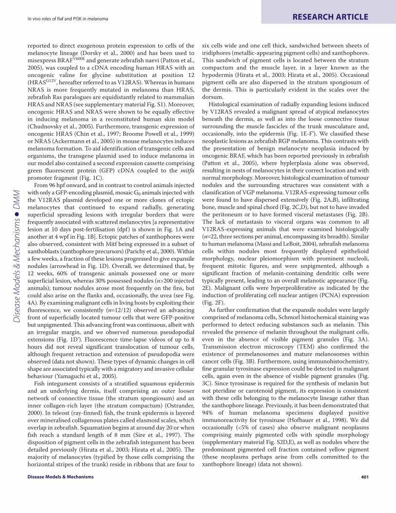
Efficient melanomagenesis requires an intact PI3K-Akt signallingpathwayThe relative inefficiency of V12RAST35S and BRAFV600E at inducingneoplasia (11% and 15%, respectively) when compared withV12RAS (90%), suggested that additional ‘hits’ (genetic orepigenetic) other than activation of Raf signalling are required forinitiation of melanoma. Together, activation of the PI3K-Aktsignalling module by V12RAS and its mutational targeting inhuman melanoma prompted us to examine whether PI3K-Aktsignalling is required for efficient melanomagenesis by oncogenicRas. To address this, we co-expressed V12RAS with an inhibitorof the PI3K pathway, a mutant form of the PI3K regulatory domainp85 (referred to elsewhere, and also herein, as Δp85) in which a 35amino acid in-frame deletion in the interSH2 domain deletes thep110 interaction domain (Rodriguez-Viciana et al., 1997)(supplementary material Fig. S4G; Fig. S5H). Expression of Δp85alone produced no overt effect on pigmentation in G0 animals (datanot shown). G0 animals expressing V12RAS alone or co-expressingV12RAS with Δp85 (V12RAS::Δp85) were followed until 12 wpfand were scored positive if they displayed one or more melanin-pigmented neoplasms, either superficial or nodular. Co-expressionof Δp85 with V12RAS (Fig. 5A; supplementary material Fig. S4G)significantly impaired V12RAS-induced tumourigenicity: weobserved an 80% reduction in neoplasia-positive animals co-expressing V12RAS and Δp85 compared with those expressingV12RAS alone (Fig. 5B) (a typical lesion induced by the RAS::Δp85construct is shown in Fig. 5C). Furthermore, nodular lesions wereonly observed in animals expressing V12RAS alone. This resultsuggests that stimulation of the PI3K-signalling module is requiredfor efficient initiation of neoplastic transformation by V12RAS.Indeed, the frequency of neoplasia induced by the V12RAS::Δp85construct was comparable to the frequency induced by V12RAST35S
and BRAFV600E, consistent with the above finding that Raf-Mek-Erk signalling is inefficient at inducing neoplasia.
Raf-Mek-Erk and PI3K-Akt signalling fail to synergise to inducemalignancyFrom the above data, we inferred that although an input from BRafis essential for neoplasia, it is not sufficient for malignancy. Sinceexpression of oncogenic Ras alone can induce malignancy, wehypothesised that a combination of Ras effectors, which shouldinclude BRaf, is required to reconstitute the full effect of V12RAS.For reasons stated above, co-activation of the PI3K-Akt pathwayseemed the most obvious candidate. To test this, we constructedplasmids allowing co-expression of BRAFV600E with eitherp110αCAAX or AktDD (see supplementary material Fig. S4D andS4F, respectively). Neither of these constructs could inducemelanoma (see Table 1). Rather, with co-expression ofp110αCAAX, we observed benign naevi at a similar frequency andlatency as for BRAF alone (14%, n=84) (Fig. 5D), whereas co-expression of AktDD resulted in no apparent lesions (n=167) (Table1). Co-expression of RlfCAAX or (C1199)Tiam1 also failed toaugment the tumourigenic effect of BRAFV600E (data not shown).In all cases, a benign histology was confirmed (data not shown).Thus, combined Raf-Mek-Erk and PI3K-Akt-signalling was notsufficient to induce malignancy, suggesting that, in addition toactivation of these two pathways, one or more additional V12RAS-dependent signals are required to reconstitute the full effect ofV12RAS.
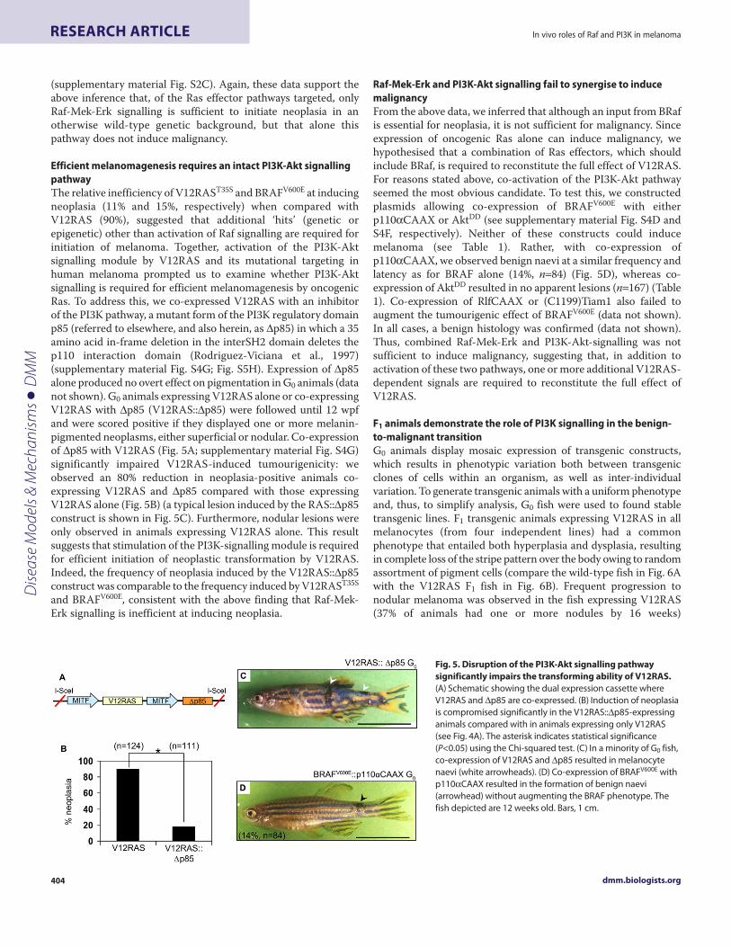
F1 animals demonstrate the role of PI3K signalling in the benign-to-malignant transitionG0 animals display mosaic expression of transgenic constructs,which results in phenotypic variation both between transgenicclones of cells within an organism, as well as inter-individualvariation. To generate transgenic animals with a uniform phenotypeand, thus, to simplify analysis, G0 fish were used to found stabletransgenic lines. F1 transgenic animals expressing V12RAS in allmelanocytes (from four independent lines) had a commonphenotype that entailed both hyperplasia and dysplasia, resultingin complete loss of the stripe pattern over the body owing to randomassortment of pigment cells (compare the wild-type fish in Fig. 6Awith the V12RAS F1 fish in Fig. 6B). Frequent progression tonodular melanoma was observed in the fish expressing V12RAS(37% of animals had one or more nodules by 16 weeks)
dmm.biologists.org404
In vivo roles of Raf and PI3K in melanomaRESEARCH ARTICLE
Fig. 5. Disruption of the PI3K-Akt signalling pathwaysignificantly impairs the transforming ability of V12RAS.(A) Schematic showing the dual expression cassette whereV12RAS and Δp85 are co-expressed. (B) Induction of neoplasiais compromised significantly in the V12RAS::Δp85-expressinganimals compared with in animals expressing only V12RAS(see Fig. 4A). The asterisk indicates statistical significance(P<0.05) using the Chi-squared test. (C) In a minority of G0 fish,co-expression of V12RAS and Δp85 resulted in melanocytenaevi (white arrowheads). (D) Co-expression of BRAFV600E withp110αCAAX resulted in the formation of benign naevi(arrowhead) without augmenting the BRAF phenotype. Thefish depicted are 12 weeks old. Bars, 1 cm.
Dise
ase
Mod
els &
Mec
hani
sms
D
MM
(supplementary material Fig. S6A). Intriguingly, the scales over thedorsum of these animals contain almost half the number ofpigmented melanocytes when compared with wild-type scales fromthe equivalent region (supplementary material Fig. S7). However,these melanocytes were more than four times larger thanmelanocytes found on wild-type scales (supplementary materialFig. S7). In contrast to V12RAS transgenic animals, and inagreement with a previous study (Patton et al., 2005), F1 animalsexpressing human BRAFV600E (from two independent lines)displayed only melanocytic hyperplasia (Fig. 6C), as apparent frombroader fused stripes and more populous hypertrophic, butcorrectly positioned, melanocytes associated with the scales overthe dorsum (Fig. 6C; supplementary material Fig. S7). We alsogenerated F1 animals co-expressing V12RAS and Δp85(V12RAS::Δp85) (three independent lines), all with a consistentphenotype [semi-quantitative reverse transcriptase (RT)-PCR wasused to confirm Δp85 expression] (supplementary material Fig.S6C). These animals also displayed an overall benign phenotypethat was analogous to the BRAFV600E-expressing line (Fig. 6D),retaining melanocyte hyperplasia but, unlike the lines expressingV12RAS alone, no marked dysplasia. Thus, animals co-expressingV12RAS and Δp85 possessed broader fused stripes (althoughinterstripes were still apparent ventrally) and more populous,correctly positioned melanocytes associated with the scales overthe dorsum (Fig. 6D; supplementary material Fig. S7). These
melanocytes were the most similar in size and distribution to thewild-type melanocytes (compare scales in Fig. 6A with those inFig. 6D; see also supplementary material Fig. S7), further confirmingtheir benign nature. Neither BRAFV600E-expressing animals, norV12RAS::Δp85 animals developed expansile nodules (no nodulesobserved in >200 animals after >15 months). However, localisedregions of progression to a pigment pattern like the one seen infish expressing V12RAS alone were noted in V12RAS::Δp85 animalsthat, nevertheless, failed to progress to VGP melanoma.
Disease Models & Mechanisms 405
In vivo roles of Raf and PI3K in melanoma RESEARCH ARTICLE
Fig. 6. PI3K-signalling is required for benign-to-malignant transition. Thepanels illustrate representative animals with an accompanying scale from thedorsum of wild-type zebrafish (A) and of each of the transgenic line series thatwere established (B-D). (B) V12RAS::GFP animals display melanocytichyperplasia and dysplasia that results in total aberration of the pigmentpattern. The melanocytes associated with the dorsal scales exhibit a morechaotic distribution compared with in other genotypes. (C) BRAFV600E animalsonly display melanocytic hyperplasia where the dorsal and lateral pigmentlines are fused together; melanocytes associated with the dorsal scales aremore populous in these fish. (D) V12RAS::Δp85 animals also exhibit thefeatures described in C, consistent with a benign neoplastic phenotype;however, stripes are generally less uniform. Bars, 1 cm (fish); 200 μm (scales).
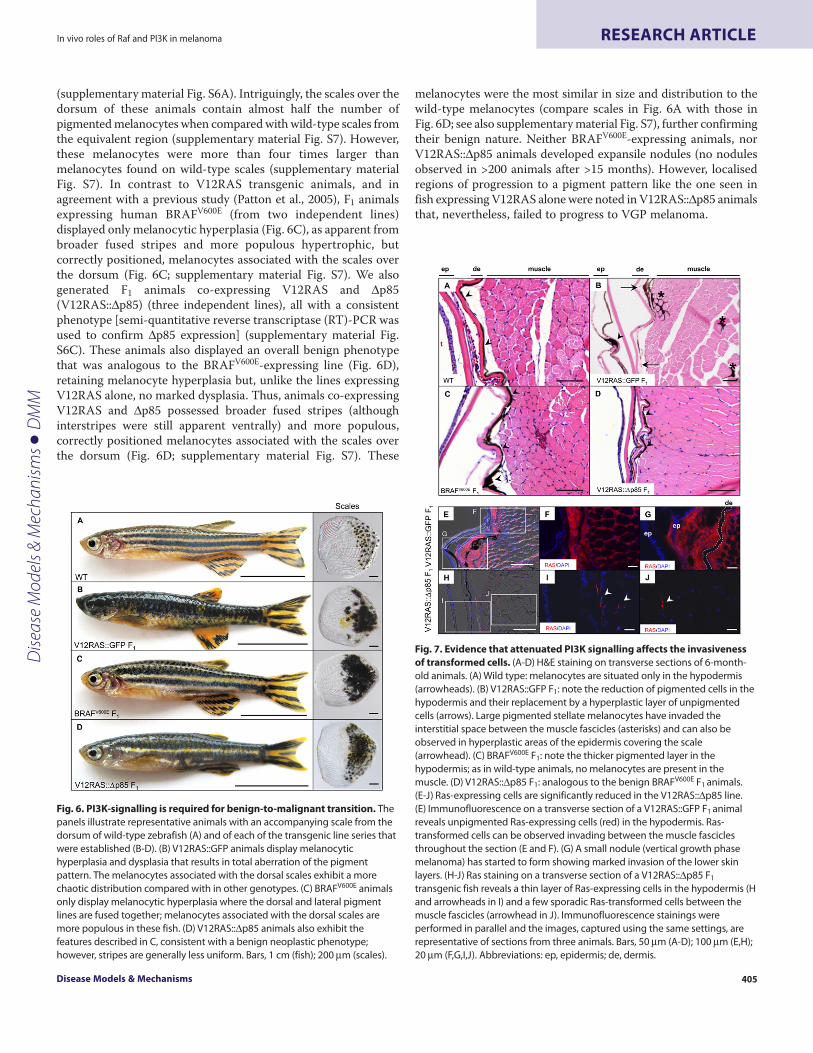
Fig. 7. Evidence that attenuated PI3K signalling affects the invasivenessof transformed cells. (A-D) H&E staining on transverse sections of 6-month-old animals. (A) Wild type: melanocytes are situated only in the hypodermis(arrowheads). (B) V12RAS::GFP F1: note the reduction of pigmented cells in thehypodermis and their replacement by a hyperplastic layer of unpigmentedcells (arrows). Large pigmented stellate melanocytes have invaded theinterstitial space between the muscle fascicles (asterisks) and can also beobserved in hyperplastic areas of the epidermis covering the scale(arrowhead). (C) BRAFV600E F1: note the thicker pigmented layer in thehypodermis; as in wild-type animals, no melanocytes are present in themuscle. (D) V12RAS::Δp85 F1: analogous to the benign BRAFV600E F1 animals.(E-J) Ras-expressing cells are significantly reduced in the V12RAS::Δp85 line.(E) Immunofluorescence on a transverse section of a V12RAS::GFP F1 animalreveals unpigmented Ras-expressing cells (red) in the hypodermis. Ras-transformed cells can be observed invading between the muscle fasciclesthroughout the section (E and F). (G) A small nodule (vertical growth phasemelanoma) has started to form showing marked invasion of the lower skinlayers. (H-J) Ras staining on a transverse section of a V12RAS::Δp85 F1
transgenic fish reveals a thin layer of Ras-expressing cells in the hypodermis (Hand arrowheads in I) and a few sporadic Ras-transformed cells between themuscle fascicles (arrowhead in J). Immunofluorescence stainings wereperformed in parallel and the images, captured using the same settings, arerepresentative of sections from three animals. Bars, 50 μm (A-D); 100 μm (E,H);20 μm (F,G,I,J). Abbreviations: ep, epidermis; de, dermis.
Dise
ase
Mod
els &
Mec
hani
sms
D
MM
These macroscopic observations gave a first indication thatattenuation of PI3K signalling had suppressed the malignantphenotype induced by oncogenic Ras. This was subsequentlyconfirmed by histology. As stated above, melanocytes in wild-typeanimals are located largely in the hypodermis (i.e. between the skindermis and muscle) (Fig. 7A). In V12RAS-expressing lines,melanocytes had expanded in the dermis and hypodermis, and werenow accompanied by a population of unpigmented cells in thehypodermis (Fig. 7B). Strikingly, pigmented stellate melanocyteswere also observed in the interstitial spaces between the musclefascicles. Occasional melanocyte invasion into epidermal layers wasalso apparent (Fig. 7B). This presentation can be equated to RGPmelanoma encompassing the entire body surface. In bothBRAFV600E-expressing lines (Fig. 7C) and V12RAS::Δp85 lines (Fig.7D), melanocytes were positioned correctly in the hypodermis.Significantly, both the presence of stellate melanocytes in betweenmuscle fascicles and invasion of the epidermis were completelyabsent in BRAFV600E-expressing lines (Fig. 7C) and largely absentin V12RAS::Δp85 lines (Fig. 7D and see below). This was analysedfurther using immunofluorescence to detect V12RAS expression,revealing the full extent of invasion of oncogenic Ras-transformedcells in animals expressing V12RAS alone compared with animalsco-expressing V12RAS and Δp85. In animals expressing V12RASalone, the new population of unpigmented cells generated in thehypodermis was shown to be V12RAS positive (Fig. 7E-G).Frequent V12RAS-positive cells were also identified betweenskeletal muscle fascicles of the trunk and in the overlying dermis(Fig. 7F,G). The presence of V12RAS-expressing cells in each ofthese locations in V12RAS::Δp85 animals was generally reducedgreatly (Fig. 7H), with only a few sporadic Ras-expressing cellsobserved between muscle fascicles (Fig. 7I,J).
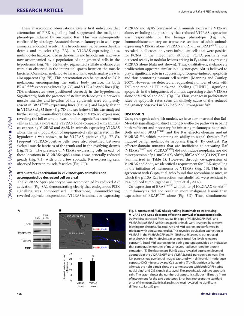
Attenuated Akt activation in V12RAS::Δp85 animals is notaccompanied by decreased cell survivalThe V12RAS::Δp85 phenotype was accompanied by reduced Aktactivation (Fig. 8A), demonstrating clearly that endogenous PI3Ksignalling was compromised. Furthermore, immunoblottingrevealed equivalent expression of V12RAS in animals co-expressing
V12RAS and Δp85 compared with animals expressing V12RASalone, excluding the possibility that reduced V12RAS expressionwas responsible for the benign phenotype (Fig. 8A).Immunohistochemistry on transverse sections of adult animalsexpressing V12RAS alone, V12RAS and Δp85, or BRAFV600E alonerevealed, in all cases, only very infrequent cells that were positivefor PCNA in the integument, although PCNA positivity wasdetected readily in nodular lesions arising in F1 animals expressingV12RAS alone (data not shown). Thus, qualitatively, melanocyteproliferation appeared similar in all genotypes. Akt is believed toplay a significant role in suppressing oncogene-induced apoptosisand thus promoting tumour cell survival (Manning and Cantley,2007). However, we detected an equivalent number of cells withTdT-mediated dUTP nick-end labelling (TUNEL), signifyingapoptosis, in the integument of animals expressing either V12RASalone or V12RAS and Δp85 (Fig. 8B). Thus, changes in proliferationrates or apoptosis rates seem an unlikely cause of the reducedmalignancy observed in V12RAS::Δp85 transgenic fish.
DISCUSSIONUsing transgenic zebrafish models, we have demonstrated that Raf-Mek-Erk signalling is distinct among Ras effector pathways in beingboth sufficient and necessary for initiating melanocyte neoplasia.Both mutant BRAFV600E and the Ras effector-domain mutantV12RAST35S, which maintains an ability to signal through Raf,induced benign melanocyte neoplasia (Fig. 4). By contrast, Raseffector-domain mutants that are inefficient at activating Raf(V12RASY40C and V12RASE37G) did not induce neoplasia; nor didmisexpression of p110αCAAX, AktDD, RlfCAAX or (C1199)Tiam1(summarised in Table 1). However, through co-expression ofV12RAS and Δp85, we identified a requirement for PI3K-signallingin the initiation of melanoma by V12RAS (Fig. 5B). This is inagreement with Gupta et al. who found that recombinant mice, inwhich the p110α-Ras interaction was abolished, were resistant toRas-induced tumourigenesis (Gupta et al., 2007).
Co-expression of BRAFV600E with either p110αCAAX or AktDD
in melanocytes did not result in more malignant lesions thanexpression of BRAFV600E alone (Fig. 5D). Thus, simultaneous
dmm.biologists.org406
In vivo roles of Raf and PI3K in melanomaRESEARCH ARTICLE
Fig. 8. Attenuated PI3K-Akt signalling in animals co-expressingV12RAS and Δp85 does not affect the survival of transformed cells.(A) Proteins extracted from caudal fin clips of V12RAS::GFP (RAS) andV12RAS::Δp85 (RAS Δp85) transgenic animals were analysed by westernblotting for phosphoAkt, total Akt and Mitf expression (performed intriplicate with equivalent results). This revealed equivalent expression ofV12RAS in the V12RAS::GFP and V12RAS::Δp85 animals, but reducedphosphoAkt in the V12RAS::Δp85 animals (total Akt levels remainedconstant). Equal Mitf expression for both genotypes provided an indicationthat comparable numbers of melanocytes had been lysed for proteinextraction. (B) The fluorescent TUNEL assay revealed equivalent levels ofapoptosis in the V12RAS::GFP and V12RAS::Δp85 transgenic animals. Theleft panels show overlays of images captured with differential interferencecontrast (DIC) microscopy and Cy3 staining (TUNEL-positive cells, red),whereas the right panels show the same sections with both DAPI (stainsnuclei blue) and Cy3 signals displayed. The arrowheads point to apoptoticcells. The graph shows the numbers of apoptotic cells per millimetre (mm)of integument for the two genotypes. Error bars represent the standarderror of the mean. Statistical analysis (t-test) revealed no significantdifference. Bars, 50 μm.
Dise
ase
Mod
els &
Mec
hani
sms
D
MM

activation of Raf-Mek-Erk and PI3K-Akt signalling does not fullyreconstitute oncogenic Ras signalling. This may explain why naeviin individuals with germ-line loss-of-function mutations in thePTEN gene are not reported to be at increased risk of progressingto melanoma. Both the Raf-Mek-Erk and PI3K-Akt signallingpathways individually induce senescence in primary cells (Chen etal., 2005; Michaloglou et al., 2005; Courtois-Cox et al., 2006), andthis effect could even be additive resulting, potentially, in thecomplete suppression of neoplasia that we observed with the BRAF-AktDD dual construct (Table 1). We propose that other Ras-inducedsignalling pathways uncouple the Raf-Mek-Erk and PI3K-Aktsignalling pathways from the induction of senescence. Recombinantmouse models have indicated essential roles for RalGDS, Tiam1and phospholipase Cε in Ras-induced skin tumour formation(Malliri et al., 2002; Bai et al., 2004; Gonzalez-Garcia et al., 2005),suggesting that multiple Ras pathways synergise to elicit neoplasia.
Although F1 transgenic zebrafish expressing BRAFV600E
developed only melanocyte hyperplasia, F1 animals expressingV12RAS displayed melanocyte hyperplasia, dysplasia and invasionof loose connective tissue (RGP melanoma) (Fig. 6B; Fig. 7B),progressing spontaneously to deeply invasive (VGP) melanoma(supplementary material Fig. S6B). As such, these animals mayserve as a model for familial atypical mole and melanoma (FAMM)syndrome (formerly dysplastic nevus syndrome), although germ-line mutations in the genes encoding the Ras proteins have notbeen linked to FAMM (Pho et al., 2006). Because PTENinactivation or Akt gain of function are observed mainly inadvanced melanoma (Haluska et al., 2006; Dahl and Guldberg,2007), we tested whether deregulated PI3K signalling was requiredfor the malignant conversion of V12RAS-expressing melanocytes.Again in the F1 setting, we were able to demonstrate in ourautochthonous melanoma model that PI3K signalling is requiredfor malignant progression, corroborating recent findings thatPTEN deficiency triggers melanoma progression in the mouse(Dankort et al., 2009). Thus, V12RAS::Δp85 F1 zebrafish effectivelyphenocopied BRAFV600E F1 zebrafish, displaying, largely, featuresof benign melanocyte neoplasia (compare Fig. 6C with 6D, andFig. 7C with 7D). Furthermore, despite reduced Akt activity (Fig.8A), which is linked to cell survival (Manning and Cantley, 2007),we did not detect a difference in apoptosis rates when comparinganimals expressing V12RAS alone with animals co-expressingV12RAS and Δp85 (Fig. 8B). Our findings, implicating PI3K inmalignant progression, are consistent with the increased PI3Kactivity detected in malignant melanoma in Xiphophoruscompared with in benign pigmented neoplasms (Wellbrock et al.,1999). Likewise, combining PTEN deficiency with mammary-specific expression of activated ErbB2 in mice, resulted in moremalignant metastatic tumours than ErbB2 expression alone(Dourdin et al., 2008). Furthermore, PTEN deficiency in theintestinal mucosa of Apc mutant mice resulted in progression ofintramucosal adenoma to full-thickness adenocarcinoma (Marshet al., 2008).
Cells expressing V12RAS alone frequently appeared to be moreprimitive than benign melanocytes (in that they wereunpigmented), and whether the increased malignancy ofmelanocytes expressing V12RAS alone is a direct effect of PI3K onmotility and invasiveness, or an indirect consequence of reversionto a more primitive melanoblast-like state (or both) merits further
investigation. A V12RAS-induced delay in differentiation might alsoexplain the reduced number of pigmented cells in dorsal scales ofV12RAS transgenic animals compared with wild-type animals oranimals with benign melanocyte neoplasia (supplementary materialFig. S7). Indeed, many GFP-expressing unpigmented cells can beobserved intermingled with pigmented cells in the dorsal scales ofV12RAS transgenic animals (data not shown).
The histopathology of melanocytic malignancies generated inzebrafish and Xiphophorus models preserve many of themicroscopic features of mouse and human frank melanoma. Thesefeatures include a variable morphology of transformed cells, withan epithelioid morphology being observed more frequently thanspindle morphology; nuclear pleiomorphism and prominentnucleoli; frequent mitotic figures; and variable amounts ofpigmentation (Broome Powell et al., 1999; Gimenez-Conti et al.,2001; Massi and LeBoit, 2004; Ackermann et al., 2005). The abilityto induce tumours efficiently in G0 zebrafish allows a relatively rapidassessment of the efficacy of a given oncogene in initiatingneoplasia. A large number of embryos can be microinjected withtransgenic constructs and analysed for tumour induction within12 weeks. Thus G0 assays resemble in vitro transformation assaysin their expediency, but – since they involve live organisms – moretruthfully reflect sporadic tumourigenesis. The high penetrance andrapid onset of melanoma in the V12RAS-induced melanomamodel described here combined with other advantages of usingzebrafish as a laboratory model organism (including optical clarity,forward genetic power, and amenability to transgenesis and smallmolecule treatment), promise to make this model a valuable toolfor gaining further insight into disease mechanism and fordeveloping new treatments.
METHODSReagentsHA-epitope-tagged human V12HRAS, V12HRAS effector-domainmutants (V12RAST35S, V12RASE37G, V12RASY40C), and HA-epitope-tagged murine (C1199)Tiam1 were all gifts from AngelikiMalliri (Paterson Institute for Cancer Research, Manchester, UK).Human BRAFV600E was a kind gift from Richard Marais (Institutefor Cancer Research, London, UK). HA-epitope-tagged murineRlfCAAX was donated by Channing Der (University of NorthCarolina, NC). Myc-epitope-tagged bovine p110αCAAX wascontributed by Andrew Gilmore (The University of Manchester,Manchester, UK). HA-epitope-tagged bovine AktDD was donatedby Jack Arbiser (Emory University, Atlanta, GA). Bovine Δp85 wasa kind gift from Julian Downward (London Research Institute,London, UK). A modified pBlueScript SK+ vector, in which themultiple cloning site had been flanked by I-SceI meganucleaserestriction sites, was kindly donated by Jochen Wittbrodt (EMBL,Heidelberg. Germany).
Constructs and generation of transgenic linesA 744-base-pair DNA fragment, proximal to the translationinitiation codon of the zebrafish homologue (isoform a) of themammalian MITF gene, was cloned by PCR and subsequentlyinserted into a pGEM-T cloning vector (Promega) (sequenceavailable upon request). Transgenes of interest, combining cDNAswith the mitfa promoter, were assembled in a modified pBlueScriptvector using standard cloning techniques. To test combinations of
Disease Models & Mechanisms 407
In vivo roles of Raf and PI3K in melanoma RESEARCH ARTICLED
iseas
e M
odel
s & M
echa
nism
s
DM
M

Ras effectors, dual expression cassettes were assembled combiningcDNAs for two Ras effectors (e.g. BRAFV600E and p110αCAAX).To assess the effect of antagonising PI3K, a V12RAS expressioncassette was combined with a Δp85 expression cassette. Successfulcloning of transgene constructs was verified in each case by (1)restriction digest analysis (data not shown), (2) sequencing (datanot shown) and (3) by transient transfection of constructs intoHEK293 cells, followed by protein extraction and immunoblottingto confirm that exogenous protein(s) were generated (seesupplementary material Fig. S4).
Transgenic animals were generated by co-injection of 1 nl ofplasmid DNA (25 ng/μl) with 10 U I-SceI meganuclease into one-cell stage AB-strain zebrafish zygotes, using a PLI-90 picoinjectormicroinjection station as described previously (Thermes et al.,2002). Subsequently, animals were raised under standard conditions(28.5°C ambient temperature, 14 hour-10 hour dark-light cycle, adiet of brine shrimp and flake food) at the Biological ServicesFacility at The University of Manchester. Individual G0 founderswere backcrossed to AB animals to generate stable transgenic lines.Animals anaesthetised with MS222 (Sigma) were photographedusing either an unmounted Canon Digital Ixus 80 IS camera or anAxiocam MR digital camera mounted on a Zeiss StereoLumarstereodissecting microscope. Images were processed in AdobePhotoshop or using Axiovision software. Fin tissue and scales werealso isolated from animals under general anaesthesia. All animalprocedures were subject to local ethical review and performedunder a Home Office Licence.
Histology and immunostainingAdult fish euthanised by exposure to excess anaesthesia were fixedin 4% paraformaldehyde (PFA), and paraffin-embedded tissuesections (5 μm) were used for H&E staining, immunofluorescenceand immunohistochemistry. H&E staining was carried out usingstandard methods (Thermo Shandon staining machine). Forimmunofluorescence, the primary antibodies used were mouseanti-Ras (1:100 dilution; BD Transduction Laboratories) and mouseanti-PCNA (1:100 dilution; Euro-Diagnostica). The secondaryantibody was Vectastain biotinylated horse anti-pan-IgG. Followingincubation with the secondary antibody, the sections wereincubated with Cy3-labeled streptavidin (1:100 dilution; Sigma) andmounted using Vectashield with DAPI (Vectorlabs) in order tovisualise nuclei. For immunohistochemistry, tissue sections werestained with mouse anti-tyrosinase (1:50 dilution; Upstate 05-647),rabbit anti-phosphoErk (1:100 dilution; Cell Signalling Technology)and rabbit anti-phosphoAkt (1:100 dilution; Cell SignallingTechnology) antibodies, as per the manufacturer’s instructions.Images were captured using a Zeiss Axioplan 2 compoundfluorescent microscope and Axiocam MR camera, and processedusing Axiovision software. Alternatively, sections were scanned, andimages were captured and processed using a Zeiss Mirax Scansystem with a �20 objective lens and a Marlin f14 6C camera. Allhistological samples were evaluated by a clinical pathologist (JivkoKamarashev).
Schmorl stainParaffin sections of fish with neoplastic lesions were deparaffinisedin xylene and rehydrated in descending concentrations of ethanol.The sections were incubated for 10 minutes with a working
solution comprising 0.75% ferric chloride and (freshly prepared)0.1% potassium ferricyanide in distilled water, and washed indistilled water. Melanin has the ability to reduce ferricyanide toferrocyanide, which in the presence of ferric ions forms PrussianBlue and, therefore, cells containing melanin are stained blue.Nuclear Fast Red was used as a counterstain (2 minutes) beforesections were dehydrated in ascending concentrations of ethanol.Images were captured using a Zeiss Axioplan 2 compoundmicroscope and Axiocam MR camera, and processed usingAxiovision software.
Transmission electron microscopyTEM was carried out in the EM facility of the Faculty of LifeSciences at The University of Manchester. Specimen processing wasperformed as described previously (Toma et al., 1999). Fish withtumour nodules were sacrificed by excess anaesthetisation, and thetumours were excised and immersed in a mixture of 3% PFA and3% glutaraldehyde in 0.1 M cacodylate buffer, pH 7.4, for 2 hoursat room temperature. The specimens were then cut into smallpieces, immersed in the same fixative overnight at 4°C, and post-fixed in 2% osmium tetraoxide for 2 hours at 0°C. The specimenswere block stained with 0.5% uranyl acetate in 50% ethanolovernight and embedded in Epon 812 resin after dehydration in agraded series of ethanol. Ultra-thin sections (70-80 nm) were cutusing a Reichert Jung Ultramicrotome. TEM observations werecarried out using a FEI Tecnai12 Biotwin microscope andphotographs were taken using a CCD camera (Eagle, FEI Company).Images were acquired using an Imacon scanner and FlexColorsoftware.
ImmunoblottingFor immunoblotting, protein was extracted from caudal fin clips(n=5 for each genotype), resolved in 4-15% gradient SDS-polyacrylamide gels (Biorad), transferred to PVDF membranes(Millipore), and incubated with the following antibodies: anti-Ras(BD Transduction Laboratories), anti-phosphoAkt (Cell SignalingTechnology), anti-total Akt (Cell Signaling Technology) and anti-MITF (DakoCytomation). The secondary antibody was either anti-mouse IgG or anti-rabbit IgG conjugated to horseradish peroxidase(HRP) (Amersham Biosciences). Proteins were visualised with ECL(enhanced chemiluminescence) plus (Amersham Biosciences), asper the manufacturer’s guidelines. All immunoblotting procedureswere performed in triplicate and generated equivalent results.
Fluorescent (F)-TUNEL assayAdult fish (n=3) from each of the V12RAS::GFP and V12RAS::Δp85transgenic lines, sacrificed by excess anaesthetisation, were fixedin 4% PFA and embedded in paraffin. Transverse sections (5 μm)were used for the F-TUNEL assay. The assay was performed usingthe ApopTag Red in situ apoptosis detection kit (ChemiconInternational), as per the manufacturer’s instructions. Apoptoticcells in the integument (i.e. superficial to the muscle) were countedfrom three sections of each animal (i.e. nine sections in total foreach genotype) and the perimeter of each section was measuredusing Axiovision software, in order to determine the number ofapoptotic cells per millimetre of integument. The measurementswere analysed for statistical significance using an independentsamples t-test.
dmm.biologists.org408
In vivo roles of Raf and PI3K in melanomaRESEARCH ARTICLED
iseas
e M
odel
s & M
echa
nism
s
DM
M
BioinformaticsProtein sequences for human and murine Ras isoforms; humanERK1 and ERK2; human AKT1, AKT2 and AKT3; human BRAF;bovine p110α; bovine Akt; murine Rlf; murine Tiam1; murine RalAand RalB; murine Rac1; and bovine p85 were used to identifyhomologues in the zebrafish genome Zv7 sequence build(http://www.ensembl.org/Danio_rerio/index.html) by performingTBLASTN or BLASTP searches. Reciprocal best-BLAST andsynteny were used to confirm orthology. Peptide sequences werealigned using ClustalW multiple alignment application of BioEditsoftware.
Cell cultureHEK293 cells were maintained in DMEM/F12 Glutamax (GIBCO)supplemented with 10% foetal calf serum (Lonza) and 1% penicillin-streptomycin (Lonza). The cells were transfected with 1 μg of theplasmid of interest using FuGene6 (Roche) transfection reagent,as per the manufacturer’s instructions. AB9 zebrafish fibroblastswere maintained in DMEM/F12 Glutamax supplemented with 15%foetal calf serum (Lonza), 1% penicillin-streptomycin (Lonza) and0.6% Fungizone (GIBCO). Cells were maintained at 28°C with 5%CO2.
Cell culture immunoblottingHEK293 cellsFor immunoblotting, protein was extracted from transfectedHEK293 cells, on the third day after transfection, resolved in 4-15% gradient SDS-polyacrylamide gels (Biorad), transferred toPVDF membranes (Millipore) and incubated with the followingantibodies: anti-HA high affinity (Roche), anti-Ras (BDTransduction Laboratories), anti-Akt (Cell Signaling Technology),anti-Erk (Cell Signaling Technology), anti-BRAF (BD TransductionLaboratories), anti-PI3K p110α (BD Transduction Laboratories),anti-PI3K p85 (Upstate Biotechnology, Inc) and anti-β-actin(Sigma). The secondary antibody was anti-rat IgG, anti-mouse IgGor anti-rabbit IgG, all of which were conjugated to HRP (AmershamBiosciences). Proteins were visualised with ECL plus (AmershamBiosciences) according to the manufacturer’s guidelines. Allimmunoblotting procedures were performed in triplicate andgenerated equivalent results.
AB9 cellsAB9 cells were starved of serum for 24 hours before treatment withIGF-1 (Gro-Pep) for 10 minutes at a final concentration of 100ng/μl. Negative control cells were left untreated. Immediately afterIGF-1 treatment, the protein was extracted, resolved in 4-15%gradient SDS-polyacrylamide gels (Biorad), transferred to PVDFmembranes (Millipore) and incubated with the followingantibodies: mouse anti-phosphoErk (Cell Signalling Technology),rabbit anti-phosphoAkt (Cell Signalling Technology) and rabbitanti-β-tubulin (Abcam). The secondary antibody was either anti-rabbit IgG or anti-mouse IgG, both of which were conjugated toHRP (Amersham Biosciences). Proteins were visualised with ECLplus (Amersham Biosciences) according to the manufacturer’sguidelines. All immunoblotting procedures were performed intriplicate and generated equivalent results.
ImmunocytochemistryAB9 zebrafish fibroblasts grown on glass coverslips were starvedof serum for 24 hours before treatment with IGF-1 (Gro-Pep) for10 minutes at a final concentration of 100 ng/μl. Negative controlcells were left untreated. Immediately after the IGF-1 treatment,cells were fixed for 30 minutes at 4°C with 4% PFA, and stainedwith mouse anti-phosphoErk (1:400 dilution; Cell SignallingTechnology) and rabbit anti-phosphoAkt (1:25 dilution; CellSignalling Technology) antibodies, as per the manufacturer’sinstructions. In order to reduce non-specific antibody binding, anextra step of avidin-biotin block (Vectorlabs) was added just beforeincubation with the primary antibody. Images were captured usinga Zeiss Axioplan 2 compound fluorescent microscope and AxiocamMR camera, and processed using Axiovision software. The aboveimmunocytochemical stainings were performed in triplicate andgenerated equivalent results.
Semi-quantitative RT-coupled PCRTotal RNA was isolated and pooled from caudal fin clips (n=3 foreach genotype) using the RNAeasy mini kit (Qiagen), and reversetranscribed with an Omniscript reverse transcription kit (Qiagen)to produce cDNA. cDNA was amplified through RT-PCR usingGoTaq polymerase (Promega) and primers that flank the wild-typep85 sequence of both bovine and zebrafish p85. Primer sequencesare available upon request.
Disease Models & Mechanisms 409
In vivo roles of Raf and PI3K in melanoma RESEARCH ARTICLE
TRANSLATIONAL IMPACT
Clinical issueDeregulated Ras signalling is observed in the vast majority of human solidneoplasias, exemplified by cutaneous melanoma. Mutational profiling suggeststhat activation of Raf-Mek-Erk signalling downstream of Ras may be crucial forinitiating melanocyte neoplasia, whereas phosphoinositide 3-kinase (PI3K)-Aktsignalling is involved in malignant progression. These hypotheses have not yetbeen fully tested in in vivo models. Treatment options for patients withadvanced (metastatic) melanoma are very limited and their prognosis remainspoor. Understanding the molecular basis of melanoma formation andprogression should identify potential targets for the development of moreeffective medicines.
ResultsThe authors generate several transgenic zebrafish melanoma models byexpressing oncogenic Ras, attenuated versions of Ras, and activated Raseffectors in their melanocytes. The ability of Ras and Ras effector mutants toinduce melanocyte neoplasia confirms an important role for Raf-Mek-Erksignalling in melanoma initiation. Germline transmission of oncogenic Rasproduced fish that were covered with dysplastic melanocytes, whichfrequently progress to melanoma, reminiscent of familial atypical mole andmelanoma (FAMM) syndrome seen in humans. Malignancy was abrogated inthese animals by co-expressing a PI3K inhibitor, confirming a role for PI3K inmelanoma progression.
Implications and future directionsThe authors created and characterise zebrafish models of melanoma throughdisruption of Ras signalling. These fish display high penetrance and a rapidonset of melanoma. These findings and other characteristics of zebrafish,including optical clarity, forward genetic power, and amenability totransgenesis and small molecule treatment, suggest the future utility of thismodel in understanding and treating human cancer.
doi:10.1242/dmm.003574
Dise
ase
Mod
els &
Mec
hani
sms
D
MM

Scale melanocyte number and size measurementsAdult animals representative of the genotypes of interest (wild-type,V12RAS::GFP, BRAFV600E and V12RAS::Δp85) were anaesthetisedwith MS222 (Sigma), and scales were then removed from theirdorsum, behind the head, using fine forceps (n=6 scales for eachof three animals per genotype). The isolated scales werephotographed using a Zeiss StereoLumar stereodissectingmicroscope and the obtained images were analysed usingAxiovision Automeasure software. This application enables themeasurement of areas with a specified density (in this case thepigment in melanocytes). The results are given as pigmented areasin μm2. The scales were then treated with 1 mg/ml epinephrine(Sigma) for 5 minutes and photographed using the samemicroscope with Axiovision sofware. Epinephrine aggregates themelanosomes in the melanocytes and facilitates cell counting bymaking individual cells distinct (Rawls and Johnson, 2001). For eachscale: (1) the melanocytes were counted and (2) the measurementof pigmented area (as measured with Axiovision Automeasure) wasdivided by the number of melanocytes on the scale, and ameasurement of μm2/melanocyte was obtained. A meanmeasurement was generated for each genotype and analysed forstatistical significance using a one-way ANOVA test.ACKNOWLEDGEMENTSThis work has been funded by a Cancer Research UK Career DevelopmentFellowship and a Cancer Research UK Phd studentship awarded to A.H. We thankMartin Humphries, Angeliki Malliri, Claudia Wellbrock and Stephen Taylor forcritically reading the manuscript. We also thank Robert Kelsh, James Lister,Stephen Johnson and Keith Hoek for helpful comments and suggestions, and JackArbiser for advice on phosphoErk and phosphoAkt immunohistochemical staining.The authors also thank the staff in the EM facility of the Faculty of Life Sciences atThe University of Manchester for their assistance, and thank the Wellcome Trust forequipment grant support to the EM facility. In addition, we thank Steve Bagley, ofthe Paterson Institute Advanced Imaging Facility, for help in acquiring histologyimages, and thank the staff of The University of Manchester BSF for maintainingthe zebrafish facility.
COMPETING INTERESTSThe authors declare no competing financial interests.
AUTHOR CONTRIBUTIONSC.M. performed the majority of experiments, analysed data and assisted in thepreparation of the manuscript; M.J. generated Ras transgenic lines and certainDNA constructs; P.W. generated certain DNA constructs and provided bioinfomaticsupport; J.K. is a dermatopathologist with expertise in melanoma and performedhistological analyses of zebrafish melanocytic neoplasms; A.K. first generated andtested dual expression cassettes; A.F.L.H. instigated and devised the study andprepared the manuscript.
SUPPLEMENTARY MATERIALSupplementary material for this article is available athttp://dmm.biologists.org/lookup/suppl/doi:10.1242/dmm.001149/-/DC1
Received 16 July 2008; Accepted 17 February 2009.
REFERENCESAckermann, J., Frutschi, M., Kaloulis, K., McKee, T., Trumpp, A. and Beermann, F.
(2005). Metastasizing melanoma formation caused by expression of activated N-RasQ61K on an INK4a-deficient background. Cancer Res. 65, 4005-4011.
Alessi, D. R., Andjelkovic, M., Caudwell, B., Cron, P., Morrice, N., Cohen, P. andHemmings, B. A. (1996). Mechanism of activation of protein kinase B by insulin andIGF-1. EMBO J. 15, 6541-6551.
Bai, Y., Edamatsu, H., Maeda, S., Saito, H., Suzuki, N., Satoh, T. and Kataoka, T.(2004). Crucial role of phospholipase Cepsilon in chemical carcinogen-induced skintumor development. Cancer Res. 64, 8808-8810.
Bennett, D. C. and Lamoreux, M. L. (2003). The color loci of mice-a genetic century.Pigment Cell Res. 16, 333-344.
Birck, A., Ahrenkiel, V., Zeuthen, J., Hou-Jensen, K. and Guldberg, P. (2000).Mutation and allelic loss of the PTEN/MMAC1 gene in primary and metastaticmelanoma biopsies. J. Invest. Dermatol. 114, 277-280.
Broome Powell, M., Gause, P. R., Hyman, P., Gregus, J., Lluria-Prevatt, M., Nagle, R.and Bowden, G. T. (1999). Induction of melanoma in TPras transgenic mice.Carcinogenesis 20, 1747-1753.
Cancer Research UK (2006). CancerStats report: Malignant Melanoma UK. London:Cancer Research UK.
Chen, Z., Trotman, L. C., Shaffer, D., Lin, H. K., Dotan, Z. A., Niki, M., Koutcher, J. A.,Scher, H. I., Ludwig, T., Gerald, W. et al. (2005). Crucial role of p53-dependentcellular senescence in suppression of Pten-deficient tumorigenesis. Nature 436, 725-730.
Chin, L., Pomerantz, J., Polsky, D., Jacobson, M., Cohen, C., Cordon-Cardo, C.,Horner, J. W., 2nd and DePinho, R. A. (1997). Cooperative effects of INK4a and rasin melanoma susceptibility in vivo. Genes Dev. 11, 2822-2834.
Chudnovsky, Y., Adams, A. E., Robbins, P. B., Lin, Q. and Khavari, P. A. (2005). Use ofhuman tissue to assess the oncogenic activity of melanoma-associated mutations.Nat. Genet. 37, 745-749.
Courtois-Cox, S., Genther Williams, S. M., Reczek, E. E., Johnson, B. W.,McGillicuddy, L. T., Johannessen, C. M., Hollstein, P. E., MacCollin, M. andCichowski, K. (2006). A negative feedback signaling network underlies oncogene-induced senescence. Cancer Cell 10, 459-472.
Dahl, C. and Guldberg, P. (2007). The genome and epigenome of malignantmelanoma. APMIS 115, 1161-1176.
Dankort, D., Curley, D. P., Cartlidge, R. A., Nelson, B., Karnezis, A. N., Damsky, W.E., Jr, You, M. J., Depinho, R. A., McMahon, M. and Bosenberg, M. (2009).Braf(V600E) cooperates with Pten loss to induce metastatic melanoma. Nat. Genet.Mar 12 [Epub ahead of print] [doi:10.1038/ng.356].
Dorsky, R. I., Raible, D. W. and Moon, R. T. (2000). Direct regulation of nacre, azebrafish MITF homolog required for pigment cell formation, by the Wnt pathway.Genes Dev. 14, 158-162.
Dourdin, N., Schade, B., Lesurf, R., Hallett, M., Munn, R. J., Cardiff, R. D. and Muller,W. J. (2008). Phosphatase and tensin homologue deleted on chromosome 10deficiency accelerates tumor induction in a mouse model of ErbB-2 mammarytumorigenesis. Cancer Res. 68, 2122-2131.
Gimenez-Conti, I., Woodhead, A. D., Harshbarger, J. C., Kazianis, S., Setlow, R. B.,Nairn, R. S. and Walter, R. B. (2001). A proposed classification scheme forXiphophorus melanomas based on histopathologic analyses. Mar. Biotechnol. 3,S100-S106.
Gonzalez-Garcia, A., Pritchard, C. A., Paterson, H. F., Mavria, G., Stamp, G. andMarshall, C. J. (2005). RalGDS is required for tumor formation in a model of skincarcinogenesis. Cancer Cell 7, 219-226.
Gordon, M. (1931). Hereditary basis of melanosis in hybrid fishes. Am. J. Cancer 15,1495-1519.
Govindarajan, B., Sligh, J. E., Vincent, B. J., Li, M., Canter, J. A., Nickoloff, B. J.,Rodenburg, R. J., Smeitink, J. A., Oberley, L., Zhang, Y. et al. (2007).Overexpression of Akt converts radial growth melanoma to vertical growthmelanoma. J. Clin. Invest. 117, 719-729.
Guldberg, P., thor Straten, P., Birck, A., Ahrenkiel, V., Kirkin, A. F. and Zeuthen, J.(1997). Disruption of the MMAC1/PTEN gene by deletion or mutation is a frequentevent in malignant melanoma. Cancer Res. 57, 3660-3663.
Gupta, S., Ramjaun, A. R., Haiko, P., Wang, Y., Warne, P. H., Nicke, B., Nye, E.,Stamp, G., Alitalo, K. and Downward, J. (2007). Binding of ras to phosphoinositide3-kinase p110alpha is required for ras-driven tumorigenesis in mice. Cell 129, 957-968.
Habets, G. G., Scholtes, E. H., Zuydgeest, D., van der Kammen, R. A., Stam, J. C.,Berns, A. and Collard, J. G. (1994). Identification of an invasion-inducing gene,Tiam-1, that encodes a protein with homology to GDP-GTP exchangers for Rho-likeproteins. Cell 77, 537-549.
Haluska, F. G., Tsao, H., Wu, H., Haluska, F. S., Lazar, A. and Goel, V. (2006).Genetic alterations in signaling pathways in melanoma. Clin. Cancer Res. 12, 2301s-2307s.
Hirata, M., Nakamura, K., Kanemaru, T., Shibata, Y. and Kondo, S. (2003). Pigmentcell organization in the hypodermis of zebrafish. Dev. Dyn. 227, 497-503.
Hirata, M., Nakamura, K. and Kondo, S. (2005). Pigment cell distributions in differenttissues of the zebrafish, with special reference to the striped pigment pattern. Dev.Dyn. 234, 293-300.
Hofbauer, G. F., Kamarashev, J., Geertsen, R., Boni, R. and Dummer, R. (1998).Tyrosinase immunoreactivity in formalin-fixed, paraffin-embedded primary andmetastatic melanoma: frequency and distribution. J. Cutan. Pathol. 25, 204-209.
Johnson, S. L., Africa, D., Walker, C. and Weston, J. A. (1995). Genetic control ofadult pigment stripe development in zebrafish. Dev. Biol. 167, 27-33.
Kelsh, R. N. (2004). Genetics and evolution of pigment patterns in fish. Pigment CellRes. 17, 326-336.
Kelsh, R. N., Brand, M., Jiang, Y. J., Heisenberg, C. P., Lin, S., Haffter, P., Odenthal,J., Mullins, M. C., van Eeden, F. J., Furutani-Seiki, M. et al. (1996). Zebrafish
dmm.biologists.org410
In vivo roles of Raf and PI3K in melanomaRESEARCH ARTICLED
iseas
e M
odel
s & M
echa
nism
s
DM
M
pigmentation mutations and the processes of neural crest development.Development 123, 369-389.
Lambert, J. M., Lambert, Q. T., Reuther, G. W., Malliri, A., Siderovski, D. P., Sondek,J., Collard, J. G. and Der, C. J. (2002). Tiam1 mediates Ras activation of Rac by aPI(3)K-independent mechanism. Nat. Cell Biol. 4, 621-625.
Malliri, A., van der Kammen, R. A., Clark, K., van der Valk, M., Michiels, F. andCollard, J. G. (2002). Mice deficient in the Rac activator Tiam1 are resistant to Ras-induced skin tumours. Nature 417, 867-871.
Manning, B. D. and Cantley, L. C. (2007). AKT/PKB signaling: navigating downstream.Cell 129, 1261-1274.
Marsh, V., Winton, D. J., Williams, G. T., Dubois, N., Trumpp, A., Sansom, O. J. andClarke, A. R. (2008). Epithelial Pten is dispensable for intestinal homeostasis butsuppresses adenoma development and progression after Apc mutation. Nat. Genet.40, 1436-1444.
Massi, G. and LeBoit, P. E. (2004). Histological Diagnosis of Nevi and Melanoma.Darmstadt: Steinkopff.
Meierjohann, S. and Schartl, M. (2006). From Mendelian to molecular genetics: theXiphophorus melanoma model. Trends Genet. 22, 654-661.
Michaloglou, C., Vredeveld, L. C., Soengas, M. S., Denoyelle, C., Kuilman, T., vander Horst, C. M., Majoor, D. M., Shay, J. W., Mooi, W. J. and Peeper, D. S. (2005).BRAFE600-associated senescence-like cell cycle arrest of human naevi. Nature 436,720-724.
Ostrander, G. K. (2000). The Laboratory Fish. San Diego, CA: Academic Press.Parichy, D. M., Ransom, D. G., Paw, B., Zon, L. I. and Johnson, S. L. (2000). An
orthologue of the kit-related gene fms is required for development of neural crest-derived xanthophores and a subpopulation of adult melanocytes in the zebrafish,Danio rerio. Development 127, 3031-3044.
Patton, E. E., Widlund, H. R., Kutok, J. L., Kopani, K. R., Amatruda, J. F., Murphey, R.D., Berghmans, S., Mayhall, E. A., Traver, D., Fletcher, C. D. et al. (2005). BRAFmutations are sufficient to promote nevi formation and cooperate with p53 in thegenesis of melanoma. Curr. Biol. 15, 249-254.
Payne, D. M., Rossomando, A. J., Martino, P., Erickson, A. K., Her, J. H.,Shabanowitz, J., Hunt, D. F., Weber, M. J. and Sturgill, T. W. (1991). Identificationof the regulatory phosphorylation sites in pp42/mitogen-activated protein kinase(MAP kinase). EMBO J. 10, 885-892.
Pho, L., Grossman, D. and Leachman, S. A. (2006). Melanoma genetics: a review ofgenetic factors and clinical phenotypes in familial melanoma. Curr. Opin. Oncol. 18,173-179.
Pollock, P. M., Harper, U. L., Hansen, K. S., Yudt, L. M., Stark, M., Robbins, C. M.,Moses, T. Y., Hostetter, G., Wagner, U., Kakareka, J. et al. (2003). High frequencyof BRAF mutations in nevi. Nat. Genet. 33, 19-20.
Rawls, J. F. and Johnson, S. L. (2001). Requirements for the kit receptor tyrosine kinaseduring regeneration of zebrafish fin melanocytes. Development 128, 1943-1949.
Rodriguez-Viciana, P., Warne, P. H., Khwaja, A., Marte, B. M., Pappin, D., Das, P.,Waterfield, M. D., Ridley, A. and Downward, J. (1997). Role of phosphoinositide 3-OH kinase in cell transformation and control of the actin cytoskeleton by Ras. Cell89, 457-467.
Sire, J. Y., Allizard, F., Babiar, O., Bourguignon, J. and Quilhac, A. (1997). Scaledevelopment in zebrafish (Danio rerio). J. Anat. 190, 545-561.
Stahl, J. M., Sharma, A., Cheung, M., Zimmerman, M., Cheng, J. Q., Bosenberg, M.W., Kester, M., Sandirasegarane, L. and Robertson, G. P. (2004). Deregulated Akt3activity promotes development of malignant melanoma. Cancer Res. 64, 7002-7010.
Thermes, V., Grabher, C., Ristoratore, F., Bourrat, F., Choulika, A., Wittbrodt, J. andJoly, J. S. (2002). I-SceI meganuclease mediates highly efficient transgenesis in fish.Mech. Dev. 118, 91-98.
Toma, H., Nakamura, K., Kuraoka, A., Tanaka, M. and Kawabuchi, M. (1999). Three-dimensional structures of c-Kit-positive cellular networks in the guinea pig smallintestine and colon. Cell Tissue Res. 295, 425-436.
Tsao, H., Atkins, M. B. and Sober, A. J. (2004). Management of cutaneous melanoma.N. Engl. J. Med. 351, 998-1012.
Wan, P. T., Garnett, M. J., Roe, S. M., Lee, S., Niculescu-Duvaz, D., Good, V. M.,Jones, C. M., Marshall, C. J., Springer, C. J., Barford, D. et al. (2004). Mechanism ofactivation of the RAF-ERK signaling pathway by oncogenic mutations of B-RAF. Cell116, 855-867.
Wellbrock, C., Fischer, P. and Schartl, M. (1999). PI3-kinase is involved in mitogenicsignaling by the oncogenic receptor tyrosine kinase Xiphophorus melanomareceptor kinase in fish melanoma. Exp. Cell Res. 251, 340-349.
Whiteman, D. C., Zhou, X. P., Cummings, M. C., Pavey, S., Hayward, N. K. and Eng,C. (2002). Nuclear PTEN expression and clinicopathologic features in a population-based series of primary cutaneous melanoma. Int. J. Cancer 99, 63-67.
Wolthuis, R. M., de Ruiter, N. D., Cool, R. H. and Bos, J. L. (1997). Stimulation of geneinduction and cell growth by the Ras effector Rlf. EMBO J. 16, 6748-6761.
Wu, H., Goel, V. and Haluska, F. G. (2003). PTEN signaling pathways in melanoma.Oncogene 22, 3113-3122.
Yamaguchi, H., Wyckoff, J. and Condeelis, J. (2005). Cell migration in tumors. Curr.Opin. Cell Biol. 17, 559-564.
Disease Models & Mechanisms 411
In vivo roles of Raf and PI3K in melanoma RESEARCH ARTICLED
iseas
e M
odel
s & M
echa
nism
s
DM
M