dissecting the transactivation domain of the transcription
TRANSCRIPT
1
Dissecting the Transactivation Domain
of the Transcription Factor c-Myb
A joint project for three MSc students
Priyanga-Dina Udayakumar
Thesis for the Master’s degree in Molecular Biosciences
Main field of study in molecular biology
30 credits
Department of Bioscience
Faculty of Mathematics and Natural Science
UNIVERSITY OF OSLO
June 2019

2
© Priyanga-Dina Udayakumar
2019
Dissecting the transactivation domain of the transcription factor c-Myb
Priyanga-Dina Udayakumar
http://www.duo.uio.no/
Trykk: Reprosentralen, Universitetet i Oslo

3
Acknowledgment
This work was performed at the Department of Biosciences, Faculty of Mathematics and
Natural Science, University of Oslo in the period from January 2019 to June 2019.
First of all, I would like to thank my supervisor Professor Odd Stokke Gabrielsen for giving
me the opportunity to join the Myb-group and participate in this interesting master project.
His knowledge and professional guidance, as well as his encouragement and positivity, have
been greatly appreciated.
Second, I would like to particularly thank my co-supervisor Marit Ledsaak. Her continuous
guidance and support in the laboratory and during the writing process have meant a lot to me.
I want to thank her for always taking the time to answer all of my questions and share her
knowledge with me.
I would like to direct a special thanks to my fellow master students Guro and Jan Ove. They
have been supportive through every step of this project, and their kindness has been
invaluable to me. A special thanks to Guro who has been a big inspiration and a great friend
for several years. I would also like to thank the other Myb-group members and laboratory
colleagues. A special thanks to Andrea, Pradip, Kirill and Signe. They have been helpful with
both practical work and the writing process, as well as creating a good social environment.
A big thanks to all my friends, especially Tuja for always giving me motivation and strength
through all these years at Blindern. Her encouraging words have really helped me through all
of my struggles.
I am also grateful to Balakumaran for his patience, support and for making the writing process
less stressful.
Finally, I would like to thank my parents, my sister, my brother and the rest of my family for
believing in me and making all of this possible.
Priyanga-Dina Udayakumar
Oslo, June 2019

4
Abstract
The transcription factor and oncoprotein c-Myb regulates many genes during blood cell
development, from stem cells to more mature cells. The protein contains three domains: (1)
DNA-binding domain (DBD), (2) transactivation domain (tAD) and (3) C-regulatory domain
(CRD). There is an extensive knowledge of DBDs for numerous TFs, contrary to the
knowledge of tADs and their function.
In this study, a joint project with three MSc students, a common generated set of tAD
mutations in c-Myb was created to identify critical residues in the domain that can be an
essential step for further studying the function of tAD and its interaction partners. The
mutants were tested by each of the students in separate systems. Two mammalian systems
were used, HEK293-c1 and CV-1 cell lines, in order to analyze how chromatinized and non-
chromatinized settings affect the activity of c-Myb. The current part of this project analyzed
the HEK293-c1 cells where the reporter is integrated and chromatinized.
The designs of the tAD mutations in this project, was inspired by a study by Staller et al [15].
Their model of tAD proposed an assembly of short linear motifs (SLMs) exposed by acidic
residues and intrinsic disorder. The SLM, LxxLL, in tAD of c-Myb is crucial for gene
expression as it interacts with the coactivator CBP/p300 through its KIX domain [90]. The
preformed mutagenesis in this project strengthened the model of tAD presented by Staller et
al. [15] as mutating the LxxLL motif or the acidic residues surrounding the motif led to a
dramatic decrease of transcriptional activation. In addition, there has been performed
mutations of basic-, hydrophobic- and acidic residues in order to study the importance of
specific residues. The transcriptional activity resulted in a higher decrease when the
preformed mutants were in the center of tAD compared to the mutants located at the N- and
C-terminal flanking regions.
5
Content
1 Introduction ........................................................................................................................ 8
1.1 The eukaryotic genome ............................................................................................... 8
1.1.1 The epigenome ..................................................................................................... 9
1.1.2 Chromatin structure and function ......................................................................... 9
1.1.3 Transcription ...................................................................................................... 10
1.1.4 Transcription factors .......................................................................................... 10
1.2 Transactivation domains ............................................................................................ 11
1.2.1 Model 1: transactivation domains as acidic domains ......................................... 11
1.2.2 Model 2: transactivation domains as specific residue-rich domains .................. 12
1.2.3 Model 3: transactivation domains as short linear motifs.................................... 13
1.2.4 Model 4: transactivation domains as SLMs embedded in intrinsically disordered
acidic domains .................................................................................................................. 14
1.2.5 Model 5: transactivation domains as domains inducing liquid-liquid phase-
transition ........................................................................................................................... 15
1.2.6 The transactivation domain of c-Myb ................................................................ 16
1.3 The transcription factor c-Myb .................................................................................. 17
1.3.1 The domains of c-Myb ....................................................................................... 18
1.3.2 Target genes and biological functions of c-Myb ................................................ 20
1.3.3 Interaction partners of c-Myb ............................................................................. 21
1.3.4 Post-translational modifications in c-Myb ......................................................... 24
1.4 Aims of the study ....................................................................................................... 26
2 Methods ............................................................................................................................ 28
2.1 Bacterial techniques ................................................................................................... 28
2.1.1 Bacterial cells growth conditions ....................................................................... 28
2.1.2 Bacterial transformation ..................................................................................... 29
2.2 Mammalian cell techniques ....................................................................................... 30
2.2.1 Storage and maintenance of mammalian cells ................................................... 30
2.2.2 Counting cells ..................................................................................................... 32
2.2.3 Seeding cells ....................................................................................................... 32
2.2.4 Transfection ........................................................................................................ 33
2.3 DNA techniques ........................................................................................................ 34

6
2.3.1 Polymerase chain reaction .................................................................................. 34
2.3.2 Annealing oligonucleotides ................................................................................ 37
2.3.3 Gel electrophoresis ............................................................................................. 38
2.3.4 Restriction enzymes for DNA digestion ............................................................ 40
2.3.5 Ligation of DNA fragments ............................................................................... 41
2.3.6 Sequencing of DNA ........................................................................................... 42
2.3.7 Plasmid DNA isolation ....................................................................................... 42
2.3.8 DNA concentration ............................................................................................ 43
2.3.9 Site-directed DNA mutagenesis ......................................................................... 43
2.4 Protein techniques...................................................................................................... 45
2.4.1 Luciferase assay ................................................................................................. 45
2.4.2 Western blotting ................................................................................................. 47
3 Results .............................................................................................................................. 50
3.1 Plasmid constructions ................................................................................................ 50
3.2 Transactivation potential ........................................................................................... 56
3.3 Western blot analysis ................................................................................................. 58
4 Discussion ........................................................................................................................ 61
4.1 Part I .......................................................................................................................... 62
4.1.1 Methodical considerations of the HEK293-c1 cell line ..................................... 62
4.2 Part II ......................................................................................................................... 63
4.2.1 1.1.1 Specific amino acid residues that affect the transcriptional activity of c-
Myb 65
4.2.2 The short linear motif LxxLL ............................................................................. 67
4.2.3 Other potential short linear motifs in the tAD of c-Myb .................................... 68
4.2.4 The order of the amino acid residues ................................................................. 70
4.2.5 Increase in activation potential ........................................................................... 70
4.2.6 Comparison of the three systems ....................................................................... 71
4.2.7 The results compared to the models ................................................................... 73
4.3 Part III: Future perspectives ...................................................................................... 75
5 References ........................................................................................................................ 77
Appendix 1: Abbreviations ...................................................................................................... 83

7

8
1 Introduction
Transactivation domains (tAD) are regions of transcription factors (TFs), which in combination
with the DNA-binding domain (DBD) can activate transcription from a promoter by contacting
the transcriptional machinery either directly or through other proteins known as coactivators.
tADs are not much studied, and therefore some information is lacking. There are different
models of the molecular mechanism of tADs, but there are still disagreements between the
scientists.
In the first chapter, the theoretical basis of transcription and epigenetics will be introduced. In
the start, there will be discussed basic knowledge of the eukaryotic genome, epigenetic
regulation and the transcription process. Subsequently, the main topic of this master project,
the tAD of the proto-oncogenic transcription factor c-Myb will be reviewed. Finally, the aims
of the study are presented at the end of this chapter. This chapter is identical in the three MSc
theses, and is written by all three students in collaboration.
1.1 The eukaryotic genome
The function of the genome is to store the genetic information of an organism. The linear
double-helix structure of eukaryotic, genomic DNA is packaged into a chromatin structure to
adapt to the size of the nucleus. The smallest unit of chromatin is the nucleosome, a
DNA-histone protein complex, formed by wrapping DNA around a complex of eight histone
proteins. The octamer contains two copies each of the core histones H2A, H2B, H3 and H4 [1].
Also present in most nuclei, the linker histone H1 associates with linker DNA, which provides
partial nuclease protection for up to 20 bp of linker DNA [2]. The nucleosomes are further
coiled to form higher-order structures like chromatin loops and fibers, and ends up with the
chromosome structure [1, 2].
It is more difficult to access the DNA strands when the double-helix is packed into a chromatin
structure. Regulation of accessibility must therefore be provided. This relates to both the
transcription-, replication- and DNA-repair process. To regulate access to the DNA, the
chromatin must have a dynamic structure. The flexibility can be altered for example by eviction

9
of histones from DNA by ATP-dependent chromatin remodeling enzymes and covalent
modifications of histones [3].
1.1.1 The epigenome
Epigenetics is the study of heritable changes in gene expression or phenotype that are stable
between cell divisions, but do not involve changes in the primary nucleotide sequence. The
combination of histone and DNA post-translational modifications and the related interacting
proteins result in the epigenome, which helps defining the transcriptional program in a given
cell [1]. The epigenetic modifications are important markers for interpreting the genome and
inducing local changes in chromatin, which leads to either permissive or suppressive effects on
gene expression and other processes.
Several molecular mechanisms contribute to epigenetic gene regulation. These include the
ATP-dependent chromatin remodeling enzymes and the histone modifier enzymes [1]. The
ATP-dependent chromatin remodeling enzymes use ATP hydrolysis to disrupt histone-DNA
interactions and the histone modifier enzymes modify nucleosomal histones [4].
1.1.2 Chromatin structure and function
Chromatin is the fibers, which has a total length of 2 meters, in which DNA and genes are
packed in the nucleus of a cell. The structure is accomplished when the negatively charged
DNA is tightly compacted with the help of the positively charged histone proteins. Chromatin
is also the physiological template of all eukaryotic genetic information and a subject to a diverse
array of post-translational modifications [5].
The specific post-translation modifications of histones are associated with an open or closed
chromatin state. For instance, histone acetylation contributes actively in the process of gene
transcription, by weakening the interactions between histones and DNA, which results in an
open chromatin state. The histone phosphorylation adds a negative charge to the histone which
results in release of nucleosome structure [6].

10
1.1.3 Transcription
The expression of genetic information of a cell starts with transcription. This process is tightly
regulated to ensure that genetic programs are adapted to cell requirements. If the transcription
is deregulated this can lead to serious diseases, including cancer [7].
The transcription process is when ribonucleic acid (RNA) is synthesized from a complementary
DNA strand through three steps. One of the RNA products is mRNA, which is a single stranded
nucleotide sequence complementary to the DNA strand. The following process is the translation
where protein is the final product. The three steps of transcription are the initiation step,
elongation step and termination step. Transcription is catalyzed by RNA polymerase enzymes
along with general and sequence-specific TFs, transcriptional repressors, coactivators,
corepressors, histone-modifying enzymes, and chromatin remodeling complexes [7, 8]. In
eukaryotes, the process starts when the preinitiation complex (PIC) assembles at the core
promoter [9]. The PIC includes RNA polymerase II (Pol II), the general TFs TFIIA, -B, -D, -E,
-F, and -H, and additional coactivators and corepressors. Pol II reads the DNA sequence of
protein coding genes, and synthesizes complementary messenger RNA (mRNA) [8, 10].
1.1.4 Transcription factors
There are two types of TFs, general and sequence-specific. Most TFs have two domains with
different functions [11]. TFs are DNA-binding proteins that influence cell fate by interpreting
the regulatory DNA within a genome. All the different TFs recognize DNA in a specific
manner, and their role is to recruit the different factors needed for transcription to start. They
bind to promoter regions in the proximity of genes or at more distant enhancers, and thereby
regulate their target genes. Depending on modifications and interaction partners, the TFs can
either activate or repress gene expression. Transcriptional repressors are divided into two
classes: general and gene specific. The different repressors might block the ability of Pol II to
interact with the coding DNA, and influence DNA compaction and thus the accessibility of
chromatin. The repressors can also recruit histone deacetylases making the chromatin more
compact, which reduce the accessibility [8, 12]. Post-translational modifications can regulate,
both rapidly and reversibly, TFs by affecting subcellular localization, stability and interactions
with other proteins [13].

11
1.2 Transactivation domains
The general practice has for several years been to distinguish between four classical models
defining different classes of tADs [14]. These models focus on the amino acid composition, and
their placement in relation to each other within the tAD. More recently, new models have been
published, and both Staller et al. and Boija et al. showed interesting findings supporting these
models in their articles from last year [15, 16]. Since there exist different types of tADs, it is
naturally to think that the transcriptional activation is likely to be mediated by several different
mechanisms [17].
This section describes the different models of how the tADs operate and what they look like.
The tAD of the c-Myb oncoprotein used as a model in this thesis will be presented later on.
1.2.1 Model 1: transactivation domains as acidic domains
This model of tADs being essentially acidic domains states that these domains tends to be rich
in D and E amino acids in the center of the domain. The acidic domains are also called acid
blobs or negative noodles, based on the formation and action of the transcriptional PIC. The
PIC forms a convoluted loop that brings the tAD into contact with the Pol II and its promoter
binding proteins [18]. It is thought that the negative noodles attach through their DBDs to the
appropriate cis-activating sequences. There are stabilizing interactions between the
carboxylates of the noodle and the hydroxyl groups of the CT7n, an appendage in the PIC [18].
Acidic domains not anchored to the DNA may be able to form a stable but inactive complex
with some essential component of the general transcriptional apparatus [19].
There have been several studies of the yeast GAL4 system and its tAD. Gill et al. did a
mutational study on this domain which showed that there is a correlation between the strength
of activation and the preponderance of negative charges [20]. The VP16 system has also been
studied in some detail. The VP16 is a herpes simplex virus protein. Sadowski et al. showed that
the hybrid protein GAL4-VP16 activates transcription remarkably efficiently in mammalian
cells when bound close to, or at large distances from the gene [21].

12
The important role of the tAD was shown in a study where various lengths of the transactivation
region in a specific yeast Gcn4 construct were deleted. The deletions resulted in a higher loss
of transcription activity compared to the wild-type, where the loss corresponded to the size of
the deleted activation region. If these findings are analyzed in the light of the acidic blob model,
it can be assumed that the deletion has removed critical acidic amino residues essential for
activation [22].
Ness S.A. stated that the acidic residues are important, and as long as the residue is acidic it
will give transcriptional activity [23]. If an acidic stretch is replaced by another acidic stretch
from any other tAD, VP16 in this case, it does not change the activity of c-Myb largely.
1.2.2 Model 2: transactivation domains as specific residue-rich
domains
Glutamine-rich domains
The human TF Sp1 utilizes glutamine-rich tADs and binds to GC-rich sequence elements.
Courey et al. found out that high glutamine content might be an important feature of the tADs,
but it is agreed upon that random glutamine-rich protein segments cannot serve as a tAD on its
own [19].
Glutamine-rich and acidic domains act by different mechanisms on the background that the Sp1
activation region can super-activate transcription, while the isolated acidic tAD inhibit
transcription [19, 24]. It was proposed that glutamine-rich domains may only interact with the
general transcriptional machinery when anchored to the DNA [19].
Proline-rich domains
The human CTF/NF-1 consists of a family of CCAAT box binding proteins that activate both
the transcription and DNA replication [17]. The CTF C-terminal region includes an unusual
type of tAD containing around 25% proline residues. This tAD activates the heterologous
promoter SV40 when fused to the DBD of Sp1. The proline-rich region in the tAD is needed
for specific interactions with other factors that play a role in the initiation or transcription. There

13
is a possibility that the domain interacts directly with components of the general transcriptional
complex such as the TFIIA, -B, -D, -E, or -F, the subunits of Pol II, or other ancillary factors
that participate in the formation of an initiation complex [17]. There is also a possibility that
proline domains will fold into a unique structure that forms protein-protein contact with the
transcription machinery.
Isoleucine-rich domains
The Drosophila tissue-specific transcription factor NTF-1, also known as Elf-1, binds
specifically to promoters of several developmentally regulated Drosophila genes [25]. In
contrast to other factors, the NTF-1 has a single tAD, which has a high percentage of
isoleucines. The isoleucines were found out to be important for the function, since changing as
few as two of the isoleucines to alanine caused its activity to be significantly disrupted [25].
It was also found that NTF-1 is likely to be activating transcription via different mechanisms in
yeast and Drosophila. The tAD in NTF-1 might therefore be an example of species-specific
tADs or even tissue-specific domains that function only in specific Drosophila cell types [25].
1.2.3 Model 3: transactivation domains as short linear motifs
Short linear motifs (SLMs) mediate molecular interactions and may be involved in recruitment
of cofactors and thus enhance transcription. SLMs are hydrophobic and conserved sequence-
specific motifs, some of which create powerful tADs as they bind proteins via a “fuzzy”
complex [26]. Warfield et al. focused on the central tAD of the yeast factor Gcn4. It appeared
to be intrinsically disordered, binding the Gal11 activator-binding domain (ABD) 1 as a helix
in this “fuzzy” complex. The complex has a purely hydrophobic protein-protein interface,
allowing the Gcn4 helix to bind Gal11 in multiple different orientations [26]. The SLM
presented by Warfield et al. is the WxxLF-motif and they focused on the mediator subunit
Gal11/Med15, which contains three activator-binding domains for the yeast TF Gcn4 [26]. The
different orientations induced by the “fuzzy” protein-protein interaction explain how different
tADs can bind to coactivators.
Brzovic et al. also looked at the “fuzzy” complex of the Gcn4-Gal11, and found out that this is
a low-affinity interaction rather than a high-specificity interaction [27]. The ABD of Gal11

14
contains a hydrophobic cleft where the hydrophobic motif of Gcn4 can bind. This interaction
also induces a helical formation that may facilitate activity [27].
The sequence LxxLL was identified in RIP-140, SRC-1 and CBP [28], and was later found in
the tAD of c-Myb [29]. Heery et al. suggested that the motif is dependent on hydrophobic
residues in helix formation in order to interact with nuclear receptors [28].
1.2.4 Model 4: transactivation domains as SLMs embedded in
intrinsically disordered acidic domains
There is a general agreement on the acidic domain model, but it has been quite unclear why
tADs are acidic. Staller et al. uncovered a role for the acidic residues based on the classic model
of acidic tADs. They presented a tAD model with the presence of a specific SLM embedded in
disordered regions, with acidic residues providing exposure to binding partners. They mainly
focused on the WxxLF motif as a SLM of the yeast TF Gcn4 [15]. Other scientists have been
hinting to the same model in earlier years, such as Lu et al. and Shen et al. [30-32].
Staller et al. used a rational mutagenesis scheme that deconvolved the function of four tAD
sequence features, namely acidity, hydrophobicity, SLMs, and intrinsically disorder regions
(IDRs). They did this by quantifying the activity of thousands of variants in vivo and simulating
their conformational ensembles using an all-atom Monte Carlo approach [15].
Their model explains why the acidic tAD are acidic, and why mutating hydrophobic residues
has the largest influence on the activity. The helices expose key hydrophobic residues, and is
therefore convenient but not essential. The distribution of charge was also shown to have a large
impact on activity [15]. Their results reconcile existing observations into a modified model of
its function: the intrinsic disorder and acidic residues keep two hydrophobic motifs from driving
collapse. The most-active variants keep their aromatic residues exposed to the solvent [15]. The
results can also be explained by electrostatic interactions as the hydrophobic binding cleft on
Gal11 is flanked by positively charged residues, enhancing Gcn4-Gal11 binding and thereby
enhance activity [27].

15
This model is a combination of model 1 and 3 regarding acidic patches and SLMs in tAD. These
apply to c-Myb as it has the motif LxxLL which is also surrounded by acidic residues, making
this an excellent experimental tAD to test this model.
1.2.5 Model 5: transactivation domains as domains inducing
liquid-liquid phase-transition
The liquid-liquid phase transition appears to be a fundamental mechanism for organizing
intracellular space. Membraneless organelles adopt round morphologies and coalesce into a
single droplet upon contact with one another. In this droplet, the organelles exhibit dynamic
exchange with the surrounding nucleoplasm and cytoplasm [33]. The first membraneless
compartments were observed in the nucleus, and then later in the cytoplasm and on the
membranes of eukaryotic cells [34].
The latest model of the tAD is that it forms phase-separated condensates with the Mediator to
activate expression. Boija et al. recently studied the tAD of diverse TFs, such as OCT4, GCN4
and the estrogen receptor (ER) [16]. The dynamic interactions between proteins are typical of
the IDR-IDR interactions that facilitates the formation of phase-separated biomolecular
condensates [16]. The transcriptional control has recently been proposed to be driven by the
formation of phase-separated condensates [35], and in addition, MED1 and BRD4 are shown
to form phase-separated condensates at super-enhancers [36]. Boija et al. showed a model
whereby TFs interact with the Mediator and activate genes by the capacity of their tADs to form
phase-separated condensates. In addition, they found that the tAD amino acids required for
phase separation with the Mediator condensates, for both OCT4 and GCN4, were also required
for gene activation in vivo [16]. They also observed that by recruiting a disordered protein to
the chromatin, diverse coactivators might form phase-separated condensates to drive oncogene
expression [16].
16
1.2.6 The transactivation domain of c-Myb
The tAD of c-Myb has been located in the middle of the protein, but it lacks a systematic
functional characterization [37]. The domain consists of clusters of acidic amino acids and a
hydrophobic region [38, 39], similar to other tADs found in other TFs (reviewed by Ptashne
[40]). The domain in c-Myb has been defined as a stretch of 52 amino acids, specifically amino
acid 275-327 [41]. Both p300 and the histone acetyltransferase (HAT) CREB-binding protein
(CBP) binds to the tAD through their kinase-inducible domain interacting domain (KIX) [42-
44]. Part of the c-Myb tAD has a constant intrinsically helical secondary structure that binds
constitutively, i.e. it does not change its shape or form in order to interact with its target [45].
Molvaersmyr et al. found out that c-Myb has two activator functions (AFs). There is one AF in
the central tAD, which acts in a constitutive fashion, and a second one in the C-terminal
regulatory domain (CRD) [46]. This double AF can help the c-Myb being a more potent
transactivator.
In this project, the tAD of c-Myb were studied by creating a set of mutations in the central
tAD. Figure 1 shows the sequence of the tAD in c-Myb, where the acidic and basic amino
acid residues are marked in red and blue respectively. Some known and hypothesized
interaction partners are also included. The sequence in this figure includes more amino acid
residues than depicted for the tAD in the c-Myb overview due to the mutations performed
during this project. Amino acid residues between 267 and 361 were mutated.

17
Figure 1 - A closer look at the transactivation domain of c-Myb. The different basic (blue) and acidic (red) patches as well
as their possible interaction partners are included.
1.3 The transcription factor c-Myb
The myb oncogene is the transforming gene of the Avian myeloblastosis virus (AMV) and E26
[41, 47, 48]. There are three closely related Myb genes that are present in vertebrate animals,
A-Myb, B-Myb, and c-Myb [41]. In humans these genes are referred to as MYBL1, MYBL2,
and MYB. They all share similarities, but are expressed in different tissues [41]. A-Myb is
required for spermatogenesis and mammary gland proliferation, while B-Myb is required in
early embryonic development [41, 49]. A-Myb and B-Myb are not oncogenic and do not have
transforming activity [50]. The biological functions of c-Myb are further discussed in section
1.3.2.
c-Myb was originally identified as the homologue of the v-Myb oncogenes, which can
transform undeveloped hematopoietic cells in tissue culture and cause acute leukemias in
animals [51]. The c-Myb protein is 75 kDa. The oncogenes v-MybAMV and v-MybE26 are both

18
altered versions of the c-Myb, with sizes of 45 kDa and 135 kDa respectively [41]. While v-
MybAMV contains several amino acid substitutions, v-MybE26 has a viral gag N-terminally and
another transcription factor (ETS) which is fused C-terminally [52].
1.3.1 The domains of c-Myb
The proto-oncogene c-Myb encodes a protein that consists of three structural and functional
domains, see figure 2. In addition to the mentioned tAD, c-Myb contains the highly conserved
N-terminal DBD and a C-terminal regulatory domain (CRD) [53]. These domains are all
involved in regulating the activity of c-Myb and contains interaction sites for DNA and other
proteins [53].
Figure 2 - Structural and functional domains of c-Myb. The c-Myb protein consists of 640 amino acid residues and the weight
is 75 kDa. The DNA-binding domain is located N-terminally and is shown here in orange with three repetitive elements: R1,
R2 and R3. The transactivating domain is located in the center of the c-Myb, shown here in blue. In the C-terminal end the
regulatory domain is located, shown in green, with its three subdomains: FAETL/LZ, TP and EVES.
The N-terminal DNA-binding domain (DBD)
The N-terminal DBD consists of three tandem direct imperfect repeats, R1, R2 and R3 [54], all
three being tryptophan-rich 51 or 52-residue repeats [55]. Howe et al. showed that the R2 and
R3-MYB repeats are absolutely required for complex formation, and the R1 repeat is
dispensable [56]. However, it is found that R1 increases the stability of the Myb-DNA complex
[55, 57]. v-Myb and variations of Myb lacking R1 can possibly affect many more genes, as
R2R3 without R1 will have a lower specificity [23]. Each repeat gives rise to a helix-turn-helix-
R1 R2 R3 FAETL/LZ TP EVES
DNA binding domain Transactivation domain C-terminal regulatory domain
1 37 193 275 325 401 566 640
N C

19
related motif with unconventional turns. It is the tryptophan residues in the repeats that will
form a hydrophobic core, which will maintain the structure of the motif [58].
The functional DBD recognizes the consensus sequence 5’-(T/C)AAC(G/T)G(A/C/T)(A/C/T)
-3’, referred to as the MYB recognition element (MRE) [54, 59, 60]. The MREs have a bipartite
structure, where the R3 binds to the first half-site and the R2 binds to the second half-site [54,
55].
The DBD is also an important site for protein-protein interactions and is also involved in
chromatin remodeling. Mo et al. showed three repeated domains in the DBD that have similar
structure as the SANT domain. The DBD binds to the tails of histone H3 and H3.3, and thereby
facilitate histone tail acetylation [61]. Recently, our laboratory studied this feature in more
detail and found that c-Myb acts as a pioneer factor and that specific histone modifications,
including H3K27ac, prevent binding of c-Myb to histone tails. This might represent a
mechanism for controlling the dynamics of pioneer factor binding to chromatin [62, 63].
The C-terminal regulatory domain (CRD)
The CRD was originally referred to as the negative regulatory domain (NRD), since
carboxyterminal sequences was found to have a negative effect on transactivation and a
negative regulatory function on c-Myb activity. It was observed that after deletion of C-terminal
regions, c-Myb obtained higher transactivational activity and increased transformation capacity
[38, 64]. The CRD contains three subdomains (see figure 2), which function independently of
each other.
The FAETL subdomain, which is located N-terminally of the CRD, is named after the region
EFAETLQLID (aa 321 to 330) [65]. This domain is required for transactivation of c-Myb and
oncogenic transformation by v-Myb [66]. The FAETL region contains a leucine rich region,
which was found to be critical for negative regulation of c-Myb [48].
The TP subdomain is a region (aa 443 to 514) with the highly conserved threonine- and proline-
rich motif TPTPFK. This domain is also implicated in negative regulation, and may mediate
folding and protein interaction [23].
The EVES subdomain is located C-terminally of the CRD, and has highly conserved amino
acids [67]. The interaction is thought to be regulated by post-translational modifications, and
20
might also affect the accessibility of the leucine zipper region on the FAETL subdomain [68,
69]. The two lysine residues, K503 and K527, are placed in the EVES subdomain and these are
modified by SUMOylation [70]. It has been shown that SUMOylation regulates the
transcription of c-Myb negatively [70, 71]. When SUMOylation is abolished by mutation, the
negative effect of the domain disappears and the region turns into a tAD. Hence, the CRD also
harbors an AF along with the tAD [46]. The AF in the CRD is SUMO-regulated (SRAF), which
can be activated upon deSUMOylation of c-Myb resulting in a highly active transcription factor.
1.3.2 Target genes and biological functions of c-Myb
MYB targets over 80 genes, where most of them are positively regulated and a few are
repressed. A cooperation with other TFs is often required, this can be for instance C/EBP and
CBP/p300 [41]. The target genes can be classified into three functional groups [52]:
1. Housekeeping genes, genes that have to function for maintenance of basic cellular functions,
they are stably expressed in all cells and are expressed under the developmental stages [72].
2. Genes involved in specific functions in specific cell types or lineages. This include the Myb-
induced myeloid protein 1 (mim-1).
3. Genes linked to oncogenicity. This includes that are involved in proliferation, survival and
differentiation.
c-Myb plays several roles in hematopoiesis, both in progenitor cells and during differentiation
[73]. In addition to having a key role in blood cell production and intestinal maintenance in
adults, the c-Myb has also been reported to be expressed in the respiratory tract, skin, and retina
[74]. Any disturbances related to expression in c-Myb might lead to diseases such as congenital
disorders and hematologic malignancies [75]. Overexpression of c-Myb has been seen in
several types of human cancers, such as breast cancer, colorectal cancer and different types of
leukemia [76-79].
As mentioned, c-Myb is involved in proliferation and differentiation, and has also been proven
involved in apoptosis [80].

21
Proliferation
Antisense inhibition of c-Myb has been employed to study how c-Myb functions in cellular
proliferation. Inhibition of c-Myb causes blocking of cell cycle progression in late G1 phase
and early S phase, and thus the proliferation of hematopoietic cells [41]. Our laboratory recently
published a study where c-Myb were knocked-down using siRNA to block endogenous MYB
mRNA. The findings show that wild-type c-Myb when rescued from knockdown rescued 766
affected genes, while cells with the c-Myb mutant D152V lost the expression of 104 genes [81].
When Fuglerud et al. studied the subset of genes incapable of interacting with the mutant c-
Myb, they found that they were involved in proliferation, growth and development of the cells.
Cells regulated by both mutant and wild-type c-Myb showed an enrichment of genes involved
in metabolism [81].
Differentiation
c-Myb is highly expressed in progenitor stages of hematopoietic cells and is down-regulated
when the cell differentiation begins. When the differentiation of myeloid or erythroid leukemia
cells is cytokine or chemically induced the c-Myb is also down-regulated [82].
Apoptosis
c-Myb is also reported to prevent apoptosis by activating the bcl-2 gene, which protects the
cancer cells from apoptosis [83].
1.3.3 Interaction partners of c-Myb
c-Myb activity is modulated by post-translational modifications and interactions with other
nuclear proteins. The interaction partners of c-Myb regulate transcription via activating regions
that interact with specific targets in the Pol II machinery [44]. The interaction partners enable
Pol II to gain access to the promoter of a gene and initiate RNA synthesis at the transcription
start site (TSS). The productive elongating transcription complex is generated, and a full-length
RNA transcript will be produced [84].
22
Several cofactors have been identified, such as UBC9 and PIAS1 [70, 85], Mi-2 (CHD3) [86]
FLASH [87], HIPK1 [88] and TIP60 [89]. This section will focus on the known and possible
protein-protein interactions most relevant for this thesis.
CBP and p300
CBP is homologue of p300 and both constitute a distinct family of HATs. When c-Myb gets
acetylated by CBP and p300, an increase in transcriptional activity can be observed [53]. They
have the same KIX domain, which is a kinase-inducible domain essential for transcriptional
activity. This domain binds to c-Myb through the NR-box LxxLL-motif in tAD, and possibly
also through the CRD [53, 90]. The KIX domain in CBP/p300 is predicted to function as a
bridge between the transcription factor and transcriptional machinery [91]. The hydrophobic
residues of the single helix of c-Myb tAD interact with the hydrophobic docking site of KIX.
More precisely, the Leu302 of c-Myb is inserted deeply into the hydrophobic groove of KIX,
having a major effect on the interactions between the KIX-domain of CBP and c-Myb [90].
Leu302 is part of the LxxLL motif studied in this thesis. Heery et al. found that different TFs
containing this motif has a key role in nuclear-receptor regulations by coactivators or
corepressors, where CBP/p300 is one of the activators [28]. Studies have shown that mutations
in critical residues of the tAD essential for CBP/p300 binding decrease transforming abilities
[92].
c-Myb does also participate in chromatin remodeling by binding to the N-terminal histone tails
of histone H3 and H3.3, which facilitates histone tail acetylation. c-Myb thus has a twofold role
where it gets activated by acetylation catalyzed by CBP/p300, while also activating
transcription by recruiting CBP and p300 to chromatin to modify the histone tails [39, 61]. Our
lab recently found strong evidence of c-Myb being able to affect chromatin remodeling [63].
They suggested through the D152V mutant c-Myb that this is the first pioneer factor where this
function is impaired without affecting the DBD. Another of our more recent studies suggest a
model where c-Myb act as a pioneer factor, binding to chromatin where it recruits CBP/p300
followed by detachment and reengaging at c-Myb recognition sites [62]. Again, mutant D152V
is taken into account, but as an assumption that it would bind to the chromatin without being
able to induce acetylation due to its weakened DNA-binding.

23
SUMO
A small ubiquitin-like modifier (SUMO) protein is covalently attached to a protein through
SUMOylation, mentioned in section 1.3.4. SUMO regulates cellular processes and is a major
repressive agent of transcriptional activity [93]. It can also interact non-covalently with
proteins through a SUMO-interacting motif (SIM), which is defined by the amino acid
sequence motif V/I-X-V/I-V/I. This SUMO-binding motif exists in nearly all proteins known
to be involved in SUMO-dependent processes, and SUMO binds in a parallel or an anti-
parallel manner [94, 95]. The sequence can be seen in c-Myb in figure 1 as LHVNIVNV.
This sequence has been mutated by Sæther et al. in a study that showed an activation of c-
Myb more than 13-fold compared to the wild-type [93].
TAF12
TAF12 is a subunit of the general transcription factor TFIID and interacts with MYB. This has
been shown to potentiate a malignant gene expression program in acute myeloid leukemia
(AML). Depletion of TAF12 also facilitates the proteasomal degradation of MYB, which
results in impaired TFIID recruitment to MYB target genes [96, 97]. Another subunit of TFIID,
TAF4, contains a single histone-fold domain (HFD) that dimerizes with the HFD of TAF12
forming a “handshake”. The dimerization was further used to study a mechanism called
“squelching”, which is a form of inhibition of transcription [24]. Squelching of TAF12 with a
non-functional TAF4 peptide can block the association between MYB and TAF12 and the rest
of the TFIID complex and phenocopy the effects of TAF12 depletion [96, 97].
TAF12 is an attractive therapeutic target in MYB-addicted malignancies, where MYB is
uniquely impaired upon depleting TAF12. This may explain why many normal tissues can
persist in a TAF12-suppressed state [97]. In the c-Myb protein, TAF12 might interact around
the sequence AAAAIQRHYNDED in the tAD, see figure 1, though the actual linear binding
motif of this cofactor is unknown.

24
TFIIF
Subunit 1 of the general transcription factor TFIIF is recruited by a motif of the tAD in the
androgen receptor (AR) that contributes to transcriptional activity. The AR is a transcription
factor that has a key role in the development of prostate cancer, and the protein-protein
interactions is therefore of potential therapeutic interest. [98].
The AR has a hydrophobic motif at positions i/i+3/i+4 (W433, L436 and F437) of the tAD
while the surface of the subunit of TFIIF contains a hydrophobic cleft. The interaction between
the proteins are facilitated by hydrophobic interactions with a significant influence of
electrostatic interactions. The relative position of hydrophobic residues in the AR motif is
common in tADs, which indicates that there might be a generic mechanism by which tADs
recruit their binding partners. This highlights the general importance of regulatory mechanisms
to provide specificity [98].
The sequence SSWHTLFTAEEGQLYG in tAD of the AR has similarities to sequence
SYPGWHSTTIADHTRPH, found in the tAD of c-Myb (see figure 1). Based on this, it might
be interesting to test whether subunit 1 of TFIIF will bind to this sequence in c-Myb or not.
1.3.4 Post-translational modifications in c-Myb
Post-translational modifications can affect the activity of c-Myb. These are defined as covalent
modifications, which alter protein function in both rapid and energetically inexpensive system
[99]. Post-translational modifications can mediate the activity if transcription factors through
different mechanism such as altering the regulation of cellular location, DNA-binding affinity,
their interaction partners and protein stability [100]. Phosphorylation, SUMOylation and
ubiquitination generally inhibit c-Myb activity, while acetylation enhances the c-Myb
activation [73].

25
Acetylation
The lysine residues K442, K445, K471, K480, and K485, are located in the CRD. They are
modified by acetylation in c-Myb, and the modifications result in ahigher binding affinity of c-
Myb to DNA and coactivators. For instance, CBP and p300 function as acetyltransferases, as
its C/H2 domain interacts directly with the CRD of c-Myb. In addition to the tAD, the CRD
therefore also contributes in recruiting CBP/p300. CBP/p300 might thus function in a
synergistically manner to enhance the transactivating capacity of c-Myb [43, 101].
Phosphorylation
Several amino acid residues are modified by phosphorylation in c-Myb. For instance, serine-
528 located near the CRD regulates c-Myb negatively [67]. Serine-11 and serine-12 located in
DBD are phosphorylated by casein kinase II (CK-II) in vitro, resulting in decreased DNA-
binding of c-Myb [102]. Serine-532, located in the CRD, is a phosphorylation site for 42 kDa
mitogen-activated protein kinase (p42mapk). When this site is substitution mutated, an increase
of c-Myb transcriptional activity will occur [67, 103]. Phosphorylation of serine-116 by Protein
Kinase A destabilizes a subtype of c-Myb-DNA complexes, which results in a reduced
expression of target genes [104]. c-Myb is also phosphorylated in the CRD by the nuclear kinase
HIPK1. This will repress the ability of c-Myb to activate the chromatin embedded target gene
mim-1 [88].
SUMOylation
There are two SUMOylation sites in the CRD of c-Myb, K527 is the principal one and K503 a
secondary one. By mutating these sites into arginine residues (2KR mutant), a large
enhancement of c-Myb-dependent transactivation is observed. IKQE, found in the EVES
sub-domain of the CRD, is the core sequence motif of these sites [70]. The CRD has a
SUMO-regulated activation function (SRAF) which is turned off by SUMO-conjugation.
SUMO thereby affects the recruitment of cofactors such as CBP/p300, leading to a weak
activation [46]. The 2KR mutation will be used for the plasmid constructs for this study.

26
Ubiquitination
The 26S proteasome is a large complex engaged in the major mechanism involved in the
degradation of wild-type c-Myb. The proteasome marks the c-Myb for degradation by
post-translational ubiquitin modification of unknown lysine residues in the CRD [105, 106].
1.4 Aims of the study
The transactivation domain (tAD) of transcription factors (TFs) is in general poorly understood
compared to the DNA-binding domain, despite tAD being responsible for an essential function
in gene activation. In this study the tAD of c-Myb was dissected in order to better understand
its function. Several different models about the tAD have been proposed, which are based on
the composition of amino acid residues and the structure of the tAD, as summarized above. A
recently published article reported a highly interesting study of a canonical activation domain
from the Saccharomyces cerevisiae TF Gcn4. They reported that the intrinsically disordered
and acidic residues keep two hydrophobic motifs from driving collapse and causing
inactivation, and that the most active variants keep their aromatic residues exposed to the
solvent [15]. This study of the c-Myb tAD is inspired by this article, as well as addressing
classical models. The overall approach has been to create a set of mutations in the tAD of c-
Myb, followed measuring their transactivation potential in different systems. The design of the
mutations are based on the model that the tAD is an assembly of linear motifs kept open by
acidic residues and intrinsic disorder. The different questions specified below will be evaluated
on the basis of the observed effects of the mutants to determine the most appropriate model for
our findings.
Through mutagenesis of amino acid residues believed to contribute to transcriptional activity,
the tAD can be dissected by revealing the effect specific residues have on gene expression. By
mutating known or hypothesized short linear motifs (SLMs) such as the well-known LxxLL
motif, potential cofactor recruiting sequences can be uncovered. Another topic of interest will
be whether the specific order of amino acids in the tAD is essential for activation of
transcription, or if a shuffled version is sufficient, as suggested by classical “acid blob” models.
Analysis of the mutants’ effect on gene expression will be studied in three separate systems to
investigate potential differences in how the mutants affect the transcriptional activity.

27
The results and discussion will address the following questions:
1. Which specific amino acid residues in c-Myb tAD affect the transcriptional activity?
2. How does the SLM LxxLL affect transactivation function? Is the LxxLL motif sufficient
to activate transcription at the same level as the wild-type tAD of c-Myb?
3. Can we by mutagenesis find evidence for novel SLMs in the tAD of c-Myb, not
previously characterized?
4. Does the order of the amino acid residues in tAD have an impact on the transcriptional
activity, or is it only the actual content of amino acids that matters, as suggested by some
classical model?
5. Is the wild-type tAD sequence giving a maximal activation effect or does some mutants
increase, rather than decrease, its activation potential?
6. Which of the many models for tAD functions matches our results best?
7. How does the difference in chromatinization affect the activity of the c-Myb tAD?
8. How conserved are the mechanism giving the c-Myb tAD its activity? Will the same
mutants affect tAD similar when expressed in a mammalian and a yeast systems?
These are general questions of interest jointly addressed by all three students working together.
Some questions will be weighted more based on each studied system and each student
preference regarding their work.

28
2 Methods
The methods used in this study have been divided into the subcategories: bacterial techniques,
mammalian cell techniques, DNA techniques and protein techniques. Each subcategory has a
short description with the following protocol. All enzymes, materials, buffers etc. can be found
in the appendices.
2.1 Bacterial techniques
The used bacterial strain for subcloning and transformation was competent DH5α Escherichia
coli (E. coli).
2.1.1 Bacterial cells growth conditions
The competent strain DH5α can uptake DNA sequences, and thereby replicate the foreign
DNA-fragment together with its own DNA. In this project, the competent cells have been
transformed with plasmids containing the selective marker of ampicillin resistance. When the
following mixture is spread on LB agar plates containing ampicillin (100 µg/mL), only the
competent cells with the wanted plasmids will survive. The cells have been cultured in LB
medium or on LB agar plates with ampicillin. The volume of cultivation has been 3 mL or 100
mL LB medium containing antibiotics for elution of plasmids by miniprep or midiprep method,
respectively. The Nucleospin ® Plasmid kit has been used for miniprep, while the Nucleosnap
® Plasmid midi kit has been used for midiprep.
Stock culture
Stock cultures of bacterial cells can be kept for many years by storing them at -80ºC. Bacterial
cells are grown overnight in LB medium with the right amount of antibiotics at 37⁰C with
shaking (~250 rpm). 1 mL of the following E. coli culture is mixed with 430 µL 50% glycerol
for a final concentration of 15%. The solution is then stored at -80ºC.

29
2.1.2 Bacterial transformation
Foreign plasmid DNA is introduced into a bacterial host cell by transformation. When the
following plasmid DNA have a bacterial origin of replication, the plasmid DNA is replicated
in the host cell. Competent DH5α cells are stored in the -80⁰C freezer, and they are thawed on
ice while plasmid DNA is added. The ice ensures a low temperature which leads to adhesion of
plasmid DNA to the bacterial cell membrane. The mixture is exposed to a short incubation time
at a high temperature, known as heat shock. The cell walls of the competent cells are altered by
introducing pores, resulting in plasmid DNA entering. The cells are then spread on LB agar
plates supplemented with the appropriate antibiotics. The correctly transformed cells will have
the selective marker of ampicillin resistance, which make them able to grow.
Bacterial transformation by heat shock
1. Thaw 50 µL competent DH5α cells for ligation or 90 µL cells for mutagenesis on ice.
2. Add plasmid DNA to the bacteria. Use 3 µL for ligation and 8 µL for mutagenesis.
3. Keep the mixture on ice for 20 minutes.
4. Heat shock: Incubate the cell solution at 42ºC for exactly 90 seconds.
5. Place the cells back on ice for about two minutes.
6. Spread the cells on agar plates supplemented with ampicillin.
7. Incubate the plates for 16-20 hours in 37ºC.
30
2.2 Mammalian cell techniques
Two mammalian cell lines were used in the following study: CV-1 and HEK293-c1. The CV-
1 cell line is derived from the kidneys of the male African green monkey known as
Cercopithecus aethiops, and was used by Guro Næs. HEK23-c1is derived from the human
embryonic kidney 293 cells (HEK293). The HEK293-c1 cells have integrated 5x Gal4
luciferase reporter, which is described in section 2.4.1. The integrated reporter gene in
HEK293-c1 carries the selective marker for pyromycin.
2.2.1 Storage and maintenance of mammalian cells
Working with mammalian cells require an environment without contaminations such as
bacteria, fungi or other cell lines. Therefore, it is important to always work under sterile
conditions when working with mammalian cell lines. The work is done in laminar flow hoods,
and all facilities and solutions are disinfected with 70% ethanol. Some reagents are autoclaved
in 121⁰C for 20 minutes.
Stocks of both CV-1 and HEK293-c1 cells are kept in cryotubes with the protective agent
DMSO. The tubes are stored in tanks containing liquid nitrogen. Cells are cultured by
transferring them to a T-75 flask containing 12 mL prewarmed Dulbecco’s modified Eagle’s
medium (DMEM) added 10% Fetal Bovine Serum (FBS) and 1% penicillin/streptomycin (P/S).
The cells are then grown in 37ºC with humidified air contain 5% CO2.
Adherent mammalian cells subcultivation
When cells grow, cell culture media is used, and they occupy more and more of the available
growth surface. This can lead to overgrowth and cell death, which is avoided by subculturing
the cells. The grown cells are transferred to fresh new growth medium after splitting them in
the appropriate fraction. Subcultivation, also known as passaging, was preformed three times a
week with 48-72 hours incubation between. The cells are normally subcultivated to a maximum
of 30 passages.
31
Prior to subcultivation, the cells are washed with phosphate buffered saline (PBS) to remove
any traces of medium, as it inhibits the effect of trypsin. Trypsin is a serine protease that breaks
down the proteins which enable the cells to bind each other and to the growth surface. Trypsin
will therefore lead to a dissociation of cells from the flask they are grown in. Finally, growth
medium supplemented with FBS is added to deactivate the enzymatic reaction of trypsin to
prevent damage of the cells. The cells are now ready to be passaged.
Subculturing of adherent mammalian cells
1. Warm DMEM and 1x trypsin for 30 minutes in 37⁰C.
2. Examine cells under light microscope.
3. Remove and discard all the media from the flask.
4. Wash cells gently by adding 10 mL of 1x PBS. Remove and discard PBS.
5. Start enzymatic reaction by adding 2.5 mL 1x trypsin, Incubate the flask for 4
minutes at 37⁰C (5% CO2).
6. Ensure detachment of cells from the growth surface by inspecting them under
microscope.
7. Stop trypsinization and avoid damage of cells by adding 9.5 mL DMEM
supplemented with FBS and P/S to the cell solution.
8. Passage cells to the appropriate fraction:
HEK293-c1: For new subcultivation after a total of 48 hours, cells should be diluted
1:4 or 1:3, and 1:6 for next subcultivation after 72 hours.
9. Add DMEM containing FBS and P/S to a total volume of 12 mL. Add 1.2 µL
pyromycin (10 µg/µL) to the solution for a final concentration of 1 µg/mL.
10. Incubate the cells in 37ºC (5% CO2) until the next passaging.

32
2.2.2 Counting cells
The number of cells and their viability can be measured using the machine Countess®
Automated Cell Counter provided by InvitrogenTM. Living cells can be distinguished from the
dead ones using trypan blue. This dye passes through the membrane of the dead cells and colors
them blue. However, the living cells contain functional membranes which are precisely
selective in the compounds passing through, so trypan blue is not absorbed, and the viable cells
appear white.
Counting adherent mammalian cells
1. Follow the protocol described under section 2.2.1, subculturing of adherent
mammalian cells, until point 7.
2. Take out a sample of 20 µL cell suspension and add 20 µL trypan blue. Mix well.
3. Apply 10 µL of the mixture to a Countess® cell counting chamber slide.
4. Adjust the focus wheel until living cells appear white.
5. Press “count cells”, note the calculations of living cells and an estimation of
percentage of viability.
2.2.3 Seeding cells
Cells are be seeded into 24 well plates 24 hours prior to transfection. The concentration is
determined by the specific experiment and cell line, the table below shows the requirements for
the HEK293-c1 cell line.

33
Seeding HEK293-c1 cells
1. Follow the protocol for counting cells in section 2.2.2.
2. Calculate the volume of cell suspension needed for the correct concentration of
cells.
Cell line Plate Cells per well Volume per well
HEK293-c1 24 wells 0.34 x 10⁵ 500 µL
3. Incubate the plates for 24 hours in 37⁰C with 5% CO2.
2.2.4 Transfection
The cells can be transfected 24 hours after seeding. Each transfection experiment was
performed in triplicates. Three biological replicates were done for each experiment, resulting
in nine repeats. Transfection is the introduction of naked nucleic acids into eukaryotic cells,
while transformation is used for bacterial work and non-animal eukaryotic cells.
For this project the reagent TransIT®-LT1, which provides a delivery of plasmid DNA in
different types of mammalian cells with high efficiency, was used. The lipid based non-
liposomal transfection reagent is suitable for both transient and stable transfection. Transfection
is carried out by the lipids of the reagent as they cover the negatively charged DNA and produce
a neutral charge, allowing cells to uptake the foreign DNA.

34
Transfection of mammalian cells
1. Incubate DMEM without serum and TransIT®-LT1 reagent for 30 minutes in
room temperature.
2. Mix the appropriate amount of plasmid DNA, TransIT®-LT1 and serum-free
DMEM gently in a microcentrifuge tube, and then incubate the tube in room
temperature for 20 minutes. These are the requirements for the transfection:
Component Amount in 24 wells plate
DNA 0.4 µg
TransIT®-LT1 0.8 µL
Serum-free DMEM 50 µL
3. Transfer the mixture carefully to the cells, and then shake the plate gently.
4. Incubate the transfected cells for 24 hours at 37ºC (5% CO2).
2.3 DNA techniques
2.3.1 Polymerase chain reaction
The polymerase chain reaction, also known as PCR, is a technique used for making numerous
copies of a specific DNA sequence. The method uses the enzymatic activity of a thermostable
DNA polymerase in order to synthesize a complementary strand of DNA from a template. The
synthesis by DNA polymerase requires two primers which enables the 3’-OH group to attach
new nucleotides to so the primers can be extended. Primers are short sequence of single-

35
stranded DNA. They are designed specifically to flank the DNA region of interest by binding
the opposite strands of the DNA template.
The polymerase chain reaction involves repeated cycles of both heating and cooling so the DNA
can be synthesized. There are three basic steps of the PCR, which are denaturing, annealing and
elongation. The polymerase, the template DNA and the primers determine the temperature and
incubation time for each step.
The denaturing step involves a highly heat temperature in order to separate, or denature, the
DNA strands. The resulting single-stranded DNA allows primers to bind to template in the next
step when the reaction is cooled, annealing. The temperature is then raised in order to let the
DNA polymerase extend the primers by adding complementary deoxynucleotides (dNTP) to
the template in a 5’ to 3’ direction, new strands of DNA are synthesized. A typical PCR reaction
consists of 25-35 cycles, this result in exponential growth of the amplified template sequence.
The components of the PCR were all mixed in a 0.2 mL PCR tube and then place in in the 2720
Thermal Cycler from life technologies. The PCR product was purified using NucleoSpin® Gel
and PCR Clean-up kit.

36
PCR-setup
Component Volume (µL)
dH2O 35.5
Template (5 ng/µL) 1
Forward primer (25 µM) 2
Reverse primer (25 µM) 2
dNTP (5 µM) 2
BSA (10 mg/mL) 0.5
Thermopol Buffer (10x) 5
Vent DNA polymerase 1
Total 50
PCR program
Step Temperature Duration
1 95⁰C 5 minutes Denaturing
2 95⁰C 30 seconds Denaturing
3 55⁰C 30 seconds Annealing
4 72⁰C 1 minute Synthesis
5 Step 2-4 29 times
6 72⁰C 10 minutes Synthesis
7 4⁰C Forever Cooling

37
2.3.2 Annealing oligonucleotides
In this method, two single-stranded oligonucleotides are annealed because of complementary
sequences and the product will have overhang. The process starts by first denaturing the oligos
to ensure breakage of all hydrogen bonds, also avoiding the formation of secondary structure
within the oligonucleotide. The method is followed by a slow decrease in temperature which
result in an efficiently annealing.
The oligonucleotides were mixed in a 0.2 mL PCR tube, and the reaction was done by the PTC-
150 MiniCycler from MJ RESEARCH. In this project, the method was used for the mini-TAD
and delta-TAD mutants. The oligos can be found in appendix 4.
Annealing oligonucleotides set-up
Content Amount (µL)
dH2O 7
NEB 2 Buffer (10x) 1
Oligo 1 (10 µM) 1
Oligo 2 (10 µM) 1
Total 10

38
Program for annealing oligonucleotides
Step Temperature Duration
1 95ºC 5 minutes
2 70ºC 5 minutes
3 Decrease of 55ºC -1ºC per minute
4 4ºC Forever
2.3.3 Gel electrophoresis
Agarose gel electrophoresis is a technique used to separate macromolecules in an electric field.
In this study, there has been used gels with 1% agarose. By loading DNA samples into wells of
the gel and applying an electric current, the samples can be separated by size. The nucleic acids
are negatively charged because of the phosphate backbone, which will lead to a migration
towards the positive electrode. The amount of charge per mass is equal for all DNA fragments,
and small fragments will therefore move faster through the gel than the large ones.
The gel is stained with a DNA-binding dye, here ethidium bromide (EtBr), in order to visualize
the DNA molecules by fluorescence under UV light. Tris-acetate-EDTA (TAE) buffer was used
as running buffer and for the preparation of the gel.
In order to determine the DNA fragment sizes, a molecular-weight size marker should always
be included as a reference. Here we have used 1 kb ladders from Invitrogen.

39
Preparation of the 1% agarose gel with ethidium bromide
1. Weigh out 1 g agarose.
2. Add 100 mL 1x TAE to the agarose and heat the mixture in a microwave until
the agarose is melted and the solution is clear.
3. Cool down the temperature by using cold water from the spring.
4. Add 1 drop of ethidium bromide (428 µg/mL).
5. Pour the solution into a tray, add the comb for making the wells and let the gel
set for around 20-30 minutes.
Visualization of DNA fragments
1. Fill an electrophoresis chamber with 1x TAE buffer, and put the prepared agarose gel
in.
2. Load the DNA samples containing gel loading dye to wells.
3. Set electrical settings to 100 V, and let the gel run for around 45-60 minutes.
4. Place the gel under UV light in the VWR® Smart to visualize the movement of the
DNA fragments.
Isolation of DNA fragments from agarose gel
Distinct bands of DNA fragments can be isolated and purified for further use after agarose gel
electrophoresis. A scalpel blade is used to slice out the fragment, then it is dissolved in binding
buffer at a given temperature to avoid denaturing of DNA. The solution is transferred to a
column which binds only DNA so other components such as agarose, enzymes and salts are
washed away. The NucleoSpin® Gel and PCR Clean-up kit was used to isolate and purify DNA
fragments from agarose gel. The concentration of the eluate was analyzed using the NanoDrop
machine as described in section 2.3.8.

40
2.3.4 Restriction enzymes for DNA digestion
Restriction endonuclease enzymes recognize specific DNA sequences, called restriction sites,
and introduce a cleavage of the DNA molecule. The restriction sites are usually palindromic.
When the enzymes bind to their restriction sites, they cut the sugar-phosphate backbone of
DNA, and normally they cleave both strands of DNA. The cleavage can occur int the center of
the double helix DNA resulting in a blunt end, or give sticky ends where the cut is asymmetrical
and short overhangs of 5’ or 3’ single stranded DNA takes place. The reaction can then be
loaded in wells of the agarose gel to visualize the results.
The used restriction enzymes are from New England Biolabs (NEB), and all digests in the
project were performed with the buffers and conditions recommended by the manufacturer.
General set-up of restriction digestion of DNA
Content Amount
dH2O
Recommended NEB buffer
(10x)
2 – 5 µL
Plasmid DNA 0.5 – 3 µg
Restriction enzyme 0.5 – 1 µL
Total 20 – 50 µL

41
2.3.5 Ligation of DNA fragments
DNA fragments can be joined together when a phosphodiester bond between the 3’ hydroxyl
and 5’ phosphate is formed, known as ligation. For the cloning processes, we used the Quick
LigationTM Kit. The used enzyme Quick Ligase performs a rapid ligation of either cohesive- or
blunt end DNA fragments in an ATP dependent manner.
The different ligation reactions were set up with a vector:insert ratio of 1:10. A ligation control
without an insert was also included. The reaction was not heat inactivated as it can reduce
transformation efficiency. The amount of insert relative to vector amount is shown in following
equation:
𝑛𝑔(𝑖𝑛𝑠𝑒𝑟𝑡) =𝑏𝑝(𝑖𝑛𝑠𝑒𝑟𝑡) ⋅ 𝑛𝑔(𝑣𝑒𝑐𝑡𝑜𝑟)
𝑏𝑝(𝑣𝑒𝑐𝑡𝑜𝑟)⋅ 10
Ligation reactions
Content Amount
Quick ligase reaction buffer (2x) 7.5 µL
Vector DNA 50 ng
Insert DNA
dH2O
Quick ligase 1 µL
Total 15 µL
Ligation of DNA fragments with Quick LigationTM Kit
1. Set up the ligation reaction described in the table above.
2. Mix the reaction gently by using a pipette.

42
3. Incubate at room temperature for 5 minutes.
4. Transform 3 µL of the reaction with 50 µL competent DH5α cells as descried in
2.1.2.
2.3.6 Sequencing of DNA
The sequencing samples were sent to GATC Biotech in Germany. The list of sequencing
primers is found in Appendix 4.
Requirements for sequencing at GATC Biotech in Germany
Content Volume (µL)
Template (80-100 ng/µL) 5
Primer (5 µM) 5
Total 10
2.3.7 Plasmid DNA isolation
The Nucleospin ® Plasmid kit has been used for miniprep, while the Nucleosnap ® Plasmid
midi kit has been used for midiprep. The protocol is followed as described by the manufacturer.
The purpose of both methods is the same, the goal is to isolate the plasmid DNA. The methods
use alkaline lysis in order to isolate DNA by breaking the cells open, and the plasmid DNA is
recovered by binding to a column. Washing steps allow removal of other unwanted factors,
such as RNA. In the final step, plasmid DNA is eluted. Plasmid DNA have been isolated form
3 mL DH5α bacteria culture or 100 mL DH5α bacteria culture for miniprep and maxiprep,
respectively.

43
2.3.8 DNA concentration
The Nano Drop 2000 UV-Vis Spectrophotometer from Thermo Scientific was used to quantify
the concentration of a DNA sample, and at the same time measure the purity. The nitrogenous
bases of DNA have an absorption maximum of UV light at 260 nm, while proteins have an
absorption maximum of 280 nm. The absorbance ratio at 260/280 nm measures the purity of
DNA sample. The purity value should be between 1.80-1.90, a deviation result indicates
contaminations. The measurements start by first applying 2 µL of the buffer the sample is
diluted in, so the background is blanked. Then 2 µL of the sample is applied and the
concentration is measured.
2.3.9 Site-directed DNA mutagenesis
Site-directed mutagenesis introduces a specific, targeted change in the double stranded DNA
helix. For this project, there has been performed different site-directed mutagenesis and
appendix 3 shows the names of the different plasmids. The template plasmid is isolated from
E. coli and has been methylated on all GATC sequences by Dam methylase. However,
methylation will not occur in the PCR generated mutant DNA. When the PCR mixture is treated
with DpnI, only the methylated sequence GATC are cleaved, resulting in destroying the
parental plasmid and ideally, contain only mutated plasmids.
The concentration of primers used were 10 µM, and a concentration of 10 ng/µL template. For
the mutagenesis in this study, the PfuUltraTM DNA polymerase from Stratagene with a high-
fidelity rate, was used. The used primers are complementary to each strand of the DNA, with
the expectation of the wanted changes of the nucleotides. The annealing temperature determines
the PCR specificity. With a high temperature, the primers cannot anneal. However, a too low
annealing temperature leads to an unspecific annealing to template. In this project the annealing
temperature varied from 55-60ºC.

44
PCR reaction set-up
Content Volume (µL)
dH2O 36
Forward primer (10 µM) 2
Reverse primer (10 µM) 2
PfuUltraTMBuffer (10x) 5
Template (10 ng/µL) 3
dNTP (5 µM) 1
PfuUltraTMDNA Polymerase 1
Total 50
PCR program for mutagenesis
Step Temperature (ºC) Duration Process
1 95 2 minutes Denaturing
2 95 30 seconds Denaturing
3 55 1 minute Annealing
4 68 10 minutes Synthesis
5 Repeat step 2-4 17 times
6 68 10 minutes Synthesis
7 4 Forever Cooling

45
2.4 Protein techniques
2.4.1 Luciferase assay
Reporter gene assays, such as luciferase assays, enable the analysis of gene regulation. In this
study, luciferase was used as the reporter gene to measure the changes in activity of the c-Myb
transactivation domain with various mutations.
Luciferase is an oxidative enzyme found in different species that produces bioluminescence.
Light is emitted by the chemical reaction occurring when luciferin is converted to oxyluciferin
by the enzyme luciferase. The produced oxyluciferin is in an electronically excited state. When
the oxyluciferin goes back to the ground state, it releases a photon of light which was analyzed
by the TD-20/20 Luminometer from TURNER DESIGNS.
Transfected cells are distinguished by stable and transient transfectants. Transiently transfected
cells have a limited time of expression of the foreign DNA sequence as it is not integrated into
the genome. For stably transfected cells, on the other hand, the gene of interest is incorporated
into the cell genome providing long-term expression. In this study, the HEK293-c1 cell line,
which have stably integrated both a reporter gene and five Gal4-recognition elements (GRE),
was used. The cells were transfected with effector plasmids encoding the different mutations,
and the DNA-binding domain of c-Myb has been replaced with the Gal4-binding domain
(GBD). The CV-1 cells, however, do not have the reporter gene integrated. The reporter plasmid
with the 3xGG-luc promoter, coding for three Myb recognition elements and the luciferase
reporter gene, was transiently transfected with the effector plasmid.
24 hours after transfection, cells are ready for the luciferase reporter assay. The experiment was
performed with nine repeats of each measurement. pCIneoB was used as a negative control and
to equalize for the total transfected DNA content within each experiment.

46
Figure 3 - Schematic overview of stable and transient synthetic luciferase reporters. A. The HEK293-c1cell line illustrates
the stably integrated luciferase reporter gene. The cell-line contains five GAL4-recognition elements (GRE) upstream the
reporter which indicates that the c-Myb TF needs a conjugating Gal4-binding domain (GBD). The nucleosomes in this line
represent the reporter gene in a chromatin setting. B. The CV-1 cell-line illustrates the transient luciferase reporter gene. The
3xGG-luc promoter, which is drawn as 3x Myb recognition elements (MRE), was used in this study. The promoter is located
upstream the reporter gene.
Procedure for luciferase reporter assay (24 wells)
1. Remove the media from each well.
2. Wash each well with 0.5 mL 1x PBS.
3. Add 100 µL 1x passive lysis buffer.
4. Shake on oscillator for 15 minutes (100-150 rpm).
5. Transfer lysates to microcentrifuge tubes and centrifuge at 13 200 x g for 2
minutes.
6. 50 µL lysate is transferred to a 3.5 mL tube from SARSTEDT and mixed with
50 µL luciferase substrate. Analyze the samples with TD-20/20 Luminometer.
7. Save the data for further statistical analysis.

47
Parameters for luciferase activity
Parameter Setting
Delay time 2 seconds
Integrate time 10 seconds
Number of replicates 1
Sensitivity level 59.8%
2.4.2 Western blotting
Western blot has been used to detect proteins. To analyze the proteins with western blotting,
cell lysate samples must be prepared. The first step is a lysis process, where cells are broken in
order to get access of the different components. Cells are then lysed by adding the 3x sodium
dodecyl sulfate gel buffer. It contains sodium dodecyl sulfate (SDS) that is a detergent with
amphiphilic properties, as it is composed of a hydrophobic tail and a polar head. The detergent
solubilizes the cell membrane.
The samples for western blot are sonicated and boiled in order to break down the tissue
completely. The samples are separated according to the protein size by sodium dodecyl sulfate
polyacrylamide gel electrophoresis (SDS-PAGE), as the smaller proteins will migrate faster
through the gel compared to the larger proteins. The SDS will bind to the proteins, and its
negative charge will ensure migration of the proteins toward the anode. SDS is a denaturing
agent, it will dissolve the tridimensional structure of the protein, the size of the protein will
become relative to the charge and the proteins are separated only by size.
The proteins can then be transferred to a polyvinylidene difluoride (PVDF) membrane by
applying an electric field over the gel. The proteins are detected by probing the membrane with
both primary and secondary antibodies in a stepwise manner. Unspecific binding of antibodies
to the membrane is avoided by adding a blocking buffer as the residual sites on the membrane
are blocked. The membrane is incubated with the primary antibody after the blocking process,

48
and the antibody binds to the protein of interest. The membrane is further added the secondary
antibody which binds to the primary. The antibodies were detected by the Odyssey CLx.
Preparing cell lysates
1. Remove the media from the wells.
2. Wash the cells with 0.5 mL 1x PBS.
3. Add 100 µL 3x SDS gel loading buffer supplemented with 10% DTT.
4. Shake for 5 minutes (150 rpm).
5. Collect each triplicate of cell suspension together in a microcentrifuge tube.
6. Sonicate samples for 30 seconds using UP 400s Ultraschallprozessor.
7. Boil samples at 95ºC for 5 minutes.
SDS-PAGE
1. Put the CriterionTM XT Bis-Tris precast gel into the chamber containing 1x XT MOPS
running buffer.
2. Load a maximum of 40 µL sample into the wells.
3. Set electrical settings to 200 V and let the gel run for around 60 minutes.
Blotting of the proteins
1. Cut out 6 pieces of filter papers from GE healthcare life sciences and the PVDF
membrane to the size of the gel.
2. Soak the PVDF membrane in methanol for 2 minutes for activation, and then equilibrate
the membrane by putting it in the anode (+) buffer.

49
3. Create the transfer sandwich for the blotting process, from anode to cathode:
a. 3 filter papers soaked in the anode (+) buffer
b. PVDF membrane which has been both activated and equilibrated
c. Polyacrylamide gel
d. 3 filter papers soaked in the cathode (-) buffer
4. Remove air bubbles and apply an electric field of 140 mA to transfer proteins to the
membrane for 1 hour.
5. Ensure that the molecular weight standard has been transferred to the membrane.
Antibody exposure and detection of proteins
1. Block the membrane using Odyssey Blocking Buffer diluted 1:1 with phosphate-
buffered saline (PBS).
2. Dilute the primary antibody of interest with blocking buffer and incubate the membrane
in primary antibody solution at 4ºC overnight with shaking.
3. Remove primary antibody and wash the membrane with 1x TBS-T for 20 minutes.
Repeat following step 3 times.
4. Dilute the secondary antibody 1:10 000 with blocking buffer and incubate the
membrane in secondary antibody in room temperature for 1 hour with shaking.
5. Rinse the membrane 6 times with 1x TBS-T and wash for 20 minutes with TBS-T.
6. Detect the proteins using Odyssey Clx.
50
3 Results
The transactivation domains of transcription factors are poorly understood beyond their ability
to activate transcription. In this project, the activity of different designed mutations in the tAD
of c-Myb were measured, in order to determine the importance of critical residues for activation.
This chapter is divided into three parts: plasmid constructions, the results achieved from
luciferase assay and western blot analysis.
3.1 Plasmid constructions
Activity of mutated c-Myb tAD was analyzed by introducing the mutants of human c-Myb in
pBluescript, followed by cloning the mutations into the pCIneoB-GBD2-hcM-2KR[194-640]
effector plasmid for final expression in mammalian cells with an integrated reporter construct.
The plasmids are listed in appendix 3.
The constructs of plasmids used in this project are described in this section. The plasmid
constructs were made by mutagenesis and subcloning, however, some plasmid constructs
already existed from previous and present members of the Gabrielsen group. The cloning
experiments carried out in this study have been analyzed on CLC Main Workbench software
from CLC bio. The software included identification of both restriction sites and primer binding
sites, as well as analysis of the sequencing results. All mentioned plasmids and oligos for this
study are presented in the appendices.
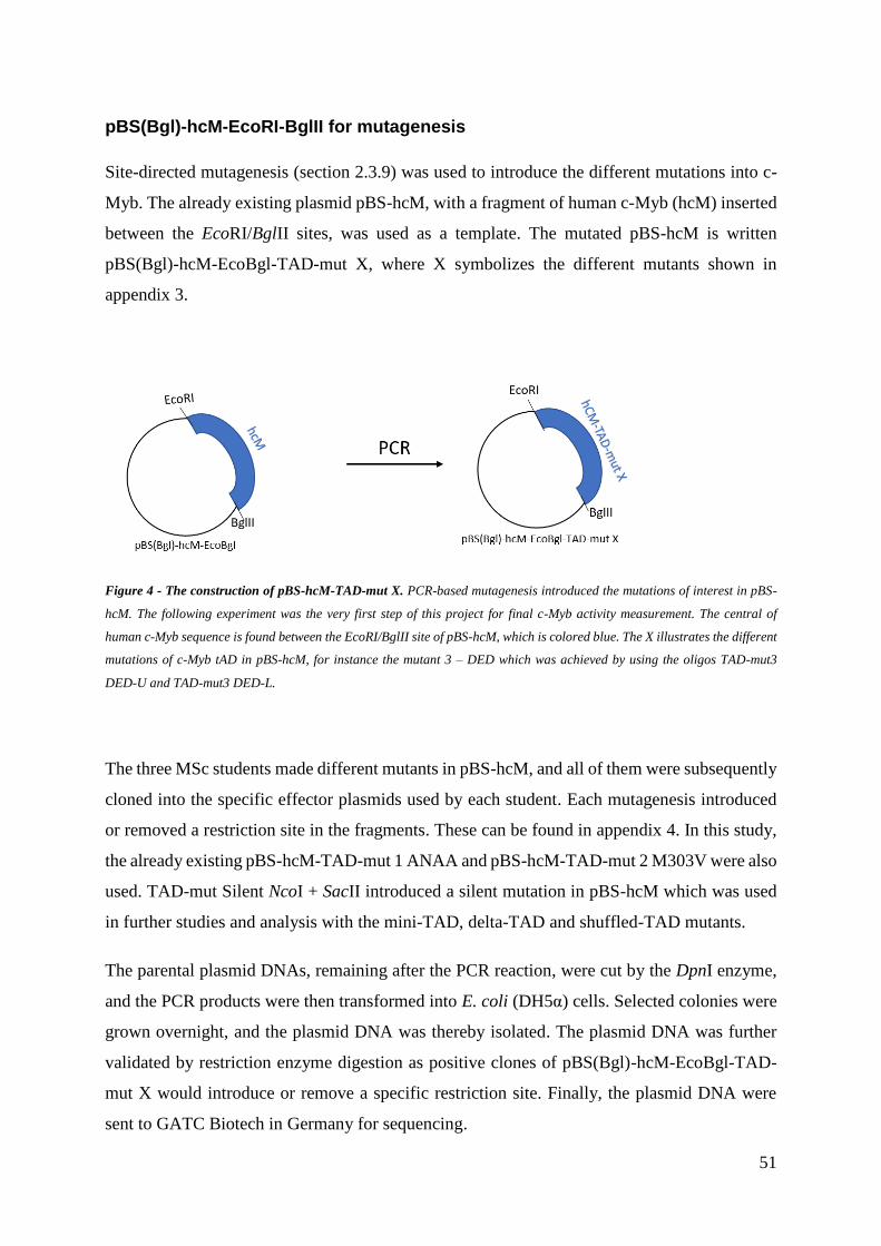
51
pBS(Bgl)-hcM-EcoRI-BglII for mutagenesis
Site-directed mutagenesis (section 2.3.9) was used to introduce the different mutations into c-
Myb. The already existing plasmid pBS-hcM, with a fragment of human c-Myb (hcM) inserted
between the EcoRI/BglII sites, was used as a template. The mutated pBS-hcM is written
pBS(Bgl)-hcM-EcoBgl-TAD-mut X, where X symbolizes the different mutants shown in
appendix 3.
Figure 4 - The construction of pBS-hcM-TAD-mut X. PCR-based mutagenesis introduced the mutations of interest in pBS-
hcM. The following experiment was the very first step of this project for final c-Myb activity measurement. The central of
human c-Myb sequence is found between the EcoRI/BglII site of pBS-hcM, which is colored blue. The X illustrates the different
mutations of c-Myb tAD in pBS-hcM, for instance the mutant 3 – DED which was achieved by using the oligos TAD-mut3
DED-U and TAD-mut3 DED-L.
The three MSc students made different mutants in pBS-hcM, and all of them were subsequently
cloned into the specific effector plasmids used by each student. Each mutagenesis introduced
or removed a restriction site in the fragments. These can be found in appendix 4. In this study,
the already existing pBS-hcM-TAD-mut 1 ANAA and pBS-hcM-TAD-mut 2 M303V were also
used. TAD-mut Silent NcoI + SacII introduced a silent mutation in pBS-hcM which was used
in further studies and analysis with the mini-TAD, delta-TAD and shuffled-TAD mutants.
The parental plasmid DNAs, remaining after the PCR reaction, were cut by the DpnI enzyme,
and the PCR products were then transformed into E. coli (DH5α) cells. Selected colonies were
grown overnight, and the plasmid DNA was thereby isolated. The plasmid DNA was further
validated by restriction enzyme digestion as positive clones of pBS(Bgl)-hcM-EcoBgl-TAD-
mut X would introduce or remove a specific restriction site. Finally, the plasmid DNA were
sent to GATC Biotech in Germany for sequencing.

52
pCIneoB-GBD2-hcM-2KR[194-640] – the basic effector plasmid
A total of X mutants were to be cloned into the vector pCIneoB-GBD2-hcM-2KR[194-640] for
further activity measurements. The construct does not contain full length c-Myb as the N-
terminal DNA-binding domain (DBD) of c-Myb is replaced by the Gal4-binding domain, but
it includes the central tAD region of c-Myb. This plasmid was not available, and therefore had
to be constructed as a combination of two similar plasmids, as illustrated in Figure 5. The
construct also contains substitution of the two SUMOylation sites K527 and K503 in the CRD
of c-Myb to arginine (2KR), which eliminated SUMO-repression and led to an increase of
activity. In addition, to simplify subcloning, a plasmid with a unique BglII site internal in the
c-Myb cDNA was needed.
pCIneoB-GBD2-hcM-2KR[194-640] was made by using pCIneoB-GBD2-hcM-HA-[233-
640], lacking a second BglII site, as the vector and pCIneo-GBD2-hcM-[194-640], spanning
the internal tAD, as the insert. Both vector and insert were digested with XhoI and NotI in order
to ligate them. The vector has one BglII site while the insert has two BglII sites. If the subcloning
of pCIneoB-GBD2-hcM-2KR[194-640] was successful, the results would show only one cut
with BglII.

53
Figure 5 - Subcloning of pCIneoB-GBD2-hcM-2KR[194-640]. A. The cloning of pCIneoB-GBD2-hcM-HA-[233-640]
(vector) and pCIneoB-GBD2-hcM-[194-640] (insert) produced the plasmid of interest. The plasmid was constructed by
digesting the vector and insert with the restriction enzymes XhoI and NotI, and then the bigger fragment of vector was ligated
with the smaller fragment of insert. The vector only contains one BglII site, while the insert has two. The positive clones should
only contain one BglII site as one is lost from the vector, but one is still kept from the insert. B. The CRD in c-Myb of pCIneoB-
GBD2-hcM-2KR[194-640] has the 2KR mutant where two lysine residues are mutated to arginine to achieve an enhancement
of activity. The mutation is marked with pink circles at the CRD. In addition, the DBD of c-Myb has been replaced with the
GBD2.
The mini-TAD, delta-TAD and shuffled-TAD mutants
pCIneoB-GBD2-hcM-2KR[194-640]-TAD-mut Silent NcoI + SacII was designed to introduce
the silent mutation NcoI+SacII flanking the central tAD in c-Myb. The construct was made to
introduce three specially designed mutations called mini-, delta- and shuffled-TAD. The mini-
TAD codes for LxxLL, the delta-TAD codes for AxxAA, and the shuffled-TAD encodes for a
segment of same size and composition as the wild-type tAD, but where the tAD-sequence had
been reordered with the same amino acids grouped together.
Mini-TAD and delta-TAD mutants were formed by annealing oligos according to the procedure
described in section 2.3.2. The oligos used for mini-TAD were mini-TAD-L and mini-TAD-U,
while delta-TAD-L and delta-TAD-U were used for delta-TAD. Shuffled-TAD was made by
gene synthesis ordered from Eurofins Genomics and received as a plasmid, pEX-A128, which
was cleaved with NcoI and SacII. The pBS(Bgl)-hcM-EcoBgl-TAD-mut Silent NcoI + SacII
plasmid was also digested with the same restriction enzymes for ligation with the designed

54
tADs, as shown in figure 6B and figure 7. The ligation mix was transformed, selected clones
were grown overnight and then the plasmid was isolated. Positive clones of pBS-hcM-mini-
TAD introduced a new SacI site, pBS-hcM-delta-TAD introduced a new NheI site and pBS-
hcM-shuffled-TAD lost a SmaI site.
Figure 6 - Mini- and delta-TAD in pBS-hcM. A. pBS-hcM-TAD-mut Silent NcoI + SacII was cut with NcoI and SacII in order
to ligate with the specially designed tADs. B. Mini-TAD, colored yellow, was produced by annealing the oligos mini-TAD-U
and mini-TAD-L and delta-TAD, colored green, was produced by annealing the oligos delta-TAD-U and delta-TAD-L. These
were individually ligated with the digested pBS-hcM-TAD-mut Silent NcoI + SacII as shown in A.
As the shuffled-TAD was received as a plasmid, it was directly cleaved with NcoI and SacII,
and then ligated with the digested pBS-hcM-TAD-mut Silent NcoI + SacII as shown in figure
7. The amino acid sequence for isolated shuffled-TAD can be found in appendix 6. pBS-hcM-
shuffled-TAD was transformed in E. coli cells, grown overnight to isolate the plasmid.

55
Figure 7 - The construction of pBS-hcM-Shuffled-TAD. The pEX-A128 was cut with NcoI and SacII to isolate shuffled-TAD,
which is colored purple. The fragment of interest was thereby ligated with the cut pBS-hcM-TAD-mut NcoI + SacII to achieve
pBS-hcM-shuffled-TAD.
Subcloning the mutants into pCIneoB-GBD2-hcM-2KR[194-640]
All the X introduced mutations in pBS-hcM were finally subcloned into pCIneoB-GBD2-hcM-
2KR[194-640], before final expression in the mammalian cell line HEK293-c1.
The cloning process for pCIneoB-GBD2-hcM-2KR[194-640]-TAD-mut X started by running
a PCR of the pBS(Bgl)-hcM-EcoBgl-TAD-mut X to introduce a MluI site in c-Myb upstream
of the mutated sequence. The PCR product contained a MluI site and a BglII site, one at each
end. The procedure was implemented by cutting the following PCR product and the pCIneoB-
GBD2-hcM-2KR[194-640] with MluI and BglII, and then ligate them, as can be seen in figure
8. The PCR product is colored blue and was ligated in c-Myb tAD of pCIneoB-GBD2-hcM-
2KR[194-640] which is colored green. The final product was the pCIneoB-GBD2-hcM-
2KR[194-640]-TAD-mut X plasmid which is the plasmid expressing the different mutants (X).

56
Figure 8 - Cloning the different mutants into pCIneoB-GBD2-hcM-2KR[194-640]. pBS-hcM with the different mutants were
run through PCR with the oligos hCM-194 (fwd) and Uni-REV. The PCR product, which is blue, and pCIneoB-GBD2-hcM-
2KR[194-640] were cut with MluI and BglII and then ligated together to achieve the final construct.
However, there were one exception for this cloning process under the PCR. The oligos TAD-
mut6 ENE-L and TAD-mut6 ENE-U have already introduced a MluI site in pBS-hcM.
Therefore, the oligo F194-EcoRI (fwd) was used rather than hcM-194 to introduce an EcoRI
site instead of a MluI site, as the addition of another MluI site would not be optimal under the
ligation.
3.2 Transactivation potential
The final constructs of the effector plasmid, pCIneoB-GBD2-hcM-2KR[194-640]-TAD-mut X,
was finally expressed in the HEK293-c1 cell line harboring an integrated luciferase reporter
gene. The different mutations of the transactivation domain were studied and compared to the
wild-type by transfecting the plasmids into the cell lines and thereby observe the outcome of
the lysates with luciferase assays. The values of transcriptional activity were calculated from
three experiments carried out in triplicates. The reporter assay data was normalized by the
Solver Add-in function in Excel (p-value < 0.5).

57
Chromatinized system
The HEK293-c1 cells were used for the monitoring transactivation in a chromatin context, as
described in section 2.4.1. pCIneoB-GBD2-hcM-2KR[194-640]-TAD-mut X does not contain
full-length c-Myb as its DBD was replaced with a Gal4-binding domain (GBD). The HEK293-
c1 cells have a stably integrated luciferase reporter gene driven by a Gal4-recognition promoter,
so the cells were transfected only with the effector plasmids containing the Gal4-hcM-fusions.
The activtiy of GBD2-hcM tAD with the different mutants were measured and compared to the
wild-type c-Myb which was, in this case, pCIneoB-GBD2-hcM-2KR[194-640]. The latter was
set to 100 and the others were given as relative to this reference. The measurements are shown
in figure 9.
Figure 9 - The measured activity of pCIneoB-GBD2-hcM-2KR[194-640]-TAD-mut X. HEK293-c1 cells were transfected
with 0.1 µg effector plasmid encoding pCIneoB-GBD2-hcM-2KR[194-640]-TAD-mut X and 0.3 µg pCIneoB empty vector. The
wild-type is pCIneoB-GBD2-hcM-2KR[194-640]. The luciferase activity was measured and normalized. The values are given
in relative luciferase units (RLU) and normalized by setting the c-Myb wild-type to 100 RLU. The error bars show ± SEM.

58
The activity of GBD2-hcM ANAA, GBD2-hcM RHY, GBD2-hcM Y284, GBD2-hcM VL, and
GBD2-hcM WHSTT mutants showed an increase of activity relative to the wild-type. The
GBD2-hcM ANAA and GBD2-hcM WHSTT mutants caused approximately a twofold increase
in transcriptional activation, while the GBD2-hcM RHY and GBD2-hcM Y284A mutants
increased the activity with about 50%. The GBD2-hcM VL mutant increased the activity with
about 75%. The effects of GBD2-hcM mini-TAD, delta-TAD, shuffled-TAD, as well as GBD2-
hcM DED, ELE, ENE and LxxLL mutants, showed a dramatic decrease of transcriptional
activity compared to the wild-type, as they were almost inactive. The GBD2-hcM M303V,
GBD2-hcM EKE and GBD2-hcM KEKRIK mutants also showed a decrease of activity, they
were reduced with about 75-70%. GBD2-hcM NcoI + SacII silent mutant showed about no
change from the wild-type in activity.
3.3 Western blot analysis
The amount of the c-Myb protein expressed was studied using western blot. The method was
performed as a control in order to determine whether the differences in activity were in fact
caused directly by mutations rather than indirectly by their effect on the amount of protein. If
the amount of protein for each mutant differed vastly from the wild-type, it may have explained
why there were alterations in c-Myb activity. I tried western analysis of the different mutants
first but did not receive nice results, so because of lack of time, kind and helpful Marit Ledsaak
tried the analysis again for me.
HEK293-c1 cells were seeded into 24 well dishes and transfected after 24 hours. For western
analysis, we used 0.2 µg effector plasmid and 0.2 µg pCIneoB empty vector to equalize for
transfection. Cells were lysed with 3x SDS gel loading buffer after 24 hours. The H141 mouse
antibody (binds aa 500-640 in c-Myb) and the anti-GAPDH goat antibody (loading control)
were used as primary antibodies.
Figure 10 shows the results for western analysis for pCIneoB-GBD2-hcM-2KR[194-640]-TAD
mut 1 – 8. Figure 11 shows the western analysis for pCIneoB-GBD2-hcM-2KR[194-640]-TAD
mut 9 – 12, as well as, pCIneoB-GBD2-hcM-2KR[194-640]-TAD mut Silent NcoI + SacII,
GBD2-hcM mini-TAD, delta-TAD and shuffled-TAD.

59
Figure 10 - Western blot of wild-type GBD2-hcM and the GBD2-hcM mutants 1-8. .034 x 105 cells were seeded into 24 well
plates. The cells were transfected with 0.2 µg wild-type GBD2-hcM or mutated GD2-hcM and equalized with 0.2 µg pCIneoB.
Cell lysates were collected 24 hours after the transfection for analyzation with SDS-PAGE and western blotting. H141 rabbit
and GAPDH goat were used as primary antibodies, rabbit IR800 and goat IR680 were used as secondary antibodies. GAPDH
was used as a loading control. H141 detects the c-Myb protein. The empty vector pCIneoB was used as a control for the blot.
Figure 11 - Western analysis of wild-type GBD2-hcM and GBD2-hcM mutants 9-12, as well as GBD2-hcM NcoI+SacII
silent, mini-TAD, delta-TAD and shuffled-tAD mutants. 034 x 105 cells were seeded into 24 well plates. The cells were
transfected with 0.2 µg wild-type GBD2-hcM or mutated GBD2-hcM and equalized with 0.2 µg pCIneoB. Cell lysates were
collected 24 hours after the transfection for analyzation with SDS-PAGE and western blotting. H141 rabbit and GAPDH goat
were used as primary antibodies, rabbit IR800 and goat IR680 were used as secondary antibodies. GAPDH was used as a
loading control. H141 detects the c-Myb protein. The empty vector pCIneoB was used as a control for the blot.
All fragments, except the empty vector, studied n figure 10 seem to have the same strength as
the wild-type. There is only weak background from pCIneoB which is understandable as this is
the empty vector without c-Myb. In figure 11, however, there seems to be stronger background

60
and some fragments are stronger than the wild-type. The fragment for was stronger compared
to the wild-type, this will be further discussed in the discussion section.

61
4 Discussion
The aim of this study was to dissect the transactivation domain (tAD) of human c-Myb.
Mutations of the different amino acid residues in the tAD of c-Myb allowed for activity studies
in three different systems: A mammalian system with a reporter gene incorporated into the
genome in the attempt to mimic a natural context of gene activation, a reporter plasmid
transfected into a mammalian cell to study transactivation of the tAD directly in a classical
reporter assay, and a yeast system to compare any activation properties dependent on
mechanisms conserved from yeast to man.
Exploring the tAD of c-Myb allows for a comparison of the different models for tAD function.
The differences in activity observed when changing a few base pairs can show whether specific
amino acids are required for activation, and thereby determine if there are residues affecting the
tAD both positively and negatively. Mapping out the tAD of c-Myb might give an indication
of how some of the mutations may disturb recruitment of cofactors such as CBP/p300 and
SUMO.
This chapter comprises three parts. The methodical considerations are included in part I. A
discussion regarding the results obtained mostly through the CV-1 cells (Non-chromatinized)
and HEK293-c1 cells (Chromatinized) can be found in part II. Here, the questions from section
1.4, aims of the study, will be discussed in a combined view of the two mammalian systems.
This is followed by a comparative discussion of the two systems, based on question 6 and 7
from the aims of the study. The models explaining the function and structure of tAD will also
be discussed, with a comparison to find if our system conforms to any one of them. In part III,
suggestions toward future perspectives are presented. While part I is different in the three
MSc theses, part II and III are identical and written by all three students in collaboration.

62
4.1 Part I
This part reflects some considerations by using a mammalian system with chromatin setting.
4.1.1 Methodical considerations of the HEK293-c1 cell line
The goal for most research studies is to work with a system that reflects the physical and
chemical conditions present in the organism of which the studied gene is from. In this project,
the human c-Myb gene was studied. The HEK293-c1 cells, acquired from the human embryonic
kidney cells, were used in this study. The cell line has stably integrated the reporter gene and
five Gal4-recognition elements (GRE). As the transfected effector plasmid had a substitution
of the DBD with Gal4-binding domain (GBD), the influences of the different mutations in the
tAD could be studied. In other words, the transiently transfected effector plasmid does not
contain full-length c-Myb, which might not be ideal because naturally occurring mediations
through the DBD can be affected by either loss or alterations.
The reporter gene is the integrated in the genome and thus has a chromatinization level which
is closer to the physiologic conditions. Obviously, c-Myb regulates its target genes embedded
in chromatin structured DNA in stem and progenitor cells. In addition, the advantage of using
stably integrated reporter gene is avoiding poor transfection efficiencies as the reporter is not
co-transfected with the effector plasmids. However, the effector plasmid containing c-Myb is
transiently transfected and is not integrated into the genome of HEK293-c1 cells, which
consequently can result in unequal distribution of c-Myb expression between the cells. There
can be an overexpression when the cells uptake multiple plasmids containing c-Myb or low
levels of c-Myb can be expressed when the transfection contains fewer plasmids, making the
results less reliable. The ideal method would be to integrate the GBD2-hcM tAD mutants in the
genome using CRISPR to ensure the right amount of c-Myb protein. CRISPR is a DNA
sequence used by the enzyme Cas9 in order to recognize and cleave specific strands for gene
editing.
The HEK293-c1 cells are close to the gene’s species of origin generating observations of actions
and reactions of a closely physiological in vitro experiment, but the cell-line does not naturally
exist in mammalian cells. Using living organisms, such as mouse models, would be the most

63
optimal. Knock-out mouse or mutant mouse, preferentially by integrating the effector gene into
the organism, can fulfill the physiological conditions as the functions of a living organism are
present. The observed results can be used to study c-Myb controlled gene expressions.
4.2 Part II
The two mammalian systems both require an effector plasmid encoding c-Myb, either the
wild-type or a mutant, to be transfected into the cells. Only the effector plasmid is needed in
the HEK293-c1 cells because of the incorporated reporter gene that includes five
Gal4-recognition elements (GREs) and the luciferase reporter gene. Because of the CV-1 cells
not having an integrated reporter gene, a reporter plasmid containing multiple Myb recognition
elements (MREs) and the luciferase reporter gene is transfected at the same time as the effector
plasmid. This gives both systems the capability to express luciferase at a level reflecting the
activity of the c-Myb tAD. Both systems use a version of pCIneoB as their effector plasmid to
express c-Myb.
Western blots have been performed in both systems, where all of the different mutants were
found to be expressed in close to identical amounts. From this we concluded that the observed
differences of activity were in fact caused by the tAD itself and not because of varying levels
of the c-Myb protein. The only exception is for the LxxLL mutant, which had a more intense
fragment than the wild-type. This will be considered when this mutant is discussed below. Part
II will discuss our findings by systematically going through the questions mentioned in the aims
of the study.
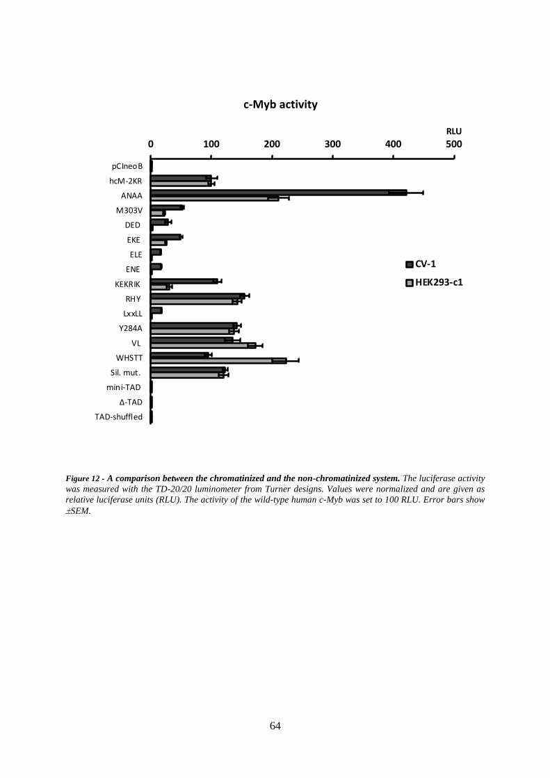
64
Figure 12 - A comparison between the chromatinized and the non-chromatinized system. The luciferase activity
was measured with the TD-20/20 luminometer from Turner designs. Values were normalized and are given as
relative luciferase units (RLU). The activity of the wild-type human c-Myb was set to 100 RLU. Error bars show
SEM.
0 100 200 300 400 500
pCIneoB
hcM-2KR
ANAA
M303V
DED
EKE
ELE
ENE
KEKRIK
RHY
LxxLL
Y284A
VL
WHSTT
Sil. mut.
mini-TAD
∆-TAD
TAD-shuffled
RLU
c-Myb activity
CV-1
HEK293-c1

65
4.2.1 1.1.1 Specific amino acid residues that affect the
transcriptional activity of c-Myb
In the remaining discussion, the two cell systems will be referred to as the
non-chromatinized/chromatinized system. The discussions are sectioned according to the
different groups of amino acids mutated: acidic, basic and hydrophobic groups.
Acidic amino acids and their effect on c-Myb activity
The most prominent observation was that the acidic amino acids in the central part of the tAD
of c-Myb is essential for activation of transcription. The mutant ELE and ENE, changing just
two acidic amino acids to alanine, led to an 80% loss of transcriptional activity in the
non-chromatinized system, and almost complete inactivation in the chromatinized system,
when compared to the wild-type. The central tAD mutations of acidic amino acids include
mutant DED, EKE, ELE and ENE.
The traditional view of tAD function has been as an acidic cluster. Gill et al. showed the
importance of the correlation between activation strength and negative charge [20]. Our data
support an important role for the acidic residues. The extreme cases were observed in the
chromatinized system where the mutants DED, ELE and ENE revealed a total loss of activity,
and EKE was reduced by 75% activity compared to the wild-type. These numbers suggest that
the acidic amino acid residues in the central part of the c-Myb tAD affect transcriptional activity
in a positive manner. The residues may be important for interactions with other proteins
required for activation, as all acidic residues of interest were located in close proximity to the
LxxLL motif essential for the recruitment of p300/CBP. Staller et. al. proposed in their
hypothesis that the negatively charged amino acids affect recruitment of cofactors positively by
keeping the short linear motif (SLM) exposed to the solvent, which would be consistent with
the presented results [15].

66
Hydrophobic amino acids and their effect on c-Myb activity
Many SLMs will contain hydrophobic residues. Therefore we targeted several of the
hydrophobic amino acids in our mutant approach. Four hydrophobic motifs were mutated:
LxxLL [298-302], Y284A, VL [314-315], and WHSTT [327-331]. The first had a very strong
negative effect and is discussed in a separate section below. The others caused some degree of
activation.
The mutant VL resulted in an increase of activity compared to the wild-type, with an increase
in transcriptional activity of about 75% for the chromatinized system, and 50% for the
non-chromatinized system. There may be two possible explanations for this effect. Either the
hydrophobic residues are part of SLMs that recruit some corepressors, or they could have an
effect on the structure of c-Myb leading to altered activation. Evidence for the latter is a work
of Dukare et al. who studied a self-interaction phenomenon occurring in v-Myb caused by
hydrophobic residues [39]. Based on their studies of conserved amino acids in v-Myb, they
made the assumption that hydrophobic regions can induce self-interactions within c-Myb as
well, and potentially reduce the accessibility of the SLMs. By mutating valine to isoleucine,
which has one more methyl group than valine, they observed that this replacement had a strong
negative effect on the transcriptional activity. In this study, mutant VL had two hydrophobic
amino acid residues changed to alanine, which has one less methyl group. This mutation could
have prevented self-interactions, which in turn would provide with a better interaction with
different coactivators such as CBP/p300.
Basic amino acids and their effect on c-Myb activity
Two basic patches have been mutated in this study, the KEKRIK and RHY, where the basic
residues have been changed to the neutral alanine (A).
Based on the non-chromatinized system, the basic amino acid residues do not appear to have a
major impact on the transcriptional activity when looking at the mutant KEKRIK. This mutant
showed a slight increase of transactivation, approximately 10% compared to the wild-type of
the c-Myb tAD. The same system had only a slight increase in the reporter activation of the
RHY mutant. In contrast, the chromatinized system showed a strong reduction in reporter
activation (70% drop) with the KEKRIK mutant. RHY on the other hand, showed an increase

67
of about 50%, both compared to the wild-type. This system showed no correlation between the
activity provided by the basic patches, as one led to an increase while the other led to a
reduction. But their position seems to be important.
The summary of the specific amino acid patches is that the non-chromatinized system was
strongly affected by many mutants, especially the negative and hydrophobic amino acid
mutations. The chromatinized system seems to be even more sensitive to the changes, as it
showed a stronger impact of changing amino acids of all properties, some of them to the point
of activity depletion.
4.2.2 The short linear motif LxxLL
The SLM LxxLL is a hydrophobic residue that when mutated to AELAA affects the tAD
severely. In the introduction, we covered how CBP/p300 increases transcriptional activity when
it is recruited, and that the KIX domain of CBP/p300 interacts with the LxxLL motif of c-Myb
[53, 90]. This motif was the only known SLM in the tAD of c-Myb and its mutation therefore
functioned as a positive control for the important contribution of SLMs to tAD function. The
LxxLL mutant destroys the ability of c-Myb to recruit the p300 cofactor, and is an example of
a mutant where we can give a precise explanation of how the specific amino acid residues affect
the transactivation of c-Myb.
Mutant LxxLL resulted in a large decrease of 80% in transcriptional activity in the non-
chromatinized system compared to wild-type. In comparison, the chromatinized system
indicated a transcriptional activation close to zero. The results in this thesis is consistent with
previous experiments suggesting the CBP/p300 interaction through the LxxLL motif.
Interestingly, for both systems, the western blot fragment for the LxxLL mutant of c-Myb was
stronger compared to the wild-type. This depicts the presence of more protein. Had there been
about equal amounts of LxxLL and wild-type protein, the luciferase-activity of the motif would
probably have been even lower.
The dramatic effect of the mutation of the LxxLL motif led us to ask whether this SLM would
be sufficient for transactivation. We therefore created the delta-TAD and mini-TAD mutants.
The mini-TAD mutant had almost the whole tAD removed except for the LxxLL motif, with

68
the ends being flanked by a few amino acids only. The transcriptional activity was virtually
zero compared to the wild-type tAD of c-Myb in both systems. This indicates that the LxxLL
motif is not sufficient to activate transcription on its own, and suggests that the motif needs
more specific amino acid residues in its proximity. Based on Staller et al., many of these amino
acid residues would need to be acidic, so that they can keep the hydrophobic motif from driving
collapse [15]. This also matches the findings in the acidic amino acid mutants, which showed
the importance of the negatively charged residues in close proximity of the hydrophobic motif
LxxLL.
To make sure that any activity observed from the mini-TAD mutant was not affected by any
background activity of c-Myb, the delta-TAD mutant would function as the background
reference. Delta-TAD mutant has the LxxLL motif changed to AxxAA also with the rest of the
tAD removed. It functions as a control for the mini-TAD mutant, to make sure there were to
interfering activity from other parts of c-Myb itself.
In addition to the LxxLL motif, the M303V point mutation have shown a reduced interaction
to p300, which results in suboptimal transactivation of c-Myb target sequences. Evidence was
presented that M303 of the c-Myb tAD is important for recruitment of p300 since it interacts
with the KIX domain, and mutations of this residue weaken this interaction [107]. As CBP/p300
increases transcriptional activity, the reduced interaction can explain why the M303V point
mutation had a decrease in transactivation. The observed activities compared to the wild-type
are reduced by 50% and 75% in the non-chromatinized and chromatinized systems,
respectively.
4.2.3 Other potential short linear motifs in the tAD of c-Myb
All of the mutations that showed a change of transcriptional activity compared to the wild-type
would be expected either affecting a SLM indirectly and its ability to recruit cofactors, or is a
potential SLM itself. SLMs tend to be preserved through evolution, and subtle changes to a
motif will most likely have an effect on the activity. The SLMs would be involved in recruiting
and interacting with cofactors, where the change in activity reflects the loss of interaction. In
the introduction, several possible interaction partners were mentioned, including TFIIF and
TAF12. Based on the recently published article from De Mol et al. the WHSTT sequence in

69
c-Myb tAD is an interesting sequence, since this might be a binding site for the general TF
TFIIF [98]. They found that TFIIF interacts with a specific motif of the androgen receptor (AR),
and a similar sequence is found in the c-Myb tAD. The motif was thought to share similarites
with WHSTT, where this mutant was used to test whether there was any cofactor recruitment
or not.
In our study, the WHSTT mutant did not affect the transactivation of the non-chromatinized
system, while the chromatinized system had a 2-fold increase compared to the wild-type. Based
on De Mol et al., removing the interaction to TFIIF could potentially decrease the c-Myb
activity. There are two potential reasons for the activity increase in c-Myb. One is that TFIIF
functions as a repressor in the c-Myb interaction, and the other being that c-Myb does not recruit
TFIIF through WHSTT. Such an interaction would need a GST-pulldown to be verified. If a
cofactor were recruited through WHSTT, its removal increased the activity of the
chromatinized system, and showed no change in the non-chromatinized system. This could
suggest a putative chromatin interacting corepressor.
The second potential interaction of interest is the TAF12 subunit of TFIID, based on the recently
published article by Xu et al.. In this article they reported that the TAF12 interacts with the tAD
in Myb and promotes activation of genes [97]. TAF12 is depicted over the RHY sequence in
figure 1, which might be a possible binding motif, though the actual linear binding motif of this
cofactor is unknown. The RHY mutant, where the sequence RHY in c-Myb tAD were mutated
to AAY, resulted in a higher activity, the opposite of what would have been expected if RHY
bound TAF12. This study did not find any SLM candidates for TAF12 based on Xu et al.. The
results in this study might indicate that RHY rather recruits a repressive factor. Further studies
are needed to determine the actual interaction partner.
Another potential SLM might be the VL mutant, where the two amino acid residues were
mutated to AA. This mutation gave an increased transcriptional activity in both systems,
indicating that the two amino acid residues might be part of a SLM. This hypothesized SLM
may function as a binding site for a corepressor, as the transcriptional activity increased by
mutating these amino acid residues. Indirect effects on the SLMs or the folding of the tAD
cannot be excluded, but this would need further mutations in the tAD of c-Myb to be verified.

70
4.2.4 The order of the amino acid residues
Model 1 presented in the introduction suggests that the tAD of TFs consists of a certain
percentage of acidic amino acid residues, and indicates that the order of the residues is not
essential. This model implies that a shuffled version with the same percentage of the different
amino acid residues should have similar gene expression as the wild-type. In order to study
whether the order of amino acid residues affected transactivation of the tAD of c-Myb, the
construct of shuffled-TAD was made. This shuffled mutant had all the original amino acids of
the c-Myb tAD, but in a different and shuffled order. The hydrophobic, acidic and basic residues
were grouped together.
The luciferase reporter assay showed that there were no transcriptional activity at all for both
systems when the shuffled mutant was transfected into the cells. This implies that the order of
amino acid residues has an impact on the transcriptional activity, and that the actual content of
amino acids is not sufficient for gene expression. Based on these results, model 1 can be rejected
as the best match for c-Myb tAD function. One interpretation could be that the shuffled-TAD
construct may not facilitate the exposure of SLMs as the hydrophobic patches have the potential
to form clusters through hydrophobic interactions. The acidic residues’ effect on transactivation
is suggested to depend on their placement in between hydrophobic residues. Having them
grouped together might affect the exposure of the hydrophobic SLMs negatively. The shuffled
tAD may enhance destabilization and degradation of c-Myb as the structure of the protein as a
whole is disturbed. A potential degradation is visualized in the western blots for both systems,
where the bands appear as shaded compared to the wild-type. Since a fragment could still be
seen in the western blot of the shuffled-TAD mutant, complete degradation cannot have
happened, though enhanced degradation appears to be present.
4.2.5 Increase in activation potential
Several mutants gave an increase of transactivation function compared to the wild-type. In
addition to the mentioned mutants RHY, VL and WHSTT, the mutants ANAA and Y284A
gave an increase in transcriptional activation. SUMO is a repressive factor of transcriptional
activity that interacts with a V/I-X-V/I-V/I motif, where V267NIV can be found in the c-Myb
tAD [93]. The ANAA mutant is known to remove the SIM VNIV, which is necessary for c-

71
Myb to be able to interact non-covalently with SUMO. Our lab studied this mutation at a
previous occasion, and revealed that destroying the SUMO-binding properties resulted in a
large increase of the transactivation potential of c-Myb [93]. The mutant ANAA’s activity
increased drastically in both systems compared to the wild-type. The non-chromatinized system
increased over 4-fold, while the chromatinized system increased 2-fold. The reason proposed
by Saether et al. was that the SUMO recruits a repressor, albeit the repressor itself has not been
identified.
An interesting observation is that all the mutations that led to an increased transcriptional
activity are located N-terminally or C-terminally of the central tAD. These mutations include
the mentioned mutants ANAA, RHY and VL, in addition to the mutant Y284A. The Y284A
mutant increased the activity of c-Myb by about 50% in both systems, about the same as the
RHY mutant. Interestingly, Y284A mutates the tyrosine of the RHY sequence, indicating that
the residue might in fact be part of a novel SLM.
The mutants leading to an increase of transcriptional activity in the N- and C-terminal regions
of tAD altered the activity between 40% and 400% in the non-chromatinized system, and
between 140 and 210% in the chromatinized system. The changes are on average larger in the
chromatinized system, as it appears to be more sensitive to mutations except for the ANAA
mutant. The increased activity suggests that these mutants are part of different motifs in which
repressing cofactors might bind, or they may have an indirect structural effect.
4.2.6 Comparison of the three systems
Three different systems were used in this joint project between the three MSc students, where
two were mammalian systems and one was a yeast system. Each mammalian system has its
own effector plasmid used to transfect in the c-Myb. The non-chromatinized cells (CV-1) were
transient transfected with an effector plasmid and a reporter plasmid at the same time to do the
assays. This system would express the mutant’s activity without being affected by
chromatinization. In the chromatinized cells (HEK293-c1), a reporter gene incorporated into a
chromatin context was used to measure how the different mutants of c-Myb affected activity in
a chromatin context. This system made it possible to emphasize effects that relate to chromatin
accessibility.

72
The idea of including the yeast system was to distinguish general effects on gene activation,
from activation that depended on specific mammalian cofactors. Difficulties occurred with the
plasmids transformed into the yeast, and because of time constrains, no quantitative reporter
data could be presented. However we managed to see effects on the His3 reporter since the
reporter here was a fusion of the lacZ gene and the His3 gene with two enzymatic activities.
The His3 activation allowed for a stamp-test, revealing differences in growth for the yeast
colonies as the consequences of the c-Myb being mutated. All mutants grew at approximately
the same rate as the wild-type, where the small differences might be a result of the stamping
being uneven. The observation was that the tAD of c-Myb in fact is able to activate in yeast,
suggesting quite conserved mechanisms for tAD function. The lack of effects of mutations may
be because they affect recruitment of mammalian cofactors which is why no differences could
be seen in yeast. Beyond this, yeast is excluded in the discussion, and it is instead presented in
section 4.3 future perspectives.
Below is discussed the effect on activity of the c-Myb tAD based on the difference in
chromatinization.
Difference in chromatinization affect the activity of tAD mutants
The main difference between the two systems is the degree of chromatinization of the reporter
gene. Hence, the two systems make it possible to see the effect of chromatinization in
mammalian cells in regards to transcriptional activity. An example is the M303V mutant, which
reduced the activity more in the chromatinized system. The mutant affects the recruitment of
CBP/p300 through the KIX domain negatively, where the chromatinized system appeared to
have a stronger activity reduction than observed in the non-chromatinized system. Identical
changes in recruitment might affect the chromatinized system more because of it being sensitive
to acetylation.
Mutant KEKRIK showed increased and decreased transcriptional activity in the non-
chromatinized and chromatinized system, respectively. All the mutated amino acid residues in
this mutant was of basic nature, which might indicate that the basic amino acid residues affect
factors related to chromatin. The mutant KEKRIK is located near the LxxLL motif, and one

73
reason for this major decrease in activity may be that the mutation has affected the KIX-LxxLL
interaction in c-Myb. The lack of acetylation would explain why the chromatinized system
shows the stronger activity loss.
The ANAA mutation is interesting because it increases over 4-fold in the non-chromatinized
system, compared to “only” about 2-fold in the chromatinized system. As opposed to the
tendency of the chromatinized system being more sensitive to the mutations, the proposed
explanation goes toward a repressor that affect the enhancer or promoter region itself, rather
than affecting the chromatin structure.
4.2.7 The results compared to the models
Our study could not conclude as to what model is right or wrong for defining the function of
the c-Myb tAD. Instead, reasonable suggestions will be presented comparing our mutants to the
more plausible models. The results will be discussed based on the two mammalian cell systems.
Model 1 can be rejected on the basis of the shuffled-TAD experiment. This thesis indicates that
the amino acid residues in the tAD of c-Myb are more specific than in the first presented models.
We will here discuss our observation in relation to two previously published models for tADs
in TFs.
Model 4: Transactivation domains as SLM embedded in an intrinsically
disordered acidic domain
This model is based on a combination of model 1 defining the acidic amino acid rich domains
and model 3 defining SLM. Model 4 describe tADs as an area with linear motifs containing
critical hydrophobic residues, in a context of acidic amino acids exposing the hydrophobic
residues. Staller et al. featured the model in a recently published article, where the tAD of the
yeast TF Gcn4 was dissected [15]. This model seems to fit our observations the best, and can
be applied to the c-Myb tAD where the motif LxxLL serve as a key SLM but which function
depends on proximal acidic residues. The mutations of these surrounding acidic residues led to
a major decrease of transactivation. The transcriptional activity is almost totally lost by
mutating either the LxxLL motif or the acidic amino acid residues.

74
In regards to the size of the motif and earlier studies, a hypothesis would be that the motif alone
is insufficient to induce activation. The mini-TAD mutant demonstrates the importance of the
positioning of the acidic residues. As expected, the motif was not sufficient, which endorse
model 4 as the proper explanation.
Model 4 applied to c-Myb can be summarized as follows: The LxxLL motif is placed in between
acidic amino acid residues, where mutants suggest that the acidic residues keep the hydrophobic
motif from driving collapse. Neither the hydrophobic motif nor the acidic residues are sufficient
for transactivation, but the combination of these lead to gene expression.
Model 5: transactivation domains function as domains that induce liquid-liquid
phase-transition
A recently published article from Bojia et al. presents a model where the tAD forms phase-
separated condensates with the mediator in order to enhance gene expression [16]. In this work,
each mutant consisted of 10 altered amino acid residues, which rendered their TFs unable to
activate transcription. Acidic domains were found necessary for the liquid-liquid phase
transition to manifest, and the gene activation was found to be affected in parallel. Our study
did not test for any liquid-droplet formation, which makes it difficult to say anything about
droplets in c-Myb. A smaller number of amino acids were altered for each mutant compared to
the 10 in Boija et al., where some cases resulted in major effects. This might indicate that the
mutants of c-Myb tAD either decrease or enhance its phase-separation capacity during
transcription. To find out whether or not liquid-liquid phase transitions play a major role in the
activity of c-Myb requires studies directly monitoring effects on such transitions.

75
4.3 Part III: Future perspectives
As of now, there are many potential interaction partners affecting the transactivation of c-Myb,
but none of them have been verified directly through this study, but performing GST-pulldown
could reveal the interaction partner(s) associated with WHSTT, RHY and VL.
Each amino acid in the tAD has an effect on how a SLM recruits cofactors. After finding the
exact interaction partners, selecting new mutations to study how the amino acid composition
affects the interaction could shed light onto the exact sequence of the SLMs. In addition, a better
understanding regarding the amino acid composition around the binding element of putative
interaction partners could help strengthen the results achieved from mutating the proximity of
LxxLL in this study. Probable candidates to study would be the KIX domain of p300, the
TAF12 interaction and the general TF TFIIF. Based on published data, some of the mutations
created through this study were expected to have a strong effect on the KIX-interactions,
namely LxxLL and M303V. The SLM recruiting TAF12 has not yet been discovered, which
makes this an interesting interaction to study. It could be favorable to include experiments, such
as co-immunoprecipitation, to study whether two interaction partners act synergistically or in a
competitive fashion.
How long the mini-TAD mutant needs to be until the transcriptional activity reaches the level
of the wild-type tAD could be explored. Through the appliance of amino acid residues one after
another to the mutant, the follow up by a luciferase reporter assay for every added amino acid
would give potential insight as to the importance of amino acids. The questions asked could be
how many of the tAD’s amino acids are needed to activate the motif, and which amino acids
affect the activity the most.
The plan for this tripartite project was to analyze how each mutant affect the activity of c-Myb
in three systems. Only two of the systems were analyzed due to complications with the yeast
system. It was possible to make both the effector plasmids and reporter plasmid for the yeast
system, and the colonies grew on selection plates. The colonies did not grow in the last
overnight process, and the cells were found to have lost one of the plasmids. If growth can be
induced in the yeast cells, followed by the ONPG-test, comparisons between the mammalian
systems and the yeast system could prove interesting when studying the cofactors of c-Myb.

76
This MSc thesis has been a start of an interesting project where the tAD of c-Myb could be
explored across different systems with differences in chromatin structures.

77
5 References
1. Arrowsmith, C.H., et al., Epigenetic protein families: a new frontier for drug
discovery. Nat Rev Drug Discov, 2012. 11(5): p. 384-400.
2. Woodcock, C.L. and R.P. Ghosh, Chromatin higher-order structure and dynamics.
Cold Spring Harb Perspect Biol, 2010. 2(5): p. a000596.
3. Zentner, G.E. and S. Henikoff, Regulation of nucleosome dynamics by histone
modifications. Nat Struct Mol Biol, 2013. 20(3): p. 259-66.
4. Boyer, L.A., et al., Essential role for the SANT domain in the functioning of multiple
chromatin remodeling enzymes. Mol Cell, 2002. 10(4): p. 935-42.
5. Jenuwein, T. and C.D. Allis, Translating the histone code. Science, 2001. 293(5532):
p. 1074-80.
6. Bannister, A.J. and T. Kouzarides, Regulation of chromatin by histone modifications.
Cell Res, 2011. 21(3): p. 381-95.
7. Soutourina, J., Transcription regulation by the Mediator complex. Nat Rev Mol Cell
Biol, 2018. 19(4): p. 262-274.
8. Kornberg, R.D., The molecular basis of eucaryotic transcription. Cell Death Differ,
2007. 14(12): p. 1989-97.
9. Sainsbury, S., C. Bernecky, and P. Cramer, Structural basis of transcription initiation
by RNA polymerase II. Nat Rev Mol Cell Biol, 2015. 16(3): p. 129-43.
10. Zhou, Q., T. Li, and D.H. Price, RNA polymerase II elongation control. Annu Rev
Biochem, 2012. 81: p. 119-43.
11. Zabidi, M.A. and A. Stark, Regulatory Enhancer-Core-Promoter Communication via
Transcription Factors and Cofactors. Trends Genet, 2016. 32(12): p. 801-814.
12. Lee, T.I. and R.A. Young, Transcription of eukaryotic protein-coding genes. Annu
Rev Genet, 2000. 34: p. 77-137.
13. Tootle, T.L. and I. Rebay, Post-translational modifications influence transcription
factor activity: a view from the ETS superfamily. Bioessays, 2005. 27(3): p. 285-98.
14. Mitchell, P.J. and R. Tjian, Transcriptional regulation in mammalian cells by
sequence-specific DNA binding proteins. Science, 1989. 245(4916): p. 371-8.
15. Staller, M.V., et al., A High-Throughput Mutational Scan of an Intrinsically
Disordered Acidic Transcriptional Activation Domain. Cell Syst, 2018. 6(4): p. 444-
455.e6.
16. Boija, A., et al., Transcription Factors Activate Genes through the Phase-Separation
Capacity of Their Activation Domains. Cell, 2018. 175(7): p. 1842-1855.e16.
17. Mermod, N., et al., The proline-rich transcriptional activator of CTF/NF-I is distinct
from the replication and DNA binding domain. Cell, 1989. 58(4): p. 741-53.
18. Sigler, P.B., Transcriptional activation. Acid blobs and negative noodles. Nature,
1988. 333(6170): p. 210-2.
19. Courey, A.J., et al., Synergistic activation by the glutamine-rich domains of human
transcription factor Sp1. Cell, 1989. 59(5): p. 827-36.
20. Gill, G. and M. Ptashne, Mutants of GAL4 protein altered in an activation function.
Cell, 1987. 51(1): p. 121-6.
21. Sadowski, I., et al., GAL4-VP16 is an unusually potent transcriptional activator.
Nature, 1988. 335(6190): p. 563-4.

78
22. Hope, I.A., S. Mahadevan, and K. Struhl, Structural and functional characterization
of the short acidic transcriptional activation region of yeast GCN4 protein. Nature,
1988. 333(6174): p. 635-40.
23. Ness, S.A., The Myb oncoprotein: regulating a regulator. Biochim Biophys Acta,
1996. 1288(3): p. F123-39.
24. Gill, G. and M. Ptashne, Negative effect of the transcriptional activator GAL4. Nature,
1988. 334(6184): p. 721-4.
25. Attardi, L.D. and R. Tjian, Drosophila tissue-specific transcription factor NTF-1
contains a novel isoleucine-rich activation motif. Genes Dev, 1993. 7(7b): p. 1341-53.
26. Warfield, L., et al., A sequence-specific transcription activator motif and powerful
synthetic variants that bind Mediator using a fuzzy protein interface. Proc Natl Acad
Sci U S A, 2014. 111(34): p. E3506-13.
27. Brzovic, P.S., et al., The acidic transcription activator Gcn4 binds the mediator
subunit Gal11/Med15 using a simple protein interface forming a fuzzy complex. Mol
Cell, 2011. 44(6): p. 942-53.
28. Heery, D.M., et al., A signature motif in transcriptional co-activators mediates
binding to nuclear receptors. Nature, 1997. 387(6634): p. 733-6.
29. Plevin, M.J., M.M. Mills, and M. Ikura, The LxxLL motif: a multifunctional binding
sequence in transcriptional regulation. Trends Biochem Sci, 2005. 30(2): p. 66-9.
30. Lu, Z., et al., Unraveling the mechanism of a potent transcriptional activator. J Biol
Chem, 2005. 280(33): p. 29689-98.
31. Shen, F., et al., Transcriptional activation domain of the herpesvirus protein VP16
becomes conformationally constrained upon interaction with basal transcription
factors. J Biol Chem, 1996. 271(9): p. 4827-37.
32. Shen, F., et al., Critical amino acids in the transcriptional activation domain of the
herpesvirus protein VP16 are solvent-exposed in highly mobile protein segments. An
intrinsic fluorescence study. J Biol Chem, 1996. 271(9): p. 4819-26.
33. Shin, Y. and C.P. Brangwynne, Liquid phase condensation in cell physiology and
disease. Science, 2017. 357(6357).
34. Banani, S.F., et al., Biomolecular condensates: organizers of cellular biochemistry.
Nat Rev Mol Cell Biol, 2017. 18(5): p. 285-298.
35. Hnisz, D., et al., A Phase Separation Model for Transcriptional Control. Cell, 2017.
169(1): p. 13-23.
36. Sabari, B.R., et al., Coactivator condensation at super-enhancers links phase
separation and gene control. Science, 2018. 361(6400).
37. Arnold, C.D., et al., A high-throughput method to identify trans-activation domains
within transcription factor sequences. Embo j, 2018. 37(16).
38. Sakura, H., et al., Delineation of three functional domains of the transcriptional
activator encoded by the c-myb protooncogene. Proc Natl Acad Sci U S A, 1989.
86(15): p. 5758-62.
39. Dukare, S. and K.H. Klempnauer, A conserved patch of hydrophobic amino acids
modulates Myb activity by mediating protein-protein interactions. Biochim Biophys
Acta, 2016. 1859(7): p. 914-21.
40. Ptashne, M., How eukaryotic transcriptional activators work. Nature, 1988.
335(6192): p. 683-9.
41. Oh, I.H. and E.P. Reddy, The myb gene family in cell growth, differentiation and
apoptosis. Oncogene, 1999. 18(19): p. 3017-33.
42. Kasper, L.H., et al., A transcription-factor-binding surface of coactivator p300 is
required for haematopoiesis. Nature, 2002. 419(6908): p. 738-43.

79
43. Tomita, A., et al., c-Myb acetylation at the carboxyl-terminal conserved domain by
transcriptional co-activator p300. Oncogene, 2000. 19(3): p. 444-51.
44. Parker, D., et al., Role of secondary structure in discrimination between constitutive
and inducible activators. Mol Cell Biol, 1999. 19(8): p. 5601-7.
45. Wright, P.E. and H.J. Dyson, Intrinsically unstructured proteins: re-assessing the
protein structure-function paradigm. J Mol Biol, 1999. 293(2): p. 321-31.
46. Molvaersmyr, A.K., et al., A SUMO-regulated activation function controls synergy of
c-Myb through a repressor-activator switch leading to differential p300 recruitment.
Nucleic Acids Res, 2010. 38(15): p. 4970-84.
47. Klempnauer, K.H., T.J. Gonda, and J.M. Bishop, Nucleotide sequence of the retroviral
leukemia gene v-myb and its cellular progenitor c-myb: the architecture of a
transduced oncogene. Cell, 1982. 31(2 Pt 1): p. 453-63.
48. Nomura, T., et al., Negative autoregulation of c-Myb activity by homodimer formation
through the leucine zipper. J Biol Chem, 1993. 268(29): p. 21914-23.
49. Tanaka, Y., et al., B-myb is required for inner cell mass formation at an early stage of
development. J Biol Chem, 1999. 274(40): p. 28067-70.
50. Zhou, Y. and S.A. Ness, Myb proteins: angels and demons in normal and transformed
cells. Front Biosci (Landmark Ed), 2011. 16: p. 1109-31.
51. Lipsick, J.S. and D.M. Wang, Transformation by v-Myb. Oncogene, 1999. 18(19): p.
3047-55.
52. Ramsay, R.G. and T.J. Gonda, MYB function in normal and cancer cells. Nat Rev
Cancer, 2008. 8(7): p. 523-34.
53. Ness, S.A., Myb binding proteins: regulators and cohorts in transformation.
Oncogene, 1999. 18(19): p. 3039-46.
54. Prouse, M.B. and M.M. Campbell, The interaction between MYB proteins and their
target DNA binding sites. Biochim Biophys Acta, 2012. 1819(1): p. 67-77.
55. Ording, E., et al., Two functionally distinct half sites in the DNA-recognition sequence
of the Myb oncoprotein. Eur J Biochem, 1994. 222(1): p. 113-20.
56. Howe, K.M., C.F. Reakes, and R.J. Watson, Characterization of the sequence-specific
interaction of mouse c-myb protein with DNA. Embo j, 1990. 9(1): p. 161-9.
57. Tanikawa, J., et al., Recognition of specific DNA sequences by the c-myb
protooncogene product: role of three repeat units in the DNA-binding domain. Proc
Natl Acad Sci U S A, 1993. 90(20): p. 9320-4.
58. Gabrielsen, O.S., A. Sentenac, and P. Fromageot, Specific DNA binding by c-Myb:
evidence for a double helix-turn-helix-related motif. Science, 1991. 253(5024): p.
1140-3.
59. Weston, K. and J.M. Bishop, Transcriptional activation by the v-myb oncogene and its
cellular progenitor, c-myb. Cell, 1989. 58(1): p. 85-93.
60. Biedenkapp, H., et al., Viral myb oncogene encodes a sequence-specific DNA-binding
activity. Nature, 1988. 335(6193): p. 835-7.
61. Mo, X., et al., Histone H3 tail positioning and acetylation by the c-Myb but not the v-
Myb DNA-binding SANT domain. Genes Dev, 2005. 19(20): p. 2447-57.
62. Fuglerud, B.M., et al., The pioneer factor activity of c-Myb involves recruitment of
p300 and induction of histone acetylation followed by acetylation-induced chromatin
dissociation. Epigenetics Chromatin, 2018. 11(1): p. 35.
63. Fuglerud, B.M., et al., A c-Myb mutant causes deregulated differentiation due to
impaired histone binding and abrogated pioneer factor function. Nucleic Acids Res,
2017. 45(13): p. 7681-7696.
64. Kalkbrenner, F., S. Guehmann, and K. Moelling, Transcriptional activation by human
c-myb and v-myb genes. Oncogene, 1990. 5(5): p. 657-61.
80
65. Fu, S.L. and J.S. Lipsick, FAETL motif required for leukemic transformation by v-
Myb. J Virol, 1996. 70(8): p. 5600-10.
66. Kanei-Ishii, C., et al., Transactivation and transformation by Myb are negatively
regulated by a leucine-zipper structure. Proc Natl Acad Sci U S A, 1992. 89(7): p.
3088-92.
67. Aziz, N., et al., Modulation of c-Myb-induced transcription activation by a
phosphorylation site near the negative regulatory domain. Proc Natl Acad Sci U S A,
1995. 92(14): p. 6429-33.
68. Karafiat, V., et al., The leucine zipper region of Myb oncoprotein regulates the
commitment of hematopoietic progenitors. Blood, 2001. 98(13): p. 3668-76.
69. Dash, A.B., F.C. Orrico, and S.A. Ness, The EVES motif mediates both intermolecular
and intramolecular regulation of c-Myb. Genes Dev, 1996. 10(15): p. 1858-69.
70. Dahle, O., et al., Transactivation properties of c-Myb are critically dependent on two
SUMO-1 acceptor sites that are conjugated in a PIASy enhanced manner. Eur J
Biochem, 2003. 270(6): p. 1338-48.
71. Bies, J., J. Markus, and L. Wolff, Covalent attachment of the SUMO-1 protein to the
negative regulatory domain of the c-Myb transcription factor modifies its stability and
transactivation capacity. J Biol Chem, 2002. 277(11): p. 8999-9009.
72. Wei, K. and L. Ma, Concept development of housekeeping genes in the high-
throughput sequencing era. Yi Chuan, 2017. 39(2): p. 127-134.
73. Greig, K.T., S. Carotta, and S.L. Nutt, Critical roles for c-Myb in hematopoietic
progenitor cells. Semin Immunol, 2008. 20(4): p. 247-56.
74. Ganter, B. and J.S. Lipsick, Myb and oncogenesis. Adv Cancer Res, 1999. 76: p. 21-
60.
75. Smith, C., Hematopoietic stem cells and hematopoiesis. Cancer Control, 2003. 10(1):
p. 9-16.
76. Kauraniemi, P., et al., MYB oncogene amplification in hereditary BRCA1 breast
cancer. Cancer Res, 2000. 60(19): p. 5323-8.
77. Torelli, G., et al., Expression of c-myb protooncogene and other cell cycle-related
genes in normal and neoplastic human colonic mucosa. Cancer Res, 1987. 47(20): p.
5266-9.
78. Pelicci, P.G., et al., Amplification of the c-myb oncogene in a case of human acute
myelogenous leukemia. Science, 1984. 224(4653): p. 1117-21.
79. Lahortiga, I., et al., Duplication of the MYB oncogene in T cell acute lymphoblastic
leukemia. Nat Genet, 2007. 39(5): p. 593-5.
80. Weston, K., Myb proteins in life, death and differentiation. Curr Opin Genet Dev,
1998. 8(1): p. 76-81.
81. Fuglerud, B.M., et al., A c-Myb mutant causes deregulated differentiation due to
impaired histone binding and abrogated pioneer factor function. Nucleic acids
research, 2017. 45(13): p. 7681-7696.
82. Melotti, P., D.H. Ku, and B. Calabretta, Regulation of the expression of the
hematopoietic stem cell antigen CD34: role of c-myb. J Exp Med, 1994. 179(3): p.
1023-8.
83. Thompson, M.A., et al., c-Myb down-regulation is associated with human colon cell
differentiation, apoptosis, and decreased Bcl-2 expression. Cancer Res, 1998. 58(22):
p. 5168-75.
84. Fuda, N.J., M.B. Ardehali, and J.T. Lis, Defining mechanisms that regulate RNA
polymerase II transcription in vivo. Nature, 2009. 461(7261): p. 186-92.
85. Alm-Kristiansen, A.H., et al., PIAS1 interacts with FLASH and enhances its co-
activation of c-Myb. Mol Cancer, 2011. 10: p. 21.

81
86. Saether, T., et al., The chromatin remodeling factor Mi-2alpha acts as a novel co-
activator for human c-Myb. J Biol Chem, 2007. 282(19): p. 13994-4005.
87. Alm-Kristiansen, A.H., et al., FLASH acts as a co-activator of the transcription factor
c-Myb and localizes to active RNA polymerase II foci. Oncogene, 2008. 27(34): p.
4644-56.
88. Matre, V., et al., HIPK1 interacts with c-Myb and modulates its activity through
phosphorylation. Biochem Biophys Res Commun, 2009. 388(1): p. 150-4.
89. Zhao, H., S. Jin, and A.M. Gewirtz, The histone acetyltransferase TIP60 interacts with
c-Myb and inactivates its transcriptional activity in human leukemia. J Biol Chem,
2012. 287(2): p. 925-34.
90. Zor, T., et al., Solution structure of the KIX domain of CBP bound to the
transactivation domain of c-Myb. J Mol Biol, 2004. 337(3): p. 521-34.
91. Dai, P., et al., CBP as a transcriptional coactivator of c-Myb. Genes Dev, 1996. 10(5):
p. 528-40.
92. Pattabiraman, D.R., et al., Mutations in multiple domains of c-Myb disrupt interaction
with CBP/p300 and abrogate myeloid transforming ability. Mol Cancer Res, 2009.
7(9): p. 1477-86.
93. Saether, T., et al., A functional SUMO-interacting motif in the transactivation domain
of c-Myb regulates its myeloid transforming ability. Oncogene, 2011. 30(2): p. 212-22.
94. Hecker, C.M., et al., Specification of SUMO1- and SUMO2-interacting motifs. J Biol
Chem, 2006. 281(23): p. 16117-27.
95. Song, J., et al., Identification of a SUMO-binding motif that recognizes SUMO-
modified proteins. Proc Natl Acad Sci U S A, 2004. 101(40): p. 14373-8.
96. Bell, C.C. and M.A. Dawson, TFIID and MYB Share a Therapeutic Handshake in
AML. Cancer Cell, 2018. 33(1): p. 1-3.
97. Xu, Y., et al., A TFIID-SAGA Perturbation that Targets MYB and Suppresses Acute
Myeloid Leukemia. Cancer Cell, 2018. 33(1): p. 13-28.e8.
98. De Mol, E., et al., Regulation of Androgen Receptor Activity by Transient Interactions
of Its Transactivation Domain with General Transcription Regulators. Structure,
2018. 26(1): p. 145-152.e3.
99. Johnson, E.S., Protein modification by SUMO. Annu Rev Biochem, 2004. 73: p. 355-
82.
100. Whitmarsh, A.J. and R.J. Davis, Regulation of transcription factor function by
phosphorylation. Cell Mol Life Sci, 2000. 57(8-9): p. 1172-83.
101. Sano, Y. and S. Ishii, Increased affinity of c-Myb for CREB-binding protein (CBP)
after CBP-induced acetylation. J Biol Chem, 2001. 276(5): p. 3674-82.
102. Luscher, B., et al., Myb DNA binding inhibited by phosphorylation at a site deleted
during oncogenic activation. Nature, 1990. 344(6266): p. 517-22.
103. Aziz, N., et al., c-Myb and v-Myb are differentially phosphorylated by p42mapk in
vitro. Oncogene, 1993. 8(8): p. 2259-65.
104. Andersson, K.B., et al., Phosphorylation-dependent down-regulation of c-Myb DNA
binding is abrogated by a point mutation in the v-myb oncogene. J Biol Chem, 2003.
278(6): p. 3816-24.
105. Bies, J. and L. Wolff, Oncogenic activation of c-Myb by carboxyl-terminal truncation
leads to decreased proteolysis by the ubiquitin-26S proteasome pathway. Oncogene,
1997. 14(2): p. 203-12.
106. Feikova, S., L. Wolff, and J. Bies, Constitutive ubiquitination and degradation of c-
myb by the 26S proteasome during proliferation and differentiation of myeloid cells.
Neoplasma, 2000. 47(4): p. 212-8.

82
107. Sandberg, M. L., S. E. Sutton, M. T. Pletcher, T. Wiltshire, L. M. Tarantino, J. B.
Hogenesch and M. P. Cooke , c-Myb and p300 regulate hematopoietic stem cell
proliferation and differentiation. Dev Cell, 2005. 8(2): 153-166.

83
Appendix 1: Abbreviations
2KR Non-SUMOylated c-Myb
µ Micro
aa Amino acid
AML Acute myeloid leukemia
AMV Avian myeloblastosis virus
AR Androgen receptor
ATP Adenosine triphosphate
bp Base pair
BSA Bovine serum albumin
cAMP Cyclic adenosine monophosphate
CBP cAMP response element-binding protein
(CREB)-binding protein
cDNA Complementary DNA
CKII Casein kinase II
CRD C-terminal regulatory domain of c-Myb
CV-1 African monkey kidney fibroblast cells
Da Dalton
DBD DNA-binding domain
dH2O Distilled water

84
DMEM Dulbecco’s modified Eagle medium
DMSO Dimethyl sulfoxide
DNA Deoxyribonucleic acid
dNTP Deoxynucleotides
DTT Dithiothreitol
E. coli Escherichia coli
E26 Avian erytolastose virus
EtBr Ethidium bromide
EVES Sub-domain of CRD in c-Myb
FBS Fetal bovine serum
Fwd Forward
g Gram
GBD Gal4-binding domain
GRE Gal4-recognition element
GTF General transcription factors
H1, H2A, H2B, H3, H4 Histone proteins
HAT Histone acetyltransferase
hcM Human c-Myb
HEK293-c1 Human embryonic kidney 293 cells
k Kilo
KIX domain Kinase-inducible domain

85
LB medium Lysogeny broth
m Milli
M Molar
mL Milliliter
MRE Myb recognition element
mRNA Messenger RNA
Mut Mutant
NEB New England Biolabs
NR box Nuclear receptor box
p300 Paralog of CBP
PAGE Polyacrylamide gel electrophoresis
pBS Bluescript SKII vector
PBS Phosphate buffered saline
PCR Polymerase chain reaction
PIAS Protein inhibitor of activated STAT
P/S Penicillin/Streptomycin
PTM Post-translational modification
PVDF membrane Polyvinylidene difluoride membrane
R1, R2, R3 Three direct repeats in c-Myb DBD
Rev Reverse
rpm Revolutions per minute

86
S. cerevisiae Saccharomyces cerevisiae
SDS Sodium dodecyl sulfate
SEM Standard error of mean
SIM SUMO-interacting motif
SRAF SUMO-regulated activation function
SUMO Small ubiquitin-related modifier
tAD Transactivation domain
TAE buffer Tris acetate-EDTA buffer
TBS-T buffer Tris-buffered saline with Tween-20
TF Transcription factor
U Unit of enzyme
UBC9 Ubiquitin conjugation enzyme 9
UTR Untranslated region of mRNA
UV light Ultraviolet light
VP16 Transcription factor of herpes virus protein
WT Wild-type
x g Centrifugal force
Amino acid Three letter code One letter code
Alanine Ala A
Arginine Arg R

87
Asparagine Asn N
Aspartic acid Asp D
Cysteine Cys C
Glutamic acid Glu E
Glutamine Gln Q
Glycine Gly G
Histidine His H
Isoleucine Ile I
Leucine Leu L
Lysine Lys K
Methionine Met M
Phenylalanine Phe F
Proline Pro P
Serine Ser S
Threonine Thr T
Tryptophan Trp W
Tyrosine Tyr Y
Valine Val V
Any amino acid a
Bases One letter code
88
Adenine A
Cytosine C
Guanine G
Thymine T
Purine (A/G) R
Pyrimidine (C/T) Y
Any nucleotide N

89
Appendix 2: Recipes
0.5 M EDTA pH 8.0 (250 mL)
46.5 g Ethylenediaminetetraacetic acid (EDTA)*2H2O (MW = 372.2 g/mol)
200 mL dH2O
Adjust pH to 8.0 with about 5 g NaOH pellets
Adjust to 250 mL with dH2O
Autoclave
1 M Tris-HCl pH 8.0 (500 mL)
60.55 g Tris base (Tris-(hydroksymetyl) aminometan MW=121.1 g/mol)
300 mL H2O
Adjust pH to 8.0 with about 20 mL concentrated HCl.
Adjust to 500 mL with H2O.
Autoclave
1 x TE / TE-buffer pH 8.0 (250 mL)
10 mM 1 M Tris-HCl pH 8.0
1 mM EDTA
Adjust to 250 mL with dH2O
Autoclave
10 % Sodium dodecyl sulfat / SDS (250 mL)

90
25 g SDS
Adjust to 250 mL with dH2O
3x SDS gel loading buffer
6 mL 10 % SDS
5 mL 85 % glycerol
0.5 mL 0.5 M Tris-HCl buffer pH 6.8
10 % DTT
A few bromphenol blue pellets
50 % Glyserol (100 mL)
50 mL glyserol
50 mL mQH2O
Autoclave
5 M NaCl (500 mL)
146.1 g NaCl (MW = 58.44 g/mol)
Adjust to 500 mL with mQH2O
Autoclave
LB-medium (1 liter)
10 g Trypton
5 g yeast extract
10 g NaCl

91
Adjust to 800 mL with dH2O
Adjust pH to 7.2 with 10 M NaOH
Adjust volume to 1 L with dH2O
Add antibiotics to medium when the temperature is below 50oC
1:1000 Ampicillin 100mg/mL
LB agar medium (400 mL)
400 ml LB-medium (without antibiotics)
6.0 g agar
Autoclave
Add antibiotics to medium when the temperature is below 50oC
1:1000 Ampicillin 100mg/mL
Around 20 mL LB agar medium is added to each petri plate
50x TAE (500 mL)
121 g Tris base
28.5 mL acetic acid
50 mL 5 M EDTA
Adjust volume to 500 mL with dH2O
Agarose gel (1%)
1 g agarose

92
100 mL 1xTAE buffer
1 drop Ethidium bromide (428 µg/mL)
Anode (+) buffer
3 g Tris
20 % methanol
Adjust volume to 1 L with dH2O
Cathode (-) buffer
500 mL anode (+) buffer
2.62 g e-Amino-n-Caproic acid
10x TBS-T
50 mL 1 M Tris-HCl pH 8.0
150 mL 5 M NaCl
2.5 mL Tween20
Adjust to 500 mL with dH2O

93
Appendix 3: Plasmids
Plasmids that were not designed by me are marked with asterisks (*).
General
pCIneoB *
c-Myb plasmids
pBS[Bgl]-hcM-EcoBgl *
pBS[Bgl]-hcM-EcoBgl-TAD-mut silent NcoI *
pBS[Bgl]-hcM-EcoBgl-TAD-mut silent SacII *
pBS[Bgl]-hcM-EcoBgl-TAD-mut silent NcoI + SacII *
pBS[Bgl]-hcM-EcoBgl-TAD-mut 1 ANAA *
pBS[Bgl]-hcM-EcoBgl-TAD-mut 2 M303V *
pBS[Bgl]-hcM-EcoBgl-TAD-mut 3 DED *
pBS[Bgl]-hcM-EcoBgl-TAD-mut 4 EKE *
pBS[Bgl]-hcM-EcoBgl-TAD-mut 5 ELE
pBS[Bgl]-hcM-EcoBgl-TAD-mut 6 ENE
pBS[Bgl]-hcM-EcoBgl-TAD-mut 7 KEKRIK
pBS[Bgl]-hcM-EcoBgl-TAD-mut 8 RHY
pBS[Bgl]-hcM-EcoBgl-TAD-mut 9 LxxLL *
pBS[Bgl]-hcM-EcoBgl-TAD-mut 10 Y284A *
pBS[Bgl]-hcM-EcoBgl-TAD-mut 11 VL *

94
pBS[Bgl]-hcM-EcoBgl-TAD-mut 12 WHSTT *
pBS[Bgl]-hcM-EcoBgl-mini-TAD
pBS[Bgl]-hcM-EcoBgl-delta-TAD
pBS[Bgl]-hcM-EcoBgl-shuffled-TAD
pCIneo-GBD2-hcM-[194-640]-2KR *
pCIneoB-GBD2-hcM-[233-640] *
pCIneoB-GBD2-hcM-2KR[194-640]
pCIneoB-GBD2-hcM-2KR[194-640]-TAD-mut 1 ANAA
pCIneoB-GBD2-hcM-2KR[194-640]-TAD-mut 2 M303V
pCIneoB-GBD2-hcM-2KR[194-640]-TAD-mut 3 DED
pCIneoB-GBD2-hcM-2KR[194-640]-TAD-mut 4 EKE
pCIneoB-GBD2-hcM-2KR[194-640]-TAD-mut 5 ELE
pCIneoB-GBD2-hcM-2KR[194-640]-TAD-mut 6 ENE
pCIneoB-GBD2-hcM-2KR[194-640]-TAD-mut 7 KEKRIK
pCIneoB-GBD2-hcM-2KR[194-640]-TAD-mut 8 RHY
pCIneoB-GBD2-hcM-2KR[194-640]-TAD-mut 9 LxxLL
pCIneoB-GBD2-hcM-2KR[194-640]-TAD-mut 10 Y284A
pCIneoB-GBD2-hcM-2KR[194-640]-TAD-mut 11 VL
pCIneoB-GBD2-hcM-2KR[194-640]-TAD-mut 12 WHSTT
pCIneoB-GBD2-hcM-2KR[194-640]-TAD-mut silent NcoI + SacII
pCIneoB-GBD2-hcM-2KR[194-640]-mini-TAD

95
pCIneoB-GBD2-hcM-2KR[194-640]-delta-TAD
pCIneoB-GBD2-hcM-2KR[194-640]-shuffled-TAD
pEX-A128 *

96
Appendix 4: Primers
Oligos that were not used by me for site-directed mutagenesis are marked with asterisks (*).
Sequencing oligos
Code Name Sequence (5’ → 3’)
S182 Uni-UP primer 06 GTAAAACGACGGCCAGTG
S183 Uni-REV primer 06 GAAACAGCTATGACCATG
S164 hcM-R1316-Seq TGGTCTCTATGAAATGGTGTTG
Oligos for site-directed mutagenesis
Code Name Sequence (5’ → 3’)
M178 TAD-mut-silent NcoI-U * ACCATTGCCGACCACACCAGACCcCATGG
AGACAGTGCACCTGTTTCC
M180 TAD-mut-silent SacII-U * GTAAATATAGTCAATGTCCCTCAGCCCGC
GGCTGCAGCCATTCAGAGACACTATAATG
M182 TAD-mut3 DED-U * GCCATTCAGAGACACTATAATGcTGcAGcC
CCTGAGAAGGAAAAGCGAAT
M184 TAD-mut4 EKE-U * GACACTATAATGATGAAGACCCaGcCAAGG
cGAAGCGAATAAAGGAATTAGAATTGC
M186 TAD-mut5 ELE-U GAGAAGGAAAAGCGAATAAAaGctTTAGcA
TTGCTCCTAATGTCAACCGA
M188 TAD-mut6 ENE-U GAATTGCTCCTAATGTCAACCGcGAAcGcGt
TAAAAGGACAGCAGGTGCT

97
M190 TAD-mut7 KEKRIK-U TATAATGATGAAGACCCTGAGgctGAggcGgc
AATAgcGGAATTAGAATTGCTCCTAATG
M192 TAD-mut8 RHY-U GCTGCCGCAGCCATTCAGgcAgcgTAcAATG
ATGAAGACCCTGAGAAGGA
M194 TAD-mut9 LxxLL-U * AAGGAAAAGCGAATAAAGGAAgcAGAgctag
cCgcAATGTCAACCGAGAATGAGC
M196 TAD-mut10 Y284A-U * CTGCCGCAGCCATTCAGAGgCAtgcCAATGA
TGAAGACCCTGAGAAGGA
M198 TAD-mut11 VL-U * AATGAGCTAAAAGGACAGCAGGcGgcgCCA
ACACAGAACCACACATGC
M200 TAD-mut12 WHSTT-U * ACATGCAGCTACCCCGGGgcGgctAGCgCCg
CCATTGCCGACCACACCAG
Complementary oligos for site-directed mutagenesis
Code Name Sequence (5’ → 3’)
M179 TAD-mut-silent NcoI-L * ACCATTGCCGACCACACCAGACCcCATGG
AGACAGTGCACCTGTTTCC
M181 TAD-mut-silent-SacII-L * GTAAATATAGTCAATGTCCCTCAGCCCGC
GGCTGCAGCCATTCAGAGACACTATAATG
M183 TAD-mut3 DED-L * GCCATTCAGAGACACTATAATGcTGcAGcC
CCTGAGAAGGAAAAGCGAAT
M185 TAD-mut4 EKE-L * GACACTATAATGATGAAGACCCaGcCAAGG
cGAAGCGAATAAAGGAATTAGAATTGC
M187 TAD-mut5 ELE-L GAGAAGGAAAAGCGAATAAAaGctTTAGcA
TTGCTCCTAATGTCAACCGA

98
M189 TAD-mut6 ENE-L GAATTGCTCCTAATGTCAACCGcGAAcGcGt
TAAAAGGACAGCAGGTGCT
M191 TAD-mut7 KEKRIK-L TATAATGATGAAGACCCTGAGgctGAggcGgc
AATAgcGGAATTAGAATTGCTCCTAATG
M193 TAD-mut8 RHY-L GCTGCCGCAGCCATTCAGgcAgcgTAcAATG
ATGAAGACCCTGAGAAGGA
M195 TAD-mut9 LxxLL-L * AAGGAAAAGCGAATAAAGGAAgcAGAgctag
cCgcAATGTCAACCGAGAATGAGC
M197 TAD-mut10 Y284A-L * CTGCCGCAGCCATTCAGAGgCAtgcCAATGA
TGAAGACCCTGAGAAGGA
M199 TAD-mut11 VL-L * AATGAGCTAAAAGGACAGCAGGcGgcgCCA
ACACAGAACCACACATGC
M201 TAD-mut12 WHSTT-L * ACATGCAGCTACCCCGGGgcGgctAGCgCCg
CCATTGCCGACCACACCAG
Introduced restriction sites by each oligo
Oligo name Restriction site
TAD-mut-silent NcoI-U
TAD-mut-silent NcoI-L
NcoI
TAD-mut-silent SacII-U
TAD-mut-silent SacII-L
SacII
TAD-mut3 DED-U PstI

99
TAD-mut3 DED-L
TAD-mut4 EKE-U
TAD-mut4 EKE-L
BseYI
TAD-mut5 ELE-U
TAD-mut5 ELE-L
HindIII
TAD-mut6 ENE-U
TAD-mut6 ENE-L
MluI
TAD-mut7 KEKRIK-U
TAD-mut7 KEKRIK-L
BbvCI
TAD-mut8 RHY-U
TAD-mut8 RHY-L
RsaI
TAD-mut9 LxxLL-U
TAD-mut9 LxxLL-L
NheI
TAD-mut10 Y284A-U
TAD-mut10 Y284A-L
SphI
TAD-mut11 VL-U
TAD-mut11 VL-L
NarI
TAD-mut12 WHSTT-U
TAD-mut12 WHSTT-L
NheI

100
Oligos for subcloning
Code Name Sequence (5’ → 3’)
S050 Uni-Rev primer GAAACAGCTATGACCATG
C244 hcM-F194 catctccgaaacgcgtGAACAGGAAGGTTATCTGC
AGGAG
F194-
EcoRI
hcM-F194-EcoRI CTCTCCGAAGAATTCGAACAGGAAGGTTA
TCTGCAGGAG
Annealing oligos for subcloning
Name Sequence (5’ → 3’)
Mini-TAD-U gGAATTAGAgcTcCTCCTAATGccc
Mini-TAD-L catggggCATTAGGAGgAgcTCTAATTCcgc
Delta-TAD-U gGAAgcAGAgctagcCgcAATGccc
Delta-TAD-L catggggCATTgcGgctagcTCTgcTTCcgc

101
Appendix 5: Materials
Material Producer Product no.
Antibiotics
Ampicillin Sigma-Aldrich A9518
Pyromycin Sigma-Aldrich P9620
Antibodies
c-Myb H141 (rabbit) Santa Cruz Biotechnology Sc-7874
GAPDH (goat) Novus Biologicals NB300-320
IR800 Rabbit LI-COR 925-32213
IR680 Goat LI-COR 925-68074
Cell culture
CountessTM chamber slides NanoEnTek EVS-050
DMEM Gibco®Invitrogen 41965-039
PBS Gibco®Invitrogen 14190-094
P/S Gibco®Invitrogen 15140-122
FBS Gibco®Invitrogen 10106-169
Trans-IT®-LT1 Mirus MIR 2305
Trypan Blue NanoEnTek EBT-001
Trypsin-EDTA Gibco®Invitrogen 25300-054
Buffers

102
Cutsmart New England Biolabs® B7204S
NEB 2 New England Biolabs® B7002S
NEB 2.1 New England Biolabs® B7202S
NEB 3.1 New England Biolabs® B7203S
PfuUltraTM buffer New England Biolabs® 600670-52
T4 DNA ligase buffer New England Biolabs® B0202S
Thermopol buffer New England Biolabs® B9004S
Quick ligase buffer New England Biolabs® B2200S
Restriction enzymes
AatII New England Biolabs® R0117S
BamHI New England Biolabs® R0136S
BbvCI New England Biolabs® R0601S
BglII New England Biolabs® R0144S
BseYI New England Biolabs® R0635S
DpnI New England Biolabs® R0176S
EcoRI New England Biolabs® R0101S
EcoRI-HF New England Biolabs® R3101S
HindIII New England Biolabs® R0104S
MluI New England Biolabs® R0198S
NarI New England Biolabs® R0191S

103
NcoI New England Biolabs® R0193S
NheI New England Biolabs® R0131S
NotI New England Biolabs® R0189S
NotI-HF New England Biolabs® R3189S
PstI New England Biolabs® R0140S
RsaI New England Biolabs® R0167S
SacI New England Biolabs® R0156S
SacII New England Biolabs® R0157S
SmaI New England Biolabs® R0141S
SphI New England Biolabs® R0182S
XhoI New England Biolabs® R0146S
Other enzymes
Pfu UltraTM DNA
polymerase
Stratagene 600670-51
T4 DNA ligase New England Biolabs® M0202S
Vent DNA polymerase New England Biolabs® M0254S
Quick ligase New England Biolabs® M2200S
Ladder
1 kb DNA ladder Invitrogen N3232S
Loading dye
Gel loading dye purple New England Biolabs® B7024S

104
Protein and DNA
techniques
CriterionTM XT Bis-Tris
precast gel
BioRad 345-0412
Grade 17 Chr Whatman
filter papers
GE healthcare 3017-915
PVDF membrane GE healthcare IPFL00010
XT MOPS running buffer BioRad 161-0788
Precision plus proteinTM
standards
BIO-RAD 161-0374
Equipment
Product Producer Model
Countess® Automated Cell
Counter
Invitrogen
Luminometer TURNER DESIGNS TD-20/20
MiniCycler MJ RESEARCH PTC-150
NanoDrop UV-Vis
Spectrophotometer
Thermo Scientific 2000
Odyssey® Clx LI-COR 9140
Thermal Cycler Life technologies 2720

105
Computer Software
Program Version Company
CLC Main Workbench 9 CLC bio
Excel 365 Microsoft Office

106
Appendix 6: Shuffled-TAD aa sequence
PAAAAILLLMYWILLVLYIAPPNQNGQQQTNPDEDEEEEDSTEEKRHKRKKHH
TCSGHSTT