dissertation approval for the doctoral dissertation …
TRANSCRIPT

UNIFORMED SERVICES UNIVERSITY, SCHOOL OF MEDICINE GRADUATE PROGRAMS Graduate Education Office (A 1045), 4301 Jones Bridge Road, Bethesda, MD 20814
DISSERTATION APPROVAL FOR THE DOCTORAL DISSERTATION IN THE NEUROSCIENCE GRADUATE PROGRAM
Title of Dissertation: "GABAJ!(eceptor-mediated activity in a model of cortical dysplasia"
Name of Candidate: Joseph Abbah Doctor of Philosophy Degree June 29, 2012
DISSERTATION AND ABSTRACT APPROVED:
DATE:
Dr. aria F. Braga DE ARTMEN,T OF ANATOMY, PHYSIOLOGY, AND GENETICS Committee C~airpers n
Dr. Sharon L. Juli o DEPARTMENT ANATOMY, PHYSIOLOGY, AND GENETICS Dissertation Advisor
<otlBfn .. Dr. Martin Do / y /" ~----DEPART ,. NTOF ANA~OMY, Y"SIOLOGY, AND GENETICS Co~t,Member - --::c-h y- ·:_---~ I -- ~---_.// w_{zq{rz.
Dr. Xin Xiang DEPARTMENT OF BIOCHEMISTRY Committee Member
;;A (____ . Dr. Jojhua Corbin CHILDREN'S NATIONAL MEDICAL CENTER AND GEORGE WASHINGTON UNIVERSITY
Eleanor S. Metcalf, Ph .D., Associate Dean II www.usuhs.mil/graded II [email protected]
Toll Free: 800-772-1747 II Commercial: 301-295-3913 I 9474 II DSN: 295-9474 II Fax: 301-295-6772

UNIFORMED SERVICES UNIVERSITY, SCHOOL OF MEDICINE GRADUATE PROGRAMS
Graduate Education Office (A 1045), 4301 Jones Bridge Road, Bethesda, MD 20814
FINAL EXAMINATION/PRIVATE DEFENSE FOR THE DEGREE OF DOCTOR OF PHILOSOPHY IN IN THE NEUROSCIENCE GRADUATE PROGRAM
Name of Student: Joseph Abbah
Date of Examination: June 29, 2012
Time:
Place:
DECISION OF EXAMINATION COMMITTEE MEMBERS:
PASS FAIL
~ ~ _x -Dr. ariaR DEPART:r4ENT OF ANATOMY, PHYSIOLOGY, AND GENETICS Committee\ Chairperson
Dc;s:b.;(~ /( DEPARTMENT OF ANATOMY, PHYSIOLOGY, AND GENETICS Dissertation Advisor
~~------- -->L....--7 --
D M . D ...,'1-\"'::::===---====;::r==--r. artm ou5 uty: ___ -· ·
DEPART-MENT OF AN~TOMY, P YSIOLOGY,_AND GENETICS Committee Member -~ ~~- -~ , X
j/'- •· , I --------------~----Dr. Xin Xiang DEPARTMENT OF BIOCHEMISTRY Committee Member
~~ c~ x -5~
Dr.Jushua Corbin CHILDREN'S NATIONAL MEDICAL CENTER AND GEORGE WASHINGTON UNIVERSITY
Eleanor S. Metcalf, Ph.D., Associate Dean II www.usuhs.mil/graded II [email protected]
Toll Free : 800-772-1747 II Commercial : 301-295-3913 I 9474 II DSN: 295-9474 II Fax: 301-295-6772

The author hereby certifies that the use of any copyrighted material in the thesis manuscript
entitled:
'GABAA receptor-mediated activity in a model of cortical dysplasia'
is appropriately acknowledged and, beyond brief excerpts, is with the permission of the
copyright owner.
Program in Neuroscience
Uniformed Services University
07/09/2012

ii
COPYRIGHT AND DISCLAIMER
The author certifies that the use of any copyrighted material in this dissertation manuscript entitled:
“GABAA receptor-mediated activity in a model of cortical dysplasia”
and aside from brief passages, it is with the permission of the copyright holder. The author holds harmless the Uniformed Services University from damage
which may arise from any copyright infringement.
The views expressed in this work are those of the author and do not reflect those of the United States Department of Defense, or the United States Governmental
Institution.
Joseph Abbah
Graduate Program in Neuroscience
Uniformed Services University of the Health Sciences

iii
Title of Dissertation: GABAA RECEPTOR-MEDIATED ACTIVITY IN A
MODEL OF CORTICAL DYSPLASIA
By
Joseph Abbah, Doctor of Philosophy, 2012
Thesis directed by: Sharon L. Juliano, Ph.D.
Professor, Department of Anatomy, Physiology and
Genetics, Neuroscience Program Director
ABSTRACT
Cortical dysplasia is a developmental abnormality characterized by
changes in the structure and function of the neocortex. This disorder is implicated
in many neuropsychiatric disorders such as epilepsy, autism and schizophrenia,
and results from failure of immature neurons to appropriately migrate to their
cortical targets. We developed a model of cortical dysplasia by administering
methylazoxymethanol (MAM), an anti-mitotic, to pregnant ferrets on gestational
day 33 leading to abnormal cortical layering, increased expression of GABAA
receptors (GABAAR) and altered distribution of neurons derived from the
ganglionic eminence (GE).
We evaluated the impact of MAM treatment on the kinetic behavior of
migrating GE cells and examined the relationship between changes in the
ambient level of GABAAR-mediated activity and alterations in neuronal migration

iv
in an organotypic cultures of postnatal day 0 to 1 (P0 - P1). In addition, we
investigated the functional implication of altered neuronal migration in the cortex
of dysplastic animals using whole-cell patch-clamp recording and western blot
assay. The normal dynamic pattern of migration of GE-derived cells alters after
MAM treatment. Cells originating in the GE of normal ferrets migrate significantly
faster and greater proportion of these cells demonstrates ‘exploratory behavior’
compared with cells in model animals. Treatment with MAM also increased the
expression of GABAAα2, GABAAα3 receptor subunits and the neuron-specific
potassium chloride co-transporter in the cortex of P0 – P1 animals suggesting
that GABAAR-mediated activity increases in our model animals. In support, the
deficit in migration in MAM-treated GE cells improves when GABAAR activity is
blocked.
Functional analysis showed that treatment with MAM significantly
increased the amplitude and frequency of GABAAR-mediated spontaneous
inhibitory post-synaptic currents (sIPSCs) in GE cells. In older MAM-treated
animals, the amplitude of sIPSCs in layer 2/3 pyramidal cells increased with no
significant effect on frequency. The amplitude and frequency of miniature IPSCs,
however, were significantly increased in the MAM-treated pyramidal cells. The
expression of GABAAα3 receptor subunit was increased following MAM treatment.
These data suggest that treatment with MAM increases GABA signaling within
the neocortex, which impairs migration and distribution of GE-derived cells to
alter the microcircuitry and function of the resultant cortex.

v
GABAA RECEPTOR-MEDIATED ACTIVITY IN A MODEL OF CORTICAL
DYSPLASIA
By
Joseph Abbah
Doctoral Dissertation submitted to the Faculty of the Neuroscience Graduate
Program at the Uniformed Services University of the Health Sciences in partial
fulfillment of the requirements for the degree of
Doctor of Philosophy, 2012.

vi
PREFACE
This thesis is based upon work performed in the laboratory of Dr. Sharon
Juliano at the Uniformed Services University of the Health Sciences between
September 3, 2007 and June 22, 2012.

vii
DEDICATION
To my beloved wife, Faith Abbah, our beautiful son, Joseph (jnr) Owoicho Adah
Joseph, and the living memory of our lovely daughter, Emily Enayi Joseph (who
left us too soon to be with the Lord) - for their love and inspiration; and to the
beautiful memories of my parents- Enayi Abah and Audu Abah- for making me
recognize the value of education very early in life.

viii
AKNOWLEDGEMENTS
This work is the product of the efforts of many individuals who made
tremendous contributions in building my capacity as a professional and scientist;
the tribute I pay here is an inadequate reflection of my personal sense of
gratitude to them and what they have meant to me.
I am deeply indebted to Dr Sharon L. Juliano for her faith in me, for her
support and mentorship, which extend beyond the confines of academia.
Throughout my time in the program, she was a steady influence and guidance to
me and my family. Although my journey into graduate school did not begin with
my chanced meeting with her in South Africa, nonetheless, she was a major
catalyst in my quest for admission; I cannot imagine under what scenario I would
have ended up at USUHS had I not met her. My personal experience with her is
both a reflection and a tiny representation of her dedication towards expanding
the culture of neuroscience to technologically and scientifically-challenged
regions of the world, sometimes at great risk to her security and life.
I want to specially thank members of my thesis committee-Dr Maria Braga
(Chair), Dr Martin Doughty, Dr Joshua Corbin, Dr Xing Xiang, for their invaluable
inputs, criticisms, suggestions and useful comments, which greatly improved the
quality of this work.
Graduate school is a challenging task made more daunting when
undertaken outside a socio-cultural comfort zone. As an international student
from Nigeria, a lot of what I had to deal with in the beginning were completely

ix
new to me and a good amount of effort was expended on adjusting rapidly to my
new environment and responsibilities. This effort was aided immensely by the
warmness with which I was received, the guidance and support that I enjoyed
from my colleagues and faculties, and the generous outpouring of
encouragement from members of the USUHS community. In particular, I like to
thank my classmates- Mike Bentley, Anton Dmitriev, Nicole Flint, Nora Hibits and
especially Megan Rose Hershfield for creating a very cordial class room
environment into which I was seamlessly integrated. Thank you guys!
My colleagues-current and former- in Dr Juliano’s lab have an
unbelievable capacity for fun that eases the stress of running lab experiments;
they are an incredible bunch of fellas to work with. I appreciate the scientific
assistance Dr Marcin Gierdalski and Dr Sylvie Poluch freely rendered; their
advice and criticisms were very useful. Dr Tom McFate and Dr John Trentini’s
empathy and support towards securing a visa for my wife will forever be
remembered. Latoya Hyson’s patient support and extra efforts in practically ‘baby
sitting’ the ferrets was exemplary and outstanding. Other members of the lab- Dr
Deb McLaughlin, Dr Alisa Schaeffer, Dr Nate Craymer, Dr Susan Schwerin, Dr
Kapinga Ngalula, Dr Kwame Affram, Dr Olu Akinola, Mara Schindell, Mitali
Charterjee, Neha Datta, were quite supportive throughout. Dr Volodymyr
Pidoplichko was very helpful with some aspects of the project.
I owe a great depth of gratitude to Dr Gamaniel Karniyus and Dr Yakubu
Ngwai who recruited me to my first job and gave me the initial opportunity to
deeply appreciate science. Dr Gamaniel’s support and mentorship has

x
consistently guided me in every step of this journey, which he, with a touch of
clairvoyance, predicted. I will always remember the tutelage I enjoyed under Dr
Habiba Usman, Dr Amos Samson, Dr Chindo Ben, and the rest of my colleagues
in Pharmacology and Toxicology Dept of NIPRD.
My gratitude goes to my family members who, in various measures, have
been a constant positive force in my life, which continues to propell me towards
greater heights. My parents, Audu Abah and Enayi Abah, who exposed me to the
values of education and applied their meager incomes towards that goal, and
who inculcated in me the ideals of hard work, commitment, dedication, and a
belief that I could achieve whatever goal I set my sights on, they are the
architects of this success. Although they are no longer here with us to bear
witness to this achievement, am certain that as they look down from heaven, they
would be extremely proud of this moment, which I want to dedicate to them. I
want to specially thank Fred Abah for his selfless investment and constant
encouragement to all of us to reach for our greatest potential. I cannot thank
Salamatu E. Abah enough for her crucial financial support from the time I was in
high school through college and beyond. She has been a defacto Mum to us. I
acknowledge with gratitude the generosity of Adah Abah whose ‘widow’s might’
assistance meant so much more to me than she could ever imagine. Alice N.
Odeh has persistently supported my efforts and maintained a sustained interest
in my progress. I want to thank my brothers Ibrahim Abba, Suleiman Abba,
Mohammed F. Abah, Alfred Abah, Ongaji Abah, Mohammed Onoja, Sunday
Oluma, Adakole Awuja, Godwin Ugana, Audu Ugana, Moses A. Odeh, Adah

xi
Adugba, Emmanuel Oche, Elijah Abah for your love, prayers and the rich
memories we shared; my sisters, Maminetu Abah, Alice Abba, Agnes Edoh,
Onyema Onum, Cecilia Abah, Margaret Abah, Fatima Okodeje, Onyoibo Abah;
my aunt, Aladi Abah; my in-laws, Agnes Abah, Helen Abah, Husseina Abba,
Stephen Edeh, Victoria A. Edeh, Cynthia Edeh and those who, as a result of
space constraints, are not mentioned here, for all you have meant to me.
I deeply appreciate the role played by John Oduh and his wife Aisha
Oduh, in laying the foundation for my professional career. I would have literally
starved and dropped out of school had it not been for their generosity when I was
in college. They have maintained the same level of commitment to me up to this
moment. I benefited immensely from Mike Onum’s humility and selfless support
at crucial periods during the program. The world needs more John Oduhs and
Mike Onums.
My friends Bernard Akpa, Tony Eti-Ukwu, Nicodemus Nanven, Tags
Zacharias, Jiru Bako, Julius Ogunleye, have been an unbelievable source of
inspiration and support to me.
The Idoma community in the DMV area is quite remarkable for the
friendship and sense of solidarity they have built over the years. I benefited from
my association and friendship with all of you. In particular, I want to thank
Matthew Abah and Deborah Abah for their fantastic support. I also want to
express my appreciation to Patrick Akatu, George Akoji, Grace Akoji, Francis
Obeya, Dr Ene Obeya and Lawrence Ogbe for their care and concern throughout

xii
this program. Pharm. Steve Onyilokwu and Beatrice O. Onyilokwu were
magnificent in their support for the family. Pharm Prince A. Adekoya’s
professional assistance will always be appreciated.
To my beautiful wife, Faith Ene Abbah, whose love and care for the family
allowed me time and space to selfishly focus on my career, I will eternally remain
grateful. Life took a different meaning, expanding in scope and richness when
you came into my life. Your unwavering support and belief in this project was
very reassuring at very tough periods and helped strengthened my resolve and
faith. I love you. Our son Joseph is a bundle of joy that sustained my spirit in the
latter stage of study. Joseph, I know even in your immature thoughts (your brain
cells are still migrating and forming networks) you have always wondered why
am always away from home, why am unavailable to play soccer with you, this is
the reason why. It is my hope that you will one day come to appreciate this and
explain to your own children. My daughter, Emily Enayi, gave me so much hope
but left us rather too soon and took a lot away from us. The cloud of gloom and
despair that enveloped us when you departed was profoundly felt, but our hearts
are lifted by the belief that you are resting with the Lord. We will always love you.
Finally, I want to thank God for making everything possible, for his
faithfulness and blessings. The early phase of my life never offered any clue that
I will be a high school graduate let alone attend graduate school in the US. But
he made all things possible. Thank you Lord.

xiii
TABLE OF CONTENTS
APPROVAL SHEET……...……………………………………….…………………….i
COPYRIGHT STATEMENT…………………………………………………………...ii
ABSTRACT………………………………………………………………………….….iii
TITLE PAGE………………………………………………………………………….....v
PREFACE……………………………………………………………………………….vi
DEDICATION............………………………………………………………………….vii
ACKNOWLEDGEMENTS……………………………………………………………viii
TABLE OF CONTENTS…………………………......………………………..…….xiii
LIST OF ABBREVIATIONS…..………………………………….………………….xvi
CHAPTER 1- INTRODUCTION……………………………………………………….1
CHAPTER 2- ALTERED KINETIC BEHAVIOR UNDERLIES REDISTRIBUTION
OF INTERNEURONS IN A MODEL OF CORTICAL DYSPLASIA: THE
INFLUENCE OF ELEVATED GABAA RECEPTOR ACTIVITY.
Title Page…..............................................................................................28
Abstract…………………………………………………………...…………….29
Introduction……………………………………………………………………..30

xiv
Materials and Methods………………………………………………………..33
Results……………………………………………………………………….....37
Discussion………………………………………………………………….......45
Acknowledgements..………………………………..…………………………52
References………………………………………………………..………….. 53
Figures…...……………………………………………………………………..64
Chapter 3- SELECTIVE DISRUPTION OF LAYER 4 FORMATION ENHANCES
GABAA RECEPTOR-MEDIATED ACTIVITY IN LAYER 2/3 OF FERRET
SOMATOSENSORY CORTEX.
Title Page…...……………………………………………………..…………...75
Abstract…………………………………………………………...……………76
Introduction……………………………………………………………………..78
Materials and Methods………………………………………………………..82
Results……………………………………………………………………….....87
Discussion………………………………………………………………….......93
Acknowledgements.................................................................................102
References………..……………………………………………..……………103
Figures……………. ……………………………………………………….....119

xv
CHAPTER 4- DISCUSSION………………………………………………………..125
REFERENCES……………………………………………………………………….140

xvi
LIST OF ABREVIATIONS
aCSF Artificial cerebrospinal fluid
AMPA α-amino-3-hydroxy-5-methyl-4-isoxazolepropionic acid
AP Action potential
BMI Bicuculline methiodide
BSA Bovine serum albumin
CGE Caudal ganglionic eminence
CNQX 6-Cyano-7-nitroquinoxaline-2,3-dion
CVZ Cortical ventricular zone
DiI 1,1’-Dioctadecyl-3,3,3’,3’-Tetramethylindocarbocyanine perchlorate
E33 Embryonic day 33 (E1= day vaginal plug is seen)
GABA Gamma (γ)-aminobutyric acid
GE Ganglionic eminence
KCC2 Potassium- Chloride Co-transporter
LGE Lateral ganglionic eminence
MAM Methylazoxymethanol acetate
MGE Medial ganglionic eminence
MK801 (5R, 10S)-(-)-5-Methyl-10,11-dihydro-5H-dibenzo[a,d]cyclophepten-
5,10-imine maleate
MZ Marginal zone
NB media Neurobasal media (containing B27, N2 and G1.2. supplements)
NKCCl Sodium Potassium Chloride Co-transpoter
NMDA N-methyl-D-aspartate

xvii
PBS Phosphate buffered saline
RA Retinoic acid
RG Radial glia cells
SCH50911 (2S)-(+)-5,5-Dimethyl-2-morpholine acetic acid
SVZ Subventricular zone
TTX Octahydro-12-(hydroxymethyl)-2-imino-5,9:7,10a-dimethano-10aH-
[1,3]dioxocino[6,5-d]pyrimidine-4,7,10,11,12-pentol citrate
VZ Ventricular zone

xviii
CHAPTER 1
1
INTRODUCTION
Cortical dysplasia (CD) is a developmental abnormality characterized by
changes in the structure and function of the cerebral cortex. This disorder is
implicated in many neuropsychiatric disorders such as intractable epilepsy,
schizophrenia, and autism, and results from failure of immature neurons to
appropriately migrate and reach their cortical targets (Taylor et al., 1971; Choi
and Mathias, 1987; Tassi et al., 2002; Calcagnotto and Baraban, 2003; Moroni et
al., 2008). By some estimates, CD is responsible for 8-12% of all clinical cases of
epilepsy that fail to respond to standard drug treatment, and 14-26% of pediatric
cases of epilepsy in which surgical intervention is indicated (Li et al., 1995; Polky,
1996; Fauser et al., 2012). During development, the cerebral cortex, which
consists of six distinct cortical layers, is constructed through a sequence of time-
dependent processes that includes generation, migration and subsequent
differentiation of two principal neuronal cell types: projection neurons, primarily
pyramidal cells, and interneurons. These two major classes of neurons derive
from distinct neurogenic regions of the developing cortex and use different
modes of migration to reach their cortical targets (Parnavelas et al., 1991; Mione
et al., 1994; Parnavelas, 2000; Nadarajah and Parnavelas, 2002; Marin and
Rubenstein, 2003; Kriegstein and Noctor, 2004; Ayala et al., 2007). Over the
years, studies conducted in invertebrates and rodents revealed some of the
molecular mechanisms underlying the complex processes of neurogenesis and
neuronal migration; these investigations demonstrate that the process of
neurogenesis and neuronal migration are similar across species (Garcia-Bellido,

2
1979; Villares and Cabrera, 1987; Ghysen and Dambly-Chaudiere, 1988;
Gonzalez et al., 1989; Jarman et al., 1993; Bertrand et al., 2002). These findings
form the basis for extrapolation of developmental events observed in lower
animals to higher organisms including humans. However, it is apparent from
recent studies that important differences exist in the process of cortical
development between lissencephallic animals such as rodents and gyrencephalic
organisms, such as ferrets and primates. For instance, gyrencephalic animals
such as ferrets and primates have protracted neurogenesis leading to a
significant increase in population of neurons generated, as well as the size and
features of the resulting cortex (Poluch et al., 2008; Fietz et al., 2010; Martinez-
Cerdeno et al., 2012). In addition, the site of origin and migratory characteristics
of interneurons in higher organisms differ from lower animals (Poluch et al.,
2008; Letinic et al., 2002; Fietz et al., 2010). These differences underscore the
need to study the process of corticogenesis under normal and diseased
conditions in animals with a convoluted cortex.
Origin of Projection neurons. Projection neurons are usually
characterized by a pyramidal morphology and use glutamate as their
neurotransmitter. It is widely accepted that despite the unique pyramidal
morphology of these cells, cortical projection neurons in different cortical layers
have different molecular signatures, and are generated sequentially from stem
and progenitor cells in neocortical germinal zones in the dorsal telecephalon
including the ventricular zone (VZ) and the subventricular zone (SVZ) (Rakic,
1988; Walsh and Cepko, 1988, 1993). Both symmetric and asymmetric division

3
of stem cells in the germinal zones and their progeny generates the repertoire of
projection neurons in the cortex (Noctor et al., 2001a, 2004). Neurogenesis
initiates when stem cells in the neuroepithelium undergo symmetric division to
generate daughter stem cells (with same molecular properties as the parent stem
cells) followed by asymmetric division to produce radial glia (RG) cells
(Haubensak et al., 2004; Noctor et al., 2004; Gotz and Huttner, 2005; Kriegstein
and Alvarez-Buylla, 2009; Hansen et al., 2011). Within the ventricular zone (VZ),
RG cells, which have stem cell properties, divide symmetrically or asymmetrically
to generate daughter cells that are also RG cells, while simultaneously
generating other daughter cells that represent committed neuronal progenitors.
Subsequently, the progenitor cells in the subventricular zone (SZ) undergo
further cycles of symmetric cell division to produce committed neuroblasts that
exit the cell cycle and migrate to their final cortical target (Gray et al., 1988;
Luskin et al., 1988; Price and Thurlow, 1988; Grove et al., 1993; Chen and
McConnell, 1995; Kornack and Rakic, 1995; McConnell, 1995; Rakic, 1995;
Mione et al., 1997; Reid et al., 1997; Qian et al., 1998 and 2000; Malatesta et al.,
2000; Miyata et al., 2001; Noctor et al., 2001a; Shen et al., 2002; Gotz et al.,
2002; Fishell and Kriegstein, 2003; Kriegstein and Gotz, 2003; Noctor et al.,
2004; Gotz and Huttener, 2005; Gal et al., 2006; Kriegstein and Alvarez-Buylla,
2009).
Mammals have six cortical layers populated by pyramidal cells and
interneurons; the position of projection neurons is determined by the time of birth,
corresponding to exit of the cell cycle (Cavines and Sidman, 1973; Cavines,

4
1982; Holt et al., 1988; Takahashi et al., 1999; Ohnuma and Harris, 2003). The
initial group of cells migrate from the VZ; the cortical hem is also likely to provide
a cohort of Cajal Retzius cells to form the preplate (Yoshida et al., 2006). This
zone is split by a surge of cortical cells to create the cortical plate, now
sandwiched between the more superficial MZ that includes Cajal Retzius cells
and the deeper subplate layer (Marin-Padilla, 1998). Subsequent waves of
cortical cells situate themselves between these two layers (in the cortical plate) in
such a way that cells occupying the deep layers of 6 and 5 are born first followed
by cells of the more superficial layers, described as an inside-out pattern of
corticogenesis (Angevine and Sidman, 1961; Rakic, 1974; Bayer and Altman,
1991; Shen et al., 2006).
The specification and progressive generation of pyramidal cells is
regulated by a molecular program involving several transcriptional factors with
distinct spatial expressions. Whereas all pyramidal neurons, irrespective of layer
identity, are specified by Neurogenin 1 and Neurogenin 2 (Neurog1 and
Neurog2), basic-helix-loop-helix transcriptional factors expressed in the VZ of the
dorsal telencephalon (Fode et al., 2000; Bertrand et al., 2002; Ross et al., 2003;
Schummanns et al., 2004; Guillemot, 2007), the molecular identity of pyramidal
cells is controlled by downstream effector genes that regulate both the layer
identity and axonal projections and targets of the different types of these cells
(Fishell and Hanashima, 2008). For instance, projection neurons destined to the
deep layers of 5 and 6 express Sox5, Fezf2, Ctip2 and Tbr1 and project to
subcortical regions of the brain (DeFelipe and Farinas, 1992; Koster and O’leary,
5
1993; Kristin et al., 2002; Arlotta et al., 2005; Chen et al., 2005a, 2005b;
Molyneaux et al., 2005, 2007; Chen et al., 2008). On the other hand, pyramidal
cells in upper cortical layers of 2 to 4 express Satb2 and project to intracortical
regions (Britanova et al., 2008).
Migration of projection neurons. Regardless of laminar position,
projection neurons born in the neocortical VZ or SVZ migrate radially, using two
migratory approaches to reach their final cortical position (Marin and Rubenstein,
2003). A subset of migrating projection neurons use radial glial support and
adopt ‘glial guided’ locomotion (Rakic, 1971, 1972; O’Rourke et al., 1992; Anton
et al., 1996; Nadarajah et al., 2001). Projection neurons that use this migratory
strategy attach to radial glial fibres that stretch across the entire cortical wall, as a
supporting framework. Other projection neurons reach the cortical plate via a
somal translocation mechanism in which cells migrate independent of radial glia
support (Morest, 1970; Rakic, 1972, O’Rourke et al., 1992; Nadarajah and
Parnavelas, 2002; Nadarajah et al., 2001). Migration of pyramidal cells proceeds
through distinct phases that include the following steps: (1) migrating cells
become physically attached to radial glia fibres (those that adopt glia-guided
locomotion), (2) development and extension of a leading process, (3)
nucleokinesis that involves translocation of the nucleus in the direction of the
leading process (4) retraction of the trailing process and (5) detachment of cell
from radial glia fiber (for glial guided migrating cells) and adoption of a cortical
position (de Rouvroit and Goffinet, 2001; Honda et al., 2003; Marin and
Rubenstein, 2003).

6
Regulation of radial migration. Each stage of the migratory process is
regulated by both environmental cues and intrinsic signaling mechanisms that act
in concert to modulate the cytoskeletal network of cells. Among the identified
factors implicated in influencing pyramidal cell migration are motogens such as
neurotrophins (NT4 and BDNF), epidermal growth factors, neurotransmitters
such as GABA acting on GABAA and GABAB receptors and glutamate acting on
both NMDA and AMPA receptors (Behar et al., 1996, 1998, 1999, 2000, 2001;
Komuro and Rakic, 1993; Kornblum et al., 1997; Nakagawa et al., 1998; Marin
and Rubenstein, 2001, 2003). Genes (e.g. Lis1, Doublecortin, Filamin), which
regulate the function of the neuronal cytoskeleton to control locomotion, and cues
that signal migrating cells to cease locomotion and detach from radial glia cells
are also crucial in regulating the migratory process (Cavinness, 1982; Reiner et
al., 1993; Hattori et al., 1994; D’Arcangelo et al., 1995; Hirotsune et al., 1995;
Ogawa et al., 1995; Eksioglu et al., 1996; Frotscher 1997; Sheppard and
Pearlman, 1997; Dulabon et al., 2000; Feng and Walsh, 2001).
Genes including Neurogenin 1 and Neurogenin 2 (Neuog1 and Neurog2)
play crucial roles in the migration of these cells (Hand et al., 2005; Ge et al.,
2006; Nguyen et al., 2006). Neurog1/2 promotes neuronal migration by inducing
expression of Dcx and p35, while simultaneously repressing the activity of RhoA,
a GTPase (Ge et al., 2006; Nobrega-Pereira et al., 2009). Neurog1/2 also
influence the migration of pyramidal cells by regulating (increasing) the
expression of Rnd2, another GTPase (Nakamura et al., 2006; Heng et al., 2008).

7
On reaching the cortical plate, migrating cortical projection neurons
assume laminar positions within the developing cortical wall and terminate
migration. The ability of pyramidal cells to find an appropriate cortical target is
regulated by Brn1 and Brn2, POU-domain transcription factors that regulate the
reelin/DAB1 pathway, and cyclin-dependent kinase (CDK5) signaling; this
pathway plays critical roles in normal cortical layer formation (McEvilly et al.,
2002; Sugitani et al., 2002; Nobrega-Pereira et al., 2009). Understandably, loss
of function of any of the key players above due to mutation leads to abnormal
cortical layering with inversion of cortex (D’Arcangelo et al., 1995; Hirotsune et
al., 1995; Ogawa et al., 1995; Ohshima et al., 1996; Chae et al., 1997; Kwon and
Tsai, 1998; Howell et al., 1999).
Interneurons. Cortical interneurons are a heterogeneous population of
cells that originate from germinal zones under the regulatory control of a
molecular program (Markram et al., 2008; Ascoli et al., 2008; Corbin and Butt,
2011). In rodents, these cells arise from three distinct germinal zones within the
ventral telencephalon: the VZs of the lateral ganglionic eminence (LGE), the
medial ganglionic eminence (MGE) and the caudal ganglionic eminence (CGE)
(Van Eden et al., 1989; DeDiego et al., 1994; De Carlos et al., 1996; Anderson et
al., 1997; Tamamaki et al., 1997; Lavdas et al., 1999; Sussel et al., 1999;
Wichterle et al., 1999; Corbin et al., 2001; Letinic et al., 2002; Nery et al., 2002;
Yuste, 2005; Wonders and Anderson, 2006; Batista-Brito and Fishell, 2009). An
overwhelming majority of cortical interneurons are generated in the GE (Corbin et
al., 2001; Wonders and Anderson, 2006). These cells express GAD65/67 and

8
make up roughly 20% of the total neuronal cell population in the neocortex and
use GABA as their primary neurotransmitter (Hendry et al., 1987; Meinecke and
Peters, 1987; Hatten, 2002; Tamamaki et al., 2003). Similar to pyramidal cells,
the concerted action of proneural genes of the bHLH motif such as Mash1,
homeodomain transcription factors such as Dlx1/2, Dlx 5/6, Nkx2.1, Nkx6.2, Lhx6
and Gli1, and other signaling molecules such as Sonic hedgehog in the GE
specify and generate the different types of interneurons destined for the
neocortex and non-cortical structures (Chiang et al., 1996; Anderson et al., 1997;
Casarosa et al., 1999; Sussel et al., 1999; Xu et al., 2004; Butt et al., 2008;
Corbin et al., 2008; Welagen and Anderson, 2011). The ability of the GE to
generate diverse interneuron subtypes is due to the expression of distinct
transcription factors in different regions of the GE (Butt et al., 2007; Corbin and
Butt, 2011). For instance, Gli1 and Nkx6.1 show restricted expression in the
dorsal part of the MGE (Corbin et al., 2008; Welagen and Anderson, 2011), this
fact has been used to explain why the dorsal MGE is the source of somatostatin-
expressing GE cells while the ventral MGE generates parvalbumin-expressing
GE cells (Fogarty et al., 2007; Wonders et al., 2008), a view not supported by
others (Yu et al., 2009). Similarly, COUPTI and COUPTII are expressed within
the caudal ganglionic eminence and regulate the identity of cells generated within
this region (Kanatani et al., 2008; Lodato et al., 2009).
Biochemically, interneurons are classified on the basis of their expression
of different calcium binding proteins such as calretinin, calbindin, and
parvalbumin, neuropeptide Y and somatostatin (Hendry et al., 1987; De Felipe,

9
1993; Fairen et al., 1984; Houser et al., 1983; Gupta et al., 2000; Hendry et al.,
1989; Jones, 1975; Kawaguchi and Kubota, 1997; Lund and Lewis, 1993; Ascoli
et al., 2008). Physiological classifications are based on intrinsic firing properties
of the cells (Butt et al., 2005) while morphological classification relies on the
distinctive appearance of the cells.
In rodents, the MGE gives rise to two broad types of interneurons: (a)
somatostatin (SST) positive cells with a characteristic burst spiking firing property
and a bitufted morphology, exemplified by the Martinotti cell, and (b) parvalbumin
(PV) positive cells with fast spiking intrinsic properties, such as basket cells and
chandelier cells (reviewed in Corbin and Butt, 2011). The cells generated in the
MGE provide the bulk of cortical interneurons and also make significant
contributions to interneurons in the hippocampus (Lavdas et al., 1999; Sussel et
al., 1999; Witchterle et al., 1999; Pleasure et al., 2000; Anderson et al., 2001).
Interneurons generated in the LGE migrate within the rostral migratory stream to
reach the olfactory bulb; other LGE-derived interneurons eventually settle in the
hippocampus, the striatum and amygdala (Corbin et al., 2001; Marin et al., 2000;
Witchterle et al., 2001; Yun et al., 2001; Kohwi et al., 2007; Young et al., 2007;
Cocas et al., 2011). The caudal ganglionic eminence (CGE) gives rise to a
diverse sub-population of interneurons that express reelin, calretinin (CR), and
neuropeptide Y (NPY) (Miyoshi and Fishell, 2011). These cells include the late
spiking neurogliaform cells, the bipolar and tripolar cells capable of adaptation in
the frequency of their firing, and the fast spiking double bouquet cells (Butt et al.,
2005; Corbin and Butt, 2011).

10
The contribution of the different GE regions to the overall population of
cortical interneurons varies across animal species. In rodents, the majority of
interneurons are generated in the MGE even though a good number of these
cells also originate from the lateral and caudal ganglionic eminences (Marin and
Rubenstein, 2001; Lopez-Bendito et al., 2004). In ferrets, however, which
demonstrates an early fusion of the LGE and MGE after midcortical
development, the majority of interneurons originate from the lateral ganglionic
eminence (Figure 1B; Poluch et al., 2008). In humans, many cortical interneurons
are produced within the CVZ (Letinic et al., 2002; Yu and Zeevic, 2011).
Similar to pyramidal cells, the laminar position of interneurons appears
dependent on the time of birth and their phenotype and reflects their source
within the GE (Miller 1985; Fairen et al., 1986; Peduzzi, 1988; Leone et al.,
2008). For these reasons, MGE-derived cells, which are born early during
development, occupy both deep and upper cortical layers, whereas cells from the
CGE, generated much later, occupy the upper cortical layers (Cavanagh and
Parnavelas, 1988 and 1989; Miyoshi et al., 2007; Rymar and Sadikot, 2007;
Miyoshi and Fishell, 2011). Thus the inside-out pattern of neurogenesis
demonstrated by MGE-derived cells is absent in the CGE. Interestingly,
interneurons and projection neurons born at a similar time period occupy the
same cortical layer (Hevner et al., 2004).
Migration of cortical interneurons. The sequence of events involved in
the migration of interneurons is similar to the process of migration of pyramidal
cells described earlier. In general, migrating interneurons display either a bipolar

11
morphology with leading and trailing processes or branched leading processes
(Anderson et al., 1997; Bellion et al., 2005; Friocourt et al., 2007; Kappeler et al.,
2006; Lavdas et al., 1999; Marin and Rubenstein, 2001; Polleux et al., 2002)
Martini et al., 2009). Migration is achieved with a step-wise development and
extension of a leading process followed by nucleokinesis and retraction of a
trailing process (Marin and Rubenstein, 2001, 2003). Interneurons travelling to
the neocortex migrate through multiple routes and complex pathways.
Interneurons first migrate within the subpallium towards the corticostriatal border;
the path taken by GE-derived cells within this region is influenced by the time of
their birth. In general, interneurons fated to lower cortical layers (early born)
migrate within the IZ of the GE where as upper cortical layers (late-born)
interneurons migrate through the VZ/SVZ of the striatum before entering the
neocortex (Anderson et al., 2001; Ang et al., 2003). As they cross the
corticostriatal boundary, migrating GE-derived cells, which are oriented
tangentially, use two major routes to reach the neocortex: a subset of
interneurons, particularly those originating from the MGE, migrate through the IZ
and SVZ; other MGE-derived cells travel through the MZ and the preplate (Anton
et al., 1996; Kriegstein and Noctor, 2004; Poluch and Juliano, 2008; Lavdas et
al., 1999; Anderson et al., 2001; Wichterle et al., 2001; Jimenez et al., 2002; Ang
et al., 2003; Tanaka et al., 2003). LGE-derived cells are also believed to travel
tangentially within the SVZ (Anderson et al., 2001).
On reaching the neocortex, migrating interneurons (in the IZ/SVZ and MZ)
switch their orientation from tangential to radial mode to reach the cortical plate

12
and take up a laminar position (Anderson et al., 2001; Nadarajah et al., 2001;
Anderson et al., 2002; Nadarajah and Parnavelas, 2002; Wichterle et al., 2001;
2003; Ang et al., 2003; Tanaka et al., 2003, 2006; Poluch and Juliano 2007,
2008). Some interneurons that migrate tangentially within the IZ/SVZ switch to
radial orientation and migrate past the cortical plate to reach the MZ before
reversing direction to migrate downwards to enter the cortical plate (Tanaka et
al., 2003). It has also been observed that populations of interneurons exhibit a
‘ventricle-directed’ migration. These cells, after reaching the neocortex, reverse
direction and migrate to the ventricle before proceeding to migrate back to the
cortical plate (Nadarajah et al., 2002). Various reports suggest that the radial
phase of interneuron migration use radial glia or axonal support (O’Rourke et al.,
1995; Denaxa et al., 2001; Letinic et al., 2002; Polleux et al., 2002; Nadarajah et
al., 2002; Ang et al., 2003; Poluch and Juliano, 2007; Yokota et al., 2007).
Functionally, interneurons play important roles in local cortical circuits and
in modulating the excitatory output of pyramidal neurons (Cobb et al., 1995;
Thomson et al., 1996; Anderson et al., 1997; Wichterle et al., 1999; Parnavelas,
2000; Tamas et al., 2000; Marin and Rubenstein, 2001).
Regulation of interneuron migration. As mentioned earlier, in addition
to cortical interneurons, the GE also provides interneurons to other telencephalic
structures (such as the hippocampus, striatum and olfactory bulb), which raises
the question as to what mechanisms ensure appropriate migration of these cells
to their ultimate target destinations. Although the full picture has yet to emerge,
available evidence suggests that the combination of intrinsic mechanisms and

13
environmental cues ensure guided migration of GE-derived cells to their final
targets (Figure 1A, Mason et al., 2001; Marin et al., 2003; Wichterle et al., 2003;
Corbin and Butt, 2011). The factors responsible for guiding interneurons from the
GE to their various targets can be roughly classified into: (a) motogenic factors
(b) repulsive signals (c) attractants (d) neurotransmistters (e) genetic factors and
(f) stop signals (Marin and Rubenstein, 2001; 2003; Ayala et al., 2007; Corbin
and Butt, 2011). Motogenic factors trigger kinetic activity in GE-derived cells
leading to random movement in all directions. For instance, hepatocyte growth
factor (HGF), a motogen, acts on µ-PAR receptors to selectively promote the
migration of parvalbumin-positive interneurons to the neocortex (Powell et al.,
2001). Thus, in loss-of-function experiments such as in µ-PAR-/- mutants,
interneuron migration is impaired leading to significant reduction in GE-derived
cells in the neocortex. Neurotrophins such as NT4 and BDNF activate TrkB
neurotrophin receptors to stimulate migration of GE cells, which underscores the
migration deficit seen when neurotrophin signaling is impaired (Brunstrom et al.,
1997; Polleux et al., 2002).
Repulsive signals regulate the movement of GE-derived cells to cross the
cortico-striatal boundary enroute to the neocortex. The ability of cortical
interneurons to sort out from GE-derived cells destined to the striatum is in part
due to the repellant action of semaphorins mediated by semaphorin-neuropillin
signaling. Cortical interneurons express two transmembrane receptors,
neuropillin 1 and neuropilin 2 (Nrp1/2), enabling them to respond to repulsive
signals of semaphorin 3A and semaphorin 3AF expressed in the striatum to thus

14
avoid settling in the striatum (Marin et al., 2001b; Tamamaki et al., 2003). Other
studies suggest a role for Slit proteins (Slit1 and Slit2), which are expressed in
the subpallium during development, and Netrin as repulsive signals to migrating
interneurons, but this claim is undermined by observations in Slit1 and Slit2
double mutants or in Slit1 and Slit2 and Netrin1 deficient animals, in which the
number of cortical interneurons were not significantly altered (Zhu et al., 1999;
Hamasaki et al., 2001; Marin et al., 2003; Wichterle et al., 2003). Recently, it has
been suggested that repulsive signals expressed in the cortical hem regulate the
switch in orientation of migrating GE cells from tangential to radial as cells
migrate to the cortical plate (Caronina-Brown and Grove, 2011).
In various in-vitro migration assays, migrating interneurons are attracted to
cortical cells as well as ectopically placed cortical explants, suggesting that
neocortical diffusible attractive factors modulate the directional movement of
interneurons (Marin et al., 2003; Poluch et al., 2008). This suggestion is
supported by observations that genetic disruption of embryonic cortex in Emx1
and Emx2 double mutants impairs the migration of interneurons, indicating that
factors intrinsic to the cortex influence interneuron migration (Shinozaki et al.,
2002). Furthermore, when normal and dysplastic GE explants are placed
together in coculture experiments, improved orientation of interneurons migrating
toward the normal cortex indicate that the neocortical environment plays an
important role in the proper migration of interneurons (Poluch et al., 2008). Taken
together, these observations provide strong evidence that the neocortical
environment is a critical determinant of proper migration of GE-derived neuronal

15
cells destined for the cortex. However, the identities of candidate neocortical
cues that modulate interneuron migration are yet to be conclusively established.
Nonetheless, there have been suggestions that GDNF and Neuregullin (Flames
et al., 2004; Pozas and Ibanez, 2005; Ghashghaei et al., 2006 ), and chemokines
via CXCR7 and CXCR4 receptors (Stumm et al., 2003; Lopez-Bendito et al.,
2008; Sanchiz-Alcaniz et al., 2011; Wang et al., 2011) are chemoatttractant to
migrating GE cells.
Neurotransmitters also regulate migration of GE-derived neurons.
Migrating interneurons express AMPA, NMDA, GABAA and GABAB and
dopamine receptors and may therefore respond to activity mediated by these
molecules (Lopez-Bendito et al., 2003; Represa and Ben-Ari, 2005; Manent et
al., 2006; Crandall et al., 2007). Indeed, the role of these agents as modulators
of interneuron migration has been demonstrated in a number of migratory models
in which the actions of these agents operated via a Ca2+-dependent mechanism
(Behar et al., 1996, 1998, 1999, 2001; Komuro and Rakic, 1992, 1993; Metin et
al., 2000; Poluch and Konig, 2002; Poluch et al., 2008; Soria and Valdemolmillos,
2002).
Proneural genes also play important roles in the specification of
interneurons and regulate migration of these cells. For instance, the
homeodomain transcription factor, Nkx2.1, expressed in the MGE, negatively
regulates the expression of Nrp receptors in migrating GE-derived cells that
control the sorting of cortical interneurons (Nobrega-Pereira et al., 2008). Thus,
in the presence of Nkx2.1, Nrp is downregulated, resulting in the failure of MGE

16
cells to respond to semaphorin signaling leading to the reduction of interneurons
entering the neocortex (Nobrega-Pereira et al., 2008, Nobrega-Pereira and
Marin, 2009). A similar role has been suggested for the homeobox transcription
factor, Dlx1/2 in regulating interneuron migration, presumably by controlling the
expression of Nrp receptors and Arx (Le et al., 2007; Cobos et al., 2005a, 2005b,
2007). The aristaless related homeobox gene, Arx, has been implicated in
modulating migration of interneurons both in rodents and humans (Kitamura et
al., 2002; Alifragis et al., 2004; Liodis et al., 2007; Friocourt et al., 2008).
Retinoic acid (RA) also influence the migration of cells leaving the GE
(Crandall et al., 2011). RA inhibits GE cell migration probably through regulation
of dopamine D2 receptor expression. Activation of dopamine D2 receptors
inhibits GE cell migration (Crandall et al., 2007). Consequently, blocking D2-
receptors abolishes the effect of RA leading to enhancement of GE cell
migration. Recently, it was demonstrated that a subset of interneurons that are
generated in the CGE depend on activity for migration and development of
axonal arbors for network integration (Natalia et al., 2011).
On reaching their target destination in the cortex, migrating GE cells
terminate migration and take up a cortical position. How this is achieved still
remains unclear. Local signals from projection neurons within the same cortical
layer and/or from the meninges may instruct migrating GE cells to take up
cortical position (Tiveron et al., 2006; Li et al., 2008; Li and Pleasure, 2011;
Lodato et al., 2011; Caronia-Brown and Grove, 2011; Zarbalis et al., 2012). The
neuron-specific potassium- chloride co-transporter, KCC2, has also been found

17
to act as a stop signal (Bortone and Polleux, 2010) (for detailed discussion on
this topic, see below).
Role of GABA in cortical development and function. Within the
cerebral cortex of adult mammals, inhibitory neurotransmission is mediated
primarily by GABA, through two receptor subtypes: the fast conducting ionotropic
GABAA receptors and the slow conducting metabotropic GABAB receptor (Tija et
al., 2008). Thus GABA signaling modulates excitatory outputs of glutamatergic
projection neurons to establish a balance between excitation and inhibition to
ensure normal cortical function (Gilbert and Wiesel 1985; Jones 1986; Cobb et
al., 1995; Thomson et al., 1996; Tamas et al., 2000; Freund, 2003; Hensch,
2005; Somogyi and Klausberger 2005; Caronia-Brown and Grove, 2011).
Consequently, alterations in GABAergic signaling have been implicated in many
neuropsychiatric disorders such as epilepsy (Luhmann et al., 1995; Loup et al.,
2000; Kobayashi and Buckmaster, 2003; Fristch et al., 2009; Gant et al., 2009;
Kang and Barnes; 2012; Ma and Prince et al., 2012), Alzheimers Disease
(Young, 1987; Fonseca et al., 1993; Lanctot et al., 2004; Limon et al., 2012),
schizophrenia (Keverne, 1999; Hashimoto et al., 2003; Lewis et al., 2004, 2005;
Bernstein et al., 2007; Chattopaddhyaya and Di Cristo, 2012; Moyer et al., 2012)
autism (Fatemi et al., 2006 , 2009; Chao et al., 2010; Oblak et al., 2010;
Chattopaddhyaya and Di Cristo, 2012; Kang and Barnes; 2012), and X-linked
lissencephaly with abnormal genitalia (Colombo et al., 2007).
Interestingly, the role of GABA as an inhibitory neurotransmitter is not
innate but evolves. During development of the neocortex, migrating neurons first

18
express GABA receptors and receive signals from striatally released GABA,
followed by subsequent activation of excitatory glutamate receptors (Owens et
al., 1999; Tyzio et al., 1999; Hennou et al., 2002). There is good evidence that
GABA plays a multifaceted role during development, in regulating DNA
synthesis, cell proliferation, migration, differentiation, development of AMPA
receptors, excitatory synapse formation and possibly acting as a stop signal for
migrating GE cells (Barbin et al., 1993; Heck et al., 2007; LoTurco et al., 1995;
Wang and Kriegstein, 2008, 2009; Bortone and Polleux, 2010; Inada et al.,
2011). It has been known for several years that GABA acts as an excitatory
neurotransmitter early in development to facilitate cell migration, but switches to
mediate inhibition as cells mature (Plotkin et al., 1997; Clayton et al., 1998;
Rivera et al., 1999). The dual role of GABA is attributed to the temporal
expression of two cation-chloride transporters, the sodium-potassium-chloride
co-transporter (NKCCl) and the potassium-chloride co-transporter (KCC2).
Migrating neurons initially express NKCCl, which imports chloride ions (Cl-) into
the cell to maintain a higher intracellular Cl- concentration (Clayton et al., 1998).
In response to GABAA activation, the outward movement of Cl- results in
depolarization of the cells and triggers Ca2+-mediated events that influence the
cytoskeleton and stimulate migration (Behar et al., 2001; Soria & Valdeolmillos,
2002). As cells mature, NKCC1 downregulates and coincides with increased
expression of KCC2 (Plotkin et al., 1997; Delpire, 2000; Li et al., 2002; Owens
and Kriegstein, 2002). KCC2 is a Cl- exporter that maintains a higher
extracellular gradient of Cl-. Consequently, GABAAR activation when KCC2 is

19
increased results in hyperpolarization and cell inhibition (Payne, 1997; Rivera et
al., 1999). In addition, GABA activity is believed to be an important factor in
promoting the expression of KCC2 during development (Ganguly et al., 1999).
The role of GABA in neuronal migration has been evaluated in various in
vitro and in vivo migratory models, with researchers finding both enhancement
and inhibition of neuronal migration (Behar et al., 1996, 1998, 1999, 2001, Heck
et al., 2007). These discrepancies may arise from differences in the composition
of cell type and the design of the various studies. In addition, many experiments
did not distinguish between effects elicited on cells arising from the GE vs CVZ.
The dose of GABAergic agonists and antagonists appears to have differential
effects, as low concentrations of GABA cause directed migration, whereas high
concentrations appear to cause random movement (Behar et al., 1996; 1998). In
a study by Heck et al. (2007) both GABAAR agonists and antagonists delivered by
elvax result in ectopic migration in the neocortex in neonatal animals. Bolteus
and Bordey (2004) demonstrated that enhancement of GABAergic activity in
acute brain slices slows the rate of cellular migration; although this study
evaluated chain migration in the olfactory system, which may use other
mechanisms than interneurons migrating into the neocortex. In addition to
differences in cell types studied and the type of assay used, the seemingly
complex and contradictory role of GABA in modulating migration of interneurons
could also be due in part to the dynamic and temporal expression of the different
subsets of GABA receptors in migrating GE cells, which determines the nature of
response of these cells to GABA (Laurie et al., 1992; Poulter et al., 1992;

20
Fritschy et al., 1994; Cuzon et al., 2006; Wang and Kriegstein, 2009; Carlson and
Yeh, 2010). Taken together, these studies indicate that GABA has an effect on
neuronal migration, but the exact process needs continued clarification.
In addition to affecting migration, GABA, as a result of KCC2 expression,
can also act as a stop signal for migrating GE cells (Bortone and Polleux, 2010).
The effect of GABA on neuronal migration is also likely to be dependent on Ca2+.
In the neocortex, stimulation of GABAA receptors activates changes in
intracellular calcium levels in tangentially migrating cells and this effect may
influence growth cone dynamics and thus migratory behavior (Behar et al., 2001;
Soria & Valdeolmillos, 2002).
The excitatory action of GABA during early neocortical development (Ben-
Ari et al., 1989; Cherubini et al., 1991) is also implicated in the establishment of
AMPA receptors and formation of synapses- an effect mediated via NMDA
signaling (Wang and Kriegstein, 2008). Recently, our lab demonstrated that
GABAAR mechanisms are important for both the migration and orientation of
tangentially migrating interneurons (Poluch et al., 2008). Taken together, these
observations underline an important role of GABA signaling during migration and
cortical development.
Cortical dysplasia. Perturbation in the process of cell generation,
migration or terminal differentiation as a result of genetic or environmental factors
can profoundly disrupt corticogenesis leading to cortical dysplasia. These
aberrations, which are often associated with abnormal GABA signaling (Rosen et
al., 1998; Roper et al., 1999; Talos et al., 2012; Cepeda et al., 2012), underlie a

21
vast number of neurological/neuropsychiatric disorders including drug-resistant
epilepsies requiring surgery, depression and schizophrenia (Chugani et al., 1990;
Palmini et al., 1991; Gleeson and Walsh, 2000; Zhu and Roper, 2000; Ross and
Walsh, 2001; Calcagnotto et al., 2002; Calcagnotto and Baraban, 2003; Colombo
et al., 2003; Lerner et al., 2009). The etiological factors underlying the
development of cortical dysplasia may either be genetic or environmental in
nature. Among the identified genetic factors are mutations in key genes that play
a pivotal role in neuronal migration such as Lis1 (which results in lissencephaly),
and Doublecortin (that leads to double cortex in females and heterotopias in
males) (des Portes et al., 1998; Feng and Walsh, 2001; Gleeson et al., 1998;
1999). Prenatal alcohol exposure, carbon monoxide intoxication from cigarette
smoke, alcohol intoxication are known environmental factors mediating cortical
dysplasia (Ginsburg and Myers, 1974a and b; Choi and Matthias, 1987;
Barinaga, 1993; Komatsu et al., 2001; Gorman et al., 2001; Autti-Ramo et al.,
2002).
MAM Model of Cortical dysplasia: In order to gain critical insight into the
underlying pathology in cortical dysplasia, many animal models of the syndrome
have been developed to mirror specific deficits of the disorder observed in
humans. Genetic models are generated through manipulation of key genes
involved in cortical development such as Lis1, Doublecortin, Reelin, Otx1, TSC1
cKO; this generates phenotypes such as smooth brain, double cortex, reeler
mouse, flathead, Tish, and Eker rats (Caviness & Yorke, 1976; Acampora et al.,
1996; Lee et al., 1997; Yeung et al., 1997; Hirotsune et al., 1995, 1998; Roberts

22
et al., 2000; Uhlmann et al., 2002; Moroni et al., 2008). Other models leading to
CD not involving genetic manipulation, such as freeze lesions and cortical
undercut, rely on physical assault to the developing brain to induce injury and
subsequent aberrant migration (Rosen et al., 1992 and 1998; Jacobs et al.,
1996). In addition, some models use toxins such as carmustine, MAM, and
radiation to disrupt cortical development in-utero in rodents (Ferrer, 1993; Ferrer
et al., 1993; Roper, 1998; Colacitti et al., 1999; Benardete and Kriegstein, 2002;
Moroni et al., 2008).
MAM is an antimitotic that transiently interferes with the cell cycle to inhibit
neurogenesis. MAM methylates the 7’ position of the guanine residue, which
results in inhibition of DNA polymerase activity, and thus reduces nucleic acid
synthesis (Matsumato and Higa, 1966). Our laboratory developed a model of
cortical dysplasia, in which we administer MAM intraperitoneally to pregnant
ferrets at specific gestational periods. Ferrets are the smallest mammal with a
gyrencephalic cortex and thus make excellent models for studying development
of the neocortex. The presence of sulci and gyri is evolutionarily significant since
these features, which expand the cortical surface area, enhance the capacity for
higher cognitive function (Kriegstein et al., 2006). These features are present in
humans and underlie the need to develop and study this disorder in more
advanced animal models.
MAM transiently disrupts cell production resulting in different types of
cortical dysplasia depending on the time of administration (Noctor et al., 1999;
Gierdalski and Juliano., 2003; Hasling et al., 2003; Gierdalski and Juliano, 2005;
23
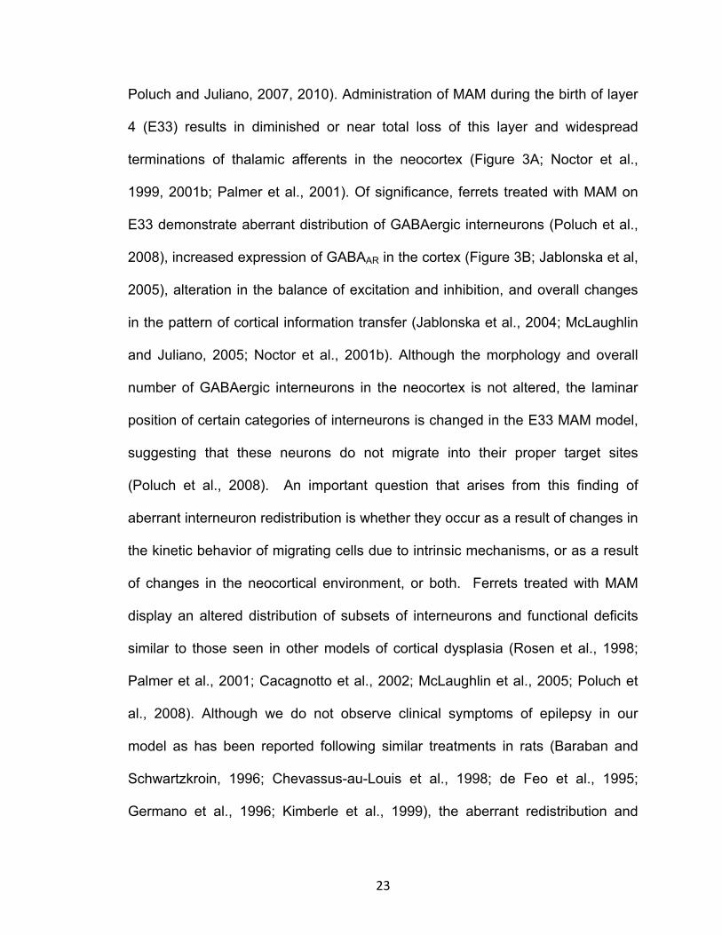
Poluch and Juliano, 2007, 2010). Administration of MAM during the birth of layer
4 (E33) results in diminished or near total loss of this layer and widespread
terminations of thalamic afferents in the neocortex (Figure 3A; Noctor et al.,
1999, 2001b; Palmer et al., 2001). Of significance, ferrets treated with MAM on
E33 demonstrate aberrant distribution of GABAergic interneurons (Poluch et al.,
2008), increased expression of GABAAR in the cortex (Figure 3B; Jablonska et al,
2005), alteration in the balance of excitation and inhibition, and overall changes
in the pattern of cortical information transfer (Jablonska et al., 2004; McLaughlin
and Juliano, 2005; Noctor et al., 2001b). Although the morphology and overall
number of GABAergic interneurons in the neocortex is not altered, the laminar
position of certain categories of interneurons is changed in the E33 MAM model,
suggesting that these neurons do not migrate into their proper target sites
(Poluch et al., 2008). An important question that arises from this finding of
aberrant interneuron redistribution is whether they occur as a result of changes in
the kinetic behavior of migrating cells due to intrinsic mechanisms, or as a result
of changes in the neocortical environment, or both. Ferrets treated with MAM
display an altered distribution of subsets of interneurons and functional deficits
similar to those seen in other models of cortical dysplasia (Rosen et al., 1998;
Palmer et al., 2001; Cacagnotto et al., 2002; McLaughlin et al., 2005; Poluch et
al., 2008). Although we do not observe clinical symptoms of epilepsy in our
model as has been reported following similar treatments in rats (Baraban and
Schwartzkroin, 1996; Chevassus-au-Louis et al., 1998; de Feo et al., 1995;
Germano et al., 1996; Kimberle et al., 1999), the aberrant redistribution and

24
position of interneurons seen in our model raises the likelihood that the inhibitory
function of these cells might be compromised, which may impair overall cortical
function. This study investigates the impact of MAM treatment on the dynamic
pattern of migration of GE cells as it relates to the basal level of GABAA activity
and function of the resulting cortex.
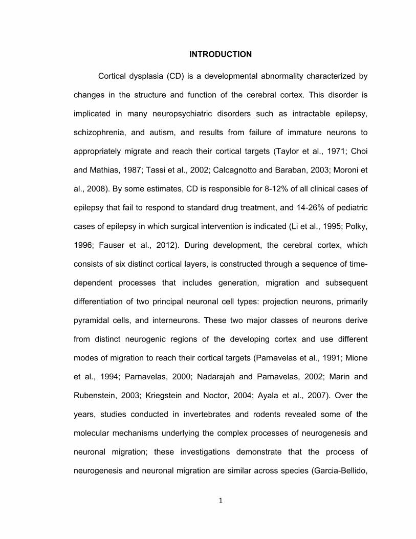
A B

25
Figure 1: (A). During development, projection neurons and interneurons are generated from
distinct neurogenic regions. Projection neurons form in the cortical ventricular zone and migrate
radially to the cortical plate. Cortical interneurons are generated in the ganglionic eminences (GE)
but migrate tangentially and radially to settle in the cortex. (B) Several extrinsic cues, including
GABA, act in concert with intrinsic signaling mechanisms to regulate the process of migration of
interneurons. The anatomic layout of the GE varies across species. Unlike rodents with three
distinct subtypes of GE, the GE in ferrets fuses at midcortical development. Broken lines signify
the corticostriatal boundary.

26
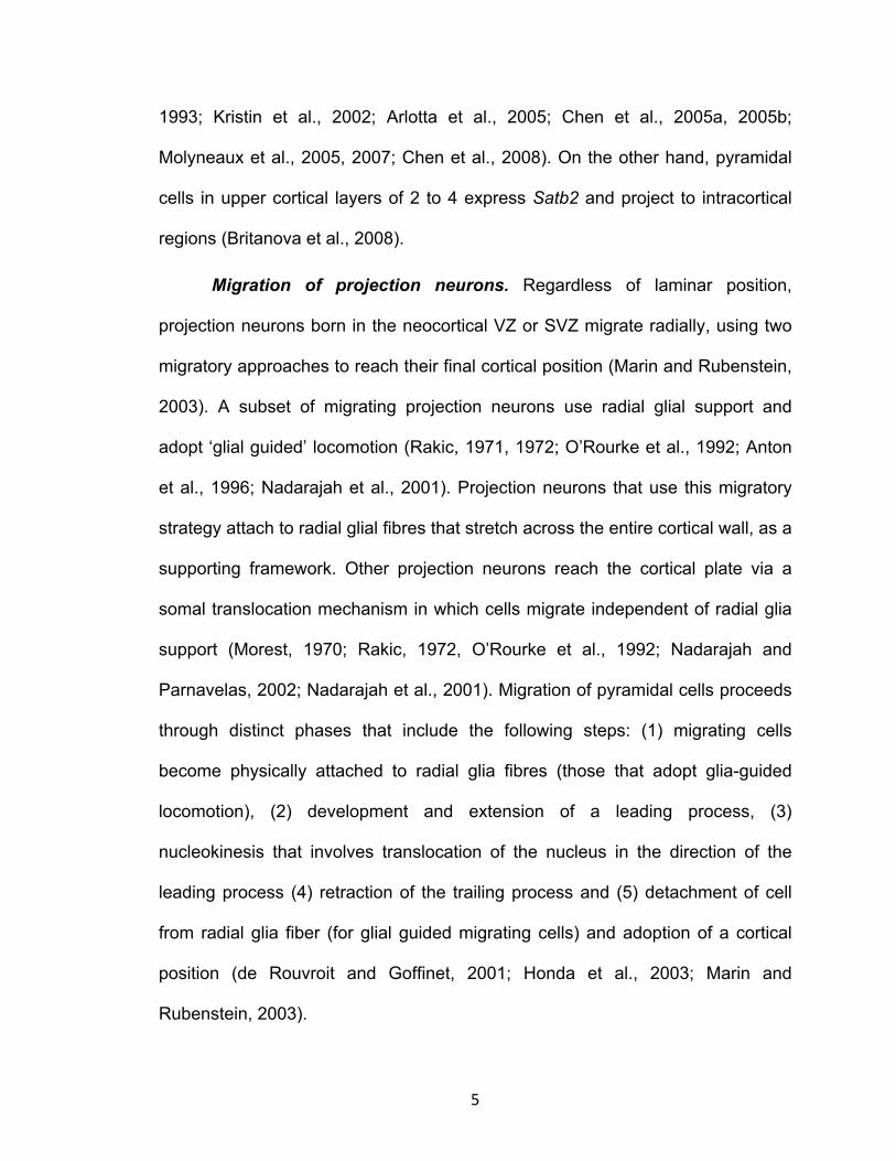
Figure 2: Pyramidal cells are generated from cortical ventricular zone and migrate radially to
different cortical layers. In this picture, cells within the CVZ were electroporated with a plasmid
producing red fluorescent protein. (A) Labeled radial glia cells that span the cortex can be seen
aposed with migrating projection neurons (B) High power view of a section of migrating projection
neurons on radial glial cells.

27
Figure 3: Administration of MAM on E33 induces several developmental malformations on the
cortex. (A) As a result of transient inhibition of neurogenesis, layer 4 cells, which are born during
this time period, are not generated leading to reduction in the thickness of cortical layer 4. (B)
After the reduction in thickness of layer 4, GABAA receptors, which are normally expressed in
high density in this region, expand to upper cortical layers.

28
CHAPTER 2
Altered kinetic behavior underlies redistribution of interneurons in a model
of cortical dysplasia: the influence of elevated GABAA receptor activity.
J. Abbah1 and S. L. Juliano1,2
1Program in Neuroscience and 2Anatomy, Physiology and Genetics, Uniformed
Services University of the Health Sciences, Bethesda, Maryland, 20814-4799
Corresponding Author: Sharon L Juliano: Anatomy, Physiology and Genetics,
Uniformed Services University of the Health Sciences, 4301 Jones Bridge Rd.,
Bethesda, Maryland, 20814-4799
Phone: 301-295-3673
Email: [email protected]
Running Title: GABA activity is critical for migration of GE cells

29
Abstract
Appropriate function of the neocortex depends on timely generation and
migration of cells produced in the germinal zones of the neocortex and ganglionic
eminence (GE). Failure to accurately complete migration results in cortical
dysplasia, a developmental syndrome implicated in many neurologic disorders.
We developed a model of cortical dysplasia in ferrets involving administration of
methylaxozymethanol acetate (MAM), an antimitotic, to pregnant ferrets on
gestational day 33, leading to dramatic reduction of layer 4 in the neocortex.
Here, using time-lapse video imaging, we investigate kinetic behavior of
migrating GE cells in ferrets and the role of GABAA activity. Treatment with MAM
significantly reduced migration speed and the relative proportion of GE cells
demonstrating exploratory behavior. Pharmacologic inhibition of GABAA
receptors (GABAAR) improved the speed of migration and exploratory ability of
migrating MAM-treated GE cells. Additionally, the expression of α2 and α3
subunits of GABAAR and the potassium chloride co-transporter (KCC2) increased
in the neocortex of MAM-treated animals. After MAM treatment, increases in
endogenous KCC2 and GABAAR combine to alter the kinetic properties and
exploratory behavior of migrating interneurons in ferrets. We show a direct
correlation between increased GABAA and KCC2 expression with impaired
migration and ability to explore the environment.
Keywords: development, ferret, KCC2, MAM, neuronal migration

30
Introduction
The laminar organization of the neocortex and its ability to function
depends on timely generation and proper migration of cells originating from the
ventricular zones of the neocortex (CVZ) and GE. Events that adversely impact
the migratory process can lead to cortical dysplasia, a developmental
abnormality characterized by aberrant cell clustering, altered gyral patterns, and
changes in electrophysiological profile (Taylor et al. 1971; Choi and Mathias
1987; Tassi et al. 2002; Calcagnotto and Baraban 2003; Moroni et al. 2008). The
aberrations underlie a vast number of neurological/neuropsychiatric disorders
including drug-resistant epilepsy, depression, and schizophrenia (Palmini et al.
1991; Gleeson and Walsh 2000; Zhu and Roper 2000; Ross and Walsh 2001;
Calcagnotto et al. 2002). Understanding the process of neuronal migration into
the neocortex is critical to comprehending the many disorders that result from
disrupted migration.
We developed a model of cortical dysplasia by administering a short
acting anti-mitotic, methylazoxy methanol (MAM), to pregnant ferrets on
gestational day (E33). Ferrets are the smallest animal with a convoluted cortex,
which make them an important model for neocortical development. The
tangential expansion of the neocortex, which assists in the formation of the sulci
and gyri, relies on proliferation of outer subventricular progenitors, a cell
population found in ferrets and humans, but absent in rodents (Fietz et al. 2010;
Lui et al. 2011; also see Martinez et al 2012 for a different point of view). Our lab
also found distinctions in the migratory patterns of interneurons in rodents and

31
ferrets, indicating the importance of studying a more developed model of
neocortex (Poluch et al. 2008).
Treatment with MAM on E33 coincides with the generation of layer 4 and
results in its dramatic reduction and in widespread re-distribution of GABAAαR
(Noctor et al. 1999; Palmer et al. 2001; Jablonska et al. 2004). Although the
morphology and overall number of interneurons in the neocortex is not altered,
the laminar position and orientation of interneurons is changed in the MAM
model, suggesting that these neurons do not migrate into their proper target sites
(Poluch et al. 2008). The aberrant redistribution of interneurons and GABAAαR
within the neocortex of MAM-treated animals raises important questions. 1) Do
changes in the distribution of cells arising from the GE occur as a result of altered
kinetic behavior of migrating GE cells, and, if so 2) are these changes influenced
by the ambient activity of GABA?
Using real-time video imaging and an in-vitro migration assay of
organotypic cultures of neonatal ferrets, we observed the dynamic movement
patterns of neurons leaving the GE and CVZ. Treatment with MAM impairs the
speed and exploratory potential of tangentially migrating interneurons leaving the
GE without any significant effects on neurons arising from the CVZ. GE cells in
MAM-treated animals exposed to GABA antagonists showed a significant
improvement in the speed of migration and the proportion of cells that display
exploratory activity. This reinforces the idea that E33 MAM treatment alters the
migration kinetics of GE cells. The changes in the dynamic movement of GE cells
are related to abnormal levels of GABAA-mediated activity, which diminishes the

32
capacity of these cells to explore the environment and effectively migrate to their
target within the neocortex.

33
Materials and Methods
Timed pregnant ferrets were obtained from Marshal Farms (New Rose, NY) and
maintained in the animal facilities of the Uniformed Services University of the
Health Sciences (USUHS). Pregnant ferrets were injected with 14mg/kg of MAM
(Midwest Research Institute, Kansas City, MO) IP on E33 under isofluorane
anesthesia (1-2%). After recovery from anesthesia, ferrets were maintained in
the animal facility until their kits were delivered. Handling of animals complied
with the USUHS Institutional Animal Care and Use Committee policy on the
humane use and treatment of animals.
Preparation of Organotypic slices: Preparation of organotypic cultures was
accomplished as previously described (Palmer et al. 2001). Postnatal day 0 to
day 1 (P0-P1) ferret kits were anesthetized with sodium pentobarbital (50mg/kg),
their brains removed and placed in ice-cold artificial cerebrospinal fluid (aCSF)
composed of (in mM): 124 NaCl, 3.2 KCl, 2.4 CaCl2, 1.2 MgCl2, 26 NaHCO3, 1.2
NaH2PO4 and 10 glucose bubbled with 95% O2 and 5% CO2 under a laminar flow
hood. Coronal slices (350-500 µm thick) were prepared from each hemisphere
using a tissue chopper (Stoelting Co., Wood Dale, Illinois). Brain slices were
transferred into 0.4 µm culture plate inserts (Millicell-CM, Bedford, MA) placed in
six-well plates containing Neurobasal media with B27, N2, and G1.2 (containing
gentamycin and glutamine) supplements. Slices were incubated at 37°C under
95% O2 and 5% CO2. In some experiments, bicuculline methiodide (BMI; Tocris
Bioscience, Park Ellisville, MO) at a final concentration of 10 µM was added to
the media.

34
Cell labeling: Two approaches were used to label migrating neurons. In
one approach, crystals of 1,l’-dioctadecyl-3,3,3_,3_-tetramethylindocarbocyanine
percholate (DiI, Invitrogen, Carlsbad, CA) were placed in the GE or CVZ using
pulled borosilicate glass pipettes. In another approach, we used electroporation
to focally transfect cells within the ventricular zone (VZ) of the GE using a
modification of the method described by Flames et al, 2004. Transfection was
accomplished using a plasmid that codes for red fluorescent protein (RFP), which
was cloned into pCAGGS expression vector (a gift from Dr Tarik Haydar).
Between 1-3 µl of plasmid DNA (2.7-3.6 µg/µl) was injected into the VZ of the GE
of organotypic slices of ferrets. The cathode of a gene paddle electrode (Harvard
Apparatus, Inc., Holliston, MA) was placed within the lateral ventricle close to the
VZ of the GE while the anode was placed closed to the pial surface in an
appropriate position. A pulse of 60V was applied four times, each lasting for 50
ms at intervals of 950 ms using a BTX ECM830 pulse generator (Gal et al 2006;
Harvard Apparatus, Inc., Holliston, MA). Slices were incubated as above for at
least 24 hrs prior to video imaging.
Video imaging: After incubating organotypic slices for 24 h (CVZ-derived)
or 48 h (GE-derived) for analysis, migrating neurons were continuously visualized
using an Axiovert 200 inverted microscope fitted with an apotome and Axiovision
software (Carl Zeiss AG, Oberkochen, Germany). The microscope was fitted with
an incubation chamber and a holder for the slices, which were maintained with
humidification at 37°C with 95% O2 and 5% CO2. Serial stacks of images taken
through the thickness of the slice were collected using a 10X objective every 30

35
min for 24 hrs. The image stacks were collapsed into a single frame prior to
analysis of migration. Migrating GE cells were analyzed after crossing the
corticostriatal boundary into the neocortex.
Analysis of migration: To measure speed, migrating cells captured in real
time were tracked using ImageJ software (http://rsb.info.nih.go/ij) and the tracked
distances measured and expressed over time (to obtain the speed in real time)
using a program written in R statistical language (R Development, 2009). Also,
the orientation of migrating GE cells (either tangential or radial) and exploratory
activities were quantified for each migrating cell. The distribution of migrating GE
cells within the neocortex in both normal and MAM-treated animals was
determined using Axiovision software. To do this, the neocortex was divided into
three roughly concentric regions (0-350 µm, 350-700 µm and >700 µm) from the
border of the GE VZ and labeled neurons found within the various bins were
counted and expressed as a percentage of the total number of cells.
Western Blot analysis: The neocortex was dissected from organotypic
slices of normal and MAM-treated P0-P1 ferret brains (prepared as described
above), frozen on dry ice and preserved at -82°C prior to use. Tissues were
homogenized using RIPA lysis buffer (Santa Cruz Biotech, Santa Cruz, CA)
followed by centrifugation at 14,000 g at 4°C. Protein concentration was
estimated using a colorimetric assay. Proteins were separated by SDS-PAGE
using 10% Bis-Tris gel and electrophoretically transferred to a PVDF membrane
(Invitrogen, Carlsbad, CA). A loading volume of 10 µl containing 1-2 µg of protein
was used for each analysis. Membranes were incubated with Casein blocking

36
buffer (PBS (0.5M NaCl) + 3% Casein + 0.5% Tween-20) overnight, followed by
affinity purified rabbit polyclonal antibodies directed against GABAAα2 (1:200;
ProSci Inc., Poway, CA), GABAAα3 (1:2000; Sigma, St. Louis, MO), KCC2 (1:500,
Millipore, Billerica, MA), and monoclonal anti-actin (1:3000, NeoMarkers,
Fremont, CA) for 24 hrs. Following several washes with PBS, protein bands were
detected using HRP-conjugated anti-rabbit secondary antibodies (1:1000,
Jackson Lab., West Grove, PA) and HRP-conjugated anti mouse (1:3000,
Thermo Scientific, Rockford, IL) and visualized using enhanced
chemiluminescence detection. Signal intensities were quantified using Image j
software (http://rsb.info.nih.go/ij).
Statistics: The chi-square distribution test was used to analyze distribution
of GE cells within the neocortex. For all other analysis, a Student’s t-test was
applied and differences evaluated at P < 0.05.

37
Results
General properties of normal and MAM- treated migrating cells leaving the
GE
We previously reported that cells leaving the GE in MAM-treated animals
exhibit abnormal characteristics, specifically showing orientations that differed
from control cells (Poluch et al., 2008). To further characterize the migratory
behavior of cells leaving the GE or the CVZ, we applied an in-vitro live video-
imaging assay to continuously view migrating cells. Our earlier work
demonstrates that at P0-P1 in the ferret, the medial and lateral GE are fused,
thus we will refer to our label as directed to the fused GE (Poluch et al. 2008).
Migrating cells exiting the GE or CVZ of organotypic slices were labeled using
electroporation with plasmids that code for RFP or injections of DiI. An example
of a slice labeled by electroporation can be seen in Figure 1A. Higher power
views of cells en route to the cortical plate are shown in Figures 1B and C. These
cells have varied orientations as well as morphology; the dynamic features
change when observed in real time. After allowing the cells to move away from
the GE or CVZ, the dynamic pattern of migration was captured in real time. GE
cells moving into the neocortex adopt different orientations and take different
routes. A number of migrating neurons course through the VZ or SVZ
(subventricular zone), many orient in a tangential direction (Figure 2A, B red
arrows). A different group of more superficially labeled GE cells display both
tangential (yellow arrows) and radial (green arrows) orientations. Many of the
radially oriented cells point in the direction of the pial surface. However, some of

38
the radially-oriented cells have their leading edges directed toward the ventricular
zone (Figure 2A, B, blue arrows). These observations correspond to our previous
report in the ferret that similar to rodents, tangentially migrating cells in the GE
follow 3 paths including one close to the VZ, one in the IZ, and one superficially
near the marginal zone (not shown in this case) (Poluch et al. 2008). We also
observed that migrating GE cells alter their morphology as they transit from point
to point, generating distinct morphological features. In static images the labeled
cells are either unipolar (the cell posses a single leading unbranched process
(Figure 2C-a), display a branched process (Figure 2C-b), are bipolar (both
leading and trailing processes Figure 2C-c), or multipolar (multiple processes
emanate from the soma of the cell (Figure 2C-d).
Although the general trend of movement for cells leaving the GE is
towards the neocortex, individual cells move in all directions and often alter their
course of direction (Movie 1). Cells may emit one or more processes and move in
the direction of the new branch (e.g. Movie 2), or emit branches and continue in
the same direction or not move at all, but only explore the environment for a time
(Movie 3). We also frequently observed nucleokinesis in which the nucleus
moves within the cytoplasm of a cell in the direction of the leading process (as
described by others such as Nadarajah et al. 2001, Martini et al. 2009, Movies 2
and 3). This process usually precedes the point-to-point translocation of the cell
during migration.
Kinetic behavior of cells exiting the GE in normal and MAM-treated slices

39
Our earlier work indicates that after treatment with MAM, in addition to
changes in orientation while migrating into the cortical plate, several subtypes of
interneurons are redistributed in the neocortex of juvenile ferrets (Poluch et al.
2008). To assess the factors contributing to the altered distribution, we evaluated
specific dynamic parameters of cells leaving the ganglionic eminence in normal
and MAM-treated animals, including the speed, orientation of movement, and
exploratory behavior (defined as extension of new processes and changes in the
direction of movement).
Speed and direction. The migratory speed of GE and CVZ cells was
measured as described in the Methods. Cells in organotypic cultures of normal
ferrets migrated significantly faster (37.435 µm ± 0.87/hr) compared with similar
cells in animals treated with MAM (32.98499 µm ± 0.73/hr) (Figure 3A). Because
migrating interneurons from the GE have different orientations (e.g. Figure 2A
and also Ang et al. 2003; Tanaka et al. 2003 and 2006), we examined the
relationship of speed to orientation during movement and assessed distinctions
between normal and MAM treatment for radial versus tangential travel. Normal
cells traveling in both radial and tangential directions migrated significantly faster
than MAM-treated cells (Figure 3B).
To investigate if changes in the speed of migration affect the final
distribution of migrating neurons, we assessed their positions in organotypic
slices of normal and MAM-treated animals after 48 hrs of incubation. We divided
the neocortex into three roughly concentric regions (0-350 µm, 350-700 µm and
greater than 700 µm) from the point of DiI injection in the GE using Axiovision

40
software (Figure 4A). The total number of migrating GE cells in each sector was
computed; the amount of cells in each sector of the normal and MAM-treated
organotypic slices were compared and presented as percentages of the total.
Although slightly more cells in normal organotypic slices migrated further away
from the GE as revealed by the higher percent in the region 700 µm from the GE,
the overall distribution across the three sectors was not significantly different
(Figure 4B). In other words, the cells leaving the MAM-treated GE moved slower,
but eventually situate at similar distances away from the GE.
Exploratory behavior of GE-derived neurons in normal and MAM-treated
slices
Migrating GE cells are influenced by a variety of signaling molecules that
induce stereotypic migratory activity, which we refer to as exploratory behavior.
Two parameters defined exploratory behavior in our study: (i) process extension
or the ability of migrating cells to extend more than one leading process and (ii)
turns or changes in direction of movement, in which a deviation from the initial
course of movement occurred. In Figure 5A, the green arrow indicates a cell
changing direction, while the red arrow shows a cell branching and also changing
its direction of movement. To determine if treatment with MAM alters the ability of
migrating GE cells to explore the neocortical environment, migrating cells were
observed during live video imaging for 24 hrs. For several examples, the path of
multiple migrating cells was traced using ImageJ. The movement was tracked for
a period of at least 8 hours and the position of each cell recorded. Figure 6A and

41
B illustrate that the general pattern of movement in the MAM-treated tracks of
cells (Figure 6B) show fewer changes in their direction when compared with the
normal patterns of movement (Figure 6A). The quantification of movement
patterns is seen in Figure 5B, which demonstrates that the tracks of normal cells
completed more changes of direction in their movement patterns (i.e., turns)
compared with the MAM-treated cells. The percent of cells displaying process
extension or turning in each slice was calculated for normal and MAM-treated
organotypic cultures. Cells were scored negative if they did not display any of
these behaviors during the observed period of time. In MAM-treated slices, fewer
cells leaving the GE extended more than one leading process in comparison with
cells in normal animals. In addition, the percent of cells that performed turns was
fewer after MAM treatment (Figure 5B).
Interference with GABAA-mediated activity affects migratory behavior of GE
cells in MAM-treated animals
The migratory behavior of interneurons entering the neocortex is regulated
by a variety of signaling molecules including GABA (Cuzon et al. 2006; Heck et
al. 2007; Lopez-Bendito et al. 2003 and 2008). We previously demonstrated that
treatment with MAM leads to a widespread redistribution of GABAAα receptors;
we also found that increasing GABAAR-mediated activity in normal organotypic
cultures interferes with the orientation of interneurons as they migrate into the
cortical plate similar to the effect of MAM treatment (Jablonska et al. 2004;
Poluch et al. 2008). These findings suggest that sensitivity to GABA is unusually

42
high after MAM treatment and inhibiting GABA signaling may alter the positioning
of neocortical interneurons. We predicted that treatment with GABAA antagonists
would shift the kinetic behavior of neurons originating from MAM-treated slices
toward normal. The speed and exploratory behavior of migrating cells in normal
and MAM-treated slices was evaluated as described previously after adding the
GABAA antagonist BMI to the media. This treatment significantly increased the
speed of migration in the MAM-treated group to be closer to normal (Figure 7A).
When organotypic slices of E33 MAM-treated animals were exposed to
BMI, the percent of migrating cells extending more than one leading process
increased and was nearly identical to those of normal migrating GE cells. This
matches our prediction that decreasing GABAA activity in MAM-treated brains
results in more normal behavior (Figure 7B). The number of turns, or alterations
in the direction of movement, did not change however, after adding BMI to the
media.
Treatment with MAM does not significantly alter the migration kinetics of
neurons originating from the cortical ventricular zone
Our previous observations indicated that tangentially migrating GABAergic
neurons were specifically disoriented in MAM-treated ferrets (Poluch et al. 2008).
To test if the changes in the kinetic behavior of migrating interneurons were
limited to the neurons originating from the GE, we examined the migration of
neurons from the CVZ to the neocortex using live-video imaging. Organotypic
slices were injected with DiI in the neocortical ventricular zone and only slices in

43
which the DiI label was confined to the VZ were used for analysis. In this regard
we limited labeling cells in the vicinity of the CVZ, but arising from the GE. We
hoped to insure that the cells migrating away originated from the CVZ. An
example of an injection site and cells migrating away from the CVZ can be seen
in Figure 8. We measured the speed and exploratory behavior of cells leaving the
CVZ in normal and MAM-treated slices. Although we found no significant
differences in the parameters analyzed between the two groups, there was a
noticeable, but statistically insignificant, decrease in the speed of migration for
the MAM-treated cells (Figure 9A-B).
Treatment with MAM increases the expression of GABAAα2, GABAAα3
receptor subunits and KCC2
Our previous studies strongly suggest that GABAA receptors and GABA
activity are increased in the neocortex of MAM-treated animals (Jablonska et al.
2004; Poluch et al. 2008). Here we also show that blockade of GABAA-mediated
activity ameliorates the deficits in dynamic activity after MAM treatment. To
further assess the role of GABA in mediating the kinetics of migrating GE cells
following treatment with MAM, we determined the level of expression of GABAAα2
and GABAAα3 receptor subunits using Western blots. GABAAα2 and GABAAα3
show high expression in both the neocortex and GE early in development, and
may play important roles in migration and other developmental events in GE cells
(Laurie et al. 1992; Poulter et al. 1992; Fritschy et al. 1994; Wang and Kriegstein
2009; Carlson et al. 2010). In our study, both α2 and α3 subunits are increased in

44
MAM- treated cortex compared to normal, furthering support for increased
GABAA-mediated activity in our model animals (Figure 10A and B).
To further evaluate the potential of altered environmental GABA to
influence migration, we established the amount of KCC2 in normal and MAM-
treated cortex. During development, KCC2 maintains a high extracellular Cl-
gradient and induces hyperpolarization of cells in response to GABA, playing a
role in the developmental switch from the GABA-induced depolarization to
hyperpolarization, which may also terminate cell migration (Rivera et al 1999;
Bortone and Polleux 2009). After MAM treatment, both monomeric and
oligomeric forms of KCC2 were increased in the neocortex as demonstrated
using Western blots (Figure 10C, right upper panel). Furthermore, ratiometric
analysis reveals a decrease in KCC2 monomer: oligomer ratio (Figure 10C, right
lower panel).
45
Discussion
The kinetic behavior of migrating interneurons, influenced by both intrinsic
and environmental factors, plays an important role in the proper positioning of
these cells in the neocortex. We developed a model of cortical dysplasia in
ferrets by interrupting the development of layer 4 cells resulting in abnormal
placement of specific populations of interneurons (Noctor et al. 1999; Poluch et
al. 2008). In this study we show that transient and layer-specific interference with
corticogenesis leads to: a) alterations in the basal activity of GABAA and in KCC2
expression b) this change directly influences the kinetic behavior of migrating GE
cells. These findings directly and uniquely implicate increased GABAAR and
KCC2 activity in impairing dynamic aspects of migration and in the ability of GE
cells to actively explore the environment during migration to the cerebral cortex.
We demonstrate that details of migration not shown before involving active
exploration are strongly influence by ambient GABAAR and KCC2 activity. This
may underlie the modified migratory behavior of later born interneurons.
Ferrets signify a valuable developmental model. As the smallest mammal with a
convoluted cortex, they represent an important link between studying
lissencephalic rodents and non-human primates. Several groups have identified
important developmental features in ferrets, including our previous findings that
many neurons populating the neocortex are generated in lateral portions of the
GE (Poluch et al. 2008). Several recent articles describe a distinct outer
subventricular zone (oSVZ) in ferrets, a region found in primates, and suggest
that this region may be important for generating cells that populate and expand

46
the gyrencephalic neocortex (Fietz et al. 2010; Reillo and Borell 2011; Reillo et
al. 2011; Liu et al. 2011). The oSVZ contains a population of cells called outer
radial glia (oRG; also called tRG), which appear to be the cell type responsible
for generating additional cortical cells leading to the expansion of the developing
cortex. This idea is contested recently by a group suggesting that the outer SVZ
in ferrets does not differ substantially from that region in the rat (Martinez-
Cerdeno et al. 2012). Nevertheless, these examples demonstrate the importance
of studying developmental processes using several animal models.
MAM treatment alters the kinetic behavior of migrating GE cells
The ability of migrating GE cells to reach appropriate positions within the
neocortex depends on the concerted action of extrinsic factors within the
neocortex and intrinsic signaling mechanisms that inhibit, facilitate, and guide
migrating neurons (Anderso et al. 1997; Powell et al. 2001; Lopez-Bendito et al.
2003 and 2008; Polleux et al. 2003; Stumm et al. 2003; Alifragis et al. 2004;
Flames et al. 2004; Cuzon et al. 2006; Cobos et al. 2007; Liodis et al. 2007;
Friocourt et al. 2008). Events leading to changes in neocortical architecture may
cause variability in the environment that alters the pattern of migration of
neuronal cells and results in abnormal cell positioning and cortical dysplasia. In
our model of cortical dysplasia, layer 4 of ferret neocortex is diminished resulting
in widespread termination of thalamocortical afferents (Palmer et al. 2004),
redistribution of GABAA receptors and parvalbumin and calbindin positive
interneurons (Jablonska et al. 2004, Poluch et al. 2008) and changes in the

47
sequence of information transfer in the somatosensory cortex (McLaughlin et al.
2005). To better understand the mechanisms contributing to the redistribution of
interneurons, we evaluated several parameters of kinetic behavior in migrating
GE cells. Cells leaving the GE in normal animals migrated significantly faster
compared to similar cells in MAM-treated animals. In addition, GE generated
neurons in MAM-treated animals exhibited less exploration of the neocortical
environment. Exploratory behavior, including extension of multiple leading
processes and changes in direction of movement, results from coordinated
signaling from various sites, which communicate cues that determine the final
location of these cells. The change in the speed of migration and exploratory
activity of interneurons following treatment with MAM therefore has important
implications on the overall positioning of cells within the neocortex.
Inhibiting GABAA-mediated activity improves migration of interneurons in
MAM-treated animals
GABA is an important influence on migrating cells, although its precise
role in the migration of telencephalic cells is complex. GABA receptors are
important during different phases of migration and mediate aspects of the
initiation, continuation, and termination of cellular movement (Behar et al. 1996,
2001; Bolteus and Bordey 2004; Heck et al. 2007; Bortone and Polleux 2009).
Cuzon and colleagues (2006) report that GABAA activity is present throughout
the path of migration of mouse GE cells into the neocortex, but increases in
activity as neurons approach and enter their cortical target. Our studies suggest

48
that MAM treatment results in increased ambient GABAA-mediated activity, which
may interfere with migration of GE cells as they move en route to the neocortex
before the final phase identified by Cuzon et al. 2006. This is supported by our
original observation of expanded GABAAα immunoreactivity in MAM-treated
cortex (Jablonska et al. 2004) and the current finding of increased GABAAα2 and
GABAAα3 receptor subunits as demonstrated by Western blot. Both α2 and α3
subunits of the GABAA receptor are expressed early during development and
may mediate the initial roles of GABA during development (Laurie et al. 1992;
Poulter et al. 1992; Fritschy et al. 1994; Carlson et al. 2010). This assists in
explaining our finding that elevated levels of GABA interfere with migration of GE
cells to slow the pace but not prevent their movement.
To investigate the involvement of GABAA-mediated transmission in the
migration of interneurons, we manipulated the level of GABAA activity in MAM-
treated organotypic cultures using BMI to reduce the ambient GABAA activity. We
predicted that limiting the increased GABAA activity would change the cellular
actions in MAM-treated cortex to be more like normal. In fact, blocking GABAA
activity in MAM-treated animals resulted in significant enhancement of the
migration speed of cells leaving the GE as well as increasing the percentage of
MAM-treated GE cells extending more than one leading process. This suggests
that reducing the effectiveness of ambient GABA in the MAM-treated ferret
encourages migratory activity to become more normal.
Increased GABA signaling interferes with migration

49
Both GABAAα2 and GABAAα3 expression are increased in MAM-treated
animals compared to controls. Does increased GABAAα expression in MAM-
treated animals alter the kinetic behavior of GE cell migration? It has been known
for several years that GABA acts as an excitatory neurotransmitter early in
development to facilitate cell migration, but switches to mediate inhibition as cells
mature (Plotkin et al. 1997; Clayton et al. 1998; Rivera et al. 1999). The dual role
of GABA is attributed to the temporal expression of two cation-chloride
transporters, sodium-potassium-chloride (NKCC1) and KCC2. Migrating neurons
initially express NKCC1, which imports chloride ions (Cl-) into the cell to maintain
a higher intracellular Cl- concentration (Clayton et al. 1998). In response to
GABAA activation, the outward movement of Cl- results in depolarization of the
cells and triggers Ca2+-mediated events that influence the cytoskeleton and
stimulate migration (Behar et al. 2001; Soria & Valdeolmillos 2002). As cells
mature, NKCC1 downregulates and coincides with increased expression of
KCC2 (Plotkin et al. 1997). KCC2 is a Cl- exporter that maintains a higher
extracellular gradient of Cl-. Consequently, GABAA activation when KCC2 is
increased results in hyperpolarization and cell inhibition (Payne, 1997; Rivera et
al. 1999). Interestingly, in dissociated hippocampal cell cultures, activation of
GABAAR induces the expression of NKCC1 and KCC2 (Ganguly et al. 2001). In
addition, pharmacologic blockade of GABAA delayed the expression of KCC2,
while increased activity of GABAAR induces precocious expression of the
transporter. Premature expression of KCC2 as a result of a heightened state of
GABAA–mediated activity, as we see here, may be responsible for the migration

50
defects observed in MAM-treated ferrets. KCC2 is implicated in several important
processes, including decreased motility as a neuron migrates, which leads to cell
slowing, stopping, and differentiating. As a consequence, a premature switch of
the GABAA response may occur and produce early arrest of migrating
interneurons. Bortone and Polleux 2009, demonstrated that depolarization
through GABAA receptors stimulates motility of GABAergic migrating neurons
while hyperpolarization of GABAA receptors, induced by KCC2 expression,
provides a stop signal for migrating cells. We observed increased expression of
both KCC2 monomer and oligomers in the neocortex of young MAM-treated
animals. The ratio of KCC2 monomer to oligomer was also reduced in MAM-
treated animals, which indicates greater activity of this transporter in our model
animals. Oligomerization of KCC2 is an important step in the activation and
function of KCC2 as a Cl- exporter (Blaesse et al. 2006). Although others have
demonstrated that GABAAR and KCC2 activity are involved in neuronal migration,
we demonstrate here that high endogenous levels of these substances directly
interfere with migration. In addition they appear specifically important in dynamic
and exploratory behavior in cells leaving the ganglionic eminence.
MAM treatment has no effect on the kinetic profile of migrating cortical VZ
cells
To determine whether the effect of MAM treatment on the kinetics of
migration is restricted to interneurons alone, we also investigated the kinetics of
migrating neurons arising from the cortical VZ. Although the migration speed of

51
CVZ cells was reduced in MAM-treated animals, the deficit was not significant.
We also did not observe any significant differences in exploratory behavior
between cells in normal animals and those that received MAM treatment,
indicating the impact might be restricted to tangentially migrating interneurons.
The differential impact of MAM-treatment on the kinetic behavior of GE and CVZ-
derived cells relative to normal cells is not surprising since these two cell
populations have different neurogenic origin and migrate under the influence of
similar but not identical signaling mechanisms (Marin and Rubenstein, 2003).
A working model. A plethora of signaling molecules influence migrating
interneurons. GABA initially acts as a stimulating signal to facilitate migration and
then acts as a stop signal to arrest the migratory process, an effect that
correlates with the sequential expression of NKCC1 and KCC2. An appropriate
level of GABA is necessary for the proper migration and positioning of
interneurons. Our data suggests that following treatment with MAM, the basal
level of GABAA receptor (GABAAR) activity increases, and the elevated level of
GABAAR activity impairs the migration of interneurons, through premature
expression of KCC2. Thus, treatments that decrease the level of GABAAR activity
enhance the migration of interneurons in part by slowing the development of
KCC2.
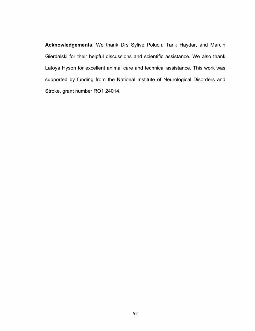
52
Acknowledgements: We thank Drs Sylive Poluch, Tarik Haydar, and Marcin
Gierdalski for their helpful discussions and scientific assistance. We also thank
Latoya Hyson for excellent animal care and technical assistance. This work was
supported by funding from the National Institute of Neurological Disorders and
Stroke, grant number RO1 24014.

53
References
Alifragis P, Liapi A, Parnavelas JG. 2004. Lhx6 regulates the migration of cortical
interneurons from the ventral telencephalon but does not specify their GABA
phenotype. J Neurosci. 24:5643-5648.
Anderson AA, Eisenstat SL, Rubenstein JL. 1997. Interneuron migration from
basal forebrain to neocortex: dependence on Dlx genes. Science. 278:474-476.
Ang ESBC, Haydar TF, Gluncic V, Rakic P. 2003. Four-dimensional migratory
coordinates of GABAergic interneurons in the developing mouse cortex. J
Neurosci. 23(13):5805-5815.
Behar TN, Li YX, Tran HT, Ma W, Dunlap V, Scott C, Barker JL. 1996. GABA
stimulates chemotaxis and chemokinesis of embryonic cortical neurons via
calcium-dependent mechanisms. J Neurosci. 16:1808-1818.
Behar TN, Smith SV, Kennedy RT, McKenzie JM, Maric I, Barker JL. 2001.
GABAB receptors mediate motility signals for migrating embryonic cortical cells.
Cereb Cortex. 11(8):744-753.
Blaesse P, Guillemin I, Schindler J, Schweizer M, Delpire E, Khiroug L, Friauf E,
Nothwang HG. 2006. Oligomerization of KCC2 correlates with development of
inhibitory neurotransmission. J Neurosci. 26(41):10407-10419.

54
Bolteus AJ, Bordey A. 2004. GABA release and uptake regulate neuronal
precursor migration in the postnatal subventricular zone. J Neurosci. 24:7623-
7631.
Bortone D, Polleux F. 2009. KCC2 expression promotes the termination of
cortical interneuron migration in a voltage-sensitive calcium-dependent manner.
Neuron. 62:53-71.
Calcagnotto ME, Paredes MF, Scott C, Baraban SC. 2002. Heterotopic neurons
with altered inhibitory synaptic function in an animal model of malformation
associated epilepsy. J Neurosci. 22(17):7596-7605.
Calcagnotto, ME, Baraban SC. 2003. An examination of calcium current function
on heterotopic neurons in hippocampal slices from rats exposed to
methylazoxymethanol. Epilepsia. 44:315-321.
Carlson VCC, Yeh HH. 2010. GABAA receptor subunit profiles of tangentially
migrating neurons derived from the medial ganglionic eminence. Cereb Cortex.
21:1792-1802.

55
Choi BH, Mathias SC. 1987. Cortical dysplasia associated with massive ectopia
of neurons and glial cells within the subarachnoid space. Acta Neuropathol
(Berl). 73:105-109.
Clayton GH, Owens GC, Wolff JS, Smith RL. 1998. Ontogeny of cation-Cl−
cotransporter expression in rat neocortex. Brain Res Dev Brain Res. 109:281-
292.
Cobos I, Borello U, Rubenstein JL. 2007. Dlx transcription factors promote
migration through repression of axon and dendrite growth. Neuron. 54:873-888.
Carlson VCC, Yeh HH. 2010. GABAA receptor subunit profiles of tangentially
migrating neurons derived from the medial ganglionic eminence. Cereb Cortex.
21:1792-1802.
Cuzon VC, Yeh PWL, Cheng Q, Yeh HH. 2006. Ambient GABA promotes cortical
entry of tangentially migrating cells derived from the medial ganglionic eminence.
Cereb Cortex. 16(10):1577-1588.
Flames N, Long JE, Garratt AN, Fischer TM, Gassmann M, Birchmeier C, Lai C,
Rubenstein JLR, Mar´ın O. 2004. Short- and long-range attraction of cortical
GABAergic interneurons by neuregulin-1. Neuron. 44:251-261.

56
Fietz SA, Kelava I, Vogt J, Wilsch-Bra¨uninger M, Stenzel D, Fish JL, Corbeil D,
Riehn A, Distler W, Nitsch R, Huttner WB. 2010. OSVZ progenitors of human and
ferret neocortex are epithelial-like and expand by integrin signaling. Nat
Neurosci. 13:690-699.
Friocourt G, Kanatani S, Tabata H, Yozu M, Takahashi T, Antypa M, Raguenes
O, Chelly J, Ferec C, Nakajima K, Parnavelas JG. 2008. Cell autonomous roles
of ARX in cell proliferation and neuronal migration during corticogenesis. J
Neurosci. 28:5794-5805.
Fristchy J-M, Paysan J, Enna A, Mohlerl H. 1994. Switch in the expression of rat
GABAA-receptor subtypes during postnatal development: an
immunohistochemical study. J Nerosci. 14(9):5302-5324.
Gal JS, Morozov YM, Ayoub AE, Chatterjee M, Rakic P, Haydar TF. 2006.
Molecular and morphological heterogeneity of neural precursors in the mouse
neocortical proliferative zones. J Neurosci. 26(3):1045-1056.
Ganguly K, Schinder AF, Wong ST, Poo M. 2001. GABA itself promotes the
Developmental-switch of neuronal GABAergic responses from excitation to
inhibition. Cell. 105:521-532.

57
Gleeson GJ, Walsh CA. 2000. Neuronal migration disorders: from genetic
diseases to developmental mechanisms. Trends Neurosci. 23:352–359.
Heck N, Kilb W, Reiprich P, Kubota H, Furukawa T, Fukuda A, Luhmann HJ.
2007. GABAA receptors regulate neocortical neuronal migration in vitro and in
vivo. Cereb. Cortex. 17:138-148.
Jablonska B, Smith AL, Palmer SL, Noctor SC, Juliano SL. 2004. GABAA
receptors reorganize when layer 4 in ferret somatosensory cortex is disrupted by
methylazoxymethanol (MAM). Cereb. Cortex. 14(4):432-40.
Laurie DJ, Wisden W, Seeburg PH. 1992. The distribution of thirteen GABAA
receptor subunit mRNAs in the rat brain. III. embryonic and postnatal
development. J Nerosci. 12(11):4151-4172.
Liodis P, Denaxa M, Grigoriou M, Akufo-Addo C, Yanagawa Y, Pachnis V. 2007.
Lhx6 activity is required for the normal migration and specification of cortical
interneuron subtypes. J Neurosci. 27:3078-3089.
López-Bendito G, Luján R, Shigemoto R, Ganter P, Paulsen O, Molnar Z. 2003.
Blockade of GABAB receptors alters the tangential migration of cortical neurons.
Cereb Cortex. 13:932-942.

58
Lopez-Bendito G, Sanchez-Alcaniz JA, Pla R, Borrell V, Pico E, Valdeolmillos M,
Marin O. 2008. Chemokine signaling controls intracortical migration and final
distribution of GABAergic interneurons. J Neurosci. 28:1613-1624.
Lui JH, Hansen DV, Kriegstein AR. 2011. Development and evolution of the
human neocortex. Cell. 146:18-36.
Mar´ın O, Rubenstein JLR. 2003. Cell migration in the forebrain. Ann Rev
Neurosci. 26:441-483.
Martinez-Cerdeno V, Cunningham CL, Camacho J, Antczak JL, Prakash AN,
Cziep ME, Walker AI, Noctor C. 2012. Comparative analysis of the subventricular
zone in rat, ferret and macaque:evidence for an outer subventricular zone in
rodents. Plos one 7(1):1-37.
Martini FJ, Valiente M, Lopez-Bendito G, Szabo G, Moya F, Valdeolmillos M,
Marin O. 2009. Biased selection of leading process branches mediates
chemotaxis during tangential neuronal migration. Development. 136:41-50.
McLaughlin DF, Juliano S. 2005. Disruption of layer 4 development alters laminar
processing in ferret somatosensory cortex. Cereb Cortex. 15:1791-1803.

59
Moroni RF, Inverardi F, Regondi MC, Panzica F, Spreafico R, Frassoni C. 2008.
Altered spatial distribution of PV-cortical cells and dysmorphic neurons in the
somatosensory cortex of BCNU-treated rat model of cortical dysplasia. Eplepsia.
49(5):872-887.
Nadarajah B, Brunstrom JE, Grutzendler J, Wong RO, Pearlman AL. 2001.
Two modes of radial migration in early development of the cerebral cortex. Nat
Neurosci. 4:143-150.
Noctor SC, Palmer SL, McLaughlin DF, Juliano SL. 2001. Disruption of layers 3
and 4 during development results in altered thalamocortical projections in ferret
somatosensory cortex. J Neurosci. 21:3184-3195.
Palmer SL, Noctor SC, Jablonska B, Juliano SL. 2001. Laminar specific
alterations of thalamocortical projections in organotypic cultures following layer 4
disruption in ferret somatosensory cortex. Eur J Neurosci. 13:1559-1571.
Palmini A, Andermann F, Olivier A, Tampieri D, Robitaille Y, Andermann E,
Wright G. 1991. Focal neuronal migration disorders and intractable partial
epilepsy: a study of 30 patients. Ann Neurol. 30:741–749.

60
Payne JA. 1997. Functional characterization of the neuronal-specific K-Cl
cotransporter: implications for [K+]o regulation. Am J Physiol. 273: C1516-
C1525.
Plotkin MD, Snyder EY, Hebert SC, and Delpire E. 1997. Expression of the Na-K-
2Cl cotransporter is developmentally regulated in postnatal rat brains: a
possible mechanism underlying GABA’s excitatory role in immature brain.
J Neurobiol. 33: 781-795.
Polleux F, Whitford KL, Dijkhuizen PA, Vitalis T, Ghosh A. 2003. Control of
cortical interneuron migration by neurotrophins and PI3-kinase signaling.
Development. 129:3147-3160.
Poluch S, Jablonska B, Juliano SL. 2008. Alteration of interneuron migration in a
ferret model of cortical dysplasia. Cereb Cortex. 18(1):78-92.
Poulter MO, Barker JL, O’Carroll AM, Lolait SJ, Mahan LC. 1992. Differential and
transient expression of GABAA receptor α-subunit mRNAs in the developing rat
CNS. J Neurosci. 12(8):2888-2900.
Powell EM, Mars WM, Levitt P. 2001. Hepatocyte growth factor/scatter factor is a
motogen for interneurons migrating from the ventral to dorsal telencephalon.
Neuron. 30:79-89.

61
R Development Core Team. 2009. R: A language and environment for statistical
computing. R Foundation for Statistical Computing, Vienna, Austria. ISBN 3-
900051-07-0, URL http://www.R-project.org.
Reillo I, Borrell V. 2011. Germinal Zones in the Developing Cerebral Cortex of
Ferret: Ontogeny, Cell Cycle Kinetics, and Diversity of Progenitors. Cereb
Cortex.
Reillo I, de Juan Romero C, Garcia-Cabezas MA, Borrell V. 2011. A role for
intermediate radial glia in the tangential expansion of the mammalian cerebral
cortex. Cereb Cortex 21: 1674-1694.
Rivera C, Voipio J, Payne JA, Ruusuvuori E, Lahtenin H, Lamsa K,
Pirvola U, Saarma M, Kaila K. 1999. The K+/Cl- co-transporter KCC2 renders
GABA hyperpolarizing during neuronal maturation. Nature. 397:251-255.
Ross EM, Walsh CA. 2001. Human brain malformations and their lessons for
neuronal migration. Annu Rev Neurosci. 24:1041-70.
Soria JM, Valdeolmillos M. 2002. Receptor-activated calcium signals in
tangentially migrating cortical cells. Cereb Cortex. 12(8):831-839.

62
Stumm RK, Zhou C, Ara T, Lazarini F, Dubois-Dalcq M, Nagasawa T, Hollt V,
Schulz S. 2003. CXCR4 regulates interneuron migration in the developing
neocortex. J Neurosci. 23:5123-5130.
Tassi L, Colombo N, Garbelli R, Francione S, Lo Russo G, Mai R, Cossu M,
Ferrario A, Galli C, Bramerio M, Citterio A, Spreafico R. 2002. Focal cortical
dysplasia: neuropathological subtypes, EEG, neuroimaging and surgical
outcome. Brain. 125:1719-1732.
Tanaka D, Nakaya Y, Yanagawa Y, Obata K, Murakami F. 2003. Multimodal
tangential migration of neocortical GABAergic neurons independent of GPI-
anchored proteins. Development. 130:5803-5813.
Tanaka D, Maekawa K, Yanagawa Y, Obata K, Murakami F. 2006.
Multidirectional and multizonal tangential migration of GABAergic interneurons in
the developing cerebral cortex. Development. 133:2167-2176.
Taylor DC, Falconer MA, Bruton CJ, Corsellis JAN. 1971. Focal dysplasia
of the cerebral cortex in epilepsy. J Neurol Neurosurg Psychiatry. 34:369-387.
Wang DD, Kriegstein AR. 2009. Defining the role of GABA in cortical
development. J Neurophys. 587(9):1873-1879.

63
Zhu WJ, Roper, SN. 2000. Reduced Inhibition in an Animal Model of Cortical
Dysplasia. J Neurosci. 20(23):8925-8931.
64
Figure 1. Images of migrating neurons leaving the ganglionic eminence (GE) of a normal ferret.
(A) Example of an organotypic culture obtained from a P0 ferret labeled by electroporation using
a plasmid that codes for red fluorescent protein (RFP). (B-C) Higher power views of cells en route
to the cortex revealing varied orientations and morphologies of migrating GE cells. B is taken
from the boxed in region of A and C is from a different normal slice culture. The arrows indicate
the path of the migrating cells. GE: ganglionic eminence; CVZ: Cortical ventricular zone

65
Figure 2. Migrating GE-derived neurons labeled with DiI. (A) Neurons migrating from the GE in
an organotypic culture of a MAM-treated animal display different orientations and positions within
the neocortex: tangentially migrating GE cells within the VZ/SVZ region (red arrows), tangentially
migrating GE cells within the intermediate zone (yellow arrows), radially-oriented GE cells in the
intermediate zone (green arrows) and radially-oriented GE cell directed towards the ventricle
(blue arrow). (B) A different organotypic culture of a MAM-treated animal labeled with DiI,
showing similar orientations of migrating cells. Arrows mark the different orientations and
positions of cells within the neocortex as described above. (C) Higher power images of cells
obtained from several labeled organotypic cultures from both normal and MAM-treated animals
(the images on the left and right were taken from normal slices); various morphologies can be
seen including those with a single leading unbranched process (a), those with single leading but
branched processes (b), bipolar cells (c), and cells with multiple leading processes (d). GE:
ganglionic eminence; LV: Lateral ventricle

66
Figure 3. Effect of MAM treatment on the speed and orientation of migrating interneurons leaving
the ganglionic eminence and traveling to the neocortex. (A) Neurons leaving the GE travel faster
in normal slices compared with those in MAM-treated slices, n= 390 cells (normal) and 467 cells
(MAM-treated). (B) Cells traveling in either the radial or tangential direction travel slower in MAM
treated slices. Tangentially migrating cells: n = 129 (normal), 105 (MAM-treated); radially
migrating cells: n = 223 (normal), 348 (MAM-treated). Significance determined using a Student’s
t-test; * p< 0.05. Error bars = Standard error of mean.

67
Figure 4. Distribution of cells leaving the GE and traveling to the neocortex. (A) To determine if
cells migrated different distances after treatment with MAM roughly concentric regions were
created in order to count the neurons that traveled up to 350 µm from the border of the GE VZ,
from 350 to 700 µm from that border, or greater than 700 µm from the center of DiI injection. (B)
After 48 hrs in culture there were no significant differences in the overall distribution of cells
leaving the GE in normal slices vs those in MAM- treated slices, A Chi-Square distribution
showed no significant differences. n=12 slices (MAM-treated), 10 slices (normal). Error bars =
Standard error of mean. GE: ganglionic eminence.

68
Figure 5. Effect of MAM treatment on the ability of migrating GE cells to extend multiple leading
processes and initiate a change in direction of movement. (A) Sequence of images of DiI-labeled
GE cells during time-lapse imaging for 8.5 h. Two cells are indicated with either a red or green
arrow and followed over time. The cell labeled with the red arrow moves diagonally from the
upper left to the approximate center of the image. At 6.5 h it branches and moves toward the
right. The cell labeled with a green arrow remains relatively still from 0 -. 4h. At this point it begins
to move upward and curves to the left, moving out of view at 8.5h. (B) The percent of migrating
cells extending new processes was evaluated in normal and MAM-treated slices. After MAM
treatment, migrating cells extended fewer new processes and displayed fewer turns/changes in
the direction of movement. n=23 slices/366 cells (MAM-treated), 21 slices/239 cells (normal); *P ≤
0.05, Student’s t-Test. Error bars = Standard error of mean.

69
Figure 6. Tracks of cells in normal and MAM treated slices. (A) The positions of migrating cells
were tracked every 30 minutes over a period of 24 h. Each cell is indicated with a different color
and a number. This is an example taken from a normal cortex. (B) The tracks of cells leaving the
GE of MAM-treated cortex are indicated here. The position of each cell is illustrated with a
different color and number. The cells leaving the normal GE show more variability in their route,
compared with those leaving the GE of MAM-treated brains. The numbers on the x and y axis
indicate the size of the area of the neocortex analyzed while the length of each track indicates a
rough estimate of the distance traveled in micrometers. The outline of ferret cortex in A and B
indicates the approximate path of cell movement (red arrow).

70
Figure 7. Administration of bicuculline (BMI) to MAM-treated slices results in an increase in the
speed and exploratory behavior of migrating cells. MAM-treated slices were placed in culture with
BMI added to the medium. (A) The addition of BMI increased the speed of migration in the treated
slices. (B) The addition of BMI also improved the ability of MAM-treated cells to extend
processes. The number of turns in the direction of movement did not increase. Significant
differences are only shown between the 2 conditions of MAM treated slices, i.e. MAM-Tx alone
(red bars) or MAM-Tx with BMI added (gray bars). The normal condition is shown for reference
(black bars). BMI treatment also eliminated significant differences between normal and MAM-Tx
for speed and extension of processes. Speed: n = 467 cells (MAM-treated), 270 cells (MAM-
treated + BMI); exploratory behavior: n = 23 slices/366 cells (MAM-treated), 16 slices/107 cells
(MAM-treated +BMI), * P ≤ 0.05, Student’s t-Test. Error bars = Standard error of mean.

71
Figure 8. Example of an injection of DiI into the neocortical ventricular zone of an organotypic
culture obtained from a MAM-treated animal. The ventricular zone is outlined with a white line.
Labeled neurons can be seen migrating toward the neocortex. The inset shows labeled migrating
cells from the same image moving into the cortex. The image in the inset was taken from the
region indicated with an arrow. LV, lateral ventricle.

72
Figure 9. Effect of MAM treatment on the speed of migration and exploratory behavior of cells
leaving the cortical ventricular zone (CVZ) to the neocortex. (A) There were no significant
differences in the speed of migration of cells leaving the CVZ from either normal or MAM-treated
slices n= 191 cells (normal) and 185 cells (E33 MAM), or (B) in the ability of cells to extend
processes or change the direction of movement or turn. Exploratory behavior: n= 13 slices/85
cells (normal) and n= 12 slices/75 cells (MAM-treated).

73
Figure 10. Expression of GABAA receptor subunits and KCC2 following treatment with MAM. (A)
Treatment with MAM results in an increase in the expression of GABAAα2, and (B) GABAAα3
receptor subunits in the neocortex as demonstrated by Western blots. (C) Both monomeric and
oligomeric forms of KCC2 are increased (top bar graph) with significant elevation in the ratio of
KCC2 oligomer:monomer (bottom graph). Protein levels were determined by densitometric
analysis using ImageJ software. For both normal and MAM-Tx animals, n = 4. Significance
determined using the Student’s t-test, * P< 0.05. Error bars = Standard error of mean.

74
Movie 1. View of cells leaving the ganglionic eminence en route to the cerebral
cortex. This movie shows cells leaving the ganglionic eminence that move
towards the cerebral cortex, which is located in the upper left of the images. It
took place over a period of 16 hours, images were obtained every 30 minutes.
Although the overall flow of movement was toward the neocortex, many cells
make turns on their way or travel in opposite directions, toward the ventricular
zone or in more lateral directions. Cells were labeled with DiI. This is taken from
a MAM-treated cortical slice at P0.
Movie 2. Cells exiting the ganglionic eminence migrating to the cerebral cortex.
There are fewer cells in this image, which allows easier viewing of details.
Individual cells stop and explore the environment without obvious movement.
Other cells change direction of movement or extend processes. Nucleokinesis
occurs in many cells. This takes place over a period of 16 hours, with images
obtained every 30 minutes. This is taken from a MAM-treated slice at P0.
Movie 3. Single cell branching and exploring. This movie shows a single cell that
exited the ganglionic eminence and moves toward the cerebral cortex. The cell
initially shifts ventrally and then changes direction toward the cortex. It also
appears to explore the environment by extending processes. This movie takes
place over a period of 9 hours, with sampling every 30 minutes. It is taken from a
normal slice at P0.

75
CHAPTER 3
Targeted disruption of layer 4 formation increases GABAA receptor-
mediated activity in upper cortical layers.
J. Abbah1 M. F. Braga1, 2 and S. L. Juliano1, 2
1Program in Neuroscience and 2Anatomy, Physiology and Genetics, Uniformed
Services University of the Health Sciences, Bethesda, Maryland, 20814-4799
Corresponding Author: Sharon L Juliano: Anatomy, Physiology and Genetics,
Uniformed Services University of the Health Sciences, 4301 Jones Bridge Rd.,
Bethesda, Maryland, 20814-4799
Phone: 301-295-3673
Email: [email protected]

76
Abstract
Cortical dysplasia is frequently associated with various clinical pathologies
including epilepsy and mental retardation. This disorder results from impaired
migration of immature neurons to their cortical targets, leading to anomalous
clustering of neural cells and changes in cortical properties. We developed a
model of cortical dysplasia by administering methylazoxymethanol (MAM), an
anti-mitotic, to pregnant ferrets on embryonic day 33 (E33) to disrupt the birth of
layer 4 cells leading to a significant reduction in the thickness of this layer and a
redistribution and increased expression of GABAA receptors. We evaluated the
impact of MAM-treatment on GABAA receptor-mediated synaptic transmission in
both migrating neurons derived from the ganglionic eminence (GE) in
organotypic cultures and on pyramidal cells in layer 2/3 of acute slices of
postnatal day 28-38 (P28-P38), using whole-cell patch-clamp recording. E33
MAM treatment results in a significant increase in the amplitude and frequency of
spontaneous GABAA receptor-mediated inhibitory post-synaptic currents (IPSCs)
in migrating GE cells. In older MAM-treated animals, the amplitude of GABAA
receptor-mediated spontaneous IPSCs in layer 2/3 pyramidal cells increased with
no changes in frequency. The amplitude and frequency of miniature IPSCs,
however, were significantly increased as a result of MAM-treatment in the older
pyramidal cells. The kinetics of GABAA receptor opening also altered following
treatment with MAM. Western blot analysis shows that the expression of
GABAAα3 receptor subunit increased in our model animals. Taken together, these
observations reinforce the idea that synaptic neurotransmission through GABAAR

77
is enhanced following treatment with MAM and coincides with our previous
finding of increased expression of GABAAα receptor expression within the upper
cortical layers. Overall, we demonstrate that small amounts of toxins delivered
during corticogenesis can result in long lasting changes in the neural architecture
that manifest as subtle disorders of information processing.
Key words: cortical dysplasia, GABA, patch-clamp, western blotting.

78
Introduction
During development of the mammalian neocortex, immature neurons
generated in the germinal zones of the neocortex and ganglionic eminences (GE)
migrate to create the six-layered, inside-out model of the neocortex (Angevine
and Sidman 1961; Rakic 1974). This laminar organization is crucial for
establishing a balance between excitation and inhibition leading to the overall
function of the neocortex. Events with adverse impact on neuronal migration
lead to cortical dysplasia (CD), a developmental abnormality characterized by
aberrant distribution and clustering of neocortical cells resulting in a plethora of
neuropathological disorders such as epilepsy, schizophrenia, and mental
retardation (Taylor et al. 1971; Choi and Mathias 1987; Palmini et al. 1991;
Gleeson and Walsh 2000; Zhu and Roper 2000; Ross and Walsh 2001; Tassi et
al. 2002; Calcagnotto et al. 2002; Calcagnotto and Baraban 2003; Colombo et al.
2003; Guerrini et al. 2003; Moroni et al. 2005, 2008). A common pathological
finding in clinical cases and animal models of CD is anomalous distribution and
clustering of neurons within the neocortex, which alters the microcircuitry and
cortical architecture (Ferrer 1993; Roper 1998; Colacitti et al. 1999; Jacobs et al.
1999; Benardete and Kriegstein, 2002; Moroni et al. 2008). Importantly, abnormal
GABA signaling frequently associates with cortical dysplasia leading to
consequent alteration in the balance of excitation and inhibition and disruption of
normal cortical function (Roper et al. 1999; Zhu and Roper 2000; Benardete and
Kriegstein 2002; Xiang et al. 2006; Moroni et al. 2008; Zhou and Roper 2009;
Brill and Huguenard 2010; Zhou and Roper 2010, 2011).

79
We developed a model of cortical dysplasia in ferrets using
methylazoxymethanol (MAM). We took advantage of the pharmacokinetic
property of MAM as a short-acting anti-mitotic, and the long developmental time
course of ferret corticogenesis to selectively disrupt the birth of layer 4 by
delivery on embryonic day 33 (E33) (Noctor et al. 1997; Noctor et al. 1999;
Palmer et al. 2001). The ferret, as the smallest mammal with a gyrencephalic
cortex, is crucial for the study of neocortical development. Because ferrets
possess an expanded neocortex with sulci, gyri, and large amounts of white
matter it is distinctive as a research subject. The protracted period of
corticogenesis and long period of cell proliferation in the large outer
subventricular zone may contribute to the expanded cerebral cortex in the ferret
compared with the rodent (Fietz et al. 2010). Distinctive features in the ferret
related to neocortical development make it essential to study developmental
processes in this animal, since aspects of neurodevelopment differ in mammals
with a convoluted versus lissencephalic cortex (Kriegstein et al. 2006; Poluch et
al. 2008).
As a likely result of the loss of layer 4 in our model, thalamic afferents that
normally synapse directly in this layer become redistributed to upper and lower
cortical layers; the capacity for entrainment and information transfer is also lost
within the somatosensory cortex (Noctor et al. 2001; Palmer et al. 2001;
McLaughlin and Juliano, 2005). Ferrets treated with MAM on E33 additionally
show an increased expression of GABAA receptors (GABAAR), which expands to
upper cortical layers, and interneurons are disorganized in their laminar positions

80
(Jablonska et al. 2004; Poluch et al. 2008). Changes in the migratory behavior of
GE-derived cells also occur (Abbah and Juliano 2012, under revision). These
changes are specific to the antimitotic MAM delivery on E33, as administration of
this drug on different embryonic dates leads to dramatically different results
(Noctor et al. 1999; Gierdalski and Juliano 2003, 2005; Poluch and Juliano 2007,
2010). An important question emerging from these observations is whether the
loss of layer 4 and subsequent redistribution of GABAAR and interneurons alters
GABA signaling within upper cortical layers and/or on the global neocortical
environment that influences the migration of late- born neurons.
Because our earlier studies in MAM-treated animals showed increased
expression of GABAAR within the neocortex as well as an abnormal pattern of
migration and distribution specifically for inhibitory cells leaving the GE, we
wanted to characterize the electrophysiological properties of both the GABAergic
cells migrating toward the neocortex as well as the functional impact of the
changed neocortical environment on responses. To investigate if changes in
cortical laminar formation alter the overall inhibitory tone within the neocortex and
also affect neuronal migration, we applied whole-cell patch-clamp recording to
measure spontaneous GABAAR-mediated inhibitory postsynaptic currents
(sIPSCs) in two types of cells. We studied cells migrating away from the GE
relatively early during ferret cortical development (P0 – P1) and pyramidal cells of
layer 2/3 of juvenile ferrets (P28 - P38). Choosing these two types of cells for
functional analysis allows us to determine the effect of early environmental
changes on migrating GABAergic cells and on inhibitory neurotransmission in the

81
somatosensory region of the resulting neocortex. Our results indicate that after
E33 MAM treatment, GABAergic synaptic neurotransmission is increased
significantly in cells migrating into the cortex from the GE and on cells populating
the cortex in the upper layers.

82
Materials and Methods
Handling of animals complied with the Animal Care and Use Committee of the
Uniformed Services University of the Health Sciences (USUHS). Pregnant ferrets
were obtained from Marshal Farms (New Rose, NY) and maintained in the
animal facilities of USUHS. On E33, ferrets were anesthetized with isofluorane
(1-2%) and injected IP with 14mg/kg of MAM (Midwest Research Institute,
Kansas City, MO). Ferrets were closely monitored and allowed to recover from
anesthesia before returning to the animal facility to await delivery of their kits.
Ferret kits were used for experiments at P0 - P1 or P28 - P38.
Preparation of Organotypic slices: Preparation of organotypic cultures
followed the method described by Palmer et al. (2001). Briefly, P0 ferret kits were
anesthetized with sodium pentobarbital (50mg/kg) and after observing
insensitivity to pain, their brains were removed and maintained in ice-cold
artificial cerebrospinal fluid (aCSF) bubbled with 95% O2 and 5% CO2 under
aseptic conditions in a laminar flow hood. The aCSF is composed of (in mM): 124
NaCl, 3.2 KCl, 2.4 CaCl2, 1.2 MgCl2, 26 NaHCO3, 1.2 NaH2PO4 and 10 glucose.
Coronal slices of the neocortex (350-500 µm thick) were generated using a tissue
chopper (Stoelting Co., Wood Dale, Illinois). Each slice was plated into a 0.4 µm
culture plate insert (Millicell-CM, Bedford, MA) and placed into six-well plates
containing Neurobasal media with B27, N2, and G1.2 (containing gentamycin
and glutamine) supplements. Slices were incubated at 37°C with 95% O2 and 5%
CO2.

83
Cell label: Migrating GE cells were labeled through electroporation using a
modification of the method described by Flames et al. (2004). Transfection was
achieved using a plasmid that codes for red fluorescent protein (RFP), which was
cloned into pCAGGS expression vector (a gift from Dr Tarik Haydar). We applied
approximately 1 µl of the plasmid DNA (5.3 µg/µl) to the ventricular region of the
ganglionic eminence (GE) of organotypic slices of ferrets. The cathode of a gene
paddle electrode (Harvard Apparatus, Inc., Holliston, MA) was placed within the
lateral ventricle close to the GE and the anode situated close to the pia in an
appropriate location; plasmid DNA was driven across the organotypic tissue wall
by electric pulse generated by a BTX ECM830 pulse generator (Harvard
Apparatus, Inc., Holliston, MA). A pulse of 60V current was delivered four times,
each lasting for 50 ms at intervals of 950 ms (Gal et al. 2006).
Preparation of acute slices: P28-P38 ferrets were anesthetized with
sodium pentobarbital (50 mg/kg i.p.) and then decapitated. Brains were quickly
removed and placed in ice-cold aCSF bubbled with 95% O2 and 5% CO2. The
parietal cortex was blocked and serial coronal sections (400 µm thick) were cut
on a vibratome (LEICA VT1000S, Buffalo Grove, IL). Slices were rapidly
transferred into a holding chamber with aerated aCSF maintained at room
temperature for 30 min to 1 h after dissection and before electrophysiological
recordings. The aCSF was composed of (in mM): 125 NaCl, 2.5 KCl, 1.0 CaCl2,
2.0 MgCl2, 25 NaHCO3, 1.25 NaH2PO4 and 25 glucose.
Recordings: Whole-cell patch-clamp recordings were performed on (a)
single neocortical pyramidal cells within layer 2/3 of the parietal cortex in acute

84
slices and (b) GE-derived cells labeled with RFP in organotypic slices after 14-20
days in culture. Pyramidal cells were identified by morphology; GE-derived cells
were identified under fluorescent microscopy using an upright microscope with
40X objective (ZEISS Axioscope 2FS PLUS). Differential interference optics
(DIC) was used for visualizing pyramidal cells. Similarly, cells migrating away
from the GE were initially identified using fluorescent microscopy but
subsequently visualized under DIC. Only GE cells that have crossed the
corticostiatal boundary into the neocortex and display characteristic elongated
leading process were selected for recording. All recordings were performed in a
submersion type recording chamber where slices were continuously perfused
with aerated aCSF composed of (in mM): 125 NaCl, 2.5 KCl, 2.0 CaCl2, 1.0
MgCl2, 25 NaHCO3, 1.25 NaH2PO4 and 25 glucose maintained at 28°C for
pyramidal cells and 37 °C for cells migrating from the GE. Slices were perfused at
a rate of 3-4ml/min. Recordings were performed using a pulled borosilicate
pipette filled with an internal solution composed of (in mM): 135 Cs-gluconate, 10
MgCl2, 0.1 CaCl2, 1 EGTA, 10 HEPES, 2 Na-ATP, 0.2 Na3 GTP, pH 7.3 (280–
290 mOsm). In some experiments, neurobiotin at a final concentration of 0.2%
was included in the internal solution for post-hoc identification. Whole-cell
recordings from the soma of neuronal cells occurred in voltage-clamp mode
using an Axopatch 200B amplifier (Axon Instruments, Foster City, CA) after a
tight seal (1 GΩ) was formed between the recording electrode (with resistance 5-
7 mΩ for pyramidal cells and 7-11 mΩ for GE-derived cells) and the cell body.
For all recordings, a threshold of 2 kHz was applied to filter signals, which were

85
recorded and digitized (Digidata1322A, Axon Instruments), stored using pClamp
software (Axon Instruments), and analyzed off-line with the Mini Analysis
Program (Synaptosoft, Inc., Leonia, NJ). The access resistance was constantly
monitored throughout the recording and cells that showed fluctuations beyond
15% were excluded from analysis.
Morphology: After recording, acute slices were immediately fixed in 4%
paraformaldehyde overnight at 4°C. After several washes with phosphate
buffered saline (PBS), slices were incubated with a solution containing 10%
methanol and 0.6% hydrogen peroxide in PBS for 30 min at room temperature.
After further washes with PBS, slices were incubated in a solution containing 2%
bovine serum albumin (BSA) and 0.75% triton X100 in PBS for 1 h, and then in
2% BSA and 0.1 % triton X100 for 15 min. Slices were then incubated in a dilute
solution of ABC elite kit overnight at 4°C. After multiple washes, cells were
visualized with peroxidase staining using DAB (Immpact DAB, Vector
Laboratories, Inc., Burlingame, CA.), and mounted in moviol.
Western blotting: Pieces of the neocortex cut from blocks of the parietal
cortex prepared as described above were frozen on dry ice and preserved at -
82°C prior to use. Protein samples were prepared first by homogenizing the
tissues using RIPA lysis buffer (Santa Cruz Biotech, Santa Cruz, CA) followed by
centrifugation at 14,000 g at 4°C. An estimate of the concentration of protein was
obtained through colorimetric assay. Separation of proteins in the samples was
accomplished by SDS-PAGE using 10% Bis-Tris gel and separated proteins
were transferred to a PVDF membrane (Invitrogen, Carlsbad, CA) using

86
electrophoresis. A loading volume of 10 to 15 µl containing 1 µg of protein was
used for each analysis. To limit non-specific binding of antibodies, membranes
were initially incubated with Casein blocking buffer (PBS [0.5M NaCl] + 3%
Casein + 0.5% Tween-20) for at least 2 h, followed by affinity purified rabbit
polyclonal antibodies directed against GABAAα2 (1:200; ProSci Inc., Poway, CA),
GABAAα3 (1:2000; Sigma, St. Louis, MO), GABAAγ2 (1:400, Sigma-Aldrich, St
Louis, MO.), and monoclonal anti-GAPDH (1:6000, Abcam, Cambridge, MA) for
24 h. After several washes with PBS, protein bands were detected using HRP-
conjugated anti-rabbit secondary antibodies (1:1000, Jackson Lab., West Grove,
PA) and HRP-conjugated anti mouse (1:6000, Thermo Scientific, Rockford, IL)
and visualized using enhanced chemiluminescence detection. Signal intensities
were quantified using Image j software (http://rsb.info.nih.go/ij).
Statistics: Student’s t-test was applied for all statistical analysis and
differences evaluated at P< 0.05.
Drugs: The following drugs were added to the recording buffer to isolate
sIPSCs and/or mIPSCs: 50 µM D-(-)-2-Amino-5-phosphonopentanoic acid (D-
APV), 20 µM 5-methyl-10,11-dihydro-5H-dibenzo[a, d] cyclohepten- 5,10-imine
[(+)-MK-801], both NMDA receptor antagonists, 20 µM (_)-(2S)-5,5-dimethyl-2-
morpholineacetic acid (SCH50911), GABAB antagonist; 10 µ M bicuculline
methiodide (BMI), a GABAA receptor antagonist; 10 µ M 6-Cyano-7-
nitroquinoxaline-2,3-dione (CNQX), an AMPA/Kainate receptor antagonist, all
from Tocris Cookson, Ballwin, MO, 1 µM Tetrodotoxin (TTX), a sodium channel
blocker; (Sigma, St. Louis, MO).

87
Results
In utero exposure to MAM increases the inhibitory tone mediated by GABAAR in
migrating cells originating from the GE.
Migrating cells leaving the GE are influenced by a variety of signaling
molecules including GABA. We previously demonstrated that the normal pattern
of migration and orientation of GE-derived neurons is altered in MAM-treated
animals, in part due to altered GABAAR-mediated activity (Poluch et al. 2008). To
determine whether the changes in neuronal migration result from increased
GABAAR activity in the neocortex, we evaluated GABAAR receptor-mediated
synaptic neurotransmission in cells leaving the GE by recording the spontaneous
inhibitory post synaptic currents (sIPSCs). Migrating cells were labeled with a
plasmid that codes for red fluorescent protein and identified under fluorescent
microscopy. Whole-cell patch-clamp recording was performed to record sIPSCs
on GE cells after allowing them to cross the corticostriatal boundary into the
neocortex (Figure 1E). sIPSCs were isolated using CNQX (10 µM), SCH50911
(20 µM) and AP-V (50 µM) and recorded at a holding potential (Vh) of -70mv. The
amplitude and frequency of sIPSCs are increased in migrating GE cells relative
to the control (Figure 1 A-D), indicating a heightened state of GABAergic
neurotransmission in this population of cells.
Treatment with MAM increases the amplitude of spontaneous IPSCs in the
pyramidal cells in layer 2/3 of parietal cortex.
Following treatment with MAM on E33, the thickness of layer 4 in ferret
somatosensory cortex diminishes leading to reduction in the overall cortical

88
thickness (Noctor et al. 1999). Additionally, the distribution of GABAAα receptor
subunits is expanded and increased in the upper cortical layers; several subtypes
of interneurons are also abnormally positioned (Jablonska et al. 2004; Poluch et
al. 2008). To examine the functional implication of the increased expression of
GABAAα receptor subunits on the inhibitory tone within the upper cortical layers,
we performed whole-cell patch-clamp recording on pyramidal cells to record
sIPSCs. sIPSCs were isolated using CNQX (10 µM), SCH50911 (20 µM) and
AP-V (50 µM) and recorded at a holding potential (Vh) of -70mv. We observed
that in P28-38 MAM-treated animals, the amplitude of sIPSCs was significantly
increased (40.08 ± 5.61 Pa, P ≤ 0.05) relative to control animals (29.45 ± 2.31
pA, P ≤ 0.05) (Figure 2 A, B, and C). Our data also indicate that the frequency of
sIPSCs was not significantly altered following treatment with MAM (Figure 2D).
The increased amplitude of sIPSCs (Figure 2C) in MAM-treated animals with no
change in frequency (Figure 2D) suggests that the change in GABAAR-mediated
activity results from enhanced GABAAR expression in the postsynaptic membrane
of layer 2/3 in the parietal cortex.
Several cells were injected with neurobiotin and reacted to visualize their
morphology. An example of the types of cells from which we recorded can be
seen in Figure 2E and F. All of the cells reported here were similar in morphology
and position within the cortex.
In MAM-treated animals, both amplitude and frequency of miniature IPSCs were
increased relative to control.

89
The increase in amplitude of sIPSCs indicates that GABAAR-mediated
activity is increased following treatment with MAM. The increase in sIPSC
amplitude may be due to changes in postsynaptic response. To determine the
underlying mechanism for increased GABAAR-mediated activity, we recorded the
action potential independent IPSCs. Miniature IPSCs (mIPSCs) were recorded in
the presence of CNQX (10 µM), SCH50911 (20 µM), AP-V (50 µM) and TTX (1
µM). Our results show that the amplitude of mIPSCs increased in MAM-treated
animals (17.31 ± 1.30, n = 24, P ≤ 0.05) relative to control (13.08 ± 0.71; n = 20,
P ≤ 0.05) (Figure 3A, B, and C). Quantitative analysis of the frequency revealed a
leftward shift in the inter-event interval indicating increased frequency of mIPSCs
generation (Figure 3D). The addition of bicuculline eliminated this response. This
result provides further indication of increased postsynaptic GABAAR synaptic
transmission in MAM-treated animals.
The kinetics of GABAA receptors are altered in MAM-treated animals
The change in frequency of mIPSCs in MAM-treated animals suggests
that alterations in presynaptic mechanisms may occur, such as alteration in
number and the configuration of interneurons, whereas changes in the amplitude
of response suggests changes in the postsynaptic membrane, probably due to
the observed increase in expression of GABAA receptors. To further evaluate the
factors responsible for these alterations, we evaluated the kinetics of GABAA
receptors. First, we analyzed the 10-90 rise time and compared control with
MAM-treated values. The 10-90% rise time provides spatial information about

90
key synaptic elements and potential alterations due to changes in presynaptic
terminal number and positioning (Xie et al. 1997; Wierenga and Wadman 1999;
Kobayashi and Buckmaster 2003; Alberto and Hirasawa 2010). In our study, the
10-90% rise time of the sIPSCs and mIPSCs was not significantly different
between normal and MAM-treated animals (Figure 4A-I and B-I). This parameter
was significantly reduced in cells migrating from the GE in our model animals
during the recordings of these cells in organotypic cultures (Figure 4C-I). We
next evaluated the decay time constant. In MAM-treated animals, the decay time
constant of sIPSCs and mIPSCs in the pyramidal cells was significantly
prolonged (Figure 4A-II and B-II). This is also true for the migrating cells
originating from the GE (Figure 4C-II). Analysis of the charge transfer (measured
as area) also reveals a significant increase in this parameter for the sIPSCs of
the pyramidal cells in layer 2-3 (Figure 4A-III) and the cells originating in the GE
(Figure 4C-III). This was also true for the recorded mIPSCs (Figure 4B-III).
Passive properties of layer 2/3 pyramidal cells and migrating GE cells are
unaffected as a result of treatment with MAM.
We measured the passive properties of both pyramidal cells of layer 2/3
and migrating neurons to evaluate whether the changes in IPSC reported is
related to intrinsic properties of the cell. Specifically, we recorded the
capacitance, membrane potential across the cell and internal and membrane
resistances. These properties provide important developmental clues relating to
the level of maturity of the cells. For this analysis, we compared data obtained

91
from normal cells leaving the GE in organotypic cultures with cells leaving the GE
after MAM treatment. We also compared pyramidal cells in normal slices with
pyramidal cells in MAM-treated slices. We found no significant differences in the
intrinsic properties between normal and MAM-treated cells for either the GE
derived cells or pyramidal cells, suggesting that the intrinsic properties are
unaffected following MAM treatment (Figure 5 A and B). In each case, there was
a reduction in the membrane resistance that was notable, but not significant.
Treatment with MAM increases the expression of GABAAα3 receptor within the
parietal cortex of ferrets.
The electrophysiological results presented so far support the idea that
GABAAR-mediated activity is increased following treatment with MAM. To further
validate the aberrant elevation of GABAAR activity in our model animals, we
performed western blot analysis to measure the level of expression of different
subtypes of the GABAAR within the somatosensory cortex. We tested several
receptor subtypes based on the likelihood of their expression in neocortex at this
stage of development in ferrets. Although we expected that GABAAα1 receptors
would be present at this age in ferret cortex, we were not able to visualize them.
GABAAα2 receptors were present equally in control and MAM-treated cortex
(Figure 6 top). The GABAAα3 receptor subunit, however, was increased in the
parietal cortex after MAM treatment (Figure 6 middle). We also tested the
presence of the γ2 subunit and found this to be slightly increased in E33 MAM-
treated cortex (Figure 6 bottom). These results indicate that GABAAR expression

92
is increased, underlying the enhanced GABAAR -mediated synaptic transmission
in MAM-treated animals.

93
Discussion
The current study uses a ferret model of cortical dysplasia to investigate
the alterations of GABAergic synaptic transmission recorded in layer 2-3
pyramidal cells of the somatosensory cortex and on migrating GE-derived cells.
We show that the basal activity mediated by GABAAR is enhanced within layer
2/3 and in cells leaving the GE in our model animals. An increased expression of
GABAAR after MAM treatment is likely to underlie the changes in synaptic
transmission. In our model, layer 4 formation is selectively disrupted by in utero
administration of MAM on E33, leading to its profound reduction in size, a
redistribution of interneurons, thalamic afferents, and GABAAR (Noctor et al.
1999; Palmer et al. 2001; Jablonska et al. 2005; Poluch et al. 2008). We also
observed that cells leaving the ganglionic eminence and traveling to the
neocortex after MAM treatment display abnormal dynamic properties while en
route (Abbah and Juliano, under revision). Our earlier studies using MAM
treatment during mid corticogenesis show a specific disruption of GABA-related
features, leading to an apparent overall increase in GABA signaling. This study
evaluated the functional implications of these findings in 2 populations of cells,
GABAergic neurons leaving the GE to become interneurons and pyramidal cells
in layer 2-3 of neocortex, presumably influenced by inhibitory synapses.
Treatment with MAM increases the amplitude of sIPSCs in layer 2/3
pyramidal cells.

94
An appropriate balance between excitation and inhibition is crucial for
normal cortical function. In cortical dysplasia, this equilibrium is disrupted
resulting in abnormal cortical activity and function. In pyramidal cells of layer 2/3
we observed an increased amplitude of sIPSCs without any significant change in
frequency. This indicates that synaptic transmission through GABAAR is
upregulated in MAM-treated animals, which is likely due to the increased GABAAα
receptor density on the post synaptic membrane previously observed in MAM-
treated animals (Jablonska et al. 2004). To further investigate the mechanism
underlying the increase in sIPSC amplitude, we evaluated the mIPSCs in the
same layer 2/3 pyramidal cells. mIPSCs are caused by the quantal release of
GABA from presynaptic terminals of GABAergic interneurons in the absence of
action potentials (Edwards et al. 1990). The number of presynaptic terminals of
interneurons and/or the postsynaptic GABAA receptor density on pyramidal cells
determines the frequency of mIPSCs (Shao and Dudek, 2005). In our study, both
amplitude and frequency of mIPSC were significantly increased in MAM-treated
animals relative to control. The increase in mIPSCs frequency and amplitude
signifies that both pre and postsynaptic mechanisms are involved in altering
GABAergic neurotransmission in our model animals. Presynaptic factors such as
alterations in the number of GABAergic interneurons and the quantal size of
release determine the frequency of response. It is possible that the GABAergic
neurons in our model are altered in some way to induce a greater number of
synapses or greater quantal release. We also know that after MAM treatment,
subpopulations of interneurons show altered distributions (Poluch et al. 2008).

95
Such redistributions may result in ectopic projections or altered synapses with
upper cortical layer pyramidal cells. Such region-specific alterations in local
circuit connections occur in other models of cortical dysplasia (Brill and
Huguenard 2010). Postsynaptic mechanisms such as the density and
internalization of postsynaptic protein modulate the size of GABAergic
neurotransmission contributing to the change in mIPSC amplitude (DeFazio and
Hablitz, 1999; Kittler et al. 2000; Loup et al. 2006; Goodkin et al. 2007; Michels
and Moss, 2007; Hartmann et al. 2008). The frequency of sIPSCs, which is the
summation of both action potential dependent and independent events, was not
altered even though the frequency of mIPSC was increased in MAM-treated
animals. A number of factors may be responsible for this, notably the nature of
the network of interneurons within layer 2/3 (Tamas et al. 1998; Bacci et al.
2003).
Although reduction in amplitude and frequency of IPSCs are frequently
associated with epilepsy and cortical dysplasia (McDonald et al. 1991; Henry et
al. 1993; Buckmaster and Dudek 1997; Hablitz and Defazio, 1998; Jacobs et al.
1999; Rocha et al. 2007), the converse occurs in other models of these diseases.
Our finding of increased GABAergic neurotransmission in layer 2/3 pyramidal
cells corresponds with a recent finding of enhanced GABAergic activity in
pediatric cortical dysplasia (Cepeda et al. 2012). Other studies demonstrate a
similar increase in GABAergic activity. A freeze lesion model of cortical dysplasia
reports increased IPSCs (Prince et al. 1997; DeFazio and Hablitz 1999; Brill and
Huguenard 2010). In other models of cortical dysplasia, however, a reduction in

96
inhibitory tone occurs, such as in utero exposure to carmustine, MAM, or
radiation in rats (Xiang et al. 2006; Zhou et al. 2009; Zhou and Roper 2010;
Karlsson et al. 2011). In these models, the development of seizures is believed to
be a consequence of reduced inhibitory drive similar to epilepitogenic foci
reported in animal models of epilepsy (Rice et al. 1996; Hirsch et al. 1999;
Cossart et al. 2001; Kobayashi and Buckmaster 2003; Shao and Dudek 2005;
Sun et al. 2007). This analysis is complicated further by evidence of elevated
GABAergic signaling in other seizure models (Gibbs et al. 1997; Nusser et al.
1998; Cossart et al. 2001; Shao and Dudek 2005; Zhan and Nadler 2009).
We did not observe any significant variation in the passive properties
between MAM-treated and sham animals indicating that the cellular intrinsic
properties remain relatively unaffected by MAM treatment. For both the cells
migrating away from the GE and the layer 2-3 pyramidal cells, however, the
membrane resistance was lower after MAM treatment, compared with control,
although the difference was not significant. It is not clear what might cause this
change, but it is possible that the membrane characteristics of MAM-treated
pyramidal cells have properties that would cause a slight difference in membrane
resistance. One possibility is that the MAM-treated brains may mature at a
different rate than the normal brains, leading to small distinctions in membrane
characteristics (Goldberg et al. 2011).
GABAA receptor-mediated activity is enhanced in migrating GE-derived
neurons after MAM treatment.

97
The orientation and distribution of interneurons is altered in E33 MAM-
treated animals, in part due to elevated GABAAR-mediated activity (Poluch et al.,
2008). Recently, we found that elevated GABAAR-mediated activity plays a role in
altering the kinetic behavior of migrating cells leaving the GE (Abbah and Juliano,
under revision). To determine whether the cells leaving the GE receive
abnormally increased GABAAR signaling after MAM treatment, we measured the
sIPSCs. Both amplitude and frequency of sIPSCs were increased in MAM-
treated animals suggesting that GABAAR synaptic transmission enhances in
MAM-treated animals. GABA signaling plays an important role in promoting
migration of GE cells and alterations in the basal level of GABAAR activity is likely
to be deleterious in the adequate migration of GE-derived cells. Specifically,
elevated GABAAR-mediated activity, while initially promoting migration of cells
derived from the GE, triggers early maturation of these cells by inducing
precocious expression of the potassium chloride co-transporter (KCC2) (Ganguly
et al., 1999; Abbah and Juliano, in revision). During development, KCC2
maintains a high extracellular Cl- gradient and induces hyperpolarization of cells
in response to GABA, playing a role in the developmental switch from the GABA-
induced depolarization to hyperpolarization, which may also terminate cell
migration (Rivera et al, 1999; Bortone and Polleux, 2009). Thus the elevated
functional responses to GABAAR synaptic transmission in cells migrating away
from the GE supports the idea that an aberrant GABAergic environment
contributes to both abnormal migration, and disrupted processing of information.

98
Kinetics of GABAA signaling is altered in MAM-treated animals.
The measure of 10-90% rise time gives relative spatial information with
respect to postsynaptic sites; changes in rise time deduce the locus of change in
presynaptic terminal number. It is widely accepted that receptors located in the
soma of postsynaptic membrane give a faster rise time compared to those
located on the dendrites (Shao and Dudek 2005). In our study there is no
significant change in the rise time in either the sIPSCs or the mIPSCs of
pyramidal cells in layer 2/3 indicating that there is no significant positional change
in the active zones and receptor clusters between normal and MAM-treated
animals (Xi et al. 1997; Alberto and Hirasawa 2011). In the cells migrating from
the GE, however, we observed a significant reduction in rise time after MAM
treatment compared to normal. Although, the reason for this change is unclear,
one factor that may be responsible is the relative position of the migrating cells in
the developing neocortex, which may affect the nature of synaptic contact they
receive. The decay time constant is influenced by the density of postsynaptic
receptors and receptor subtype composition (Xi et al. 1997; Shao and Dudek
2005); prolongation of decay time constant that we observed in both the sIPSCs
and mIPSCs in MAM-treated animals is consistent with increased expression of
GABAAR. The amplitude of the IPSCs and decay time constant determines the
charge transfer. In our study, the charge transfer was significantly increased for
both sIPSC and mIPSC in cells leaving the GE and layer 2/3 pyramidal cells
following treatment with MAM. This is consistent with the increased amplitude
and decay time that we reported.

99
Expression of GABAA receptor subunits are altered in MAM-treated
animals.
Our results show that GABAergic neurotransmission is enhanced in the
neocortex of MAM-treated animals, probably as a result of increased expression
of GABAAR. We also demonstrated that GABAAR are increased after E33 MAM
treatment, primarily in the upper cortical layers using receptor binding and
immunohistochemistry (Jablonska et al. 2004). Here we also find an increase in
the amplitude of sIPSCs and mIPSCs in pyramidal cells of layer 2/3 and a
prolonged decay time. To further confirm that increased GABAAR receptor
expression is responsible for the enhanced GABAergic neurotransmission, we
applied western blot techniques to measure the level of various subunits of
GABAA receptors. Our data shows that GABAAα3 expression is increased in
dysplastic cortex compared to control. The mammalian GABAAR is made of five
subunits generated from 19 possible subunits (α1-6, β1-3, γ1-3, δ, ε , θ , ρ, and
ρ1-3) - (Olsen and Sieghart 2008). Compelling evidence indicates that these
subunits have differences in temporal expression and regional distribution during
development (Garrett et al. 1990; McKernan et al. 1991; Araki et al. 1992; Laurie
et al. 1992; Fritschy et al. 1994; Pirker et al. 2000; Yu et al. 2006; Hashimoto et
al. 2009; Carlson et al. 2010). GABAAα3 subunit is expressed early in
development and mediates a number of the developmental activities of GABA
(Laurie et al. 1992; Poulter et al. 1992; Fritschy et al. 1994; Carlson et al. 2010).
The increased expression of GABAAα3 appears to underlie the enhanced

100
GABAergic neurotransmission reported here. We also observed increased
expression of GABAAα2 and GABAAα3 subunits in the neocortex of MAM-treated
animals at P0 (Abbah and Juliano, under review). A possible explanation for
increased GABAAR expression in layer 2/3 of parietal cortex in MAM-treated
animals is the redistribution of thalamic afferents, which associate closely with
GABAAR during development (Noctor et al. 2001; Palmer et al. 2001; Jablonksa
et al. 2004). In normal animals, thalamic afferents make synaptic contacts
primarily in layer 4 (Jones and Burton 1976). This ensures that afferent
information to the neocortex is first received in layer 4 before being transferred to
the upper layers and lower layers 5 and 6 (White 1989; Senft and Woolsey 1991;
Johnson and Alloway 1996). Thus, in the presence of diminished layer 4, the
thalamic afferents and associated GABAAR become redistributed to upper cortical
layers; this thalamic redistribution may account for the increase in GABAergic
signaling in this region. Others report a close correlation between the expression
of GABAAR and projection of thalamic afferents in the somatosensory and visual
cortices of rodents (Schlaggar et al. 1994, Broide et al. 1996). Thalamic afferents
also play a significant role in the expression and regional distribution of GABAAR
subunits within the visual and somatosensory cortical areas of rodents (Paysan
et al. 1997). In rats, the expression of the α1 subunit, the predominant subunit of
GABAAR in adult animals, is highest within the primary somatosensory (S1) and
visual (V1) cortical areas; these regions also receive substantial terminations of
thalamic afferents (Fritschy et al. 1994; Paysan et al. 1994). In S1, the
distribution of α1 receptors mirrors the arrangement of the barrels, the cortical

101
representation of whiskers. On the other hand, α2, α3 and α5 subunits have low
expression in S1 but high expression in the association areas of the
somatosensory cortex. Interestingly, perturbation of the thalamic afferents early
during development differentially impacts the density and regional distribution of
various α subunits within the parietal cortex (Paysan et al. 1997).

102
Acknowledgements: We thank Drs Volodymyr Pidoplichko and Sylive Poluch
for their helpful discussions and scientific assistance. We also thank Latoya
Hyson for excellent animal care and technical assistance. This work was
supported by funding from the National Institute of Neurological Disorders and
Stroke, grant number RO1 24014.

103
References
Alberto CO, Hirasawa M (2010) AMPA receptor-mediated miniature EPSCs have
heterogenous time courses in orexin neurons. Biochem Biophys Res Comm
400:707-712.
Angevine JB, Sidman RL (1961) Autoradiographic study of cell migration during
histogenesis of cerebral cortex in the mouse. Nature 192:766-768.
Araki T, Kiyama H, Tohyama M (1992) GABAA receptor subunit messenger
RNAs show differential expression during cortical development in the rat brain.
Neuroscience 51(3):583-591.
Bacci A, Rudolph U, Huguenard JR, and Prince DA (2003) Major differences in
inhibitory synaptic transmission onto two neocortical interneuron subclasses.
J Neurosci 23: 9664-9674.
Benardete EA, Kriegstein AR (2002) Increased excitability and decreased
sensitivity to GABA in an animal model of dysplastic cortex. Epilepsia 43:970-
982.
Bortone D, Polleux F (2009) KCC2 expression promotes the termination of
cortical interneuron migration in a voltage-sensitive calcium-dependent manner.
Neuron 62:53-71.

104
Brill J, Huguenard JR (2010) Enhanced infragranular and supragranular input
onto layer 5 pyramidal neurons in a rat model of cortical dysplasia. Cereb. Cortex
20:2926-2938.
Broide RS, Robertson, RT, Leslie FM (1996) Regulation of α7 Nicotinic
acetylcholine receptors in the developing rat somatosensory cortex by
thalamocortical afferents. J Neurosci 16(9):2956-2971
Buckmaster PS, Dudek FE (1997) Neuron loss, granule cell axon reorganization,
and functional changes in the dentate gyrus of epileptic kainate-treated rats. J
Comp Neurol 385:385-404.
Calcagnotto ME, Baraban SC (2003) An examination of calcium current function
on heterotopic neurons in hippocampal slices from rats exposed to
methylazoxymethanol. Epilepsia 44:315-321.
Calcagnotto ME, Paredes MF, Scott C, Baraban SC (2002) Heterotopic neurons
with altered inhibitory synaptic function in an animal model of malformation
associated epilepsy. J Neurosci 22(17):7596-7605.

105
Carlson VCC, Yeh HH (2010) GABAA receptor subunit profiles of tangentially
migrating neurons derived from the medial ganglionic eminence. Cereb Cortex.
21:1792-1802.
Cepeda C, Andre VM, Hauptman JS, Yamazaki I, Huynh MyN, Chang JW, Chen
JY, Fisher RS, Vinters HV, Levine MS, Mathern GW (2012) Enhanced
GABAergic network and receptor function in pediatric cortical dysplasia TypIIB
compared with tuberous sclerosis complex. Neurobiol Dis 45:310-321.
Choi BH, Matthias SC (1987) Cortical dysplasia associated with massive ectopia
of neurons and glial cells within the subarachnoid space. Acta Neuropathol (Berl)
73:105-109.
Colacitti C, Sancini G, DeBiasi S, Franceschetti S, Caputi A, Frassoni C,
Cattabeni F, Avanzini G, Spreafico R, Di Luca M, Battaglia G (1999) Prenatal
methylazoxymethanol treatment in rats produces brain abnormalities with
morphological similarities to human developmental brain dysgeneses. J
Neuropathol Exp Neurol 58:92-106.
Colombo N, Tassi L, Galli C, Citterio A, Lo Russo G, Scialfa G, Spreafico R
(2003) Focal cortical dysplasias: MR imaging, histopathologic and clinical
correlations in surgically treated patients with epilepsy. Am J Neuroradiol 24:724-
733.

106
Cossart R, Dinocourt C, Hirsch JC, Merchan-Perez A, De Felipe J, Ben-Ari Y,
Esclapez M, Bernard C (2001) Dendritic but not somatic GABAergic inhibition is
decreased in experimental epilepsy. Nat Neurosci 4:52-62.
DeFazio RA, Hablitz JJ (1999) Reduction of zolpidem sensitivity in a freeze
lesion model of neocortical dysgenesis. J Neurophysiol 81:404-407.
Edwards FA, Konnerth A, Sakmann B (1990) Quantal analysis of inhibitory
synaptic transmission in the dentate gyrus of rat hippocampal slices:a patch-
clamp study. J Physiol 430:213-249.
Ferrer I (1993) Experimentally induced cortical malformations in rats. Childs Nerv
Syst 9:403-407.
Fietz SA, Kelava I, Vogt J, Wilsch-Bra¨uninger M, Stenzel D, Fish JL, Corbeil D,
Riehn A, Distler W, Nitsch R, Huttner WB (2010) oSVZ progenitors of human and
ferret neocortex are epithelial-like and expand by integrin signaling. Nat Neurosci
13:690-699.
Flames N, Long JE, Garrat AN, Fsicher TM, Gassmann M, Birchmeier C, Lai C,
Rubenstein JLR, Marin O (2004) Short- and long-range attraction of cortical
GABAergic interneurons by neuregullin-1. Neuron 44:251-261.

107
Fristchy J-M, Paysan J, Enna A, Mohlerl H (1994) Switch in the expression of rat
GABAA-receptor subtypes during postnatal development: an
immunohistochemical study. J Nerosci 14(9):5302-5324.
Gal JS, Morozov YM, Ayoub AE, Chatterjee M, Rakic P, Haydar TF (2006)
Molecular and morphological heterogeneity of neural precursors in the mouse
neocortical proliferative zones. J Neurosci 26(3):1045-1056.
Garrett KM, Saito N, Duman RS, Abel MS, Ashton RA, Fujimori S, Beer B,
Tallmann JF, Vitek MP, Blume AJ (1990) Differential expression of γ -
aminobutyric acidA receptor subunits. Mol Pharmacol 371652-657.
Ganguly K, Schinder AF, Wong ST, Poo M (2001) GABA itself promotes the
Developmental-switch of neuronal GABAergic responses from excitation to
inhibition. Cell 105:521-532.
Gibbs JW, III, Shumate MD, and Coulter DA (1997) Differential epilepsy
associated alterations in postsynaptic GABA(A) receptor function in dentate
granule and CA1 neurons. J Neurophysiol 77:1924-1938.
Gierdalski M, Juliano SL (2003) Factors affecting the morphology of radial glia.
Cereb Cortex 13(6):572-579.
108
Gierdalski M, Juliano SL (2005) Endogenous neuregulin restores radial glia in a
(ferret) model of cortical dysplasia. J Neurosci 25(37):8498-84504.
Gleeson GJ, Walsh CA (2000) Neuronal migration disorders: from genetic
diseases to developmental mechanisms. Trends Neurosci 23:352-359.
Goldberg EM, Jeong H-Y, Kruglikov I, Tremlay R, Lazarenko RM, Rudy B (2011)
Rapid developmental maturation of neocortical FS cell intrinsic excitability. Cereb
Cortex 21:666-682.
Goodkin HP, Sun CS, Yeh JL, Mangan PS, Kapur J. 2007. GABA(A) receptor
internalization during seizures. Epilepsia. 48:109-113.
Guerrini R, Sicca F, Parmeggiani L (2003) Epilepsy and malformations of the
cerebral cortex. Epileptic Disord 5:S9-S26.
Hablitz JJ, Defazio T (1998). Excitability changes in freeze-induced neo- cortical
microgyria. Epilepsy Res. 32: 75-82.
Hartmann K, Bruehl C, Golovko T, Draguhn A (2008) Fast homeostatic plasticity
of inhibition via activity-dependent vesicular filling. PLoS ONE. 3:e2979.

109
Hashimoto T, Nguyen QL, Rotaru D, Keenan T, Arion D, Beneyto M, Gonzalez-
Burgos G, Lewis DA (2009) Protracted developmental trajectories of GABAA
Henry TR, Frey KA, Sackellares JC, Gilman S, Koeppe RA, Brunberg JA, Ross
DA, Berent S, Young AB, Kuhl DE (1993) In vivo cerebral metabolism and central
benzodiazepine receptor binding in temporal lobe epilepsy. Neurology 43:1998-
2006.
Hirsch JC, Agassandian C, Merchán-Pérez A, Ben-Ari Y, DeFelipe J, Esclapez
M, Bernard C. 1999. Deficit of quantal release of GABA in experimental models
of temporal lobe epilepsy. Nat Neurosci. 2:499-500.
Jacobs KM, Kharazia VN, Prince DA (1999) Mechanisms underlying
epileptogenesis in cortical malformations. Epilepsy Res 36:165-188.
Jablonska B, Smith AL, Palmer SL, Noctor SC, Juliano SL (2004) GABAA
receptors reorganize when layer 4 in ferret somatosensory cortex is disrupted by
methylazoxymethanol (MAM). Cereb Cortex 14(4):432-440.
Jones EG, Burton H (1976) Areal differences in the laminar distribution of
thalamic afferents in cortical fields of the insular, parietal and temporal regions of
primates. J Comp Neurol 168:197-247.

110
Johnson MJ, Alloway KD (1996) Cross-correlation analysis reveals laminar
differences in thalamocortical interactions in the somatosensory system. J
Neurophysiol 75:1444-1457.
Karlsson A, Lindquist C, Malmegren K, Asztely F (2011) Altered spontaneous
synaptic inhibition in an animal model of cerebral heterotopias. Brain Res
1383:54-61.
Kittler JT, Delmas P, Jovanovic JN, Brown DA, Smart TG, Moss SJ (2000)
Constitutive endocytosis of GABA(A) receptors by an association with the
adaptin AP2 complex modulates inhibitory synaptic currents in hippocampal
neurons. J Neurosci 20:7972-7977.
Kobayashi M, Buckmaster PS (2003) Reduced inhibition of dentate granule cells
in a model of temporal lobe epilepsy. J Neurosci 23:2440-2452.
Kriegstein A, Noctor S, Martinez-Cerdeno (2006) Patterns of neural stem and
progenitor cell division may underlie evolutionary cortical expansion. Nat Rev
Neurosci 7:883-890.
Laurie DJ, Wisden W, Seeburg PH (1992) The distribution of thirteen GABAA
receptor subunit mRNAs in the rat brain. III. embryonic and postnatal
development. J Nerosci 12(11):4151-4172.

111
Loup F Picard F, Andre VM, Kehrli P, Yonekawa Y, Wieser HG, Fritschy JM
(2006) Altered expression of alpha3-containing GABA(A) receptors in the
neocortex of patients with focal eoileosy. Brain 129:3277-3289.
McDonald JW, Garofalo EA, Hood T, Sackellares JC, Gilman S, McKeever PE,
Troncoso JC, Johnston MV (1991) Altered excitatory and inhibitory amino acid
receptor binding in hippocampus of patients with temporal lobe epilepsy. Ann
Neurol 29:529-4.
McKernan RM, Quirk K, Prince R, Cox PA, Gillard NP, Ragan CL, Whiting P
(1991) GABAA receptor subtypes immunopurified from rat brain with a subunit-
specific antibodies have unique pharmacological properties. Neuron 7:667-76.
McLaughlin DF, Juliano S (2005) Disruption of layer 4 development alters
laminar processing in ferret somatosensory cortex. Cereb Cortex 15:1791-1803.
Michels G, Moss SJ (2007) GABA(A) receptors: properties and trafficking. Crit
Rev Biochem Mol Biol. 42:3-14.
Moroni R, Regondi MC, Inverardi F, Garbelli R, Spreafico R, Frassoni C (2005)
BCNU-treated rat as a model of cortical dysplasia. In National Congress of the
Italian Society for Neuroscience, October 1-4, 2005, Ischia, Italy. p. 647.
112
Moroni RF, Inverardi F, Regondi MC, Panzica F, Spreafico R, Frassoni C. (2008)
Altered spatial distribution of PV-cortical cells and dysmorphic neurons in the
somatosensory cortex of BCNU-treated rat model of cortical dysplasia. Eplepsia
49(5):872-887.
Noctor SC, Palmer SL, McLaughlin DF, Juliano SL (2001) Disruption of layers 3
and 4 during development results in altered thalamocortical projections in ferret
somatosensory cortex. J Neurosci 21:3184-3195.
Noctor SC, Scholnicoff NJ, Juliano SL (1997) Histogenesis of ferret
somatosensory cortex. J Comp Neurol 387:179-193.
Noctor SC, Palmer SL, Hasling T, Juliano SL (1999) Interference with the
development of early generated neocortex results in disruption of radial glia and
abnormal formation of neocortical layers. Cereb Cortex 9:121-136.
Nusser Z, Hajos N, Somogyi P, Mody I (1998) Increased number of synaptic
GABA(A) receptors underlies potentiation at hippocampal inhibitory synapses.
Nature 395:172-77.
Olsen RW, Sieghart W (2008) International Union of Pharmacology. LXX.
Subtypes of gamma-aminobutyric acid(A) receptors: classification on the basis of

113
subunit composition, pharmacology, and function. Update. Pharm Rev 60(3):243-
260.
Palmer SL, Noctor SC, Jablonska B, Juliano SL (2001) Laminar specific
alterations of thalamocortical projections in organotypic cultures following layer 4
disruption in ferret somatosensory cortex. Eur J Neurosci 13:1559-1571.
Palmini A, Andermann F, Olivier A, Tampieri D, Robitaille Y, Andermann E,
Wright G (1991) Focal neuronal migration disorders and intractable partial
epilepsy: a study of 30 patients. Ann Neurol 30:741-749.
Paysan J, Bolz J, Mohler H, Fritschy J-M (1994) GABAA receptor α1 subunit, an
early marker for area specification in developing rat cerebral cortex. J Comp
Neurol 350:133-149.
Paysan J, Kossel A, Bolz J, Fritschy J-M (1997) Area specific regulation of γ -
aminobutyric acid type A receptor subtypes by thalamic afferents in developing
rat cortex. Proc Natl Acad Sci USA 94:6995-7000.
Poulter MO, Barker JL, O’Carroll AM, Lolait SJ, Mahan LC (1992) Differential and
transient expression of GABAA receptor α-subunit mRNAs in the developing rat
CNS. J Neurosci 12(8):2888-2900.

114
Poluch S, Juliano SL (2007) A normal radial glial scaffold is necessary for
migration of interneurons during neocortical development. Glia 55(8):822-830.
Poluch S, Jablonska B, Juliano SL (2008) Alteration of interneuron migration in a
ferret model of cortical dysplasia. Cereb Cortex 18(1):78-92.
Poluch and Juliano (2010) Populations of radial glial cells respond differently to
reelin and neuregulin1 in a ferret model of cortical dysplasia. PLoS One 5(10):1-
17
Pirker S, Schwarzer C, Wieselthaler A, Sieghart W, Sperk G (2000) GABAA
receptors: immunocytochemical distribution of 13 subunits in the adult rat brain.
Neuroscience 101(4):815-850.
Prince DA, Jacobs KM, Salin PA, Hoffman S, Parada I (1997) Chronic focal
neocortical epileptogenesis: does disinhibition play a role? Can J Physiol
Pharmacol. 75: 500-507.
Rakic P (1974) Neurons in rhesus monkey visual cortex: systematic relation
between time of origin and eventual disposition. Science 183: 425-427.
Rice A, Rafiq A, Shapiro SM, Jakoi ER, Coulter DA, DeLorenzo RJ (1996) Long-
lasting reduction of inhibitory function and gamma-aminobutyric acid type A

115
receptor subunit mRNA expression in a model of temporal lobe epilepsy. Proc
Natl Acad Sci USA. 93:9665-69.
Rivera C, Voipio J, Payne JA, Ruusuvuori E, Lahtenin H, Lamsa K, Pirvola U,
Saarma M, Kaila K (1999) The K+/Cl- co-transporter KCC2 renders GABA
hyperpolarizing during neuronal maturation. Nature 397:251-255.
Rocha L, Suchomelová L, Mares P, Kubová H (2007) Effects of LiCl/pilocarpine-
induced status epilepticus on brain mu and benzodiazepine receptor binding:
regional and ontogenetic studies. Brain Res 1181:104-17.
Roper SN (1998) In-utero irradiation of rats as a model of human cerebrocortical
dysgenesis: a review. Epilepsy Res 32:63-74.
Roper SN, Eisenschenk S, King MA (1999) Reduced density of parvalbumin- and
calbindin D28-immunoreactive neurons in experimental cortical dysplasia.
Epilepsy Res 37:63-71.
Ross EM, Walsh CA (2001) Human brain malformations and their lessons for
neuronal migration. Annu Rev Neurosci 24:1041-70.
Schlaggar BL, O’Leary DDM (1994) Early development of the somatotopic map
and barrel patterning in rat romatosensory cortex. J Comp Neurol 346:80-96.

116
Shao LR, Dudek FE (2005) Changes in mIPSCs and sIPSCs after kainate
treatment: evidence for loss of inhibitory input to dentate granule cells and
possible compensatory responses. J Neurophysiol 94:952-960.
Senft SL, Woolsey TA (1991) Growth of thalamic afferents into mouse barrel
cortex. Cereb Cortex 1:308-335.
Sun C, Mtchedlishvili Z, Bertram EH, Erisir A, Kapur J (2007) Selective loss of
dentate hilar interneurons contributes to reduced synaptic inhibition of granule
cells in an electrical stimulation-based animal model of temporal lobe epilepsy. J
Comp Neurol 500:876-93.
Tamas G, Somogyi P, and Buhl EH (1998) Differentially interconnected networks
of GABAergic interneurons in the visual cortex of the cat. J Neurosci 18:4255-
4270.
Tassi L, Colombo N, Garbelli R, Francione S, Lo Russo G, Mai R, Cossu M,
Ferrario A, Galli C, Bramerio M, Citterio A, Spreafico R (2002) Focal cortical
dysplasia: neuropathological subtypes, EEG, neuroimaging and surgical
outcome. Brain 125:1719-1732.

117
Taylor DC, Falconer MA, Bruton CJ, Corsellis JAN (1971) Focal dysplasia of the
cerebral cortex in epilepsy. J Neurol Neurosurg Psychiatry 34:369-387.
Wierenga CJ and Wadman WJ (1999) Miniature inhibitory postsynaptic currents
in CA1 pyramidal neurons after kindling epileptogenesis. J Neurophysiol
82:1352-1362.
White EL (1989) Cortical circuits: synaptic organization of the cerebral cortex-
structure, function and theory. Boston, MA: Birkhauser.
Xiang H, Chen HX, Yu XX, King MA, Roper SN (2006) Reduced excitatory drive
in interneurons in an animal model of cortical dysplasia. J Neurophysiol. 96:569-
578.
Xie X, Liaw JS, Baudry M, Berger TW (1997) Novel expression mechanism for
synaptic potentiation: Alignment of presynaptic release site and postsynaptic
receptor. Proc. Natl. Acad. Sci. USA 94 (1997) 6983-6988.
Yu Z-Y, Wang W, Fritschy J-M, Witte OW, Redecker C (2006) Changes in
neocortical and hippocampal GABAA receptor subunit distribution during brain
maturation and aging. Brain Res. 1099:73-81.

118
Zhan R-Z, Nadler V (2009) Enhanced tonic GABA current in normotopic and hilar
ctopic dentate granule cells after pilocarpine-induced status epilepticus. J
Neurophysiol 102:670-681.
Zhou FW, Chen HX, Roper SN (2009) Balance of inhibitory and excitatory
synaptic activity is altered in fast-spiking interneurons in experimental cortical
dysplasia. J Neurophysiol 102:2514-2525.
Zhou FW, Roper SN (2010) Densities of glutamatergic and GABAergic pre-
synaptic terminals are altered in experimental cortical dysplasia. Epilepsia
51:1468-1476.
Zhou FW, Roper SN (2011) Altered Firing rates and patterns in interneurons in
experimental cortical dysplasia. Cereb Cortex 21:1645-1658.
Zhu WJ, Roper, SN (2000) Reduced Inhibition in an Animal Model of Cortical
Dysplasia. J Neurosci 20(23):8925-8931.

119
Figure 1. In MAM-treated animals, both amplitude and frequency of sIPSC is increased in
migrating GE cells within the developing cortex. sIPSCs were recorded in a voltage clamp mode
at a holding potential of -70mV and isolated using CNQX (10 µm), SCH50911 (20 µm), AP-V (50
µm) and TTX (1µm). Representative traces of sIPSCs recorded in a cell leaving the GE in normal
(A) or MAM-treated cortex. (B). sIPSCs in both normal and MAM-treated GE cells were blocked
by bath application of 10 µm bicuculline. (C) The amplitude of the sIPSCs is significantly
increased in MAM-treated animals relative to control. (D) The cumulative probability plots of
sIPSC frequency show a left-ward shift following treatment with MAM, indicating a reduction in
inter-event interval and increase in frequency. (E) A schematic diagram of an organotypic slice
illustrating the positioning of the RFP construct in a pipette and the migratory path of the cells
exiting the GE. (F) Image of a cell migrating away from the GE during whole-cell patch-clamp
recording. An arrow points to the recording electrode. For group analysis of amplitude and
frequency of sIPSC, n= 16 cells (normal) and 21 cells (MAM-treated), *P< 0.05, Student T-test.
Error bars = Standard error of mean.

120
Figure 2: Amplitude but not frequency of spontaneous IPSCs (sIPSCs) are increased in
pyramidal cells of layer 2/3 of ferret parietal cortex. sIPSC were recorded in voltage clamp mode
at a holding potential of -70mV in the presence of CNQX (10 µm), SCH50911 (20 µm), AP-V (50
µm). (A) Representative traces of recorded sIPSCs in pyramidal cells of layer 2/3 of normal
cortex. (B) Representative traces of recorded sIPSC in pyramidal cells of layer 2/3 of a MAM-
treated cortex. sIPSCs in both normal and MAM-treated pyramidal cells were blocked by bath
application of 10 µm of bicuculline. (C) The amplitude of sIPSC is significantly increased in MAM-
treated animals relative to control. (D) The cumulative probability curve of the frequency of
sIPSCs indicates that the inter-event interval was not altered following treatment with MAM. For
group analysis of amplitude and frequency of sIPSC, n= 40 cells (normal) and 24 cells (MAM-
treated), * P< 0.05, Student T-test. Error bars = Standard error of mean. (E) Example of a
pyramidal cell in parietal cortex during whole-cell patch-clamp recording. (F) Post-hoc
identification of pyramidal cell in layer 2/3 of the parietal cortex after whole-cell patch-clamp
recording.
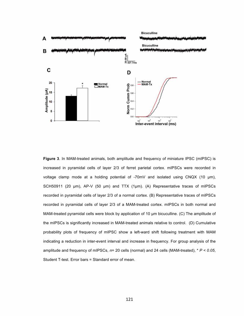
121
Figure 3. In MAM-treated animals, both amplitude and frequency of miniature IPSC (mIPSC) is
increased in pyramidal cells of layer 2/3 of ferret parietal cortex. mIPSCs were recorded in
voltage clamp mode at a holding potential of -70mV and isolated using CNQX (10 µm),
SCH50911 (20 µm), AP-V (50 µm) and TTX (1µm). (A) Representative traces of mIPSCs
recorded in pyramidal cells of layer 2/3 of a normal cortex. (B) Representative traces of mIPSCs
recorded in pyramidal cells of layer 2/3 of a MAM-treated cortex. mIPSCs in both normal and
MAM-treated pyramidal cells were block by application of 10 µm bicuculline. (C) The amplitude of
the mIPSCs is significantly increased in MAM-treated animals relative to control. (D) Cumulative
probability plots of frequency of mIPSC show a left-ward shift following treatment with MAM
indicating a reduction in inter-event interval and increase in frequency. For group analysis of the
amplitude and frequency of mIPSCs, n= 20 cells (normal) and 24 cells (MAM-treated), * P < 0.05,
Student T-test. Error bars = Standard error of mean.

122
Figure 4. Kinetics of GABAA receptors are altered following treatment with MAM. (A) In pyramidal
cells, the 10-90 rise time of the sIPSCs was not significantly altered following treatment with MAM
(I). However, the decay time constant was significantly prolonged (II); the charge transfer was
also significantly increased in MAM-treated animals (III). n= 40 cells (normal) and 24 cells (MAM-
treated), *P< 0.05, Student T-test. (B) Analysis of GABAR kinetics of the mIPSCs indicates that
the 10-90% rise time was unchanged in MAM-treated animals (I). The decay time constant,
however, was significantly prolonged (II) and the charge transfer also significantly decreased (III).
n= 20 cells (normal) and 24 cells (MAM-treated), *P< 0.05, Student T-test. (C) Kinetics of GABAR
in migrating GE cells. (I) The 10-90% rise time of recorded sIPSC in migrating GE cells was
shortened in MAM-treated animals relative to control. The decay time constant (II) and charge
transfer (III) however, increased significantly. n= 16 cells (normal) and 21 cells (MAM-treated),
*P< 0.05, Student T-test. Error bars = Standard error of mean

123
Figure 5. Treatment with MAM did not significantly alter the passive properties of both pyramidal
cells of layer 2/3 and migrating GE-derived cells. (A) Group data from MAM-treated pyramidal
cells (n= 14) normalized relative to control animals. (B) Group data from MAM-treated migrating
GE cells (n= 13) normalized relative to control animals. P< 0.05, Student T-test. Error bars =
Standard error of mean.

124
Figure 6. Expression of GABAA receptor subunits following treatment with MAM. Treatment with
MAM results in a significant increase in the expression of GABAAα3 receptor subunits and slight
elevation of GABAAγ2 receptor subunits, but those of GABAAα2 subunits were unchanged, in the
neocortex as demonstrated by Western blots. Protein bands were detected through enhanced
chemlumniscence and quantitative analysis of the bands performed by densitometric analysis
using image J software for both normal

125
CHAPTER 4
DISCUSSION
Cortical dysplasia (CD) is a developmental abnormality characterized by changes
in structure and function of the cerebral cortex. CD often results from the failure
of immature neurons to migrate appropriately to their cortical targets. This failure
compromises the establishment of the six-layered structure of the cortex and
impairs its ability to process information and perform its role to integrate and
command. The lack of a satisfactory procedure for managing disorders due to
CD underlies the need for development of alternative approaches; this requires
critical insight into the mechanisms underlying neuronal migration to comprehend
the many disorders that result from disrupted migration. Although recent studies
improve our understanding of neuronal migration, significant gaps in knowledge
still remain. Moreover, most of these studies were performed in lower animals
and rodents, which have important differences compared with higher
gyrencephalic mammals like humans and thus necessitate the study of neuronal
migration in more advanced animals.
We developed a model of cortical dysplasia in ferrets by interrupting the
development of layer 4 cells, which results in abnormal placement of specific
populations of interneurons and the redistribution of thalamic afferents and
GABAA receptors (Noctor et al., 1999; Jablonska et al, 2005; Poluch et al., 2008).
We demonstrate that the dynamic movement of migrating GE-derived cells is
impaired in a ferret model of cortical dysplasia in part due to enhanced GABAA

126
receptor (GABAAR) transmission. We also demonstrated that changes in cortical
lamination in our model animals impact the microcircuit in upper cortical layers to
alter the balance between excitation and inhibition.
Treatment with MAM alters the kinetic behavior of GE-derived cells
In our first set of experiments, we investigated the impact of MAM-treatment on
the dynamic pattern of movement of both GE cells and CVZ cells. During
development, a well regulated process of neuronal migration involving both
intrinsic factors and environmental cues ensures that projection neurons and
interneurons that make up each layer of the cortex reach their appropriate targets
to create the normal laminar pattern of the cortex (Anderson et al, 1997; Powell
et al, 2001; Lopez-Bendito et al., 2003 and 2008; Polleux et al, 2003; Stumm et
al., 2003; Alifragis et al., 2004; Flames et al., 2004; Cuzon et al, 2006; Cobos et
al., 2007; Liodis et al., 2007; Friocourt et al., 2008). Changes in the proper
function of any of the numerous factors that guide migration can lead to abnormal
cell positioning and CD. In our model of CD, the neocortical environmental is
altered as a result of redistribution of thalamic afferents, GABAA receptors, and a
sub-population of interneurons, following the loss of layer 4 (Noctor et al., 1999;
Jablonska et al., 2005; Poluch et al., 2008). To better understand the
mechanisms contributing to the redistribution of interneurons, we evaluated
several parameters of kinetic behavior in migrating GE cells including speed and
exploratory tendencies. Cells leaving the GE in normal animals migrated
significantly faster compared to similar cells in MAM-treated animals. In addition,

127
GE generated neurons in MAM-treated animals exhibited less exploration of the
neocortical environment. Exploratory behavior, including extension of multiple
leading processes and changes in direction of movement, results from
coordinated signaling from various sites, which communicate cues that determine
the final location of these cells. The change in the speed of migration and
exploratory activity of interneurons following treatment with MAM therefore has
important implications on the overall positioning of cells within the neocortex.
Is the impact of MAM treatment restricted to cells generated in the GE?
To determine if the effect of E33 MAM treatment on the kinetics of migration is
restricted to interneurons alone, we also investigated the dynamic pattern of
migration of neurons arising from the cortical VZ. Although the migration speed of
CVZ cells was reduced in MAM-treated animals, the deficit was not significant.
We also did not observe any significant differences in exploratory behavior
between cells arising from the CVZ in normal animals and those receiving E33
MAM treatment, indicating that the impact might be restricted to tangentially
migrating GABAergic interneurons. The differential impact of MAM-treatment on
the kinetic behavior of GE and CVZ-derived cells relative to normal cells is not
surprising since these two cell populations have different neurogenic origin and
migrate under the influence of similar but not identical signaling mechanisms
(Marin and Rubenstein, 2003). For instance, CVZ cells, which are derived from
RG cells, rely predominantly on the structural support of RG for their radial
migration (Noctor et al., 2001, 2004). Migration of GE-derived cells is largely

128
independent of radial glia support. In addition, different transcription programs
regulate migration of CVZ and GE-derived cells. Furthermore, different signaling
mechanisms provide positional guidance to these two population of cells within
the neocortex (for details, see above).
Does treatment with MAM alter the functional properties of migrating GE cells?
Next, we investigated the impact of MAM treatment on the GABAA receptor
activity of migrating GE cells. The orientation and distribution of interneurons is
altered in MAM-treated animals in part due to elevated GABAA-mediated activity
(Poluch et al, 2008). To determine whether migrating GE cells receive
abnormally increased GABAAR signaling, we recorded sIPSCs using whole-cell
patch-clamp recording. Both the amplitude and frequency of sIPSCs were
increased in MAM-treated animals suggesting that GABAAR-mediated synaptic
transmission is enhanced in our model animals. sIPSC amplitude is influenced by
presynaptic factors such as number of inhibitory interneurons in synaptic contact
with migrating GE cells as well as the density of GABAAR expressed on the
membrane of GE cells migrating to the neocortex (Braga et al., 2003; Shao and
Dudek 2005). The frequency is determined by number of presynaptic GABAergic
neurons. The GE cells that we recorded from were still in transit enroute to their
target destination in the neocortex. Consequently, synaptic contacts with these
cells are transient and different regions of the neocortical environment or route of
transit influence the level of GABA signaling. In our model, loss of layer 4 leads
to redistribution of thalamic afferents and associated GABAAR; this alters the
129
neocortical environment precisely with respect to GABA signaling to influence
migration and positioning of migrating GE cells. The increase in both amplitude
and frequency of sIPSCs suggests that both pre-synaptic mechanisms and
postsynaptic GABAAR expression contribute to the elevated GABAAR-mediated
increase in inhibitory synaptic neurotransmission.
Abnormal elevation in GABAAR-mediated activity may underlie the altered
migration pattern in GE-derived cells.
The increase in amplitude and frequency of sIPSCs in cells migrating away from
the GE suggests that inhibitory neurotransmission received by GE-derived cells
is elevated in E33 MAM-treated animals. In the second set of experiments, we
examined the relationship between the abnormal increase in GABAAR-mediated
activity and the alteration in GE cell migration. First, we evaluated the basal level
of GABAAR by measuring the expression of GABAAα2 and GABAAα3 subunits in
the neocortex. The mammalian GABAAR is made of five subunits generated from
19 possible subunits (α1-6, β1 -3, γ1 -3, δ, ε , θ , ρ , and ρ1 -3) - a pentameric
hetero-oligomer (Olsen and Sieghart, 2008). There is compelling evidence based
on work from various labs indicating that these subunits have different temporal
expression and regional distribution during development (Garrett et al., 1990;
Fritschy et al., 1994, Yu et al., 2006; Araki et al., 1992; Laurie et al., 1992;
Hashimoto et al., 2009; McKernan et al., 1991; Pirker et al., 2000). Both α2 and
α3 subunits of the GABAA receptor are expressed early during development and
may mediate the initial roles of GABA during development that includes

130
regulation of DNA synthesis, cell proliferation, migration, differentiation,
development of AMPA receptors, excitatory synapse formation and possibly
acting as a stop signal for migrating GE cells (Poulter et al., 1992; Barbin et al.,
1993; Heck et al., 2007; LoTurco et al., 1995; Wang and Kriegstein, 2008, 2009;
Bortone and Polleux, 2010; Inada et al., 2011). In our study, GABAAα2 and
GABAAα3 expression are increased in the neocortex of young (P0 – P1) MAM-
treated animals compared to controls. We did not observe any noticeable
expression of the α1 subunit in the neocortex of both normal and MAM-treated
animals at this age. This is not surprising since this subunit typically show
delayed expression during development (Araki et al., 1992; Laurie et al., 1992).
The expression of the α1 subunit is accompanied by decrease in the expression
of the α2 and α3 subunits; this transition occurs as animals mature (Fristchy et
al., 1992).
GABA initially acts as an excitatory neurotransmitter during development
to facilitate cell migration, but switches to mediate inhibition as cells mature
(Plotkin et al., 1997; Clayton et al., 1998; Rivera et al., 1999). The dual role of
GABA is due to the sequential expression of NKCC1 and KCC2. Migrating
neurons initially express NKCCl, which imports chloride ions (Cl-) into the cell to
maintain a higher intracellular Cl- concentration (Clayton et al., 1998). In
response to GABAAR activation, the outward movement of Cl- results in
depolarization of the cells and triggers Ca2+-mediated events that influence the
cytoskeleton and stimulate migration (Behar et al., 2001; Soria & Valdeolmillos,
2002). As cells mature, NKCCl downregulates and coincides with increased

131
expression of KCC2 (Plotkin et al., 1997). KCC2 is a Cl- exporter that maintains a
higher extracellular gradient of Cl-. Consequently, GABAAR activation when KCC2
is increased results in hyperpolarization and cell inhibition (Payne, 1997; Rivera
et al., 1999). Interestingly, in dissociated hippocampal cell cultures, activation of
GABAAR induces the expression of NKCCl and KCC2 (Ganguly et al., 2001). In
addition, pharmacologic blockade of GABAA delayed the expression of KCC2,
while increased activity of GABAAR induces precocious expression of the
transporter. Premature expression of KCC2 as a result of a heightened state of
GABAAR-mediated activity, as we see here, may be responsible for the migration
defects observed in MAM-treated ferrets. KCC2 is implicated in several important
processes, including decreased motility as a neuron migrates, which leads to cell
slowing, stopping, and differentiating. As a consequence, a premature switch of
the GABAA response may occur and produce early arrest of migrating
interneurons. Bortone and Polleux (2009) demonstrated that depolarization
through GABAA receptors stimulates motility of GABAergic migrating neurons
while hyperpolarization via GABAAR, induced by KCC2 expression, provides a
stop signal for migrating cells. We observed an increased expression of both
KCC2 monomer and oligomers in the neocortex of young MAM-treated animals.
The ratio of KCC2 monomer to oligomer was also reduced in MAM-treated
animals, which indicates greater activity of this transporter in our model animals.
Oligomerization of KCC2 is an important step in the activation and function of
KCC2 as a Cl- exporter (Blaesse et al., 2006). These overall changes in KCC2
expression, as a consequence of increased GABAAR actions, may result in a

132
slowing of interneuron migration, causing early differentiation and maturation of
GE-derived cells.
Does blocking GABAAR activity ameliorate the deficit in interneuron migration in
MAM-treated animals?
GABA is an important influence on migrating cells, although its precise role in the
migration of telencephalic cells is complex. GABA receptors are important during
different phases of migration and mediate aspects of the initiation, continuation,
and termination of cellular movement (Behar et al., 1996, 2001; Bolteus and
Bordey, 2004; Heck et al., 2007; Bortone and Polleux, 2009). Cuzon and
colleagues (2006) report that ambient GABAA activity is present throughout the
path of migration of mouse GE cells into the neocortex, but increases in activity
as neurons approach and enter their cortical target. Our studies suggest that
MAM treatment results in increased ambient GABAAR-mediated activity, which
may interfere with migration of GE cells as they move en route to the neocortex.
The region between the cortical striatal junction and developing cortical plate is
identified by Cuzon et al (2006) as a site of normally high levels of GABA, which
act as an important factor in the proper movement of GE derived cells. This is
supported by our original observation of expanded GABAAα immunoreactivity in
MAM-treated cortex (Jablonska et al., 2004) and the current finding of increased
GABAAα3 receptor subunit as demonstrated by Western blot. Both α2 and α3
subunits of the GABAA receptor are expressed early during development and
may mediate the initial roles of GABA during development (Laurie et al., 1992;

133
Poulter et al., 1992; Fritschy et al., 1994; Carlson et al., 2010). This assists in
explaining our finding that elevated levels of GABA interfere with migration of GE
cells to slow the pace but not prevent their movement.
To investigate the involvement of GABAAR-mediated transmission in the
migration of interneurons, we manipulated the level of GABAAR activity in MAM-
treated organotypic cultures using BMI to reduce the ambient GABAAR activity.
We predicted that limiting the abnormal increased GABAAR activity would change
the cellular actions in MAM-treated cortex to be more like normal. In fact,
blocking GABAAR activity in MAM-treated animals resulted in significant
enhancement of the migration speed of cells leaving the GE as well as the
percentage of MAM-treated GE cells extending more than one leading process.
This suggests that reducing GABAAR-mediated sgnalling in the MAM-treated
ferret encourages migratory activity to become more normal.
What is the functional implication of impaired migration of GE-derived
neurons?
The data we present here indicates that elevated GABAAR-mediated activity in
MAM-treated animals plays a role in the impaired migration of GE-derived cells.
An important question that arises from this finding is what impact these changes
have on GABAergic neurotransmission in the resulting cortex, which then
influences function. We addressed this question by applying whole-cell patch
clamp recording to measure IPSCs in pyramidal cells in layer 2/3. We observed
increased amplitude of sIPSCs in pyramidal cells of layer 2/3 without any

134
significant change in frequency. This indicates that GABAAR-mediated activity is
higher in MAM-treated animals. To investigate the mechanism underlying the
increase in sIPSC amplitude, we evaluated the mIPSC in layer 2/3 pyramidal
cells. mIPSCs are caused by quantal release of GABA from presynaptiic
GABAergic interneurons in the absence of action potentials and thus reflect
release probability of GABA (Edwards et al., 1990). The number of presynaptic
terminals of interneurons and/or postsynaptic GABAAR density on pyramidal cells
determines the amplitude and frequency of mIPSC (Shao and Dudek, 2005). In
our study, both amplitude and frequency of mIPSC were significantly increased in
MAM-treated animals relative to control. The increase in mIPSC frequency and
amplitude that we observed here signifies that both pre and postsynaptic
mechanisms may be involved.
We also observed changes in the kinetics of opening of GABAAR in our
model animals. The 10-90% rise time gives relative spatial information with
respect to postsynaptic sites and changes in rise time could be used to deduce
the locus of change in presynaptic terminal number. It is widely accepted that
receptors located in the soma of postsynaptic membrane give a faster rise time
compared to those located on the dendrites (Shao and Dudek, 2005). In our
study there is no significant change in the rise time in the sIPSC and mIPSC of
pyramidal cells of layer 2/3 indicating no significant positional change in the
active zones and receptor clusters between normal and MAM-treated animals
(Xie et al., 1997; Alberto and Hirasawa, 2011). In the migrating GE cells,
however, we observed a significant reduction in rise time. Although, the reason
135
for this change is unclear, one factor that may be responsible is the relative
position of the migrating GE cells in the emerging neocortex that may affect the
nature of synaptic contact they receive. The decay time constant is influenced by
the density of postsynaptic receptors (Xi et al., 1997, Shao and Dudek, 2005);
prolongation of decay time constant that we observed in both sIPSC and mIPSC
in MAM-treated animals is consistent with increased expression of GABAA
receptors. The amplitude of IPSC and decay time constant determines the
charge transfer. In our study, the charge transfer was significantly increased in
the sIPSC and mIPSC recorded in both migrating GE cells and layer 2/3
pyramidal cells following treatment with MAM suggests. This is consistent with
the increased amplitude and decay time that we reported.
Expression of GABAA receptor subunits is altered in dysplastic cortex
The result we present above suggests that GABAergic neurotransmission
is enhanced in the neocortex of MAM-treated animals, probably as a result of the
increased expression of GABAA receptors. This is evidenced by the increased
amplitude of sIPSCs and mIPSCs in pyramidal cells of layer 2/3 and the
prolonged decay time in animals P28-38. To further confirm that increased
GABAAR expression is responsible for the enhanced GABAergic
neurotransmission, we applied western blot to measure the level of various
subunits of GABAAR in the older cortex. Our data shows that GABAAα3 expression
was significantly increased in our model animals, as it was in the P0 animals. We
also observed a slight increase in the expression of GABAAγ2 and no change in

136
the GABAAα2 subunits in dysplastic cortex compared to control. The increase in
GABAAα3 receptor subunit may underscore the elevated GABA transmission in
pyramidal cells of MAM-treated juvenile ferrets. The GABAAγ2 receptor subunit is
presumed to also be important for synaptic transmission and for the postsynaptic
clustering of GABAAR during the development of synapses (Schweitzer et al.,
2003). In this regard, it could clearly play a role in the abnormal synaptic
transmission observed here. It is not clear why we did not observe an increase or
change in the α1 receptors in either the P0 – P1 or P28 – P38 animals, as we
would expect these to increase as the animal matures. It may be that the ferret
neocortex is still relatively immature, even at P28 – P38, and does not express
high levels of this receptor until a later date. In any case, the obvious increase
and probable change in the composition of at least 2 GABAAR subtypes can
clearly underlie the change in kinetics that we observed in our recordings.
Conclusion and future directions.
In this set of experiments, we demonstrated that in utero admistration of
MAM on E33 leading to development of CD, selectively interfers with migration of
GE-derived cells by altering their kinetic properties. We also showed that the
aberrant migration of MAM-treated GE cells results from anomalous increase in
GABAAR synaptic transmission within the neocortical environment. This increase
appears to enhance the rate of maturation of GE derived cells by inducing the
precocious expression of KCC2 (Figure 4). As a result, GE-derived neurons
migrate slowly and terminate at aberrant sites within the neocortex; resulting in a

137
change in cortical microcircuitry that impairs information processing within the
cortex. This finding provides evidence that abnormal elevation in GABA signaling
during neocortical maturation may underlie the development of CD.
Several neuropsychiatric disorders such as epilepsy, schizophrenia,
mental retardation and autism result from abnormal cortical development. Most of
these disorders present with impaired GABA function due to loss of inhibitory
interneurons resulting in unrestrained excitatory drive in the neocortex. This
alters cortical information processing leading to disease phenotypes. At present,
the factors responsible for loss of inhibitory interneurons are unknown, although it
has been suggested that selective vulnerability of these populations of cells may
be responsible. In cortical dysplasia due to defective migration of specific
population of cells, aberrant positioning, rather than loss of inhibitory
interneurons, impairs cortical function. Therefore, ameliorating the deficit in
migration is an appealing approach to management of CD. We postulate that an
optimal level of GABA activity is central to proper migration of GE cells and a
reduction, as well as increase, in GABA signaling compromises neuronal
migration to contribute to disease phenotype. GABA also plays a multifaceted
role during development, including regulation of DNA synthesis and intracellular
Ca2+ levels; these processes may influence aspects of neuronal migration and
their functional integration into circuits. A fascinating, but yet unexplored role of
GABA during development, is its involvement in regulating gene transcription to
influence proliferation and specification of interneurons. This area of thought
deserves greater investigation and exploration. In light of strong evidence linking

138
alterations of GABA signaling to several neuropsychiatric disorders, an intriguing
question/possibility is whether early intervention involving restoring/maintaining
an optimum level of GABA activity will reduce the severity and improve treatment
outcome.
In the future, it will be interesting to specifically evaluate the migratory
properties of the GE cells derived from different regions of the LGE or CGE in our
model. In addition, functional analysis of other regions of the neocortex of
juvenile ferrets will provide further evidence as to whether similar alterations in
IPSCs occur in other regions of the MAM-treated neocortex.

139
Figure 4: Schematic illustration of the cellular mechanisms contributing to defective migration and
increase GABAergic neurotransmission in the MAM model of cortical dysplasia. During
development, GABA initially acts as an excitatory neurotransmitter due to outward movement of
chloride ion that leads to cell depolarization and cell migration in response to GABAAR activation.
As cells matured, increased expression of KCC2 reverses the direction of chloride ion movement
to cause depolarization. MAM treatment triggers increased GABAAR activity to promote premature
expression of KCC2 to cause deficit in GE cell migration.

140
Reference:
Acampora D, Mazan S, Avantaggiato V, Barone P, Tuorto F, Lallemand Y, Brulet
P, Simeone A (1996) Epilepsy and brain abnormalities in mice lacking the Otx1
gene. Nat Genet 14:218-222.
Alberto CO, Hirasawa M (2010) AMPA receptor-mediated miniature EPSCs have
heterogenous time courses in orexin neurons. Biochem Biophys Res Comm
400:707-712.
Alifragis P, Liapi A, Parnavelas JG (2004) Lhx6 regulates the migration of cortical
interneurons from the ventral telencephalon but does not specify their GABA
phenotype. J Neurosci 24:5643-5648.
Anderson SA, Eisenstat DD, Shi L, Rubenstein JL (1997) Interneuron migration
from basal forebrain to neocortex: dependence on Dlx genes. Science 278:474-
476.
Anderson SA, Marin O, Horn C, Jennings K, Rubenstein JLR (2001) Distinct
cortical migrations from the medial and lateral ganglionic eminences.
Development 128:353-363.
141
Anderson SA, Kaznowski EC, Horn C, Rubenstein JLR, McConnell SK (2002)
Distinct origins of neocortical projection neurons and interneurons in vivo. Cereb
Cortex 12:702-709.
Angevine JB, Sidman RL (1961) Autoradiographic study of cell migration during
histogenesis of cerebral cortex in the mouse. Nature 192:766-768.
Ang ESBC, Hadar TF, Gluncic V, Rakic P (2003) Four-dimensional migratory
coordinates of GABAergic interneurons in the developing mouse cortex. J
Neurosci 23(13):5805-5815.
Anton ES, Cameron RS, Rakic P (1996) Role of neuron-glial junctional domain
proteins in the maintenance and termination of neuronal migration across the
embryonic cerebral wall. J Neurosci 16:2283-2293.
Arlotta P, Molyneaux BJ, Chen J, Inoue J, Kominami R, Macklis JD (2005)
Neuronal subtype-specific genes that control corticospinal motor neuron
development in vivo. Neuron 45: 207-221.
Araki T, Kiyama H, Tohyama M (1992) GABAA receptor subunit messenger
RNAs show differential expression during cortical development in the rat brain.
Neuroscience 51(3):583-591.
142
Ascoli GA, Alonso-Nanclares L, Anderson SA, Barrionuevo G, Benavides-
Piccione R, Burkhalter A, Buzsa´ ki G, Cauli B, Defelipe J, Fairen A, Feldmeyer
D, Fishell G, Frenac Y, Freund TF, Gardner D, Gardner EP, Goldberg JH,
Helmstaedter M, Hestrin S, Karube F, Kisvarday ZF, Lambolez B, Lewis DA,
Marin O, Markram H, Munoz A, Packer A, Petersen CC, Rockland KS, Rossier J,
Rudy B, Somogyi P, Staiger JF, Tamas G, Thomson AM, Toledo-Rodriquez M,
Wang Y, West DC, Yuste R (2008) Petilla terminology: nomenclature of features
of GABAergic interneurons of the cerebral cortex. Nat Rev Neurosci 9:557-568.
Autti-Ramo I, Autti T, Kettumen S, Salonen O, Valanne L (2002) MRI findings in
children with school problems who had been exposed prenatally to alcohol. Dev
Med and Child Neurol 44:98-106.
Ayala R, Shu T, Tsai L-H (2007) Trekking across the brain: the journey of
neuronal migration. Cell 128:29-43.
Baraban SC, Schwartzkroin PA (1996) Flurothyl seizure susceptibility in rats
following prenatal methylazoxymethanol treatment. Epilepsy Res 23:189-194.
Barbin G, Pollard H, Gaiarsa JL, Ben-Ari Y (1993) Involvement of GABAA
receptors in the outgrowth of cultured hippocampal neurons. Neurosci Lett
152:150-154.

143
Barinaga M (1993) Carbon monoxide: killer to brain messenger in one step.
Science 259: 309.
Batista-Brito R, Fishell G (2009) The developmental integration of cortical
interneurons into a functional network. Curr Top Dev Biol 87:81-118.
Bayer SA, Altman J (1991) Neocortical Development. (Raven Press, New York).
Behar TN, Li YX, Tran HT, Ma W, Dunlap V, Scott C, Barke JL (1996) GABA
stimulates chemotaxis and chemokinesis of embryonic cortical neurons via
calcium-dependent mechanisms. J Neurosci 16:1808-1818.
Behar TN, Schaffner AE, Scott CA, O’Connell C, Barker JL (1998) Differential
response of cortical plate and ventricular zone cells to GABA as a migration
stimulus. J Neurosci 18:6378-6387.
Behar TN, Scott CA, Greene CL, Wen X, Smith SV, Maric D, Liu Q-Y, Colton CA,
Barker JL (1999) Glutamate acting at NMDA receptors stimulates embryonic
cortical neuronal migration. J Neurosci 19(11):4449-4461.
Behar TN, Schaffner AE, Scott CA, Greene CL, Barker JL (2000) GABA receptor
antagonists modulate postmitotic cell migration in slice cultures of embryonic rat
cortex. Cereb Cortex 10:899-909.

144
Behar TN, Smith SV, Kennedy RT, McKenzie JM, Maric I, Barker JL (2001)
GABAB receptors mediate motility signals for migrating embryonic cortical cells.
Cereb Cortex 11(8):744-753.
Bellion A, Baudoin JP, Alvarez C, Bornens M, Métin C (2005) Nucleokinesis in
tangentially migrating neurons comprises two alternating phases: forward
migration of the Golgi/centrosome associated with centrosome splitting and
myosin contraction at the rear. J Neurosci 25:5691-5699.
Benardete EA, Kriegstein AR (2002) Increased excitability and decreased
sensitivity to GABA in an animal model of dysplastic cortex. Epilepsia 43:970-
982.
Ben-Ari Y, Cherubini E, Corradetti R, Gaiarsa JL (1989) Giant synaptic potentials
in immature rat CA3 hippocampal neurons. J Physiol 416:303-325.
Bernstein HG, Krause S, Krell D, Dobrowolny H, Wolter M, Stauch R, Ranft K,
Danos P, Jirikowski GF, Bogerts (2007) Strongly reduced number of arvalbumin-
immunoreactive projection neurons in the mammillary bodies in schizophrenia:
Further evidence for limbic neuropathology. Ann N Y Acad Sci 1096:120-127.

145
Bertrand N, Castro DS, Guillemot F (2002) Proneural genes and the specification
of neural cell types. Nature Rev Neurosci 3:517-530.
Blaesse P, Guillemin I, Schindler J, Schweizer M, Delpire E, Khiroug L, Friauf E,
Nothwang HG (2006) Oligomerization of KCC2 correlates with development of
inhibitory neurotransmission. J Neurosci 26(41):10407-10419.
Bolteus AJ, Bordey A (2004) GABA release and uptake regulate neuronal
precursor migration in the postnatal subventricular zone. J Neurosci 24:7623-
7631.
Bortone D, Polleux F (2009) KCC2 expression promotes the termination of
cortical interneuron migration in a voltage-sensitive calcium-dependent manner.
Neuron 62:53-71.
Braga MFM, Aroniadou-Anderjaska V, Xie J, Li H (2003) Bidirectional
modulation of GABA release by presynaptic glutamate receptor 5 Kainate
receptors in the basolateral amygdala. J Neurosci 23(2):442-452.
Brill J, Huguenard JR (2010) Enhanced infragranular and supragranular synaptic
input onto layer 5 pyramidal neurons in a rat model of cortical dysplasia. Cereb
Cortex 20:2926-2938.

146
Britanova O, de Juan Romero C, Cheung A, Kwan KY, Schwark M, Gyorgy
A,Vogel T, Akopov S, Mitkovski M, Agoston D, Sestan N, Molnar Z, Tarabykin V
(2008) Satb2 is a postmitotic determinant for upper-layer neuron specification in
the neocortex. Neuron 57:378-392.
Brunstrom JE, Gray-Swain MR, Osborne PA, Pearlman AL (1997) Neuronal
heterotopias in the developing cerebral cortex produced by neurotrophin-4.
Neuron 18:505-517.
Butt SJ, Fuccillo M, Nery S, Noctor S, Kriegstein A, Corbin JG, Fishell G (2005)
The temporal and spatial origins of cortical interneurons predict their
physiological subtype. Neuron 48:591-604.
Butt SJ, Cobos I, Golden J, Kessaris N, Pachnis V, Anderson S (2007)
Transcriptional regulation of cortical interneuron development. J Neurosci
27(44):11847-11850.
Butt SJ, Sousa VH, Fuccillo MV, Hjerling-Leffler J, Miyoshi G, Kimura S, Fishell
G (2008) The requirement of Nkx2-1 in the temporal specification of cortical
interneuron subtypes. Neuron 59:722-732.
147
Calcagnotto ME, Paredes MF, Scott C, Baraban SC (2002) Heterotopic neurons
with altered inhibitory synaptic function in an animal model of malformation
associated epilepsy. J Neurosci 22(17):7596-7605.
Calcagnotto ME, Baraban SC (2003) An examination of calcium current function
on heterotopic neurons in hippocampal slices from rats exposed to
methylazoxymethanol. Epilepsia 44:315-321.
Carlson VCC, Yeh HH (2010) GABAA receptor subunit profiles of tangentially
migrating neurons derived from the medial ganglionic eminence. Cereb Cortex
21:1792-1802.
Caronia-Brown G, Grove EA (2011) Timing of cortical interneuron migration is
influenced by the cortical hem. Cereb Cortex 21(4):748-755.
Casarosa S, Fode C, Guillemot F (1999) Mash1 regulates neurogenesis in the
ventral telencephalon. Development 126:525-534.
Cavanagh ME, Parnavelas JG (1988) Development of somatostatin
immunoreactive neurons in the rat occipital cortex: a combined
immunocytochemical-autoradiographic study. J Comp Neurol 268:1-12.

148
Cavanagh ME, Parnavelas JG (1989) Development of vasoactive-Intestinal
polypeptide-immunoreactive neurons in the rat occipital cortex: a combined
immunohistochemical-autoradiographic study. J Comp Neurol 284:637-645.
Caviness VS, Sidman RL (1973) Time of origin or corresponding cell classes in
the cerebral cortex of normal and reeler mutant mice: an autoradiographic
analysis. J Comp Neurol 148:141-151.
Caviness, VS (1982) Neocortical histogenesis in normal and reeler mice: a
developmental study based upon [3H]thymidine autoradiography. Brain Res
256:293-302.
Caviness VS, Yorke CH (1976) Interhemispheric neocortical connections of the
corpus callosum in the reeler mutant mouse: a study based on anterograde and
retrograde methods. J Comp Neurol 170:449-459.
Cepeda C, Andre VM, Hauptman JS, Yamazaki I, Huynh MyN, Chang JW, Chen
JY, Fisher RS, Vinters HV, Levine MS, Mathern GW (2012) Enhanced
GABAergic network and receptor function in pediatric cortical dysplasia TypIIB
compared with tuberous sclerosis complex. Neurobiol Dis 45: 310-321.
149
Chae T, Kwon YT, Bronson R, Dikkes P, Li E, Tsai LH (1997) Mice lacking p35, a
neuronal specific activator of Cdk5, display cortical lamination defects, seizures,
and adult lethality. Neuron 18:29-42.
Chao HT, Chen H, Samaco RC, Xue M, Chahrour M, Yoo J, Neul JL, Gong S, Lu
H-C, Heintz N, Ekker M, Rubenstein JLR, Noebels JL, Rosenmund C, Zoghbi HY
(2010) Dysfunction in GABA signaling mediates autism-like stereotypies and Rett
syndrome phenotypes. Nature 468:263-269.
Chattopaddhyaya B, Di Cristo G (2012) GABAergic circuit dysfunctions in
neurodevelopmental disorders. Front Psych 3(51):1-9.
Chen B, Schaevitz LR, McConnell SK (2005a) Fezl regulates the differentiation
and axon targeting of layer 5 subcortical projection neurons in cerebral cortex.
Proc Natl Acad Sci USA 102:17184-17189.
Chen JG, Rasin MR, Kwan KY, Sestan N (2005b) Zfp312 is required for
subcortical axonal projections and dendritic morphology of deep-layer pyramidal
neurons of the cerebral cortex. Proc. Natl Acad Sci USA 102: 17792-17797.
Chen B, Wang SS, Hattox AM, Rayburn H, Nelson SB, McConnell SK (2008)
The Fezf2-Ctip2 genetic pathway regulates the fate choice of subcortical

150
projection neurons in the developing cerebral cortex. Proc Natl Acad Sci USA
105:11382-11387.
Chenn A, McConnell SK (1995) Cleavage orientation and the asymmetric
inheritance of Notch1 immunoreactivity in mammalian neurogenesis. Cell 82:631-
641.
Cherubini E, Gaiarsa JL, Ben-Ari Y (1991). GABA: an excitatory transmitter in
early postnatal life. Trends Neurosci 14(12):515-519.
Chevassus-au-Louis N, Ben-Ari Y, Vergnes M (1998) Decreased seizure
threshold and more rapid rate of kindling in rats with cortical malformation
induced by prenatal treatment with methylazoxymethanol. Brain Res 812:252-
255.
Chiang C, Litingtung Y, Lee E, Young KE, Corden JL, Westphal H, Beachy PA
(1996) Cyclopia and defective axial patterning in mice lacking Sonic hedgehog
gene function. Nature 383:407-413.
Choi BH, Matthias SC (1987) Cortical dysplasia associated with massive ectopia
of neurons and glial cells within the subarachnoid space. Acta Neuropathol (Berl)
73:105-109.

151
Chugani HT, Shields WD, Shewmon DA, Olson DM, Phelps ME, Peacock WJ
(1990) Infantile spasms: I. PET identifies focal cortical dysgenesis in cryptogenic
cases for surgical treatment. Ann Neurol 27:406-413.
Clayton GH, Owens GC, Wolff JS, Smith RL (1998) Ontogeny of cation-Cl-
cotransporter expression in rat neocortex. Brain Res Dev Brain Res 109:281-
292.
Cobb SR, Buhl EH, Halasy K, Paulsen O, Somogyi P (1995) Synchronization of
neuronal activity in hippocampus by individual GABAergic interneurons. Nature
378:75-78.
Cobos I, Broccoli V, Rubenstein JL (2005a) The vertebrate ortholog of Aristaless
is regulated by Dlx genes in the developing forebrain. J Comp Neurol 483:292-
303.
Cobos I, Calcagnotto ME, Vilaythong AJ, Thwin MT, Noebels JL, Baraban SC,
Rubenstein JL (2005b) Mice lacking Dlx1 show subtypespecific loss of
interneurons, reduced inhibition and epilepsy. Nat Neurosci 8:1059-1068.
Cobos I, Borello U, Rubenstein JL (2007) Dlx transcription factors promote
migration through repression of axon and dendrite growth. Neuron 54:873-888.

152
Cocas LA, Georgala PA, Mangin JM, Clegg JM, Kessaris N, Haydar TF, Gallo V,
Price DJ, Corbin JG (2011) Pax6 is required at the telencephalic pallial-subpallial
boundary for the generation of neuronal diversity in the postnatal limbic system. J
Neurosci 6:5313-5324.
Colacitti C, Sancini G, DeBiasi S, Franceschetti S, Caputi A, Frassoni C,
Cattabeni F, Avanzini G, Spreafico R, Di Luca M, Battaglia G. (1999) Prenatal
methylazoxymethanol treatment in rats produces brain abnormalities with
morphological similarities to human developmental brain dysgeneses. J
Neuropathol Exp Neurol 58:92-106.
Colombo N, Tassi L, Galli C, Citterio A, Lo Russo G, Scialfa G, Spreafico R
(2003) Focal cortical dysplasias: MR imaging, histopathologic and clinical
correlations in surgically treated patients with epilepsy. Am J Neuroradiol 24:724-
733.
Colombo E, Collombat P, Colasante G, Bianchi M, Long J, Mansouri A,
Rubenstein JLR, Broccoli V (2007). Inactivation of Arx, the murine ortholog of the
X-linked lissencephaly with ambiguous genitalia gene, leads to severe
disorganization of the ventral telencephalon with impaired neuronal migration and
differentiation. J Neurosci 27:4786-4798.

153
Corbin JG, Nery S, Fishell G (2001) Telencephalic cells take a tangent: non-
radial migration in the mammalian forebrain. Nat Neurosci 4:1177-1182.
Corbin JG, Gaiano N, Juliano SL, Poluch S, Stancik E, Haydar TF (2008)
Regulation of neural progenitor cell development in the nervous system. J
Neurochem 106:2272-2287.
Corbin JG, Butt SJB (2011) Developmental mechanisms for the generation of
telencephalic interneurons. Dev Neurol 71(8):710-732.
Crandall JE, McCarthy DM, Araki KY, Sims JR, Ren JQ, Bhide PG (2007)
Dopamine receptor activation modulates GABA neuron migration from the basal
forebrain to the cerebral cortex. J Neurosci 27:3813-3822.
Crandall JE, Goodman T, McCarthy MD, Gregg D, Bhide PG, Grager UC,
McCaffery P (2011) Retinoic acid influences neuronal migration from the
ganglionic eminence to the cerebral cortex. J Neurochem 119:723-735
Cuzon VC, Yeh PWL, Cheng Q, Yeh HH (2006) Ambient GABA promotes
cortical entry of tangentially migrating cells derived from the medial ganglionic
eminence. Cereb Cortex 16(10):1577-1588.

154
D’Arcangelo G, Miao GG, Chen SC, Soares HD, Morgan JI, Curran T (1995) A
protein related to extracellular matrix proteins deleted in the mouse mutant
reeler. Nature 374:719-723.
De Carlos JA, L´opez-Mascaraque L, Valverde F (1996) Dynamics of cell
migration from the lateral ganglionic eminence in the rat. J Neurosci 16:6146-
6156.
DeDiego I, Smith-Fernandez A, Fairen A (1994) Cortical cells that migrate
beyond area boundaries: characterization of an early neuronal population in the
lower intermediate zone of prenatal rats. Eur J Neurosci 6:983-997.
DeFelipe J, Farinas I (1992) The pyramidal neuron of the cerebral cortex:
morphological and chemical characteristics of the synaptic inputs. Prog Neurobio
39:563-607.
De Felipe J (1993) Neocortical neuronal diversity: chemical heterogeneity
revealed by colocalization studies of classic neurotransmitters, neuropeptides,
calcium-binding proteins, and cell surface molecules. Cereb.Cortex 3:273-289.
de Feo MR, Mecarelli O, Ricci GF (1995) Seizure susceptibility in immature rats
with microencephaly induced by prenatal exposure to methylazoxymethanol
acetate. Pharmacol. Res. 31:109-114.
155
Delpire E (2000) Cation-chloride cotransporters in neuronal communication.
News Physiol Sci 15:309-312.
Denaxa M, Chan CH, Schachner M, Parnavelas JG, Karagogeos D (2001) The
adhesion molecule TAG-1 mediates the migration of cortical interneurons from
the ganglionic eminence along the corticofugal fiber system. Development 128:
4635-4644.
de Rouvroit CL, Goffinet AM (2001) Neuronal migration. Mech Dev 105:47-56.
des Portes V, Francis F, Pinard JM, Desguerre I, Moutard ML, Snoeck I, Meiners
LC, Capron F, Cusmai R, Ricci S, Motte J, Echenne B, Ponsot G, Dulac O,
Chelly J, Beldjord C (1998) Doublecortin is the major gene causing X-linked
subcortical laminar heterotopia (SCLH). Hum. Mol Genet 7:1063-1070.
Dulabon L, Olson EC, Taglienti MG, Eisenhuth S, McGrath B, Walsh CA,
Kreidberg JA, Anton ES (2000) Reelin binds alpha3beta1 integrin and inhibits
neuronal migration. Neuron 27:33-44.
Edwards FA, Konnerth A, Sakmann B (1990) Quantal analysis of inhibitory
synaptic transmission in the dentate gyrus of rat hippocampal slices: a patch-
clamp study. J Physiol 430:213-249.

156
Eksioglu YZ, Scheffer IE, Cardenas P, Knoll J, DiMario F, Ramsby G, Berg M,
Kamuro K, Berkovic SF, Duyk GM, Parisi J, Huttenlocher PR, Walsh CA (1996)
Periventricular heterotopia: an X-linked dominant epilepsy locus causing aberrant
cerebral cortical development. Neuron 16:77-87.
Fairen A, DeFelipe J, Regidor J (1984) Nonpyramidal neurons: general account.
In Cereb. Cortex. (1) (ed. A. Peters and E. G. Jones), pp. 201-253. New York:
Plenum Press.
Fairen A, Cobas A, Fonseca M (1986) Times of generation of glutamic acid
decarboxylase immunoreactive neurons in mouse somatosensory cortex. J
Comp Neurol 251:67-83.
Fatemi SH, ReutimanTJ, Folsom TD, Rooney RJ, Patel DH Thuras PD (2010)
mRNA and protein levels for GABAAα4, α5, β1 and GABABR1 receptors are
altered in brains from subjects with autism. J Autism Dev Disord 40:743-750.
Fatemi SH, Reutiman TJ, Folsom TD, Thuras PD (2009) GABAA receptor down
regulation in brains of subjects with autism. J Autism Dev Disord 39:223-230.
Fauser S, Zentner J (2012) Management of cortical dysplasia in epilepsy. Adv
Tech Stand Neurosurg 38:137-163.

157
Feng Y, Walsh CA (2001) Protein-protein interactions, cytoskeletal regulation
and neuronal migration. Nat Rev Neurosci 2:408-416.
Ferrer I (1993) Experimentally induced cortical malformations in rats. Childs Nerv
Syst 9:403-407.
Ferrer I, Alcantara S, Z`ujar MJ, Cinos C (1993) Structure and pathogenesis of
cortical nodules induced by prenatal X-irradiation in the rat. Acta Neuropathol
85:205-212.
Fietz SA, Kelava I, Vogt J, Wilsch-Bra¨uninger M, Stenzel D, Fish JL, Corbeil D,
Riehn A, Distler W, Nitsch R, Huttner WB (2010) oSVZ progenitors of human and
ferret neocortex are epithelial-like and expand by integrin signaling. Nat Neurosci
13:690-699.
Fishell G, Kriegstein AR (2003) Neurons from radial glia: the consequences of
asymmetric inheritance. Curr Opin Neurobiol 13: 34-41.
Fishell G, Hanashima C (2008) Pyramidal neurons grow up and change their
mind. Neuron 57:333-338.

158
Flames N, Long JE, Garratt AN, Fischer TM, Gassmann M, Birchmeier C, Lai C,
Rubenstein JLR, Mar´ın O (2004) Short- and long-range attraction of cortical
GABAergic interneurons by neuregulin-1. Neuron 44:251-261.
Fode C, Ma Q, Casarosa S, Ang S-L, Anderson DJ, Guillemot F (2000) A role for
neural determination genes in specifying the dorsoventral identity of
telencephalic neurons. Genes Dev 14:67-80.
Fogarty M, Grist M, Gelman D, Marı´n O, Pachnis V, Kessaris N (2007) Spatial
genetic patterning of the embryonic neuroepithelium generates GABAergic
interneuron diversity in the adult cortex. J Neurosci 27:10935-10946.
Fonseca M, Soriano E, Ferrer I, Martinez A, Tunon T (1993) Chandelier cell
axons identified by parvalbumin-immunoreactivity in the normal human temporal
cortex and in Alzheimer’s disease. Neuroscience 55:1107-1116.
Freund TF (2003) Interneuron diversity series: rhythm and mood in perisomatic
inhibition. Trends Neurosci 26:489-495.
Friocourt G, Liu JS, Antypa M, Rakic S, Walsh CA Parnavelas JG (2007) Both
doublecortin and doublecortin-like kinase play a role in cortical interneuron
migration. J Neurosci 27:3875-3883.

159
Friocourt G, Kanatani S, Tabata H, Yozu M, Takahashi T, Antypa M, Raguenes
O, Chelly J, Ferec C, Nakajima K, Parnavelas JG (2008) Cell autonomous roles
of ARX in cell proliferation and neuronal migration during corticogenesis. J
Neurosci 28:5794-5805.
Fristch B, Qashu F, Figueiredo TH, Aroniadou-Anderjaska V, Rogawski MA,
Braga MFM (2009). Pathological alterations in GABAergic interneurons and
reduced tonic inhibition in the basolateral amygdala during epileptogenesis.
Neuroscience 163(1):415-429.
Fristchy J-M, Paysan J, Enna A, Mohlerl H (1994) Switch in the expression of rat
GABA-receptor subtypes during postnatal development: an
immunohistochemical study. J Nerosci 14(9):5302-5324.
Frotscher M (1997) Dual role of Cajal-Retzius cells and reelin in cortical
development. Cell Tissue Res 290:315-322.
Gal JS, Morozov YM, Ayoub AE, Chatterjee M, Rakic P, Haydar TF (2006)
Molecular and morphological heterogeneity of neural precursors in the mouse
neocortical proliferative zones. J Neurosci 26(3):1045-1056.

160
Ganguly K, Schinder AF, Wong ST, Poo M (2001) GABA itself promotes the
Developmental-switch of neuronal GABAergic responses from excitation to
inhibition. Cell 105:521-532.
Gant JC, Thibault O, Blalock EM, Yang J, Bachstetter A, Kotick J, Schauwecker
PE, Hauser KF, Smith GM, Mervis R, Li Y, Barnes GN (2009) Decreased number
of interneurons and increased seizures in neuropilin 2 deficient mice: implications
for autism and epilepsy. Epilepsia 50:629-645.
Garrett KM, Saito N, Duman RS, Abel MS, Ashton RA, Fujimori S, Beer B,
Tallmann JF, Vitek MP, Blume AJ (1990) Differential expression of γ -
aminobutyric acidA receptor subunits. Mol Pharmacol 371652-657.
Garcia-Bellido A (1979) Genetic analysis of the achaete-scute system of
Drosophila melanogaster. Genetics 91:491-520.
Ge W, He F, Kim KJ, Blanch B, Coskun V, Nguyen L, Wu X, Zhao J, Heng JI-T,
Martinowich K, Tao J, Wu H, Castro D, Sobeih M, Corfas G, Gleeson JG,
Greenberg ME, Guillemot F, Sun YE (2006) Coupling of cell migration with
neurogenesis by proneural bHLH factors. Proc Natl Acad Sci USA.103:1319-
1324.
161
Germano IM, Zhang YF, Sperber EF, Moshe SL (1996) Neuronal migration
disorders increase susceptibility to hyperthermia-induced seizures in developing
rats. Epilepsia 37:902-910.
Ghashghaei HT, Weber J, Pevny L, Schmid R, Schwab MH, Lloyd KC,Eisenstat
DD, Lai C, Anton ES. 2006. The role of neuregulin-ErbB4 interactions on the
proliferation and organization of cells in the subventricular zone. Proc Natl Acad
Sci U S A. 103:1930-1935.
Ghysen A, Dambly-Chaudiere C (1988) From DNA to form: the achaete-scute
complex. Genes Dev 2:495-501.
Gierdalski M, Juliano SL (2003) Factors affecting the morphology of radial glia.
Cereb Cortex 13(6):572-579.
Gierdalski M, Juliano SL (2005) Endogenous neuregulin restores radial glia in a
(ferret) model of cortical dysplasia. J Neurosci 25(37):8498-84504.
Gilbert CD, Wiesel TN (1985) Intrinsic connectivity and receptive field properties
in visual cortex. Vision Res 25:365-374.
Ginsberg MD, Myers RE (1974) Experimental carbon monoxide encephalopathy
in the primate. I. Physiologic and metabolic aspects. Arch Neurol 30:202-208.

162
Ginsberg MD, Myers RE, McDonagh BF (1974) Experimental carbon monoxide
encephalopathy in the primate. II. Clinical aspects, neuropathology, and
physiologic correlation. Arch Neurol 30:209-216.
Gleeson JG, Allen KM, Fox JW, Lamperti ED, Berkovic S, Scheffer I, Cooper EC,
Dobyns WB, Minnerath SR, Ross MH, Walsh CA (1998) doublecortin, a brain-
specific gene mutated in human X-linked lissencephaly and double cortex
syndrome, encodes a putative signaling protein. Cell 92: 63-72.
Gleeson JG, Minnerath, SR, Fox JW, Allen KM, Luo RF, Hong SE, Berg MJ,
Kuzniecky R, Reitnauer PJ, Borgatti R, Mira AP, Guerrini R, Holmes GL, Rooney
CM, Berkovic S, Scheffer I, Cooper EC, Ricci S, Cusmai R, Crawford TO, Leroy
R, Andermann E, Wheless JW, Dolbyns WB, Walsh CA (1999) Characterization
of mutations in the gene doublecortin in patients with double cortex syndrome.
Ann Neurol 45:146-153.
Gleeson GJ, Walsh CA (2000) Neuronal migration disorders: from genetic
diseases to developmental mechanisms. Trends Neurosci 23:352-359.
Gorman DF, Huang YL, Williams C (2001) Prolonged exposure to one percent
carbon monoxide causes a leucoencephalopathy in un-anaesthetised sheep.
Toxicology 165: 97-107.

163
Gonzalez F, Romani S, Modolell J, Campuzano S (1989) Molecular analysis of
the asense gene, a member of the achaete-scute complex of Drosophila
melanogaster, and its novel role in optic lobe development. EMBO J 8:3553-
3562.
Gotz M, Huttner WB (2005) The cell biology of neurogenesis. Nat Rev Neurosci
6:777-788.
Götz M, Hartfuss E, Malatesta P (2002) Radial glial cells as neuronal precursors:
a new perspective on the correlation of morphology and lineage restriction in the
developing cerebral cortex of mice. Brain Res Bull 57:777-788.
Gray GE, Glover JC, Majors J, Sanes JR (1988) Radial arrangement of clonally
related cells in the chicken optic tectum: lineage analysis with a recombinant
retrovirus. Proc Natl Acad Sci USA 85:7356-7360.
Grove EA, Williams BP, Li D-Q, Hajihosseini M, Friedrich A, Price J (1993)
Multiple restricted lineages in the embryonic rat cerebral cortex. Development
117:553-561.
Guillemot F (2007) Spatial aand temporal specification of neural fates by
transcription factor codes. Development 134:3771-3780.

164
Gupta A, Wang Y, Markram H (2000) Organizing principles for a diversity of
GABAergic interneurons and synapses in the neocortex. Science 287:273-278.
Hamasaki T, Goto S, Nishikawa S, Ushio Y (2001) A role of netrin-1 in the
formation of the subcortical structure striatum: repulsive action on the migration
of late-born striatal neurons. J. Neurosci 21:4271-4280.
Hand R, Bortone D, Mattar P, Nguyen L, Heng JI, Guerrier S, Boutt E, Peters E,
Barnes AP, Parras C, Schuurmans C, Guillemot F, Polleux F (2005)
Neurogenin2 specifies the migration properties and the dendritic morphology of
pyramidal neurons in the neocortex. Neuron 48:45-62.
Hansen DV, Rubenstein JLR, Kriegstein, AR (2011) Deriving excitatory neurons
of the neocortex from pluripotent stem cells. Neuron 70:645-660.
HashimotoT,Volk DW, Eggan SM, Mirnics K, Pierri JN, Sun Z, Sampson AR,
Lewis DA (2003) Gene expression deficits in a subclass of GABA neurons in the
prefrontal cortex of subjects with schizophrenia. J Neurosci 23:6315-6326.
Hashimoto T, Nguyen QL, Rotaru D, Keenan T, Arion D, Beneyto M, Gonzalez-
Burgos G, Lewis DA (2009) Protracted developmental trajectories of GABAA

165
receptor α1 and α2 subunit expression in primate prefrontal cortex. Biol
Psychiatry 65:1015-1023.
Hasling TA, Gierdalski M, Jablonska B, Juliano SL (2003) A radialization factor in
normal cortical plate restores disorganized radial glia and disrupted migration in a
model of cortical dysplasia. Eur J Neurosci 17:467-480.
Hatten ME (2002) New directions in neuronal migration. Science 297:1660-1663.
Hattori M, Adachi H, Tsujimoto M, Arai H, Inoue K (1994) Miller-Dieker
lissencephaly gene encodes a subunit of brain platelet activating factor
acetylhydrolase. Nature 370:216-218.
Haubensak W, Attardo A, Denk W, Huttner WB (2004) Neurons arise in the basal
neuroepithelium of the early mammalian telencephalon: a major site of
neurogenesis. Proc Natl Acad Sci USA 101: 3196-3201.
Heck N, Kilb W, Reiprich P, Kubota H, Furukawa T, Fukuda A, Luhmann HJ
(2007) GABAA receptors regulate neocortical neuronal migration in vitro and in
vivo. Cereb Cortex 17:138-148.

166
Hendry SH, Schwark HD, Jones EG, Yan J (1987) Numbers and proportions of
GABA-immunoreactive neurons in different areas of monkey cerebral cortex. J
Neurosci. 7:1503-1519.
Hendry SHC, Jones EG, Emsin PC, Lawson DEM, Heizmann CW, Streit P
(1989) Two classes of cortical GABA neurons defined by differential calcium
binding protein immunoreactivities. Exp Brain Res 76:467-472.
Heng JI, Nguyen L, Castro DS, Zimmer C, Wildner H, Armant O, Skowronska-
Krawczyk D, Bedogni F, Matter JM, Hevner R, Guillemot F (2008) Neurogenin 2
controls cortical neuron migration through regulation of Rnd2. Nature 455:114-
118.
Hennou, S., Khalilov, I., Diabira, D., Ben-Ari, Y., Gozlan, H. (2002) Early
sequential formation of functional GABAA and glutamatergic synapses on CA1
interneurons of the rat fetal hippocampus. Eur J Neurosci 16:197-208.
Hensch TK (2005) Critical period plasticity in local cortical circuits. Nat Rev
Neurosci 6:877-888.
Hevner RF, Daza RA, Rubenstein JL, Stunnenberg H, Olavarria JF, Englund C
(2003) Beyond laminar fate: toward a molecular classification of cortical
projection/pyramidal neurons. Dev Neurosci 25:139-151.
167
Hevner RF, Daza RA, Englund C, Kohtz J, Fink A (2004) Postnatal shifts of
interneuron position in the neocortex of normal and reeler mice: evidence for
inward radial migration. Neuroscience 124:605-618.
Hirotsune S, Takahara T, Sasaki N, Hirose K, Yoshiki A, Ohashi T, Kusakabe M,
Murakami Y, Muramatsu M, Watanabe S, Nakao K, Katsuki M, Hayashizaki Y
(1995) The reeler gene encodes a protein with an EGF-like motif expressed by
pioneer neurons. Nat Genet 10:77-83.
Hirotsune S, Fleck MW, Gambello MJ, Bix GJ, Chen A, Clark GD, Ledbetter DH,
McBain CJ, Wynshaw-Boris A (1998) Graded reduction of Pafah1b1 (Lis1)
activity results in neuronal migration defects and early embryonic lethality. Nat
Genet 19:333-339.
Holt CE, Bertsch TW, Ellis HM, Harris WA (1988). Cellular determination in the
Xenopus retina is independent of lineage and birth date. Neuron 1:15-26.
Honda T, Tabata H, Nakajima K (2003) Cellular and molecular mechanisms of
neuronal migration in neocortical development. Cell and Dev Biol 14:169-174.
168
Houser CR, Hendry SH, Jones EG, Vaughn JE (1983) Morphological diversity of
immunocytochemically identified GABA neurons in the monkey sensory-motor
cortex. J. Neurocytol. 12:617-638.
Howell BW, Herrick TM, Cooper JA (1999) Neronal position in the developing
brain is regulated by mouse diasabled-1. Nature 389:733-737.
Inada H, Watanabe M, Uchida T, Ishibashi H, Wake H, Nemoto T, Yanagawa Y,
Fukuda A, Nabekura J (2011) GABA regulates the multidirectional tangential
migration of GABAergic interneurons in living neonatal mice. PLoS One 6(12):1-
11.
Jablonska B, Smith AL, Palmer SL, Noctor SC, Juliano SL (2004) GABAA
receptors reorganize when layer 4 in ferret somatosensory cortex is disrupted by
methylazoxymethanol (MAM). Cereb Cortex 14(4):432-440.
Jacobs KM, Gutnick MJ, Price DA (1996) Hyperexcitability in a model of cortical
maldevelopment. Cereb Cortex 6:514-523.
Jarman AP, Grau Y, Jan LY, Jan YN (1993) atonal is a proneural gene that
directs chordotonal organ formation in the Drosophila peripheral nervous system.
Cell 73:1307-1321.

169
Jimenez DL, Opez-Mascaraque L, Valverde F, De Carlos JA (2002) Tangential
migration in neocortical development. Developmental Biology 244:155-169.
Jones EG (1975) Varieties and distribution of non-pyramidal cells in the somatic
sensory cortex of the squirrel monkey. J Comp Neuro 160:205-267.
Jones EG (1986) Neurotransmitters in the cerebral cortex. J Neurosurg 65:135-
153.
Kang J-Q, Barnes G (2012) A Common Susceptibility Factor of Both Autism and
Epilepsy: Functional deficiency of GABAA Receptors. J Autism Dev Disord.
Kanatani S, Yozu M, Tabata H, Nakajima K (2008) COUPTFII is preferentially
expressed in the caudal ganglionic eminence and is involved in the caudal
migratory stream. J Neurosci 28:13582-13591.
Kappeler C, Saillour Y, Baudoin JP, Tuy FP, Alvarez C, Houbron C, Gaspar P,
Hamard G, Chelly J, Métin C, Francis F (2006) Branching and nucleokinesis
defects in migrating interneurons derived from doublecortin knockout mice. Hum
Mol Genet 15:1387-1400.
Kawaguchi Y, Kubota Y (1997) GABAergic cell subtypes and their synaptic
connections in rat frontal cortex. Cereb Cortex 7:476-486.

170
Keverne EB (1999) GABA-ergic neurons and the neurobiology of schizophrenia
and other psychoses. Brain Res Bull 48:467-473.
Kimberle MJ, Bryan JH, David AP (1999). Focal epileptogenesis in a rat model of
polymicrogyria. J Neurophysiol 81:159-173.
Kitamura K, Yanazawa M, Sugiyama N, Miura H, Iizuka-Kogo A, Kusaka M,
Omichi K, Suzuki R, Kato-Fukui Y, Kamiirisa K, Matsuo M, Kamijo S, Kasahara
M, Yoshioka H, Ogata T, Fukuda T, Kondo I, Kato M, Dobyns WB, Yokoyama M,
Morohashi K (2002) Mutation of ARX causes abnormal development of forebrain
and testes in mice and X-linked lissencephaly with abnormal genitalia in humans.
Nat Genet 32:359-369.
Kobayashi M, Buckmaster PS (2003) Reduced inhibition of dentate granule cells
in a model of temporal lobe epilepsy. J Neurosci 23:2440-2452.
Kohwi M, Petryniak MA, Long JE, Ekker M, Obata K, Yanagawa Y, Rubenstein
JL, Alvarez-Buylla A (2007) A subpopulation of olfactory bulb GABAergic
interneurons is derived from Emx1- and Dlx5/6-expressing progenitors. J
Neurosci 27:6878-6891.

171
Komatsu S, Sakata-Haga H, Sawada K., Hisano, S., Fukui, Y. (2001) Prenatal
exposure to ethanol induces leptomeningeal heterotopia in the cerebral cortex of
the rat fetus. Acta Neuropathol 101:22-26.
Komuro H, Rakic P (1992) Selective Role of N-Type calcium channels in
neuronal migration. Science 257: 806-809.
Komuro H, Rakic P (1993) Modulation of neuronal migration by NMDA receptors.
Science 260:95-97.
Komuro H, Rakic P (1995) Dynamics of granule cell migration: a confocal
microscopic study in acute cerebellar slice preparations. J Neurosci. 15(2):1110-
1120.
Kornblum, H, I., Hussain, R. J., Bronstein, J. M., Gall, C. M., Lee, D. C., Seroogy,
K. B. (1997). Prenatal ontogeny of the epidermal growth factor receptor and its
ligand, transforming growth factor alpha, in the rat brain. J Comp Neurol 380:243-
246.
Kornack DR, Rakic P (1995) Radial and horizontal deployment of clonally related
cells in the primate neocortex: relationship to distinct mitotic lineages. Neuron 15,
311-321.
172
Koster SE, O’Leary DDM (1993) Axons of early generated neurons in cingulated
cortex pioneer the corpus callosum. J Neurosci 14(11):6608-6620.
Kriegstein AR, Götz M (2003) Radial glia diversity: a matter of cell fate. Glia
43:37-43.
Kriegstein AR, Noctor SC (2004) Patterns of neuronal migration in the embryonic
cortex. Trends Neurosci 27(7):4392-399.
Kriegstein A, Noctor S, Martinez-Cerdeno (2006) Patterns of neural stem and
progenitor cell division may underlie evolutionary cortical expansion. Nat Rev
Neurosci 7:883-890.
Kriegstein A, Alvarez-Buylla A (2009) The glial nature of embryonic and adult
neural stem cells. Annu Rev Neurosci 32:149-184.
Kristin LW, Paul D, Anirvan G (2002) A molecular control of cortical dendrite
development. Annu Rev Neurosci 25:127-49.
Kwon YT, Tsai LH (1998) A novel disruption of cortical development in p35 (-/-)
mice distinct from reeler. J Comp Neurol 395:510-522.

173
Lanctot KL, Herrmann N, Mazzotta P, Khan LR, Ingber N (2004) GABAergic
function in Alzheimer’s disease: Evidence for dysfunction and potential as a
therapeutic target for the treatment of behavioural and psychological symptoms
of dementia. Can J Psychiatry 49:439-453.
Laurie DJ, Wisden W, Seeburg PH (1992) The distribution of thirteen GABAA
receptor subunit mRNAs in the rat brain. III. embryonic and postnatal
development. J Nerosci 12(11):4151-4172.
Lavdas AA, Grigoriou M, Pachnis V, Parnavelas JG (1999) The medial ganglionic
eminence gives rise to a population of early neurons in the developing cerebral
cortex. J Neurosci 19:7881-7888.
Le TN, Du G, Fonseca M, Zhou QP, Wigle JT, Eisenstat DD (2007) Dlx
homeobox genes promote cortical interneuron migration from the basal forebrain
by direct repression of the semaphorin receptor neuropilin-2. J Biol Chem
282:19071-19081.
Lee KS, Schottler F, Collins JL, Lanzino G, Couture D, Rao A, Hiramatsu KI,
Goto Y, Hong SC, Caner H, Yamamoto H, Chen ZF, Bertram E, Berr S, Omary
R, Scrable H, Jackson T, Goble J, Eisenman L (1997) A genetic animal model of
human neocortical heterotopia associated with seizures. J Neurosci 17:6236-
6242.

174
Leone DP, Srinivasan K, Chen B, Alcamo E, McConnell SK (2008) The
determination of projection neuron identity in the developing cerebral cortex. Curr
Opin Neurobiol 18:28-35.
Lerner JT, Salamon N, Hauptman JS, Velasco TR, Hemb M, Wu JY, Sankar R,
Donald Shields W, Engel J Jr, Fried I, Cepeda C, Andre VM, Levine MS, Miyata
H, Yong WH, Vinters HV, Mathern GW (2009) Assessment and surgical
outcomes for mild type I and severe type II cortical dysplasia: a critical review
and the UCLA experience. Epilepsia 50:1310-1335.
Letinic K, Zoncu R, Rakic P (2002) Origin of GABAergic neurons in the human
neocortex. Nature 417:645-649.
Lewis DA, Cruz D, Eggan S, Erickson S (2004) Postnatal development of
prefrontal inhibitory circuits and the pathophysiology of cognitive dysfunction in
schizophrenia. Ann N Y Acad Sci 1021:64-76.
Lewis DA, Hashimoto T, Volk DW (2005) Cortica inhibitory neurons and
schizophrenia. Nat Rev Neurosci 6:312-324.
Li LM, Fish DR, Sisodiya SM, SHorvon SD, Alsanjari N, Stevens JM (1995) High
resolution magnetic resonance imaging in adults with partial or secondary

175
generalized epilepsy attending a tertiary referral unit. J Neurol Neurosurgerg
Psychiatry 59:384-387.
Li H, Tornberg J, Kaila K, Airaksinen MS, Rivera C (2002) Patterns of cation-
chloride cotransporter expression during embryonic rodent CNS development.
Eur J Neurosci 16:2358-2370.
Li G, Adesnik H, Li J, Long J, Nicoll RA, Rubenstein JL, Pleasure SJ (2008)
Regional distribution of cortical interneurons and development of inhibitory tone
are regulated by Cxcl12/Cxcr4 signaling. J Neurosci 28:1085-1098.
Li G, Pleasure SJ (2011) Exciting information for inhibitory neurons. Neuron
69(4): 585-587.
Limon A, Reyes-Ruiz JM, Miledi R (2012) Loss of functional GABAA receptors in
the Alzheimer diseased brain. Proc Natl Acad Sci USA 109(25):10071-10076.
Liodis P, Denaxa M, Grigoriou M, Akufo-Addo C, Yanagawa Y, Pachnis V (2007)
Lhx6 activity is required for the normal migration and specification of cortical
interneuron subtypes. J Neurosci 27:3078-3089.
Lodato S, Tomassy GS, De Leonibus E, Uzcategui YG, Andolfi G, Armentano M,
Touzot A, Gatelu, JM, Arlotta P, Menendez de la Prida L, Studer M (2011) Loss

176
of COUP-TFI alters the balance between caudal ganglionic eminence- and
medial ganglionic eminence-derived cortical interneurons and results in
resistance to epilepsy. J Neurosci 31:4650-4662.
López-Bendito G, Luján R, Shigemoto R, Ganter P, Paulsen O, Molnar Z (2003)
Blockade of GABAB receptors alters the tangential migration of cortical neurons.
Cereb Cortex 13:932-942.
López-Bendito G, Sturgess K, Erdelyi F, Szabo G, Molnar Z, Paulsen O (2004)
Preferential origin and layer destination of GAD65-GFP cortical interneurons.
Cereb Cortex 14:1122-1133.
López-Bendito G, Sanchez-Alcaniz JA, Pla R, Borrell V, Pico E, Valdeolmillos M,
Marin O (2008) Chemokine signaling controls intracortical migration and final
distribution of GABAergic interneurons. J Neurosci 28:1613-1624.
LoTurco JJ, Owens DF, Heath MJ, Davis MB, Kriegstein AR (1995) GABA and
glutamate depolarize cortical progenitor cells and inhibit DNA synthesis. Neuron
15:1287-1298.
Loup F, Wieser HG, Yonekawa Y, Aguzzi A, Fritschy JM (2000) Selective
alterations in GABAA receptor subtypes in human temporal lobe epilepsy. J
Neurosci 20:5401-5419.

177
Luhmann HJ, Mittmann T, van Luijtelaar G, Heinemann U (1995) Impairment of
intracortical GABAergic inhibition in a rat model of absence epilepsy. Epilepsy
Res 22:43-51.
Lund JS, Lewis DA (1993) Local circuit neurons of developing and mature
macaque prefrontal cortex: Golgi and immunocytochemical characteristics. J
Comp Neurol 328:282-312.
Luskin MB, Pearlman AL, Sanes JR (1988) Cell lineage in the cerebral cortex of
the mouse studied in-vivo and in-vitro with a recombinant retrovirus. Neuron 1:
635-647.
Ma Y, Prince DA (2012) Functional alterations in GABAergic fast-spiking
interneurons in chronically injured epileptogenic neocortex. Neurobiol Dis 47:102-
113.
Malatesta P, Hartfuss E, Gotz M (2000) Isolation of radial glial cells by
fluorescent-activated cell sorting reveals a neuronal lineage. Development
127:5253-63.

178
Manent JB, Jorquera I, Ben-Ari Y, Aniksztejn L, Represa A (2006) Glutamate
acting on AMPA but not NMDA receptors modulates the migration of
hippocampal interneurons. J Neurosci 26:5901-5909.
Mar´ın O, Anderson SA, Rubenstein JL (2000) Origin and molecular specification
of striatal interneurons. J Neurosci 20:6063-6076.
Mar´ın O, Rubenstein JLR (2001) A long, remarkable journey: tangential
migration in the telencephalon. Nat Rev Neurosci 2:780-790.
Mar´ın O, Yaron A, Bagri A, Tessier-Lavigne M, Rubenstein JLR (2001) Sorting
of striatal and cortical interneurons regulated by semaphorin/neuropilin
interactions. Science 293:872-75.
Marín O, Andrew S, Plump AS, Flames N, Sánchez-Camacho C, Tessier-
Lavigne M, John LRR (2003) Directional guidance of interneuron migration to the
cerebral cortex relies on subcortical Slit1/2-independent repulsion and cortical
attraction. Development 130:1889-1901.
Marín O, Rubenstein JLR (2003) Cell migration in the forebrain. Ann Rev
Neurosci 26: 441-483.

179
Marin-Padilla, M (1998) Cajal-Retzius cells and the development of the
neocortex. Trends Neurosci 21:64-71.
Markram H, Toledo-Rodriquez M, Wang Y, Gupta A, Silberberg G, Wu C (2008)
Interneurons of the neocortical inhibitory system. Nat Rev Neurosci 5(10):793-
807.
Martínez-Cerdeño V, Cunningham CL, Camacho J, Antczak JL, Prakash AN,
Cziep ME, Walker AI, Noctor SC (2012) Comparative Analysis of the
Subventricular Zone in Rat, Ferret and Macaque: Evidence for an Outer
Subventricular Zone in Rodents. PLoS One 7(1):1-36.
Martini FJ, Valiente M, López-Bendito G, Szabó G, Moya F, Valdeolmillos M,
Marín O (2009) Biased selection of leading process branches mediates
chemotaxis during tangential neuronal migration. Development 136: 41-50.
Mason HA, Ito S, Corfas G (2001) Extracellular signals that regulate the
tangential migration of olfactory bulb neuronal precursors: inducers, inhibitors,
and repellants. J Neurosci 21:7654-7663.
Matsumoto H, Higa HH (1966) Studies on methylazoxymethanol, the aglycone of
cycasin: methylation of nucleic acids in vitro. Biochem J 98:20-22C.

180
McConnell SK (1995) Constructing the cerebral cortex: neurogenesis and fate
determination. Neuron 15:761-768.
McEvilly RJ, de Diaz MO, Schonemann MD, Hooshmand F, Rosenfeld MG
(2002) Transcriptional regulation of cortical neuron migration by POU domain
factors. Science 295:1528-1532.
McKernan RM, Quirk K, Prince R, Cox PA, Gillard NP, Ragan CL, Whiting P (1
991) GABAA receptor subtypes immunopurified from rat brain with a subunit-
specific antibodies have unique pharmacological properties. Neuron 7:667-76
McLaughlin DF, Juliano S (2005) Disruption of layer 4 development alters
laminar processing in ferret somatosensory cortex. Cereb Cortex 15:1791-1803.
Meinecke DL, Peters A (1987) GABA immunoreactive neurons in rat visual
cortex. J Comp Neuro 261:388-404.
Me´tin C, Denizot J-P, Ropert N (2000) Intermediate zone cells express calcium-
permeable AMPA receptors and establish close contact with growing axons. J
Neurosci 20(2):696-708.

181
Miller MW (1985) Cogeneration of retrogradely labeled corticocortical projection
and GABA-immunoreactive local circuit neurons in cerebral cortex. Brain Res
355:187-192.
Mione MC, Cavanagh JF, Harris B, Parnavelas JG (1997) Cell fate specification
and symmetrical/asymmetrical divisions in the developing cerebral cortex. J
Neurosci 17:2018-2029.
Miyata T, Kawaguchi A, Okano H, Ogawa M (2001) Asymmetric inheritance of
radial glial fibers by cortical neurons. Neuron 31:727-741.
Miyoshi G, Butt SJ, Takebayashi H, Fishell G (2007) Physiologically distinct
temporal cohorts of cortical interneurons arise from telencephalic Olig2-
expressing precursors. J Neurosci 27:7786-7798.
Miyoshi G, Fishell G (2011) GABAergic Interneuron lineages selectively sort into
specific cortical layers during early postnatal development. Cereb Cortex 21:845-
852.
Molyneaux BJ Arlotta P, Hirata T, Hibi M, Macklis JD (2005) Fezl is required for
the birth and specification of corticospinal motor neurons. Neuron 47:817- 831.

182
Molyneaux BJ, Arlotta P, Menezes JR, Macklis JD (2007). Neuronal subtypes
specification in the cerebral cortex. Nat Rev Neurosci 8:427-437.
Morest DK (1970) A study of neurogenesis in the forebrain of opossum pouch
young. Z Anat Entwicklungsgesch 130:265-305.
Moroni RF, Inverardi F, Regondi MC, Panzica F, Spreafico R, Frassoni C. (2008)
Altered spatial distribution of PV-cortical cells and dysmorphic neurons in the
somatosensory cortex of BCNU-treated rat model of cortical dysplasia. Eplepsia
49(5):872-887.
Moyer CE, Delevich KM, Fish KN, Asafu-Adjei JK, Simpson AR, Dorph-Petersen
K-A, Lewis DA, Sweet RA (2012) Reduced glutamate decarboxylase 65 protein
within the primary auditory cortex inhibitory boutons in schizophrenia. Biol
Psychiatry
Nadarajah B, Brunstrom JE, Grutzendler J, Wong RO, Pearlman AL (2001) Two
modes of radial migration in early development of the cerebral cortex. Nat
Neurosci 4:143-150.
Nadarajah B, Parnavelas JG (2002) Modes of neuronal migration in the
developing cerebral cortex. Nat Rev Neurosci 3:423-32.

183
Nakagawa T, Sasahara M, Hayase Y, Haneda M, Yasuda H, Kikkawa R,
Higashiyama S, Hazama F (1998) Neuronal and glial expression of heparin-
binding EGF-like growth factor in central nervous system of prenatal and early-
postnatal rat. Brain Res Dev Brain Res 108:263-272.
Nakamura K, Yamashita Y, Tamamaki N, Katoh H, Kaneko T, Negishi M (2006)
In vivo function of Rnd2 in the development of neocortical pyramidal neurons.
Neurosci Res 54:149-153.
Natalia V, Garcia De Marco, Karayannis T, Fishell G (2011) Neuronal activity is
required for the development of specific cortical interneuron subtypes. Nature
472:351-356.
Nery S, Fishell G, Corbin JG (2002) The caudal ganglionic eminence is a source
of distinct cortical and subcortical cell populations. Nat Neurosci 5:1279-1287.
Nguyen L, Besson A, Heng JI, Schuurmans C, Teboul L, Parras C, Philpott A,
Roberts JM, Guillemot F(2006) p27kip1 independently promotes neuronal
differentiation and migration in the cerebral cortex. Genes Dev 20:1511-1524.
Nóbrega-Pereira S, Kessaris N, Du T, Kimura S, Anderson SA, Marín O (2008)
Postmitotic Nkx2-1 controls the migration of telencephalic interneurons by direct
repression of guidance receptors. Neuron 59:733-45.

184
Nóbrega-Pereira S, Marín O (2009). Transcriptional control of interneuron
migration in the developing mouse brain. Cereb Cortex 19:107-113.
Noctor SC, Palmer SL, Hasling T, Juliano SL (1999) Interference with the
development of early generated neocortex results in disruption of radial glia and
abnormal formation of neocortical layers. Cereb Cortex 9:121-136.
Noctor SC, Flint AC, Weissman TA, Dammerman RS, Kriegstein AR (2001a)
Neurons derived from radial glial cells establish radial units in neocortex. Nature
409:714-720.
Noctor SC, Palmer SL, McLaughlin DF, Juliano SL (2001b) Disruption of layers 3
and 4 during development results in altered thalamocortical projections in ferret
somatosensory cortex. J Neurosci 21:3184-3195.
Noctor SC, Martinez-Cerdeno V, Ivic L, Kriegstein AR (2004) Cortical neurons
arise in symmetric and asymmetric division zones and migrate through specific
phases. Nat Neurosci 7:136-144.
Oblak L, Gibbs TT, Blatt GJ (2010) Decreased GABA- B receptors in the
cingulate cortex and fusiform gyrus in Autism. J Neurochem 114:1414-1423.

185
Ogawa M, Miyata T, Nakajima K, Yagyu K, Seike M, Ikenaka K, Yamamoto H,
Mikoshiba, K (1995) The reeler gene-associated antigen on Cajal-Retzius
neurons is a crucial molecule for laminar organization of cortical neurons. Neuron
14:899-912.
Ohnuma S, Harris WA (2003) Neurogenesis and the cell cycle. Neuron 40:199-
208.
Ohshima T, Ward JM, Huh CG, Longenecker G, Veeranna, Pant HC, Brady RO,
Martin LJ, Kulkarni AB (1996) Targeted ddisruption of the cyclin-dependent
kinase 5 gene results in abnormal corticogenesis, neuronal pathology and
perinatal death. Proc Natl Acad Sci USA 93(11):173-178.
O’Leary DDM, Koester SE (1993) Development of projection neuron types, axon
pathways, and patterned connections of the mammalian cortex. Neuron 10:991-
1006.
Olsen RW, Sieghart W (2008) International Union of Pharmacology. LXX.
Subtypes of gamma-aminobutyric acid(A) receptors: classification on the basis of
subunit composition, pharmacology, and function. Update. Pharm Rev 60(3):243-
260.

186
O'Rourke NA, Dailey ME, Smith SJ, McConnell SK (1992) Diverse migratory
pathways in the developing cerebral cortex. Science 258:299-302.
O’Rourke NA, Sullivan DP, Kaznowski CE, Jacobs AA, McConnell SK (1995)
Tangential migration of neurons in the developing cerebral cortex. Development
121:2165-2176.
Owens DF, Liu X, Kriegstein AR (1999) Changing properties of GABA(A)
receptor-mediated signaling during early neocortical development. J
Neurophysiol 82:570-583.
Owens DF, Kriegstein AR (2002) Is there more to GABA than synaptic inhibition?
Nat Rev Neurosci 3:715-727.
Palmer SL, Noctor SC, Jablonska B, Juliano SL (2001) Laminar specific alterations
of thalamocortical projections in organotypic cultures following layer 4 disruption
in ferret somatosensory cortex. Eur J Neurosci 13:1559-1571.
Palmini A, Andermann F, Olivier A, Tampieri D, Robitaille Y, Andermann E,
Wright G (1991) Focal neuronal migration disorders and intractable partial
epilepsy: a study of 30 patients. Ann Neurol 30:741-749.

187
Parnavelas JG, Barfield JA, Franke E, Luskin MB (1991) Separate progenitor
cells give rise to pyramidal and nonpyramidal neurons in the rat telencephalon.
Cereb Cortex 1:463-468.
Parnavelas JG (2000) The origin and migration of cortical neurones: new vistas.
Trends Neurosci 23:126-131.
Payne JA (1997) Functional characterization of the neuronal-specific K-Cl
cotransporter: implications for [K+]o regulation. Am J Physiol 273: C1516-C1525.
Peduzzi JD (1988) Genesis of GABA-immunoreactive neurons in the ferret visual
cortex. J Neurosci 8:920-931.
Pirker S, Schwarzer C, Wieselthaler A, Sieghart W, Sperk G (2000). GABAA
receptors: immunocytochemical distribution of 13 subunits in the adult rat brain.
Neuroscience 101(4):815-850.
Pleasure SJ, Anderson S, Hevner R, Bagri A, Marin O, Lowenstein DL,
Rubenstein JLR (2000) Cell migration from the ganglionic eminences is required
for the development of hippocampal GABAergic interneurons. Neuron 28:727-
740.

188
Plotkin MD, Snyder EY, Hebert SC, and Delpire E (1997) Expression of the Na-
K-2Cl cotransporter is developmentally regulated in postnatal rat brains: a
possible mechanism underlying GABA’s excitatory role in immature brain. J
Neurobiol 33: 781-795.
Polkey CE (1996) Cortical dysplasia: resective surgery in children. In: Guerrini,
R., Andermann, F., Canapicchi, R., Roger, J. Zifkin, B. G., Pfanner, P., editors.
Dysplasias of cerebral cortex and epilepsy. Philadelphia: Lippinctt-Raven, 435-
439.
Polleux F, Whitford KL, Dijkhuizen PA, Vitalis T, Ghosh A (2002) Control of
cortical interneuron migration by neurotrophins and PI3-kinase signaling.
Development 129:3147-3160.
Poluch S, Konig N (2002) AMPA receptor activation induces GABA release from
neurons migrating tangentially in the intermediate zone of embryonic rat
neocortex. Eur J Neurosci 16:350-354.
Poluch S, Juliano SL (2007) A normal radial glial scaffold is necessary for
migration of interneurons during neocortical development. Glia 55(8):822-830.
Poluch S, Jablonska B, Juliano SL (2008) Alteration of interneuron migration in a
ferret model of cortical dysplasia. Cereb Cortex 18(1):78-92.

189
Poluch and Juliano (2010) Populations of radial glial cells respond differently to
reelin and neuregulin1 in a ferret model of cortical dysplasia. PLoS One 5(10):1-
17
Poulter MO, Barker JL, O’Carroll AM, Lolait SJ, Mahan LC (1992) Differential and
transient expression of GABAA receptor α -subunit mRNAs in the developing rat
CNS. J Neurosci 12(8):2888-2900.
Powell EM, Mars WM, Levitt P (2001) Hepatocyte growth factor/scatter factor is a
motogen for interneurons migrating from the ventral to dorsal telencephalon.
Neuron 30:79-89.
Pozas E, Ibanez CF (2005) GDNF and GFRalpha1 promote differentiation and
tangential migration of cortical GABAergic neurons. Neuron 45:701-713.
Price J, Thurlow L (1988) Cell lineage in the rat cerebral cortex: a study using
retroviral-mediated gene transfer. Development 104: 473-482.
Qian X, Goderie SK, Shen Q, Stern JH, Temple S (1998) Intrinsic programs of
patterned cell lineages in isolated vertebrate CNS ventricular zone cells.
Development 125:3143-3152.

190
Qian X, Shen Q, Goderie SK, He W, Capela A, Davis AA, Temple S (2000)
Timing of CNS cell generation: a programmed sequence of neuron and glial cell
production from isolated murine cortical stem cells. Neuron 28:69-80.
Rakic P (1971) Guidance of neurons migrating to the fetal monkey neocortex.
Brain Res 33:471-476.
Rakic P (1972) Mode of cell migration to the superficial layers of fetal monkey
neocortex. J Comp Neurol 145(1):61-83.
Rakic P (1974) Neurons in rhesus monkey visual cortex: systematic relation
between time of origin and eventual disposition. Science 183: 425-427.
Rakic P (1988) Specification of cerebral cortical areas. Science 241:170-176.
Rakic P (1995) A small step for the cell, a giant leap for mankind: a hypothesis of
neocortical expansion during evolution. Trends Neurosci 18:383-388.
Reid CB, Tavazoie SF, Walsh CA (1997) Clonal dispersion and evidence for
asymmetric cell division in ferret cortex. Development 124:2441-2450.

191
Reiner O, Carrozzo R, Shen Y, Wehnert M, Faustinella F, Dobyns WB, Caskey
TC, Ledbetter DH (1993) Isolation of a Miller-Dieker lissencephaly gene
containing G protein beta-subunit-like repeats. Nature 364:717-721.
Represa A, Ben-Ari Y (2005) Trophic actions of GABA on neuronal development.
Trends Neurosci 28:278-283.
Rivera C, Voipio J, Payne JA, Ruusuvuori E, Lahtenin H, Lamsa K, Pirvola U,
Saarma M, Kaila K (1999) The K+/Cl- co-transporter KCC2 renders GABA
hyperpolarizing during neuronal maturation. Nature 397:251-255.
Roberts MR, Bittman K, Li WW, French R, Mitchell B, LoTurco JJ, D’Mello SR
(2000) The flathead mutation causes CNS-specific developmental abnormalities
and apoptosis. J Neurosci 20:2295-2306.
Roper SN (1998) In-utero irradiation of rats as a model of human cerebrocortical
dysgenesis: a review. Epilepsy Res 32:63-74.
Roper SN, Eisenschenk S, King MA (1999). Reduced density of parvalbumin-
and calbindin D28-immunoreactive neurons in experimental cortical dysplasia.
Epilepsy Res 37:6371.
192
Rosen GD, Press DM, Sherman GF, Galaburda AM (1992) The development of
induced cerebrocortical microgyria in the rat. J Neuropathol Exp Neurol 51:601-
611.
Rosen GD, Jacobs KM, Prince DA (1998) Effects of neonatal freeze lesions on
expression of parvalbumin in rat neocortex. Cereb Cortex 8:753-761.
Ross EM, Walsh CA (2001) Human brain malformations and their lessons for
neuronal migration. Annu Rev Neurosci 24:1041-70.
Ross SE, Greenberg ME, Stiles CD (2003) Basic helix-loop-helix factors in
cortical development. Neuron 39:13-25.
Rymar VV, Sadikot AF (2007) Laminar fate of cortical GABAergic interneurons
is dependent on both birthdate and phenotype. J Comp Neurol 501:369-380.
Sanchez-Alcaniz JA, Haege S, Mueller W, Pla R, Mackay F, Schulz S, Lopez-
Bendito G, Stumm R, Marin O (2011) Cxcr7 controls neuronal migration by
regulating chemokine responsiveness. Neuron 69:77-90
Schuurmans C, Armant O, Nieto M, Stenman J, Britz O, Klenin N, Brown C,
Langevin L-M, Seibt J, Tang H, Cunningham, JM, Dyck R, Walsh C, Campbell K,
Polleux F, Guillemot F (2004) Sequential phases of neocortical fate specification

193
involve neurogenin-dependent and -independent pathways. EMBO J 23:2892-
2902.
Shao LR, Dudek FE (2005) Changes in mIPSCs and sIPSCs after kainate
treatment: evidence for loss of inhibitory input to dentate granule cells and
possible compensatory responses. J Neurophysiol 94:952-960.
Shen Q, Zhong W, Jan YN, Temple S (2002) Asymmetric Numb distribution is
critical for asymmetric cell division of mouse cerebral cortical stem cells and
neuroblasts. Development 129:4843-4853.
Shen Q, Wang Y, Dimos JT, Fasano CA, Phoenix TN, Lemischka IR, Ivanova
NB, Stifani S, Morrisey EE, Temple S (2006) The timing of cortical neurogenesis
is encoded within lineages of individual progenitor cells. Nat Neurosci 9:743-751.
Sheppard AM, Pearlman AL (1997) Abnormal reorganization of preplate neurons
and their associated extracellular matrix: an early manifestation of altered
neocortical development in the reeler mutant mouse. J Comp Neurol 378:173-
179.
Shinozaki K, Miyagi T, Yoshida M, Miyata T, Ogawa M, Aizawa S, Suda Y (2002)
Absence of Cajal-Retzius cells and subplate neurons associated with defects of

194
tangential cell migration from ganglionic eminence in Emx1/2 double mutant
cerebral cortex. Development 129:3479-3492
Somogyi P, Klausberger T (2005) Defined types of cortical interneuron structure
space and spike timing in the hippocampus. J Physiol 562:9-26.
Soria JM, Valdeolmillos M (2002) Receptor-activated calcium signals in
tangentially migrating cortical cells. Cereb Cortex 12:831-839.
Stumm RK, Zhou C, Ara T, Lazarini F, Dubois-Dalcq M, Nagasawa T, Hollt, V,
Schulz S (2003) CXCR4 regulates interneuron migration in the developing
neocortex. J Neurosci 23:5123-5130.
Sugitani Y, Nakai S, Minowa O, Nishi M, Jishage K, Kawano H, Mori K, Ogawa
M, Noda T (2002) Brn-1 and Brn-2 share crucial roles in the production and
positioning of mouse neocortical neurons. Genes Dev 16:1760-1765.
Sussel L, Mar´ın O, Kimura S, Rubenstein JL (1999) Loss of Nkx2.1 homeobox
gene function results in a ventral to dorsal molecular re-specification within the
basal telencephalon: evidence for a transformation of the pallidum into the
striatum. Development 126:3359-3370.

195
Takahashi T, Goto T, Miyama S, Nowakowski RS, Caviness VSJr (1999)
Sequence of neuron origin and neocortical laminar fate: relation to cell cycle of
origin in the developing murine cerebral wall. J Neurosci 19:10357-10371.
Talos DM, Sun H, Kosaras B, Joseph A, Folkerth RD, Poduri A, Madsen J, Black
PM, Jensen FE (2012) Altered Inhibition in Tuberous Sclerosis and Type IIb
Cortical Dysplasia. Ann Neurol 71:539-551.
Tamamaki N, Fujimori KE, Takauji R (1997) Origin and route of tangentially
migrating neurons in the developing neocortical intermediate zone. J Neurosci
17:8313-8323.
Tamamaki N, Yanagawa Y, Tomioka R, Miyazaki J, Obata K, Kaneko T (2003)
Green fluorescent protein expression and colocalization with calretinin,
parvalbumin, and somatostatin in the GAD67-GFP knock-in mouse. J Comp
Neurol 467:60-79.
Tamas G, Buhl EH, Lorincz A, Somogyi P (2000) Proximally targeted GABAergic
synapses and gap junctions synchronize cortical interneurons. Nat Neurosci
3:366-371.

196
Tanaka D, Nakaya Y, Yanagawa Y, Obata K, Murakami F (2003) Multimodal
tangential migration of neocortical GABAergic neurons independent of GPI-
anchored proteins. Development 130:5803-5813.
Tanaka D, Maekawa K, Yanagawa Y, Obata K, Murakami F (2006)
multidirectional and multizonal tangential migration of GABAergic interneurons in
the developing cerebral cortex. Development 133:2167-2176.
Tassi L, Colombo N, Garbelli R, Francione S, Lo Russo G, Mai R, Cossu M,
Ferrario A, Galli C, Bramerio M, Citterio A, Spreafico R (2002) Focal cortical
dysplasia: neuropathological subtypes, EEG, neuroimaging and surgical
outcome. Brain 125:1719-1732.
Taylor DC, Falconer MA, Bruton CJ, Corsellis JAN (1971) Focal dysplasia of the
cerebral cortex in epilepsy. J Neurol Neurosurg Psychiatry 34:369-387.
Thomson AM, West DC, Hahn J, Deuchars J (1996) Single axon IPSPs elicited
in pyramidal cells by three classes of interneurones in slices of rat neocortex. J
Physiol (Lond) 496:81-102
Tija CJ, Stephen JM, Rachel J (2008) GABAA receptor trafficking and its role in
the dynamic modulation of neuronal inhibition. Nat Rev Neuroscience 9:331-343.

197
Tiveron MC, Rossel M, Moepps B, Zhang YL, Seidenfaden R, Favor J, Konig N,
Cremer H (2006) Molecular interaction between projection neuron precursors
and invading interneurons via stromal-derived factor 1 (CXCL12)/CXCR4
signaling in the cortical subventricular zone/intermediate zone. J Neurosci
26:13273-13278.
Tyzio R, Represa A, Jorquera I, Ben-Ari Y, Gozlan H, Aniksztejn L (1999) The
establishment of GABAergic and glutamatergic synapses on CA1 pyramidal
neurons is sequential and correlates with the development of the apical dendrite.
J Neurosci 19:10372-10382.
Uhlmann EJ, Wong M, Baldwin RL, Bajenaru ML, Onda H, Kwiatkowski DJ,
Yamada K, Gutmann DH (2002) Astrocyte-specific TSC1 conditional knockout
mice exhibit abnormal neuronal organization and seizures. Ann Neurol 52:285-
296.
Van Eden CG, Mrzljak L, Voorn P, Uylings HB (1989) Prenatal development of
GABAergic neurons in the neocortex of the rat. J Comp Neurol 289:213-227.
Villares R, Cabrera CV (1987) The achaete-scute gene complex of D.
melanogaster: conserved domains in a subset of genes required for
neurogenesis and their homology to myc. Cell 50:415-424.

198
Walsh C, Cepko CL (1988) Clonally related cortical cells show several migration
patterns. Science 241:1342-1345.
Walsh C, Cepko CL (1993) Clonal dispersion in proliferative layers of the
developing cerebral cortex. Nature 362: 632-635.
Wang DD, Kriegstein AR (2008) GABA Regulates Excitatory Synapse formation
in the Neocortex via NMDA Receptor Activation. J Neurosci 28(21):5547-5558.
Wang DD, Kriegstein AR (2009) Defining the role of GABA in cortical
development. J Neurophys 587(9):1873-1879.
Wang Y, Li G, Stanco A, Long JE, Crawford D, Potter GB, Pleasure SJ, Behrens
T, Rubenstein JLR (2011) CXCR4 and CXCR7 have distinct functions in
regulating interneuron migration. Neuron 69:61-76.
Welagen J, Anderson S (2011) Origins of neocortical interneurons in mice. Dev
Neurobiol 1:10-17.
Wichterle H, Garcia-Verdugo JM, Herrera DG, Alvarez-Buylla A (1999) Young
neurons from medial ganglionic eminence disperse in adult and embryonic brain.
Nat Neurosci 2:461-466.

199
Wichterle H, Turnbull DH, Nery S, Fishell G, Alvarez-Buylla A (2001) In utero fate
mapping reveals distinct migratory pathways and fates of neurons born in the
mammalian basal forebrain. Development 128:3759-3771.
Wichterle H, Alvarez-Dolado M, Erskine L, Alvarez-Buylla A (2003) Permissive
corridor and diffusible gradients direct medial ganglionic eminence cell migration
to the neocortex. Proc Natl Acad Sci USA 100:727-732.
Wonders CP, Anderson SA (2006) The origin and specification of cortical
interneurons. Nat Rev Neurosci 7:687-696.
Wonders CP, Taylor L, Welagen J, Mbata IC, Xiang JZ, Anderson SA (2008) A
spatial bias for the origins of interneuron subgroups within the medial ganglionic
eminence. Dev Biol 314:127-136.
Xie X, Liaw JS, Baudry M, Berger TW (1997) Novel expression mechanism for
synaptic potentiation: alignment of presynaptic release site and postsynaptic
receptor. Proc Natl Acad Aci USA 94:6983-6988.
Xu Q, Cobos I, De La Cruz E, Rubenstein JL, Anderson SA (2004) Origins of
cortical interneuron subtypes. J Neurosci 24:2612-2622.

200
Yeung RS, Katsetos CD, Klein-Szanto A (1997) Subependymal astrocytic
hamartomas in the Eker rat model of tubero sclerosis. Am J Pathol 151:1477.
Yokota Y, Gashghaei HT, Han C, Watson H, Campbell KJ, Anton ES (2007)
Radial glial dependent and independent dynamics of interneuronal migration in
the developing cerebral cortex. PLoS One 2(8):1-10.
Yoshida M, Assimacopoulos S, Jones KR, Grove EA (2006) Massive loss of
Cajal-Retzius cells does not disrupt neocortical layer order. Development
133(3)537-545.
Young AB (1987) Cortical amino acidergic pathways in Alzheimer’s disease. J
Neural Transm Suppl 24:147-152.
Young KM, Fogarty M, Kessaris N, Richardson WD (2007) Subventricular zone
stem cells are heterogeneous with respect to their embryonic origins and
neurogenic fates in the adult olfactory bulb. J Neurosci 27:8286-8296.
Yu X, Jakovcevski I, Mo Z, Zeevic N (2009) Cortical origin of calretinin-
expressing neurons in the human fetal forebrain. Soc Neurosci Abstract
39:807.12/A12.

201
Yu X, Zeevic N (2011). Dorsal radial glial cells have the potential to generate
cortical interneurons in human but not in mouse brain. J Neurosci 31:2413-2420.
Yu Z-Y, Wang W, Fritschy J-M, Witte OW, Redecker C (2006) Changes in
neocortical and hippocampal GABAA receptor subunit distribution during brain
maturation and aging. Brain Res 1099:73-81.
Yun K, Potter S, Rubenstein JL (2001) Gsh2 and Pax6 play complementary roles
in dorsoventral patterning of the mammalian telencephalon. Development
128:193-205.
Yuste R (2005) Origin and classification of cortical interneurons. Neuron
48(4):524-527.
Zarbalis K, Choe Y, Siegenthaler JA, Orosco LA, Pleasure SJ (2012) Meningeal
defects alter the tangential migration of cortical interneurons in Foxc1hith/hith
mice. Neural Dev 7:2.
Zhu WJ, Roper, SN (2000) Reduced Inhibition in an Animal Model of Cortical
Dysplasia. J Neurosci 20(23):8925-8931.

202
Zhu Y, Li H, Zhou L, Wu JY, Rao Y (1999) Cellular and molecular guidance of
GABAergic neuronal migration from an extracortical origin to the neocortex.
Neuron 23:473-485.