dissociated movements in craniosynostosis: hypothesis revived · result in dissociated movementsif...
TRANSCRIPT

British Journal ofOphthalmology 1993; 77: 563-568
Dissociated eye movements in craniosynostosis: ahypothesis revived
H Cheng, M A Burdon, G A Shun-Shin, S Czypionka
AbstractA characteristic pattern of dissociated eyemovements was observed in a large proportionof our patients with a variety of cranio-synostosis syndromes. These anomalies simu-late overaction of the inferior oblique andunderaction of the superior oblique muscleswhich, however, cannot fully explain theabnormalities. In a number of cases,excyclorotation of the muscle cone wasobserved, with the upper pole of the eye tiltedaway from the midline. It is postulated thatsuch excyclorotation of the eyes will lead todissociated eye movements which can beexplained on physiological grounds accordingto Hering's law. This paper presents a reviewof our patients and evidence to support thishypothesis.(BrJ7 Ophthalmol 1993; 77: 563-568)
The Radcliffe Infirmary,Oxford Eye Hospital,OxfordH ChengM A BurdonG A Shun-ShinS CzypionkaCorrespondence to:Mr Hung Cheng, TheRadcliffe Infirmary, OxfordEye Hospital, WoodstockRoad, Oxford OX2 6HE.Accepted for publication29 April 1993
Craniosynostosis is a term used to describe thepremature closure of cranial sutures which leadsto the cessation of growth perpendicular to theline of the suture, but not parallel to it.Modern treatment has improved the outlook of
these conditions and attention is being drawn todefects which, hitherto, have subordinated theirimportance to that of the grosser primary abnor-malities.Of the numerous types described,' the
syndromes of Crouzon, Apert, Pfeiffer, andcraniofrontonasal dysplasia occur most fre-
quently in our clinic. Coronal synostosis, charac-teristic of these patients, results in lack of bonegrowth in the anteroposterior direction andbrachycephaly. Mid-facial hypoplasia andunderdevelopment of the base of the skull lead toshallowing of the orbit and proptosis, whilecompensatory lateral expansion of the craniumpredisposes to hypertelorism and orbital diver-gence.The many ocular abnormalities that have been
described23 in this group of patients can bedivided into three main groups: those involvingthe optic nerve, those due to proptosis or expo-sure, and motility abnormalities includingsquints, of which exotropia is common; Thus,Pruzansky3 and Choy4 both reported a 50%prevalence, or more, of exophoria in patientswith mid-face hypoplasia, and Morax5 reportedthat 89% had exotropia or vertical deviation. The'V' syndrome was 'almost constant' in hisreported cases, ascribed to overaction of one orboth inferior oblique muscles. Other abnor-malities reported are the absence of vertical rectior obliques.'2
In the course of reviewing our patients, we-have also observed the frequent occurrence ofmotility abnormalities which simulate over andunderaction of the inferior and superior obliquemuscles respectively, but which cannot beexplained entirely by such an assumption. These-dissociated movements have a pattern which isbest illustrated by the detailed description of onecase.
Figure I The casedescribed showing the eyes inthe primary position (d),looking straight up anddown (c, e), and lookingfrom side to side -fixingwith the right eye (a, g) andfixing with the left eye (b, f).
563
on May 10, 2020 by guest. P
rotected by copyright.http://bjo.bm
j.com/
Br J O
phthalmol: first published as 10.1136/bjo.77.9.563 on 1 S
eptember 1993. D
ownloaded from
Cheng, Burdon, Shun-Shin, Czypionka
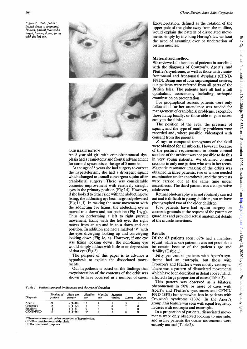
Figure 2 Top, patientlooked down to command.Bottom, patient followed atarget, looking down, fixing ywith the left eye.
CASE ILLUSTRATIONAn 8-year-old girl with craniofrontonaplasia had a craniotomy and frontal advarfor coronal synostosis at the age of 5 monAt the age of 5 years she had surgery t
the hypertelorism; she had a divergeniwhich changed to a small convergent squcraniofacial surgery. There was conscosmetic improvement with relativelyeyes in the primary position (Fig Id). Hif she looked to either side with the abducfixing, the adducting eye became grossly(Fig ILa, f). In making the same movem(the adducting eye fixing, the abductinmoved to a down and out position (FigThus on performing a left to rightmovement., fixing with the left eye, t]moves from an up and in to a downposition. In addition she had a marked'the eyes diverging looking up and cornlooking down (Fig Ic, e). However, ifwas fixing looking down, the non-fixwould simply adduct with little or no delof that eye (Fig 2).The purpose of this paper is to ad
hypothesis to explain the dissociatedments.Our hypothesis is based on the findi)
excyclorotation of the contents of theoashown to have occurred in a number c
Table 1 Patients grouped by diagnosis and the type ofdeviation
Total no of Mean age Manifest Manifest ManifestDiagnosis patients (range) eso exo vertical Latent
Apert's 24 9 (1-18) 12 7 1 3Crouzon's 15 12 (1-32) 3 5 0 7Pfeiffer's 5 6 (4-8) 0 4 0 1CFND/FND 19 13 (1-36) 6* 3 2 8
*Three were exotropic before correction of hyperelorism.CFND=craniofrontonasal dysplasia.FND=frontonasal dysplasia.
Excyclorotation, defined as the rotation of the/ j upper pole of the globe away from the midline,
would explain the pattern of dissociated move-ments simply by invoking Hering's law withoutthe need of assuming over or underaction of
- certain muscles.
Material and methodWe reviewed all the notes of patients in our clinicwith the diagnosis of Crouzon's, Apert's, andPfeiffer's syndrome, as well as those with cranio-frontonasal and frontonasal dysplasia (CFND/FND). Being one of four supraregional centres,our patients were referred from all parts of theBritish Isles. The patients have all had a fullophthalmic assessment, including orthopticexamination on presentation.For geographical reasons patients were only
followed if further attendance was needed formanagement of craniofacial problems, except forthose living locally, or those able to gain accesseasily to the clinic.The position of the eyes, the presence of
squint, and the type of motility problems wererecorded and, where possible, videotaped withconsent from the parents.X rays or computed tomograms of the skull
were obtained for all subjects. However, becauseof the postural requirements to obtain coronal
isal dys- sections ofthe orbits it was not possible to do thisncement in very young patients. We obtained coronaliths. sections in only one patient who was in her teens.) correct Magnetic resonance imaging of the orbits wast squint obtained in three patients, two of whom neededtint after examination under anaesthesia, and the two tests;iderable were carried out at the same time understraight anaesthesia. The third patient was a cooperativelowever, adolescent.-ting eye Retinal photography was not routinely carriedelevated out and is difficult in young children, but we haveent with photographed two of the older children.ig eye is Five patients have had squint surgery onIb, g). cosmetic grounds at the request of the parents orpursuit guardians and provided actual anatomical detailshe right of the muscle insertions.and out'V' withiverging Resultsone eye Of the 63 patients seen, 68% had a manifesting eye squint, while in one patient it was not possible topression be certain because of the patient's age and
debility (Table 1).Ivance a Fifty per cent of patients with Apert's syn-I move- drome had an esotropia, but those with
Crouzon's and Pfeiffer's were mostly exotropic.ngs that There was a pattern of dissociated movementsirbit was which have been described in detail above, whichA cases. affected a large proportion of cases (Table 2).
This pattern was observed as a bilateralphenomenon in 50% or more of cases withApert's and Pfeiffer's syndromes and CFND/
No FND (53%) but somewhat less in patients withfixau Crouzon's syndrome (13%). In the Apert's1 group, this feature was seen with equal frequency0 in cases with esotropia and exotropia.00 In a proportion of patients, dissociated move-
ments were only observed looking to one side,and in five patients the ocular movements wereentirely normal (Table 2).
564
on May 10, 2020 by guest. P
rotected by copyright.http://bjo.bm
j.com/
Br J O
phthalmol: first published as 10.1136/bjo.77.9.563 on 1 S
eptember 1993. D
ownloaded from

Dissociated eye movements in craniosynostosis: a hypothesis revived
Table 2 Patients grouped by diagnosis and the type ofmovement disorder
Upshoot ofUpshoot/ adducting eye
Typical downshoot or downshoot 'V' with RandomTotal dissociation to one side ofabducting dissociated 'V' 'A' movements
Diagnosis no seen to both sides only eye nwvements only pattern nofixation Full
Apert's 24 12 2 6 14 2 0 1 1Crouzon's 15 2 3 8 7 0 1 0 1Pfeiffer's 5 3 1 1 3 0 0 0 0CFND/FND 19 10 0 4 7 1 1 0 3
IMAGINGFour patients had magnetic resonance imaging ofthe orbit, such that coronal and horizontal viewswere reconstructed. Varying degrees of orbitaldeformation and excyclorotation of the extra-ocular muscles were demonstrated (Fig 3).One teenage patient had a computed tomo-
gram which also clearly showed excyclorotationofher extraocular muscles.
FUNDUS DETAILSTwo patients had fundus photographs, showingpseudo-ectopia of the fovea and excyclorotationof the retinal vessels (Fig 4). Additionally, twocases were recorded to have extorsion of vesselson clinical examination.
SURGICAL FINDINGSFive patients came to cosmetic squint surgery,comprising two cases of Apert's, one ofCrouzon's syndromes, and one each with cranio-frontonasal and frontonasal dysplasia.Of the three cases with Crouzon's syndrome
and frontonasal dysplasia, all had excyclorotationof the horizontal recti. Two of them also hadexcyclorotated vertical recti with a more anteriorinsertion of the vertical muscles in one of them.The third case (Crouzon's) was said to have amissing inferior rectus and superior obliquemuscle in the eye undergoing squint surgery,which was performed by a trained and accreditedspecialist. Subsequently, a magnetic resonanceimage showed both muscles to be present, butthe inferior rectus was seen to be displacedmedially (Fig 5). With dynamic reconstruction ofthe images, the entire course ofthe muscles couldbe traced well forward almost to the point ofinsertion.
Figure 4 Fundus photograph ofthe right eye showingmarked exorotation ofthe eye, illustrated by the rotatedretinal vessels.
One of the cases of Apert's syndrome wasreported to have normal muscle insertions, butthe rotation was not looked for specifically. Herproblem was ptosis and convergent squint andboth eyes were recorded as having markedupshoots on adduction.
DiscussionOur findings suggest that excyclorotation of theorbital contents occurs not uncommonly incraniosynostosis with hypertelorism. Ourhypothesis is that excyclorotation, causingmalalignment of the axes of movement, will
Figure 5 Postoperative magnetic resonance image ofa caseofCrouzon's syndrome reported at squint surgery to havemissing inferior rectus (IR) and superior oblique (SO)muscles. Note that both muscles are present but the IR is
displaced nasally.
Figure 3 Magneticresonance image ofthe orbitsofa patient showingconsiderable distortion of theshape ofthe orbit andmarked exorotation oftheextraocular muscles. Thedistortion ofthe orbits isasymmetrical.
565
on May 10, 2020 by guest. P
rotected by copyright.http://bjo.bm
j.com/
Br J O
phthalmol: first published as 10.1136/bjo.77.9.563 on 1 S
eptember 1993. D
ownloaded from

Cheng, Burdon, Shun-Shin, Czypionka
RE
MR
_N*X- 40
Vectors
Figure 6 Left eye fixinglooking left. Right eye showsextreme upshoot. Opposingvectors are cancelled out.
Figure 7 Right eye fixinglooking left. Left eye isdown and out.
0rMVR
Vectors
LE
xO~~~~~~~~~~~~~~~~~~~~~~~~Q)SR
x
Resultant
result in dissociated movements if Hering's lawstill applied.Thus excyclorotation through 45 degrees
requires the combined action of the superiorrectus (SR) and lateral rectus (LR) for abduction,whose contralateral synergists are the oppositeinferior oblique (IO) and medial rectus (MR)respectively (Fig 6). By using the same argu-ment, on looking left with the right eye fixing,adduction is the resultant action of the right MRand inferior rectus (IR), whose contralateralsynergists will be the left LR and superioroblique (SO), which in abduction will have verylittle vertical action (Fig 7); the result is a left eyein the down and out position. The adduction ofthe non-fixing eye when the other eye is followingan object looking down, is not explicable bypostulating SO underaction or using any otherexplanation, bearing in mind that both eyes candepress on looking down when executing avoluntary movement (Fig 2). When fixing on anobject, the excyclorotated left eye is depressed by
so
SR
O LIR
Resultant
the combined action of the IR and LR whosecontralateral synergists are the SO and MRrespectively. The MR in the excyclorotatedposition will have both adducting and elevatingactions. The elevation will be opposed by thedepressing action ofthe SO which in an adductedposition will have no significant abducting role.Thus the 'resultant' produces adduction of thenon-fixing eye (Fig 8). If it was a superior obliquepalsy alone and if the eye were not excyclo-rotated, depression of the left fixing eye shouldnot result in adduction with little or no depres-sion of the non-fixing right eye. On voluntary upand down gaze, the prime movers are thesuperior and inferior recti, hence both eyes moverelatively symmetrically albeit with an exag-gerated 'V' pattern owing to the displacement ofthe muscles (Fig 9).
EVIDENCE IN SUPPORT OF THE HYPOTHESISThe anatomical evidence comes from:
1 imaging the excyclorotation of the extra-ocular muscles in five patients with anomalouseye movements;
2 four patients who were found to haveexcyclorotation ofthe insertions ofthe muscles atsurgery;
3 in two patients, fundus photography wasavailable to show the excyclorotation of fundusdetails with pseudo-ectopia ofthe macula (Fig 4).In two patients retinal vessels were recorded to beexcyclorotated on ophthalmoscopy where photo-graphy was not obtainable.An abnormal head posture was not a feature of
these patients nor, indeed, was it a characteristicof the group. In the patient with demonstrablerotation of retinal vessels there was no apparentattempt to compensate for the rotation by headtilting even when fixing, which implies an adap-tation at the cortical level.
Using physiological principles and invokingHering's law, the anomalous pattern of eyemovements can be explained by the excyclorota-tion of the globe and its extraocular muscles.Without this model of excyclorotation, we wouldneed to postulate over or underaction of certainmuscles, especially the obliques. While ourhypothesis explains the observations, it does notexclude the possibility that the obliques may beover and underacting in certain instances: norcan one ignore the reports of missing muscles.While there are postulates for the overaction ofthe obliques,'3'4 and one accepts overaction ofinferior obliques as a common phenomenon,there is no concrete evidence, such as electro-myography, to support the hypothesis. Histo-logical abnormality of the muscles as a cause ofthe anomaly must also be allowed for, since thereis one report of structural abnormalities inApert's syndrome.'5 However, this is unlikely tobe an important factor for the group as a wholesince Crouzon's syndrome and craniofrontonasaldysplasia are not normally associated with softtissue abnormalities. Another enigma is theabsence of ptosis in cases where the SR werereported to be missing.679 Since the levatorpalpebrae superioris is derived embryologicallyfrom the SR,'6 the latter's absence will require apostulate of secondary atrophy after the levator
566
on May 10, 2020 by guest. P
rotected by copyright.http://bjo.bm
j.com/
Br J O
phthalmol: first published as 10.1136/bjo.77.9.563 on 1 S
eptember 1993. D
ownloaded from

Dissociated eye movements in craniosynostosis: a hypothesis revived
was differentiated. While there is strong circum-stantial evidence that muscle anomalies exist incraniofacial dysostosis and the evidence is irre--futable in cases where there is confirmation fromcomputed tomography or where the globe'ssurface is explored by an encircling procedure,8the possibility exists, however slight, that amissing muscle may be in another site throughextreme rotation ofthe globe, as was found in oneof our cases.The review of our patients suggests that the
pattern ofdissociated movement described aboveis quite common (Table 1) and any explanationmust take this relative frequency into account:50% or more of our patients with Apert's orPfeiffer's syndromes show the typical dissocia-tion as described in our case illustration. There-fore, the absence or atrophy ofmuscles is likely toplay a small part in the causation of theseanomalous movements. While simple overactionof the IO and underaction of the SO wouldexplain most of the anomaly if they existtogether, it is still not possible to explain on thatbasis alone, the adduction (without depression)
4,,
ResultantI=>'
of the non-fixing eye when the other eye fixes onan object looking down (Fig 2).The theory of sagittalisation" has been
advanced to explain some aspects of the 'A' and'V' phenomenon though there is no proof thatthis is the underlying mechanism in cases thathave no craniofacial abnormality. This theorypostulates that the two obliques may be insertedinto the globe at different angles to the sagittalplane. If the IO was inserted at a smaller anglethan the SO, the torsional imbalance, throughcompensatory action of other muscles, wouldlead to the 'V' phenomenon. In most of our casesthe mid-facial hypoplasia would result in theopposite shift of the 10, which could be expectedto arise more posteriorly than the tendon of theSO, thus making a larger angle with the sagittalplane and would have the opposite effect.Another distorting possibility is the alteration ofthe direction of vectors by displacement of thefulcrum through contact between the rim of theorbit and the inferior rectus muscle. 8 As Morax'pointed out, this theory is inadequate, as the 'V'syndrome is unchanged after surgery, and theanomalies can be seen in 'teleorbitism withoutexorbitism'.
Ifexcyclorotation ofthe orbital contents wouldlead to a 'V' on elevation, then the oppositewould lead to an 'A' syndrome. Though suchinstances are rare, one case has been describedwhere the muscles were shown on computedtomography to be incyclorotated'9 and this find-ing complements our hypothesis.While there is awareness of excyclorotation of
the globe in craniosynostosis5 the pattern ofdissociation that we have described has not beensufficiently emphasised, nor the fact that thepattern can only be fully appreciated by perform-ing the same movements with the two eyes fixingalternatively. Previously, overaction of the in-ferior oblique, with or without underaction ofthesuperior oblique, was frequently used as anexplanation. While this possibility remains, wehave shown that in cases where the excyclorota-tion is marked, the dissociated movements can beexplained simply by applying Hering's law ofequal innervation.
We are indebted to Dr P Anslow, DrG Ashworth, Mr L Benjamin,Mr J Elston, and Mr M D Poole for help with the preparation ofthis paper, Mrs P Lewis for orthoptic assessment, and Mrs A SGray for typing the manuscript.
1 Cohen MM, Craniosynostosis. Diagnosis, evaluation andmanagement. New York: Raven Press, 1986; 13: 413.
2 Fries PD, Katowitz JA. Congenital craniofacial anomalies ofophthalmic importance. Surv Ophthalmol 1990; 35: 87-119.
3 Pruzansky S, Miller MT. Ocular defects in craniofacialsyndromes. In: Rennie WA, ed. Goldberg's genetic andmetaboliceye disease. Boston: Little, Brown, 1986; 9: 241-55.
4 Choy AE, Margolis S, Breinin GM, McCarthy JG. Analysis ofthe pre-operative and post-operative extraocular musclefunction in surgical translocation of bony orbits. In: Con-verse JM, et al. eds. The symposium on diagnosis and treatmentofcraniofacial anomalies. St Louis: Mosby, 1979: 128.
5 Morax S. Oculo-motor disorders in craniofacial malforma-tions.J Max-fac Surg 1984; 12: 1-10.
6 Diamond GR, Katowitz JA, Whitaker LA, Quinn GE,Schaffer DB. Variations in extraocular muscle number andstructure in craniofacial dysostosis. Br J Ophthalmol 1980;64:416-8.
7 Weinstock EJ, Hardesty HH. Absence of superior recti incraniofacial dysostosis. Arch Ophthalmol 1965; 74: 152-83.
8 Snir M, Gilad E, Ben-Sira I. An unusual extraocular muscleanomaly in a patient with Crouzon's disease. Brj Ophthal-mol 1982; 66: 253-7.
9 Cuttone J, Brazis P, Miller M, Folk S. Absence of the superiorrectus muscle in Apert's syndrome. J Pediatr OphthalmolStrabismus 1979; 16: 349.
4-X-
Resultant
-o- 1,) so
IS
0~0
Vectors
Figure 8 Left eyefixinglooking down. The non-fixing right eye is adducted.The vertical actions havecancelled out.
Figure 9 Looking up anddown to command, showingan exaggerated 'V'.
Vectors
567
-J .,L-
IV
on May 10, 2020 by guest. P
rotected by copyright.http://bjo.bm
j.com/
Br J O
phthalmol: first published as 10.1136/bjo.77.9.563 on 1 S
eptember 1993. D
ownloaded from

Cheng, Burdon, Shun-Shin, Czypionka
10 Walker JW, Russell-Eggitt I, Taylor D. Ocular motilityproblems in craniofacial dysostosis. In: Kaufmann II, ed.Trans 18th Meeting ofEuropean Strabismus Association 1989:245-56.
11 Lee JP. Congenital muscular defects. Eye 1992; 6: 181-3.12 Pollard Z. Bilateral superior oblique muscle palsy associated
with Apert's syndrome. Am J Ophthalmol 1988; 106:337-40.
13 Urrets-Zavalia A, Jr. Significance of congenital cyclo-vertical motor defects of the eyes. BrJ Ophthalmol 1955; 39:11.
14 Urrets-Zavalia A. Reaction to dissociations and primary insuf-ficiencies of the vertical acting muscles: a discussion of thepathogenesis of the A+V syndromes. Trans Am AcadOphthalmol Otolaryngol 1981: 324.
15 Margolis S, Pachter BR, Breinin GM. Structural alterations ofextraocular muscle associated with Apert's syndrome.BrJ7 Ophthalmol 1977; 61: 683-9.
16 Mann IG. Development of the human eye. 2nd ed. London:BMA, 1949.
17 Gobin MH. Sagittalisation of the oblique muscles as a possiblecause for the 'A', 'V' and 'X' phenomena. Br J Ophthalmol1968; 52: 13-8.
18 Ortiz-Monasterio F, Fuente del Campo A, Limon-Brown E.Mechanism and correction of V syndrome in craniofacialdysostosis. Symposium on plastic surgery in the orbital region.St Louis: Mosby, 1976: 247-54.
19 Millar JE, Gado H. Computerised axial tomography instrabismus patients. In: Ravault AP, Lenk M, eds. TransFifth Int Orthoptic Congress. France: LIPS, 1983: 393-400.
History ofophthalmology
The invention of spectacles
The benefits of viewing the world through glasshave long been recognised, and the fact that theemperor Nero invariably watched the gladiators'events through a large emerald held to his eye isoften quoted as an example. There are, however,several explanations for this, none of whichinclude the principles of optics. Firstly, it wasrumoured that Nero disliked the colour of blood(ofwhich there was always plenty), and secondly,he loved to show off his wealth. Possibly he wasusing his emerald as sunglasses against the glarebut, sadly, it never occurred to him to have itattached to a frame as a primitive pair ofspectacles. Myopia was certainly recognised inhis time, with short sighted slaves being sold at adiscount. It is recorded that Dionysius in 460 BC,was myopic, and that his terrified courtiers (hewas a tyrant) all feigned the same affliction inorder to pacify him.The credit for securing lenses in front of the
eye may go to the Chinese, who were apparentlyseen by Marco Polo in 1270 to be sporting framedlenses attached to the head by weighted cordshanging over the ears. In Britain, it was left toRoger Bacon to moot the concept of 'using glasslenses to aid those who are old and have weaksight' (his own words). The Italians woulddisagree, and give the credit to Armati, who diedin 1317, largely because his tombstone bears theinscription 'the inventor of spectacles.'
Religious paintings of the fifteenth centuryshow St Hieronymous and St Donatus availingthemselves of the devices, and apparently thechurch saw nothing heretical in their use. Itcertainly condoned them in 1623 when de Valdez(who happened to be an officer of the inquisition)published a superb monograph on their use atthat time.De Valdez reports that the 'refractionists' of
his day did brisk trade, and began by asking thepatient's age. As a rule of thumb, a man between30 and 40 would require glasses oftwo varas (thevara approximates to one dioptre), a woman
would get a stronger lens. The 'optician' wouldthen inquire - if it were not obvious from thecustomer's dress and demeanour - whetherleather, brass, silver, or gold frames wererequired. Common sense was present in the1600s, as the optician would warn that glasseswere not a 'cure-all,' and that a lens that was tooweak was better than one that was too stroing.'Opera glasses' that were held for short periods ofdistant vision were available, and glasses toprotect from the 'winds of winter and brightlights of summer' came in yellow, brown, red,green, and blue.De Valdez touches upon the debate about
noseglasses versus earframes, etc. King Philiphad his glasses set into 'temple pieces' whichattached to his hat and steadied the spectacles onthe royal nose, but de Valdez notes this isimpossible for the common man, who needs to becontinually removing his hat from politeness.Although glasses were ridiculed at certain
times in history, snobbery reared its ugly head inEngland in the 1700s. Then the gentry pur-chased glasses as a sign of intelligence andrefinement, whether they needed them or not,but the lower classes - however much theystumbled into doors and dropped things - did notdare wear them in public. This was just as well,since they couldn't have afforded them anyway.Until the seventeenth century spectacles were sovaluable as to be separately bequeathed in one'swill, presumably regardless ofthe visual acuity ofone's beneficiaries.
FIONA ROMAN
Oliver GH. The history of the invention and discovery of spectacles.London: BMA, 1913.
Sorsby A. A short history ofophthalmology. London: Staples Press,1948: 68-76.
Ten Doesschate G. Some historical notes on spectacles andberyllus. BrJ Ophthalmol 1946; 30: 660-2.
Wood CA. The first scientific work on spectacles. In: Packard FR,ed. Annals of medical history. London: Hoeber, 1940; 2:150-5.
568
on May 10, 2020 by guest. P
rotected by copyright.http://bjo.bm
j.com/
Br J O
phthalmol: first published as 10.1136/bjo.77.9.563 on 1 S
eptember 1993. D
ownloaded from