distinct assemblage of planktonic ciliates dominates both
TRANSCRIPT
MARINE ECOLOGY PROGRESS SERIESMar Ecol Prog Ser
Vol. 526: 1–9, 2015doi: 10.3354/meps11256
Published April 22
INTRODUCTION
The spatial variability of microbial eukaryotes inmarine systems has been explored in only a few stud-ies (reviewed in Dolan 2005, Foissner 2006). Bates etal. (2013) showed that protist communities in generalare not globally distributed since composition di vergesconsiderably across large geographic distances. Con-versely, Rodríguez-Martínez et al. (2013) showed thatparticular microbial lineages (e.g. MAST-4) can befound only under specific environmental conditions.
© Inter-Research 2015 · www.int-res.com*Corresponding author: [email protected]
FREEREE ACCESSCCESSFEATURE ARTICLE
Distinct assemblage of planktonic ciliatesdominates both photic and deep waters on the
New England shelf
Jean-David Grattepanche1, Luciana F. Santoferrara2, George B. McManus2, Laura A. Katz1,3,*
1Department of Biological Sciences, Smith College, Northampton, Massachusetts 01063, USA2Department of Marine Sciences, University of Connecticut, Groton, Connecticut 06340, USA
3Program in Organismic and Evolutionary Biology, University of Massachusetts, Amherst, Massachusetts 01003, USA
ABSTRACT: Microbes are critical members of marineecosystems, given their roles as both primary producersand consumers in food webs. Despite their importance,data on biogeographical patterns of microbial eukary-otes are limited. Past studies have generally targetedeither all eukaryotes or broad clades like Rhizaria andAlveolata. For this study, we focus more narrowly onoligotrich and choreotrich ciliates (both members ofthe class Spirotrichea) as these lineages play majorroles in marine food webs. We assess distribution pat-terns of abundant ciliate community members along a163 km transect off the coast of New England, USA.Over 3 d, we sampled ciliates at 23 stations from shal-low waters (<30 m depth) to beyond the continentalshelf (>800 m). We used a community DNA finger-printing technique, denaturing gradient gel electropho -resis (DGGE), to assess patterns for abundant commu-nity members and found 2 overlapping assemblages ofciliates: one common in samples from inshore to off-shore (up to 180 km from the coast) and from the sur-face to 850 m deep; and a second that is generally re-stricted to offshore waters. The distributions of these 2assemblages correspond with distance from the coastbut not with the environmental factors that we meas-ured, including depth, temperature, degree of stratifi-cation, phytoplankton fluorescence and accessory pig-ment composition (a proxy for phytoplankton composi-tion). The presence of these ciliate assemblages as deepas 850 m suggests they may have a broader impact onmarine food webs than just photic zone herbivory.
KEY WORDS: Ciliate diversity · Oligotrich · Choreo -trich · DGGE · Inshore-offshore · Surface-deep waters
Resale or republication not permitted without written consent of the publisher
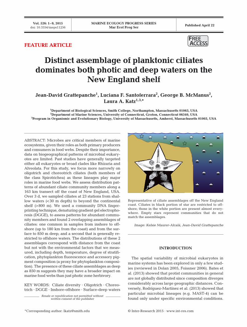
Representative of ciliate assemblages off the New Englandcoast. Ciliates in black portion of star are restricted to off-shore; those in the white portion are present almost every-where. Empty stars represent communities that do notmatch the assemblages.
Image: Kelsie Maurer-Alcalá, Jean-David Grattepanche

Mar Ecol Prog Ser 526: 1–9, 2015
Overall, the spatial scale of biogeographic patternsfor microbial species is unclear (Fenchel et al. 1997,Dolan 2005) as some have argued for cosmopolitandistributions (e.g. Finlay 2002, Massana et al. 2004,Weisse et al. 2008, Bates et al. 2013), while others sug-gest there is considerable endemism (e.g. Hillebrandet al. 2001, Foissner 2006, Simon et al. 2008, Bik et al.2012). The distribution of co-occurring assemblagesof microbial eukaryotes is not well known either. Bycomparison, for microbial prokaryotes, DNA-based di -versity studies have shown scales of community varia -bility that correlate with ocean circulation and season-ality (e.g. Fuhrman et al. 2006, Hewson et al. 2006).
Ciliates in the clades Oligotrichia and Choreo trichia(Class Spirotrichea) are major components of marinefood webs, where they feed upon phytoplankton andbacteria and in turn are fed upon by copepods andother small metazoans (Azam et al. 1983, Fenchel1988). The diversity and spatial distribution of thesemarine grazers has been poorly studied despite theircritical roles within the ‘black box’ of marine microbialfood webs (Sherr & Sherr 2002, Calbet & Saiz 2005).
Analyses using microscopy have yielded evidencefor correlations between species abundance and en -vironmental parameters. Using mouth size as a para -meter to group tintinnid ciliates, Dolan et al. (2013)showed that morphospecies distribution is related tothe distribution of prey size in 2 Medi terranean gyresand in the California Current. Morphospecies rich-ness in tintinnids has also been shown to correlatewith phytoplankton concentration (Santoferrara &Alder 2012).
A number of studies have shown that the morpho -species concept is a biased proxy for biodiversityamong ciliates given the frequent detection of crypticspecies in analyses using molecular markers (e.g.Katz et al. 2005, Bickford et al. 2007). In oligotrich andchoreotrich ciliates, molecular tools have been usedto reveal changes in distributions of phylotypes atsmall spatial scales (~1 km between stations; Grat-tepanche et al. 2014). Analyses of these 2 cladeswithin a large temperate estuary indicate that bio-geographical patterns result from a complex interac-tion of abiotic and biotic factors (Tamura et al. 2011).
We analyzed common community members of theciliate orders Oligotrichia and Choreotrichia (ClassSpirotrichea) and assessed patterns along a 163 kmtransect off the coast of New England, USA, from Nar-ragansett Bay (Rhode Island) to the shelf break, sam-pling at the surface and ~5 m above the ocean floor (forall except the 3 stations after the shelf break; see Fig. 1).Over 3 d, we sampled ciliates at 23 stations from shal-low waters (<30 m) to beyond the continental shelf
(>800 m). We assessed spatial variability using smallsubunit rRNA (SSU rDNA) amplicons and denaturinggradient gel electrophoresis (DGGE), and we gener-ated DNA sequences for common community membersby excising bands from DGGE gels. Given the struc-ture of the water column during the summer, we hy-pothesized that ciliates would show a spatial gradientinshore to offshore and reduced diversity below thephotic zone (given the potential hypoxia and lack ofphytoplankton) as compared to the uppers layers.
MATERIALS AND METHODS
Sampling
We sampled on board the R/V ‘Cape Hatteras’ fromjust outside Narragansett Bay (RI, USA) to the shelfbreak, collecting at 23 stations, each 6 km apart(Fig. 1, see Table S1 in Supplement 1 at www.int-res.com/articles/suppl/m526p001_supp/), during 3 dfrom 6 to 8 July (around 2 h between samplings dur-ing the 40 h transect). For every station, 4 depthswere sampled: surface, pycnocline, chlorophyll max-imum depth and deep layers (Fig. 1 and Dataset S1 inthe supplementary material). A CTD profiler (Sea-Bird Electronics) mounted on a rosette measured thetemperature, salinity, chlorophyll fluorescence andoxygen, and allowed us to identify the pycnocline(strongest density gradient) and the chlorophyll max-imum depth. On the shelf, the deep samples weretaken ca. 5 m off the bottom. For stations beyond theshelf break (21, 22, 23), the deepest samples werebetween 100 and 200 m, with an extra sample at850 m at the furthest station (Stn 23, ca. 100 m abovethe bottom).
Pigment composition (HPLC)
To estimate the phytoplankton composition, 250 mlfrom each chlorophyll maximum depth sample wasfiltered on a GF/F filter and stored in the dark at−80°C until extraction and analysis by high-perfor-mance liquid chromatography (HPLC) according toVan Heukelem & Thomas (2001).
Sample filtration and DNA extraction
For each sample, 1 l of seawater was collected in aNiskin bottle and filtered in series through 80 µmmesh to remove metazoans and other large organ-
2

Grattepanche et al.: Ciliate assemblage in photic and deep waters
isms, then onto 10 and 2 µm polycarbonate filters tosep arate the micro- and nanosize fractions. We usedsequential filtration on 10 and 2 µm pore sizes to assess the smallest ciliates, which are generallyunderestimated in microscope-based studies. We alsofiltered water samples through separate 3 µm nitro-cellulose filters to compare with the size fractiona-tion. All filters were immediately placed in 0.5 ml ofDNA preparation buffer (100 mM NaCl, Tris-EDTAat pH 8, and 0.5% sodium dodecyl sulfate [SDS])and stored at 4°C until DNA extraction. A standard phenol-chloroform extraction protocol (Sambrook etal. 1989, Ausubel et al. 2002) was carried out toextract and purify the DNA using a method adaptedto filters (Costas et al. 2007). The final DNA pelletwas air dried and re-suspended in 50 µl Tris-EDTA(ph 8.0) and 0.1 µl RNase.
DNA amplification
The DNA from the filters of each station was am -plified under conditions aimed at minimizing PCR re-combination. PCR was carried out using 20 µl of mas-ter mix, composed of 4 µl 5× GC buffer (NEB), 0.5 mMMgCl, 50 mM BSA, 50 µM of each deoxy nucleosidetriphosphate (dNTP), 0.25 pM of each primer, and
0.1 µl Phusion polymerase (NEB). We used SSU rDNAprimers specific to choreotrich and oligotrich ciliates(152+ and 528-GC from Doherty et al. 2007, Tamuraet al. 2011). These target regions were amplified byPCR under the following conditions: 98°C for 3 min,30 cycles of 98°C for 15 s, 58°C for 15 s, and 72°C for1 min, and a final extension at 72°C for 10 min.
DGGE analysis
For each sample, 5 PCR products amplified from a1/10 dilution of genomic DNA were pooled prior toDGGE. Each station (8 samples: 2 and 10 µm samplesfrom surface, pycnocline, chlorophyll maximum, anddeep layer, respectively) was run on an independentDGGE gel. DGGE gels (6% acrylamide gel) con -taining a linear denaturant gradient from 35 to 55%(100% denaturant corresponds to 7 M urea and 40%deionized formamide) were run at 245 V for 5 minfollowed by an incubation at 45 V for at least 15 husing a Dcode universal mutation system (Bio-Rad).We verified the robustness of DGGE by replicatingseveral gels using PCR reactions that had been runon different days. The brightest bands and all com-mon bands were excised from the gels, amplified by10 cycles of PCR following the aforementioned cycling
3
Boston
Hartford
New York
Station 1
Station 23
Atlantic Ocean
New Haven
Providence
Albany
Long Island Sound
Longitude (°N) 74 73 72 71 70 69
Latit
ude
(°W
)
43
42
41
40
39
38
USA
Fig. 1. Station locations during thecruise carried out in July 2012 from justoutside Narragansett Bay (Stn 1) to theshelf break (Stn 23). Isobaths are repre-sented with intervals of 50 m depth

Mar Ecol Prog Ser 526: 1–9, 2015
conditions and sequenced by the Sanger method(sequences are available at GenBank under acces-sion numbers KR056176-KR056216). We assignedtaxonomy to each DGGE band sequenced using aBLAST approach and confirmed this assignmentusing a phylogenetic tree (Fig. 2). However, giventhe relative shortness of our sequences and the lowinterspecific variability within the oligotrich andchoreotrich ciliates (e.g. Santoferrara et al. 2015), werecognize the uncertainty in these assignments,which should be viewed as the closest morphos-pecies from sequences in GenBank. In order to com-pare the DGGEs from each station, we created a setof markers representing known phylotypes to run ongels (Grattepanche et al. 2014).
DGGE gels were photographed and band intensitymeasured using Kodak molecular imaging software(Carestream Health) after staining for 30 min in200 ml of Tris-acetate-EDTA (TAE) buffer with 20 µlof SYBR Gold (Invitrogen). The ciliate communitystructure obtained by DGGE was analyzed with FastUniFrac software (Hamady et al. 2010) based onband pattern and intensity. For Fast UniFrac inputs,we used a star tree (phylogenetic tree with same distance between each phylotype or DGGE bandand the root), a ‘map’ of phylotypes (i.e. presence/absence or abundance table for each DGGE band orphylotype at each sample or station; see Dataset S2 inthe supplementary material) presented at each sam-ple or station, and a ‘map’ of the environmental con-ditions for each sample (see Dataset S1 in the supple-mentary material). To compare environment patterns,we performed the same analysis using environmen-tal parameters (temperature, salinity, oxygen andchlorophyll fluorescence) as starting points. In theDGGE bands cluster analysis, phylotypes are con -sidered as variables of each sample/station. In theenvironment clustering analysis, the environmentalparameters are considered as the variables of eachsample/station. To avoid sample clustering relatedonly to depth, we performed the clustering analysiswithout this para meter. The samples or stations wereclustered using the unweighted pair group methodwith arithmetic mean (UPGMA). The robustness ofthe UPGMA clusters was tested with jackknife analy-sis based on 50 permutations. The phylotype distri-bution obtained by DGGE was analyzed with hier -archical clustering analyses of Euclidean distanceusing MATLAB (version R2012a). We confirmed thepresence of the 2 ciliate assemblages by repeatingDGGE with 1 representative sample from each sta-tion to yield results similar to those described byGrattepanche et al. (2014).
Statistical analysis
To compare the structure given by the environ-mental parameters to the community structure, sam-ple clustering analysis was performed using FastUniFrac (Hamady et al. 2010), and hierarchical clus-tering of the Euclidean dissimilarity index in MAT-LAB was used to identify the ciliate assemblages.
Repeatability of DGGE
We replicated PCR and DGGE gels multiple timesto ensure that DGGE methods were robust enoughto capture the ciliate community. Repeating theDGGE with the pooled PCR products generatedthe same band pattern for each sample replicated.We confirm here the robustness of DGGE, which wehave also assessed in another study (Grattepancheet al. 2014).
RESULTS AND DISCUSSION
Overall patterns in the DGGE
DGGE allows the abundant members of a commu-nity to be quantified and subsequently identified byDNA sequencing. We used DGGE to analyze com-munities from a total of 190 samples (23 stations, atleast 4 depths, 2 size fractions). Amplification withspecies-specific primers of samples collected sequen-tially on 10 and 2 µm filters generated a profileof common community members at stations 6 kmapart (Fig. S1 and Dataset S2 in the supplementarymaterial). We observed a total of 38 unique bands inDGGE gels, each corresponding to a single sequence(i.e. phylotype) of a common community member.Individual bands were present in as few as 2 and upto as many as 178 of the 190 total samples (91 of the97 samples if pooling the 2 size fractions; Fig. S1,Dataset S2). Across all depths, the number of abun-dant phylotypes per station varied from 2 to 22, withgenerally more bands at offshore sites (Fig. S1, Data-set S2).
Two assemblages of common ciliates: ‘common’and ‘offshore’
Our analyses revealed the presence of 2 assem-blages of co-occurring ciliates. One consists of 6 ciliate species that are found in many, but not all,
4

Grattepanche et al.: Ciliate assemblage in photic and deep waters 5
Fig. 2. Phylogenetic tree used toconfirm the BLAST taxonomicassignment. The taxa in colorrepresent the sequenced DGGEbands (Table S1 in Supple-ment 1). The scale bar repre-sents the number of differencesper base pair. The maximumlikelihood tree built usingRaxML and a Newick formattedversion is available in the sup-plementary material (www.int-res.com/articles/ suppl/ m526
p001_ supp/)

Mar Ecol Prog Ser 526: 1–9, 2015
samples ranging from inshore to offshore and fromsurface to deep waters (i.e. ‘common’ assemblage).The second assem blage overlapped the first one atoffshore stations and includes the 6 ‘common’ speciesplus 4 additional species (Fig. 3, Figs. S1 & S2 in Supplement 1 at www.int-res.com/articles/suppl/m526 p001_supp/). The presence of 2 assemblages isevident in hierarchical clustering analyses of DGGEbands (Fig. S2). For this study, we used previouslydesigned primers that show considerable specificityfor oligotrich and choreotrich ciliates as they capturerepresentatives of most known genera (Doherty etal. 2007, Tamura et al. 2011, Grattepanche et al.2014, Santoferrara et al. 2014). We recognize thatno primer can capture all taxa, and that we likelymissed some cryptic species (e.g. those not distin-guished by variation in the targeted V1–V2 region),
and unknown clades (those not included when wede signed primers). Only 1 band (39) represented anon-target organism, being 99% similar to the dino-flagellate Heterocapsa triquetra GU 594638, thus in -dicating some level of non-specificity in our primers(Doherty et al. 2007, Tamura et al. 2011, Gratte -panche et al. 2014).
Sequencing common community members revealedthat 7 of the 10 bands are 99% identical to knownmorphospecies. The remaining 3 are no closer than97% to any previously reported sequences. The com-mon assemblage (bands 4, 7, 10, 14, 20 and 41) con-sists of 3 tintinnids, 1 aloricate choreotrich, and 2 oli -go trichs (Fig. 3, Table S1). The offshore assemblageincorporated 4 additional members, 1 tintinnid, 1aloricate choreotrich, and 2 oligotrichs. Surprisingly,1 phylotype of the common assemblage (band 7) is
6
Fig. 3. Distribution of the 2 ciliate assemblages from inshore to the shelf break assessed by denaturing gradient gel electro -phoresis (DGGE). The ‘common’ assemblage (open stars) was observed at many but not all locations, and was joined by an off-shore assemblage (black stars) in many stations >90 km from the shore and greater than 67 m depth (Figs. S1 & S2 in Supple-ment 1 at www.int-res.com/articles/suppl/m526p001_supp/). The colored lines represent the different layers, and the dots, thesamples: (red) surface, (turquoise) pycnocline, (green) chlorophyll maximum and (purple) deep (see Fig. 4 for more details).The boxes around the station numbers represent the stratification level: (yellow) stratified water, (blue) mixed water, and (orange) intermediate stratification levels. The closest morphospecies members of the ‘common’ and ‘offshore’ ciliate assem-blages observed by DGGE (the 850 m sample lane for Stn 23 is used in this figure) are represented inside the stars. The 6 morphospecies listed in black print are the members of the ‘common’ ciliate assemblage; morphospecies listed in grey printare the 4 additional morphospecies found in the ‘offshore’ ciliates assemblage. The values in parentheses refer to the band
numbers from DGGE gels (Table S1 in Supplement 1)

Grattepanche et al.: Ciliate assemblage in photic and deep waters
closely related to Halteria grandinella, an exclusivelyfreshwater ciliate. This suggests the possibility ofpreviously undiscovered biodiversity in marine sys-tems. While the ciliate H. grandinella has not beendocumented in marine samples (Agatha 2011), weobserved a phylotype with a V1–V2 sequence amongour DGGE sequences with 99% similarity (Table S1);this phylotype might be another Halteriidae such asPelagohalteria cirrifera, which has been reported inthe North Atlantic (Agatha 2011) and which lacks aDNA sequence in GenBank.
Environmental correlates for presence/absence ofassemblages
The presence of the 2 ciliate assemblages doesnot correlate with any of the environmentalparameters we measured, even though the envi-ronmental para meters themselves are structuredby both water layer and location (Fig. 4). A FastUniFrac cluster analysis (Hamady et al. 2010)using temperature, oxygen, chlo rophyll fluores-cence and salinity for all samples revealed that
7
St15 C
St2 DSt3 D
St23 P
0.1Deep
Surface
Intermediatestratification
Pycnocline
Deep Chlorophyll Maximum
0.2
St1
St2
St3
St10St11St13
St9
St1
2St6
St7
St8
St14
St15
St4
St5
St16
St17
St2
3
St18
St19
St20
St21
St22
0.01Stratified water
Mixed water
St1
St3
St4
St2
St6St7
St8
St9St
10
St12
St17
St20
St11
St13
St14
St15
St16 S
t18
St1
9
St21
St22
St23
St5
0.2
a c
db
Fig. 4. Clustering of samples (station and depth) using Fast UniFrac (Hamady et al. 2010) by environmental variables and cili-ate diversity shows different patterns. (a) Samples clustered according to water layers (surface, pycnocline, chlorophyll maxi-mum and deep layers) using environmental parameters (temperature, salinity, oxygen and fluorescence), (b) stations (i.e. alllayers from the same station grouped in 1 ‘sample’) clustered by nature of the water mass (mixed or stratified) using the sameenvironmental parameters, (c) samples clustered by DGGE band pattern do not correlate with layers and (d) clustering ofDGGE bands by station is also unrelated to stratification level or pigment composition. The presence of the ‘common’ (whitestar) and the ‘offshore’ (black star) communities (Fig. 3, and Fig. S1 in Supplement 1) and the different phytoplankton commu-nities (dark gray dot: inshore; gray dot: middle shore; light gray and white dots: 2 offshore communities) assessed by HPLCpigment composition (Fig. S5 in Supplement 1) are indicated. Networks drawn to scale; the scale bar shows the distance be-tween clusters in UniFrac units (distance matrix where 0 = identical samples, 0.5 = samples composed of different ciliates)

Mar Ecol Prog Ser 526: 1–9, 2015
each depth layer is distinct, with few exceptions(Fig. 4a). Similarly, the samples from each station(i.e. the temperature, chlorophyll fluorescence,salinity and oxygen across all depths) cluster on thebasis of stratification level: inshore and offshore sta -tions are marked by mixed water masses, while in -ter mediate stations are stratified, i.e. dynamic ver-sus more stable environment (Fig. 4b, and Fig. S3in Supplement 1).
In contrast to environmental parameters, no clearpatterns emerged in analyses of the communitystructure based on DGGE when we considered rel-ative abundances (brightness of bands; Dataset S2in the supplementary material), presence/absence ofphylotypes as related to the water layers (Fig. 4c),level of stratification (Fig. 4d) or size fraction (Fig. S4in Supplement 1). In other words, the ‘common’and ‘offshore’ assemblages are not clearly related tothe water masses (temperature, oxygen and pres-sure/depth) or to the estimated phytoplankton bio-mass and com position (chlorophyll concentration andaccessory pigment composition; Fig. 4, and Fig. S5in Supplement 1). Our data suggest that there isa widespread ciliate community offshore and thatsome members are excluded from nearshore waters,giving us the distinct ‘offshore’ versus ‘common’assemblages.
Given that the oligotrich and choreotrich ciliatesare believed to feed principally on phytoplankton,the detection of a highly diversified ciliate assem-blage in deep samples, even at 850 m, is unex-pected. Wickham et al. (2011) also observed oligo -trich and choreotrich ciliates in morphologicalana lyses of deep samples (up to 500 m), but thesewere less diverse than the upper layers community.This suggests that these diverse ciliates may alsofeed on bacteria (Sherr & Sherr 1987) or other heterotrophic protists (Sherr & Sherr 2002) belowthe photic zone. Alternatively, we may be detectingcysts at deep levels, though it would be surprisingto find the offshore assemblage containing rela-tively constant abundances of both cysts and feed-ing stages throughout the water column, as is ob -served at all depths for Stn 23 (Fig. 3, and Fig. S1 inSupplement 1). It is also possible that the presenceof these ciliate assemblages in deep samples isdue to sinking organic particles or, given that wesampled so near the shelf break (Stns 21 to 23), tocirculation patterns. Due to the structure of oursampling (40 h outbound), we were unable to assesspatterns relating to the tidal cycles, unlike in ourprevious study, that followed a single water mass(Grattepanche et al. 2014).
Synthesis
The presence of the common ciliate assemblage inour analyses of ciliate communities along a 163 kmtransect suggests that some phylotypes are widelyadaptable to various environments and/or have alarge niche space. The common assemblage occursat various stations and layers representing gradientsin light, temperature, phytoplankton abundance,nutrients, etc. Similarly, the distribution of the off-shore assemblage does not correspond with the envi-ronmental parameters that we measured (e.g. depth,phytoplankton fluorescence, temperature), exceptfor distance from the coast. An alternative explana-tion could be that these ciliate assemblages followthe unified neutral theory of biodiversity, as observedby Dolan et al. (2007) for tintinnid morphospecies.The unified neutral theory holds that local assem-blages are put together from species randomlyselected from a larger metacommunity of ecologi-cally similar organisms. Open questions here includewhether the common community is stable over timeand in regions well beyond the shelf, and what drivesthe loss of assemblage members in some areas.
Acknowledgements. We thank the crew of the R/V ‘CapeHatteras’, A. M. Oliverio, J. Andrade, K. Brien, A. Liefeld,D. Biemesderfer, D. Tian, and A. Gordon, for assistance during the cruise and in the lab. This work was supported bythe National Science Foundation (Grants OCE-1129734 toL.A.K. and OCE1130033 to G.B.M.), Smith College and theUniversity of Connecticut.
LITERATURE CITED
Agatha S (2011) Global diversity of aloricate Oligotrichea(Protista, Ciliophora, Spirotricha) in marine and brackishsea water. PLoS ONE 6: e22466
Ausubel FM, Brent R, Kingston RE, Moore DD, Seidman J,Smith JA, Struhl K (2002) Short protocols in molecularbiology: a compendium of methods from current proto-cols in molecular biology, Vol 2. Wiley, New York, NY
Azam F, Fenchel T, Field JG, Gray JS, Meyer-Reil LA,Thingstad F (1983) The ecological role of water-columnmicrobes in the sea. Mar Ecol Prog Ser 10: 257−263
Bates ST, Clemente JC, Flores GE, Walters WA, Parfrey LW,Knight R, Fierer N (2013) Global biogeography of highlydiverse protistan communities in soil. ISME J 7: 652−659
Bickford D, Lohman DJ, Sodhi NS, Ng PKL and others (2007)Cryptic species as a window on diversity and conserva-tion. Trends Ecol Evol 22: 148−155
Bik HM, Sung W, De Ley P, Baldwin JG, Sharma J, Rocha-Olivares A, Thomas WK (2012) Metagenetic communityanalysis of microbial eukaryotes illuminates biogeo-graphic patterns in deep-sea and shallow water sedi-ments. Mol Ecol 21: 1048−1059
Calbet A, Saiz E (2005) The ciliate-copepod link in marineecosystems. Aquat Microb Ecol 38: 157−167
8

Grattepanche et al.: Ciliate assemblage in photic and deep waters
Costas BA, McManus G, Doherty M, Katz LA (2007) Use ofspecies-specific primers and PCR to measure the distri-butions of planktonic ciliates in coastal waters. LimnolOceanogr Methods 5: 163−173
Doherty M, Costas BA, McManus GB, Katz LA (2007) Cul-ture-independent assessment of planktonic ciliate diver-sity in coastal northwest Atlantic waters. Aquat MicrobEcol 48: 141−154
Dolan JR (2005) An introduction to the biogeography ofaquatic microbes. Aquat Microb Ecol 41: 39−48
Dolan JR, Ritchie ME, Ras J (2007) The ‘neutral’ communitystructure of planktonic herbivores, tintinnid ciliates ofthe microzooplankton, across the SE Tropical PacificOcean. Biogeosciences 4: 297−310
Dolan JR, Landry MR, Ritchie ME (2013) The species-richassemblages of tintinnids (marine planktonic protists)are structured by mouth size. ISME J 7: 1237−1243
Fenchel T (1988) Marine plankton food chains. Annu RevEcol Syst 19: 19−38
Fenchel T, Esteban GF, Finlay BJ (1997) Local versus globaldiversity of microorganisms: cryptic diversity of ciliatedprotozoa. Oikos 80: 220−225
Finlay BJ (2002) Global dispersal of free-living microbialeukaryote species. Science 296: 1061−1063
Foissner W (2006) Biogeography and dispersal of micro-organisms: A review emphasizing protists. Acta Proto-zool 45: 111−136
Fuhrman JA, Hewson I, Schwalbach MS, Steele JA, BrownMV, Naeem S (2006) Annually reoccurring bacterialcommunities are predictable from ocean conditions. ProcNatl Acad Sci USA 103: 13104−13109
Grattepanche JD, Santoferrara LF, Andrade J, Oliverio AM,McManus GB, Katz LA (2014) Distribution and diversityof oligotrich and choreotrich ciliates assessed by mor-phology and DGGE in temperate coastal waters. AquatMicrob Ecol 71: 211−221
Hamady M, Lozupone C, Knight R (2010) Fast UniFrac: facil-itating high-throughput phylogenetic analyses of micro-bial communities including analysis of pyrosequencingand PhyloChip data. ISME J 4: 17−27
Hewson I, Steele JA, Capone DG, Fuhrman JA (2006) Tem-poral and spatial scales of variation in bacterioplanktonassemblages of oligotrophic surface waters. Mar EcolProg Ser 311: 67−77
Hillebrand H, Watermann F, Karez R, Berninger UG (2001)Differences in species richness patterns between unicel-lular and multicellular organisms. Oecologia 126: 114−124
Katz LA, McManus GB, Snoeyenbos-West OLO, Griffin A,Pirog K, Costas B, Foissner W (2005) Reframing the‘Everything is everywhere’ debate: evidence for high
gene flow and diversity in ciliate morphospecies. AquatMicrob Ecol 41: 55−65
Massana R, Castresana J, Balague V, Guillou L and others(2004) Phylogenetic and ecological analysis of novel mar-ine stramenopiles. Appl Environ Microbiol 70: 3528−3534
Rodríguez-Martínez R, Rocap G, Salazar G, Massana R (2013)Biogeography of the uncultured marine pico eukaryoteMAST-4: temperature-driven distribution patterns. ISMEJ 7: 1531−1543
Sambrook J, Fritsch EF, Maniatis T (1989) Molecular clo -ning, Vol 2. Cold Spring Harbor Laboratory Press, NewYork, NY
Santoferrara L, Alder V (2012) Abundance and diversity oftintinnids (planktonic ciliates) under contrasting levels ofproductivity in the Argentine Shelf and Drake Passage.J Sea Res 71: 25−30
Santoferrara LF, Grattepanche JD, Katz LA, McManus GB(2014) Pyrosequencing for assessing diversity of eukary-otic microbes: analysis of data on marine planktonic cili-ates and comparison with traditional methods. EnvironMicrobiol 16: 2752−2763
Santoferrara LF, Tian M, Alder VA, McManus GB (2015)Discrimination of closely related species in tintinnid cili-ates: new insights on crypticity and polymorphism in thegenus Helicostomella. Protist 166: 78−92
Sherr EB, Sherr BF (1987) High rates of consumption of bacteria by pelagic ciliates. Nature 325: 710−711
Sherr EB, Sherr BF (2002) Significance of predation by protists in aquatic microbial food webs. Antonie vanLeeuwenhoek 81: 293−308
Simon EM, Nanney DL, Doerder FP (2008) The ‘Tetrahymenapyriformis’ complex of cryptic species. Biodivers Conserv17: 365−380
Tamura M, Katz LA, McManus GB (2011) Distribution anddiversity of oligotrich and choreotrich ciliates across anenvironmental gradient in a large temperate estuary.Aquat Microb Ecol 64: 51−67
Van Heukelem L, Thomas CS (2001) Computer-assistedhigh-performance liquid chromatography method devel-opment with applications to the isolation and analysis ofphytoplankton pigments. J Chromatogr A 910: 31−49
Weisse T, Struder-Kypke MC, Berger H, Foissner W (2008)Genetic, morphological, and ecological diversity of spa-tially separated clones of Meseres corlissi Petz & Foiss-ner, 1992 (Ciliophora, Spirotrichea). J Eukaryot Micro-biol 55: 257−270
Wickham SA, Steinmair U, Kamennaya N (2011) Ciliate distributions and forcing factors in the Amundsen andBellingshausen Seas (Antarctic). Aquat Microb Ecol 62: 215−230
9
Editorial responsibility: Ronald Kiene, Mobile, Alabama, USA
Submitted: October 6, 2014; Accepted: February 23, 2015Proofs received from author(s): April 8, 2015