dmd 38:187–199, 2010 printed in u.s.a. exploration of...
TRANSCRIPT

Exploration of Catalytic Properties of CYP2D6 and CYP3A4Through Metabolic Studies of Levorphanol and Levallorphan□S
Britta Bonn, Collen M. Masimirembwa, and Neal Castagnoli, Jr.
Department of Chemistry, Medicinal Chemistry, University of Gothenburg, Gothenburg, Sweden (B.B.); Discovery DMPK,AstraZeneca Research and Development, Molndal, Sweden (B.B., C.M.M.); African Institute of Biomedical Science andTechnology, Harare, Zimbabwe (C.M.M.); and Department of Chemistry, Virginia Tech and the Edward Via College of
Osteopathic Medicine, Blacksburg, Virginia (N.C.)
Received May 31, 2009; accepted September 29, 2009
ABSTRACT:
CYP2D6 and CYP3A4, two members of the cytochrome P450 su-perfamily of monooxygenases, mediate the biotransformation of avariety of xenobiotics. The two enzymes differ in substrate speci-ficity and size and characteristics of the active site cavity. The aimof this study was to determine whether the catalytic properties ofthese isoforms, reflected by the differences observed from crystalstructures and homology models, could be confirmed with exper-imental data. Detailed metabolite identification, reversible inhibi-tion, and time-dependent inhibition were examined for levorphanoland levallorphan with CYP2D6 and CYP3A4. The studies weredesigned to provide a comparison of the orientations of sub-strates, the catalytic sites of the two enzymes, and the subsequentoutcomes on metabolism and inhibition. The metabolite identifica-
tion revealed that CYP3A4 catalyzed the formation of a variety ofmetabolites as a result of presenting different parts of the sub-strates to the heme. CYP2D6 was a poorer catalyst that led to amore limited number of metabolites that were interpreted in termsto two orientations of the substrates. The inhibition studiesshowed evidence for strong reversible inhibition of CYP2D6 but notfor CYP3A4. Levallorphan acted as a time-dependent inhibitor onCYP3A4, indicating a productive binding mode with this enzymenot observed with CYP2D6 that presumably resulted from closeinteractions of the N-allyl moiety oriented toward the heme. All theresults are in agreement with the large and flexible active site ofCYP3A4 and the more restricted active site of CYP2D6.
CYP3A4 and CYP2D6 are two members of the cytochrome P450(P450) superfamily of monooxygenases that catalyze the biotransfor-mation of a variety of xenobiotics (Guengerich, 2003). CYP3A4 hasa large, flexible active site (Ekroos and Sjogren, 2006) resulting inbroad substrate selectivity. CYP2D6 is more substrate-selective be-cause of its smaller active site and the presence of specific amino acidresidues that may contribute to substrate-enzyme interactions (Koy-mans et al., 1992; de Groot et al., 1997; Lewis et al., 1997).
To gain further structural information of the P450s, efforts havebeen directed toward the crystallization of these enzymes. The crys-tallization of soluble bacterial P450s (Poulos et al., 1987) provided anopportunity to conduct homology modeling studies of human P450susing these bacterial crystal structures as templates (Lewis, 1999; DeRienzo et al., 2000). The crystallizations of human CYP3A4 in 2004(Williams et al., 2004; Yano et al., 2004) and CYP2D6 in 2006(Rowland et al., 2006) allow for an extension of homology modelingto include direct comparisons with the human enzymes. A comparisonof crystal structures and homology models has been published re-
cently (Kjellander et al., 2007). This comparison involved dockingsolutions of a set of compounds for which specific interactions withthe surrounding amino acid residues had been identified by compu-tational methods. In addition, the opportunity to predict sites ofmetabolism for the different structures was investigated. The com-pound class used for this study was a family of morphinanyl opioidanalgesics. These compounds are known substrates for both CYP2D6and CYP3A4 that presumably adopt different orientations in theactive sites of the two isoforms (Kirkwood et al., 1997; Yue and Sawe,1997; von Moltke et al., 1998; Benetton et al., 2004; Hutchinson et al.,2004). CYP3A4 preferentially catalyzes the oxidative N-demethyl-ation, and CYP2D6 catalyzes the oxidative O-demethylation of se-lected members of this class of compounds. Attempts to dock some ofthese probe molecules into the active sites of CYP3A4 as defined byits X-ray crystal structure led to ambiguous results. The large andflexible active site cavity of this enzyme resulted in orientations withdifferent parts of the molecules in close proximity to the heme. Thisin turn led to a variety of solutions for the predicted sites of metab-olism. Because the experimentally determined sites of metabolismreported in the article by Kjellander et al. (2007) considered majormetabolic pathways only, a more detailed characterization of themetabolic profiles of two compounds of the series has been pursued.The experimental results were expected to provide information about
Article, publication date, and citation information can be found athttp://dmd.aspetjournals.org.
doi:10.1124/dmd.109.028670.□S The online version of this article (available at http://dmd.aspetjournals.org)
contains supplemental material.
ABBREVIATIONS: P450, cytochrome P450; TDI, time-dependent inhibition; AMMC, 4-aminomethyl-7-methoxycoumarine; BFC, 7-benzyloxy-4-trifluoromethylcoumarine; HPLC, high-performance liquid chromatography; MS, mass spectrometry; XIC, extracted ion chromatogram; TIC, totalion current chromatogram; NR, normalized ratio; Clint, intrinsic clearance.
0090-9556/10/3801-187–199$20.00DRUG METABOLISM AND DISPOSITION Vol. 38, No. 1Copyright © 2010 by The American Society for Pharmacology and Experimental Therapeutics 28670/3540905DMD 38:187–199, 2010 Printed in U.S.A.
187
http://dmd.aspetjournals.org/content/suppl/2009/10/01/dmd.109.028670.DC1Supplemental material to this article can be found at:
at ASPE
T Journals on June 1, 2018
dmd.aspetjournals.org
Dow
nloaded from

the different orientations the molecules adopt in CYP2D6 andCYP3A4 as part of an effort to characterize further the differencesbetween the two isoforms, regarding interactions and active sitecavities, observed with computational methods.
The two compounds selected for this study (Fig. 1) were levorpha-nol (1) and levallorphan (2). Levorphanol, an N-methylmorphinanylderivative, serves as the prototype for this family of opioid analgesics.The second compound, levallorphan (2), was selected because of thepresence of the N-allyl side chain that might be involved in hydro-phobic interactions in the active site cavity that could result in animproved affinity. Furthermore, compounds bearing the allylaminylgroup present in levallorphan have been reported to undergo meta-bolic activation leading to time-dependent inhibition (TDI) of theenzyme (Fontana et al., 2005). Consequently, the TDI properties oflevorphanol and levallorphan were compared using the two enzymes.In addition, the reversible inhibition potential was determined for thecompounds as a measure of the affinity for the two P450 isoforms. Itwas anticipated that these results would give further information onthe productive orientations of these two molecules in the active sitesof CYP3A4 and CYP2D6. Also included in this article are dockingsof the two compounds based on the crystal structures of CYP3A4 andCYP2D6.
Materials and Methods
Materials. The recombinant enzymes used in these studies (human P450sCYP2D6 and CYP3A4 coexpressed with NADPH-P450 reductase in bacterial mem-branes) were supplied from Cypex Ltd. (Dundee, Scotland, UK). Levorphanol tartrate,levallorphan tartrate, NADPH, and GSH were purchased from Sigma-Aldrich (St.Louis, MO), and 4-aminomethyl-7-methoxycoumarine (AMMC) and 7-benzyloxy-4-trifluoromethylcoumarine (BFC) were from BD Gentest (Woburn, MA). Propranololand troleandomycin were supplied from Sigma-Aldrich. HO-Bufuralol, bufuralol, and1�-HO-midazolam were purchased from SAFC Corp. (St. Louis, MO), and midazolamwas from Lipomed AG (Arlesheim, Switzerland). All of the other chemicals were ofanalytical grade and of the highest quality available.
Bioanalytical Equipment and Analytical Methods. High-performanceliquid chromatography/mass spectrometry (HPLC/MS) and HPLC/MS2 wereconducted for the metabolite identification studies. The HPLC system was anAgilent 1100 Series (Hewlett-Packard GmbH, Waldbronn, Germany) coupledto an HTC PAL autosampler (CTC Analytics AG, Zwingen, Switzerland). Theanalytical column used for chromatographic separation of the metabolites wasa HyPURITY C18 column (100 � 2.5 mm, 5 �m; Thermo Fisher Scientific,Waltham, MA). The mobile phases consisted of 0.1% formic acid in water (A)and 0.1% formic acid in acetonitrile (B). The gradient was increased linearlyfrom 5% solvent B to 50% solvent B in 8 min at a flow rate of 0.75 ml/min.The HPLC system was connected to a Sciex API 4000 quadrupole massspectrometer with electrospray ionization interface (Applied Biosystems, Con-cord, ON, Canada). Positive ion mass spectra were recorded over the massrange m/z 80 to 400, followed by product ion scans on peaks of interest. Thecollision energy used was 40 eV. The Analyst 1.4.1 software (Applied Bio-systems) was used for analysis and storage of data.
Additional MS2 experiments using a Waters (Wythenshawe, Manchester,UK) quadrupole time-of-flight Premiere instrument were performed to obtainaccurate mass data. The MS2 analyses were performed on the metabolites
observed on the Sciex API 4000 quadrupole mass spectrometer (AppliedBiosystems). The collision energy was ramped from 20 to 40 eV, and the conevoltage was set to 35 V. For chromatographic separation, a Waters (Milford,MA) ACQUITY UPLC was used with a Waters ACQUITY UPLC C18 column(50 � 2.1 mm, 1.18 �m). The same mobile phases as on the Agilent 1100series instrument (Hewlett-Packard GmbH) were used. The gradient of 5 to90% B was achieved in 5 min; the flow rate was 0.75 ml/min. MassLynxversion 1.4 software (Waters) was used for analysis and storage of data.
The analysis of HO-bufuralol and 1�-HO-midazolam for the IC50 deter-minations was performed with HPLC/MS on the Sciex API 4000 instru-ment (Applied Biosystems) described above. The multiple reaction moni-toring transitions used were 2778.1/185.7 for HO-bufuralol and 342.0/202.7 for HO-midazolam. The column used for chromatographic separationwas a HyPURITY C18 column (50 � 2.5 mm, 5 �m; Thermo FisherScientific).
Metabolite Identification in Incubations with Recombinant Enzymes.The metabolic profiles of levorphanol (1) and levallorphan (2) were investi-gated with recombinant CYP2D6 and CYP3A4. The compounds (10 �M) weremixed with enzyme (100 pmol/ml) and phosphate buffer (0.1 M, pH 7.4). Eachmixture was preincubated for 10 min at 37°C, and the reaction was theninitiated by addition of NADPH (1 mM). Zero time samples were taken beforethe addition of NADPH. After 60 min, the incubations were quenched with theaddition of an equal amount of ice-cooled solution of 0.8% formic acid inacetonitrile. The samples were centrifuged, and the supernatant was diluted 1:1in water before analysis. The 0- and 60-min samples were used for identifi-cation of metabolites.
In the case of levallorphan (2), incubations were also performed in thepresence of GSH to capture any reactive species. The same incubation condi-tions as above were used with the addition of GSH (5 mM).
To quantify the turnover of the parent compound, intrinsic clearance (Clint)values for levorphanol and levallorphan were determined at 1 �M. Thecompounds were mixed with recombinant P450 (50 pmol/ml) and potassiumphosphate buffer (0.1 M, pH 7.4). After 10-min preincubation, the reaction wasinitiated by addition of NADPH (1 mM), and the incubations were continuedfor 7, 15, 20, and 30 min. Zero time point samples were taken just beforeaddition of NADPH. All the incubations were performed in microtiter plates at37°C. The reaction was quenched with two parts ice-cooled acetonitrile con-taining 0.8% formic acid. The samples were centrifuged at 2737g for 20 minat 4°C, and the supernatant was diluted 1:2 with water before analysis.
IC50 Determination. For determination of the potential of the test com-pounds to inhibit reversibly CYP2D6 and CYP3A4, full IC50 curves wereobtained with recombinant P450s. The inhibition of the catalytic activity of theenzymes was studied using bufuralol and midazolam as probe substrates forCYP2D6 and CYP3A4, respectively. Serial dilutions of the test compoundswere done in 50% acetonitrile (50.00, 16.70, 5.50, 1.90, 0.60, 0.20, 0.07, and0.02 �M). These solutions were added to a master mix containing enzyme (5pmol/ml), potassium phosphate buffer (0.2 M, pH 7.4), and the probe sub-strates bufuralol (10 �M) for CYP2D6 and midazolam (3 �M) for CYP3A4.Also included were blank samples, without test compound and substrate, andincubations containing only the substrate (100% activity). The incubationmixtures were preincubated for 10 min, and the reactions were initiated byaddition of NADPH (1 mM), except for the blank samples. After incubating for15 min, the reactions were quenched with ice-cooled acetonitrile containing0.8% formic acid. All the incubations were performed in microtiter plates at37°C. The plates were centrifuged at 2737g for 20 min at 4°C, and thesupernatants were diluted 1:2 in water before analysis. The CYP2D6 sampleswere diluted again 1:10. Standard curves of 1�-HO-midazolam and HO-bufuralol were prepared in the incubation matrix and were precipitated in thesame way as the samples. The formation of 1�-HO-midazolam and HO-bufuralol were analyzed with HPLC/MS.
Time-Dependent Inhibition. Levorphanol and levallorphan were tested fortheir TDI properties. A time-dependent inhibitor is a compound that inhibitsthe enzyme in an irreversible or quasi-irreversible manner after metabolicactivation. In this assay the activities of recombinant CYP2D6 and CYP3A4were measured with the fluorescent probe substrates AMMC and BFC after 0,5, 10, and 20 min of preincubation with the test compound. For the determi-nation of the extent of enzyme inactivation, the enzymes were incubated at37°C with the test compound (1.5, 12.5, and 25 �M) in the presence and
FIG. 1. Structures of levorphanol (1) and levallorphan (2).
188 BONN ET AL.
at ASPE
T Journals on June 1, 2018
dmd.aspetjournals.org
Dow
nloaded from

absence of NADPH (1 mM for CYP3A4 and 0.4 mM for CYP2D6). Controlincubations were included without any test compound present. After preincu-bation, a 20-�l aliquot of the preincubation mixture was transferred to a wellcontaining BFC (50 �M) or AMMC (60 �M), NADPH (1 mM for CYP3A4and 0.4 mM for CYP2D6), and phosphate buffer (0.2 M) at a final volume of200 �l. To decrease the impact of reversible inhibition, substrate concentra-tions at 4� Km were used to saturate the enzyme. A 1/20 dilution of thepreincubation mixture into the incubation with substrate was also tested tofurther rule out a reversible inhibition component. The final protein concen-tration was 1.25 pmol/ml CYP3A4 and 20 pmol/ml CYP2D6. The higherenzyme concentration with CYP2D6 was used to obtain a sufficient signal-to-noise ratio when measuring the fluorescence of the AMMC metabolite. Theincubations were quenched after 15 min with a stop solution containing 80%acetonitrile and 20% Tris. The catalytic activities were determined from theamount of metabolite formed from the probe substrates by measuring thefluorescence at 390/460 nm for AMMC and at 405/535 nm for BFC. This wasdone using a SpectraMax Gemini XS fluorescence detector (Molecular De-vices, Sunnyvale, CA). The percentage of remaining activity was calculated bydividing the counts in the samples with the counts in the control incubations(100% activity). Included in the assays were positive controls known to act astime-dependent inhibitors on the two enzymes. These were propranolol forCYP2D6 (Narimatsu et al., 2001) and troleandomycin for CYP3A4 (Yamazakiand Shimada, 1998).
Dockings of Levorphanol and Levallorphan into Crystal Structures ofCYP2D6 and CYP3A4. Dockings of levorphanol and levallorphan inCYP2D6 and CYP3A4 were performed to visualize possible orientations in theactive site cavities. The protein structures used were the ligand-free CYP2D6crystal structure (Protein Data Bank code 2f9q) (Rowland et al., 2006), and theCYP3A4 crystal structure cocrystallized with erythromycin (Protein DataBank code 2j0d) (Ekroos and Sjogren, 2006). In the study by Kjellander et al.(2007), these two crystal structures were evaluated regarding site of metabo-lism predictions of compounds in the same family as levallorphan and levor-phanol. Consequently, these structures were chosen for the purpose of thisstudy. The CYP2D6 crystal structure has some unresolved regions; however,these are assumed not to affect the environment of the active site. Because theaim was to visualize possible orientations of the compounds, settings wereused to restrict the dockings based on knowledge of the two enzyme active sitecavities. The substrates were drawn in ISISdraw (Symyx Technologies Inc.,
Sunnyvale, CA) and exported as sdf-files. The two- to three-dimensionalconversion was made with CORINA (Molecular Networks GmbH, Erlangen,Germany; http://www.mol-net.de). For the dockings in CYP2D6 substratemolecules were protonated because the electrostatic interactions are believedto be important for the orientation in the CYP2D6 active site cavity. As in thestudy by Kjellander et al. (2007), neutral molecules were used for the dockingsin CYP3A4.
The dockings were performed with the docking module GLUE in programGRID version 22.2.2 (Molecular Discovery, Pinner, Middlesex, UK; http://moldiscovery.com). This program is based on calculations of GRID-molecularinteraction fields (Goodford, 1985). Specific probes related to the two com-pounds studied were chosen for analysis of the active site of the enzyme. InCYP2D6 these were H (hydrogen), OH2 (water), DRY (hydrophobic),N1� (sp3 aminyl cation), C3 (methyl), C1 � (sp2 CH aromatic or vinylic),and OH (phenol or carboxyl). For CYP3A4 the same probes were usedexcept for N1�, which was exchanged for N: (sp3 N with a lone pair). Forthe analysis a distance of 1 Å between the grid points was used. To restrictthe dockings, a box estimated to include the heme and active site was used(�16 � 20 � 23 Å). In the dockings possible interactions with surroundingwater molecules were not considered. For the binding affinity estimation,contributions from electrostatic interactions were included, as well as apenalty for loss of rotatable torsions. Different conformations of the ligandswere generated in GLUE using random search by rotating a maximum offive rotatable bonds. A maximum number of binding sites was set to 100,and an energy cutoff to �100 kcal/mol was included. Because the aim wasto provide possible productive binding modes, the resulting docking poseswere filtered, and only those that were within 6 Å from any atom of theheme were selected for further analysis.
Results
Identification of Levorphanol Metabolites. An analysis of theproduct ion spectrum (Fig. 2) of protonated levorphanol (1H�) wasundertaken to provide comparative data that would be useful in theelucidation of the structures of metabolites derived from this com-pound. This spectrum (as well as the corresponding spectra of lev-allorphan and the derived metabolites) could be obtained only at ahigh collision dissociation energy (40 eV) because of the stability of
FIG. 2. HPLC/electrospray ionization/MS product ion spectrum of protonated levorphanol (1H�) obtained at 40 eV.
189EXPLORATION OF CATALYTIC PROPERTIES OF CYP2D6 AND CYP3A4
at ASPE
T Journals on June 1, 2018
dmd.aspetjournals.org
Dow
nloaded from

the MH� species. The molecular formulas for the major product ionswere obtained from accurate mass analysis (Table 1). Structureassignments and attempts to rationalize the formation of the majorproduct ions are detailed in the supplemental data.
Two informative product ions present in this spectrum, at m/z201.1277 (C14H17O�) and 199.1120 (C14H15O�), are likely to cor-respond to ii� and iii�, respectively. Pathways to rationalize theformation of these product ions (Scheme 1) start with ring-openingand proton rearrangements of 1H�. Migration of a proton from theN-methyl group to C(9) with simultaneous ring opening leads to 1a�
that loses ethene and formaldimine to yield ii� [path (a)]. Alterna-tively, cleavage of the C(9)-N bond accompanied by proton migrationfrom the benzylic C(10) atom to nitrogen leads to ib�. Subsequentfragmentation with neutral losses of ethene and methylamine leads toiii� [path (b)]. The product ions observed in this spectrum are alsopresent in the product ion spectrum of protonated levallorphan (seebelow). Furthermore, the corresponding spectra obtained from several
metabolites of levorphanol and levallorphan display product ionsequivalent to ii� and iii�.
Levorphanol Metabolites Formed in the Presence of CYP3A4and CYP2D6. The extracted ion chromatograms (XICs) obtainedfrom incubations of levorphanol with CYP3A4 and CYP2D6 of all themetabolite peaks are shown in Fig. 3. (See supplemental data for allthe total ion current chromatograms.) The product ions are recorded inTable 2. Peaks corresponding to six metabolites (3-8) were observedin the XICs for CYP3A4, and three of these metabolites (4, 6, and 8)were formed in the presence of CYP2D6. The m/z for MH� of 3, 4,and 5 (m/z 274) was 16 m/z units greater than 1H� (m/z 258); the m/zfor MH� of 6 (m/z 244) was 14 m/z units less than that of 1H�; andthe m/z for MH� of 7 and 8 (m/z 260) was 2 m/z units greater than1H�. The suggested metabolic pathways of levorphanol withCYP3A4 and CYP2D6 are summarized in Scheme 2.
Based on the principal product ions, the structure of the ion withMH� at m/z 274.1799 is assigned to the C(8)-hydroxylated species 3(Scheme 2). The product ion present at m/z 256.1709, correspondingto loss of water, supports an aliphatic, as opposed to aromatic, site ofhydroxylation. The proposed position of the hydroxylation at C(8) isbased on the structures of the product ions at m/z 215.1067 (xii�),173.0596 (xiii�), and 157.0659 (v� via xi�) (see Supplemental DataScheme S2 for details).
The m/z values of the ions observed in the product ion spectrum ofthe compound assigned the structure 4H� correspond to those ob-served in the product ion spectrum of 1H� with the addition of 16 m/zunits. The structure of this metabolite is suggested to be a dihydroxy-phenyl species (4 in Scheme 2, unknown regiochemistry) because theproduct ion spectrum shows no loss of water. Proposed structures ofthe other product ions are consistent with this assignment (see Sup-plemental Data Scheme S3).
OH
NH+CH2
H
H (a)
H+
OH NH+
H
H
HNCH2C2H4
C+
OH
H
OH
NH+CH3
H
H
(b)
H+
OH NH+
H
H
H2NCH3C2H4
C+
OH
H
1H+ ia+
C17H24NO+
Exact mass: 258.1858Found: 258.1842 (6.2 ppm)
ii+
C14H17O+
Exact mass: 201.1279Found: 201.1277 (1.0 ppm)
1H+ ib+iii+
C14H15O+
Exact mass: 199.1123Found: 199.1120 (1.5 ppm)
SCHEME 1. Pathways proposed to account for the product ions at m/z 201.1277 (ii�) and 199.1120 (iii�).
TABLE 1
Comparison of calculated and experimentally determined masses for the productions observed in the product ion spectrum of protonated levorphanol (1H�)
Product Iona FoundMass
SuggestedFormula Exact Mass Difference
(ppm)
ia� and ib� 258.1842 C17H24NO� 258.1858 6.2ii� 201.1277 C14H17O� 201.1279 1.0iii� 199.1120 C14H15O� 199.1123 1.5iv� 159.0813 C11H11O� 159.0810a 1.9v� 157.0646 C11H9O� 157.0653a 4.5vii� 145.0659 C10H9O� 145.0653a 4.1ix� 133.0647 C9H9O� 133.0653a 4.5
a Proposed structures and fragmentation pathways to rationalize product ions with theseformulas will be found in the supplemental data.
190 BONN ET AL.
at ASPE
T Journals on June 1, 2018
dmd.aspetjournals.org
Dow
nloaded from

The product ion spectrum of the third metabolite assigned withMH� 274.1793 shows the same ions as observed with 1H� togetherwith two additional ions at m/z 257.1777 and m/z 256.1703. The ionat m/z 256.1703, corresponding to loss of water, is consistent with asecond aliphatic hydroxylation product. However, the ion at m/z257.1777 corresponds to a loss of 17 m/z units from the parent. Theonly reasonable assignment for a neutral loss of 17 m/z units from thissystem is the hydroxyl radical (�OH). Loss of �OH suggests that theoxidation of 1 has taken place on a position that can fragment to givea relatively stable radical cation. Therefore, the suggested structure ofthis metabolite is the N-oxide (5 in Scheme 2) (see Supplemental DataScheme S4 for details). This assignment is supported by the longer
retention time of this metabolite compared with that of the parent.Both 3 and 5 may be unresolved diastereomeric mixtures.
The metabolite with MH� at m/z 244.1696 is a compound with 14 m/zunits less than that of protonated levorphanol (1H�). This loss of 14 m/zunits corresponds to loss of a methylene group and suggests that thestructure of this metabolite is the secondary amine (6 in Scheme 2). Thisproposal is supported by the observed product ions (Table 2) that corre-spond to those found in the product ion spectrum of 1H� and all of whichinvolve pathways (a) and (b) outlined in Scheme 1.
The remaining two metabolites have an MH� at m/z 260.1636,2 m/z units greater than that of 1H�. Based on the proposedstructures of 3, 4, and 6, these metabolites are likely to result from
FIG. 3. XICs obtained from extracts of incubation mixtures of levorphanol (10 �M) in the presence of NADPH-supplemented recombinant CYP3A4 (black) and CYP2D6(red).
191EXPLORATION OF CATALYTIC PROPERTIES OF CYP2D6 AND CYP3A4
at ASPE
T Journals on June 1, 2018
dmd.aspetjournals.org
Dow
nloaded from

a combination of N-demethylation (�14 m/z units) and hydroxy-lation (�16 m/z units). The product ions formed from the proton-ated 7 species resemble those observed from 3H�, suggesting thatthis is the N-demethylated metabolite of 3 (7 in Scheme 2). Theproduct ions at m/z 242.1613 (loss of water), 215.1097 (losses ofethene and methylamine), and 173.0591 (subsequent loss of cyclo-propane) are consistent with structure 7. The product ion spectrumof the second species with MH� at m/z 260 was very weak.Because there is no evidence for loss of water in the spectrum, this
compound may be the N-demethylated dihydroxyphenyl species 8(Scheme 2).
Because no authentic standards were available, only approximateestimates of the quantities of the metabolites formed, from integratedion intensities, were possible. Even if this is not a proper way ofquantification it gives an opportunity to compare the distribution ofmetabolites for the compound studied. The MS response area and acalculation of the relative MS area (%MS) of all the metabolites andthe parent are presented in Table 3. CYP3A4 proved to be a moreeffective catalyst with approximately 43% of the substrate convertedto six metabolites, including 20% conversion to the aliphatic carbinol3 as a major route. CYP2D6 was less active (33% conversion), witharomatic hydroxylation to give 4 as the dominant pathway (27%). TheClint values at 1 �M were �0.2 �l/min/pmol P450 (t1/2 �40 min) inboth CYP2D6 and CYP3A4, indicating a low turnover for the com-pound in the two enzymes.
Identification of Levallorphan Metabolites. The product ionspectrum of protonated levallorphan (2H� at m/z 284.1986; see Sup-plemental Data Fig. S9) was essentially identical to that of protonatedlevorphanol (1H�) shown in Fig. 2. This behavior makes clear thatcollision-induced dissociation of 1H� and 2H� proceeds via the sametwo initial fragmentation reactions [(a) and (b)] shown for 1H� inScheme 1. The only unique product ions formed from 2H� are thering-opened ions xxxa� and xxxb� (Scheme 3) that replace ia� andib� formed from 1H�.
Incubations of Levallorphan with CYP3A4 and CYP2D6. Thetotal ion current chromatograms (TICs) of incubation extracts ob-
OH
N
H
OH
N
H
H
OH
N
H
OH
N
HOH
OH
N
H
H
OH
N+
H
O-
OH
OH
OH
N
H
H
OH
Levorphanol (1)
6 7
3
84
5
3A4
3A4
3A4
3A4
2D6
3A42D6
3A42D6
SCHEME 2. Proposal for the in vitro metabolic pathways of levorphanol (1) catalyzed by CYP3A4 and CYP2D6.
TABLE 2
Molecular mass, responsible P450, and MH�-generated product ions oflevorphanol (1), levallorphan (2), and their metabolites
Compound P450 MH� atm/z Product Ions �10% Relative Abundance
Levorphanol (1) 258 201, 199, 159, 157,a 145, 1333 3A4 274 256, 215, 199, 197, 173, 157,a 1454 3A4/2D6 274 217, 215, 175, 173,a 161, 1495 3A4 274 257, 256, 201, 199, 159, 157,a 145, 1336 3A4/2D6 244 201, 199, 159, 157,a 145, 1337 3A4 260 242, 215, 199, 173, 157,a 1458 3A4/2D6 260 202, 200, 158,a 157, 134, 133Levallorphan (2) 284 201, 199, 159, 157,a 145, 1336 3A4/2D6 244 201, 199, 159, 157,a 145, 1339 3A4 300 282, 215, 199, 197, 173, 157,a 14510 3A4/2D6 300 217, 215, 175, 173,a 161, 14911 3A4/2D6 316 298, 231, 215,a 213, 189, 173, 16112 3A4 316 231, 215, 213, 197,a 16913 3A4 258 215, 187, 159, 146, 145,a 133, 107
a Base peak.
192 BONN ET AL.
at ASPE
T Journals on June 1, 2018
dmd.aspetjournals.org
Dow
nloaded from

tained with levallorphan (2) in the presence of CYP3A4 revealed sixmetabolite peaks; the corresponding chromatograms with CYP2D6showed only three metabolite peaks, all of which were present in theCYP3A4 tracing. The XICs of these analytes are shown in Fig. 4.
The retention time and product ion spectra of the CYP3A4- andCYP2D6-generated species observed at m/z 244 were identical tothose of the N-demethylated metabolite 6 of levorphanol. Conse-quently, this compound must be the N-deallylated metabolite of le-vallorphan (6, Scheme 4). The other five levallorphan metaboliteswere identified as 9 through 13 (see below). In addition to 6, com-pounds 10 and 11 were common metabolites of CYP3A4 andCYP2D6. The ions obtained from the product ion spectra are reportedin Table 2.
A summary of the metabolic pathways of levallorphan includingstructures of metabolites is depicted in Scheme 4. Metabolites 9 and10 have MH� at m/z 300.1934/300.1955, 16 m/z units greater than
that of levallorphan, suggesting formation of hydroxylated metabo-lites. The structure proposed for 9 is the C(5)-cyclohexylcarbinol (9,Scheme 4). This assignment is based on the same analysis that led tothe assignment of 3 for the levorphanol metabolite because the prod-uct ions of 9H� correspond to the product ions observed for 3H�. Inparticular, the loss of 18 m/z units (water) to give m/z 282.1799supports the proposed aliphatic hydroxylation site. The product ionspectrum of the second � 16 metabolite displays ions correspondingto those observed for 4, the aromatic hydroxylated metabolite oflevorphanol. That leads to structure 10 for this metabolite (Scheme 4).
Compounds assigned structures 11 and 12 (MH� at m/z 316.1920)have a mass difference compared with levallorphan of �32 m/z units,indicating bis-hydroxylations. The MS2-generated product ions of 11correspond to the product ions observed with the carbinol metabolite9 of levallorphan except for the increase of 16 Da for the secondhydroxyl group. The presence of a product ion at m/z 161.0612 in theMS2 of 11, corresponding to v� (see Supplemental Data Scheme S2)with the addition of 16 m/z units, suggests that the second hydroxy-lation is positioned in the aromatic ring [C(10) or C(13)] (11, Scheme4). The proposed structure of 12 is shown in Scheme 4. This com-pound is also a bis-hydroxylated metabolite of levallorphan that, inthis case, seems to involve two oxidations of the cyclohexyl ring (ringC). This conclusion is based on comparisons of the product ionspresent in the MS2 tracing of 12 with those formed in the MS2 of 9and 10 (see Supplemental Data Scheme S7 for details).
The compound assigned structure 13 has MH� at m/z 258.1472,26 m/z units less than the parent 2H�. This mass corresponds toN-deallylation and hydroxylation of the parent followed by furtheroxidation of a carbinol group to a carbonyl group. The MS2
fragmentation pattern differs from those of other compounds in theseries. This leads to the suggestion that the carbonyl group is positioned� to the nitrogen atom resulting in the lactam 13 (Scheme 4). Thisproposal is supported by the observed product ions that are discussed indetail in the supplemental data (Scheme S8).
In addition to the aforementioned metabolites, three weak signalswere observed at m/z 314 (data not shown). The mass difference of 30
(a)
H+
OH NH+
H
H
OH
NH+
H
H
(b)
H+
OH NH+
H
H
C19H26NO+
Exact mass: 284.2014Found: 284.1986 (9.9 ppm)
2H+
xxxb+
xxxa+
SCHEME 3. Formation of product ions xxxa� andxxxb� from 2H�.
TABLE 3
MS response areas and %MS of levorphanol, levallorphan, and the observedmetabolites after 60-min incubations with recombinant CYP2D6 and CYP3A4
MS Response Area(107 cps) %MSa
Compound 3A4 2D6 3A4 2D6
Levorphanol (1) 7.40 7.91 57 673 2.54 3.15 204 0.40 3.1 275 0.77 0.41 66 0.72 0.41 5.6 3.47 0.16 1.28 0.39 0.38 3.1 3.2Levallorphan (2) 5.59 7.71 39 846 1.90 0.299 13 3.29 2.41 1710 1.29 0.240 9 2.611 0.819 0.52 5.7 5.612 0.53 3.713 0.417 2.9
a %MS calculated from MS response area divided by the summed areas of all the metabolitesand parent.
193EXPLORATION OF CATALYTIC PROPERTIES OF CYP2D6 AND CYP3A4
at ASPE
T Journals on June 1, 2018
dmd.aspetjournals.org
Dow
nloaded from

m/z units compared with the parent compound suggests double hy-droxylations and further oxidation to a carbonyl group. No structuralassignments are offered for these three secondary metabolites.
The absolute MS response area for each metabolite of levallorphantogether with the %MS are presented in Table 3. The main speciesdetected in the HPLC/MS analysis is the parent drug, particularly inthe CYP2D6 incubations. Based on the %MS, the major metaboliteformed with CYP3A4 results from hydroxylation in the cyclohexylring. The three CYP2D6-generated metabolites appear to be formed inapproximately equal amounts. The Clint values at 1 �M were �0.2�l/min/pmol P450 (t1/2 �40 min) in CYP2D6 and 0.82 �l/min/pmolP450 (t1/2 8.5 min) in CYP3A4, indicating a higher turnover forlevallorphan in CYP3A4.
IC50 Determinations. To determine the reversible inhibition po-tentials of levorphanol (1) and levallorphan (2) on CYP3A4 andCYP2D6, full IC50 curves were obtained. The IC50 values for levor-phanol were �50 �M in the CYP3A4 assay and 11.5 �M in theCYP2D6 assay. The corresponding values for levallorphan were �50�M in the CYP3A4 assay and 0.65 �M in the CYP2D6 assay. Theseresults indicate that both compounds act as relatively strong reversibleinhibitors of CYP2D6, but neither of them shows any inhibition ofCYP3A4.
Time-Dependent Inhibition. The two test compounds also wereanalyzed for their potential TDI properties. After preincubation witheach test compound in the presence and absence of NADPH, theenzyme activity was measured in incubations with the probe sub-strates AMMC (CYP2D6) and BFC (CYP3A4). The results for lev-allorphan showed a decrease in CYP2D6 activity at higher concen-trations both in samples preincubated with and without NADPH. Thisresult suggests that levallorphan is a strong, reversible inhibitor. Nochanges were obtained with the 1/20 dilution compared with the resultsfrom the 1/10 dilution that are reported here. From the results of lev-allorphan incubated with CYP2D6, a concentration-dependent loss ofactivity was observed but it was not time-dependent. To diminish thistime-independent component the activity was normalized to the incuba-tions carried out in the absence of NADPH. The normalized ratios (NRs)were calculated according to the following equation:
NR �Ainh/Actrl
Ainh/Actrlwo NADPH
where Ainh is the catalytic activity after preincubation with inhibitorpresent and Actrl is the activity in the control incubations withoutinhibitor. A compound is considered to be a time-dependent inhibitor
FIG. 4. XICs obtained from extracts of in-cubation mixtures of levallorphan (10 �M)in the presence of NADPH-supplementedrecombinant CYP3A4 (black) and CYP2D6(red).
194 BONN ET AL.
at ASPE
T Journals on June 1, 2018
dmd.aspetjournals.org
Dow
nloaded from

if the NR is less than 0.7. This is an empirical value based on theunderstanding that the value for a compound not acting as a time-dependent inhibitor should be approximately 1.0. Allowing for anexperimental error of 20%, it could be higher or lower. Hence there isa gray zone for NR � 0.7 to 0.9.
The NRs for the two test compounds, after 20 min of preincubations,together with those of the positive controls, are presented in Fig. 5. Completetime curves are presented in the Supplemental Data Figs. S17 andS18. From the NR values it was apparent that levallorphan acts as atime-dependent inhibitor on CYP3A4 (NR 0.45 at 12.5 �M and NR0.27 at 25 �M) but not on CYP2D6. Levorphanol, which lacks theallylic side chain, did not act as a time-dependent inhibitor on eitherCYP3A4 or CYP2D6.
It is noteworthy that levallorphan shows strong reversible inhibition(IC50 � 0.65 �M) but no TDI of CYP2D6. The opposite results observedwith CYP3A4 could indicate differences in how the two isoenzymes interactwith this substrate. The difference in the TDI properties with CYP3A4 forlevallorphan and levorphanol supports the involvement of the allylic sidechain of levallorphan in the mediation of the observed TDI.
Studies with Glutathione. The presence of the allylic side chain inlevallorphan and the fact that this compound acts as a time-dependentinhibitor of CYP3A4 led to glutathione trapping studies that weredesigned to trap possible metabolically generated reactive species.The result could indicate a possible relationship between the TDI andthe anticipated bioactivation of levallorphan to reactive species.
TICs obtained with levallorphan incubations in the presence ofGSH and CYP3A4 or CYP2D6 are presented in Fig. 6. In MS2
tracings of the CYP3A4 incubations, a large peak with the retentiontime 4.18 min at m/z 589 was observed. This mass corresponds to the
mass of 2 � 307 � H2 � H�, consistent with a glutathionyl conjugateof levallorphan that had undergone an initial 2-electron (net loss ofH2) oxidation (Scheme 5). The suggested structure, 16, reflects theproposed 1,4-addition reaction of GSH with the eneiminiumyl metab-olite 15� formed by initial oxidation (hydroxylation followed bydehydration of the protonated carbinol 14H� oxygen atom) of thecarbon atom � to the nitrogen atom of levallorphan. Less likely wouldbe 1,2-addition because the terminal carbon atom of the eneiminiumylmoiety is sterically less hindered than the internal �-bond. In addition,the proposed 1,4-addition product 16 will be more stable than thecorresponding 1,2-addition product because of the conjugation of thenitrogen lone pair with the �-bond. The principal fragment ion ob-served in the MS2 of MH� at m/z 589 was at m/z 282 as expectedbecause the glutathionyl group is in conjugation with the eneaminylsystem and should readily eliminate following protonation of sulfur togive the eneiminiumyl fragment (m/z 282) with glutathione as theneutral loss (Scheme 6). The second diagnostic fragment ion observedat m/z 460 corresponds to the expected neutral loss of pyroglutamic(129 Da) that is characteristic of glutathionyl adducts (see Supple-mental Data Scheme S9 for details).
In addition to 16H� two minor peaks at 0.7 and 5.14 min arepresent in both the CYP3A4 and CYP2D6 incubations. These peakshave MH� at m/z 605, corresponding to a glutathionyl conjugate ofhydroxylated levallorphan (2 � 305 � 16 � H�). The MS2 tracingsof these peaks were weak, and no structural assignments were at-tempted. Possible metabolites could result from epoxidation of eitherthe allyl or the aromatic moiety followed by conjugation with GSH.These peaks are considered to be minor metabolites and are notconsidered further.
OH
N
H
OH
N
H
H
OH
N
H
OH
N
HOH
OH
N
HOH
OH
OH
OH
NH
HO
Levallorphan (2)
M4 (6) M11 (13)
M7 (9)
M9 (11)M8 (10)
3A4
3A4
3A4
2D6
3A42D6
3A42D6
3A4
OH
N
HOH
OHM10 (12)
SCHEME 4. Proposed in vitro metabolic pathways of levallorphan (2) catalyzed by CYP2D6 and CYP3A4.
195EXPLORATION OF CATALYTIC PROPERTIES OF CYP2D6 AND CYP3A4
at ASPE
T Journals on June 1, 2018
dmd.aspetjournals.org
Dow
nloaded from
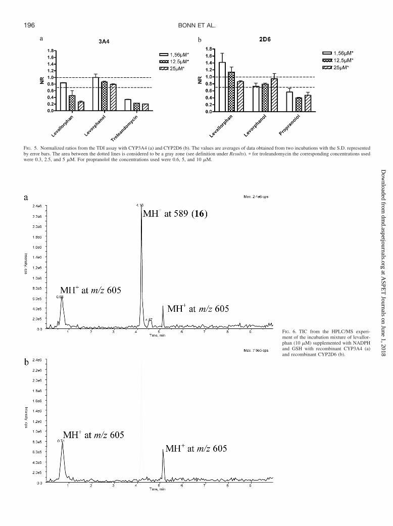
FIG. 5. Normalized ratios from the TDI assay with CYP3A4 (a) and CYP2D6 (b). The values are averages of data obtained from two incubations with the S.D. representedby error bars. The area between the dotted lines is considered to be a gray zone (see definition under Results). � for troleandomycin the corresponding concentrations usedwere 0.3, 2.5, and 5 �M. For propranolol the concentrations used were 0.6, 5, and 10 �M.
FIG. 6. TIC from the HPLC/MS experi-ment of the incubation mixture of levallor-phan (10 �M) supplemented with NADPHand GSH with recombinant CYP3A4 (a)and recombinant CYP2D6 (b).
196 BONN ET AL.
at ASPE
T Journals on June 1, 2018
dmd.aspetjournals.org
Dow
nloaded from

Dockings of Levorphanol and Levallorphan into CrystalStructures of CYP2D6 and CYP3A4. In CYP2D6 three poses eachof levorphanol and levallorphan were obtained. Comparing the cal-culated interaction energies, two poses are indicated to be the mostfavorable. These are presented in Fig. 7, a and b, and represent anorientation of levallorphan and levorphanol with the aromatic ring/phenol toward the heme. For levallorphan the distance between thepositively charged nitrogen and the negatively charged Glu216 was4.64 Å, and the distance to the oxy species in the heme was 3.55 Å.For levorphanol these distances were 7.50 and 2.87 Å, respectively. Theother two poses of levallorphan were with the allyl group pointing towardthe heme, and an additional pose is with the aromatic moiety toward theheme. Levorphanol also showed an additional pose with the aromatic ringclosest to the heme and one with the pyridine ring pointing toward theheme. Docking energies and information about the orientation of eachdocking pose are available in the Supplemental Data Table S1.
In contrast to the limited number of docking poses in CYP2D6, thedockings in CYP3A4 revealed 19 poses of levallorphan and levor-phanol. The calculated interaction energies did not differ much be-tween these poses, and there was no obvious ranking among them.The poses represent orientations of the substrates presenting manydifferent parts of the molecule to the heme. Some examples arevisualized in Fig. 8. In general, most of the levallorphan poses werefar away from the heme compared with levorphanol. These posesmight be favored by hydrophobic interactions with the phenyl alaninecluster in CYP3A4. Docking energies and information about the
orientation of each docking pose are available in the SupplementalData Table S1.
Interaction energies between each docking pose and the surround-ing amino acid residues were calculated with the energy calculationmethod described previously (Kjellander et al., 2007). These data areavailable in Supplemental Data Tables S2 and S3.
Discussion
The rationales for the present study included the possibility of exploringorientations of the two selected compounds, levallorphan and levorphanol, inthe active sites of CYP2D6 versus CYP3A4 in vitro and in silico. Ourapproach involved metabolic profiling studies on levallorphan and levorpha-nol with the key tool being the characterization of metabolites using recom-binant enzymes. The inhibition potentials of levallorphan and levorphanol onCYP2D6 and CYP3A4 were also examined in an effort to assess theiraffinities for these enzymes. In addition, their TDI properties were exploredin part because of the presence of the allylic side chain in levallorphan, astructural feature often associated with TDI (Fontana et al., 2005). Weanticipated that the results of these experiments would provide useful insightsinto how these compounds would be oriented in the enzyme active sitecavities of the two P450 isoforms to give productive binding modes. Finally,glutathionyl trapping experiments were performed on levallorphan to deter-mine whether the formation of glutathionyl conjugates correlated with theobserved TDI of this allylamine derivative.
CYP2D6 catalyzed only the aromatic hydroxylation and N-demeth-ylation/N-deallylation pathways, consistent with an orientation of the
OH
N
HCYP3A4
OH
N
H
O+H
H
OH
N+
H
OH
N
H
SG
OH2
-SG
214H+
15+16
gluthionyl conjugate of levallorphan
SCHEME 5. Proposed mechanism for theformation of the glutathionyl conjugate 16of levallorphan.
OH
N
H
S+H
G
16H+
OH
N+
H
GSH
xxxxxiii+
C19H24NO+
Exact mass: 282.1858Found: 282*
SCHEME 6. Proposed mechanism to account for the observed frag-ment xxxxxiii� in the MS2 of 16H�. � no accurate mass availablefor the GSH adduct and its product ion.
197EXPLORATION OF CATALYTIC PROPERTIES OF CYP2D6 AND CYP3A4
at ASPE
T Journals on June 1, 2018
dmd.aspetjournals.org
Dow
nloaded from

substrate with the aromatic moiety or the nitrogen atom pointingtoward the heme. These orientations were also those obtained from thedocking experiments. The orientation with the aromatic moiety to-ward the heme was the most favorable with respect to calculatedinteraction energies. The CYP2D6 substrate/inhibitor pharmacophoreis proposed to consist of interactions with a basic nitrogen atom of thesubstrate and acidic amino acid residues of the enzyme (Glu216 andAsp301) that fix the substrate at a defined distance from the site ofmetabolism. In addition, a hydrophobic region is suggested to bepresent near the site of oxidation. Site-directed mutagenesis studieshave documented the influence of the hydrophobic amino acids,Phe120 and Phe483, on the regioselectivity of some CYP2D6 sub-strates (Flanagan et al., 2004; Lussenburg et al., 2005). The impor-tance of the interactions between the basic nitrogen and acidic aminoacid residues in the active site also have been supported by site-directed mutagenesis studies (Guengerich et al., 2003; Paine et al.,2003). In addition, Upthagrove and Nelson (2001) have shown arelationship between substrate affinity and pKa of the substrate. Fromthis knowledge of CYP2D6 and its substrates it could be hypothesizedthat an orientation of the ligand with the nitrogen pointing away fromthe heme is favored, and consequently N-dealkylation is unlikely tooccur. The orientation with the aromatic ring in levorphanol/levallor-phan toward the heme is consistent with the proposed CYP2D6pharmacophore, whereas the orientation with the nitrogen toward theheme does not fit this analysis. However, considering the amount ofmetabolites formed, the N-dealkylation is only a minor pathway inCYP2D6. In the best pose obtained from the dockings of levallorphanthe basic nitrogen is 4.6 Å from the Glu216. In addition, Phe120seems to interact with the hydrophobic core structure of the ligand.For levorphanol a similar picture is obtained, but the nitrogen in the
ligand is rather far away from the acidic amino acid residue in theactive site. However, this picture is a static view of the protein, and inreality the side chains of the amino acids are flexible and are allowedto move in the active site.
Comparing the extent of metabolism of levorphanol and levallor-phan with the selected isoenzymes made clear that both compoundswere more extensively metabolized by CYP3A4 than CYP2D6. TheCYP3A4 metabolites observed indicated that both compounds couldbe oriented in different ways in the active site, resulting in thepresentation of different regions of the molecule to the heme.CYP3A4 has a larger and flexible active site in which hydrophobicinteractions with the ligand will probably dominate (see Fig. 8). Incontrast to CYP2D6, N-dealkylation reactions are more common withCYP3A4 as a result of an orientation with the nitrogen atom in theligand oriented toward the heme (Coutts et al., 1994). These resultsalso are consistent with the large number of energetically expectabledocking solutions obtained with CYP3A4.
Besides metabolic profiling of compounds in different P450s, theinhibition potentials of the compounds are of interest. P450 inhibitioncan result from a nonproductive binding mode of a substrate and maygive an indication of the orientation in the active site. The potential toinhibit CYP2D6 and CYP3A4 in a reversible manner was determinedfor levorphanol and levallorphan. From the resulting IC50 valuesneither of the two compounds proved to inhibit CYP3A4. On the otherhand, both compounds inhibited CYP2D6. If applying the CYP2D6substrate/inhibitor pharmacophore to the interactions of these twocompounds, the preferred orientation would be with the oxygen atomof the aromatic moiety pointing toward the heme and the nitrogenatom pointing away from the heme. Interactions between the basic
FIG. 7. Levallorphan (a) and levorphanol (b) docked in active site cavity ofCYP2D6 crystal structure (Protein Data Bank code 2f9q).
FIG. 8. Levallorphan and levorphanol docked in active site cavity of CYP3A4crystal structure (Protein Data Bank code 2j0d).
198 BONN ET AL.
at ASPE
T Journals on June 1, 2018
dmd.aspetjournals.org
Dow
nloaded from

nitrogen atom and the acidic amino acid residues in the active sitecavity may favor a nonproductive orientation of the substrate thatcould, in theory, result in enzyme inhibition. From the dockings,however, it is difficult to draw any conclusions about the inhibitory orproductive binding modes of the ligands. In this case the high affinityof levallorphan for CYP2D6, reflected by its low IC50 value, might bethe result of hydrophobic interactions between the allyl chain and theprotein, as well as electrostatic interactions with the acidic aminoacids. In CYP3A4, on the other hand, both compounds were exten-sively metabolized but did not inhibit the substrate in a reversiblemanner. This result is clearly consistent with the different character ofthe active sites of CYP2D6 and CYP3A4 as discussed above.
The potential TDI properties of levallorphan and levorphanol alsowere examined. As anticipated, levorphanol, which lacks the allylicside chain, did not show any TDI properties, whereas the allylaminylsubstrate levallorphan proved to be a time-dependent inhibitor. Adifference in the TDI properties of levallorphan with the two isoen-zymes should give an indication of binding modes in CYP2D6 andCYP3A4 because the assay reports on the TDI properties of the testcompound that presumably involve the formation of reactive species.That is, a metabolite is formed that in turn can form adducts with theprotein. The mechanism of inactivation is not considered here, onlythe formation of the reactive metabolite. Studies on the TDI propertiesestablished that levallorphan inactivated CYP3A4 but not CYP2D6,suggesting that a reactive species is formed in the presence ofCYP3A4 but not in the presence of CYP2D6. From the glutathionyltrapping experiment a large peak, corresponding to a glutathionyladduct (16) at the allylic side chain of levallorphan, was present inincubations with CYP3A4. The suggested adduct position of theglutathionyl moiety is consistent with that previously reported in theliterature for allylamines that cause time-dependent inactivation ofP450 (Fontana et al., 2005). The absence of any large peak corre-sponding to a glutathionyl adduct of levallorphan in CYP2D6 incu-bations correlates with the lack of TDI properties of levallorphan onthis enzyme. This is in agreement with the discussion that the envi-ronment in the active site cavity of CYP2D6 orients the ligands withthe basic nitrogen pointing away from the heme.
In summary, studies of the metabolism and inhibition potentials oflevorphanol and levallorphan with CYP2D6 and CYP3A4 have led toinformation on how these compounds interact with these two isoen-zymes of P450. Rationalizations of the fragmentation pathways ob-served in the product ion spectra for the metabolites formed fromlevorphanol and levallorphan have provided an opportunity to char-acterize the orientations of these molecules in the respective enzymeactive sites. From the metabolites formed it may be concluded thatsubstrates may have several productive orientations in the active siteof CYP3A4. In CYP2D6, on the other hand, the compounds orientwith the aromatic moiety or the nitrogen atom pointing toward theheme. The N-dealkylated metabolites, not following the CYP2D6pharmacophore, were only minor compared with those formed inCYP3A4. Furthermore, the TDI properties observed with levallorphanand the formation of a glutathionyl conjugate of this compound withCYP3A4 but not with CYP2D6 strongly indicate that levallorphanadopts a productive binding mode in CYP3A4 that is not allowed inCYP2D6. These results support the hypothesis that the acidic aminoacid residues in the CYP2D6 active site cavity favor an orientation ofthe substrate with the nitrogen pointing away from the heme, whereasorientations with the nitrogen atom toward the heme are commonlyobserved in CYP3A4. These results are also consistent with thedockings and support previous conclusions reached from computa-tional studies of the enzymes in which CYP3A4 is suggested to havea more flexible active site with hydrophobic interactions as major
determinants for ligand-enzyme affinity, whereas CYP2D6 has a morerestricted active site with electrostatic interactions with the ligand asan important factor.
Acknowledgments. We thank Ismael Zamora, Professor KristinaLuthman, and Richard Thompson for valuable discussions and criticalreading of the manuscript.
References
Benetton SA, Borges VM, Chang TK, and McErlane KM (2004) Role of individual humancytochrome P450 enzymes in the in vitro metabolism of hydromorphone. Xenobiotica 34:335–344.
Coutts RT, Su P, and Baker GB (1994) Involvement of CYP2D6, CYP3A4, and other cyto-chrome P-450 isozymes in N-dealkylation reactions. J Pharmacol Toxicol Methods 31:177–186.
de Groot MJ, Bijloo GJ, Martens BJ, van Acker FA, and Vermeulen NP (1997) A refinedsubstrate model for human cytochrome P450 2D6. Chem Res Toxicol 10:41–48.
De Rienzo F, Fanelli F, Menziani MC, and De Benedetti PG (2000) Theoretical investigation ofsubstrate specificity for cytochromes P450 IA2, P450 IID6 and P450 IIIA4. J Comput AidedMol Des 14:93–116.
Ekroos M and Sjogren T (2006) Structural basis for ligand promiscuity in cytochrome P450 3A4.Proc Natl Acad Sci U S A 103:13682–13687.
Flanagan JU, Marechal JD, Ward R, Kemp CA, McLaughlin LA, Sutcliffe MJ, Roberts GC,Paine MJ, and Wolf CR (2004) Phe120 contributes to the regiospecificity of cytochrome P4502D6: mutation leads to the formation of a novel dextromethorphan metabolite. Biochem J380:353–360.
Fontana E, Dansette PM, and Poli SM (2005) Cytochrome p450 enzymes mechanism basedinhibitors: common sub-structures and reactivity. Curr Drug Metab 6:413–454.
Goodford PJ (1985) A computational procedure for determining energetically favorable bindingsites on biologically important macromolecules. J Med Chem 28:849–857.
Guengerich FP (2003) Cytochromes P450, drugs, and diseases. Mol Interv 3:194–204.Guengerich FP, Hanna IH, Martin MV, and Gillam EM (2003) Role of glutamic acid 216 in
cytochrome P450 2D6 substrate binding and catalysis. Biochemistry 42:1245–1253.Hutchinson MR, Menelaou A, Foster DJ, Coller JK, and Somogyi AA (2004) CYP2D6 and
CYP3A4 involvement in the primary oxidative metabolism of hydrocodone by human livermicrosomes. Br J Clin Pharmacol 57:287–297.
Kirkwood LC, Nation RL, and Somogyi AA (1997) Characterization of the human cytochromeP450 enzymes involved in the metabolism of dihydrocodeine. Br J Clin Pharmacol 44:549–555.
Kjellander B, Masimirembwa CM, and Zamora I (2007) Exploration of enzyme-ligand interac-tions in CYP2D6 & 3A4 homology models and crystal structures using a novel computationalapproach. J Chem Inf Model 47:1234–1247.
Koymans L, Vermeulen NP, van Acker SA, te Koppele JM, Heykants JJ, Lavrijsen K, Meul-dermans W, and Donne-Op den Kelder GM (1992) A predictive model for substrates ofcytochrome P450-debrisoquine (2D6). Chem Res Toxicol 5:211–219.
Lewis DF (1999) Homology modelling of human cytochromes P450 involved in xenobioticmetabolism and rationalization of substrate selectivity. Exp Toxicol Pathol 51:369–374.
Lewis DF, Eddershaw PJ, Goldfarb PS, and Tarbit MH (1997) Molecular modelling of cyto-chrome P4502D6 (CYP2D6) based on an alignment with CYP102: structural studies onspecific CYP2D6 substrate metabolism. Xenobiotica 27:319–339.
Lussenburg BM, Keizers PH, de Graaf C, Hidestrand M, Ingelman-Sundberg M, Vermeulen NP,and Commandeur JN (2005) The role of phenylalanine 483 in cytochrome P450 2D6 isstrongly substrate dependent. Biochem Pharmacol 70:1253–1261.
Narimatsu S, Arai T, Masubuchi Y, Horie T, Hosokawa M, Ueno K, Kataoka H, Yamamoto S,Ishikawa T, and Cho AK (2001) Inactivation of rat cytochrome P450 2D enzyme by a furthermetabolite of 4-hydroxypropranolol, the major and active metabolite of propranolol. BiolPharm Bull 24:988–994.
Paine MJ, McLaughlin LA, Flanagan JU, Kemp CA, Sutcliffe MJ, Roberts GC, and Wolf CR(2003) Residues glutamate 216 and aspartate 301 are key determinants of substrate specificityand product regioselectivity in cytochrome P450 2D6. J Biol Chem 278:4021–4027.
Poulos TL, Finzel BC, and Howard AJ (1987) High-resolution crystal structure of cytochromeP450cam. J Mol Biol 195:687–700.
Rowland P, Blaney FE, Smyth MG, Jones JJ, Leydon VR, Oxbrow AK, Lewis CJ, Tennant MG,Modi S, Eggleston DS, et al. (2006) Crystal structure of human cytochrome P450 2D6. J BiolChem 281:7614–7622.
von Moltke LL, Greenblatt DJ, Grassi JM, Granda BW, Venkatakrishnan K, Schmider J,Harmatz JS, and Shader RI (1998) Multiple human cytochromes contribute to biotransforma-tion of dextromethorphan in-vitro: role of CYP2C9, CYP2C19, CYP2D6, and CYP3A.J Pharm Pharmacol 50:997–1004.
Williams PA, Cosme J, Vinkovic DM, Ward A, Angove HC, Day PJ, Vonrhein C, Tickle IJ, andJhoti H (2004) Crystal structures of human cytochrome P450 3A4 bound to metyrapone andprogesterone. Science 305:683–686.
Yamazaki H and Shimada T (1998) Comparative studies of in vitro inhibition of cytochromeP450 3A4-dependent testosterone 6beta-hydroxylation by roxithromycin and its metabolites,troleandomycin, and erythromycin. Drug Metab Dispos 26:1053–1057.
Yano JK, Wester MR, Schoch GA, Griffin KJ, Stout CD, and Johnson EF (2004) The structureof human microsomal cytochrome P450 3A4 determined by X-ray crystallography to 2.05-Aresolution. J Biol Chem 279:38091–38094.
Yue QY and Sawe J (1997) Different effects of inhibitors on the O- and N-demethylation ofcodeine in human liver microsomes. Eur J Clin Pharmacol 52:41–47.
Address correspondence to: Britta Bonn, Discovery DMPK, AstraZenecaR&D Molndal, SE-43181 Molndal, Sweden. E-mail: [email protected]
199EXPLORATION OF CATALYTIC PROPERTIES OF CYP2D6 AND CYP3A4
at ASPE
T Journals on June 1, 2018
dmd.aspetjournals.org
Dow
nloaded from