dna damage & repair reagents - … · endo iii flare™ assay kit catalog number: 4045-100-fk...
TRANSCRIPT

DNA Damage & Repair Reagents
Endo III FLARE™ Assay Kit
Catalog Number 4045-100-FK

Endo III FLARE™ Assay Kit
Catalog Number: 4045-100-FK
Reagent Kit for the Analysis of DNA Damage in Single CellsUsing the CometAssay™ and E. coli Endonuclease III.
This package insert must be read in its entirety before using this product.
FOR RESEARCH USE ONLY.NOT FOR USE IN DIAGNOSTIC PROCEDURES.

TABLE OF CONTENTSContents Page
INTRODUCTION . . . . . . . . . . . . . . . . . . . . . . . . . . . . . . . . . . . . . . . . . . . . . . . . . . . . . . . . . . . . . . . . . 2REAGENTS PROVIDED 3ADDITIONAL REAGENTS AND MATERIALS REQUIRED . . . . . . . . . . . . . . . . . . . . . . . . . . . . . . . . . 3PROCEDURE 4
Reagent Preparation . . . . . . . . . . . . . . . . . . . . . . . . . . . . . . . . . . . . . . . . . . . . . . . . . . . . . . . . . 4Sample Preparation 6Cell Preparation. . . . . . . . . . . . . . . . . . . . . . . . . . . . . . . . . . . . . . . . . . . . . . . . . . . . . . . . . . . . . 6Tissue Preparation 6Method for Cryopreservation of Cells Prior to FLARE Assay . . . . . . . . . . . . . . . . . . . . . . . . . . 7
FLARE ASSAY PROTOCOL 7CONTROLS . . . . . . . . . . . . . . . . . . . . . . . . . . . . . . . . . . . . . . . . . . . . . . . . . . . . . . . . . . . . . . . . . . . . 9DATA ANALYSIS 9TROUBLESHOOTING GUIDE. . . . . . . . . . . . . . . . . . . . . . . . . . . . . . . . . . . . . . . . . . . . . . . . . . . . . . 10WARNINGS 12APPENDICES . . . . . . . . . . . . . . . . . . . . . . . . . . . . . . . . . . . . . . . . . . . . . . . . . . . . . . . . . . . . . . . . . . 12
A. Reagents and Buffer Composition 12B. Suggestions for Assay Optimization 13
REFERENCES 14
MANUFACTURED FOR AND DISTRIBUTED BY:R&D Systems, Inc. TELEPHONE: (800) 343-7475614 McKinley Place NE (612) 379-2956Minneapolis, MN 55413 FAX: (612) 656-4400United States of America E-MAIL: [email protected]
R&D Systems Europe, Ltd.19 Barton Lane TELEPHONE: +44 (0)1235 529449Abingdon Science Park FAX: +44 (0)1235 533420Abingdon, OX14 3NB E-MAIL: [email protected] Kingdom
R&D Systems GmbHBorsigstrasse 7 TELEPHONE: +49 (0)6122 9098065205 Wiesbaden-Nordenstadt FAX: +49 (0)6122 909819Germany E-MAIL: [email protected]
R&D Systems Europe77 boulevard Vauban FREEPHONE: +0800 90 72 4959800 LILLE FAX: +0800 77 16 68France E-MAIL: [email protected]

INTRODUCTIONThe Endonuclease III FLARE (Fragment Length Analysis using Repair Enzymes) Assay Kitprovides the reagents to screen for DNA damage in single cells using E. coli Endonuclease III(Thymine Glycol-DNA Glycosylase) in conjunction with the CometAssay™ single cell gelelectrophoresis kit. To assess the type of DNA damage by a putative mutagen, drug, ortreatment regimen, cells are harvested after treatment and immobilized in a layer of lowmelting point agarose on the FLARE Slide. The cells are gently lysed and then incubated withEndonuclease III, which is a DNA glycosylase with an associated AP lyase activity that cleavesby �-� elimination, producing a single nucleotide gap in the DNA. Endonuclease III catalyzesthe excision of the following forms of DNA damage:
� Cis- and trans- thymine glycol � 5-hydroxycytosine and 5-hydroxyuracil� 5,6-dihydrothymine � 5-hydroxy-6-hydrothymine� 5,6-dihydroxydihydrothymine � 5,6-dihydrouracil� Urea � Alloxan� 5-hydroxy-5-methylhydantoin � Uracil glycol� Methyltartronylurea � 5-hydroxy-6-hydrouracil� 6-hydroxy-5,6-dihydropyrimidines � AP sites
The slides are then immersed in an alkali solution to unwind the DNA strands, followed byalkaline gel electrophoresis. The denatured, cleaved DNA fragments migrate out of the cellunder the influence of an electric field, whereas undamaged supercoiled DNA remains withinthe confines of the nuclear cell membrane. Evaluation of the DNA “comet” tail shape andmigration pattern after staining with a fluorescent DNA intercalating dye or silver stainingallows for assessment of the extent of DNA damage. The type of DNA damage is inferred fromthe substrate specificity of Endonuclease III listed above.
Qualitative data may be generated if the comets are scored according to categories of small tolarge comet tail DNA content. Quantitative and statistical data can readily be generated byanalysis of the results with image analysis software that can calculate tail length and tailmoment (see Data Analysis section).
The FLARE Slide promotes adherence of low melting point agarose, eliminating the timeconsuming and unreliable traditional method of preparing base layers of agarose. These slidesshorten assay time and permit the rapid and reliable analysis of large numbers of samples in astandard format. SYBR� Green I is included in the kit for DNA visualization with improvedsensitivity compared to ethidium bromide.
2

REAGENTS PROVIDED
Component Amount Provided Storage Temperature
E. coli Endonuclease III 100 UnitsAliquot and freeze at � -80° C.
Avoid repeated freeze-thaw cycles.
25X FLARE Buffer 1 40 mL Room temperature
REC™ Dilution Buffer 10 mL � -20° C in a manual defrost freezer
100X BSA Additive 100 �L � -20° C in a manual defrost freezer
FLARE Slides 25 each* Room temperature
Lysis Solution 2 x 500 mL Room temperature
Comet LMAgarose 15 mL Room temperature
SYBR Green Inucleic acid gel stain
5 �L � -20° C in a manual defrost freezer
*Additional FLARE Slides (100 each), Catalog # 3950-300-02.
ADDITIONAL REAGENTS AND MATERIALS REQUIRED
Reagents� 10X PBS, Ca2+ and Mg2+ free.� NaOH pellets.� Dimethylsulfoxide (optional).� Ethanol.� Glycerol.� 500 mM EDTA, pH 8.0.� Tris-HCl (pH 7.5).� TE Buffer (10 mM Tris-HCl, pH 7.5, 1 mM EDTA).� p-Phenylenediamine dihydrochloride.� Deionized water.� 3M NaCl.
Equipment� Adjustable pipettes and pipette tips (1 - 20 �L, 20 - 200 �L, 200 - 1000 �L pipettors).� Serological pipettes.� Boiling water bath and 37° C water bath.� Horizontal electrophoresis apparatus.� Epifluorescence microscope equipped with a Fluorescein filter.� 2 liter beaker.� 2 - 8° C refrigerator.� Humidity chamber.
3

PROCEDURE
Reagent Preparation
Reagents marked with an asterisk (*) should be prepared immediately before use. Weargloves, lab coat and eye protection when handling any chemical reagents. The volumesgiven for each reagent are based on processing samples of up to 4 cm2, immobilized on glassslides. Different configurations of chamber slides, culture plates, free floating sections and theuse of glass coverslips may require adjustments to the stated volumes.
1. 1X PBS, Ca2+ and Mg2+ freeDilute 10X PBS with deionized water to prepare 1X PBS. Store at room temperature.
2. Lysis SolutionFor up to 10 slides (3 samples per slide) prepare:
Lysis Solution 40 mL
DMSO 4 mL (optional)
Chill at 2 - 8° C, or on ice, for at least 20 minutes before use. The addition of DMSO isoptional and is required only for samples containing heme, such as blood cells or tissuesamples.
3. Comet LMAgaroseThe Comet LMAgarose is ready to use once molten. Loosen the cap to allow forexpansion, then heat the bottle in a boiling water bath for 5 minutes or until the agarose ismolten. (Caution: Microwaving is not recommended). Place the bottle in a 37° C waterbath for 10 minutes to cool. The LMAgarose will remain molten indefinitely at 37° C.
4. Alkali Solution, pH 12.1*Prepare on the day of use. Standardizing the pH meter with pH 10.0 standard bufferimmediately before making the Alkali Solution is recommended. In a 2 liter beaker,combine:
3M NaCl 200 mL
500 mM EDTA, pH 8.0 4 mL
Deionized water 1700 mL
Add 5 N NaOH dropwise with stirring until the pH of the solution reaches 12.1. Adddeionized H2O to a final volume of 2 liters. Store 100 mL (if using 1 Coplin jar) at roomtemperature and store the remainder at 2 - 8° C.
5. Initial Endonuclease III Dilutions*The final concentration of Endonuclease III to be used in the FLARE assay must beoptimized for your particular cell line and type of exposure to maximize the difference incomet size between experimental samples treated with Endo III and those exposed toreaction buffer only. Typical final dilutions for Endonuclease III are 1:100, 1:500, and1:1,000. We recommend making initial dilutions (e.g. 1:5 and 1:10) in REC Dilution Buffer.The long term stability of diluted Endonuclease III has not been established, therefore,immediate use of the diluted enzyme is recommended.
4

6. 1X FLARE Buffer 1Prepare 150 mL of 1X FLARE Buffer 1 (sufficient for 10 slides):
25X FLARE Buffer 1 6 mL
Deionized water 144 mL
7. Endonuclease III FLARE Reaction BufferPrepare 1 mL of Endonuclease III FLARE Reaction Buffer (sufficient for 10 samples):
25X FLARE Buffer 1 40 �L
100X BSA Additive 10 �L
Deionized water 950 �L
Store at 2 - 8° C for up to 1 week or freeze at � -20° C in a manual defrost freezer.
8. Working Endonuclease III Enzyme Solutions*Each sample area on the FLARE Slide requires 100 �L of working Endonuclease IIIEnzyme Solution prepared as follows (for 1 sample area):
Endonuclease III FLARE Reaction Buffer 100 �L
Endonuclease III Enzyme (undiluted or diluted) 1 �L
Place on ice and use immediately.
9. SYBR Green I Staining SolutionPrepare SYBR Green I Staining Solution from the SYBR Green I nucleic acid gel stainprovided (10,000X concentrate in DMSO).
SYBR Green I nucleic acid gel stain 1 �L
TE Buffer, pH 7.5(TE: 10 mM Tris-Cl (pH 7.5), 1mM EDTA)
10 mL
The diluted stock is stable for several weeks when stored at 2 - 8° C in the dark.
10. Anti-fade SolutionPrepare if fading of samples occurs. In a 50 mL tube, mix until dissolved:
p-Phenylenediamine dihydrochloride 500 mg
1X PBS 4.5 mL
Adjust the pH to 7.5 - 8.0 with dropwise addition of 10 N NaOH. Add 1X PBS to increasethe volume to 5 mL, then add 45 mL of glycerol for a final volume of 50 mL. Vortex themixture thoroughly and apply 10 �L per sample, covering samples with coverslips. Nailpolish may be used to seal the coverslip. Re-staining of slides is not recommended. Storeanti-fade solution at � -20° C in a manual defrost freezer up to 1 month. Darkening of thesolution may occur.
5

Sample Preparation
Cell samples should be prepared immediately before starting the assay, although success hasbeen obtained using cryopreserved cells (see below). Cell samples should be handled underdimmed or yellow light to prevent DNA damage from ultraviolet light. Buffers should be chilledto 2 - 8° C or on ice to inhibit endogenous damage occurring during sample preparation and toinhibit repair in the unfixed cells. PBS must be calcium and magnesium free to inhibitendonuclease activity. The appropriate controls should also be included (see below). Optimalresults are usually obtained with 500 - 1000 cells per FLARE Slide sample area. Using 50 �Lof a cell suspension at 1 x 10⁵ cells per mL combined with 500 �L of LMAgarose will providethe correct agarose concentration and cell density for optimal results when plating 75 �L persample.
Cell Preparation
Note: 1X PBS must be Ca2+ and Mg2+ free.
Suspension CellsCell suspensions are harvested by centrifugation. Resuspend cells at 1 x 10⁵ cells/mL inice cold 1X PBS. The media used for cell culture can reduce the adhesion of the agaroseon the Comet Slides.
Adherent CellsTrypsinize cells with Trypsin-EDTA (0.25% Trypsin, 1 mM EDTA). First, wash themonolayer of cells with sterile 1X PBS, warmed to 37° C. Add enough Trypsin-EDTA tocoat the entire monolayer of cells. Incubate the flask for 2 minutes at 37° C or until thecells easily detach upon tapping of the flask. Add 10 mL of complete media (containingfetal bovine serum) to inactivate the trypsin. Transfer the cells into a centrifuge tube.Perform cell count and then pellet cells. Wash once in ice cold 1X PBS. Resuspend at1 x 105 cells/mL in ice cold 1X PBS.
Tissue PreparationNote: 1X PBS must be Ca2+ and Mg2+ free.
Place a small piece of tissue into 1 - 2 mL of ice cold 20 mM EDTA in 1X PBS. Using smalldissecting scissors, mince the tissue into very small pieces and let stand for 5 minutes.Recover the cell suspension, avoiding transfer of debris. Count cells, pellet bycentrifugation, and resuspend at 1 x 10⁵ cells/mL in ice cold 1X PBS.
For blood rich organs (e.g., liver, spleen), chop the tissue into large pieces (1 - 2 mm3), letsettle for 5 minutes then aspirate and discard medium. Add 1 - 2 mL of ice cold 20 mMEDTA in 1X PBS, mince the tissue into very small pieces and let stand for 5 minutes.Recover the cell suspension, avoiding transfer of debris. Count cells, pellet, andresuspend at 1 x 10⁵ cells/mL in ice cold 1X PBS.
6

Method for Cryopreservation of Cells Prior to FLARE Assay
Note: 1X PBS must be Ca2+ and Mg2+ free.
Certain cells (e.g. lymphocytes) may be successfully cryopreserved prior to performing theCometAssay (see reference 7). A pilot study should be performed to determine ifcryopreservation is appropriate for the cells in use.
1. Centrifuge cells at 200 x g for 5 minutes.
2. Resuspend the cell pellet at 1 x 10⁷ cells/mL in 10% (v/v) dimethylsulfoxide, 40%(v/v) medium, 50% (v/v) fetal calf serum.
3. Transfer aliquots of 2 x 10⁶ cells into freezing vials.
4. Freeze at � -70° C with -1° C per minute freezing rate.
5. Recover cells by submerging in a 37° C water bath until the last trace of ice hasmelted.
6. Transfer to 15 mL of prechilled 40% (v/v) medium, 10% (w/v) dextrose, 50% (v/v) fetalcalf serum.
7. Centrifuge at 200 x g for 10 minutes at 2 - 8° C.
8. Resuspend in ice cold 1X PBS and proceed with FLARE Assay.
FLARE ASSAY PROTOCOLModifications may be necessary for use of the Endonuclease III FLARE assay kit with othertypes of slides. The protocol utilizes alkali electrophoresis conditions at pH 12.1 to detectsingle stranded DNA breaks, double-stranded DNA breaks, apurinic sites, and apyrimidinicsites. Prior to performing the FLARE assay, a cell viability assay should be performed todetermine the dose of the test substance that gives at least 75% viability. False positives mayoccur when high doses of cytotoxic agents are used.
The FLARE assay requires approximately 3 hours to complete. Once the cells or tissues havebeen prepared, the procedure is not labor intensive. The Lysis Solution can be chilled and theLMAgarose melted while the cell and tissue samples are being prepared.
All steps are performed at room temperature (18 - 24° C) unless otherwise specified.Work under dimmed or yellow light to prevent damage from UV.
1. Prepare Lysis Solution (Reagent Preparation, step 2).
2. Melt LMAgarose (Reagent Preparation, step 3).
3. Refer to the Sample Preparation section. Combine cells at 1 x 10⁵/mL with moltenLMAgarose (37° C) at a ratio of 1:10 (v/v) and immediately pipette 75 �L onto the FLARESlide. Use the side of the pipette tip to spread the agarose/cells over the sample area.
Comet LMAgarose 500 �L
Cells in 1X PBS (Ca2+ and Mg2+ free) 50 �L
Note: If sample is not spreading evenly on the slide, warm the slide to 37° C beforeapplication.
7

4. Place the FLARE slide flat at 2 - 8° C in the dark (in a refrigerator) for 10 minutes.A 0.5 mm clear ring will appear at the edge of the sample area. Increasing gelling time to30 minutes improves adherence of samples in high humidity environments.
5. Immerse the slide in prechilled Lysis Solution and leave on ice or at 2 - 8° C for30 - 60 minutes.
6. Tap off excess buffer from the slide and immerse in freshly prepared 1X FLARE Buffer 1(Reagent Preparation, step 6) at room temperature to equilibrate the sample. Change the1X FLARE Buffer 1 solution three times over a 15 minute period.
7. Add 100 �L of working Endonuclease III Enzyme Solution (Reagent Preparation, step 8) toeach sample area. Remember to include a buffer-only control. Carefully place slides in ahumidity chamber and incubate at 37° C for 30 - 60 minutes. Example: For sample area 1on a FLARE slide, apply buffer only; sample area 2, 1:100 dilution of Endonuclease III; andsample area 3, 1:500 dilution of Endonuclease III.
8. Transfer the FLARE Slides to a Coplin jar containing Alkali Solution at pH 12.1 (ReagentPreparation, step 4) and incubate for 30 minutes at room temperature in the dark. ChangeAlkali Solution once.
9. Transfer slides from the Alkali Solution to a horizontal electrophoresis apparatus. Place theslides flat onto a gel tray and align equidistant from the electrodes. Carefully pour the AlkaliSolution until the level just covers slides. Set the voltage to about 1 Volt/cm. Performelectrophoresis for 20 - 40 minutes.
Note: Since the Alkali Electrophoresis Solution is a non-buffered system, temperaturecontrol is highly recommended. Testing has shown great temperature fluctuations whenconducting the alkali electrophoresis at ambient temperature. To improve temperaturecontrol, the use of a large electrophoresis apparatus (25 - 30 cm between electrodes) isrecommended along with recirculation of the electrophoresis solution. Alternatively,performing the electrophoresis at cooler temperatures (e.g. 16° C or 4° C) will diminishbackground damage, ensure sample adherence at high pH, and significantly improvereproducibility. Choose the method that is most convenient for your laboratory and alwaysuse the same conditions, power supply, and electrophoresis chamber for comparativeanalyses.
10.Gently tap off excess solution and dip the slide in 70% ethanol for 5 minutes.
11.Air dry samples. Drying brings all the cells to a single plane which facilitates observation.At this stage samples may be stored at room temperature with desiccant prior to scoring.
12. Place 50 �L of diluted SYBR Green I (Reagent Preparation, step 9) onto each circle ofdried agarose for 1 - 5 minutes.
Note: The CometAssay Silver Staining Kit (Catalog # TA4251) is also available. Silverstaining allows visualization of comets on any transmission light microscope andpermanently stains the samples for archiving and long term storage. It is recommendedthat samples be dried before silver staining.
13.View the slide by epifluorescence microscopy (SYBR Green I maximum excitation andemission are 494 nm/521 nm, respectively. A Fluorescein filter is adequate). If fadingoccurs, apply 50 �L of anti-fade solution (Reagent Preparation, step 10).
8

CONTROLSControls should be included in every experiment. Two samples of untreated cells incubatedwith Endonuclease III and Endonuclease III FLARE Reaction Buffer 1 should always beprocessed to control for endogenous levels of damage within cells and damage that may occurduring sample preparation. Each FLARE slide has three sample areas providing a convenientformat for comparing samples with and without Endonuclease III treatment.
If you require a sample that will be positive for comet tails, treat cells with freshly prepared10 - 100 �M hydrogen peroxide or 5 - 25 �M KMnO4 for 20 minutes at 2 - 8° C. Hydrogenperoxide or KMnO4 treatment will generate significant oxidative damage/DNA adducts in themajority of cells. Note that the dimensions and characteristics of the comet tail, as aconsequence of hydrogen peroxide or KMnO4 treatment, may be different from those inducedby the damage under investigation.
DATA ANALYSISWhen excited (425 - 500 nm), the DNA-bound SYBR Green I emits green light. In healthy cells,the fluorescence is confined to the nucleoid: undamaged DNA is supercoiled and thus does notmigrate very far from the nucleoid under the influence of an electric current. In cells that haveaccrued damage to the DNA, the alkali treatment unwinds the DNA, releasing fragments thatmigrate from the cell when subjected to an electric field. The negatively charged DNA migratestoward the anode and the extrusion length reflects increasing relaxation of supercoiling, whichis indicative of damage. When using alkaline electrophoresis conditions, the distribution ofDNA between the tail and the head of the comet is used to evaluate the degree of DNAdamage. The characteristics of the comet tail including length, width, and DNA content mayalso be useful in assessing qualitative differences in the type of DNA damage.
Qualitative AnalysisThe comet tail can be scored according to DNA content (intensity). The control (untreatedcells) should be used to determine the characteristics of data for a healthy cell. Scoringcan then be made according to nominal, medium, or high intensity tail DNA content. Atleast 75 cells should be scored per sample.
Quantitative AnalysisThere are several image analysis systems that are suitable for quantitation of CometAssaydata. The more sophisticated systems include a microscope, camera, and computeranalysis package. These systems can be set up to establish the length of DNA migration,image length, nuclear size, and calculate the tail moment. At least 75 randomly selectedcells should be analyzed per sample.
A list of commercial software packages is available from R&D Systems.
9

TROUBLESHOOTING GUIDE
Problem Cause Action
Majority of cells in untreatedcontrol sample withoutEndonuclease III treatmenthave large comet tails.
Unwanted damage to cellsoccurred in culture or in samplepreparations.
Intracellular endonucleaseactivity.
LMAgarose too hot.
Check morphology of cells toensure healthy appearance.
Handle cells or tissues gently toavoid physical damage.
Keep cells on ice and preparecell samples immediately beforecombination with moltenLMAgarose.
Cool LMAgarose to 37° Cbefore adding cells.
Majority of cells in untreatedcontrol sample withoutEndonuclease III treatmenthave small to medium comettails.
Endogenous oxidative damageor endonuclease activity isdamaging DNA during samplepreparation.
Ensure lysis solution wasthoroughly chilled before use.
Add DMSO to any cell samplethat may contain heme groups.
Ensure PBS used is calciumand magnesium free.
Work under dimmed lightconditions or under yellow light.
No evidence of comet tail inpositive control (e.g. 100 �Mhydrogen peroxide for20 minutes on ice).
No damage to DNA.
Sample was not processedcorrectly.
Use freshly prepared reagentsto induce damage.
Ensure each step in protocolwas performed correctly. Failureto lyse, denature in alkali, or toproperly performelectrophoresis may generatepoor results.
Comet tails present but notsignificant in positive control.
Insufficient denaturation inAlkali Solution.
Insufficient electrophoresis time.
Buffer level duringelectrophoresis too high.
Increase time in Alkali Solutionup to 1 hour.
Increase time of electrophoresisup to 20 minutes. Maintainvoltage at 1 volt/cm.
Remove some buffer from theelectrophoresis chamber. If toomuch alkali buffer is present,migration will not occur.
10
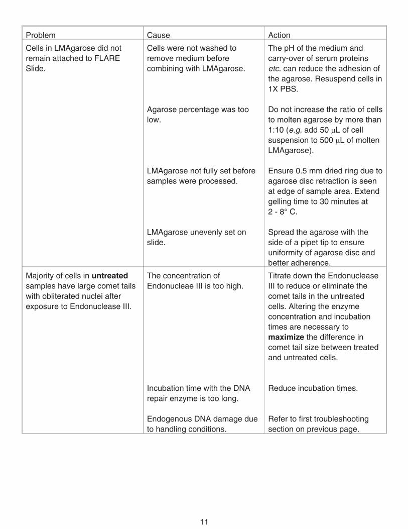
Problem Cause Action
Cells in LMAgarose did notremain attached to FLARESlide.
Cells were not washed toremove medium beforecombining with LMAgarose.
Agarose percentage was toolow.
LMAgarose not fully set beforesamples were processed.
LMAgarose unevenly set onslide.
The pH of the medium andcarry-over of serum proteinsetc. can reduce the adhesion ofthe agarose. Resuspend cells in1X PBS.
Do not increase the ratio of cellsto molten agarose by more than1:10 (e.g. add 50 �L of cellsuspension to 500 �L of moltenLMAgarose).
Ensure 0.5 mm dried ring due toagarose disc retraction is seenat edge of sample area. Extendgelling time to 30 minutes at2 - 8° C.
Spread the agarose with theside of a pipet tip to ensureuniformity of agarose disc andbetter adherence.
Majority of cells in untreatedsamples have large comet tailswith obliterated nuclei afterexposure to Endonuclease III.
The concentration ofEndonucleae III is too high.
Incubation time with the DNArepair enzyme is too long.
Endogenous DNA damage dueto handling conditions.
Titrate down the EndonucleaseIII to reduce or eliminate thecomet tails in the untreatedcells. Altering the enzymeconcentration and incubationtimes are necessary tomaximize the difference incomet tail size between treatedand untreated cells.
Reduce incubation times.
Refer to first troubleshootingsection on previous page.
11

WARNINGS� FOR RESEARCH USE ONLY. NOT FOR USE IN DIAGNOSTIC PROCEDURES.� The physical, chemical, and toxicological properties of the products contained within the
FLARE Assay Kit may not have been fully investigated. Therefore, the use of gloves, labcoats, and eye protection while using any of these chemical reagents is recommended.R&D Systems assumes no liability for damage resulting from handling or contact withthese products.
� Lysis Solution contains 1% sodium lauryl sarcosinate which is an irritant. In case of eye orskin contact, wash thoroughly under running water. In case of ingestion, rinse mouth withwater and seek medical advice.
� SYBR Green I contains DMSO. Please refer to Material Safety Data sheets (MSDS).
APPENDICES
A. Reagents and Buffer Composition
25X FLARE Buffer 1250 mM HEPES-KOH, pH 7.42.5 M KCI250 mM EDTA
REC Dilution Buffer10 mM HEPES-KOH, pH 7.4100 mM KCI0.1 mg/mL BSA50% glycerol
100X BSA additiveProprietary stabilizer reagent
Lysis Solution2.5 M sodium chloride100 mM EDTA, pH 1010 mM Tris base1% sodium lauryl sarcosinate1% Triton X-100
Comet LMAgarose1% low melting point agarose1X PBS
12

B. Suggestions for Assay Optimization
For consistent results, the Endonuclease III FLARE Kit requires optimization orconsideration of the following parameters:
A) Degree of drug/agent exposure
The exposure of your particular cell line to the drug/agent under study must be such thatthe tail moment of the comets in the cells in the absence of Endonuclease III besignificantly less than that in the cells exposed to Endonuclease III. Too high an exposurewill create comets that mask any incremental increase in comet size induced by the actionof the enzyme. Conversely, too little exposure may require very high levels ofEndonuclease III for an observable effect on tail moment.
B) Temperature during drug/agent exposure and subsequent FLARE module stepsDNA repair is inhibited by low temperatures. Lower drug/agent exposures andEndonuclease III enzyme are required if the cells are maintained at 2 - 8° C. If your studiesinvolve measurement of DNA repair at physiological temperatures, it is likely that higherdrug/agent doses and units of enzyme will be needed for significant changes in tailmoment. It is important to regulate cell temperature, at least through the cell lysis stage, forconsistent results between experiments.
C) Units of Endonuclease III
We recommend that you titrate the Endonuclease III, using 1:100, 1:500, and 1:1000 finaldilutions of enzyme per sample area, to optimize the differences in tail moment betweenuntreated and Endonuclease III-treated cells. If necessary, up to 10 �L of undilutedenzyme can be applied in 100 �L of Endonuclease III FLARE Reaction Buffer.
D) Incubation times with Endonuclease IIIOptimum temperature for Endonuclease III is 37° C. Vary the incubation time at 37° C upto 1 hour with Endonuclease III to optimize the differences in tail moment betweenuntreated and Endonuclease III-treated cells.
E) Health of your cells
Your cells must be at least 95% viable as measured by Trypan Blue exclusion. Adherentcells should be gently trypsinized prior to analysis. Note that extensive trypsinization mayinduce non-specific DNA damage and repair and, therefore, high background.
13

REFERENCES1. Nia, A.B. et al. (2001), Carcinogenesis 22:395.
2. Lemay, M. et al. (1999), BioTechniques 27(4):846.
3. Angelis, K.J. et al. (1999), Electrophoresis 20:2133.
4. Morris, E.J. et al. (1999), BioTechniques 26:282.
5. Malyapa, R.S. et al. (1998), Radiation Res. 149:396.
6. Henderson, L. et al. (1998), Mutagenesis 13:89.
7. Visvardis, E.E. et al. (1997), Mutation Res. 383:71.
8. Fairbairn, D.W. et al. (1995), Mutation Res. 339:37.
9. Collins, A.R. et al. (1995), Mutation Res. 336:69.
10. Singh, N.P. et al. (1988), Exp. Cell Res. 175:184.
11. Östling, O. and K.J. Johanson, (1984), Biochem. Biophys. Res. Commun. 123:291.
SYBR Green I Nucleic acid gel stain is a registered trademark of Molecular Probes, Inc. and is sold under license from
Molecular Probes, Inc. under US patent numbers 5436134 and 5658751. For use in a comet assay for internal research
and development only, where research and development use expressly excludes the use of this product for providing
medical, diagnostic or any other testing analysis or screening services or providing clinical information or clinical analysis, in
return for compensation on a per-test basis, and research and development use expressly excludes incorporation of this
product into another product for commercialization even if such other product would be commercialized for research and/or
development use.
CometAssay, FLARE and REC are trademarks of Trevigen, Inc.
14

NOTES
12.02 750101.1 4/04
15