dna sequencing by the sanger method -...
TRANSCRIPT

Sequencing

What is Sequencing A lab technique used to find out the
sequence of nucleotide bases in a DNA
molecule or fragment.
It is a deciphering of the exact order of
base sequence in a nucleotide
Sequence.
Examples are dideoxy sequencing and
maxam-gilbert sequencing.

Method of the Sequencing There are two methods of sequencing
Maxam and Gilbert method (the manual or
chemical sequencing )
And
Sanger method using dideoxynucleotide
(modern sequencing)

Sanger method is more efficient and uses fewer toxic chemicals and lower amounts of radioactivity than the method of Maxam and Gilbert, it rapidly became the method of choice.

DNA SEQUENCING
Principles & methods

Topic Outline
Principles of DNA Sequencing
Methods of DNA Sequencing

Principles of DNA Sequencing
Cycle sequencing (one cycle)
Denaturation
Annealing Extension/ termination

Partial copies of DNA fragments made with DNA polymerase
Collection of DNA fragments that terminate with A,C,G or T using ddNTP or chemical.
Separate by gel electrophoresis
Read DNA sequence

Components For Reaction DNA fragment to be sequenced
DNA Template
Oligonucleotide Primer
Enzyme: DNA Polymerase
Deoxynucleotide triphosphate: dNTPs [dATP,
dTTP, dCTP, dGTP]
Dideoxy nucleotides triphosphate (ddNTP):
[ddA, ddC, ddG and ddT]

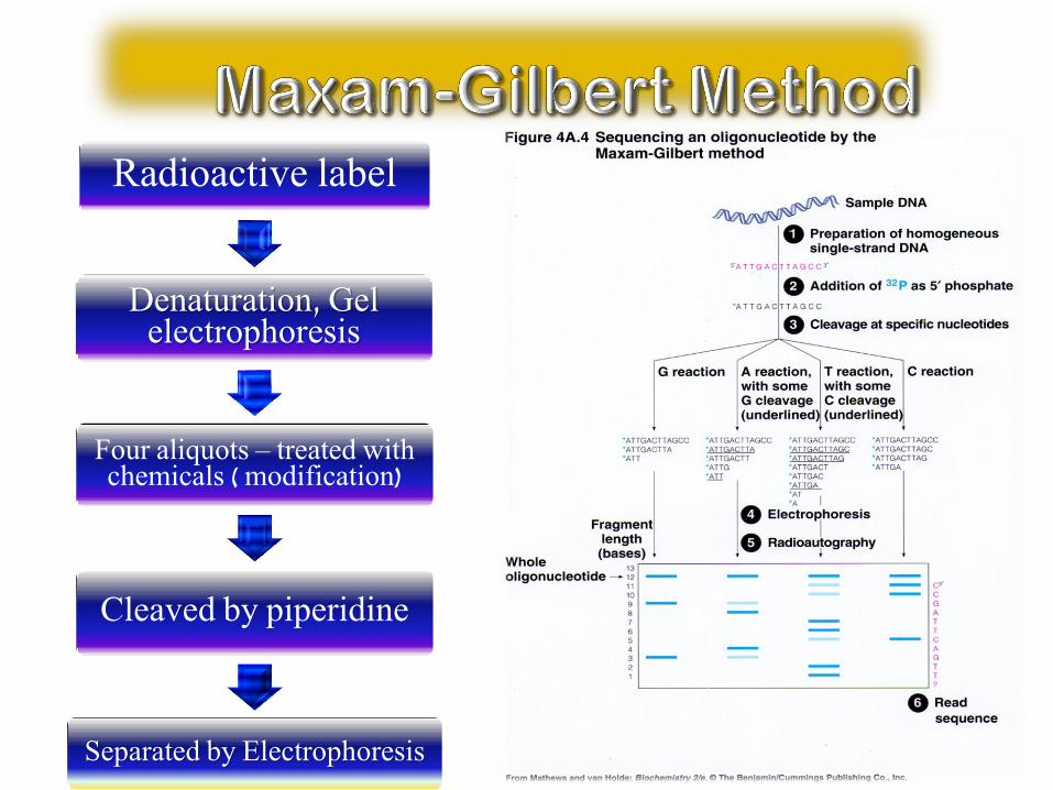
Radioactive label
Denaturation, Gel electrophoresis
Four aliquots – treated with chemicals ( modification)
Cleaved by piperidine
Separated by Electrophoresis

Add in DNA polymerase I,
dNTPs, ddNTPs
Extension /synthesis of complementary
strands
Synthesis termination
Dead-end product
Run gel divide by size

DNA sequencing by the Sanger method
The standard DNA sequencing technique is the Sanger method,
named for its developer, Frederick Sanger, who shared the 1980
Nobel Prize in Chemistry. This method begins with the use of
special enzymes to synthesize fragments of DNA that terminate
when a selected base appears in the stretch of DNA being
sequenced. These fragments are then sorted according to size
by placing them in a slab of polymeric gel and applying an
electric field -- a technique called electrophoresis. Because of
DNA's negative charge, the fragments move across the gel toward
the positive electrode. The shorter the fragment, the faster it
moves. Typically, each of the terminating bases within the
collection of fragments is tagged with a radioactive probe for
identification.

DNA sequencing example
Problem Statement: Consider the following DNA
sequence (from firefly luciferase). Draw the sequencing
gel pattern that forms as a result of sequencing the
following template DNA with ddNTP as the capper.
atgaccatgattacg...
Solution:
Given DNA template: 5'-atgaccatgattacg...-3'
DNA synthesized: 3'-tactggtactaatgc...-5'

DNA sequencing example
Given DNA template: 5'-atgaccatgattacg...-3'
DNA synthesized: 3'-tactggtactaatgc...-5'
Gel pattern: +-------------------------+
lane ddATP | W | | || |
lane ddTTP | W | | | | | |
lane ddCTP | W | | | |
lane ddGTP | W || | |
+-------------------------+
Electric Field +
Decreasing size
where "W" indicates the well position, and "|"
denotes the DNA bands on the sequencing gel.


Running the reaction of all the dideoxy nucleotides
using different dyes generates this type of diagram in same lane.

• Sometimes the spacing between the bands is hard to measure.
• Thus use machine to run and read the electrophoresis.
• Capillary electrophoresis: the fragments are piped through a tiny glass-fiber capillary during the electrophoresis step, and they come out the far
end in size-order.

Chemical cleave method
Sequence small fragments of DNA
The radioactive labelling is done on the dsDNA.
Division of aliquots is done by methylation or removal of base.
Requires DNA
Breaks DNA at different nucleotides
Enzymatic cleave method
Sequencing small fragments are problematic.
The radioactive labelling is done on the ssDNA.
Allow high throughput automated sequencing techniques.
Allow Real Time detection.
Requires DNA synthesis
Termination of chain elongation


• Enzymatic plus-minus method by Sanger - 1975 - using DNA polymerase
• chemical degradation method by Maxam and Gilbert - 1977
• Sanger DNA sequencing method using polyacrylamid gels
• semiautomated methods for running and analysing sequencing gels
• automated methods like: PCR cycle sequencing or fluorescent DNA sequencing

• DNA phage X174, 5386 bp (plus-minus method)
• SV40 DNA 5243 bp (Maxam-Gilbert method)
• recombinant plasmid pBR322, 4362 bp (Maxam-Gilbert method)
• cloning experiments
• PCR products can be sequenced directly
• chromosome mapping
• genome sequencing projects
• generating protein information

Comparison of the DNA Sequencing Methods
Maxam-Gilbert Method Sanger Method
Manual sequencing Automatic sequencing
(Chain-termination
method)
More effort needed in
running gels
Uses automated technology
Time consuming Easier, faster (1 lane per
sample instead of 4)
Radioactivity is used to label
the fragments
Uses special fluorescent dyes
to label the DNA
fragments
1
2
3
4

A sequencing gel
This picture is a radiograph. The dark color of the lines is
proportional to the radioactivity from 32P labeled adenonsine
in the transcribed DNA sample.

The codon table
5’-Base Middle Base 3’-Base
U(=T) C A G
U(=T) Phe Ser Tyr Cys U(=T)
Phe Ser Tyr Cys C
Leu Ser Term Term A
Leu Ser Term Trp G
C Leu Pro His Arg U(=T)
Leu Pro His Arg C
Leu Pro Gln Arg A
Leu Pro Gln Arg G
A Ile Thr Asn Ser U(=T)
Ile Thr Asn Ser C
Ile Thr Lys Arg A
Met Thr Lys Arg G
G Val Ala Asp Gly U(=T)
Val Ala Asp Gly C
Val Ala Glu Gly A
Val Ala Glu Gly G

Translating the DNA sequence
This entry AAG in the table is Lysine (Lys).
Therefore the second amino acid is Lysine.
The first few residues, and their DNA sequence, are as follows
(color coded to indicate the correct location in the
codon table):
Met Lys Leu Gly Arg … ...
AUG AAG CUG GGC CGG GCC GUG C..
This procedure is exactly what cells do when they synthesize
proteins based on the mRNA sequence. The process of translation
in cells occurs in a large complex called the ribosome.

Automated procedure for DNA sequencing
A computer read-out of the gel generates a “false color” image
where each color corresponds to a base. Then the intensities are
translated into peaks that represent the sequence.

High-throughput seqeuncing: Capillary electrophoresis
The human genome project
has spurred an effort to
develop faster, higher
throughput, and less
expensive technologies
for DNA sequencing.
Capillary electrophoresis
(CE) separation has many
advantages over slab gel
separations. CE separations are faster and are capable of producing
greater resolution. CE instruments can use tens and even
hundreds of capillaries simultaneously. The figure show a simple
CE setup where the fluorescently-labeled DNA is detected as it
exits the capillary.
Laser
PMT
Focusing
lens
Sheath flow cuvette
Sheath flow
Collection Lensc Collection Lensc
Beam block
filter

DNA sequencing. • Dideoxy analogs of normal nucleotide triphosphates (ddNTP)
cause premature termination of a growing chain of nucleotides.
ACAGTCGATTG
ACAddG
ACAGTCddG
ACAGTCGATTddG
• Fragments are separated according to their sizes in gel electrophoresis. The lengths show the positions of “G” in the original DNA sequence.

Nucleotides and phosphodiester bond.
Phosphodiester bond

Sequencing cDNA libraries.
• mRNA is pooled from the tissues which express genes.
• cDNA libraries are prepared by copying of mRNA with reverse transcriptase.
• Expressed Sequence Tags (EST) – partial sequences of expressed genes.
• Comparing translated ESTs to annotated proteins – annotation of genes.