does excessive memory load attenuate activation in the
TRANSCRIPT
Does excessive memory load attenuate activation in the prefrontalcortex? Load-dependent processing in single and dual tasks:
functional magnetic resonance imaging study
Susanne M. Jaeggi,a,* Ria Seewer,a Arto C. Nirkko,b Doris Eckstein,a Gerhard Schroth,c
Rudolf Groner,a and Klemens Gutbrodb
a Department of Psychology, University of Bern, CH-3000 Bern 9, Switzerlandb Department of Neurology, University Hospital of Bern, CH-3010 Bern, Switzerland
c Department of Neuroradiology, University Hospital of Bern, CH-3010 Bern, Switzerland
Received 22 March 2002; revised 3 November 2002; accepted 11 December 2002
Abstract
Functional magnetic resonance imaging was used to investigate the relationship between cortical activation and memory load in dualtasks. An n-back task at four levels of difficulty was used with auditory–verbal and visual–nonverbal material, performed separately as singletasks and simultaneously as dual tasks. With reference to single tasks, activation in the prefrontal cortex (PFC) commonly increases withincremental memory load, whereas for dual tasks it has been hypothesized previously that activity in the PFC decreases in the face ofexcessive processing demands, i.e., if the capacity of the working memory’s central executive system is exceeded. However, our resultsshow that during both single and dual tasks, prefrontal activation increases continuously as a function of memory load. An increase ofprefrontal activation was observed in the dual tasks even though processing demands were excessive in the case of the most difficultcondition, as indicated by behavioral accuracy measures. The hypothesis concerning the decrease in prefrontal activation could not besupported and was discussed in terms of motivation factors. Similar changes in load-dependent activation were observed in two other regionsoutside the PFC, namely in the precentral gyrus and the superior parietal lobule. The results suggest that excessive processing demands indual tasks are not necessarily accompanied by a diminution in cortical activity.© 2003 Elsevier Science (USA). All rights reserved.
Keywords: Functional magnetic resonance imaging (fMRI); Working memory; Dual-task processing; Memory load; n-back task; Prefrontal cortex
Introduction
Working memory (WM) refers to an on-line informationprocessing system and implies temporary storage and trans-fer of information in the service of higher order cognitivefunctions such as language comprehension, planning, andproblem solving. According to Baddeley (1986), WM con-sists of several components and supports active mainte-nance of information as well as executive control processes.A central executive system (CES) is considered responsible
for the control and the transfer of information from and tothe verbal and spatial “slave systems” (phonological loopand visuospatial sketchpad) and is seen as being involved inthe allocation and coordination of attentional resources. It isassumed that the capacity of the CES is limited.
Various functional imaging studies with positron emis-sion tomography and functional magnetic resonance imag-ing (fMRI) provided strong evidence of prefrontal cortex(PFC) involvement in a wide variety of tasks related toworking memory (for a review, see D’Esposito et al., 1998,or Fletcher and Henson, 2001). It has been suggested thatthere is a functional specialization within the PFC in rela-tion to WM regarding the type of processes operating on thememoranda. While the ventrolateral PFC (VLPFC; BA 44/
* Corresponding author. Department of Psychology, Muesmattstrasse45, University of Bern, CH-3000 Bern 9, Switzerland. Fax: �41-31-631-8212.
E-mail address: [email protected] (S.M. Jaeggi).
NeuroImage 19 (2003) 210–225 www.elsevier.com/locate/ynimg
1053-8119/03/$ – see front matter © 2003 Elsevier Science (USA). All rights reserved.doi:10.1016/S1053-8119(03)00098-3
45/47) is concerned with active maintenance and updatingof information, i.e., with tasks conducted by the phonolog-ical loop and the visuospatial sketchpad, the dorsolateralPFC (DLPFC; BA 9/46) is seen as mediating active manip-ulation and monitoring, i.e., executive control (Petrides,1994; Owen et al., 1998; Fletcher and Henson, 2001). How-ever, activation of additional brain regions has commonlybeen observed during the performance of WM tasks (e.g.,posterior parietal cortex, as well as premotor and supple-mentary motor regions), suggesting that WM functions aresubserved by multiple brain regions.
The present study focused on the role of WM, especiallyconcerning executive control processes and their neuralrepresentations.
The dual-task paradigm has been widely used as aneffective experimental tool to investigate such executivecontrol processes as well as their limits, since the concurrentperformance of two tasks requires distribution of attentionalresources to different simultaneous processes, which is oneof the main functions attributed to the CES.
Lesion studies have shown that patients with prefrontaldamage are generally impaired in performing dual taskswhile performance in single tasks is relatively preserved(Baddeley et al., 1991, 1997; McDowell et al., 1997).
On the basis of functional imaging data, D’Esposito et al.(1995) suggested that the PFC is critical for the CES. Theycompared single-task performance of two non-WM taskswith a concurrent performance of both tasks, which wasexpected to engage WM and, especially, the CES. Activa-tion of the DLPFC occurred only during the dual-task con-dition and not when either task was performed alone. Thesefindings have been attributed to the supplementary engage-ment of WM in this condition and were taken as evidencefor a neural basis of the CES in the PFC. This interpretationis consistent with the idea that the simultaneous perfor-mance of two tasks compared to single-task performancerequires additional mental resources, which could beadopted by novel areas specialized for dual-task-specificprocesses not required in either single task, such as taskcoordination or shifting attention.
However, several studies failed to elicit such a “surplus”activation in the DLPFC during dual tasks compared tosingle tasks (Klingberg, 1998; Goldberg et al., 1998, Ad-cock et al., 2000; Bunge et al., 2000). In all of these studies,the single tasks per se engaged WM and the DLPFC wasalready activated under these single-task conditions. Thereis evidence that if the DLPFC is already activated in thesingle tasks, activation does not increase further during theperformance of a secondary task. For example, Klingberg(1998) did not find additional activation in the PFC duringdual tasks, relative to the activation during the single tasks.Thus, he could not observe a separate cortical area thatcould be associated with a specific cognitive process, takingplace only during dual tasks. More recent studies have alsoshown no evidence for an additionally activated site for apossible CES expressed in the recruitment of novel prefron-
tal (or any other) regions during dual-task performancerelative to the performance in the single tasks. Rather,dual-task processing was associated with a stronger andmore extensive magnitude of activation in regions activatedby either single task (Adcock et al., 2000; Bunge et al.,2000). Therefore, additional resources required for the per-formance of dual tasks seem to be reflected as enhancedactivation in the same brain regions that subserve perfor-mance in either single task. Finally, Goldberg et al. (1998)reported even a decrease of PFC activation during theirdual-task condition compared to the activation pattern undertheir single-task conditions. In consideration of these re-sults, D’Esposito (2001) concluded that “under dual taskconditions activation in the PFC would increase as a resultof greater demands on processing, up to some level ofasymptote, before attenuating,” i.e., if the capacity of theCES is exceeded, cortical activity in the PFC may decrease.
Additional theoretical considerations concerning theseresults were made by Goldberg et al. (1998). They consid-ered capacity limits of the CES as well as the concept ofcognitive workload, a concept characterized as the differ-ence between the expected and the actual performance of atask due to an increase in task difficulty (Gopher andDonchin, 1986). An increase in cognitive workload may beassociated with a decrement of performance, and WMshould be especially susceptible to manipulations of work-load, since WM is thought to be of limited capacity. Thus,Goldberg and colleagues suppose that the impact of exces-sive workload on WM could lead to an attenuation ofprefrontal activation, which could indicate a breakdown inneural networks. Because of the substantial increase ofcognitive workload under dual-task conditions, the capacitylimits of the CES are quite quickly reached, leading to thereported decrements of accuracy together with decreases infMRI signal intensity in some cortical sites that are assumedto be related to the CES (mainly in the DLPFC).
Another slightly different explanation goes back to arecent fMRI study reported by Just et al. (2001) in whichactivations in dual and single tasks were compared in dif-ferent cortical areas. They hypothesized that if two taskscompete for a common resource pool, i.e., for the samebrain regions, there should be less activation in those re-gions during the concurrent performance of the two tasksthan the sum of activation under the two single-task condi-tions. However, concerning the PFC, Just et al. (2001)reported similar activation for the dual-task condition com-pared with the summed activation of both single tasks. Buteven though both single tasks activated the PFC, activationin that area was minimal, leaving the question open ofwhether dual-task activation would decrease further if thePFC was more substantially activated.
Thus, it seems that activation in the PFC during theconcurrent performance of two tasks can exceed the activa-tion during the single-task conditions, as long as the singletasks do not compete for the same resources, or as long as
211S.M. Jaeggi et al. / NeuroImage 19 (2003) 210–225
memory load in the single tasks only induces minimal PFCactivation.
A crucial question is now whether the enhanced prefron-tal activation during dual tasks in the studies cited abovewas actually due to an increase of WM demands or ratherreflected an unspecific increase in “mental effort” orarousal, due to greater task difficulty. This question of taskdifficulty and its confounding with WM load was directlyaddressed in a study by Barch et al. (1997). They system-atically varied cognitive workload and task difficulty byincreasing task difficulty independent of WM demands byusing degraded stimuli to enhance task difficulty and delayvariation to enhance WM load. In fact, they found a doubledissociation between regions recruited in WM tasks(DLPFC, Broca’s area, and parietal cortex) and those re-cruited in task difficulty (primarily anterior cingulate).Thus, the DLPFC seems indeed to be involved in WMprocessing. Other imaging studies supply further evidencethat the prefrontal region is quantitatively more activated ifthe WM load is increased (Braver et al., 1997; Cohen et al.,1997; Jonides et al., 1997; Owen et al., 1996; Petrides et al.,1993a, 1993b; Schumacher et al., 1996; Smith and Jonides,1997). However, the processing demands in these studies,expressed in accuracy, never fell below 75%, which couldindicate that the subjects never reached the limit of theirprocessing capacity. Therefore, it is not clear whether acti-vation would still increase in such a case.
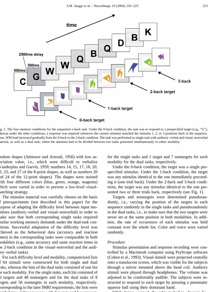
The present study investigated cortical activation withfunctional MRI during the systematic variation of memoryload both in single and in dual tasks. To this end we used avariation of the n-back paradigm adapted from Gevins andCutillo (1993) at four levels of difficulty with auditory–verbal and visual–nonverbal material (see Fig. 1). This taskgenerates a specific response pattern in behavioral perfor-mance as well as in the fMRI signal and has reliably pro-duced activation in WM-related cortical regions (Braver etal., 1997; Cohen et al., 1997; Jonides et al., 1997). Then-back paradigm requires a continuous monitoring of se-quentially presented stimuli, and subjects must answer pos-itively whenever the current stimulus matches the stimulusoccurring n positions back in the sequence. The task com-bines maintenance as well as active manipulation, i.e., ex-ecutive processes, because of the necessity to continuouslyencode, update, and discard the information held in WMwith the presentation of each new stimulus. The value of nis regarded as proportional to memory load (Braver et al.,1997).
On the basis of earlier findings several hypotheses weretested.
First, for the single tasks it was predicted that corticalactivity increases in proportion to the WM demands, i.e., weexpect a linear relationship between activity and WM load,especially in the PFC (Braver et al., 1997; Cohen et al.,1997; Jonides et al., 1997; Owen et al., 1996; Petrides et al.,1993a, 1993b; Schumacher et al., 1996; Smith and Jonides,1997). The pattern of activation should not differ quantita-
tively and qualitatively in most areas (i.e., no increasingactivation and no recruitment of additional areas with in-creasing WM demands) with the exception of the DLPFC(BA 46/9) and Broca’s area (BA 44/45) where, by compar-ing the 1-back with the 2-back condition, we expect anincrease in cortical activation due to novel task demands(updating and rehearsal).
Second, for the dual tasks the hypothesis formulated byD’Esposito (2001; see above) was tested: initially, activa-tion in the PFC should gradually increase due to the aug-mentation of processing demands on the WM; but, whenprocessing demands reach a level where the subject is un-able to perform the task with an accuracy above chancelevel, we predict an attenuation of prefrontal activation.
Third, comparing the activation pattern in the summedsingle tasks with the corresponding dual tasks, it is predictedthat the greater the sum of the activation with increasingmemory load during the single tasks in a given area (here,in the DLPFC), the smaller the portion of activation result-ing from the dual tasks in comparison with the sum ofactivation in the single tasks (Just et al., 2001). Thus, thereshould be an interaction between the sum of activations inthe single tasks and those of the corresponding dual taskswith increasing memory load, i.e., in the 1-back condition,the sum of the activations of the two single tasks should besmaller than the activation in the corresponding dual task,whereas in the 3-back condition, the summed activations ofthe two single tasks should exceed the activation of the dualtask.
Materials and methods
Subjects
Six subjects (three female, three male; age range 24 to 27years; mean 25.3) participated in this study. All were as-sessed as right-handed (Oldfield, 1971) with no seeing,hearing, or neurological disorders and normal structuredMRI scans. All participants gave written informed consentaccording to a protocol approved by the local ethics com-mittee. All subjects were given identical practice with thetask and were scanned only after reaching a criterion levelof performance (�75% accuracy in the 2-back single task).
Task design
MaterialsThe n-back paradigm was used at four levels of difficulty
with auditory–verbal and visual–nonverbal material, con-structed as single tasks as well as dual tasks.
The auditory–verbal material consisted of 10 consonants(b, c, d, g, h, k, p, q, t, and w), which were spoken by twomales and two females. The voice was varied in order toprevent a low-level auditory-matching strategy. The visual–nonverbal stimuli were drawn from a set of 10 abstract
212 S.M. Jaeggi et al. / NeuroImage 19 (2003) 210–225
random shapes (Attneave and Arnoult, 1956) with low as-sociation value, i.e., which were difficult to verbalize(Vanderplas and Garvin, 1959; numbers 14, 15, 17, 18, 20,22, 23, and 27 of the 8-point shapes, as well as numbers 20and 24 of the 12-point shapes). The shapes were stainedwith four different colors (blue, green, orange, magenta)which were varied in order to prevent a low-level visual-matching strategy.
The stimulus material was carefully chosen on the basisof preexperiments (not described in this paper) for thepurpose of adapting the difficulty level between input mo-dalities (auditory–verbal and visual–nonverbal) in order tomake sure that both corresponding single tasks requiredsimilar working memory demands under the dual-task con-ditions. Successful adaptation of the difficulty level wasachieved as the behavioral data (accuracy and reactiontimes) in the corresponding tasks were comparable in bothmodalities (e.g., same accuracy and same reaction times inthe 2-back condition in the visual–nonverbal and the audi-tory–verbal tasks).
For each difficulty level and modality, computerized listsof 64 stimuli were constructed for both single and dualtasks, whereas the lists of the dual tasks consisted of one listfor each modality. For the single tasks, each list consisted of16 targets and 48 nontargets and for the dual tasks of 8targets and 56 nontargets in each modality, respectively.Corresponding to the later fMRI requirements, the lists weredivided into eight sequences with 2 targets and 6 nontargets
for the single tasks and 1 target and 7 nontargets for eachmodality for the dual tasks, respectively.
Under the 0-back condition, the target was a single pre-specified stimulus. Under the 1-back condition, the targetwas any stimulus identical to the one immediately preceed-ing it (one trial back). Under the 2-back and 3-back condi-tions, the target was any stimulus identical to the one pre-sented two or three trials back, respectively (see Fig. 1).
Targets and nontargets were determined pseudoran-domly, i.e., varying the position of the targets in eachsequence randomly in the single tasks and pseudorandomlyin the dual tasks, i.e., to make sure that the two targets werenever set at the same position in both modalities. In addi-tion, the rate of occurrence of each stimulus was heldconstant over the whole list. Color and voice were variedrandomly.
ProcedureStimulus presentation and response recording were con-
trolled by a Macintosh computer using PsyScope software(Cohen et al., 1993). Visual stimuli were projected centrallyonto a translucent screen, which was visible for the subjectsthrough a mirror mounted above the head coil. Auditorystimuli were played through headphones. The volume wasadjusted to be comfortably audible. The subjects were in-structed to respond to each target by pressing a pneumaticsqueeze ball using their dominant hand.
While being scanned, the subjects had to alternate be-
Fig. 1. The four memory conditions for the sequential n-back task. Under the 0-back condition, the task was to respond to a prespecified target (e.g., “G”),whereas under the other conditions, a response was required whenever the current stimulus matched the stimulus 1, 2, or 3 positions back in the sequence.Thus, WM load increased gradually from the 0-back to the 3-back condition. The task was performed as single task with auditory–verbal and visual–nonverbalmaterial, as well as a dual task, where the attention had to be divided between two tasks presented simultaneously in either modality.
213S.M. Jaeggi et al. / NeuroImage 19 (2003) 210–225
tween two different task conditions (two different n-backtasks) every 24 s, eight times, in a total of 6.4 min. Accord-ing to the required task contrasts all subjects completedseven different task alternations. All task alternations weretrained beforehand in a practice session. Alternation wasautomatically triggered by the computer. In order to analyzeonly data with steady-state task performance (and steady-state MRI signal) all task alternation started with an addi-tional constructed warm-up task (identical to one of the taskalternations). Data from this warm-up task were discardedfrom further data analysis.
The order of the task alternations was varied randomlyfor each subject. Four task alternations consisted of single-task combinations (auditory 1-back vs 2-back, auditory0-back vs 3-back, visual 0-back vs 2-back, visual 1-back vs2-back) and three task alternations consisted of dual-taskcombinations (0-back vs 1-back, 0-back vs 2-back, 1-backvs 3-back). Stimuli were presented sequentially for 500 mswith a 2500-ms interstimulus interval (ISI). Under the dual-task conditions the auditory and visual stimuli were pre-sented at the same time. Task alternations were indicatedcentrally on the screen, presenting specified instructionsigns (0 for the 0-back task, Roman numbers for the othertasks (I, II, III), respectively) during the ISI before eachalternation (2000-ms duration, 250-ms ISI before and afterthe instruction sign).
Subject performance during scanning was monitored interms of reaction time and accuracy (number of target lettersidentified correctly). In addition, subjects answered a ques-tionnaire in which they rated the subjectively experienceddemands of each task on a five-point scale from small toexcessive demands.
Data acquisition
Imaging was performed as previously described (Nirkkoet al., 2001). In short, we used a 1.5-T whole-body MRIscanner (Magnetom Vision, Siemens Medical Systems, Er-langen, Germany) with its standard whole-body gradientsystem and circularly polarized head coil. To restrain headmotion in all directions, the subjects were fixed with boththe standard lateral pads and a smooth U-shaped plasticdental plate functioning as an improved bite bar. Aftershimming, whole brain fMRI was performed with a bloodoxygenation level dependent (BOLD) echo-planar imaging(EPI) sequence (matrix 128 � 128 � 30 slices � 68measurements, resulting in 1.56 � 1.56 � 4 mm � TR 6 s;TE 82 ms). Phase-encoding direction was anteroposterior,thus preserving brain symmetry. Slice angulation was par-allel to a line connecting the base of the genu corpori callosiand the confluens sinuum, resulting in an angle of about20–30° with respect to the bicommissural (AC–PC) line(Nirkko et al., 2001). Between fMRI experiments, standard-ized sets of high-resolution structural images were acquiredfor later coregistration.
Data analysis
Behavioral dataBehavioral data (accuracy and reaction times) were an-
alyzed in order to evaluate the subjects’ compliance with thetask along with the effectiveness of the manipulation ofWM load in single and dual tasks and further, to controldifferences in performance across modality (auditory–ver-bal vs visual–nonverbal). The subjects’ performance wasevaluated using two-way repeated-measures ANOVA withcondition (single vs dual task) and load (1-back to 3-back)as within-subjects factors. Differences in performanceacross modality (auditory–verbal vs visual–nonverbal) wereanalyzed using a two-way repeated-measures ANOVA withmodality and load (1-back and 2-back) as within-subjectsfactors.
Post hoc tests for significant main effects and contrastsfor significant interactions were performed throughout andwere corrected for multiple comparisons (Bonferroni cor-rection).
fMRI evaluationWe used a methodology of fMRI evaluation respecting
interindividual cortical variability and allowing direct sta-tistical comparison between experimental tasks and brainregions. To compare the amount of activation in a givenarea across experimental tasks, anatomical defined regionsof interest (ROI) were drawn for each subject using theparcellation scheme described by Rademacher et al. (1992).In order to examine not just the location of activation butalso how the experimental tasks affected the magnitude ofthe activation in each of these regions, it was important touse an a priori, independent method of defining the ROIs(Nirkko et al., 2001; Michael et al., 2001). This method useslimiting sulci as anatomical landmarks to segment corticalregions. The schematic drawing in the center of Fig. 3displays the set of ROIs that were defined.
Specifically designed software was used to delineate theROIs manually on each EPI slice of each subject (Nirkko,2000). Delineation was done by two staff research assistantsafter extensive training in the Rademacher parcellationscheme. To optimize reliability we created a standard ana-tomical atlas using the EPI images of one of our subjects asan explicit guide to the delineation of individual sulci fol-lowing the suggestions of Rademacher et al. (1992), Cavi-ness et al. (1996), Ono et al. (1990), and Damasio (1995).The interrater reliability of this ROI-defining procedurebetween the two trained staff members was evaluated for allROIs in one task for two subjects. The reliability measurewas obtained by calculating correlations between the tworaters based on percentage signal change relative to thecontrol task average. An interrater reliability measure of r �0.93 in one subject and of r � 0.82 in the other subjectreflects a high degree of reliability in the ROI-definingprocedure.
Seven volumes of interest (VOI; each composed of cor-
214 S.M. Jaeggi et al. / NeuroImage 19 (2003) 210–225
responding ROIs in several slices) of the lateral and medialcerebral surface were specified in each hemisphere (seeschematic drawing in the center of Fig. 3).
Laterally, the DLPFC was defined as the middle frontalgyrus (F2; BA 6,8,9, and 46) with the exception of theposterior-most part (frontal eye fields) to exclude activationrelated to eye movements. The inferior frontal VOI includedF3o and F3t (BA 44 and 45). The temporal VOI comprisedthe superior (T1a and T1p) and middle (T2a and T2p)temporal gyri (BA 21 and 37). These gyri were combinedinto one VOI because previous studies of other cognitiveprocesses (e.g., language) have often found activation cen-tered in the superior temporal sulcus between them (Kelleret al., 2001; Michael et al., 2001). The parietal VOI con-sisted of the superior parietal lobule (BA 21 and 37). Theprecentral VOI corresponded to the precentral gyrus (PRG;BA 4). Medially, the supplementary motor area VOI con-sisted of parcellation unit SMC (BA 6) and finally, theanterior cingulate VOI was CGa (BA 24).
No maximum z score values or counts of significantpixels were used for quantitative evaluation since both sta-tistical z values and thresholded pixel counts are highlynonlinear and depend not only on the signal increase, butalso on the number of measurements and the amount ofnoise and thus invalidate averaging of interindividual, in-tertask, and even interregion comparisons: for instance, ahigher z score can result from a region with a smaller signalchange, but less noise. This is evident from the formula tocalculate z scores, but in essence also holds for other sta-tistical values like t tests. Using the formula for the z scorefor independent samples,
z �m1 � m0
�V1
n1�
V0
n0
,
where m1 and m0 are the mean values for activation and rest,V1 and V0 are the variances, and n1 and n0 are the numberof respective samples, and given the nonlinear characteris-tics of the variance calculation itself, it can be seen that thescore is nonlinearily and inversely related to the variances (ameasure of the noise contained in the data) and the numberof samples and that the z score of the average (or sum) oftwo values does not equal the average (or sum) of the twoz scores. Moreover, selecting “activated pixels” above acertain threshold is a nonlinear operation by definition.While this is valid and very useful as an endpoint forvisualization of significant areas, using activated pixels as adeparting point for further analysis is highly problematicbecause of these relationships: in particular, averaging orotherwise evaluating counts of activated pixels for compar-ison between tasks, as in comparing whether the sum of theactivation of two tasks exceeds the activation of anothertask, is mathematically and statistically invalid: as a realisticexample, assuming a homogenously activating region of100 independent pixels, a standard deviation of 2%, 30
measurements for activation and 30 for baseline, and a zscore threshold of 2.0, the number of activated pixels in thatregion will remain 0 between a signal change of 0% and 0.8,then steeply rise from 8 pixels at 0.9% signal change to 96pixels at 1.2% signal change, above which all 100 pixelswill activate. This demonstrates the highly nonlinear behav-ior of activated pixel counts (higher z score thresholds andlarger number of measurements will even increase the non-linearity). Now if, in this example, the relation between twocomponent tasks and a combined “additive task” were to beassessed, and this region activated 0.5% in each componenttask and 1.0% in the additive task, resulting pixel countswill be 0 for the components and 36 for the additive task,resulting in the interpretation that the additive task with 36activated pixels demands much more than the sum of thecomponent tasks with 0 pixels each, i.e., reveals a newregion specific to the combination of the two tasks, but notto the component tasks themselves. Conversely, if eachcomponent would activate 1.2% and the additive task 2.4%,pixel counts would be 96 for the components and 100 forthe additive task, resulting in the interpretation that theadditive task is not additive at all, but demands no more thaneach component task. In both cases, the interpretation iswrong because statistical end results were used in an invalidway as a starting point for further calculations. The correctinterpretation results from using the mean values of thesignal changes themselves, which in both cases shows thatthe additive task activation correspond to the sum of eachcomponent task, whether it is 0.5 � 0.5 � 1.0 or 1.2 � 1.2� 2.4. With these signal change values as starting point,further calculations and statistical tests can be done (despitebeing aware of the possibility of a mild nonlinearity of theBOLD response itself (Friston et al., 2000), in practiceprobably only significant at very high activation levels).With thresholded pixel counts, or signal changes based onactivated pixels only, instead of unselected pixels, the cal-culations are strongly false and mathematically invalid inthe first place. To avoid these problems, we used the per-centage signal change relative to the control task average,which in each VOI from all subjects was subjected to thetypical statistical procedure, i.e., testing the null hypothesisof no change by calculation of z scores. In addition, theresulting percentage signal change (between activation andcontrol task), as determined for each VOI, allowed foraveraging between subjects and further statistical compari-son between tasks and regions. Minimal statistical signifi-cance was set at the conventional P � 0.05 (corrected formultiple comparisons with the Bonferroni correction);higher significance levels are labeled in the correspondingtable legend. From the resulting percentage signal changesin the seven realized task alternations, it was possible tocalculate the percentage signal change of other contrasts.For example, from the task alternations visual 0-back vs2-back and 1-back vs 2-back, the contrast visual 0-back vs1-back could be derived, due to the fact that the two primaltask alternations share one common task (2-back). Since the
215S.M. Jaeggi et al. / NeuroImage 19 (2003) 210–225
“process-specific” theory says that the DLPFC is involvedin processes such as monitoring and higher-level planningand not sensitive for different types of information (Fletcherand Henson, 2001; D’Esposito and Postle, 1999; Petrides,1994, 1995), we assumed that there are no differences inDLPFC activation across modality (auditory–verbal vs vi-sual–nonverbal). In that case, the results of the realizedcontrasts in auditory–verbal and visual–nonverbal singletasks could be summed up in one line of single tasks withincreasing load (1-back to 3-back task as activation taskswith the 0-back task as control). To control the differencesin the percentage signal change across modality (auditory–verbal vs visual–nonverbal), we compared the contrast1-back vs 2-back, which was realized under both the audi-tory–verbal and the visual–nonverbal conditions, using atwo-way repeated-measures ANOVA (SPSS for Windows,Release 10.0) with hemisphere and modality as within-subjects factors for each VOI.
To compare signal changes in single and dual tasks, aswell as to test a possible interaction between these condi-tions, repeated-measures ANOVAs were performed, withcondition (single vs dual tasks), load (1-back to 3-back), andhemisphere (left vs right) as within factors for each VOI.
Additionally, to directly test our hypotheses concerningthe load-dependent changes in the percentage signal changeseparately for single and dual tasks, repeated-measuresANOVAs were performed, with load (1-back to 3-back) andhemisphere (left vs right) as within factors for each VOI.
In analogy to Just et al. (2001), the comparison of theactivation pattern of the dual tasks with the sum of thecorresponding single tasks was analyzed using a three-wayrepeated-measures ANOVA with condition (sum of the sin-gle tasks vs dual tasks), load (1-back to 3-back), and hemi-sphere (left vs right) as within-subjects factors for eachVOI.
Significant main effects and interactions were followed
by post hoc tests and contrasts and were corrected formultiple comparisons (Bonferroni correction).
Results
Behavioral data for single and dual tasks
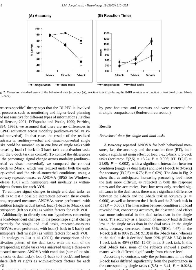
A two-way repeated ANOVA for both behavioral mea-sures, i.e., the accuracy and the reaction time (RT), indi-cated a significant main effect of load, i.e., 1-back to 3-backtasks (accuracy: F(2,5) � 13.24; P � 0.006; RT: F(2,5) �21.09; P � 0.002), with a significant interaction betweencondition (single vs dual tasks) and load (1-back to 3-back)for accuracy (F(2,5) � 6.73; P � 0.029). The data in Fig. 2show that, as anticipated, increasing processing load madethe task more difficult, as reflected in both the responsetimes and the accuracies. Post hoc tests only reached sig-nificance in the dual tasks: there was a significant differencebetween the 1-back and the 3-back task in accuracy (P �0.000), as well as between the 1-back and the 2-back task inRT (P � 0.000). The interaction between condition and loadrevealed that, concerning accuracy, the observed load effectwas more substantial in the dual tasks than in the singletasks. The accuracy as a function of memory load declinedless in the single tasks than in the dual tasks. In the singletasks, accuracy decreased from 89% (SEM: 4.67) in the1-back task to 80% (SEM: 9.13) in the 3-back task, whereasin the dual tasks it declined from 90% (SEM: 5.74) in the1-back task to 45% (SEM: 12.08) in the 3-back task. In thisdual 3-back task, none of the subjects showed a perfor-mance which differed significantly from chance level.
According to contrasts, only the performance in the dual2-back tasks differed significantly from the performance inthe corresponding single tasks (t(5,5) � 3.41; P � 0.028).
As predicted, there was no significant difference in per-
Fig. 2. Means and standard errors of the behavioral data (accuracy (A); reaction time (B)) during the fMRI session as a function of task load (from 1-backto 3-back).
216 S.M. Jaeggi et al. / NeuroImage 19 (2003) 210–225
formance across input modality of the stimuli (auditory–verbal vs visual–nonverbal), either in accuracy (F(1,5) �0.90; P � 0.387) in RT (F(1,5) � 5.54; P � 0.065).
Imaging data
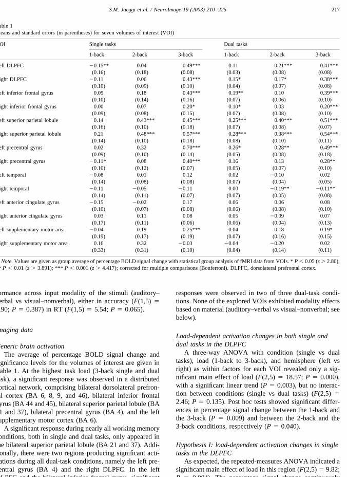
Generic brain activationThe average of percentage BOLD signal change and
significance levels for the volumes of interest are given inTable 1. At the highest task load (3-back single and dualtask), a significant response was observed in a distributedcortical network, comprising bilateral dorsolateral prefron-tal cortex (BA 6, 8, 9, and 46), bilateral inferior frontalgyrus (BA 44 and 45), bilateral superior parietal lobule (BA21 and 37), bilateral precentral gyrus (BA 4), and the leftsupplementary motor cortex (BA 6).
A significant response during nearly all working memoryconditions, both in single and dual tasks, only appeared inthe bilateral superior parietal lobule (BA 21 and 37). Addi-tionally, there were two regions producing significant acti-vations during all dual-task conditions, namely the left pre-central gyrus (BA 4) and the right DLPFC. In the leftDLPFC and the bilateral inferior frontal gyrus, significant
responses were observed in two of three dual-task condi-tions. None of the explored VOIs exhibited modality effectsbased on material (auditory–verbal vs visual–nonverbal; seebelow).
Load-dependent activation changes in both single anddual tasks in the DLPFC
A three-way ANOVA with condition (single vs dualtasks), load (1-back to 3-back), and hemisphere (left vsright) as within factors for each VOI revealed only a sig-nificant main effect of load (F(2,5) � 18.57; P � 0.000),with a significant linear trend (P � 0.003), but no interac-tion between conditions (single vs dual tasks) (F(2,5) �2.46; P � 0.135). Post hoc tests showed significant differ-ences in percentage signal change between the 1-back andthe 3-back (P � 0.009) and between the 2-back and the3-back conditions, respectively (P � 0.040).
Hypothesis I: load-dependent activation changes in singletasks in the DLPFC
As expected, the repeated-measures ANOVA indicated asignificant main effect of load in this region (F(2,5) � 9.82;P � 0.004). The percentage signal change continuously
Table 1Means and standard errors (in parentheses) for seven volumes of interest (VOI)
VOI Single tasks Dual tasks
1-back 2-back 3-back 1-back 2-back 3-back
Left DLPFC �0.15** 0.04 0.49*** 0.11 0.21*** 0.41***(0.16) (0.18) (0.08) (0.03) (0.08) (0.08)
Right DLPFC �0.11 0.06 0.43*** 0.15* 0.17* 0.38***(0.10) (0.09) (0.10) (0.04) (0.07) (0.08)
Left inferior frontal gyrus 0.09 0.18 0.43*** 0.19** 0.10 0.39***(0.10) (0.14) (0.16) (0.07) (0.06) (0.10)
Right inferior frontal gyrus 0.00 0.07 0.20* 0.10* 0.03 0.20***(0.09) (0.08) (0.15) (0.07) (0.08) (0.10)
Left superior parietal lobule 0.14 0.43*** 0.45*** 0.25*** 0.40*** 0.51***(0.16) (0.10) (0.18) (0.07) (0.08) (0.07)
Right superior parietal lobule 0.21 0.48*** 0.57*** 0.28*** 0.38*** 0.54***(0.14) (0.10) (0.18) (0.08) (0.10) (0.11)
Left precentral gyrus 0.02 0.32 0.70*** 0.26* 0.28** 0.49***(0.09) (0.10) (0.14) (0.05) (0.08) (0.18)
Right precentral gyrus �0.11* 0.08 0.40*** 0.16 0.13 0.28**(0.10) (0.12) (0.07) (0.05) (0.07) (0.10)
Left temporal �0.08 0.01 0.12 0.02 �0.10 0.02(0.14) (0.08) (0.08) (0.07) (0.04) (0.05)
Right temporal �0.11 �0.05 �0.11 0.00 �0.19** �0.11**(0.14) (0.11) (0.07) (0.07) (0.05) (0.08)
Left anterior cingulate gyrus �0.15 �0.02 0.17 0.06 0.06 0.08(0.10) (0.07) (0.08) (0.06) (0.08) (0.10)
Right anterior cingulate gyrus 0.03 0.11 0.08 0.05 �0.09 0.07(0.17) (0.11) (0.06) (0.06) (0.04) (0.13)
Left supplementary motor area �0.04 0.19 0.25*** 0.04 0.18 0.19*(0.19) (0.17) (0.19) (0.07) (0.16) (0.15)
Right supplementary motor area 0.16 0.32 �0.03 �0.04 �0.20 0.02(0.33) (0.31) (0.10) (0.04) (0.14) (0.11)
Note. Values are given as group average of percentage BOLD signal change with statistical group analysis of fMRI data from VOIs. * P � 0.05 (z � 2.80);** P � 0.01 (z � 3.891); *** P � 0.001 (z � 4.417); corrected for multiple comparisons (Bonferroni). DLPFC, dorsolateral prefrontal cortex.
217S.M. Jaeggi et al. / NeuroImage 19 (2003) 210–225
increased from the 1-back to the 3-back condition (1-back:0.13% (SEM: 0.12); 2-back: 0.05% (SEM: 0.13); 3-back:0.46% (SEM: 0.08)). The linear trend was significant (P �0.016). Post hoc tests showed only a significant differencein percentage signal change between the 1- and the 3-backtasks (P � 0.048) (Fig. 3).
Hypothesis II: load-dependent activation changes in dualtasks in the DLPFC
Similar to the single tasks, the repeated-measuresANOVA showed a significant main effect of load (F(2,5) �13.17; P � 0.002). The percentage signal change increasedlinearly from the 1-back to the 3-back condition (1-back:0.13% (SEM: 0.02); 2-back: 0.19% (SEM: 0.06); 3-back:0.39% (SEM: 0.07)). The linear trend was significant (P �0.012). Post hoc tests showed significant differences inpercentage signal change between the 1-back and the 3-back(P � 0.036), as well as between the 2-back and the 3-backtasks (P � 0.004).
Hypothesis III: load-dependent activation changes in dualtasks compared with the summed activations of thecorresponding single tasks
Since no VOI showed a significant difference in thepercentage signal change across modality (see below), thesum of the single tasks was computed as doubled activationof the corresponding single task. A three-way repeated-measures ANOVA with condition (sum of the single tasksvs dual tasks), load (1-back to 3-back), and hemisphere aswithin-subjects factors showed that the DLPFC produced asignificant main effect of load (F(2,5) � 14.49; P � 0.001)as well as a significant interaction between condition andload (F(2,5) � 5.55; P � 0.024). The increase of theactivation change as a function of task load was more substan-tial in the single tasks than in the dual tasks (see Fig. 4).
However, the contrasts showed no significant differencesbetween the activation of the sum of single tasks and theactivation of the corresponding dual tasks under the 1-backand the 2-back conditions (Fig. 5). Under the 3-back con-
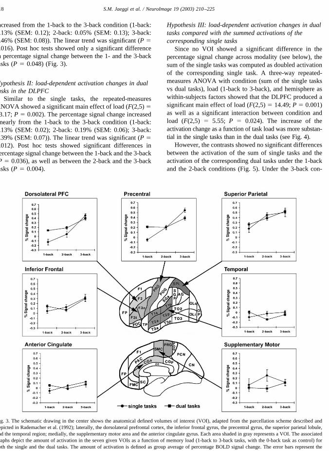
Fig. 3. The schematic drawing in the center shows the anatomical defined volumes of interest (VOI), adapted from the parcellation scheme described anddepicted in Rademacher et al. (1992); laterally, the dorsolateral prefrontal cortex, the inferior frontal gyrus, the precentral gyrus, the superior parietal lobule,and the temporal region; medially, the supplementary motor area and the anterior cingulate gyrus. Each area shaded in gray represents a VOI. The associatedgraphs depict the amount of activation in the seven given VOIs as a function of memory load (1-back to 3-back tasks, with the 0-back task as control) forboth the single and the dual tasks. The amount of activation is defined as group average of percentage BOLD signal change. The error bars represent thestandard error of the mean.
218 S.M. Jaeggi et al. / NeuroImage 19 (2003) 210–225
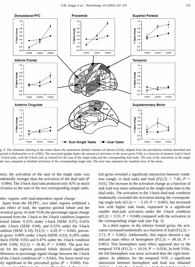
dition, the activation of the sum of the single tasks wastendentially stronger than the activation of the dual task (P� 0.084). The 3-back dual task produced only 42% as muchactivation as the sum of the two corresponding single tasks.
Other regions with load-dependent signal changeApart from the DLPFC, two other regions exhibited a
main effect of load, the superior parietal lobule and theprecentral gyrus. In both VOIs the percentage signal changeincreased from the 1-back to the 3-back condition [superiorparietal lobule: 0.22% under 1-back (SEM: 0.07), 0.42%under 2-back (SEM: 0.04), and 0.52% under the 3-backcondition (SEM: 0.10); F(2,5) � 4.29; P � 0.045; precen-tral gyrus: 0.08% under 1-back (SEM: 0.03), 0.20% under2-back (SEM: 0.05) and 0.47% under the 3-back condition(SEM: 0.09); F(2,5) � 18.36; P � 0.000]. The post hoctests for the superior parietal lobule showed significantdifferences in percentage signal change between the 1-backand the 2-back conditions (P � 0.042). The linear trend wasonly significant in the precentral gyrus (P � 0.006). Fur-thermore, in addition to the main effect of load, the precen-
tral gyrus revealed a significant interaction between condi-tion (single vs dual task) and load (F(2,5) � 7.45; P �0.01). The increase in the activation change as a function oftask load was more substantial in the single tasks than in thedual tasks. The activation in the 1-back dual-task conditiontendentially exceeded the activation during the correspond-ing single task (t(5,5) � �3.10; P � 0.085), but increasedless with higher task loads, expressed in a significantsmaller dual-task activation under the 3-back condition(t(5,5) � 3.55; P � 0.048) compared with the activation inthe corresponding single task (see Fig. 3).
In a third region, in the inferior frontal gyrus, the acti-vation increased tendentially as a function of load (F(2,5) �3.71; P � 0.062). Additionally, this region showed a sig-nificant main effect of hemisphere (F(1,5) � 49.16; P �0.001). This hemisphere main effect appeared also in thetemporal VOI (F(1,5) � 10.16; P � 0.024). In both VOIs,the left hemisphere was more activated than the right hemi-sphere. In addition, for the temporal VOI, a significantinteraction between hemisphere and load was obtained(F(2,5) � 4.32; P � 0.044), in which the left hemisphere
Fig. 4. The schematic drawing in the center shows the anatomical defined volumes of interest (VOI), adapted from the parcellation scheme described anddepicted in Rademacher et al. (1992). The associated graphs depict the amount of activation in the seven given VOIs as a function of memory load (1-backto 3-back tasks, with the 0-back task as control) for the sum of the single tasks and the corresponding dual tasks. The sum of the activations in the singletasks was computed as doubled activation of the corresponding single task. The error bars represent the standard error of the mean.
219S.M. Jaeggi et al. / NeuroImage 19 (2003) 210–225
exhibited an increasing percentage signal change with aug-mented load, whereas the right hemisphere did not.
Differences in percentage signal change across inputmodality (auditory–verbal vs visual–nonverbal)
No VOI was obtained exhibiting a significant main effectof modality in the ANOVA hemisphere � modality on thepercentage signal change (0.01 � F(1,5) � 0.97; 0.373 � P� 0.982). Thus, it was possible to sum up the results of therealized contrasts in verbal and nonverbal single tasks (1-back to 3-back task with the 0-back task as control) for ourfurther data analysis. Two VOIs exhibited a significant maineffect of hemisphere. In both regions, in the inferior frontalVOI and in the temporal VOI, the mean activation wasstronger in the left hemisphere than in the right hemisphere(inferior frontal: F(1,5) � 49.16; P � 0.001; temporal:F(1,5) � 10.16; P � 0.024).
Discussion
Summary of main results with reference to activations inthe DLPFC
When subjects perform single tasks as well as dual tasksat different levels of difficulty, comparable activation pat-terns for both conditions seem to emerge in the DLPFC,indicating that both single and dual tasks enhance activationwith increasing load in that region. Thus, it appears that
there are specific cortical areas, mainly prefrontal, which arecritical for load-dependent processing, regardless ofwhether a task is performed as a single task or concurrentlywith a secondary task.
Hypothesis I: activation patterns in the DLPFC andbehavioral performance resulting from the single tasks
With reference to our initial hypothesis concerning thesingle tasks, the expected relationship between activation inPFC and WM load was found to yield a monotous increase,although not with a strictly linear proportionality: as hy-pothesized, the smallest activation occurred under the1-back condition and the strongest under the 3-back condi-tion (see Fig. 3). But although the linear trend was signifi-cant, this relationship was most probably based on thesignificant increase from the 2-back to the 3-back condition.Additionally, the signal change was unexpectedly smallunder the 2-back condition and even negative under the1-back condition (see Fig. 3). Thus, even though our datashowed a monotonous increase in activation in the DLPFC,we failed to find substantial signal changes in the n-backsingle task at lower levels of load. In part, this is concordantwith the literature (e.g., Braver et al., 1997; Callicott et al.,1999), where prominent activation in other regions, but littleactivation in the DLPFC was found in low-level single WMtasks, and especially in single 1-back tasks (Callicott et al.,1999, where a relevant increase in DLPFC activation is seenonly under the 2-back condition). We explain these results
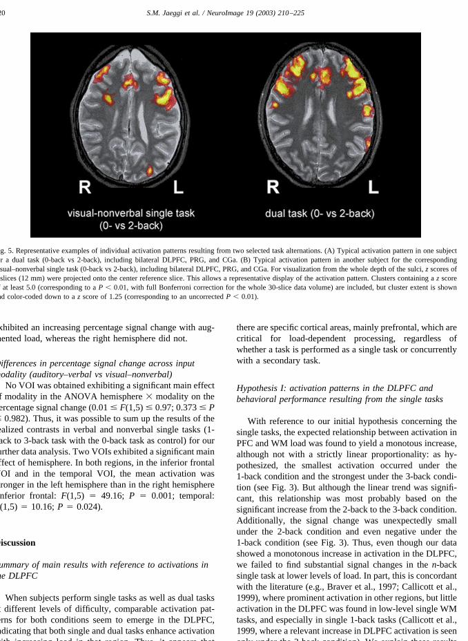
Fig. 5. Representative examples of individual activation patterns resulting from two selected task alternations. (A) Typical activation pattern in one subjectfor a dual task (0-back vs 2-back), including bilateral DLPFC, PRG, and CGa. (B) Typical activation pattern in another subject for the correspondingvisual–nonverbal single task (0-back vs 2-back), including bilateral DLPFC, PRG, and CGa. For visualization from the whole depth of the sulci, z scores of3 slices (12 mm) were projected onto the center reference slice. This allows a representative display of the activation pattern. Clusters containing a z scoreof at least 5.0 (corresponding to a P � 0.01, with full Bonferroni correction for the whole 30-slice data volume) are included, but cluster extent is shownand color-coded down to a z score of 1.25 (corresponding to an uncorrected P � 0.01).
220 S.M. Jaeggi et al. / NeuroImage 19 (2003) 210–225
in terms of the chosen criterion level of performance (atleast 75% accuracy in the 2-back single task) in order toobtain a certain level of accuracy under the dual 1- and2-back conditions, which might have led to that pattern. Theselection of high-performing subjects could have resulted inthe possibility that they were not challenged enough in theserelatively simple 1- and 2-back tasks, suggesting that in thiscase there was not sufficient demand on the WM networks.The behavioral performance confirms the observed signalchanges: the accuracy was very high and comparable underboth the 1- and the 2-back conditions and only decreasedunder the 3-back task (see Fig. 2), indicating that the firsttwo tasks were very easy to perform for our subjects indeed.The 0-back condition used as control task might also havecontributed to the low activation patterns under the 1-and2-back conditions: all three tasks were probably too similarand too easy to execute for our high-performing subjectsand therefore did not yield sufficient signal changes.
Alternatively, the chosen VOI-based methodology forfMRI data analysis could have contributed to the difficultyof detecting substantial DLPFC activation changes in thosesimple tasks, where signal changes are expected to be con-strained to a relatively small volume, because the chosenVOIs were relatively large. However, the observed signalchange under the dual 1-back condition, which was alsoexpected to be of small size, was substantial and thereforespeaks against a failure of detecting signal changes due tothe chosen methodology.
Hypothesis II: activation patterns in the DLPFC andbehavioral performance resulting from the dual tasks
Since single and dual tasks evoked comparable activa-tion patterns, the hypothesis concerning the dual tasks couldnot be confirmed, given that we could not provide clearevidence for an attenuation or even just saturation of PFCactivation under the dual-task condition even in the mostdifficult task (dual 3-back); rather, activation still increased(see Fig. 3) despite behavioral performance with an errorrate corresponding to random selection. Thus, the assump-tion made by D’Esposito (2001) that prefrontal activationpossibly decreases in the face of extreme processing de-mands, i.e., due to capacity limits of the CES, could not besupported. Instead, our results suggest that increased diffi-culty in dual tasks leads to a stronger activation in the PFCeven if the processing demands are excessive. However, theincrease in the dual tasks tended to be smaller than theincrease in the single tasks, thus supplying some kind ofevidence for a beginning saturation in the BOLD response.But still, the interaction between single- and dual-task ac-tivation was not significant and, additionally, the increase insignal change between the 2- and the 3-back tasks was alsosignificant.
A possible explanation for the continuative increase inactivation could be that memory load was not increasedenough, implicating that our subjects did not reach their
limit of performance, i.e., that their processing capacity wasnot exceeded. Yet, taking the behavioral data into account,this argument would not apply, since performance droppedto chance level for all the subjects during the dual 3-backcondition. In contrast to the activation patterns, which werethe same for single and dual tasks, there was a significantdifference in the behavioral performance, i.e., the subjectsproduced longer RTs and more errors in the dual taskscompared to the single tasks. Furthermore, subjective ratingconcerning the demands for the dual 3-back condition re-vealed that each subject experienced this condition as ex-tremely difficult; hence, it can be concluded that the de-mands were excessive indeed. In summary, the DLPFCactivation did not yet show saturation at a level of difficultywhere the behavioral accuracy was already at chance level,as judged from the error rate.
Therefore, we conclude the following: the activation andthe accuracy do not decrease or saturate accordingly; rather,they seem to behave quite independently, suggesting thatthe observed signal changes did not only represent WMprocesses only, but in addition some kind of “mental effort”or “willful attention” (Frith and Dolan, 1996; Ingvar, 1994)in order to cope with the task demands, attributable toexecutive functioning subscribed to the PFC. Postinterviewswith our subjects clearly indicated that they tried hard inorder to succeed in the given tasks. Such a notion, i.e., thehigh motivation of the subjects, could also explain thesimilar increasing activation curves in parietal and precen-tral regions and could also be taken into account for thecapacity-unconstrained activation patterns in those regions.
Alternatively, our subjects could be considered as havingaccomplished selectively only one task under the dual-taskcondition, trying to ignore the stimuli of the secondary taskin order to minimize interference. Such a process, i.e.,selectively responding to one task only, despite our explicitrequest to allocate attention equally to both tasks, wouldhave been a comprehensible strategy in order to accomplishat least one of the tasks sufficiently and is also an often-ascribed process attributed to executive functioning sub-scribed to the PFC (Klingberg and Roland, 1997). Never-theless, the high accuracy achieved under the dual 1-backand 2-back conditions speaks for a high probability that thesubjects executed the task in the intended mode. Addition-ally, the reaction times as well as the accuracy under the3-back condition would have been comparable to those ofthe 3-back single task if the subjects had paid attention toone task only, which was not the case; rather, in the 3-backdual task the RTs significantly exceeded that achieved in thecorresponding single task, whereas accuracy was reliablysmaller.
Another reason why we could not support the hypothesisabout a possible attenuation in the face of excessive demandcould lie in the fact that this hypothesis was inferred on thebasis of results from different studies (D’Esposito et al.,1995; Goldberg et al., 1998), with different subjects, usingdifferent tasks, and also measuring different psychological
221S.M. Jaeggi et al. / NeuroImage 19 (2003) 210–225
constructs. This speaks to the possibility that specific taskproperties and the associated task demands may play animportant role in determining activation patterns. In partic-ular, the task used by Goldberg et al. (1998) also involvedappreciable amounts of cognitive reasoning (Wisconsincard sorting test), which might also put demands on over-lapping regions of the DLPFC and, thus, interfere with WM.
Hypothesis III: comparison of the activation patterns inthe summed single tasks with the activation in thecorresponding dual tasks
When we evaluated single- and dual-task activations totest our third hypothesis, the predicted interaction betweenWM load and the summed activations in the single tasks,compared with the activation in the corresponding dualtasks, could be confirmed: under the most difficult condition(3-back), the sum of activation changes in these single tasksexceeded the activation changes resulting from the 3-backdual task, whereas under the simpler 1-back and the 2-backconditions, the summed single-task activations were similarto the activations in the corresponding dual task (see Fig. 4).
These results are in agreement with findings by Just et al.(2001), who reported similar activation patterns in the pre-frontal areas for their summed single tasks compared to theactivation in the dual task, where the dual tasks could beperformed “without compromising accuracy,” as in our1-back and 2-back tasks. A direct comparison between sin-gle and dual n-back task with excessive behavioral demandsresulting in degradation of accuracy like our dual 3-backtask has not been assessed before to our knowledge. Also,corresponding to the relatively low DLPFC activation levelsin our 1-back and 2-back tasks, the activation Just et al.(2001) found in the DLPFC “was minimal in both single-and dual-task conditions,” so that “the comparison of signalintensities in the prefrontal areas could not be made becausemany participants failed to show reliable activation in thoseareas.” In contrast, the sum of our single-task activationsexceeded the dual-task activations under the 3-back condi-tion, although only at the tendency level. If accepted forreal, this result can be interpreted in accordance with Navonand Gopher (1979) or Klingberg and Roland (1997), statingthat concurrent tasks interfere with each other if they de-pend on a common resource pool, i.e., if they demandactivation of the same part of the cortex. Such an interfer-ence is indicated by increasing RT along with increasingerror rate and, additionally, as was the case in our experi-ment, by the smaller activation during the dual task com-pared to the total of activation during the single tasks underthe 3-back condition. These smaller activations in the dualtasks could represent the physiological homologue of theabove-mentioned behavioral cost (increasing RT and errorrate).
Other studies also indicate that the BOLD response isrelatively smaller in certain cortical areas if a secondary taskis performed concurrently with a primary task, if both de-
pend on resources of the same cortical area (e.g., Vander-berghe et al., 1997, or Rees et al., 1997). Just et al. (2001)suggested that any observed underadditivity in the dual taskcould be explained with a certain biological constraint in theamount of activation that can be distributed to an area at agiven time. This constraint could be another manifestationof a generally assumed limit of attention which can beallocated to tasks at one time (e.g., Broadbent, 1957; Kah-neman, 1973), resulting most evidently in the decrement ofperformance if several demanding tasks are required to beperformed simultaneously. As a physiological reflection,this limit of attention might result in the relatively smalleractivation of certain cortical areas involved with these tasks.
Just et al. (2001) mention the possibility that any ob-served underadditivity in prefrontal areas could be attrib-uted to the nonlinearity of the BOLD response. There isevidence that, at high levels of demand, a saturation of theBOLD response may occur, i.e., leading to the lesser mea-surable activation in the involved areas (Friston et al.,2000). In our experiment, the criterion of a highly demand-ing task has been reached, at least in the case of the dual3-back condition. However, although a certain underaddi-tivity was observed during the dual tasks in our experiment,indicating a smaller increase under the dual-task conditioncompared with the single-task condition, the activation stillincreased with incremental memory load, therefore speak-ing against a saturation based solely on the BOLD response.Moreover, the observed maximal signal change even underthe 3-back condition (0.39%) is only a fraction of what weobserve with identical methodology in the motor cortexduring motor tasks (up to 2.33%, Nirkko et al., 2001). Itseems unlikely that the BOLD response should be saturatedat such a lower threshold in these almost neighboring re-gions of the DLPFC. We conclude that the observed under-additivity reflects a neuronal phenomenon, not a limitationof the vascular or physical/technical BOLD response.
Regions apart from the DLPFC responding tomanipulations of memory load
Apart from the DLPFC there are a few regions thatsimilarly show load-dependent signal change: bilateral pre-central gyrus (BA 4), bilateral superior parietal lobule (BA39/40), left superior and middle temporal gyri (BA 21/37),and, to a certain extent, inferior frontal cortex (BA 44/45)(see Fig. 3).
These regions were commonly activated as a function ofmemory load in most studies, which used the n-back task asWM paradigm and, regarding the premotor and inferior–frontal regions, the assumption has been previously made(e.g., Braver et al., 1997, Smith and Jonides, 1997, orRypma et al., 1999) that these regions house anterior speechareas and, concerning WM, that premotor and supplemen-tary motor regions in cooperation with Broca’s area mediatemaintenance processes, especially subvocal articulatoryprocesses such as verbal rehearsal. The load sensitivity
222 S.M. Jaeggi et al. / NeuroImage 19 (2003) 210–225
observed in these regions might presumably reflect the in-crease in number of rehearsal items associated with thecontinuous augmentation of WM load.
Parietal cortex did not only produce the strongest overallsignal change, but also exhibited increased activation withaugmented WM load which is in accordance with studies byPaulesu et al. (1993), Smith and Jonides (1997), or Salmonet al. (1996), suggesting that the parietal cortex is critical forphonological storage processes.
With regard to temporal regions we observed a load-dependent activation pattern for the left temporal cortex,which, similar to parietal regions, could account for phono-logical storage processes. According to Smith and Jonides(1997) there is some evidence that inferotemporal regions inconnection with posterior parietal areas might mediate stor-age and rehearsal for object information in addition toverbal information. However, temporal activations observedin our experiment were very small (see Table 1) but stillshowed a significant left-lateralized, load-dependent signalchange that speaks for the involvement of verbal and/orobject storage processes.
Hence, there seems to be a network of cortical regionsmediating WM tasks, including inferior frontal, parietal,and temporal areas, supporting a WM model like the oneproposed by Baddeley (1986) consisting of different sub-components responsible for different processes carried outin specific cortical areas.
Differences in activation between input modalities withinthe PFC
WM processes in our experiment seem to induce activa-tion in specific prefrontal areas regardless of the input mo-dality (auditory/visual) or the type of the stimulus material(verbal/nonverbal) held in WM, supporting the “process-specific” theory proposed by Petrides (1994) or Owen et al.(1996). Still, there is evidence for a certain type of “mate-rial-specific” processing in the PFC, specifically concerningthe VLPFC, where verbal processing, as well as processingof objects, is more likely to be reflected as activations in theleft hemisphere (Broca’s area) (e.g. Smith et al., 1997;Jonides et al., 1997), compared to spatial processing mainlyevoking activations in homologous areas in the right. In ourexperiment, the left VLPFC was more activated under bothsingle-task conditions (shapes and consonants) than theright VLPFC, speaking for a lateralized stimulus-based pro-cessing mechanism.
One explanation for these results is that our subjects canbe considered as having used the same encoding, maintain-ing, or manipulation strategies across both stimulus types,e.g., that subjects verbalized and rehearsed the abstractshapes as they did the verbal material. Indeed, posttestinterviews revealed that all subjects tended to verbalize theshapes despite our efforts to select material designed to bedifficult to verbalize. In that case, the processing mecha-nisms for nonverbal and verbal material were likely to be
the same, and left-lateralization of the VLPFC was less dueto the stimulus material, but rather to similar processingmechanisms, which could also explain the similar activa-tions in the auditory and the visual tasks in the analyzedVOIs. Other, more posterior regions (i.e., occipital areas), inwhich modality-specific differences could be expected,were not explicitly explored. Under the dual-task condition,however, both tasks apparently competed for the same pro-cessing system, most probably for the phonological loop,assumed to be responsible for verbal rehearsal. Such acompetition for resources might additionally have led to theobserved interferences between the two tasks.
Conclusion
In sum, our results show that single and dual taskssimilarly enhance activation in the DLPFC with increasingload; therefore, the intended interaction between the mem-ory load and the activation in single and in dual tasks couldnot be observed. This interaction, however, was significantwhen the sum of the activation in the single tasks wascompared with the activation in the corresponding dual task,i.e., the increase in activation was smaller in the dual tasksin comparison with the increase of activation in the summedsingle tasks. Under the 3-back condition, the activation ofthe sum of both single tasks exceeded the activation in thedual task for not less than 42%. However, this finding couldbe expected, since the effects concerning the increase inactivation resulting from single-task performance due toaugmented memory load apparently became more substan-tial when the doubled activation of these tasks was taken asreference for the activation in the dual tasks. Further inves-tigations with such methods would have address the ques-tion of which type of evaluation would make more sense,i.e., either to choose an evaluation comparing single tasksand dual tasks as independent measures or, rather, to com-pare the activation in dual tasks with the sum of two singletasks which, in reality, were executed independently. It mustbe considered that, presumably, the performance of a singletask demands entirely different processes compared with theprocesses involved in the performance of a dual task.
Our study provides some informative evidence that be-havioral measures (i.e., accuracy) are not necessarily relatedto activation in involved regions in a direct mode, sinceactivation still increased even as performance was at chancelevel in the most difficult task. Task difficulty, as expressedin task load, and the related mental effort to execute thetasks seem to play a crucial role for the observed activationpatterns, mainly in a distributed network comprising theDLPFC, the superior parietal lobule, and precentral regions.We conclude, that this motivation factor probably plays themain role in decreases at higher levels of load reported byother authors (e.g., Goldberg et al., 1998), especially if thesubjects fail to execute the task appropriately anymore and,consequently, is not trying to achieve their best performance
223S.M. Jaeggi et al. / NeuroImage 19 (2003) 210–225
anymore. But, of course, such factors should be controlledfor, e.g., by posttest interviews.
We suggest that the load-dependent activation changeswe observed did not exclusively represent WM processesper se, but rather a more global attentional, somehow mo-tivational network (Frith and Dolan, 1996; Ingvar, 1994)related to strategic task processing. These processes may notbe related to the decrement of behavioral performance, but,explaining our data, seem to rely on the same neural sub-strates that mediate the executive component of WM.
Consequently, further studies will be needed to deter-mine carefully how and under which conditions dual-taskprocessing affects activation in specific cortical areas, inorder to clarify the role of capacity-constrained regions inthe human brain, as well as to separate WM demands fromthose needed in more global attentional networks. Thecoregistration and evaluation of behavioral data are crucialin that context, as psychologically relevant reference func-tion in order to interpret activation data. Furthermore, morestudies are needed to clarify the question of how dual-taskprocessing is related to single-task processing, along withthe reasons for possible occurrences of any deviations in theobserved activation patterns between dual and single tasks.We suggest that the direct comparison of parametricallyvaried single tasks with analogously varied dual-task con-ditions might be a fruitful way to succeed.
Acknowledgments
This work was funded in part by the Swiss NationalScience Foundation (NFP-38: Grant 4038-044053). The au-thors thank Martin Buschkuhl for assistance in program-ming and data analysis and the anonymous reviewer forhelpful comments on an earlier draft of the manuscript.
References
Adcock, R.A., Constable, R.T., Gore, J.C., Goldman-Rakic, P.S., 2000.Functional neuroanatomy of executive processes involved in dual-taskperformance. Proc. Natl. Acad. Sci. USA 97, 3567–3572.
Attneave, F., Arnoult, M.D., 1956. Methodological considerations in thequantitative study of shape and pattern perception. Psychol. Bull. 53,221–227.
Baddeley, A.D., 1986. Working Memory. Clarendon Press, Oxford, En-gland.
Baddeley, A.D., Bressi, S., Della Sala, S., Logie, R., Spinnler, H., 1991.The decline of working memory in Alzheimer’s disease. A longitudinalstudy. Brain 114, 2521–2542.
Baddeley, A.D., Della Sala, S., Papagano, C., Spinnler, H., 1997. Dual-taskperformance in dysexecutive and nondysexecutive patients with a fron-tal lesion. Neuropsychology 11, 187–194.
Barch, D.M., Braver, T.S., Nystrom, L.E., Forman, S.D., Noll, D.C.,Cohen, J.D., 1997. Dissociating working memory from task difficultyin human prefrontal cortex. Neuropsychologia 35J.D., 1373–1380.
Braver, T.S., Cohen, J.D., Nystrom, L.E., Jonides, J., Smith, E.E., Noll,D.C., 1997. A parametric study of prefrontal cortex involvement inhuman working memory. NeuroImage 5, 49–62.
Broadbent, D.E., 1957. A mechanical model for human attention andimmediate memory. Psychol. Rev. 64, 205–215.
Bunge, S.A., Klingberg, T., Jacobsen, R.B., Gabrieli, J.D.E., 2000. Aresource model of the neural basis of executive working memory. Proc.Natl. Acad. Sci. USA 97, 3573–3578.
Callicott, J.H., Mattay, V.S., Bertolino, A., Finn, K., Coppola, R., Frank,J.A., Goldberg, T.E., Weinberger, D.R., 1999. Physiological character-istics of capacity constraints in working memory as revealed by func-tional MRI. Cereb. Cortex 9, 20–26.
Caviness Jr., V.S., Meyer, J., Makris, N., Kennedy, D.N., 1996. MRI-basedtopographic parcellation of human neocortex: an anatomically specifiedmethod with estimate of reliability. J. Cogn. Neurosci. 8, 566–587.
Cohen, J.D., MacWhinney, B., Flatt, M.R., Provost, J., 1993. PsyScope: anew graphic interactive environment for designing psychology exper-iments. Behav. Res. Methods Instrum. Comput. 25, 257–271.
Cohen, J.D., Perlstein, W.M., Braver, T.S., Nystrom, L.E., Noll, D.C.,Jonides, J., Smith, E.E., 1997. Temporal dynamics of brain activationduring a working memory task. Nature 386, 604–608.
Damasio, H., 1995. Human Brain Anatomy in Computerized Images.Oxford Univ. Press, New York.
D’Esposito, M, 2001. Working Memory, in: Cabeza, R., Kingstone, A.(Eds.), Handbook of Functional Neuroimaging of Cognition. MITPress, Cambridge, MA, pp. 293–327.
D’Esposito, M., Postle, B.R., 1999. The dependence of span and delayed-response performance on prefrontal cortex. Neuropsychologia 37,1303–1315.
D’Esposito, M., Aguirre, G.K., Zarahn, E., Ballard, D., Shin, R.K., Lease,J., 1998. Functional MRI studies of spatial and nonspatial workingmemory. Cogn. Brain Res. 7, 1–13.
D’Esposito, M., Detre, J.A., Alsop, D.C., Shin, R.K., Atlas, D., Grossman,M., 1995. The neural basis of the central executive system of workingmemory. Nature 378, 279–281.
Fletcher, P.C., Henson, R.N.A., 2001. Frontal lobes and human memory.Insights from functional neuroimaging. Brain 124, 849–881.
Friston, K.J., Mechelli, A., Turner, R., Price, C.J., 2000. Non-linear re-sponses in fMRI: the balloon model, Volterra kernels, and other he-modynamics. NeuroImage 12, 466–477.
Frith, C., Dolan, R., 1996. The role of the prefrontal cortex in highercognitive functions. Brain Res. Cogn. Brain Res. 5, 175–181.
Gevins, A.S., Cutillo, B.C., 1993. Neuroelectric evidence for distributedprocessing in human working memory. Electroencephalogr. Clin. Neu-rophysiol. 87, 128–143.
Goldberg, T.E., Berman, K.F., Fleming, K., Ostrem, J., Van Horn, J.D.,Esposito, G., Mattay, V.S., Gold, J.M., Weinberger, D.R., 1998. Un-coupling cognitive workload and prefrontal cortical physiology: a PETrCBF study. NeuroImage 7, 296–303.
Gopher, D. and Donchin, E. 1986. Workload—an examination of theconcept, in: Boff, K.R., Kaufman, L., Thomas, J.P. (Eds.), Handbook ofHuman Perception and Performance, Vol. 2, Cognitive Processes andPerformance, Wiley, New York, pp. 41-1–41-49.
Ingvar, D.H., 1994. The will and the brain: cerebral correlates of wilfulacts. J. Theor. Biol. 171, 7–12.
Jonides, J., Schumacher, E.H., Smith, E.E., Lauber, E.J., Awh, E.,Minoshima, S., Koeppe, R.A., 1997. Verbal working memory loadaffects regional brain activation as measured by PET. J. Cogn. Neuro-sci. 9, 462–475.
Just, M.A., Carpenter, P.A., Keller, T.A., Emery, L., Zajac, H., Thulborn,K.R., 2001. Interdependence of nonoverlapping cortical systems indual cognitive tasks. NeuroImage 14, 417–426.
Kahneman, D., 1973. Attention and Effort. Prentice Hall, EnglewoodCliffs, NJ.
Keller, T.A., Carpenter, P.A., Just, M.A., 2001. The neural bases ofsentence comprehension: an fMRI examination of syntactic and lexicalprocessing. Cereb. Cortex 11, 223–237.
Klingberg, T., 1998. Concurrent performance of two working memorytasks: potential mechanisms of interference. Cereb. Cortex 8, 593–601.
224 S.M. Jaeggi et al. / NeuroImage 19 (2003) 210–225
Klingberg, T., Roland, P., 1997. Interference between two concurrent tasksis associated with activation of overlapping field in the cortex. Cognit.Brain Res. 6, 1–8.
McDowell, S., Whyte, J., D’Esposito, M., 1997. Working memory impair-ments in traumatic brain injury: evidence from a dual-task paradigm.Neuropsychologia 35, 1341–1353.
Michael, E.B., Keller, T.A., Carpenter, P.A., Just, M.A., 2001. fMRIinvestigation of sentence comprehension by eye and by ear: modalityfingerprints on cognitive processes. Hum. Brain Mapp. 13, 239–252.
Navon, D., Gopher, D., 1979. On the economy of the human-processingsystem. Psychol. Rev. 86, 214–255.
Nirkko, A.C., 2000. A small software utility for fully automated downloadand evaluation of fMRI data. NeuroImage 11, S919.
Nirkko, A.C., Ozdoba, C., Redmond, S.M., Burki, M., Schroth, G., Hess,C.W., Wiesendanger, M., 2001. Different ipsilateral representations fordistal and proximal movements in the sensorimotor cortex: activationand deactivation patterns. NeuroImage 13, 825–835.
Oldfield, R.C., 1971. The assessment and analysis of handedness: theEdinburgh inventory. Neuropsychologia 9, 97–113.
Ono, M., Kubik, S., Abernathey, C.D., 1990. Atlas of the Cerebral Sulci.Thieme, Stuttgart.
Owen, A.M., Evans, A.C., Petrides, M., 1996. Evidence for a two-stagemodel of spatial working memory processing within the lateral frontalcortex: a positron emission tomography study. Cereb. Cortex 6, 31–38.
Owen, A.M., Stern, C.E., Look, R.B., Tracey, I., Rosen, B.R., Petrides, M.,1998. Functional organization of spatial and nonspatial working mem-ory processing within the human lateral frontal cortex. Proc. Natl.Acad. Sci. USA 95, 7721–7726.
Paulesu, E., Frith, C.D., Frackowiak, R.S.J., 1993. The neural correlates ofthe verbal component of working memory. Nature 362, 342–344.
Petrides, M., 1995. Impairments on non-spatial self-ordered and externallyordered working memory tasks after lesions of the mid-dorsal part ofthe lateral frontal cortex in the monkey. J. Neurosci. 15, 359–375.
Petrides, M., 1994. Frontal lobes of working memory: evidence frominvestigations of the effects of cortical excisions in nonhuman primates.
in: Boller, F., Grafman, J (Eds.), Handbook of Neuropsychology, Vol.9. Elsevier, New York, pp. 59–82.
Petrides, M., Alivisatos, B., Meyer, E., Evans, A.C., 1993a. Dissociation ofhuman mid-dorsolateral from posterior dorsolateral frontal cortex inmemory processing. Proc. Natl. Acad. Sci. USA 90, 873–877.
Petrides, M., Alivisatos, B., Meyer, E., Evans, A.C., 1993b. Functional acti-vation of the human prefrontal cortex during the performance of verbalworking memory tasks. Proc. Natl. Acad. Sci. USA 90, 878–882.
Rademacher, J., Galaburda, A.M., Kennedy, D.N., Filipek, P.A., CavinessJr., V.S., 1992. Human cerebral cortex: localization, parcellation, andmorphometry with magnetic resonance imaging. J. Cogn. Neurosci. 4,352–374.
Rees, G., Frith, C.D., Lavie, N., 1997. Modulating irrelevant motionperception by varying attentional load in an unrelated task. Science278, 1616–1619.
Rypma, B., Prabhakaran, V., Desmond, J.E., Glover, G.H., Gabrieli,J.D.E., 1999. Load-dependent roles of frontal brain regions in themaintenance of working memory. NeuroImage 9, 216–226.
Salmon, E., Van der Linden, M., Collette, F., Delfiore, G., Maquet, P.,Degueldre, C., Luxen, A., Franck, G., 1996. Regional brain activityduring working memory tasks. Brain 119, 1617–1625.
Schumacher, E.H., Lauber, E., Awh, E., Jonides, J., Smith, E.E., Koeppe,R.A., 1996. PET evidence for an amodal verbal working memorysystem. NeuroImage 3, 79–88.
Smith, E.E., Jonides, J., Koeppe, R.A., 1997. Dissociating verbal andspatial working memory using PET. Cereb. Cortex 6, 11–20.
Smith, E.E., Jonides, J., 1997. Working memory: a view from neuroimag-ing. Cogn. Psychol. 33, 5–42.
Vanderberghe, R., Duncan, J., Dupont, P., Ward, R., Poline, J.B., Bormans,G., Michiels, J., Mortelmans, L., Orban, G.A., 1997. Attention to oneor two features in left or right visual field: a positron emission tomog-raphy study. J. Neurosci. 17, 3739–3750.
Vanderplas, J.M., Garvin, E.A., 1959. The association value of randomshapes. J. Exp. Psychol. 3, 147–154.
225S.M. Jaeggi et al. / NeuroImage 19 (2003) 210–225