domains in microbial 3-1,4-glycanases: sequence …mmbr.asm.org/content/55/2/303.full.pdf · ·...
TRANSCRIPT

Vol. 55, No. 2MICROBIOLOGICAL REVIEWS, June 1991, p. 303-3150146-0749/91/020303-13$02.00/0Copyright © 1991, American Society for Microbiology
Domains in Microbial 3-1,4-Glycanases: Sequence Conservation,Function, and Enzyme Families
N. R. GILKES,1 B. HENRISSAT,2 D. G. KILBURN,' R. C. MILLER, JR.,' AND R. A. J. WARREN'*Department of Microbiology, University of British Columbia, Vancouver, British Columbia, Canada V6T I W5,1and Centre de Recherches sur les Macromolecules Vegetales, Centre National de la Recherche Scientifique,
B.P. 53X, 38041 Grenoble, France2
INTRODUCTION ......................................................................... 303STRUCTURAL ELEMENTS IN CELLULASES AND XYLANASES ...............................................303
Linkers ......................................................................... 303Repeated Sequences ......................................................................... 305Cellulose-Binding Domains ......................................................................... 306Catalytic Domains ......................................................................... 308
EVOLUTION OF CELLULASES AND XYLANASES ..................................................................310CONCLUSIONS ......................................................................... 312ACKNOWLEDGMENTS ......................................................................... 312REFERENCES ......................................................................... 312
INTRODUCTION
Cellulose is a polysaccharide composed of ,-D-glucopyr-anosyl units joined by 1,4-glycosidic bonds. In xylan, therepeating unit is the P-1,4-D-xylopyranosyl residue. Al-though chemically similar, these two polysaccharides adoptdifferent conformations. Cellulose molecules have a fullyextended, flat conformation (37, 64) and are tightly packedinto microfibrils to form a fibrous, naturally crystalline,insoluble material. Xylan molecules are twisted and aremore flexible than cellulose chains (102), and the backbone issubstituted with arabinose, glucuronic acid, or methylglucu-ronic acid. Chitin resembles cellulose since it is composed ofN-acetyl-2-amino-2-deoxy-f3-D-glucopyranosyl residues joinedby 1,4-glycosidic bonds. Like cellulose, chitin chains have anextended conformation and form insoluble and crystallinemicrofibrils (12). The microbial conversion of cellulose andxylan to soluble products requires several types of enzyme:endoglucanases (1,4-p-D-glucan glucanohydrolase; EC3.2.1.4), cellobiohydrolases (1,4-p-D-glucan cellobiohydrolase;EC 3.2.1.91), xylanases (1,4-p-D-xylan xylanohydrolase; EC3.2.1.8), and P-xylosidases (1,4-p-D-xylan xylohydrolase; EC3.2.1.37). Microorganisms capable of degrading lignocelluloseusually produce complex, extracellular cellulase systems com-prising combinations of these enzymes (22, 23). Microorgan-isms that degrade chitin also produce a variety of enzymes.Cellulases, xylanases, and chitinases all hydrolyze P-1,4-gly-cosidic bonds between pyranose units, but they show subtledifferences in specificity. Structural features common to suchenzymes may be related to their general catalytic activities asglycosidases. Those peculiar to each may be related to theirspecificities, i.e., exoglycanase or endoglycanase, cellulase orxylanase. Such features should be reflected in the amino acidsequences of the enzymes.Gene cloning and DNA sequencing have allowed rapid
determination of the amino acid sequences of cellulases andxylanases (la, 7). Analysis and comparison of the sequenceshave revealed conserved stretches which are common toboth cellulases and xylanases. The conserved sequences
* Corresponding author.
occur in discrete domains connected by linkers which allowthe domains to function independently (30, 39, 61, 111, 113).The conserved sequences can be used to group the enzymesinto families (5, 8, 54, 55, 62). It seems that the variousP-1,4-glycanases arose from a few progenitor sequences bymutation and domain shuffling. In this context, it should benoted that some enzymes show a mixed specificity: enzymesthat hydrolyze P-1,4 bonds in cellulosic substrates may alsohydrolyze xylan, chitin, and related substrates at significantrates (41).
This review summarizes the domain organizations ofcellulases and xylanases analyzed to date. It also comparesthe enzymes with other proteins which interact with variouspolysaccharides.
STRUCTURAL ELEMENTS IN CELLULASES ANDXYLANASES
Linkers
Proteolytic cleavage of cellulases into separate catalyticand cellulose-binding fragments first demonstrated the pres-ence of true domains within these enzymes (42, 111, 113).The primary sites of cleavage in an exoglucanase and anendoglucanase from Cellulomonas fimi (42) and in twocellobiohydrolases from Trichoderma reesei (111) are withinor adjacent to short sequences of amino acids rich in prolineor hydroxyamino acids or both. The short sequences appearto be linkers joining discrete catalytic domains and cellulose-binding domains (CBDs) in these enzymes. Similar se-quences occur in other cellulases and xylanases (Table 1).The sequences vary considerably in length (6 to 59 aminoacids for those reported to date) and in their proline andhydroxyamino acid contents. Some of them contain runs ofhydroxyamino acids or consecutive repeats of shorter se-quences of amino acids or both. Some are relatively rich inaspartate or glutamate or both. There is some sequenceidentity between linkers in enzymes from the same organ-ism, but, except for CenA and Cex from C. fimi andEngXCA from Xanthomonas campestris (44), there is little if
303
on July 3, 2018 by guesthttp://m
mbr.asm
.org/D
ownloaded from

304 GILKES ET AL.
cn e~n C)'
o00= m e oo CO
._ 0) NN
(A
0)
0)$.C0
1-O
COCO0)
aOCLCO
m
CO
0
0)0)w
0
CO).4-0
0)c
ca0)
*-OCtIr.L;4
410
0)00)COLI)*0)
02
00e
COr.
0000)
o
r.
toCZ
00
0
MICROBIOL. REV.
o Neno -4N
(ONNr-~r-q r N4ren0o
'It m CA W- ON N 'I en mN oo 7- r- £0 -t It r- 1* 't It
ON ONN- 100,)-400C ONs 0) r- ON 00 00IRtr-P-4r- IR fW 11O N 000ON
00oo N N O N 00 n 00 tn C)" t CON e 0 00 OCNoo O C""r-4r--4 r--4 r-4 r~~~~~~-.4 r
N 'I Rt :t "t ) "t It m N O N 0 00 N Nr '0I N U)W 000- ,-4, -4, r-4 Ol0 OO
00 C') I'll N- N- N- 'IC 0) 'IC P 'I 0 "-4--4 C') 0) 10 ": 0) tn ON 00'-4
UU VILT;<U bU
6 0O X
)x M 0 0
Q Q Q ao X 0 rA r
CZ CZ Ct CZ t CZQ 0 0 0 O
0"0"
0 ._ 3 0
COO
3
CCOQ°
O0
z
C6O0.5
COC
C0&daa > '
0 ~ ~
CO 0) o o CO V
CO CO C OCOCOC CO
C QQ O O C
0 0 0 0C 0 co
r
S isSE E EEEzE S E X g # fln ' E
(If un 't 'IC " 't O) m r- W) CO r °= t - W) 'Iem 00-40>no N'fH \Ot
C) " m m m U) m 't m 'It " " " eq en
C')"-
CO
u u: u: co u: ce
0)0) 0)C Oct co Cd cdQQc)¢ Q~LQQ< >< Uco0)0) 0)0) C 0) 00)
Qt co,05 ro_ _ r
COCOCOCO CO U~~~~~~CO)GOUC1
CO CO)
COCOCOCOC CO CO CO COCe
00t 0 0 CO CO 0 00S~~" "0 "0"0XE.;.
S~~CEE d CO COCO
on July 3, 2018 by guesthttp://m
mbr.asm
.org/D
ownloaded from

VOL. 55, 1991
o t o o o 00 0
'-4
r0 4 00 'IO
'IO4 r- en I\00 Itr-C e e r- - qr-
0
H00
0
n
H:FPZ4
H0
~00NN
:C: S
v) v>
¢4
_I-I-I
P64:4L4N
N
E-
EO > O <
'- N -
00 Vn lq
00-
00
NEa(A W <wa
U~~~~~~~
0 w Z
a)u '0O oE;~ 0 r,
00la C)~0 O.0'
ts-tSCi =1%C)
.
0.
I, o. .%
-0.
DOMAINS IN MICROBIAL 0-1,4-GLYCANASES 305
a)s
.0
L)
C0
V
0.^
0t C
*.- 0*L
._
O
._-
C .*Qa)
Qa)-
any sequence identity between linkers from different organ-isms (Table 1).Spore germination-specific polypeptide 270-11 from the
slime mold Dictyostelium discoideum contains two se-quences, each about 100 amino acids long, which are rich inproline and hydroxyamino acids. The sequences are charac-terized by contiguous repeats of the tetrapeptide TETP(Table 1) (43). The walls of D. discoideum spores containcellulose, and it is possible that polypeptide 270-11 is in-volved in cellulose hydrolysis during germination (see be-low) (43).
It must be emphasized, however, that some cellulases donot contain obvious linker sequences like those given inTable 1. Such enzymes may not lack discrete domains,because CenA from C. fimi (102a) and XynA from Pseudo-monas fluorescens subsp. cellulosa (30), from which thelinkers have been deleted, have catalytic and cellulose-binding properties similar to those of the wild-type enzymes.The linker sequences, especially those rich in proline, are
similar to proline-rich linkers connecting different functionaldomains in other proteins (Table 1). Such sequences inimmunoglobulin Al (16), ribosomal protein L12 (17), and thepyruvate dehydrogenase complex (92) form flexible, ex-tended hinge regions between different domains, and thosein outer membrane protein TonB (27) and procyclin (94)form extended structures. It seems likely, therefore, that thelinker sequences in cellulases form extended, flexible hingesbetween domains.Some of the linkers in cellulases are also relatively rich in
arginine, glutamine, glutamate, and hydrophobic amino ac-ids. In this they are similar to Q-linkers, a recently proposedclass of interdomain linkers found in a number of bacterialregulatory and sensory transduction proteins (118). Q-link-ers are rich in glutamine, arginine, glutamate, serine, andproline and contain some hydrophobic amino acids (Table1).
Repeated Sequences
Repeated sequences ranging in length from 20 to 150amino acids occur in a number of cellulases (Table 2). Themost striking example is a highly conserved sequence of ca.24 amino acids which occurs twice, in close proximity, infive endoglucanases, a xylanase, and a partial open readingframe from Clostridium thermocellum and in an endogluca-nase from Clostridium cellulolyticum. The identity is >70%.In CelA, CelB, CelCCA, CelD, CelH, and the open readingframe, the repeats are very close to the C termini of thepolypeptides; in CelE and XynZ, the repeats are in themiddle of the polypeptides (5, 29, 45, 46, 49, 58, 120). InCelA, CelB, CelE, and the open reading frame, the repeatsare preceded by linker sequences; in XynZ, the repeat isfollowed by a linker sequence. The function(s) of the re-peated sequences is not known, but they are not required forthe catalytic activity of CelE (49) or CelH (120).CelB of alkalophilic Bacillus strain N4 has a repeat of 61
amino acids at its C terminus. The repeats are 28 amino acidsapart and are 90% identical. The polypeptide also has twolinker sequences, each of which overlaps the N-terminalsequence of a repeat (35). CelB and CelA of strain N4 havevery similar sequences throughout, but CeLA has only asingle copy, again at its C terminus, of the repeated sequencein CelB (35).Endoglucanase CenB from C. fimi contains three contig-
uous repeats of a sequence of 98 amino acids which are>60% identical and separated by linkers. The repeats join an
000:HOC1 '-4 't
" -4 IR
'-4 "~V-4
on July 3, 2018 by guesthttp://m
mbr.asm
.org/D
ownloaded from
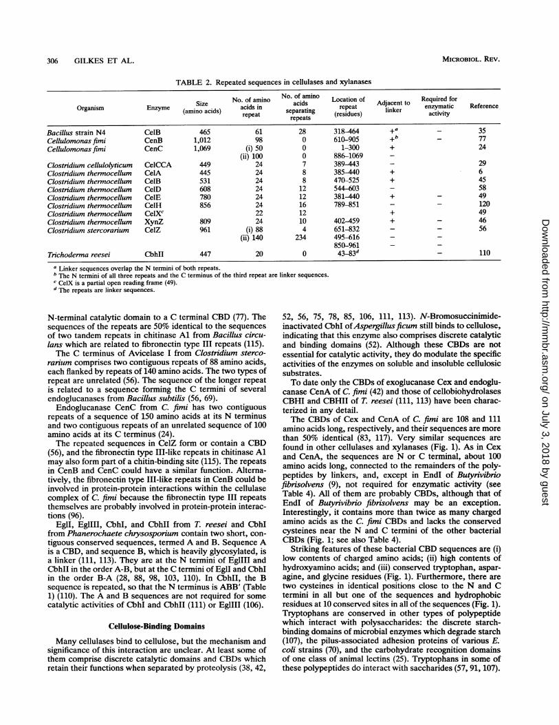
TABLE 2. Repeated sequences in cellulases and xylanases
No. of amino No. of amino Location of A Required forOrganism Enzyme Size acids in acids repeat Adjacent to enzymatic Reference
(amino acids) repeat separating (residues) linker activityOrganismEnzyme (amino acids) repeat repeats (eius
Bacillus strain N4 Ce1B 465 61 28 318-464 +a _ 35Cellulomonas fimi CenB 1,012 98 0 610-905 +b 77Cellulomonas fimi CenC 1,069 (i) 50 0 1-300 + 24
(ii) 100 0 886-1069 -Clostridium cellulolyticum CeICCA 449 24 7 389-443 - 29Clostridium thermocellum CelA 445 24 8 385-440 + 6Clostridium thermocellum CelB 531 24 8 470-525 + 45Clostridium thermocellum CelD 608 24 12 544--603 - 58Clostridium thennocellum CelE 780 24 12 381-440 + - 49Clostridium thermocellum CelH 856 24 16 789-851 - - 120Clostnidium thermocellum CelXC 22 12 + 49Clostridium thermocellum XynZ 809 24 10 402-459 + - 46Clostridium stercorarium CelZ 961 (i) 88 4 651-832 - - 56
(ii) 140 234 495-616 - -850-961 - -
Trichoderma reesei CbhlI 447 20 0 43-83d- 110
a Linker sequences overlap the N termini of both repeats.b The N termini of all three repeats and the C terminus of the third repeat are linker sequences.c CeIX is a partial open reading frame (49).d The repeats are linker sequences.
N-terminal catalytic domain to a C terminal CBD (77). Thesequences of the repeats are 50% identical to the sequencesof two tandem repeats in chitinase Al from Bacillus circu-lans which are related to fibronectin type III repeats (115).The C terminus of Avicelase I from Clostridium sterco-
rarium comprises two contiguous repeats of 88 amino acids,each flanked by repeats of 140 amino acids. The two types ofrepeat are unrelated (56). The sequence of the longer repeatis related to a sequence forming the C termini of severalendoglucanases from Bacillus subtilis (56, 69).Endoglucanase CenC from C. fimi has two contiguous
repeats of a sequence of 150 amino acids at its N terminusand two contiguous repeats of an unrelated sequence of 100amino acids at its C terminus (24).The repeated sequences in CelZ form or contain a CBD
(56), and the fibronectin type III-like repeats in chitinase Almay also form part of a chitin-binding site (115). The repeatsin CenB and CenC could have a similar function. Alterna-tively, the fibronectin type III-like repeats in CenB could beinvolved in protein-protein interactions within the cellulasecomplex of C. fimi because the fibronectin type III repeatsthemselves are probably involved in protein-protein interac-tions (96).
EglI, EgIIII, CbhI, and CbhII from T. reesei and CbhIfrom Phanerochaete chrysosporium contain two short, con-
tiguous conserved sequences, termed A and B. Sequence Ais a CBD, and sequence B, which is heavily glycosylated, isa linker (111, 113). They are at the N termini of EgIIII andCbhII in the order A-B, but at the C termini of EglI and CbhIin the order B-A (28, 88, 98, 103, 110). In CbhII, the Bsequence is repeated, so that the N terminus is ABB' (Table1) (110). The A and B sequences are not required for somecatalytic activities of CbhI and CbhII (111) or EglIII (106).
Cellulose-Binding Domains
Many cellulases bind to cellulose, but the mechanism andsignificance of this interaction are unclear. At least some ofthem comprise discrete catalytic domains and CBDs whichretain their functions when separated by proteolysis (38, 42,
52, 56, 75, 78, 85, 106, 111, 113). N-Bromosuccinimide-inactivated CbhI ofAspergillusficum still binds to cellulose,indicating that this enzyme also comprises discrete catalyticand binding domains (52). Although these CBDs are notessential for catalytic activity, they do modulate the specificactivities of the enzymes on soluble and insoluble cellulosicsubstrates.To date only the CBDs of exoglucanase Cex and endoglu-
canase CenA of C. fimi (42) and those of cellobiohydrolasesCBHI and CBHII of T. reesei (111, 113) have been charac-terized in any detail.The CBDs of Cex and CenA of C. fimi are 108 and 111
amino acids long, respectively, and their sequences are morethan 50% identical (83, 117). Very similar sequences arefound in other cellulases and xylanases (Fig. 1). As in Cexand CenA, the sequences are N or C terminal, about 100amino acids long, connected to the remainders of the poly-peptides by linkers, and, except in EndI of Butyrivibriofibrisolvens (9), not required for enzymatic activity (seeTable 4). All of them are probably CBDs, although that ofEndI of Butyrivibrio fibnisolvens may be an exception.Interestingly, it contains more than twice as many chargedamino acids as the C. fimi CBDs and lacks the conservedcysteines near the N and C termini of the other bacterialCBDs (Fig. 1; see also Table 4).
Striking features of these bacterial CBD sequences are (i)low contents of charged amino acids; (ii) high contents ofhydroxyamino acids; and (iii) conserved tryptophan, aspar-agine, and glycine residues (Fig. 1). Furthermore, there aretwo cysteines in identical positions close to the N and Ctermini in all but one of the sequences and hydrophobicresidues at 10 conserved sites in all of the sequences (Fig. 1).Tryptophans are conserved in other types of polypeptidewhich interact with polysaccharides: the discrete starch-binding domains of microbial enzymes which degrade starch(107), the pilus-associated adhesion proteins of various E.coli strains (70), and the carbohydrate recognition domainsof one class of animal lectins (25). Tryptophans in some ofthese polypeptides do interact with saccharides (57, 91, 107).
MICROBIOL. REV.306 GILKES ET AL.
on July 3, 2018 by guesthttp://m
mbr.asm
.org/D
ownloaded from

DOMAINS IN MICROBIAL P-1,4-GLYCANASES 307
The CBDs of Cex and CenA of C. fimi bind the enzymesto cellulose (42), as do similar sequences in an endogluca-nase, xylanases XynA and XynB, and an arabinofuranosi-dase from P. fluorescens subsp. cellulosa (30, 39, 50, 61).Cex hydrolyzes both cellulose and xylan (41), and the aminoacid-sequence of the catalytic domain of Cex is similar tosequences in several xylanases (see Table 6). Cellulose isusually associated with hemicelluloses such as xylan whenfound in nature. It is not surprising that some enzymeshydrolyze both cellulose and xylan and that some xylanasesbind to cellulose.The CBDs of CbhI, CbhII, EglI, and EglIll of T. reesei
correspond to the conserved A sequences of the enzymes(Table 3) (28, 88, 98, 106, 110, 111, 113). They contain 33amino acids, in contrast to the approximately 100-amino-acid bacterial CBDs (Fig. 1). They can be N or C terminal(Table 4). The identical residues include four cysteines,which form two disulfide bridges, two glutamines, and fouraromatic residues. The CBD of CbhII was prepared bychemical synthesis and shown by two-dimensional nuclearmagnetic resonance to be wedge shaped and to contain twodisulfide bridges (65). Surprisingly, the catalytic domain ofEgIIII contains a sequence of ca. 100 amino acids which isvery similar to the bacterial CBDs (76a).
Polypeptide 270-11 from D. discoideum contains twosequences of about 100 amino acids each, which are similarin sequence to the bacterial CBDs (43, 76a). It is notsurprising that such sequences occur in both procaryotic andeucaryotic polypeptides, given the sequence similaritiesbetween the catalytic domains of some bacterial and fungalcellulases and between some bacterial cellulases and sporegermination-specific polypeptide 270-6 of D. discoideum(Table 5).
Proteolytic removal of the N-terminal half of the CBD ofCenA of C. fimi, which leaves only a single cysteine in theCBD, does not prevent the truncated enzyme from bindingto cellulose (40). This suggests that a disulfide bridge be-tween the two cysteines of this CBD is not essential forbinding. The sequences of the C-terminal segments of thebacterial CBDs and the T. reesei CBDs are not related (Fig.1; Table 3). The interactions between CBDs and cellulosehave not been elucidated. It will be interesting to seewhether the bacterial and T. reesei CBDs adsorb to cellulosein a similar manner and at the same sites.
Since CBDs in both groups are N or C terminal and areattached to catalytic domains of different specificities, theyappear not to be determinants of specificity. An interestingexample is provided by the endoglucanases from C. fimi andMicrobispora bispora, which display significant homologyboth in their catalytic cores and in their binding domains. Inthe M. bispora enzyme the binding domain is at the Cterminus; in the C. fimi enzyme it is at the N terminus. Sinceboth enzymes are endoglucanases, the location of the bind-ing domain at the N or C terminus of the catalytic domainclearly does not determine the endo versus exo specificity ofthese enzymes. There are differences in the sequences of theCBDs within each group, however, and further analysis isrequired to determine their exact contributions to enzymefunction. The cellulases of Clostridium thermocellum char-acterized to date apparently lack CBDs, but they form a
multienzyme complex, the cellulosome, which binds in tototo cellulose. Cell-associated cellulosomes can bind Clostrid-ium thermocellum itself to cellulose (66). Such binding maybe mediated by a noncatalytic component of the cellulosome(66). Cellulosomelike enzyme aggregates have not beenobserved in T. reesei and the bacteria which produce en-
C
m
CCD
CD pC
0
CD
CD~CD > CD
CD CD..
~ptT1i
CD
C )
CDOC
'0 0
0DCDCD
0 O
CD CD}
CD (-D
3I ri S
C
On C-D:
0 0 Wo) o
U,+C
CD 4; CD
CD V :3
o g. oCD
CD0
CD
CA.
wxxww00000
> -3x "n En En cn -
.- -D - -
-3C 0 _ 0
6) (31 (31 '.3Z) (3
< v H inHenIO n
6)ZZv>ZZaWZU
<Z'.13>
tIn c n>uz cn c ntnlFw>10 lo V3oo 3
>I 10 m 0 aI 0 '0
0(31(3
< <
3 03 1-0
owwmm1woowo
00000 Z Z c
I
'3 '3 (o -3
Z (3o >1Z(31)Z Z
:3 :-' << m r
3 --' 3; ~C.3 En
< I I I t: <t: t::
:01 0 :P
3"I H-< I HCH 0H0 0' 0 H
310
cn O CA C)g -3 C)
6C)
>ooUlO OoD OO~~it
(1 OD W O) % (A)VI w N)oo n L) -i
I- ()
*
< O C)'3 C) C)'.3 :3PO to
6) > ) C)'< Z 0 0U)
'3'. tn iO t'3 '3 W O> :II < < < <
III1-t3 P < 0 r-
F z So :to I I :t I
z w
H6 n
3 '3 '3 '3 '.3
0XlZZZZ2'.3(31
C/j z zzo cnuo
C) c Z .Z
i310 31'3F3 t3
Z cn n tn a
-3 '.3 - 3J
: 3 (31 0F
t7: Z 0 Z Z
aC) Cl) Cl)c
:t t- t-t3> >g
1-H~H < Ht- 4 1< N
DOZ--Z Z:0Z Z :3Ch
ti UE: £.1 0 a a a 0 ol
>: Zcn Z Ko E3KOUtn o w Z cn t3 o 0 t3 cn0
cn IU Utn2:n E>n
:ZZtncool..3..3
VOL. 55, 1991
on July 3, 2018 by guesthttp://m
mbr.asm
.org/D
ownloaded from

308 GILKES ET AL.
TABLE 3. Amino acid sequences of fungal CBD
Enzymea Sequenceb Reference
PcCbhI T v p q W G Q C G G I G Y t G s T T C A S p y T C h v L N P Y Y S Q C y 103TrCbhI T Q S H y G Q C G G I G Y S G P T v C A S G T T C Q Y L N P Y Y S Q C L 28TrCbhII c s S V W G Q C G G q n W S G P T c C A S G S T C v Y S N D Y Y S Q C L 110TrEgIl T Q T H W G Q C G G I G Y S G c k T C T S G T T C Q Y S N D Y Y S Q C L 88TrEglIll q Q T V W G Q C G G I G W S G P T n C A p G S A C s t L N P Y Y a Q C i 98a Pc, P. chrysosporium enzyme; Tr, T. reesei enzyme.bAmino acid residues are indicated in the single-letter code. Boldface capital letters indicate conservation; lightface capital letters indicate partial conservation;
lowercase letters indicate nonconservation. Symbols: *, N terminus of the mature enzyme; ***, C terminus.
zymes with CBDs. Perhaps these organisms are relativelystatic and are better served by diffusible enzymes which bindto the substrate than by multienzyme aggregates or adhesionof cells to the substrate or both.
Catalytic Domains
As with CBDs, catalytic domains have been delineated insome cellulases by proteolysis (38, 40, 42, 52, 56, 75, 78, 106,111, 113). Other cellulases for which discrete domains havenot been identified can be truncated by proteolysis withoutloss of catalytic activity (14, 18, 76, 93, 108). Still others,including two xylanases, can be truncated without loss ofcatalytic activity by deleting ends of the genes encodingthem (32, 46, 48, 50, 69, 72, 120, 123). These enzymes mayalso have discrete catalytic domains.More than 60 cellulase and xylanase genes have been
sequenced. Other than the amino acid sequences deducedfrom the nucleotide sequences of the genes, little, if any-thing, is known about the enzymes encoded by many ofthem. However, sequence identity between characterizedand uncharacterized enzymes is a strong indication of func-tional domains in the uncharacterized enzymes. Linkersequences join the domains of a number of the characterizedenzymes (28, 30, 50, 77, 83, 110, 117, 119). The amino acid
sequences between or next to putative linkers in newlysequenced enzymes can be analyzed for similarities to thesequences of known domains in other cellulases and xyla-nases. However, enzymes such as endoglucanase CelC ofClostridium thermocellum (101) and an endoglucanase ofCellulomonas uda (81), which contain neither repeated se-quences nor putative linkers, may comprise catalytic do-mains only. Their amino acid sequences are similar to thoseof the catalytic domains of other ceilulases (Table 5).
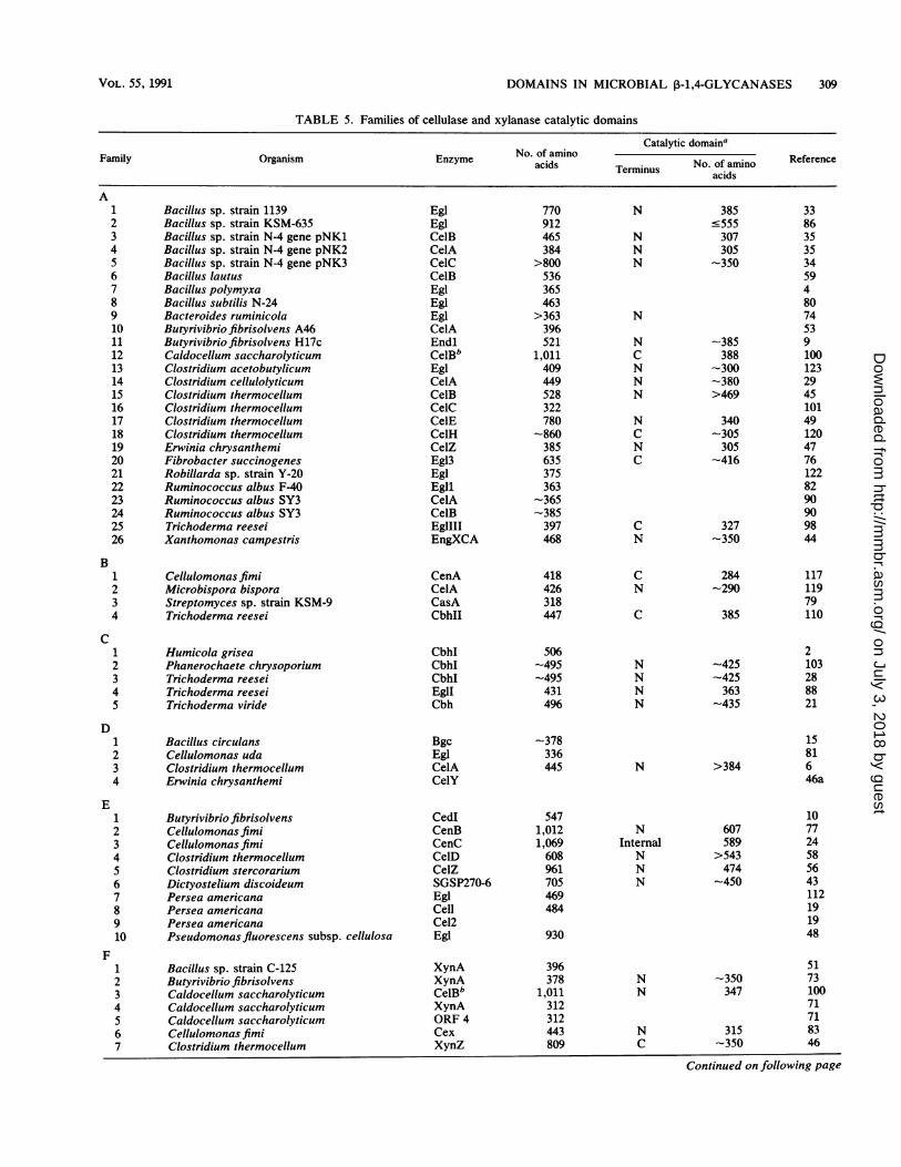
Cellulases and xylanases can be grouped into families ofrelated enzymes on the basis of amino acid sequence iden-tities in their putative catalytic domains (5, 8, 54, 55, 62).This grouping is confirmed and extended by hydrophobiccluster analysis, which reveals similarities in apparent sec-ondary structures with proteins of very low sequence iden-tity, even when domains are separated by variable segmentsof widely differing sizes (54, 55). Hydrophobic cluster anal-ysis is especially useful for cellulases and xylanases, withtheir discrete domains and repeated sequences and linkers ofvarious lengths.Sequence identity in the catalytic domains of cellulases
and xylanases has been reviewed recently (5, 8, 54, 55). Theknown sequences can be grouped into nine families (Table5), which are quite distinct (5, 54, 55, 84, 97). Families A, B,F, and H contain fungal and bacterial enzymes. Family E
TABLE 4. Characteristics of CBDs
No. of:Required Charged Rfr
Organism' Enzyme Location for Tr-Rfr(terminus) catalytic Amino amino Thponine- esacids onines and Trp Cy- ncactivity acids ____ ens tophans teines
AButyrivibriofibrisolvens EglI C + 97 6 9 21 4 1 9Cellulomonasfimi CenA N - 111 3 3 27 6 2 117Cellulomonas fimi CenB C - 101 2 1 33 5 2 77Cellulomonas fimi Cex C - 108 3 1 28 5 2 83Cellulomonas flavigena ORF X NDb 106 5 3 27 5 2 1Microbispora bispora CelA C - 104 3 3 26 5 2 119Pseudomonasfluorescens subsp. cellulosa CeIA C - 103 6 2 18 5 2 48Pseudomonasfluorescens subsp. cellulosa CelB N - 100 4 4 24 5 2 39Pseudomonasfluorescens subsp. cellulosa XynA N ND 104 4 3 25 5 2 50Pseudomonas fluorescens subsp. cellulosa XynB/XynCc N - 97 4 5 23 5 2 61
BPhanerochaete chrysosporium CbhI C ND 33 1 0 8 1 4 103Trichoderma reesei CbhI C - 33 1 0 6 0 4 28Trichoderma reesei CbhII N - 33 0 1 9 2 4 110Trichoderma reesei EglI C ND 33 2 1 8 1 4 88Trichoderma reesei EgIIII N - 33 0 0 5 2 4 98a A, Bacterial enzymes; B, fungal enzymes.bND, Not determined.c Identical sequences in two enzymes.
MICROBIOL. REV.
on July 3, 2018 by guesthttp://m
mbr.asm
.org/D
ownloaded from
DOMAINS IN MICROBIAL P-1,4-GLYCANASES 309
TABLE 5. Families of cellulase and xylanase catalytic domains
Catalytic domainaFamily Organism Enzyme No. of amino TerminusNo. of amino Reference
acids Terminus No. of aminoFamilyOrganism Enzyme acids Terminus ~~~~~~~~~~~~~~acids
A1 Bacillus sp. strain 1139 Egl 770 N 385 332 Bacillus sp. strain KSM-635 Egl 912 -555 863 Bacillus sp. strain N-4 gene pNK1 CelB 465 N 307 354 Bacillus sp. strain N-4 gene pNK2 CelA 384 N 305 355 Bacillus sp. strain N-4 gene pNK3 CelC >800 N -350 346 Bacillus lautus CelB 536 597 Bacillus polymyxa Egl 365 48 Bacillus subtilis N-24 Egl 463 809 Bacteroides ruminicola Egl >363 N 7410 Butyrivibrio fibrisolvens A46 CelA 396 5311 Butyrivibriofibrisolvens H17c Endl 521 N -385 912 Caldocellum saccharolyticum CelBb 1,011 C 388 10013 Clostridium acetobutylicum Egl 409 N -300 12314 Clostridium cellulolyticum CelA 449 N -380 2915 Clostridium thermocellum CelB 528 N >469 4516 Clostridium thermocellum CelC 322 10117 Clostridium thermocellum CelE 780 N 340 4918 Clostridium thermocellum CelH -860 C -305 12019 Erwinia chrysanthemi CelZ 385 N 305 4720 Fibrobacter succinogenes Egl3 635 C -416 7621 Robillarda sp. strain Y-20 Egl 375 12222 Ruminococcus albus F-40 Egll 363 8223 Ruminococcus albus SY3 CelA -365 9024 Ruminococcus albus SY3 CelB -385 9025 Trichoderma reesei EglIII 397 C 327 9826 Xanthomonas campestris EngXCA 468 N -350 44
B1 Cellulomonasfimi CenA 418 C 284 1172 Microbispora bispora CelA 426 N -290 1193 Streptomyces sp. strain KSM-9 CasA 318 794 Trichoderma reesei CbhII 447 C 385 110
C1 Humicola grisea CbhI 506 22 Phanerochaete chrysoporium CbhI -495 N -425 1033 Trichoderma reesei CbhI -495 N -425 284 Trichoderma reesei EglI 431 N 363 885 Trichoderma viride Cbh 496 N -435 21
D1 Bacillus circulans Bgc -378 152 Cellulomonas uda Egl 336 813 Clostridium thermocellum CelA 445 N >384 64 Erwinia chrysanthemi CelY 46a
E1 Butyrivibrio fibrisolvens CedI 547 102 Cellulomonasfimi CenB 1,012 N 607 773 Cellulomonas fimi CenC 1,069 Internal 589 244 Clostridium thermocellum CelD 608 N >543 58S Clostridium stercorarium CelZ 961 N 474 566 Dictyostelium discoideum SGSP270-6 705 N -450 437 Persea americana EgI 469 1128 Persea americana Cell 484 199 Persea americana Cel2 1910 Pseudomonas fluorescens subsp. cellulosa Egl 930 48
F1 Bacillus sp. strain C-125 XynA 396 512 Butyrivibrio fibrisolvens XynA 378 N -350 733 Caldocellum saccharolyticum CelBb 1,011 N 347 1004 Caldocellum saccharolyticum XynA 312 71S Caldocellum saccharolyticum ORF 4 312 716 Cellulomonas fimi Cex 443 N 315 837 Clostridium thermocellum XynZ 809 C -350 46
Continued on following page
VOL. 55, 1991
on July 3, 2018 by guesthttp://m
mbr.asm
.org/D
ownloaded from

310 GILKES ET AL.
TABLE 5-Continued
Catalytic domainaFamily Organism Enzyme acids Terminus No. of amino Reference
acids
8 Cryptococcus albidus Xyn 311 139 Pseudomonasfluorescens subsp. cellulosa XynA 585 C 345 5010 Pseudomonasfluorescens subsp. cellulosa XynB 555 C 272 6111 Thermoascus aurantiacus Xyn 269 105
G1 Bacillus circulans Xyn 185 1212 Bacillus pumilis XynA 201 363 Bacillus subtilis Xyn 182 874 Clostridium acetobutylicum XynB 234 124
H1 Aspergillus aculeatus Egl 237 842 Erwinia carotovora CelS 232 97
I1 Ruminococcus flavefaciens CelA 352 114a Deduced from positions of putative linkers, sequence comparison, and truncation experiments.b Catalytic domain 1 (family F, no. 3) catalytic domain 2 (family A, no. 12) of the bifunctional cellulase of Caldocellum saccharolyticum.
contains bacterial enzymes and plant enzymes, therebyraising the possibility of a lateral transfer. It includes sporegermination-specific polypeptide 270-6 from D. discoideum,whose spore coats contain cellulose. Polypeptides 270-6 and270-11 could be involved in cellulose hydrolysis during sporegermination (43). The avocado cellulases in family E appearto be involved in fruit ripening (19). At present, family Ccontains only fungal enzymes and families D and G containonly bacterial enzymes. Cellulases and xylanases varywidely in the numbers of amino acids they contain, but theircatalytic domains tend to be more uniform in size (Table 5).It should be noted, however, that relatively few catalyticdomains have been identified other than by sequence relat-edness to known domains and the presence of adjacentlinkers.
All enzymes reported to have exoglycosidase activity fallinto families which have members with only endoglycosi-dase activity (Table 5). In other words, enzymes with similarsequences have different specificities. This suggests thatexoglycosidase versus endoglycosidase activity may be aconsequence of fine details of three-dimensional structurerather than of overall conformation. The only catalyticdomain for which the three-dimensional structure is knownis that of cellobiohydrolase II from T. reesei (95). The activesite is in an enclosed tunnel through which a cellulosemolecule threads. Two aspartyl residues located in themiddle of the tunnel may be catalytic residues (95). CbhII isin family B of cellulases and xylanases, which containsexoglucanases and endoglucanases (5, 54). Modeling ofother enzymes in the family, with the structure of CbhII as aguide, should give useful insights into their possible struc-tures. Realistic comparisons, however, will require determi-nation of the three-dimensional structures of other enzymes.
Hydrolysis of the 3-1,4-glycosidic bond with retention orinversion of anomeric configuration may be a better indica-tor of similarity than enzyme specificity (116). Hydrolysiswith retention of configuration requires a quite differentmechanism than does hydrolysis with inversion (104). CenAof C. fimi and CbhII of T. reesei are in family B, and bothcause inversion of configuration (63, 116). CenB of C. fimi,which is in family E, also hydrolyzes with inversion of
configuration (77). The other enzymes which have beencharacterized, Cex of C. fimi and CbhI of T. reesei, hydro-lyze with retention of configuration (63, 116) but are infamilies F and C, respectively.At least one cellulase has two catalytic domains. CelB of
Caldocellum saccharolyticum has an N-terminal exogluca-nase domain and a C-terminal endoglucanase domain whichbelong to different families. A linker connects each catalyticdomain to a central amino acid sequence of unknown func-tion (99, 100) which is related to a sequence found at the Ctermini of several endoglucanases from B. subtilis (72, 80)and within avicelase I from Clostridium stercorarium (56)and endoglucanase CenB from C. fimi (76a). XynZ of Clos-tridium thermocellum contains a centrally located repeatedsequence flanked by linkers, with a xylanase catalytic do-main at the C terminus of the polypeptide and a sequence of401 amino acids of unknown function at the N terminus (46).
All CBDs described to date are N or C terminal. Catalyticdomains from each family are found in various combinationswith other conserved sequences, such as CBDs and repeatedsequences. This gives rise to a number of different types ofprimary structures in cellulases and xylanases. It is possiblethat further families remain to be identified.
Hydrolysis by glycosyl hydrolases often involves generalacid catalysis, usually promoted by aspartate or glutamateresidues or both. Active-site residues are usually highlyconserved during evolution. The catalytic domains of cellu-lases and xylanases have been analyzed for conservedaspartates and glutamates in an attempt to target catalyticresidues in the families (54, 55). In family A, two particularresidues emerged as candidates. Site-directed mutations intwo of the enzymes in family A support the involvement ofthe targeted residues in the active site (3, 90a).
EVOLUTION OF CELLULASES AND XYLANASES
It is obvious that cellulases and xylanases evolved bydomain shuffling, with subsequent modifications of the do-mains. This is well illustrated by the fact that catalyticdomains from different families are associated with the same
MICROBIOL. REV.
on July 3, 2018 by guesthttp://m
mbr.asm
.org/D
ownloaded from

DOMAINS IN MICROBIAL ,B-1,4-GLYCANASES 311
I"
A
2
311-
4
B
2
C
.43,
10 11
4
D E F
11
,2
'4
G H IFIG. 2. Unrooted phylogenetic trees for the various related sequences and families of domains. Trees A through F, respectively, are for
the families A through F of catalytic domains in Table 5; the enzymes are designated by the numbers in the table. Tree G is for the CBDsof the T. reesei enzymes in Table 3: 1, PcCbhI; 2, TrCbhI; 3, TrCbhII; 4, TrEglI; and 5, TrEgIII. Tree H is for the terminal domains withrepeated sequences in the Clostridium enzymes in Table 2: 4, CeICCA; 5, CelA; 6, CelB; 7, CelD; 8, CelF; 9, CelH; 10, CelX; and 11, XynZ.Tree I is for the bacterial CBDs in Fig. 1: 1, Endl; 2, CenA; 3, CenB; 4, Cex; 5, CelA; 6,CflX; 7, EndA; 8, EndB; 9, XynA; and 10, XynB/C.
type of CBD (Fig. 1; Table 5). Although at first sight thecatalytic domain families B, C, and F appear to containenzymes of different types, it must be emphasized again thatthe specificities of many cellulases and xylanases are notabsolute.
It is striking that a given organism can possess enzymesfrom several families. Clostridium thermocellum, for exam-
ple, has enzymes with catalytic domains from four of the sixfamilies (Table 5). In contrast, only one type of CBD hasbeen found in a given organism to date, and there appear tobe fewer CBD families than catalytic domain families. How-ever, relatively few CBDs have been identified as such.Amino acid sequences allowing binding may be more con-strained than those with catalytic activity because the latter
VOL. 55, 1991
on July 3, 2018 by guesthttp://m
mbr.asm
.org/D
ownloaded from

312 GILKES ET AL.
confer the subtle differences in specificity and mechanismwithin this group of enzymes.
Unrooted phylogenetic trees were computed (67) for thevarious families of domains (Fig. 2). Given the complexity ofsome of these trees, it will be interesting to see whether allmembers of a given family of catalytic domains do indeedhydrolyze with inversion or retention of anomeric configura-tion.The diversity of the linker sequences stands in contrast to
the families of related domains. Presumably, the linkersserve to optimize the activities or roles of the domains theyjoin. Some of them may participate in enzyme-enzymeinteraction, which could explain their varied compositionsand lengths. Repeated copies of sequences such as
PX2otX2LX2LX2LXLX2NXaXa (where ao is M, I, L, or V)are thought to participate in protein-protein interactions(31).
CONCLUSIONS
Microbial cellulases and xylanases comprise various com-
binations of discrete functional elements: catalytic domains,CBDs, linkers connecting such domains, and repeated se-
quences of amino acids. The enzymes can be grouped intofamilies on the basis of conserved amino acid sequences inthe catalytic domains and by hydrophobic cluster analysis.There are conserved sequences in some of the other ele-ments, especially the CBDs, which are present in enzymes
from different catalytic domain families. The enzymes ap-
pear to have arisen from a limited number of progenitorsequences by fusion or shuffling, or both, of domains. Thebinding domains have some features in common with otherproteins that interact with polysaccharides, such as lectins,chitinases, and amylases.Knowledge of the mechanisms of action and of the three-
dimensional structures of microbial ,-1,4-glycanases isneeded to corroborate and extend the conclusions drawnfrom analysis of the amino acid sequences of these enzymes.
Catalytic domains within a family would be expected to havesimilar conformations and mechanisms of action. For exam-
ple, they should all hydrolyze the glycosidic bond withretention or inversion of configuration. The use of site-directed mutagenesis to change the conserved amino acids ofthe CBDs, especially the aromatic residues, could givecritical insights into the ways in which the enzymes interactwith cellulose.
ACKNOWLEDGMENTS
N.R.G., D.G.K., R.C.M., and R.A.J.W. thank the Natural Sci-ences and Engineering Research Council of Canada for support.
B.H. thanks S. Altschul for providing the MSA program.
We are greatly indebted to Carrie Hirsch for typing the manu-
script with care and precision.
REFERENCES
1. Al-Tawheed, A. R. 1988. M.Sc. thesis. Trinity College, Dublin,Ireland.
la.Aubert, J.-P., P. Beguin, and J. Millet (ed.). 1988. Biochemis-
try and genetics of cellulose degradation. FEMS Symp. 43:1-
428.2. Azevedo, M. D., M. S. S. Felipe, S. Astolfi-Filho, and A.
Radford. 1990. Cloning, sequencing and homologies of the
cbh-1 (exoglucanase) gene of Humicola grisea var. ther-
moidea. J. Gen. Microbiol. 136:2569-2576.3. Baird, S. D., M. A. Hefford, D. A. Johnson, W. L. Sung, M.
Yaguchi, and V. Seligy. 1990. The glu residue in the conserved
asn-glu-pro sequence of two highly divergent endo-,B-1,4-glu-
canases is essential for enzymatic activity. Biochem. Biophys.Res. Commun. 169:1035-1039.
4. Baird, S. D., D. A. Johnson, and V. Seligy. 1990. Molecularcloning, expression, and characterization of endo-3-1,4-gluca-nase genes from Bacillus polymyxa and Bacillus circulans. J.Bacteriol. 172:1576-1586.
5. Beguin, P. 1990. Molecular biology of cellulose degradation.Annu. Rev. Microbiol. 44:219-248.
6. Beguin, P., P. Cornet, and J.-P. Aubert. 1985. Sequence of acellulase gene of the thermophilic bacterium Clostridium ther-mocellum. J. Bacteriol. 162:102-105.
7. Beguin, P., N. R. Gilkes, D. G. Kilburn, R. C. Miller, Jr., G. P.O'Neill, and R. A. J. Warren. 1987. Cloning of cellulase genes.Crit. Rev. Biotechnol. 6:129-162.
8. Beguin, P., J. Millet, S. Chauvaux, E. Yague, P. Tomme, andJ.-P. Aubert. 1989. Genetics of bacterial cellulases, p. 57-72.In M. P. Coughlan (ed.), Enzyme systems for lignocellulosedegradation. Elsevier Applied Science, London.
9. Berger, E., W. A. Jones, D. T. Jones, and D. R. Woods. 1989.Cloning and sequencing of an endoglucanase (endl) gene fromButyrivibriofibrisolvens H17c. Mol. Gen. Genet. 219:193-198.
10. Berger, E., W. A. Jones, D. T. Jones, and D. R. Woods. 1990.Sequencing and expression of a cellodextrinase (cedl) genefrom Butyrivibriofibrosolvens H17c cloned in Escherichia coli.Mol. Gen. Genet. 223:310-318.
11. Bhandari, D. G., B. A. Levine, I. P. Trayer, and M. E. Yeadon.1986. 'H-NMR study of mobility and conformational con-straints within the proline-rich N-terminal of the LC1 alkalilight chain of skeletal myosin. Correlation with similar seg-ments in other protein systems. Eur. J. Biochem. 160:349-356.
12. Blackwell, J. 1982. The macromolecular organization of cellu-lose and chitin, p. 403-428. In R. M. Brown, Jr. (ed.), Celluloseand other natural polymer systems. Plenum Press, New York.
13. Boucher, F., R. Morosoli, and S. Durand. 1988. Completenucleotide sequence of the xylanase gene from the yeastCryptococcus albidus. Nucleic Acids Res. 16:9874.
14. Boyer, M. H., J. P. Chambost, M. Magnan, and J. Caltaneo.1984. Carboxymethylcellulase from Erwinia chrysanthemi. II.Purification and partial characterization of an endo-,-1,4-glucanase. J. Biotechnol. 1:241-252.
15. Bueno, A., C. R. Vazquez de Aldana, J. Correa, and F. del Rey.1990. Nucleotide sequence of 1,3-1,4-0-glucanase-encodinggene in Bacillus circulans WL-12. Nucleic Acids Res. 18:4248.
16. Burton, J., S. G. Wood, A. Pedyczak, and I. Z. Siemion. 1989.Conformational preferences of sequential fragments of thehinge region of human IgA1 immunoglobulin molecule. II.Biophys. Chem. 33:39-45.
17. Bushuev, V. N., A. T. Gudkov, A. Liljas, and N. F. Sepetov.1989. The flexible region of protein L12 from bacterial ribo-somes studied by nuclear magnetic resonance. J. Biol. Chem.264:4498-4505.
18. Calza, R. E., D. C. Irwin, and D. B. Wilson. 1985. Purificationand characterization of two P-1,4-endoglucanases from Ther-momonospora fusca. Biochemistry 24:7797-7804.
19. Cass, L. G., K. A. Kirven, and R. E. Christoffersen. 1990.Isolation and characterization of a cellulase gene family mem-ber expressed during avocado fruit ripening. Mol. Gen. Genet.223:76-86.
20. Chen, R., W. Schmidmayr, C. Kramer, U. Chen-Schmeisser,and U. Henning. 1980. Primary structure of a major outermembrane protein II* (ompA protein) of Escherichia coli K-12.Proc. Natl. Acad. Sci. USA 77:4592-4596.
21. Cheng, C., N. Tsukagoshi, and S. Udaka. 1990. Nucleotidesequence of the cellobiohydrolase gene from Trichodermaviride. Nucleic Acids Res. 18:5559.
22. Coughlan, M. P. 1985. The properties of fungal and bacterialcellulases with comment on their production and application.Biotechnol. Genet. Eng. Rev. 3:39-109.
23. Coughlan, M. P., and L. G. Ljungdahl. 1988. Comparativebiochemistry of fungal and bacterial cellulolytic systems.FEMS Symp. 43:11-30.
24. Coutinho, J. B., B. Moser, D. G. Kilburn, R. A. J. Warren, andR. C. Miller, Jr. 1991. Mol. Microbiol., in press.
MICROBIOL. REV.
on July 3, 2018 by guesthttp://m
mbr.asm
.org/D
ownloaded from

DOMAINS IN MICROBIAL 1-1,4-GLYCANASES 313
25. Drickamer, K. 1988. Two distinct classes of carbohydrate-recognition domains in animal lectins. J. Biol. Chem. 263:9557-9560.
26. Erni, B., B. Zanolari, P. Graff, and H. P. Kocher. 1989.Mannose permease of Escherichia coli. Domain structure andfunction of the phosphorylating subunit. J. Biol. Chem. 264:18733-18741.
27. Evans, J. S., B. A. Levine, I. P. Trayer, C. J. Dorman, andC. F. Higgins. 1986. Sequence-imposed structural constraintsin the TonB protein of E. coli. FEBS Lett. 208:211-216.
28. Fagerstam, L. G., G. Pettersson, and J. A. Engstrom. 1984. Theprimary structure of a 1,4-,3-glucan cellobiohydrolase from thefungus Trichoderma reesei QM9414. FEBS Lett. 167:309-315.
29. Faure, E., A. Belaich, C. Bagnara, C. Gaudin, and J.-P.Belaich. 1990. Sequence analysis of the Clostridium cellulolyti-cum celCCA endoglucanase gene. Gene 65:51-58.
30. Ferreira, L. M. A., A. J. Durrant, J. Hall, G. P. Haziewood,and H. J. Gilbert. 1990. Spatial separation of protein domainsis not necessary for catalytic activity or substrate binding in axylanase. Biochem. J. 269:261-264.
31. Field, J., H.-P. Xu, T. Michaeli, R. Ballester, P. Sass, M.Wigler, and J. Colicelli. 1990. Mutations of the adenyl cyclasegene that block RAS function in Saccharomyces cerevisiae.Science 247:464-467.
32. Fukumori, F., T. Kudo, and K. Horikoshi. 1987. Truncationanalysis of an alkaline cellulase from an alkalophilic Bacillusspecies. FEMS Microbiol. Lett. 40:311-314.
33. Fukumori, F., T. Kudo, Y. Narahashi, and K. Horikoshi. 1986.Molecular cloning and nucleotide sequence of the alkalinecellulase gene from the alkalophilic Bacillus sp. strain 1139. J.Gen. Microbiol. 132:2329-2335.
34. Fukumori, F., T. Kudo, N. Sashihara, Y. Nagata, K. Ito, and K.Horikoshi. 1989. The third cellulase of alkalophilic Bacillus sp.strain N-4: evolutionary relationships within the cel genefamily. Gene 76:289-298.
35. Fukumori, F., N. Sashihara, T. Kudo, and K. Horikoshi. 1986.Nucleotide sequences of two cellulase genes from alkalophilicBacillus sp. strain N-4 and their strong homology. J. Bacteriol.168:479-485.
36. Fukusaki, E., W. Panbangred, W. Shinmyo, and H. Okada.1984. The complete nucleotide sequence of the xylanase gene(xynA) of Bacillus pumilis. FEBS Lett. 171:197-201.
37. Gardner, K. H., and J. Blackwell. 1974. The structure of nativecellulose. Biopolymers 13:1975-2001.
38. Ghangas, G. S., and D. B. Wilson. 1988. Cloning of theThermomonospora fusca endoglucanase E2 gene in Strepto-myces lividans: affinity purification and functional domains ofthe cloned gene product. Appl. Environ. Microbiol. 54:2521-2526.
39. Gilbert, H. J., J. Hall, G. P. Hazlewood, and L. M. A. Ferreira.1990. The N-terminal region of an endoglucanase from Pseu-domonas fluorescens subspecies cellulosa constitutes a cellu-lose-binding domain that is distinct from the catalytic centre.Mol. Microbiol. 4:759-767.
40. Gilkes, N. R., D. G. Kilburn, R. C. Miller, Jr., and R. A. J.Warren. 1989. Structural and functional analysis of a bacterialcellulase by proteolysis. J. Biol. Chem. 264:17802-17808.
41. Gilkes, N. R., M. L. Langsford, D. G. Kilburn, R. C. Miller,Jr., and R. A. J. Warren. 1984. Mode of action and substratespecificities of cellulases from cloned bacterial genes. J. Biol.Chem. 259:10455-10459.
42. Gilkes, N. R., R. A. J. Warren, R. C. Miller, Jr., and D. G.Kilburn. 1988. Precise excision of the cellulose binding do-mains from two Cellulomonasfimi cellulases by a homologousprotease and the effect on catalysis. J. Biol. Chem. 263:10401-10407.
43. Giorda, R., T. Ohmachi, D. R. Shaw, and H. L. Ennis. 1990. Ashared internal threonine-glutamic acid-threonine-proline re-peat defines a family of Dictyostelium discoideum spore ger-mination specific proteins. Biochemistry 29:7264-7269.
44. Gough, C. L., J. M. Dow, J. Keen, B. Henrissat, and M. J.Daniels. 1990. Nucleotide sequence of the gene encoding themajor endoglucanase of Xanthomonas campestris pv. campes-
tris. Gene 89:53-59.45. Grepinet, O., and P. Beguin. 1986. Sequence of the cellulase
gene of Clostridium thermocellum coding for endoglucanase B.Nucleic Acids Res. 14:1791-1799.
46. Grepinet, O., M.-C. Chebrou, and P. Beguin. 1988. Nucleotidesequence and deletion analysis of the xylanase gene (xynZ) ofClostridium thermocellum. J. Bacteriol. 170:4582-4588.
46a.Guiseppi, A. 1988. Ph.D. thesis. Universite d'Aix-Marseille I,Aix-Marseille, France.
47. Guiseppi, A., B. Cami, J.-L. Aymeric, G. Ball, and N. Cruezet.1988. Homology between endoglucanase Z of Erwinia chrysan-themi and endoglucanases of Bacillus subtilis and alkalophilicBacillus. Mol. Microbiol. 2:159-164.
48. Hall, J., and H. J. Gilbert. 1988. The nucleotide sequence of acarboxymethylcellulase gene from Pseudomonas fluorescenssubsp. cellulosa. Mol. Gen. Genet. 213:112-117.
49. Hall, J., G. P. Hazlewood, P. J. Barker, and H. J. Gilbert. 1988.Conserved reiterated domains in Clostridium thermocellumendoglucanases are not essential for catalytic activity. Gene69:29-38.
50. Hall, J., G. P. Hazlewood, N. S. Huskisson, A. J. Durrant, andH. J. Gilbert. 1989. Conserved serine-rich sequences in xyla-nase and cellulase from Pseudomonas fluorescens subspeciescellulosa: internal signal sequence and unusual protein proc-essing. Mol. Microbiol. 3:1211-1219.
51. Hammamoto, T., H. Honda, T. Kudo, and K. Horikoshi. 1987.Nucleotide sequence of the xylanase A gene of alkalophilicBacillus sp. C-125. Agric. Biol. Chem. 51:953-955.
52. Hayashida, S., K. Mo, and A. Hosada. 1988. Production andcharacteristics of Avicel-digesting and non-Avicel-digestingcellobiohydrolases from Aspergillus ficum. Appl. Environ.Microbiol. 54:1523-1529.
53. Hazlewood, G. P., K. Davidson, J. I. Laurie, M. P. M.Romaniec, and H. J. Gilbert. 1990. Cloning and sequencing ofthe celA gene encoding endoglucanase A of Butyrivibriofibri-solvens strain A46. J. Gen. Microbiol. 136:2089-2097.
54. Henrissat, B., M. Claeyssens, P. Tomme, L. Lemesle, and J.-P.Mornon. 1989. Cellulase families revealed by hydrophobiccluster analysis. Gene 81:83-95.
55. Henrissat, B., and J. P. Mornon. In Trichoderma cellulases:biochemistry, genetics, physiology and applications, in press.Springer-Verlag, New York.
56. Jauris, S., K. P. Rucknagel, W. H. Schwarz, P. Kratzsch, K.Bronnenmeir, and W. L. Staudenbauer. 1990. Sequence anal-ysis of the Clostridium stercorarium celZ gene encoding athermoactive cellulase (Avicelase I): identification of catalyticand cellulose-binding domains. Mol. Gen. Genet. 223:258-267.
57. Johnson, L. N., J. Cheetham, P. J. McLaughlin, K. R. Acharya,D. Barford, and D. C. Phillips. 1988. Protein-oligosaccharideinteractions: lysozyme, phosphorylase, amylases. Curr. Top.Microbiol. Immunol. 139:81-134.
58. Joliff, G., P. Beguin, and J.-P. Aubert. 1986. Nucleotidesequence of the cellulase gene celD encoding endoglucanase Dof Clostridium thermocellum. Nucleic Acids Res. 14:8605-8613.
59. J0rgensen, P. L., and C. K. Hansen. 1990. Multiple endo-P-1,4-glucanase-encoding genes from Bacillus lautus PL236 andcharacterization of the celB gene. Gene 93:55-60.
60. Katsuragi, N., N. Takizawa, and Y. Murooka. 1987. Entirenucleotide sequence of the pullulanase gene of Klebsiellaaerogenes W70. J. Bacteriol. 169:2301-2306.
61. Kellett, L. E., D. M. Poole, L. M. A. Ferreira, A. J. Durrant,G. P. Hazlewood, and H. J. Gilbert. 1990. Xylanase B and anarabinofuranosidase from Pseudomonas fluorescens subsp.cellulosa contain identical cellulose-binding domains and areencoded by adjacent genes. Biochem. J. 272:369-376.
62. Knowles, J., P. Lehtovaara, and T. Teeri. 1987. Cellulasefamilies and their genes. Trends Biotechnol. 5:255-261.
63. Knowles, J. K. C., P. Lehtovaara, M. Murray, and M. L.Sinnott. 1988. Sterochemical course of the action of the cello-bioside hydrolases I and II of Trichoderma reesei. J. Chem.Soc. Chem. Commun. 1988:1401-1402.
64. Kolpak, F. J., and J. Blackwell. 1976. Determination of the
VOL. 55, 1991
on July 3, 2018 by guesthttp://m
mbr.asm
.org/D
ownloaded from

314 GILKES ET AL.
structure of cellulose II. Macromolecules 9:273-278.65. Kraulis, P. M., M. G. Clore, M. Nilges, T. A. Jones, G.
Pettersson, J. Knowles, and A. M. Gronenborn. 1989. Determi-nation of the three-dimensional solution structure of the C-ter-minal domain of cellobiohydrolase I from Trichoderma reesei.A study using nuclear magnetic resonance and hybrid distancegeometry-dynamical simulated annealing. Biochemistry 28:7241-7257.
66. Lamed, R., and E. A. Bayer. 1988. The cellulosome of Clos-tridium thermocellum. Adv. Appl. Microbiol. 33:1-46.
67. Lipman, D. J., S. F. Altschul, and J. D. Kececioglu. 1989. A toolfor multiple sequence alignment. Proc. Natl. Acad. Sci. USA86:4412-4415.
68. Liu, Y. S. V., T. L. K. Low, A. Infante, and F. W. Putnam.1976. Complete covalent structure of a human IgAj immuno-globulin. Science 193:1017-1020.
69. Lo, A. M., R. M. MacKay, V. M. Seligy, and G. E. Willick.1988. Bacillus subtilis P-1,4-endoglucanase products from in-tact and truncated genes are secreted into the extracellularmedium by Escherichia coli. Appl. Environ. Microbiol. 54:2287-2292.
70. Lund, B., F. Lindberg, and S. Normark. 1988. Structure andantigenic properties of the tip-located P pilus proteins ofuropathogenic Escherichia coli. J. Bacteriol. 170:1887-1894.
71. Luthi, E., D. R. Love, J. McAnulty, C. Wallace, P. A. Caughey,D. Saul, and P. L. Bergquist. 1990. Cloning, sequence analysis,and expression of genes encoding xylan-degrading enzymes
from the thermophile "Caldocellum saccharolyticum." Appl.Environ. Microbiol. 56:1017-1024.
72. MacKay, R. M., A. Lo, G. Willick, M. Zuker, S. Baird, M.Dove, F. Moranelli, and V. Seligy. 1986. Structure of a Bacillussubtilis endo-,-1,4-glucanase gene. Nucleic Acids Res. 14:9159-9170.
73. Mannarelli, B. M., S. Evans, and D. Lee. 1990. Cloning,sequencing, and expression of a xylanase gene from theanaerobic ruminal bacterium Butyrivibrio succinogenes. J.Bacteriol. 172:4247-4254.
74. Matsushita, O., J. B. Russell, and D. B. Wilson. 1990. Cloningand sequencing of a Bacteroides ruminicola B14 endoglucanasegene. J. Bacteriol. 172:3620-3630.
75. McGavin, M., and C. W. Forsberg. 1989. Catalytic and sub-strate-binding domains of endoglucanase 2 from Bacteroidessuccinogenes. J. Bacteriol. 171:3310-3315.
76. McGavin, M. J., C. W. Forsberg, B. Bell, A. W. Crosby, D.Dignard, and D. Y. Thomas. 1989. Structure of the cel-3 gene
from Fibrobacter succinogenes S85 and characteristics of theencoded gene product, endoglucanase 3. J. Bacteriol. 171:5587-5595.
76a.Meinke, A. Personal communication.77. Meinke, A., C. Braun, N. R. Gilkes, D. G. Kilburn, R. C.
Miller, Jr., and R. A. J. Warren. 1991. Unusual sequence
organization in CenB, an inverting endoglucanase from Cellu-lomonasfimi. J. Bacteriol. 171:308-314.
78. Mo, K., and S. Hayashida. 1988. Conversion of Geotrichumcandidum endocellulase I to endocellulase II by limited prote-
olysis. Agric. Biol. Chem. 52:1683-1688.79. Nakai, R., S. Horinouchi, and T. Beppu. 1988. Cloning and
nucleotide sequence of a cellulase gene casA, from an alkalo-philic Streptomyces strain. Gene 65:229-238.
80. Nakamura, A., T. Uozumi, and T. Beppu. 1987. Nucleotidesequence of a cellulase gene of Bacillus subtilis. Eur. J.
Biochem. 164:317-320.81. Nakamura, K., N. Misawa, and K. Kitamura. 1986. Sequence
of a cellulase gene of Cellulomonas uda CB4. J. Biotechnol.
4:247-254.82. Ohmiya, K., T. Kajino, A. Kato, and S. Shimizu. 1989. Struc-
ture of a Ruminococcus albus endo-1,4-0-glucanase gene. J.
Bacteriol. 171:6771-6775.83. O'Neill, G. P., S. H. Goh, R. A. J. Warren, D. G. Kilburn, and
R. C. Miller, Jr. 1986. Structure of the gene encoding the
exoglucanase of Cellulomonasfimi. Gene 44:325-330.
84. Ooi, T., A. Shinmyo, H. Okada, S. Murao, T. Kawaguchi, and
M. Arai. 1990. Complete nucleotide sequence of a gene coding
for Aspergillus aculeatus cellulase (F1-CMCase). Nucleic Ac-ids Res. 18:5884.
85. Owolabi, J. B., P. Beguin, D. G. Kilburn, R. C. Miller, Jr., andR. A. J. Warren. 1988. Expression in Escherichia coli of theCellulomonas fimi structural gene for endoglucanase B. Appl.Environ. Microbiol. 54:518-523.
86. Ozaki, K., S. Shikata, S. Kawai, S. Ito, and K. Okamoto. 1990.Molecular cloning and nucleotide sequence of a gene foralkaline cellulase from Bacillus sp. KSM-635. J. Gen. Micro-biol. 136:1327-1334.
87. Paice, M. G., R. Bourbonnais, M. Desrochers, L. Jurasek, andM. Yaguchi. 1986. A xylanase gene from Bacillus subtilis:nucleotide sequence and comparison with B. pumilis gene.Arch. Microbiol. 144:201-206.
88. Penttila, M., P. Lehtovaara, H. Nevalainen, R. Bhikhabhai, andJ. Knowles. 1986. Homology between cellulase genes of Tri-choderma reesei: complete nucleotide sequence of the en-doglucanase I gene. Gene 45:253-263.
89. Perham, R. N., and L. J. Packman. 1989. 2-Oxo acid dehydro-genase multienzyme complexes: domains, dynamics, and de-sign. Ann. N.Y. Acad. Sci. 573:1-20.
90. Poole, D. M., G. P. Haziewood, J. I. Laurie, P. J. Barker, andH. J. Gilbert. 1990. Nucleotide sequence of the Ruminococcusalbus SY3 endoglucanase genes celA and celB. Mol. Gen.Genet. 223:217-223.
90a.Py, B., I. Bortoli-German, J. Haiech, M. Chippaux, and F.Barras. 1991. Cellulase EGZ of Erwinia chrysanthemi: struc-tural organization and importance of His-98 and Glu-133 resi-dues for catalysis. Protein Eng. 4:325-333.
91. Quiocho, F. A. 1986. Carbohydrate-binding proteins: tertiarystructures and protein-sugar interactions. Annu. Rev. Bio-chem. 55:287-315.
92. Radford, S. E., E. D. Laue, R. N. Perham, S. R. Martin, and E.Appella. 1989. Conformational flexibility and folding of syn-thetic peptides representing an interdomain segment of poly-peptide chain in the pyruvate dehydrogenase multienzymecomplex of Escherichia coli. J. Biol. Chem. 264:767-775.
93. Robson, L. M., and G. H. Chambliss. 1986. Cloning of theBacillus subtilis DLG 0-1,4-glucanase gene and its expressionin Escherichia coli and Bacillus subtilis. J. Bacteriol. 165:612-619.
94. Roditi, I., H. Schwarz, T. W. Pearson, R. P. Beecroft, M. K.Liu, J. P. Richardson, H.-J. Buhring, J. Pleiss, R. Bulow, R. 0.Williams, and P. Overath. 1989. Procyclin gene expression andloss of the variant surface glycoprotein during differentiation ofTrypanosoma brucei. J. Cell Biol. 108:737-746.
95. Rouvinen, J., T. Bergfors, T. Teeri, J. K. C. Knowles, and T. A.Jones. 1990. Three-dimensional structure of cellobiohydrolaseII from Trichoderma reesei. Science 249:380-386.
96. Ruoslahti, E. 1988. Fibronectin and its receptors. Annu. Rev.Biochem. 57:375-413.
97. Saarilahti, H. T., B. Henrissat, and E. T. Palva. 1990. CelS: anovel endoglucanase identified from Erwinia carotovorasubsp. carotovora. Gene 90:9-14.
98. Saloheimo, M., P. Lehtovaara, M. Penttila, T. T. Teeri, J.Stahlberg, G. Johansson, G. Pettersson, M. Claeyssens, P.Tomme, and J. C. Knowles. 1988. EGIII, a new endoglucanasefrom Trichoderma reesei: the characterization of both gene andenzyme. Gene 63:11-21.
99. Saul, D. J., L. C. Williams, R. W. Grayling, L. W. Chamley,D. R. Love, and P. L. Bergquist. 1990. celB, a gene coding fora bifunctional cellulase from the extreme thermophile "Caldo-cellum saccharolyticum." Appl. Environ. Microbiol. 56:3117-3124.
100. Saul, D. J., L. C. Williams, D. R. Love, L. W. Chamley, andP. L. Bergquist. 1989. Nucleotide sequence of a gene fromCaldocellum saccharolyticum encoding for exocellulase andendocellulase activity. Nucleic Acids Res. 17:439.
101. Schwarz, W. H., S. Schimming, K. P. Rucknagel, S. Burg-schwaiger, G. Kreil, and W. L. Staudenbauer. 1988. Nucleotidesequence of the celC gene encoding endoglucanase C ofClostridium thermocellum. Gene 63:23-30.
102. Settineri, W. J., and R. H. Marchessault. 1965. Derivation of
MICROBIOL. REV.
on July 3, 2018 by guesthttp://m
mbr.asm
.org/D
ownloaded from

DOMAINS IN MICROBIAL 3-1,4-GLYCANASES 315
possible chain conformations for poly(3-1,4-anhydroxylose). J.Polym. Sci. Part C 11:253-264.
102a.Shen, H., M. Schmuck, I. Pilz, N. R. Gilkes, D. G. Kilburn,R. C. Miller, Jr., and R. A. J. Warren. J. Biol. Chem., in press.
103. Sims, P. F. G., C. James, and P. Broda. 1988. The identifica-tion, molecular cloning and characterization of a gene fromPhanerochaete chrysosporfum that shows strong homology tothe exo-cellobiohydrolase I gene from Trichoderma reesei.Gene 74:411-422.
104. Sinnott, M. L. 1987. Glycosyl group transfer, p. 259-296. InM. I. Page and A. Williams (ed.), Enzyme mechanisms. RoyalSociety of Chemistry, London.
105. Srinivasa, B. R., P. J. Vithayathil, R. P. Roy, and K. R.Swaminathan. 1990. Significance of structural homology ofThermoascus aurantiacus xylanase with the exoglucanase ofCellulomonasfimi. J. Protein Chem. 9:337-338.
106. Stahlberg, J., G. Johansson, and G. Pettersson. 1988. A bind-ing-site-deficient, catalytically active, core protein of endoglu-canase III from the-culture filtrate of Trichoderma reesei. Eur.J. Biochem. 173:'179-183.
107. Svensson, B., H. Jespersen, M. R. Sierks, and E. A. Macgregor.1989. Sequence homology between putative raw-starch bindingdomains from different starch-degrading enzymes. Biochem. J.264:309-311.
108. Taylor, K. A., B. Crosby, M. McGavin, C. W. Forsberg, andD. Y. Thomas. 1987. Characteristics of the endoglucanaseencoded by a cel gene from Bacteroides succinogenes ex-pressed in Escherichia coli. Appl. Environ. Microbiol. 53:41-46.
109. Teather, R. M., and J. D. Erfle. 1990. DNA sequence of aFibrobacter succinogenes mixed-linkage ,B-glucanase (1,3-1,4-P-D-glucan 4-glucanohydrolase) gene. J. Bacteriol. 172:3837-3841.
110. Teeri, T. T., P. Lehtovaara, S. Kauppinen, I. Salovuori, and J.Knowles. 1987. Homologous domains in Trichoderma reeseicellulolytic enzymes: gene sequence and expression of cello-biohydrolase II. Gene'51:43-52.
111. Tomme, P., H. Van Tilbeurgh, G. Pettersson, J. Van Damme, J.Vandekerckhove, J. Knowles, T. Teeri, and M. Claeyssens.1988. Studies of the cellulolytic system of Trichoderma reeseiQM 9414. Analysis of domain function in two cellobiohydro-lases by limited proteolysis. Eur. J. Biochem. 170:575-581.
112. Tucker, M. L., M. L. Durbin, M. T. Clegg, and L. N. Lewis.1987. Avocado cellulase: nucleotide sequence of a putativefull-length cDNA clone and evidence for a small gene family.Plant Mol. Biol. 9:197-203.
113. Van Tilbeurgh, H., P. Tomme, M. Claeyssens, R. Bhikhabhai,and G. Pettersson. 1986. Limited proteolysis of the cellobiohy-drolase I from Trichoderma reesei. Separation of functionaldomains. FEBS Lett. 204:223-227.
114. Wang, W., and J. A. Thomson. 1990. Nucleotide sequence ofthe celA gene encoding a cellodextrinase of Ruminococcusflavefaciens FD-1. Mol. Gen. Genet. 222:265-269.
115. Watanabe, T., K. Suzuki, W. Oyanagi, K. Ohnishi, and H.Tanaka. 1990. Gene cloning of chitinase Al from Bacilluscirculans WL-12 revealed its evolutionary relationship to Ser-ratia chitinase and to the type III homology units of fi-bronectin. J. Biol. Chem. 265:15659-15665.
116. Withers, S. G., D. Dombroski, L. A. Berven, D. G. Kilburn,R. C. Miller, Jr., R. A. J. Warren, and N. R. Gilkes. 1986.Direct 'H N.M.R. determination of the stereochemical courseof hydrolyses catalyzed by glucanase components of the cel-lulase complex. Biochem. Biophys. Res. Commun. 139:487-494.
117. Wong, W. K. R., B. Gerhard, Z. M. Guo, D. G. Kilburn,R. A. J. Warren, and R. C. Miller, Jr. 1986. Characterizationand structure of an endoglucanase gene cenA of Cellulomonasfimi. Gene 44:315-324.
118. Wootton, J. C., and M. H. Drummond. 1989. The Q-linker: aclass of interdomain sequences found in bacterial multidomainregulatory proteins. Protein Eng. 2:535-543.
119. Yablonsky, M. D., T. Bartley, K. 0. Elliston, S. K. Kahrs, Z. P.Shalita, and D. E. Eveleigh. 1988. Characterization and cloningof the cellulase complex of Microbispora bispora. FEMSSymp. 43:249-266.
120. Yague, E., P. Beguin, and J.-P. Aubert. 1990. Nucleotidesequence and deletion analysis of the cellulase-encoding genecelH of Clostridium thermocellum. Gene 89:61-67.
121. Yang, R. C. A., C. R. MacKenzie, and S. A. Narang. 1988.Nucleotide sequence of a Bacillus circulans xylanase gene.Nucleic Acids Res. 16:7187.
122. Yoshigi, N., H. Taniguchi, and T. Sasaki. 1990. Cloning andsequencing of the endo-cellulase cDNA from Robillarda sp.Y-20. J. Biochem. 108:388-392.
123. Zappe, H., W. A. Jones, D. T. Jones, and D. R. Woods. 1988.Structure of an endo-0-1,4-glucanase gene from Clostridiumacetobutylicum P262 showing homology with endoglucanasegenes from Bacillus spp. Appl. Environ. Microbiol. 54:1289-1292.
124. Zappe, H., W. A. Jones, and D. R. Woods. 1990. Nucleotidesequence of a Clostridium acetobutylicum P262 xylanase gene(xynB). Nucleic Acids Res. 18:2179.
VOL. 55, 1991
on July 3, 2018 by guesthttp://m
mbr.asm
.org/D
ownloaded from