domestication and molecular evolution of the watermelon
TRANSCRIPT

Szent István University
Gödöllő Hungary
Domestication and molecular evolution of the watermelon
(Citrullus lanatus)
PhD Thesis
Zoltán Tóth
Gödöllő 2013

2
PhD School Plant Sciences Szent István University Gödöllő, Hungary, 2103
Head László Heszky DSc, MHASci
Institute of Genetics and Biotechnology Szent István University Gödöllő, Hungary, 2103
Field of Sciences Agricultural Sciences PhD program Plant Genetics, Biotechnology and Breeding László Heszky DSc, MHASci
Institute of Genetics and Biotechnology Szent István University Gödöllő, Hungary, 2103
Advisor Gábor Gyulai, DSc Professor Institute of Genetics and Biotechnology Szent István University Gödöllő, Hungary, 2103
………………………… ………………………… Dr. Gábor Gyulai Dr. László Heszky
Advisor Head of the PhD Program
………………………. Dr. László Heszky
Head of the PhD School
3
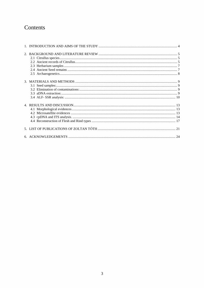
Contents 1. INTRODUCTION AND AIMS OF THE STUDY ........................................................................................... 4 2. BACKGROUND AND LITERATURE REVIEW ........................................................................................... 5
2.1 Citrullus species ........................................................................................................................................ 5 2.2 Ancient records of Citrullus ...................................................................................................................... 5 2.3 Herbarium samples ................................................................................................................................... 7 2.4 Ancient Seed remains ............................................................................................................................... 7 2.5 Archaeogenetics ........................................................................................................................................ 8
3. MATERIALS AND METHODS ...................................................................................................................... 9
3.1 Seed samples:............................................................................................................................................ 9 3.2 Elimination of contaminations: ................................................................................................................. 9 3.3 aDNA extraction: ...................................................................................................................................... 9 3.4 ALF- SSR analysis: ................................................................................................................................ 10
4. RESULTS AND DISCUSSION ...................................................................................................................... 13
4.1 Morphological evidences ........................................................................................................................ 13 4.2 Microsatellite evidences ......................................................................................................................... 13 4.3 cpDNA and ITS analysis. ....................................................................................................................... 14 4.4 Reconstruction of Flesh and Rind types ................................................................................................. 17
5. LIST OF PUBLICATIONS OF ZOLTAN TÓTH .......................................................................................... 21 6. ACKNOWLEDGEMENTS ............................................................................................................................ 24
4
1. INTRODUCTION AND AIMS OF THE STUDY
The aDNA samples extracted from remains of plants and animals supply unique materials for
the analysis of post-mortem DNA degradation (Brown, 1999; Threadgold and Brown, 2003),
domestication and microevolution (Salamini et al., 2002; Gugerli et al., 2005; Janick and
Paris, 2006). Analysis of aDNAs also provides crucial data concerning crop domestication
events that have occurred during previous centuries (Gyulai et al., 2001, 2006; Bacsó et al.
2004; Bisztray et al., 2004; Vaughan et al., 2007; Schlumbaum et al., 2008). Sequences of
intact aDNA fragments (Szabó et al., 2005; Lágler et al., 2005; Gyulai et al., 2006), and
complete genomes (and mitomes) (Cooper et al., 2001; Pääbo et al., 2004) of the extinct
organisms were also reconstructed by tools of archaeogenetics.
For safe aDNA analysis the most important step is to eliminate both the exogenously and
endogenously infected seeds, because bacterial or fungal DNA-remains can contaminate the
plant DNA being studied. The surface sterilization seeds and incubation for a month in tissue
culture provided optimal aseptic source for aDNA extraction (Tóth et al., 2008). Possible
DNA cross contamination from the laboratory investigations was also excluded by this aseptic
treatment.
Fossilized samples of Bangiomorpha pubescens (a red alga) from Canada proved that
chloroplasts had developed more than 1.2 billion years ago (Butterfield, 2000). Fossilization
coupled with charcoalification leaved floral morphology of ancient Nymphaeales perfectly
preserved at a site in Sayreville (NJ, USA) from the earliest Upper Cretaceous time
(Turonian, ca. 90 million years b.p.) (Crepet et al., 2004). Fossils of basal angiosperms
(Archaefructus sp) were also discovered from lower early Cretaceous period in China (Zhou
et al., 2003). Extinct angiosperm species (e.g. Pinus tuzsoni Greguss; syn. Pinuxylon
tarnocziense Tuzson) were identified from 20 million year old (Lower Miocene) site at
Ipolytarnóc (Hungary) (Andreánszky, 1966; Greguss, 1972; Erdei et al., 2007; Hably, 2006;
Süss, 2007).
The aims of the study presented were to analyse of aDNA fragments and sequences (ITS,
SSR, cpDNA, and lcyB gene) of 800-, 600- and 170-year-old Citrullus specimens together
with a comparison to modern cultivars (1 to 44) with the final aim of molecular and
morphological reconstruction of ancient Citrullus genotypes.

5
2. BACKGROUND AND LITERATURE REVIEW
2.1 Citrullus species
The monotypic genus Citrullus of family Cucurbitaceae is comprised of only four diploid (2n
= 4x = 22; 4.25 - 4.54 x 10⁸ bp; 0.42 pg DNA) species, including the annual watermelon
(Citrullus lanatus), the perennial colocynth (syn.: bitter apple) (Citrullus colocynthis), and
two wild species, growing in Kalahari Desert (Africa): the perennial, monoecious Citrullus
ecirrhosus with bitter-tasting fruit; and the recently identified, annual Citrullus rehmii (De
Winter, 1990) with pink and olive green spotted rind, mandarin sized non-edible fruits
(Robinson and Decker-Walters, 1997; Sarafis, 1999; Dane and Lang, 2004).
Species watermelon (C. lanatus) comprises two subspecies of domesticated watermelon (C.
lanatus lanatus) with its green fleshed, wild form growing in Namib desert (Sarafis, 1999),
and its wild ancestral citron melon (syn.: African tsamma) with also white flesh (C. lanatus
citroides) (Nakai, 1916; Kanda, 1951; Hanelt, 2001). Domesticated watermelon includes
diverse varieties, cultivars, feral forms, mutants (e.g. egusi melon: C. lanatus mucospermum;
Gusmini et al., 2004) and new crossed hybrids (e.g. the first seedless triploid hybrid
watermelon developed by Kihara 1951; and the first commercial ‘Allsweet’-type hybrid cv.
‘Sangria’ developed by Tom V. Williams, Syngenta Seeds, 1985) (Maynard et al., 2007)
The primary gene centre for watermelon is in South-West Africa, the domestication might
have occurred in Northern Africa implied by excavations of six thousand (Barakat, 1990) and
five thousand (Wasylikowa and van der Veen, 2004) year-old seed remains. Colocynth grown
as medicinal plant, citron as fodder crop, and the domesticated watermelon as fresh fruit
production have a history of production in the World.
2.2 Ancient records of Citrullus
The most ancient image of watermelon from Pharaohs tomb is known form 3,100 – 2,100
B.C. (Old Kingdom) (Manniche, 1989; Janick et al., 2007). Hieroglypf of watermelon is
known from 1,550 B.C. (Warid, 1995). The first figures of colocynth (C. colocynthis) (known
in Arabic as handel) were carved into the cedar wood in Solomon's temple (960 – 586 B.C.),
which is the only poisonous (medicinal) plant displayed in the temple (I. Kings 6:18a, Bible)
prior to a notes of Exodus from the time 480 year earlier recalling watermelon eaten in Egypt

6
(1,440 B.C.) (IV. Num. Moses 11:5, Bible). The first painting of colocynth remained in
excellent color form in the ancient Dioscorides codex (Dioscorides 1st CENT., and 512 A.D.)
The Greeks and Romans traveling to Egypt must to have known of watermelons probably
without discriminating it from colocynth and citron melon (Cox and van der Veen, 2009).
Pliny II. wrote about a ‘wild’ (probably the current colocynth) and two types of ‘cultivated’
colocynth (probably the current watermelon) one with pale green, and the other with grass
green rind, as it has been written: „…Another kind of wild gourd is called Colocynthis. The
fruit is smaller than the cultivated one, and full of seeds. The pale variety is more useful than
the grass-green one…” (Pliny 23-79 A.D.; Gilmore, 1919; Blake, 1981).
Six hundred years later, when the Iberian Peninsula was conquered by the Berbers (Moors)
led by Tarik Ibn Ziyad in 711 A.D., new watermelon types might enter Europe as recorded in
the ancient record of Book of Agricuture (Al-Awwam, 1158). In this book two cultivated
forms were compared, a black seed type (with dark-green rind which turns black when it
ripens) and a red seed type (with green rind which turns to yellow when it ripens) (Blake,
1981).
By 800 A.D. watermelons became popular in India and by 1,100 A.D. in China. The first
records of the name of watermelon in Hungarian ‘görög dinnye’ means ‘melon from Greece’
is known from 1395 (Finály, 1892). However, the first record on the name melon (‘dinna’) is
known from the 1000’s recorded in an ancient certificate (Szamota and Zolnai, 1902-1906)
without discriminating cucumbers from melons and watermelons.
Watermelon might have also been introduced to Europe through Crusades (Fischer, 1929) led
by either Richard I. the Lionheart (the 3rd Crusades, 1187-1192), or Endre II. the Hungarian
King of Árpád Dynasty who led the 5th Crusades (1217-1221). Watermelon spread through
Europe quickly and became very popular and commonly cultivated fruit of the Renaissance
Europe, with the second color illustration on the frescos in the Villa Farnesina, Rome, Italy,
1517 A.D. painted by Giovanni Martini da Udina (Janick and Paris, 2006). Watermelon
reached the New World after Columbus’ second voyage in 1493 and dispersed quickly among
American natives (Blake, 1981). One of the most ancient forms of small, round fruit with
thin, green rind, red flesh and black seeds has survived up to the recent times (Gilmore, 1919).

7
2.3 Herbarium samples
One of the oldest watermelon herbarium sample is available from G Bauhin’s (1560-1624)
collections (personal communication, Mark Spencer, The Natural History Museum, London,
UK), who named it Anguira citrullus about a hundred year time earlier than Linnaeus. No
watermelon herbarium sample remained from C Linnaeus (1753) collections, who named
watermelon as Cucurbita citrullus, and clocynth as Cucumis colocynthis (personal
communication, Arne Anderberg, The Linnean Herbarium, Swedish Museum of Natural
History, Stockholm, S).
2.4 Ancient Seed remains
The oldest plant remains with proven human activity have revealed only cereal seeds as wild
barley (H. spontaneum) and wild emmer (Triticum dicoccoides) from 19,000 b.p. at Ohalo II.,
river Jordan (Nadel et al., 2004, 2006; Piperno et al., 2004). The 17,310±310 b.p. site in
Korea (Chungbuk National University, South Korea) revealed the first ancient rice (Oryza
sativa) seed remains with extractable amount of aDNA (Suh et al., 2000).
The first Cucurbit seeds were excavated from the Spirit Cave (Hoabinh, Thailand) including
cucumber type Cucumis seeds at least 9,180 ± 360 b.p. as analyzed by C14 of bamboo
charcoal (Gorman, 1969).
The oldest, 6,000-year old Citrullus (watermelon, C. l. lanatus) seeds were excavated in
Helwan (Egypt, Africa), at a site 4.000 B.C. (Barakat, 1990). About 5,000-year old seeds
were excavated in Uan Muhuggiag, Lybia, Africa from a site 3.000 B.C. (Wasylikowa and
van der Veen, 2004). Large quantity of watermelon seeds were deposited in the Pharaoh’s
tombs of Pyramids as in Thebes (New Kingdom: 1,550-1,070 B.C.; stored in Agricultural
Museum, Dokki, Giza, Egypt) (Warid, 1995) and in the pyramid of Tutankhamum ca. 1,330
B.C. (Hepper, 1990; Kroll, 2000; Vartavan and Amorós, 1997). Watermelon seed remains
were also excavated from 1,550 B.C. in an old temple near Semna, Nubia (van Zeist, 1983).
Ancient watermelon seeds of the study presented were excavated at sites from 13th CENT.
(Debrecen, Hungary), and 15th CENT. (Budapest, Hungary) (Gyulai et al., 2006), and
collected form a herbal seed collection from 19th CENT. (Pannonhalma) (Vörös, 1971).

8
2.5 Archaeogenetics
The aDNA samples extracted from remains of plants and animals supply unique materials for
the analysis of post-mortem DNA degradation (Brown, 1999; Threadgold and Brown, 2003),
domestication and microevolution (Gugerli et al., 2005; Gyulai et al., 2006; Janick and Paris,
2006). Analysis of aDNAs also provides crucial data concerning crop domestication events
that have occurred during previous centuries (Gyulai F et al., 1992; Bacsó et al., 2004;
Bisztray et al., 2004; Vaughan et al., 2007; Schlumbaum et al., 2008). Sequences of intact
aDNA fragments (Szabó et al., 2005; Lágler et al., 2005), and complete genomes (mitomes)
(Cooper et al., 2001; Pääbo et al., 2004) of the extinct organisms were also reconstructed by
tools of archaeogenetics.
The aDNA (ancient DNA) samples recovered from excavated remains of plants and animals
supply unique materials for tracking domestication (Gugerli et al., 2005), microevolution
(Gyulai et al., 2006), migration (Dane and Liu, 2006) and the analysis of post-mortem DNA
degradation (Brown, 1999; Threadgold and Brown, 2003). A numbers of amplifications of
intact sequences of aDNA samples (Szabó et al., 2005; Lágler et al., 2005) and complete
genomes (Cooper et al., 2001; Pääbo et al., 2004) of the extinct organisms have also been
reported.

9
3. MATERIALS AND METHODS
3.1 Seed samples:
800-year-old seed remains of watermelon (Citrullus l. lanatus) were excavated at a site from
the 13th CENT. (Debrecen, Hungary). In total, 95,133 seed of 206 plant species were
identified. Of them 251 watermelon seeds were determined. The 600 year-old seeds were
excavated at a site from the 15th CENT. (8th well, Mansion Teleki, King’s Palace of Árpád
Dinasty, Buda Hill, Budapest, Hungary) (54,415 watermelon seeds in total) (Gyulai et al.,
2006). Sediment samples were processed by seed sorting and identified in the laboratory
according to Schermann (1966). The 19th CENT. (ca. 1836) seeds were collected from the
oldest botanical seed collection of Hungary (Pannonhalma) (Vörös, 1971). The collection is
recently exhibited at the Hungarian Agricultural Museum, Budapest (Hungary) (Hartyányi
and Nováky, 1975). For comparative analysis, forty-four modern Citrullus species and
varieties were included.
3.2 Elimination of contaminations:
Ancient seeds were surface sterilized by washing with regular detergent (for 3 min) and rinsed
three times with distilled water (for 3 min) followed by soaking in ethanol (70% v/v for 1
min) and a bleaching agent (8% NaOCl w/v, for 1 min) with a final rinses with sterile distilled
water according to general aseptic culture technique (Gyulai et al., 1992). Aseptic seeds were
incubated for seven days in tissue culture media to eliminate seeds contaminated either by
bacteria or fungi (Gyulai et al., 2006). Exogenously and endogenously contaminated seeds
infected by fungi and bacteria were eliminated from further analyses. Seeds of the modern
varieties were also surface sterilized.
3.3 aDNA extraction:
Aseptic seeds were ground in an aseptic mortar with liquid nitrogen followed by the DNA
extraction with CTAB (cethyltrimethylammonium bromide) protocol according to Biss et
al.,(2003), Yang, (1997) and Cooper and Poinar, (2000). The aDNA isolation was carried out
in a laminar air flow cabinets of archaeobotanical lab of the St Stephanus University, Gödöllő.
Seed DNA of modern cultivars (0.1 g) was also extracted in CTAB buffer, followed by an
RNase-A treatment (Sigma, R-4875) for 30 min at 37°C in each case. The quality and
quantity of extracted DNA were measured (2 µl) by a NanoDrop ND-1000 UV-Vis
spectrophotometer (NanoDrop Technologies, Delaware, USA – BioScience, Budapest,
10
Hungary). DNA samples were adjusted to concentration of 30 ng/µl with ddH2O and
subjected to PCR amplification (Gyulai et al., 2006).
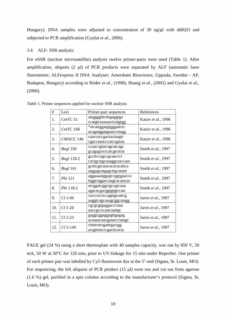
3.4 ALF- SSR analysis:
For nSSR (nuclear microsatellite) analysis twelve primer-pairs were used (Table 1). After
amplification, aliquots (2 µl) of PCR products were separated by ALF (automatic laser
fluorometer; ALFexpress II DNA Analyser; Amersham Bioscience, Uppsala, Sweden - AP,
Budapest, Hungary) according to Röder et al., (1998), Huang et al., (2002) and Gyulai et al.,
(2006).
Table 1. Primer sequences applied for nuclear SSR analysis PAGE gel (24 %) using a short thermoplate with 40 samples capacity, was run by 850 V, 50
mA, 50 W at 50°C for 120 min, prior to UV-linkage for 15 min under ReproSet. One primer
of each primer pair was labelled by Cy5 fluorescent dye at the 5’-end (Sigma, St. Louis, MO).
For sequencing, the left aliquots of PCR product (15 µl) were run and cut out from agarose
(1.6 %) gel, purified in a spin column according to the manufacturer’s protocol (Sigma, St.
Louis, MO).
# Loci Primer-pair sequences References
1. CmTC 51 attggggtttctttgaggtga ccatgtctaaaaactcatgtgg
Katzir et al., 1996
2. CmTC 168 *atcattggatgtgggattctc acagatggatgaaaccttagg
Katzir et al., 1996
3. CMACC 146 caaccaccgactactaagtc cgaccaaacccatccgataa
Katzir et al., 1996
4. Bngl 339 ccaaccgtatcagcatcagc gcagagctctcatcgtcttctt
Smith et al., 1997
5. Bngl 118-2 gccttccagccgcaaccct cactgcatgcaaaggcaaccaac
Smith et al., 1997
6. Bngl 161 gctttcgtcatacacacacattca atggagcatgagcttgcatattt
Smith et al., 1997
7. Phi 121 aggaaaatggagccggtgaacca ttggtctggaccaagcacatacac
Smith et al., 1997
8. Phi 118-2 atcggatcggctgccgtcaaa agacacgacggtgtgtccatc
Smith et al., 1997
9. Cl 1-06 caccctcctccagttgtcattcg aaggtcagcaaagcggcatagg
Jarret et al., 1997
10. Cl 1-20 cgcgcgtgaggaccctata aaccgcctcaatcaattgc
Jarret et al., 1997
11. Cl 2-23 gaggcggaggagttgagag acaaaacaacgaaacccatagc
Jarret et al., 1997
12. Cl 2-140 ctttttcttctgatttgactgg actgtttatcccgacttcacta
Jarret et al., 1997

11
LycB gene (lycopene β-cyclase) gene were probed by primer pairs designed by ‘Promer-3’
program based on the sequences of (NCBI EF183522, and EF183521) Bang et al., (2007)
(Table 2).
Table 2. Primer sequences applied for lcyB gene analysis The cpDNA were probed at two loci trnaVAL-rps12 (AJ970307; Al Jabani et al., 1994) and
ycf9-orf62 (AY522531, ay522537 and AY522539; Dane and Liu, 2007) (Table 3).
Table 3. Primer sequences applied for cpDNA For ITS analysis primers complementary to the evolutionary conserved spacer regions of the
nuclear ribosomal (rDNA) gene cluster of ITS1-5.8S-ITS2 (internal transcribed spacer) were
used. Primer pairs were designed by ‘Promer-3’ program based on the sequences of
AJ488232 (Hsiao et al., 1995) (ITSL and ITS4): ITS L: 5’-cgcgtttacaaacaaattgtcc-3’; ITS 4/1:
5’-acactacggtggttgatccg -3’; ITS4/2: 5’-gtccccccaaaggatgacgc-3’
PCR amplification: Hot Start PCR (Erlich et al., 1991) was combined with Touchdown PCR
(Don et al., 1991) using AmpliTaq GoldTM Polymerase. Reactions were carried out in a total
volume of 25 µl containing genomic DNA of 30-50 ng, 1 x PCR buffer (2.5 mM MgCl2),
dNTPs (200 µM each), 20 pmol of each primer and 1.0 U of Taq polymerase. Touchdown
PCR was performed by decreasing the annealing temperature by 1.0 oC / per cycle with each
of the initial 12 cycles (PE 9700, Applied Biosystems), followed by a ‘touchdown’ annealing
temperature for the remaining 25 cycles at 56 oC for 30 s with a final cycle of 72 oC for 10
min (transgene detection) and hold at 4 oC. The regular PCR cycles developed for prokariotic
cpDNA (Demesure et al., 1995; Dane et al., 2007) were performed as follows: initial
denaturing step at 94 oC for 5 min, followed by 35 cycles of 94 oC / 1 min - 55 oC / 1 min - 65 oC / 2 min, followed by a final extension step at 65 oC for 10 min and hold at 4 oC. A negative
control which contained all the necessary PCR components except template DNA was
# Loci Primer pair sequences Reference
1. LCYB 314 cctgttcttctggagttctt gaaaaagtgagtggtgtgagga
Bang et al., 2007 2. LCYB 1134
aatgatggtgtgaccattcaag cttacaatccaggctaccagg
# Loci primer-pair sequences References
1. clp12 agttcgagcctgattatccc gatgaacgctggcggcatgc
Al-Janabi et al., 1994
2. ycf 9 aattagagggaggggtctcttgc ataataggctagctctgcactgatg
Dane et al., 2004

12
included in the PCR runs. A minimum of three independent DNA preparations of each sample
was used. Amplifications were assayed by agarose (1.8 %, SeaKem LE, FMC) gel
electrophoresis (Owl system), stained with ethidium bromide (0.5 ng/µl) running at 80 V in 1
X TBE buffer. Each successful reaction with scorable bands was repeated at least twice.
Transilluminated gels were analyzed by the ChemilImager v 5.5 computer program (Alpha
Innotech Corporation - Bio-Science Kft, Budapest, Hungary). A negative control which
contained all the necessary PCR components except template DNA was included in the PCR
runs. Fragments were purified in a spin column (Sigma 5-6501) according to the
manufacturer’s protocol and subjected for sequencing.
Sequencing: PCR fragments were isolated from the agarose gel with a spin column (Sigma,
56501) and subjected to automated fluorescent DNA sequencing (ABI PRISM 3100 Genetic
Analyzer, Applied Biosystems, Hungary). Multiple Sequence Alignments (MSA). MSAs
were carried out by either BioEdit Sequence Alignment Editor (NCSU, USA) (Hall 1999),
CLUSTALW (Thompson et al., 1994) software programs or the on-line MULTALIN
computer program (http:// npsa_pbil.ibcp.fr/cgibin/npsa_automat.pl?page=/
NPSA/npsa_multalinan.html). BLAST (Basic Local Alignment Search Tool) analysis
(Altschul et al., 1997) was carried out by a computer program of NCBI (National Center for
Biotechnology Information).
Distance trees based on DNA sequences were edited by either MEGA4 (Tamura et al., 2007)
program. For MEGA4 the following steps were applied: Bootstrap Test of Phylogeny (1000);
Neighbor-Joining; Gaps (Complete deletions); Substitution model (Nucleotide Maximum
Composite Likelihood) according to Tamura et al., (2007). Diagrams were edited by
Microsoft Office Excel program (9625 West 76th Street, Eden Prairie, MN 55344, USA).
Cluster analysis was carried out by either or MEGA4 (Tamura et al., 2007) or SPSS-11
program package using the Jaccard Similarity Index (Jaccard, 1908) (Average Linkage, within
group) based on the presence versus absence of SSR alleles and nucleotides of DNA.
Data analysis: PIC value (polymorphism index content) of each SSR was calculated using the
formula of Anderson et al., (1993): PIC = 1 - ∑n-ipi2, where pi is the frequency of the ith
allele. Cluster analysis was carried out by the SPSS-11 program package using the Jaccard
Similarity Index (Jaccard, 1908) (Average Linkage, within group) based on the presence
versus absence of SSR alleles.

13
4. RESULTS AND DISCUSSION
4.1 Morphological evidences
Watermelon seeds excavated at both medieval sites appeared to be extremely well preserved
(Fig. 1) due to anaerobic conditions at site Debrecen (13th CENT.), and in the slime of a deep
well in Budapest (15th CENT.) (Gyulai et al., 2006). The herbarium sample seeds form the
19th CENT. were stored under precise conditions in glass containers (Vörös, 1971).
Figure 1. Seed morphology (groups and individuals) of ancient seeds (Citrullus l. lanatus) from the 13th CENT. (Debrecen, Hungary); 15th CENT. (Budapest, Hungary) and 18th CENT. (Citrullus l. citroides) (Pannonhalma, Hungary) (scale bar 1cm)
4.2 Microsatellite evidences
Microsatellites (SSR) of nuclear DNA (nDNA) analysis revealed a sum of 701 fragments of
23 SSR alleles at 12 SSR loci among the medieval (13th and 15th CENT.), 19th CENT. and
modern Citrullus specimens. Molecular dendrogram (fig. 2) based on nSSR fragment
diversity loci revealed that 13th CENT. sample (# 45) showed close genetic similarity to
modern watermelon cv. ‘Kecskeméti vh’ (#36) and the 15th CENT. sample (Budapest)
showed close similarity to cv. ‘Csárdaszállás’ (# 14). As expected from seed morphology, the
19th CENT. Citrullus showed close molecular similarity to modern citron melons (C. l.
citroides) (# 4-6) with white flesh color.
Allelic diversity of microsatellites were reliably detected in aDNAs of 300 – 1,100-year old
seagrass (Posidonia oceanica) (Raniello and Procaccini, 2002). SSRs were used to
morphologically reconstruct 600-year old melon (Cucumis staivus) (Szabó et al., 2005) and
millet (Panicum miliaceum) (Lágler et al., 2005; Gyulai et al., 2006). SSR analysis was also
applied to herbarium samples of common reed (Phargmites australis) of about 100-year-old
to track plant invasion in North America (Saltonstall, 2003).
a b c

14
Figure 2. Molecular (SSR) dendrogram (SPSS 16) of current varieties of colocynth (Citrullus colocynthis, # 1-3), citron melon (Citrullus lanatus citroides, # 4-6) and watermelon (Citrullus lanatus lanatus, # 7-44) compared to archaeological and herbarium samples (# 45-47)
4.3 cpDNA and ITS analysis.
Chloroplast genome specific primers provides highly sensitive methods for analyzing cpDNA
in the total DNA samples without using the former tedious ultracentrifuge separations (Al-
Jabani et al., 1994; Demesure et al., 1995 ; Dane et al., 2007). The level of SNP
polymorphism of cpDNA in plant species varies from nil, as in pearl millet (Pennisetum
glaucum) (Gepts and Clegg, 1989), compared to low level in soybean (Glycine soya) (Xu et
al., 2002), chestnut (Castanea sativa) (Fineschi et al., 2000) and pear (Pyrus ssp.) (Katayama
and Uematsu 2003), with high polymorphisms in wild beet (Beta vulgaris ssp maritima)
(Forcioli et al., 1998) and several tree species such as Prunus (Mohanty et al., 2001) and olive
(Olea europaea; Besnard et al., 2002). Citrullus species were found to also have high SNP
polymorphism with 6 SNP (1.73 %) along the tRNA-VAL – rps12 sequence (346 nt) (fig. 3).

15
Figure 3. Sequence analysis of cpDNA on tRNA-Val - rps12 loci of current varieties of colocynth (Citrullus colocynthis, # 1-3), citron melon (Citrullus lanatus citroides, # 4-6) and watermelon (Citrullus lanatus lanatus, # 7-44) compared to archaeological and herbarium samples (# 45-47).

16
The morphological reconstruction revealed that 13th CENT. watermelon might have been a
red flesh type (carrying the homozygote recessive alleles of lycb/ lycb gene) with green rind
similar to modern watermelon cv. ‘Kecskeméti vh’ (#36) (based on SSR similarities). The
15th CENT. watermelon might have been a yellow flesh type (carrying the homozygote
dominant alleles of lycB/ lycB gene) with striped rind similar to modern watermelon cv.
‘Csárdaszállás’ (# 14) (based on SSR similarities). As expected from seed morphology, the
19th CENT. Citrullus showed close molecular similarity to modern citron melons (C. l.
citroides) (# 4-6) with white flesh color (based on SSR similarities). The ITS-analysis of
rDNA supplied a further watermelon-specific marker by separating watermelons (C. lanatus
lanatus) from citrons (C. lanatus citroides) and colocynths (C. colocynthis). The rDNA
sequences of modern cultivars of colocynth (#1-3), citron melons (#4-6) and the 19th CENT.
Citrullus showed the same SNP pattern at the ITS1-5.8S-ITS2 locus, however modern citron
melon cv. ‘De Banat’ (#5) was found to carry a watermelon-specific rDNA allele at
heterozygote form, which might indicate an evolutionary step from bitter tasted citron
towards watermelons. In contrast, modern watermelon cv. ‘Túrkeve’ (#20) was found to carry
a citron-specific rDNA allele at heterozygote form, which indicates that watermelon cv.
‘Túrkeve’ (#20) is the most ancient watermelon type among the accessions studied (fig. 4).
Figure 4. Molecular (ITS1-5.8S-ITS2) dendrogram (MEGA 4) of current varieties of colocynth (Citrullus colocynthis, # 1-3), citron melon (Citrullus lanatus citroides, # 4-6) and watermelon (Citrullus lanatus lanatus, # 7-44) compared to archaeological and herbarium samples (# 45-47).

17
4.4 Reconstruction of Flesh and Rind types
Flesh color of watermelons varies from white to yellow - canary yellow - salmon yellow and
orange mainly due to pigment compositions of xanthophylls. The pink - red - purple colors
varies mainly due to pigment compositions of lycopenes. Genes coding for white flesh color
(w) were QTL-mapped (quantitative trait loci) on chromosome (syn.: linkage group) 6
(Hashizume et al., 1996). Genes responsible for yellow and red color were mapped on
chromosome 2. These gene loci indicate different transition colors between yellow and red
(canary yellow, pale yellow) (Hashizume et al., 2003). QTL responsible for red flesh color
had another locus on chromosome 8. This locus showed genetic linkage with QTL for high
sugar (brix value) content (Hashizume et al., 2003). This result strongly indicate the reason of
over numbered red flesh watermelons compared to cultivars with white and yellow flesh
colors, as selection for sweeter watermelons during domestication has been coupled with
selection for red flesh color at the same time (Hashizume et al., 2003). Further genetic loci for
color determination were recently determined, namely Y (red, dominant), yo (orange,
recessive), y (salmon yellow, recessive), C (canary yellow, dominant) and c (red, recessice),
respectively (reviewed in Bang et al., 2007).
The enzyme LYCB (lycopene β-cyclase) encoded by lycb gene play a central role in plant
color development by converting lycopene to carotenoids with ring structure. SNP (single
nucleotide polymorphism) markers in lycb gene, which discriminates yellow and red flesh
watermelons were developed recently (Bang et al., 2007).
The SNPs at the 1182th base pair (bp) were also found to be A=T (Adenine = Thymine) in all
CY (canary yellow) watermelons with homozygote dominant (lycB/lycB) genotype.
However, all red flesh watermelons had G≡C (Guanine ≡ Cytosine) bp at the 1182th bp
indicating a homozygote recessive genotype (lycb/lycb). The heterozygotes (lycB/lycb) also
encodes for dominant CY flesh type carrying both alleles (T=A and G ≡ C). No heterozygote
was found in the samples studied. The other SNP at the 518th nt (Bang et al., 2007) with the
opposite way of substitutions (G ≡ C in canary yellow, and T=A in red) were also found to be
identical. Amino acid sequence analysis of LYCB enzyme revealed that this SNP resulted in
an aminoacid substitutios from Ph to Val at the 226th amino acid locus, which might be impair
the catalytic function of lycB gene (Bang et al., 2007).
The 19th CENT. (#47) and 15th CENT. (#46) samples and all modern colocynths (#1-3), citrons
(#4-6), and yellow flesh watermelons (# 7-14) had the homozygote dominant CY-allele at

18
both SNP loci of lycB gene with G≡C bp (base pair) at the 518th nt, and T=A at 1182th nt,
respectively. The 13th CENT. (#45) sample and all red flesh modern watermelons (# 15 - 44)
had the recessive homozygote allele (lycb) at both SNP loci with T=A at the 518th nt and G≡C
at the 1182th nt, respectively (fig. 5). Red flesh watermelon appeared also in the painting of
Still Life with Melons and Carafe of White Wine (1603 A.D.) painted by Caravaggio (Janick,
2004; Janick et al., 2007). No colocynths and citrons were found with red flesh color
indicating that the recessive
allele of lycb gene
developed later during the
domestication.
Figure 5. Molecular (lycb) dendrogram (MEGA 4) of current varieties of colocynth (Citrullus colocynthis, # 1-3), citron melon (Citrullus lanatus citroides, # 4-6) and watermelon (Citrullus lanatus lanatus, # 7-44) compared to archaeological and herbarium samples (# 45-47). Watermelons fruit shapes are round to cylindrical. Unexpectedly, the most ancient, 5000 year
old record in Pharaohs tomb (3.100 – 2.100 B.C., Old Kingdom) shows not a round but an
elongated watermelon with green strips (Manniche, 1989; Janick et al., 2007). Fruit rind
(exocarp) color varies from pale green to dark green, with or without whitish strips, or small
whitish spots.
The most ancient European color wall paintings (1517) show watermelons with pale green
rinds (Janick et al., 2007) which indicate an ancient rind type, as a QTL locus (gs) responsible
for dark-green rind was found to be dominant over the light-green rind (Hashizume et al.,
2003).

19
SUMMARY
Watermelon seeds excavated at both medieval sites analyzed in the study presented appeared
to be extremely well preserved due to anaerobic conditions at site Debrecen (13th CENT.), and
in the slime of a deep well in Budapest (15th CENT.) covered by water, apparently used as
dust holes in the Middle Ages (Gyulai et al., 2006). The herbarium sample seeds form the 19th
CENT. were stored under precise conditions in glass containers (Vörös, 1971).
Molecular dendrogram of the study presented based on 701 SSR fragments in total identified
at eleven nuclear microsatellite (nSSR) loci revealed that middle age samples show close
lineages to ancient varieties currently growing in Hungary with red flesh colour. Allelic
diversity of microsatellites were reliably detected in aDNAs of 300 – 1,100-year old seagrass
(Posidonia oceanica) (Raniello and Procaccini, 2002). SSRs were used to morphologically
reconstruct 600-year old melon (Cucumis melo) (Szabó et al., 2005a) and millet (Panicum
miliaceum) (Lágler et al., 2005; Lágler, Gyulai et al., 2006). SSR analysis was also applied to
herbarium samples of common reed (Phargmites australis) of about 100-year-old to track
plant invasion in North America (Saltonstall, 2003). Results of seed morphology correlated
strongly to molecular results. The 13th -14th CENT. sample (Debrecen) showed similarity to
cv. ‘Kecskeméti vöröshéjú’; the 15th CENT. sample (Budapest) showed similarity to cv.
‘Belyj dlinnij’ (# 12). These results also reflect the preferential cultivation of red flesh – and
not yellow flesh- watermelon in the Middle Age of Hungary. Red flesh watermelon also
appeared in the painting of Still Life with Melons and Carafe of White Wine (1603 A.D.)
painted by Caravaggio (Janick, 2004; Janick et al., 2007). Molecular data obtained might
provide further tools for watermelon breeders. The 170-year-old herbarium sample
(Pannonhalma, Hungary) showed close molecular similarity to citron melon (Citrullus lanatus
citroides) cv.‘Újszilvás’ which reflects the importance of citron melon as fodder in the
Middle-Age Hungary.
Watermelons are divided into several morphological types; based on fruit weight as personal
size with to 2.7 kg / 6 lbs, icebox type to 6.8 kg/15 lbs, and picnic type above 6.8 kg/15 lbs.
Fruit shapes are round to cylindrical. Unexpectedly, the most ancient, 5000 year old record in
Pharaohs tomb (3.100 – 2.100 B.C., Old Kingdom,) shows not round but elongated fruit with
green strips (Manniche, 1989; Janick et al., 2007). Fruit rind (exocarp) varies from thin to
thick and brittle to tough with colors from pale green to dark green, with or without whitish

20
strips, or small whitish spots. The most ancient European color wall paintings (1517) show
watermelons with pale green rind (Janick et al., 2007) which indicate an ancient rind type, as
a QTL locus (gs) responsible for dark-green rind was found to be dominant over the light-
green rind (Hashizume et al., 2003).
Flesh color of watermelons varies from white; to yellow - canary yellow - salmon yellow -
orange mainly due to pigment compositions of xanthophylls. The pink - red - purple colors
mainly due to pigments of lycopenes. Genes coding for white flesh color (w) were QTL-
mapped (quantitative trait loci) on chromosome (syn.: linkage group) 6 (Hashizume et al.,
1996). Genes responsible for yellow and red color were mapped on chromosome 2. These
gene loci indicate the transition colors between yellow and red (canary yellow, pale yellow)
(Hashizume et al., 2003). QTL responsible for red flesh color had another locus on
chromosome 8. This locus showed genetic linkage with QTL for high sugar content
(Hashizume et al., 2003). This result strongly indicate the reason of over numbered red flesh
watermelons compared to cultivars with white and yellow flesh colors, as selection for
sweeter watermelons during domestication has been coupled with selection for red flesh color
at the same time (Hashizume et al., 2003). Some further genetic loci for color determination
were recently determined by breeding tools (crossings), namely Y (red, dominant), yo
(orange, recessive), y (salmon yellow, recessive), C (canary yellow, dominant) and c (red,
recessive), respectively (reviewed in Bang et al., 2007).
The enzyme LCYB (lycopene β-cyclase) encoded by lcyb gene play a central role in plant
color development by converting lycopene to carotenoids with ring structure. SNP (single
nucleotide polymorphism) markers in lcyb gene (NCBI EF183521) were which discriminated
yellow and red flesh watermelons (Bang et al., 2007). The 19th CENT. and 15th CENT.
samples along with modern colocynts, citrons, and modern (# 7-15) yellow flesh watermelons
(Citrullus lanatus lanatus) showed CY-type SNPs at both loci 518th (G≡C) and 1182th (T=A)
of lcyb gene. The 13th CENT. sample and all red flesh modern watermelons (# 16 - 44)
showed the red-type SNPs at both loci 518th (T=A) and 1182th (G≡C) of lcyb gene. No
colocynts and citrons were found with red flesh color.

21
5. LIST OF PUBLICATIONS OF ZOLTAN TÓTH Peer Reviewed Publications
1. Gyulai G, Z Szabó, B Wichmann, A Bittsánszky, LWaters Jr, Z Tóth, F Dane (2012) Conservation genetics - Heat Map analysis of nuSSRs of aDNA of archaeological watermelons (Cucurbitaceae, Citrullus l. lanatus) compared to current varieties. Genes, Genomes and Genomics 6 (SI1): 86-96.
2. Başli Ag, G Gyulai, Z Tóth, A Güner, Z Szabó, Vl Stakhov, L Murenyetz, Sg Yashina, L Heszky, Sv Gubin (2009) Light and Scanning Electron Microscopic Analysis of Silene stenophylla Seeds Excavated from Pleistocene-Age (Kolyma). Anadolu Univ J Sci and Technol 10:161-167.
3. Güner A, G Gyulai, Z Tóth, Ga Başli, Z Szabó, F Gyulai, L Heszky (2009) Grape (Vitis vinifera) seeds from Antiquity and the Middle Ages Excavated in Hungary - LM and SEM analysis. Anadolu Univ J Sci Technol 10:205-213.
4. Gyulai G, M Humphreys, R Lágler, Z Szabó, Z Tóth, A Bittsánszky, F Gyulai, L Heszky (2006) Seed remains of common millet from the 4th (Mongolia) and 15th (Hungary) centuries; AFLP, SSR, and mtDNA sequence recoveries. Seed Science Research 16:179-191. (IF: 1,892)
5. Bittsánszky A, G Gyulai, M Humphreys, G Gullner, Zs Csintalan, J Kiss, Z Szabó, R Lágler, Z Tóth, H Rennenberg, L Heszky and T Kőmíves (2006) RT-PCR analysis and stress response capacity of transgenic gshI-poplar clones (Populus x canescens) in response to paraquat exposure. Z Naturforschung 61c:699-730. (IF: 0,756)
6. Gyulai G, Z Tóth, Z Szabó, F Gyulai, R Lágler, L Kocsis, L Heszky (2009) Domestication Events of Grape (Vitis vinifera) from Antiquity and the Middle Ages in Hungary. Hung Agric Resi 2009/4.
7. Tóth Z, G Gyulai, L Horváth, Z Szabó, L Heszky (2007) Watermelon (Citrullus l. lanatus) production in Hungary from the Middle Ages. Hung Agric Res 2007/4: 14-19.
8. Lágler R, G Gyulai, Z Szabó, Z Tóth, A Bittsánszky, L Horváth, J Kiss, F Gyulai, L Heszky (2006) Molecular diversity of common millet (P. miliaceum) compared to archaeological samples excavated from the 4th and 15th centuries. Hung Agric Res 2006/1:14-19.
English Book Chapters International (1)
9. Gyulai G, Z Tóth, A Bittsánszky (2011) Flesh color reconstruction from aDNAs of Citrullus seeds from the 13th, 15th, and 19th cents (Hungary). In. Plant Archaeogenetics. Ed. by G Gyulai. Chapter 7. pp. 69-87. Nova Sci Publisher Inc., New York, USA. ISBN 978-1-61122-644-7.
10. Gyulai G, Z Tóth, A Bittsánszky, Z Szabó, G Gullner, J Kiss, T Kőmíves and L Heszky (2008) Gene up-regulation by DNA demethylation in 35S-gshI-transgenic poplars (Populus x canescens). in: Genetically Modified Plants: New Research Trends. Eds. T Wolf and J Koch, Nova Science Publisher, Inc. USA, Chapter 8, pp. 173-191. ISBN 978-1-60456-696-3.
Hungarian Scientific Journals (6) 11. Tóth Z, Gyulai G, Szabó Z, Heszky L (2008) Sejtmagi mikroszatellita és cpDNS szekveniák diverzitása
görögdinnyében (C. lanatus). Agr Vidékfejl Szemle 2008/3(1): 1-5. 12. Tóth Z, Gyulai G, Szabó Z, Horváth L, Gyulai G, Heszky L (2007) Mikroszatellita lokuszok evolúciója a
görögdinnyében (Citrullus lanatus) a középkor óta; (CT)3 deléció a (CT)26 nSSR-ban. Agrártud Közl 27: 125-134.
13. Szabó Z, Gyulai G, Tóth Z, Heszky L (2007) SNP elemzés az rDNA ITS-5.8S-ITS2 lokuszán a mai és a középkori sárgadinnyében (Cucumis melo). Agrártud Közl 27: 27: 120-124
14. Bittsánszky A, Gyulai G, Tóth Z, Horváth M, Fekete I, Szabó Z, Heltai Gy, Gullner G, Kőmíves T, Heszky L (2008) Molekuláris nyárfanemesítés (Populus x canescens) ökoremediációs alkalmazásra. Agr Vidékfejl Szemle vol. 3. 2008/2:184-189.
15. Lágler R, Gyulai G, Szabó Z, Tóth Z, Heszky L (2007) A köles (Panicum miliaceum) SSR- és ISSR szekvencia-stabilitása a 4. és 15. századi régészeti leletektől a mai fajtákig. Agrártud Közl 27: 10-19.
16. Horváth L, Gyulai G, Szabó Z, Lágler R, Tóth Z, Heszky L (2007) Morfológiai diverzitás sárgadinnyében (Cucumis melo); egy középkori típus fajtarekonstrukciója. Agrártud Közl 27: 84-90.
Peer Reviewed Proceedings English (4) 17. Tóth Z, G Gyulai, Z Szabó, F Gyulai, L Heszky (2008a) New Citrullus haplotypes at the tRNA-Val – rps12
locus of cpDNA.1 Cucurbitaceae 2008, Proceedings of the IXth EUCARPIA meeting on genetics and breeding of Cucurbitaceae (Pitrat M, ed), INRA, Avignon (France), May 21-24th, 2008, pp.335-310.
18. Tóth Z, G Gyulai, Z Szabó, A Bittsánszky, L Heszky (2008b) Genotype (nSSR) and haplotype (cpDNA) identification in watermelons (Citrullus l. lanatus). Gen. Meet. EUCARPIA, Valencia, Spain, pp. 253-257.

22
19. Szabó Z, G Gyulai, Z Tóth, A Bittsánszky, L Heszky (2008) Sequence diversity at the loci of nuclear SSRs and ITS1-5.8S-ITS2 of rDNA of 47 melon (Cucumis melo) cultivars and an extinct landrace excavated from the 15th century. General Meeting EUCARPIA, Valencia, Spain, pp. 244-249.
20. Szabó Z, G Gyulai, Z Tóth, L Heszky (2008) Morphological and molecular diversity of 47 melon (Cucumis melo) cultivars compared to an extinct landrace excavated from the 15th Century. Cucurbitaceae 2008, Proceedings of the IXth EUCARPIA meeting on genetics and breeding of Cucurbitaceae (Pitrat M, ed), INRA, Avignon (France), May 21-24th, 2008, pp.313-321.
Hungarian (4) 21. Tóth Z, Gyulai G, Kenéz Á, Szabó Z, Bittsánszky A, Lágler R, Gyulai F, Horváth L, Heszky L (2009)
Molekuláris domesztikáció a Citrullus nemzetségben az ITS (ITS1-5.8s-ITS2), NSSR, SNP (lcyb) és cpDNS (ycf9-orf62; trnval-rps12) lokuszokon. Hagyomány és haladás a növénynemesítésben. XV. Növénynemesítési Tudományos Napok, Budapest, ISBN: 978-963-508-575-0, pp. 507-511.
22. Tóth Z, G Gyulai, Z Szabó, L Heszky (2008c) Az nSSR és cpDNS lokuszok evolúciója a görögdinnyében (Citrullus lanatus). 4. Erdei Ferenc Tud Konf., Kecskemét, aug.27-28. (ed Ferenc Á.), II. kötet pp.866-870.
23. Kenéz Á, Gyulai G, Tóth Z, Szabó Zoltán, Lágler R, Heszky L, Gyulai F (2009) Római Kori (Keszthely-Fenékpuszta, (5. sz.) Növényleletek Azonosítása: I. Egyszikűek. Hagyomány és haladás a növénynemesítésben. XV. Növénynemesítési Tudományos Napok, Budapest, ISBN: 978-963-508-575-0, pp. 233-237.
24. Bittsánszky A, Gyulai G, Gullner G, Tóth Z, Kiss J, Szabó Z, Heszky L, Kőmíves T (2009) Paraquat-toleráns nyárfa in vitro szelekciója és molekuláris jellemzése. Hagyomány és haladás a növénynemesítésben. XV. Növénynemesítési Tudományos Napok, Budapest, ISBN: 978-963-508-575-0, pp. 31-35.
Peer Reviewed Presentations and Posters English (6) 22. Tóth Z, Gyulai G, A Başli, R Lágler, A Güner, Z Szabó, A Kis, A Bittsánszky, L Heszky (2007) Morphological and molecular reconstruction of 15th and
18th cent. watermelons (C. lanatus). IWGP14, 14th Int Symp Work Group for Palaeoethnobotany, 17-23 June, 2007, Kraków, Poland. 23. Tóth Z, G Gyulai, L Horváth, F Gyulai (2006) Molecular and morphological reconstruction of a medieval melon (Cucumis melo). 36th International
Symposium on Archaeometry (ISA 2006), május 2 - 6, Quebec City, Canada. 24. Lágler R, Gyulai G, A Güner, Z Tóth, A Kis, Z Szabó, Ga Başli, A Bittsánszky, L Heszky (2007) Morphological and molecular analysis of ancient
common millet (P. miliaceum) seeds. IWGP14, 14th Int Symp Work Group for Palaeoethnobotany, 17-23 June, 2007, Kraków, Poland. 25. Stakhov V, Gyulai G, Szabó Z, Kovács L, Murenyetz L, Lagler R, Tóth Z, Yashina S, Bittsánszky A, Heszky L, Gubin S (2007) Pleistocene-Age Silene
stenophylla seeds excavated in Russia – A Scanning Electron Microscopic Analysis. Botany and Plant biology Joint Congress, Chicago, Illiois, USA, July 7-11. ID: 2131
26. Szabó Z, Gyulai G, Ga Başli, Z Tóth, A Güner, R. Lágler, L Kovács, A Kis, A Bittsánszky, L Kocsis, L Heszky, F Gyulai (2007) Analysis of Grape (Vitis vinifera) Seeds from Antiquity and the Middle Ages Excavated in Hungary. 14th Int Symp Work Group for Palaeoethnobotany, 17-23 June, 2007, Kraków, Poland.
27. Szabó Z, Gyulai G, Kovács L, Tóth Z, Lagler R, Bittsanszky A, Kocsis L (2007) Ancient DNA analysis and morphology of grape seeds from antiquity and the middle agesexcavated in Hungary. Botany and Plant biology Joint Congress, Chicago, Illiois, USA, July 7-11. ID: 2119
Hungarian (14) 28. Tóth Z, Gyulai G, Szabó Z, Gyulai F, Horváth L, Kiss J, Heszky L (2008) Haplotípusok azonosítása görögdinnyében (Citrullus lanatus). XIVdik
Növénynemesítési Tudományos Napok, MTA Budapest, 2008. március 12. p. 14. 29. Tóth Z, Gyulai G, Lágler R, Szabó Z, Güner A, Basli Ga, Bittsánszky A, Kis A, Heszky L (2007) A régészeti genetika születése Magyarországon – (ct)3
deléció és (ct)4 inverzió a görögdinnye (C. lanatus) (ct)26-30 nSSR lókuszán a középkor óta. VII. Magyar Genetikai Kongresszus – 14. Sejt- és Fejlődésbiológiai Napok, Balatonfüred, Balatonfüred, április 15-17. P111, 178-179
30. Tóth Z, Gyulai G, Szabó Z, Mórocz S, Hajósné N M, Lágler R, Kálmán L, Kiss J, Bock I, Bittsánszky A, Koncz S, Bottka S, Heszky L (2006) SSR és cpSSR lókuszok Q-PCR és ALF-SSR elemzése kukorica (Zea mays L.) vonalakban és hibridekben, XII. Növénynemesítési Tudományos Napok, Budapest, p. 175.
31. Horváth L, Gyulai G, Szabó Z, Lágler R, Tóth Z, Bittsánszky A, Lehoczky P, Gyulai F, Heszky L (2007) Morfológiai diverzitás a sárgadinnyében (Cucumis melo); egy középkori típus fajtarekonstrukciója. XIII. Növénynemesítési Tudományos Napok, p.79.Budapest.
32. Kis A, Gyulai G, Lágler R, Szabó Z, Gubin Sv, Tóth Z, Stakhov Vl, Bittsánszky A, Yashina Sg, Heszky L (2007) Pleisztocén kori (Jégkorszak) Silene magvak morfológiai és molekuláris elemzése. VII. Magyar Genetikai Kongresszus-14dik Sejt- és Fejlődésbiológiai Napok, Balatonfüred, április 15-17. P064, 134.
33. Kis A, Gyulai G, Lágler R, Szabó Z, Gubin, Tóth Z, Stakhov Vl, Bittsánszky A, Yashina Sg, Heszky L (2007) Morfológiai és molekuláris elemzés jégkorszaki Silene magokban. XIII. Növénynemesítési Tudományos Napok, p.78.Budapest.
34. Lágler R, Gyulai G, Szabó Z, Bittsánszky A, Tóth Z, Kiss J, Gyulai F, Horváth L, Bock I, Holly L, Heszky L (2006) mtDNS RFLP-PCR, SSR és ISSR elemzés középkori köles (Panicum miliaceum L.) magvak DNS mintáiban, XII. Növénynemesítési Tudományos Napok, Budapest, p. 120.
35. Lágler R, Gyulai G, Szabó Z, Tóth Z, Lehoczky P, Bittsánszky A, Horváth L, Heszky L (2007) A köles (Panicum miliaceum) SSR- és ISSR szekvencia-stabilitás a kölesben (Panicum miliaceum) a középkortól napjainkig. XIII. Növénynemesítési Tudományos Napok, p.80. Budapest.
36. Lágler R., Gyulai G., Tóth Z., Szabó Z., Kis A., Bittsánszky A., Heszky L. (2007). A régészeti genetika születése Magyarországon – a köles (P. miliaceum) SSR- s ISSR szekvencia-stabilitása a 4.- és 15. századtól napjainkig. VII. Magyar Genetikai Kongresszus-14dik Sejt- és Fejlődésbiológiai Napok, Balatonfüred, április 15-17. P067, 138-139
37. Lehoczky P, Gyulai G, Szabó Z, Lágler R, Tóth Z, Kis A, Bittsánszky A, Bock I, Heszky L (2007) SNP-elemzés mai és középkori sárgadinnye (C. melo) rDNS ITS1-5.8S-ITS2 lókuszán. VII. Magyar Genetikai Kongresszus, Balatonfüred – 14dik Sejt- és Fejlődésbiológiai Napok, Balatonfüred, április 15-17. P109, 176-177
38. Lehoczky P, Gyulai G, Tóth Z, Lágler R, Szabó Z, Bittsánszky A, Bock I, Heszky L (2007) Az rDNS ITS1-5.8S-ITS2 lókusz SNP-elemzése mai és középkori sárgadinnyében (Cucumis melo). XIII. Növénynemesítési Tudományos Napok, p.81. Budapest.
39. Szabó Z, Gyulai G, Lágler R, Tóth Z, Bittsánszky A, Lehoczky P, Heszky L (2007) A sárgadinnye (Cucumis melo) A sárgadinnyében (Cucumis melo) sejtmagi mikroszatellita lókuszainak evolúciója a középkor óta. XIII. Növénynemesítési Tudományos Napok, p.66. Budapest.

23
40. Szabó Z, Gyulai G, Lágler R, Tóth Z, Kis A, Bittsánszky A, Heszky L (2007) A részégeti genetika születése Magyarországon – a (ctt)25 nSSR lókusz evolóciója a sárgadinnyében (C. melo) a középkor óta. VII. Magyar Genetikai Kongresszus – 14dik Sejt- és Fejlődésbiológiai Napok, Balatonfüred, Balatonfüred, április 15-17. P110, 177-178.
41. Szabó Z, Gyulai G, Tóth Z, Lágler R, Kiss J, Bittsánszky A, Gyulai F, Horváth L, Bock I, Holly L, Heszky L (2006) Sejtmagi mikroszatellita allélek diverzitása és ALF-SSR elemzése sárgadinnyében (Cucumis melo): molekuláris mikroevolúció a középkor óta, XII. Növénynemesítési Tudományos Napok, Budapest, p. 158.

24
6. ACKNOWLEDGEMENTS
I would like to express my sincere thanks to my supervisor, Prof. Dr. G. Gyulai whose
encouragement, approachability and willingness to help throughout the course of the PhD study has
been very much appreciated.
I also would like to extend my thanks to Prof. Dr. F. Gyulai for the contribution in the analysis of
archaeological Citrullus seed samples.
I am pleased to acknowledge the contribution of Prof. Dr. L Heszky head of the PhD School, and
Prof. Dr. E. Kiss, Head of the Institute of Genetics and Biotechnology, and the staff especially Dr.
Z. Szabó, Dr. A. Bittsánszky, and Predoc R. Lágler in the Szent István University, Gödöllő,
Hungary.
My particular thanks are due to my family for making my study so enjoyable.