doppler prediction of adverse perinatal outcome in ... · of lubchenco and colleagues in 19631...
TRANSCRIPT

http://dx.doi.org/10.5455/2320-1770.ijrcog20150223 Volume 4 · Issue 1 Page 119
International Journal of Reproduction, Contraception, Obstetrics and Gynecology
Mahale N et al. Int J Reprod Contracept Obstet Gynecol. 2015 Feb;4(1):119-130
www.ijrcog.org pISSN 2320-1770 | eISSN 2320-1789
Research Article
Doppler prediction of adverse perinatal outcome
in intrauterine growth restriction
Nina Mahale1, Bandana Khanal
1, Ajit Mahale
2*, Merwyn Fernandes
2,
Pallavi Rao2, Chanabasappa Chavadi
2
INTRODUCTION
Disturbance of normal fetal growth can result in
abnormal weight, body mass or body proportion at birth.
Growth is a dynamic process, and only the comparison of
absolute measurements with gestational age reference
ranges allows the detection of discrepancies between
expected and actual growth. The landmark observations
of Lubchenco and colleagues in 19631 showed that the
classification of neonates by birth weight percentile has a
significant prognostic advantage because it improves the
detection of neonates with intrauterine growth restriction
(IUGR) who are at increased risk for adverse health
status throughout life.2-4
The identification of pregnancies at risk for preventable
perinatal handicap is a primary goal of the obstetric care
provider. Pregnancies in which adverse intrauterine
1Department of Obstetrics & Gynecology, KMC Mangalore Manipal University, Mangalore, Karnataka, India
2Department of Radiodiagnosis KMC Mangalore Manipal University, Mangalore, Karnataka, India
Received: 28 December 2014
Accepted: 19 January 2015
*Correspondence:
Dr. Ajit Mahale,
E-mail: [email protected]
Copyright: © the author(s), publisher and licensee Medip Academy. This is an open-access article distributed under
the terms of the Creative Commons Attribution Non-Commercial License, which permits unrestricted non-commercial
use, distribution, and reproduction in any medium, provided the original work is properly cited.
ABSTRACT
Background: Objective of current study was to determine and compare the diagnostic performance of Doppler
ultrasonography of the fetal Middle Cerebral Artery (MCA) and Umbilical Artery (UA) for prediction of adverse
perinatal outcome in suspected intrauterine growth restriction (IUGR).
Methods: Fifty singleton pregnancies in third trimester of pregnancy with suspected intrauterine growth restriction
were examined with Doppler ultrasonography of fetal MCA and UA.
Results: Twenty patients of the fifty included patients had at least one major or minor adverse outcome. Major
adverse outcome included perinatal deaths which included both intrauterine deaths and early neonatal deaths, hypoxic
ischemic encephalopathy, intraventricular hemorrhage, periventricular leukomalacia, pulmonary hemorrhage,
necrotizing enterocolitis and septicemia. Minor outcomes included cesarean section for fetal distress, Apgar score
below 7 at 5 minutes and admission to Neonatal Intensive Care Unit (NICU) for treatment. MCA PI is the most
sensitive(90%) index in predicting any adverse perinatal outcome i.e. including both major and minor outcomes,
Positive Predictive Value (PPV) and specificity being greatest for MCA/UA PI (96.6%, 93.7%). For the major
adverse outcome most sensitive (86.6%) most specific (91.4%) and with highest PPV (81.2%) and NPV (94.1%), is
MCA/UA PI. Ratio of MCA/UAPI is more sensitive (90%) than PI of both the arteries alone for overall prediction of
adverse perinatal outcome.
Conclusions: Thus we conclude that the Doppler studies of the multiple vessels in the fetoplacental unit can help in
the monitoring of the compromised fetus and can help us predicting neonatal morbidity. This may be helpful in
determining the optimal time of deliveries in pregnancies complicated by IUGR.
Keywords: Adverse outcome, Doppler, Pregnancy IUGR
DOI: 10.5455/2320-1770.ijrcog20150223

Mahale N et al. Int J Reprod Contracept Obstet Gynecol. 2015 Feb;4(1):119-130
International Journal of Reproduction, Contraception, Obstetrics and Gynecology Volume 4 · Issue 1 Page 120
conditions result in failure of the fetus to reach its growth
potential constitute such a high risk group. Next to
prematurity, IUGR is the second most leading cause of
perinatal mortality.5
Compared to the appropriately grown
counterparts, perinatal mortality rates in growth restricted
neonates are 6-10 times greater; perinatal mortality rates
as high as 120 per 1000 for all cases of IUGR and 80 per
1000 after exclusion of anomalous infants have been
reported. As many as 53% of preterm still births and 26%
of term still births are growth restricted.6
In survivor, the
incidence of intrapartum asphysxia may be as high as
50%.6
Prevention of some perinatal complications that
lead to adverse outcomes in growth restricted fetus is
possible with appropriate perinatal identification and
management.
An important goal in contemporary obstetrics is to
identify the growth restricted; fetus at risk for perinatal
morbidity and mortality. Traditionally, serial sonographic
evaluations of fetal biometry and amniotic fluid volume
has been used along with antepartum Fetal Heart Rate
(FHR) testing to assess the condition of a potentially
growth restricted fetus.7
Doppler ultrasound has become available as a non-
invasive technique for identifying fetal growth restriction
associated with an abnormal uteroplacental and/or fetal
circulation.8-10
Because a growth restricted fetus subjected to
compromised blood flow is particularly at high risk for
hypoxia, Doppler flow velocimetry studies have been
advanced as an important technique for distinguishing the
compromised small fetus from a small fetus that is
unlikely to experience, serious perinatal complications.1
METHODS
The study included 50 pregnant women in third trimester
suspected of IUGR (n=50).
Type of study and duration
i. Study design: Longitudinal
ii. Type of study: Prospective
iii. Place of study: KMC Mangalore
iv. Duration of study: Nov 2008 to March 2013
Inclusion criteria
i. Pregnant women in third trimester suspected of
having IUGR
ii. Giving informed consent
iii. Reliable dates and first trimester
ultrasonography
iv. High risk pregnancies (preeclampsia, anemia,
heart disease etc. with the clinical manifestation
of growth restriction)
v. Other medical illness complicating pregnancy
Exclusion criteria
i. Wrong dates
ii. Multiple gestation
iii. Congenital anomalies
Studies of various fetal vessels were performed using
color Doppler ultrasound (GE Voluson-730 Expert,
Philips-Envisor HD and GE Logic-5) curvilinear probe
with a high pass filter.
The following vessels were studied with the mother in a
recumbent position during fetal inactivity and apnea.
1. Umbilical Artery (UA) (Figure 1)
2. Middle Cerebral Artery (MCA) (Figure 10)
The above vessels were located in the standard plane:
The umbilical artery measurements were made from free
loop of cord midway between the placental and
abdominal wall insertion.
The middle cerebral artery was located in a transverse
plane at the level of the lesser wing of the sphenoid bone
with sample gate placed on proximal portion of the
vessel.
Flow velocity wave forms, the resistance index,
pulsatility index, systolic/diastolic ratio of umbilical
artery, middle cerebral artery were noted.
1. Doppler study is considered abnormal when
resistance and pulsatility index of umbilical artery
(>2 SD), middle cerebral artery (<5th
percentile, and
uterine artery (>2 SD) for the gestational age
according to the standard reference values; the
reference value of umbilical artery P.I. and
cerebroumbilical ratio are according to Dandolo
Gramellini et al.12
and MCA PI ratio & Umbilical
artery RI reference values were taken according to
Kurmanavicius et al.11
2. The ratios examined were considered abnormal
when PI of MCA/UA <1.12
Fetal outcome
Fetal outcome was studied under major and minor
adverse outcomes.
1. Major adverse outcomes were
Perinatal deaths - including intrauterine and early
neonatal deaths. Major complications like hypoxic
ischemic encephalopathy, intraventricular hemorrhage,
periventricular leukomalacia, pulmonary hemorrhage,
necrotizing enterocolitis and septicemia.

Mahale N et al. Int J Reprod Contracept Obstet Gynecol. 2015 Feb;4(1):119-130
International Journal of Reproduction, Contraception, Obstetrics and Gynecology Volume 4 · Issue 1 Page 121
2. Minor outcomes include-cesarean delivery for fetal
distress, APGAR score below 7 at 5 minutes,
admission to NICU for treatment.
The patients are followed by serial Doppler assessment
and non-stress test and the results of the last Doppler
examination within 10 days of delivery are considered, in
the subsequent correlation with perinatal outcomes.
Figure 1: Normal umbilical artery waveform.
Figure 2: Umbilical artery waveform showing absent
and diastolic flow.
Figure 3: Waveform showing reserved and diastolic
flow.
Figure 4: Umbilical artery - Doppler waveform showing and systole (Sys) diastole (D) are identified in green note
that diastole is less at 20 weeks (yellow ellipse) than at 36 weeks (red ellipse).
Figure 5: Umbilical artery shows absent diastolic flow
and reverse diastolic flow.
Figure 6: Normal uterine artery waveform in
pregnant women.

Mahale N et al. Int J Reprod Contracept Obstet Gynecol. 2015 Feb;4(1):119-130
International Journal of Reproduction, Contraception, Obstetrics and Gynecology Volume 4 · Issue 1 Page 122
Figure 7: Abnormal uterine artery waveform.
Figure 8: Normal uterine artery waveform during 24
weeks of gestation.
Figure 9: Waveform from the uterine artery at 24
weeks of gestation in a pregnancy with impaired
placentation; in early diastole there is notch (yellow
arrow) and in late diastole there is decreased flow
(orange arrow).
Figure 10: Normal middle cerebral artery waveform.
Figure 11: Abnormal middle cerebral artery
waveform.
RESULTS
In our study the mean maternal age was 27.24 years.
primigravidas comprised 66% (n=33) of the studied
population and 34% 9 (n=17) were multiparas. The
maximum gestational age at which the delivery occurred
was 32-36 weeks of gestation. Even when the parental
resources were poor leading to non-affordability of NICU
care, pregnancy was terminated by induction for maternal
indications like severe preeclampsia.
Preeclampsia with IUGR formed the majority of the
study population (58%, n=29), followed by IUGR alone
(36%, n=18), and anemia with IUGR (6%, n=13) (Table
1).
Table 1: Patient distribution.
Frequency Percentage
Preeclampsia with IUGR 29 58.0
Anaemia with IUGR 3 6.0
IUGR 18 36.0
Total 50 100.0
Figure 12: Patient distribution.
Modes of delivery
Out of 50 pregnancies, 10 (20%) had spontaneous onset
of labour and delivered. 38% (n=18) induced for various
reasons and 44% (n=22) were taken up for caesarean
section (Table 2).
Preeclampsia
with IUGR
58% N=29 Anemia with
IUGR 6%
N=3
IUGR 36%
n=18

Mahale N et al. Int J Reprod Contracept Obstet Gynecol. 2015 Feb;4(1):119-130
International Journal of Reproduction, Contraception, Obstetrics and Gynecology Volume 4 · Issue 1 Page 123
Table 2: Modes of delivery.
Frequency Percentage
Induced 18 36.0
LSCS 22 44.0
Spontaneous 10 20.0
Total 50 100.0
Figure 13: Modes of delivery.
Incidence of meconium stained liquor
Out of fifty pregnancies 54% (n=27) had Meconium
stained amniotic fluid at delivery (Figure 14).
Figure 14: Meconium.
Indication for LSCS
Out of these 22 LSCS six were electively taken up and 16
were emergency out of which 10 (45%) were done for
fetal distress, 7 (32%) for severe uncontrolled
preeclampsia and 5(23%) for various other reasons
(Table 3).
Table 3: Indication for LSCS.
Frequency Percentage
Fetal distress 10 45
Severe preeclampsia 7 32
Others 5 23
Total 22 100.0
Figure 15: Indication for LSCS.
Term and preterm
There were a significant number of preterm babies in our
study (60%, n=30). This included one case of intrauterine
death which was delivered at 33 weeks (Table 4).
Table 4: Term and preterm.
Frequency Percentage
Preterm babies 30 60.0
Term babies 20 40.0
Total 50 100.0
Figure 16: Term and preterm.
Admission to NICU
Out of 49 live births 37 babies were admitted in NICU
for various reasons. Out of that 6 babies expired and the
cause of death was septicemia and very low birth weight.
Of the remaining, 21 babies required admission in NICU
for more than 5 days for various reasons (Table 5).
Table 5: Admission to NICU.
Frequency Percentage
No 12 24.0
Yes 37 76.0
Total 49 100.0
Induced 36%
(n=18)
LSCS 44%
(N=22)
Sppmtaneous
20% (n=10)
MSAF
54%
n=27
Clear
46%
n=23
10%
7%
5%
Fetal distress Severe preeclampsia Others
Preterm
babies 60%
(n=30) Term babies
40%
(n=20)67%

Mahale N et al. Int J Reprod Contracept Obstet Gynecol. 2015 Feb;4(1):119-130
International Journal of Reproduction, Contraception, Obstetrics and Gynecology Volume 4 · Issue 1 Page 124
Figure 17: Admission to NICU.
Neonatal complications
Out of the 49 live births 25 babies developed some
complications. The majority were septicemia (44%,
n=11) following which was hyperbilirubinemia (36%,
n=9) followed by hypoglycemia (12%, n=3). There were
one each of thrombocytopenia and hypothermia. Out of
all these babies which were admitted in NICU 3 babies
expired of septicemia. In our study major complications
like necrotizing enterocolitis (NEC) intraventricular
hemorrhage (IVH), pulmonary hemorrhage and
periventricular leukomalacia did not occur (Table 6).
Table 6: Neonatal complications.
Complication Frequency Percentage
Hyperbilirubinemia 9 36
Hypoglycemia 3 12
Hypothermia 1 4
Septicemia 11 44
Thrombocytopenia 1 4
Total 25 100
Figure 18: Neonatal complications.
Birth weight distribution
Maximum of the babies had birth weight between 1-15
kg at birth including one intrauterine death (IUD) which
had birth weight of 1 kg. Two babies weighed less than
1kgs (Table 7).
Table 7: Birth weight distribution.
Birth weight
(kg) Frequency
<1 2
1-1.5 20
1.5-2 13
2-2.5 15
Total 50
Figure 19: Birth weight distribution.
APGAR at 5 minutes
Of all the live births (n=49) 23 babies had APGAR at five
minutes below 7.
Table 8: APGAR at 5 minutes.
Frequency Percentage
Abnormal (<7) 23 47.0
Normal (≥ 7) 26 53.0
Total 49 100.0
Figure 19: APGAR at 5 minutes.
Perinatal outcome
Considering the final outcome, out of fifty pregnancies
one had an intrauterine death and 49 were live births. Out
of these 43 babies has good outcome and were discharged
n=12
(24%)
n=37
(76%)
1 2
02468
1012 9
3
1
11
1
0
5
10
15
20
<1 1-1.5 1.5-2 2-2.5
2
20
13 15
Fre
qu
ency
Birth weight (kg)
Abnormal
>n=23
(47%) Normal ≥7
n=26
(53%)

Mahale N et al. Int J Reprod Contracept Obstet Gynecol. 2015 Feb;4(1):119-130
International Journal of Reproduction, Contraception, Obstetrics and Gynecology Volume 4 · Issue 1 Page 125
home despite of being admitted in NICU for various
reasons. During the course of treatment in NICU 6 babies
had early neonatal death and septicemia accounted for 5
deaths. One of the baby who expired had only
hyperbilirubinemia during the stay in NICU (Table 9).
Table 9: Perinatal outcome.
Frequency Percentage
Early neonatal death 6 12.0
Intrauterine fetal death 1 2.0
Survival 43 86.0
Total 50 100.0
Figure 20: Perinatal outcome.
Umbilical artery RI in prediction of major and minor
outcome
Sensitivity: 60.15%
Specificity: 76.66%
Positive predictive value: 63.15%
Negative predictive value: 74.19%
Diagnostic accuracy: 74%
Table 10: Umbilical artery RI in prediction of major
and minor outcome.
Umbilical
artery RI
Perinatal outcome
(major and minor)
Present Absent Total
Abnormal 12 7 19
Normal 8 23 31
Total 20 30 50
Umbilical artery PI in prediction of major and minor
outcome
Sensitivity: 55%
Specificity: 86.66%
Positive predictive value: 73.33%
Negative predictive value: 74.28%
Diagnostic accuracy: 74%
Table 11: Umbilical artery PI in prediction of major
and minor outcome.
Umbilical
artery PI
Perinatal outcome
(major and minor)
Present Absent Total
Abnormal 11 4 15
Normal 9 26 35
Total 20 30 50
Abnormal umbilical artery RI in prediction of major
and minor outcome
Sensitivity: 80%
Specificity: 86.66%
Positive predictive value: 80%
Negative predictive value: 86.6%
Diagnostic accuracy: 84%
Table 12: Abnormal umbilical artery RI in prediction
of major and minor outcome.
Umbilical
artery
waveform
Perinatal outcome
(major and minor)
Present Absent Total
Abnormal 16 4 20
Normal 4 26 30
Total 20 30 50
Abnormal umbilical artery waveforms (Figure 5)
Raised S/D ratio:14
Absent diastolic flow: 2 (Figure 2)
Reversed end diastolic flow: 4 (Figure 3)
Out of the 14 raised S/D ratio flow 8 had major
adverse outcome that included 5 cases of septicemia
and three cases of early neonatal death
Out of the two absent diastolic flow-emergency
LSCS was done for non-reactive NST for one case.
Though the baby had suffered septicemia it
recovered and survived. But the other one for whom
an elective LSCS was done the baby succumbed to
death though the baby only had suffered from
hyperbilirubinemia during the stay at NICU
0 10 20 30 40 50
Earthy Neonatal Death
Intrauterine fetal death
Survival
6
1
43
Mahale N et al. Int J Reprod Contracept Obstet Gynecol. 2015 Feb;4(1):119-130
International Journal of Reproduction, Contraception, Obstetrics and Gynecology Volume 4 · Issue 1 Page 126
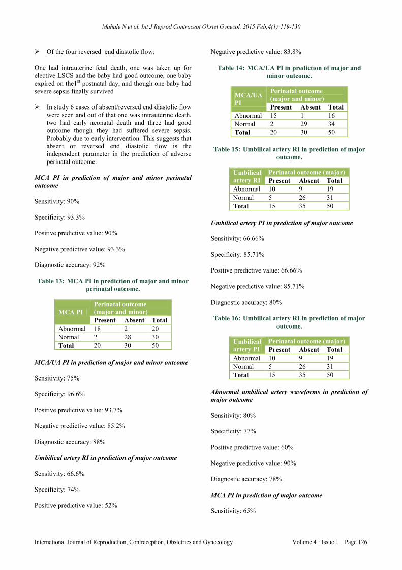
Of the four reversed end diastolic flow:
One had intrauterine fetal death, one was taken up for
elective LSCS and the baby had good outcome, one baby
expired on the1st postnatal day, and though one baby had
severe sepsis finally survived
In study 6 cases of absent/reversed end diastolic flow
were seen and out of that one was intrauterine death,
two had early neonatal death and three had good
outcome though they had suffered severe sepsis.
Probably due to early intervention. This suggests that
absent or reversed end diastolic flow is the
independent parameter in the prediction of adverse
perinatal outcome.
MCA PI in prediction of major and minor perinatal
outcome
Sensitivity: 90%
Specificity: 93.3%
Positive predictive value: 90%
Negative predictive value: 93.3%
Diagnostic accuracy: 92%
Table 13: MCA PI in prediction of major and minor
perinatal outcome.
MCA PI
Perinatal outcome
(major and minor)
Present Absent Total
Abnormal 18 2 20
Normal 2 28 30
Total 20 30 50
MCA/UA PI in prediction of major and minor outcome
Sensitivity: 75%
Specificity: 96.6%
Positive predictive value: 93.7%
Negative predictive value: 85.2%
Diagnostic accuracy: 88%
Umbilical artery RI in prediction of major outcome
Sensitivity: 66.6%
Specificity: 74%
Positive predictive value: 52%
Negative predictive value: 83.8%
Table 14: MCA/UA PI in prediction of major and
minor outcome.
MCA/UA
PI
Perinatal outcome
(major and minor)
Present Absent Total
Abnormal 15 1 16
Normal 2 29 34
Total 20 30 50
Table 15: Umbilical artery RI in prediction of major
outcome.
Umbilical
artery RI
Perinatal outcome (major)
Present Absent Total
Abnormal 10 9 19
Normal 5 26 31
Total 15 35 50
Umbilical artery PI in prediction of major outcome
Sensitivity: 66.66%
Specificity: 85.71%
Positive predictive value: 66.66%
Negative predictive value: 85.71%
Diagnostic accuracy: 80%
Table 16: Umbilical artery RI in prediction of major
outcome.
Umbilical
artery PI
Perinatal outcome (major)
Present Absent Total
Abnormal 10 9 19
Normal 5 26 31
Total 15 35 50
Abnormal umbilical artery waveforms in prediction of
major outcome
Sensitivity: 80%
Specificity: 77%
Positive predictive value: 60%
Negative predictive value: 90%
Diagnostic accuracy: 78%
MCA PI in prediction of major outcome
Sensitivity: 65%

Mahale N et al. Int J Reprod Contracept Obstet Gynecol. 2015 Feb;4(1):119-130
International Journal of Reproduction, Contraception, Obstetrics and Gynecology Volume 4 · Issue 1 Page 127
Specificity: 80.5%
Positive predictive value: 65%
Negative predictive value: 93.3%
Diagnostic accuracy: 82%
Table 17: Abnormal umbilical artery waveforms in
prediction of major outcome.
Umbilical
artery
waveforms
Perinatal outcome (major)
Present Absent Total
Abnormal 12 8 20
Normal 3 27 30
Table 18: MCA PI in prediction of major outcome.
MCA PI Perinatal outcome (major)
Present Absent Total
Abnormal 13 7 20
Normal 2 28 30
Total 15 35 50
MCA/UA PI in prediction of major outcome
Sensitivity: 86.6%
Specificity: 91.4%
Positive predictive value: 81.2%
Negative predictive value: 94.1%
Diagnostic accuracy: 90%
Table 19: MCA/UA PI in prediction of major
outcome.
MCA/UA
PI
Perinatal outcome (major)
Present Absent Total
Abnormal 13 3 16
Normal 2 32 34
Total 15 35 50
Doppler indices in prediction of adverse perinatal
outcome
The Table 20 depicts the efficacy of various Doppler
parameters in prediction of adverse perinatal outcome.
Statistical analysis shows that MCA PI is the most
sensitive parameter in the prediction of adverse perinatal
outcome (90%), specificity being the highest for
MCA/UAPI (96.6%). Positive predictive value is highest
for MCA/UA PI (93.7%) and the negative predictive
value highest being for the MCA PI (93.3%).
Table 20: Doppler indices in prediction of adverse
perinatal outcome.
Sensitivity Specificity PPV NPV
Umbilical A RI 60.1 76.6 63.1 74.1
Umbilical A PI 55 86.6 73.3 74.2
Umbilical A S/D 80 86.6 80 86.6
MCA PI 90 93.3 90 93.3
PIMCA/UA 75 96.6 93.7 85.2
Efficacy of Doppler in predicting adverse perinatal
outcome (major)
The Table 21 depicts the comparison of our study with
the study by BN Lakhhar et al. for various parameters to
predict the major perinatal outcome.
Table 21: Efficacy of Doppler in predicting adverse perinatal outcome (major).
Sensitivity (%) Specificity (%) PPV (%) NPV (%)
BNL Present BNL Present BNL Present BNL Present
UARI 58 66.6 71.7 74 35 52 86.8 83.8
UAPI 58 66.6 56.5 85.71 35 66.6 86.8 85.71
UA S/D 75 80 41.3 77.1 25 60 86.8 90
MCA PI 66.6 65 71.7 80.5 47 65 90 93.5
MCA/UAPI 66.6 86.6 73.9 91.4 40 81.2 89.4 94.1
UA-Umbilical artery, MCA-Middle cerebral artery, RI-Resistive index, PI-Pulsatility index, S/D-Systolic/diastolic ratio
MCA/UA PI was the most sensitive (86.6%), most
specific (91.4%), with highest positive predictive value
(81.2%) and negative predictive value (94.1%) in
prediction of major perinatal outcome which was
comparable to the study by BN Lakhhar et al. where
sensitivity was highest for MCA/UA PI (66.6%) and with
highest specificity (73.9%).
Efficacy of PI in predicting adverse perinatal outcome
(major & minor)
In considering Pulsatility Index (PI) for prediction of
major and minor adverse outcome sensitivity was highest
for MCA PI (90%) whereas in a study by Dandolo Gram
et al it was for MCA/UA PI (68%) and in a study by BN
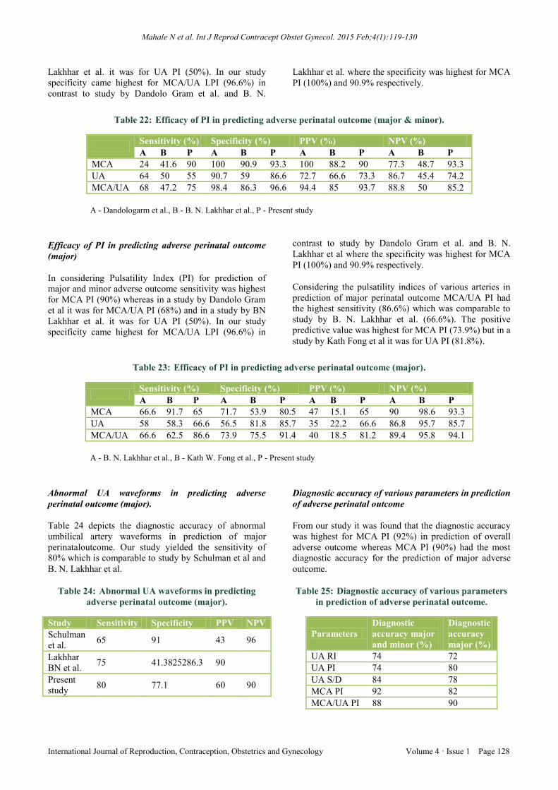
Mahale N et al. Int J Reprod Contracept Obstet Gynecol. 2015 Feb;4(1):119-130
International Journal of Reproduction, Contraception, Obstetrics and Gynecology Volume 4 · Issue 1 Page 128
Lakhhar et al. it was for UA PI (50%). In our study
specificity came highest for MCA/UA LPI (96.6%) in
contrast to study by Dandolo Gram et al. and B. N.
Lakhhar et al. where the specificity was highest for MCA
PI (100%) and 90.9% respectively.
Table 22: Efficacy of PI in predicting adverse perinatal outcome (major & minor).
Sensitivity (%) Specificity (%) PPV (%) NPV (%)
A B P A B P A B P A B P
MCA 24 41.6 90 100 90.9 93.3 100 88.2 90 77.3 48.7 93.3
UA 64 50 55 90.7 59 86.6 72.7 66.6 73.3 86.7 45.4 74.2
MCA/UA 68 47.2 75 98.4 86.3 96.6 94.4 85 93.7 88.8 50 85.2
A - Dandologarm et al., B - B. N. Lakhhar et al., P - Present study
Efficacy of PI in predicting adverse perinatal outcome
(major)
In considering Pulsatility Index (PI) for prediction of
major and minor adverse outcome sensitivity was highest
for MCA PI (90%) whereas in a study by Dandolo Gram
et al it was for MCA/UA PI (68%) and in a study by BN
Lakhhar et al. it was for UA PI (50%). In our study
specificity came highest for MCA/UA LPI (96.6%) in
contrast to study by Dandolo Gram et al. and B. N.
Lakhhar et al where the specificity was highest for MCA
PI (100%) and 90.9% respectively.
Considering the pulsatility indices of various arteries in
prediction of major perinatal outcome MCA/UA PI had
the highest sensitivity (86.6%) which was comparable to
study by B. N. Lakhhar et al. (66.6%). The positive
predictive value was highest for MCA PI (73.9%) but in a
study by Kath Fong et al it was for UA PI (81.8%).
Table 23: Efficacy of PI in predicting adverse perinatal outcome (major).
Sensitivity (%) Specificity (%) PPV (%) NPV (%)
A B P A B P A B P A B P
MCA 66.6 91.7 65 71.7 53.9 80.5 47 15.1 65 90 98.6 93.3
UA 58 58.3 66.6 56.5 81.8 85.7 35 22.2 66.6 86.8 95.7 85.7
MCA/UA 66.6 62.5 86.6 73.9 75.5 91.4 40 18.5 81.2 89.4 95.8 94.1
A - B. N. Lakhhar et al., B - Kath W. Fong et al., P - Present study
Abnormal UA waveforms in predicting adverse
perinatal outcome (major).
Table 24 depicts the diagnostic accuracy of abnormal
umbilical artery waveforms in prediction of major
perinataloutcome. Our study yielded the sensitivity of
80% which is comparable to study by Schulman et al and
B. N. Lakhhar et al.
Table 24: Abnormal UA waveforms in predicting
adverse perinatal outcome (major).
Study Sensitivity Specificity PPV NPV
Schulman
et al. 65 91 43 96
Lakhhar
BN et al. 75 41.3825286.3 90
Present
study 80 77.1 60 90
Diagnostic accuracy of various parameters in prediction
of adverse perinatal outcome
From our study it was found that the diagnostic accuracy
was highest for MCA PI (92%) in prediction of overall
adverse outcome whereas MCA PI (90%) had the most
diagnostic accuracy for the prediction of major adverse
outcome.
Table 25: Diagnostic accuracy of various parameters
in prediction of adverse perinatal outcome.
Parameters
Diagnostic
accuracy major
and minor (%)
Diagnostic
accuracy
major (%)
UA RI 74 72
UA PI 74 80
UA S/D 84 78
MCA PI 92 82
MCA/UA PI 88 90

Mahale N et al. Int J Reprod Contracept Obstet Gynecol. 2015 Feb;4(1):119-130
International Journal of Reproduction, Contraception, Obstetrics and Gynecology Volume 4 · Issue 1 Page 129
Diagnostic accuracy of various PI in prediction of
adverse perinatal outcome
The Table 26 shows the diagnostic accuracy of various PI
in prediction of overall perinatal outcome. MCA/UA PI
had the highest diagnostic accuracy (90%) which was
comparable to the study by Gramellini et al. (90%). It
shows that the diagnostic accuracy of the MCA/UA PI
ratio is better predictor of adverse outcome than PI of
each alone.
Table 26: Diagnostic accuracy of various PI in
prediction of adverse perinatal outcome.
Study UA PI
(%)
MCA PI
(%)
MCA/UA PI
(%)
A 83.3 78.8 90
B 80 82 90
A - Gramellini et al., B - Present study
UA PI for prediction of major & minor outcome
The Table 27 shows the UA PI prediction of adverse
perinatal outcome, the sensitivity being 56% which is
comparable to study by KF et al. and B. N. Lakhhar et al.
(44.7% and 50% respectively).
Table 27: UA PI for prediction of major & minor
outcome.
Study Sensitivity
(%)
Specificity
(%)
KF et al. 44.7 86.6
B. N. Lakhhar et al. 50 59
Present study 56 52
DISCUSSION
In normal pregnancy, as the gestation advances the three
indices namely, the S/D ratio, the Pulsatility Index (PI)
and the Resistive Index (RI), decrease in umbilical artery.
But, in IUGR, first there is decrease in diastolic flow in
the umbilical artery due to increase in the resistance of
small arteries and arterioles of the tertiary villi. This
raises the S/D ratio, PI and the RI of the umbilical artery.
As the placental insufficiency worsens, the diastolic flow
decreases, becomes absent and later reverses. In milder
form of placental insufficiency, the decrease in diastolic
velocity remains constant even with advancing gestation.
The prevalence of perinatal death in fetuses with
absent/reversed end diastolic flow velocity is reported to
be over 40%.14
In our study it is found to be 50% Yoon et
al.15
demonstrated in their study that absent umbilical
artery waveform is a strong and independent predictor of
adverse perinatal outcome.
As already described, fetal middle cerebral artery (MCA)
is a low resistance circulation throughout pregnancy and
accounts for 7% of total fetal cardiac output.16
MCA reacts earlier and more sensitively than Common
Carotid Artery (CCA) to hypoxia and ischemia.17
The
MCA impedance varies during gestation according to
Mari et al;13
with a parabolic pattern during pregnancy
and does not change significantly after delivery.
Increase in diastolic flow with decreased pulsatility index
shows the brain sparing effect taking place in
compromised fetuses. Arbeille et al.12
found that the
cerebroplacentan ratio is constant during pregnancy
especially after 30 weeks and suggested one as the cut off
value; all values less than one are considered abnormal.
This ratio shown to have higher sensitivity (86.6%) when
compared with PI of MCA alone (65%), which is
comparable to the study by B. N. Lakhhar et al. (66.6%).
In the analysis of adverse perinatal outcome including
both major and minor, MCA PI had the highest
sensitivity (90%) in contrast to study by Dandolo Gram
et al. which had highest sensitivity for MCA/UA PI
(68%) and to the study by B. N. Lakhhar et al. which had
highest sensitivity for UA PI (50%). In our study,
specificity and positive predictive value were highest for
MCA/UA PI (96.6% and 93.7% respectively) and the
negative predictive value being highest for MCA PI
(93.3%).
We found that ratio of PI of MCA/UA is more sensitive
(86.6%) than MCA PI alone (65%) in predicting major
adverse neonatal outcome in contrast to study by Fong et
al (1999), which had shown sensitivity of 91.7% for
MCA PI alone than that for MCA/UA PI (62.5%). Our
study is comparable to study by B. N. Lakhhar et al.
which showed similar results.
In a study by K. W. Fong et al. (1999), it was concluded
that a normal MCA PI is helpful to identify the fetuses
without a major adverse perinatal outcome, hence once
the umbilical artery PI is abnormal, it is better to perform
the MCA PI to know the extent of brain sparing, stressing
the importance of studying two vessels in the Doppler
had shown sensitivity of 91.7% for MCA PI alone than
that for MCA/UA PI (62.5%). Our study is comparable to
study by Lakhhar BN et al. which showed similar results.
In a study by K. W. Fong et al. (1999), it was concluded
that a normal MCA PI is helpful to identify the fetuses
without a major adverse perinatal outcome, hence once
the umbilical artery PI is abnormal, it is better to perform
the MCA PI to know the extent of brain sparing, stressing
the importance of studying two vessels in the Doppler.
Our study was concurring with most of the findings done
in earlier studies hence emphasizing the role of Doppler
in predicting adverse perinatal outcome.
ACKNOWLEDGEMENTS
We would like to thank Manipal University, Kasturba
medical college, departments of obstetrics & gynecolgy
and radiodiagnosis.

Mahale N et al. Int J Reprod Contracept Obstet Gynecol. 2015 Feb;4(1):119-130
International Journal of Reproduction, Contraception, Obstetrics and Gynecology Volume 4 · Issue 1 Page 130
Funding: No funding sources
Conflict of interest: None declared
Ethical approval: Not required since it was done as a
routine protocol for antenatal ultrasound on patients in
OPD & hospital admissions
REFERENCES
1. Giampaolo Mandruzzato, Aris Antsaklis, Francesc
Botet, Frank A. Chervenak, Francisc Figueras, Amos
Grunebaum, et al. Intrauterine restriction (IUGR). J
Perinat Med. 2008;36(4):277-81.
2. Adre J. du Plessis. Cerebral blood flow and
metabolism in the developing fetus. Clin Perinatol.
2009:36(3):531-48.
3. Scifres CM, Stamilio D, Macones GA, Odibo AO.
Predicting perinatal mortality in preterm intrauterine
growth restriction. Am J Perinatal. 2009;26(10):723-
8.
4. Kingdom JC, Burrell SJ, Kaufmann P. Pathology and
clinical implications of abnormal umbilical artery
Doppler waveforms. Ultrasound Obstet Gynecol.
1997;9(4):271-86.
5. Wolfe HM, Gross TL, Sokol RJ. Recurrent small for
gestational age birth: perinatal risks and outcomes.
Am J Obstet Gynecol. 1987;157(2):288-93.
6. Gardosi J, Mul T, Mongelli M, Fagan D. Analysis of
birth weight and gestational age in antepartum
stillbirths. Br J Obstet Gynecol. 1998;105(5):524-30.
7. Seeds JW. Impaired fetal growth: ultrasonic
evaluation and clinical management. Obstet Gynecol.
1984;64(4):577-84.
8. Trudinger BJ, Giles WB, Cook CM. Flow velocity
waveforms in the maternal uteroplacental and fetal
umbilical circulations. Am J Obstet Gynecol.
1985;152(2):155-63.
9. 9.Fleischer A, Schulman H, Farmakides G, Bracero
L, Blattner P, Randolph G. Umbilical artery velocity
waveforms and intrauterine growth retardation. Am J
Obstet Gynecol. 1985;151(4):502-5.
10. Campbello S, Diaz-Recasens J, Griffin DR, Cohen-
Overbeek TE, Pearce JN, Willson K, et al. New
Doppler technique for assessing utero placental
blood flow. Lancet. 1983;321(8326):675-7.
11. Kurmanavicius J, Florio I, Wisser J, Hebisch G,
Zimmermann R, Müller R, et al. Reference resistance
indices of the umbilical, fetal middle cerebral and
uterine arteries at 24-42 weeks of gestation.
Ultrasound Obstet Gynecol. 1997;10(2):112-20.
12. .Gramellini D, Folli MC. Cerebral-umbilical Doppler
ratio as a predictor of adverse perinatal outcome.
Obstet Gynecol. 1992;79:416-20.
13. Mari G, Deter RL. Middle cerebral artery flow
velocity waveforms in normal and small-for-
gestational-age fetuses. Am J Obstet Gynecol.
1992;166:1262-70.
14. Madazli R. Prognostic factors for survival of growth-
restricted fetuses with absent end-diastolic velocity
in the umbilical artery. J Perinatol. 2002
Jun;22(4):286-90.
15. Yoon BH, Lee CM, Kim SW. An abnormal
umbilical artery waveform; a strong and independent
predictor of adverse perinatal outcome in patients
with preeclampsia. Am J Obstet Gynecol.
1994;171:713-21.
16. Giles WB. Vascular Doppler techniques. Obstet
Gynecol Clin North Am. 1999 Dec;26(4):595-606,
vi.
17. Meyberg GC, Solomayer EF, Grishke EM, Bastert
G. Does the measurement of four fetal arteries
provide more information than the measurement of
just two arteries in perinatal Doppler sonography?
Ultrasound Obstet Gynecol. 1999;13:407-14.
18. Lakhkar BN, Rajagopal KV, Gourisankar PT.
Doppler prediction of adverse perinatal outcome in
PIH and IUGR. Indian J Radiol Imaging.
2006;16:109-16.
DOI: 10.5455/2320-1770.ijrcog20150223
Cite this article as: Mahale N, Khanal B, Mahale A,
Fernandes M, Rao P, Chavadi C. Doppler prediction
of adverse perinatal outcome in intrauterine growth
restriction. Int J Reprod Contracept Obstet Gynecol
2015;4:119-30.