dorsal hippocampus and classical fear conditioning to … et... · learning more complex stimulus...
TRANSCRIPT

Dorsal Hippocampus and Classical Fear Conditioningto Tone and Context in Rats: Effects of Local NMDA-Receptor Blockade and Stimulation
Tobias Bast, Wei-Ning Zhang, and Joram Feldon*
Behavioral Neurobiology Laboratory, Swiss FederalInstitute of Technology Zurich,Schwerzenbach, Switzerland
ABSTRACT: Consistent with the importance of the hippocampus inlearning more complex stimulus relations, but not in simple associativelearning, the dorsal hippocampus has commonly been implicated in clas-sical fear conditioning to context, but not to discrete stimuli, such as atone. In particular, a specific and central role in contextual fear condi-tioning has been attributed to mechanisms mediated by dorsal hippocam-pal N-methyl-D-aspartate (NMDA)-type glutamate receptors. The presentstudy characterized the effects of blockade or tonic stimulation of dorsalhippocampal NMDA receptors by bilateral local infusion of the noncom-petitive NMDA receptor antagonist MK-801 (dizocilpine maleate;6.25�g/side) or of NMDA (0.7�g/side), respectively, on classical fearconditioning to tone and context in Wistar rats. Freezing was used tomeasure conditioned fear. Regardless of whether conditioning was con-ducted with tone-shock pairings or unsignaled footshocks (background orforeground contextual conditioning), both NMDA and MK-801 infusionbefore conditioning resulted in reduced freezing during subsequent ex-posure to the conditioning context. Freezing during subsequent tonepresentation in a new context, normally resulting from conditioning withtone-shock pairings, was not impaired by MK-801 but was strongly re-duced by NMDA infusion before conditioning; this freezing was alsoreduced by NMDA infusion before tone presentation (in an experimentinvolving NMDA infusions before conditioning and subsequent tone pre-sentation to assess the role of state-dependent learning). It was assessedwhether unspecific infusion effects (altered sensorimotor functions, statedependency) or infusion-induced dorsal hippocampal damage contrib-uted to the observed reductions in conditioned freezing. Our data suggestthat formation of fear conditioning to context, but not tone, requiresNMDA receptor-mediated mechanisms in the dorsal hippocampus. Asindicated by the effects of NMDA, some dorsal hippocampal processesmay also contribute to fear conditioning to tone. The role of the dorsalhippocampus and local NMDA receptor-mediated processes in fear con-ditioning to tone and context is discussed in comparison with ventralhippocampal processes. © 2003 Wiley-Liss, Inc.
KEY WORDS: freezing; glutamate; intracerebral microinfusion; learn-ing and memory; MK-801
INTRODUCTION
In classical fear conditioning in rats, a conditionedstimulus (CS) is paired with an inescapable aversive un-conditioned stimulus (US), such as an electrical foot-shock, so that it elicits conditioned fear responses, such asfreezing. A discrete stimulus, such as a tone, as well as theenvironmental context, can serve as CS. With a discreteCS, fear conditioning is simple associative learning. Witha contextual CS, it may also involve relational learning toform a unified representation of environmental stimuliand their mutual relations. This relational learning hasbeen linked to processes underlying the formation of spa-tial and human declarative (in particular, episodic) mem-ory and, in contrast to simple associative learning, hascommonly been associated with the hippocampus (Nadeland Willner, 1980; Eichenbaum, 1996; Fanselow, 2000;Anagnostaras et al., 2001; Kandel, 2001; Morris, 2001).
Recent studies yielded strong evidence that the ventralhippocampus is not only involved in contextual fear con-ditioning, but also in simple fear conditioning to tone(Maren, 1999; Richmond et al., 1999; Bast et al.,2001b,d; Zhang et al., 2001). It is still commonly held,however, that the dorsal hippocampus, which indeedmay differ functionally from the ventral hippocampus(Moser and Moser, 1998; Zhang et al., 2002), contrib-utes to contextual fear conditioning by supporting a uni-fied context representation, but is not required for fearconditioning to tone (Fanselow, 2000; Anagnostaras etal., 2001; Gale et al., 2001; Rudy and O’Reilly, 2001;Wallenstein and Vago, 2001; but see Maren et al., 1997).In particular, fear conditioning to context, similar to spa-tial and episodic-like learning (Morris et al., 1989; Steeleand Morris, 1999; Lee and Kesner, 2002; but see Cain,1997), has been suggested to require processes mediatedby N-methyl-D-aspartate (NMDA)-type glutamate re-ceptors in the dorsal hippocampus, and prevalent con-cepts of fear conditioning to context imply a centralcontribution of NMDA receptor-mediated synaptic plas-ticity in the dorsal hippocampus to context representa-
The current address of Tobias Bast is Department of Neuroscience, Uni-versity of Edinburgh, 1 George Square, Edinburgh EH8 9JZ, United King-dom.Grant sponsor: Swiss Federal Institute of Technology Zurich.*Correspondence to: J. Feldon, Behavioral Neurobiology Laboratory, SwissFederal Institute of Technology Zurich, Schorenstrasse 16, CH 8603Schwerzenbach, Switzerland. E-mail: [email protected] for publication 24 June 2002DOI 10.1002/hipo.10115
HIPPOCAMPUS 13:657–675 (2003)
© 2003 WILEY-LISS, INC.
tion (Fanselow et al., 1994; Young et al., 1994; Anagnostaras et al.,2001; Stiedl et al., 2000; Gale et al., 2001). However, although werecently provided respective data for the ventral hippocampus(Zhang et al., 2001), it remains to be demonstrated directly thatblockade of NMDA receptors in the rat dorsal hippocampus im-pairs only fear conditioning to context, but not to tone.
The four experiments (experiments 1–4) described in thepresent article further examined the role of the dorsal hippocampusand local NMDA receptor-mediated processes in classical fear con-ditioning to tone and context in Wistar rats. Freezing was used asa measure of conditioned fear. NMDA receptor-mediated signal-ing in the dorsal hippocampus was manipulated by local microin-fusion of the noncompetitive NMDA receptor antagonist MK-801 (dizocilpine), which blocks the pore of the receptor channel,or the prototypic and selective NMDA receptor agonist NMDA(Collingridge and Lester, 1989). MK-801 impairs any type ofNMDA receptor-mediated signaling. NMDA tonically stimulatesNMDA receptor-mediated transmission and thereby disrupts pro-cesses depending on the temporal and synaptic order of NMDAreceptor activation, such as NMDA receptor-mediated synapticplasticity (Martin et al., 2000). Moreover, by inducing strong ex-citation of the local neuronal network, NMDA in the dorsal hip-pocampus may also interfere with coordinated dorsal hippocampalneurotransmission not primarily mediated by NMDA receptors.Prevalent concepts of the role of the dorsal hippocampus and localNMDA receptor-mediated signaling in classical fear conditioning(see above) would predict that both MK-801 and NMDA in thedorsal hippocampus impair fear conditioning to context, but notto tone. Part of the presented results has previously been publishedin a preliminary form (Bast et al., 2001a).
MATERIALS AND METHODS
Subjects
One hundred-forty male adult Wistar rats (Zur:Wist[HanIbm];Research Unit Schwerzenbach, Schwerzenbach, Switzerland),weighing �250–300 g, and �10 weeks old at surgery, were in-cluded in the four experiments of the present study (experiments 1,2, and 4: 40 rats each; experiment 3: 20 rats). Animals were housedunder a reversed light-dark cycle (lights on: 19:00–07:00) in atemperature (21 � 1°C)- and humidity (55 � 5%)-controlledroom and were allowed free access to food and water. All ratsreceived bilateral implantation of guide cannulae aimed at thedorsal hippocampus. Before surgery, rats were housed in groups offour per cage; after surgery, they were individually caged. Begin-ning 3 days before surgery and thereafter until the beginning of thebehavioral experiments, all rats were handled daily. All experimen-tal procedures were carried out during the dark phase of the cycle.The Principles of Laboratory Animal Care (NIH publication no.86-23, revised 1985) and Swiss regulations for animal experimen-tation were followed.
Implantation of Guide Cannulae forIntracerebral Microinfusions
Rats were anesthetized with 1 ml of Nembutal (sodium pento-barbital, 50 mg/ml, Abbott Laboratories, North Chicago, IL) perkg body weight, with heads placed in a stereotactic frame (KopfInstruments, Tujunga, CA). After application of a local anesthetic(Lidocaine), the scalp was incised to expose the skull; bregma andlambda were aligned in the same horizontal plane. A pair of guidecannulae (9-mm, 26-gauge, stainless steel) in a Perspex holder wereimplanted through small holes (1.5-mm diameter) drilled on eachside of the skull. The tips of the guide cannulae were aimed at thefollowing coordinates above the dorsal hippocampus (in mm): 3.0posterior and �1.5 lateral to bregma, and 2.5 ventral to dura.These coordinates have been used in previous studies examiningthe behavioral effects of dorsal hippocampal infusions (Zhang etal., 2000, 2002). The guide cannulae were fixed with dental ce-ment for which three small stainless screws, previously screwedinto the skull, served as anchors. Stainless steel stylets (34-gauge),which extended 0.5 mm beyond the tips of the guide cannulae,were placed inside the guide cannulae to prevent occlusion. Aftersurgery, the health of the rats was checked daily and lost styletswere replaced. The behavioral experiments commenced 7 daysafter surgery.
Intracerebral Microinfusions and Drugs
For microinfusions into the dorsal hippocampus, rats were man-ually restrained, and the stylets were removed from the guide can-nulae. Infusion cannulae (34-gauge, stainless steel), connected viaflexible polyetheretherketone (PEEK) tubing to 10-�l Hamiltonmicrosyringes mounted on a microinfusion pump (KD scientificor WPI sp200i), were then inserted into the guide cannulae. Thetips of the infusion cannulae protruded 1.5 mm from the guidecannulae into the dorsal hippocampus, thus aiming at a final dor-soventral coordinate of 4 mm below dura. The rats were bilaterallyinfused with NMDA (0.7 �g; C5H9NO4; Sigma, Switzerland) orMK-801 (6.25 �g; dizocilpine maleate; C16H15NC4H4O4;Merck, Sharp & Dohme, UK) in 0.5 �l vehicle (0.9% saline) orwith 0.5 �l vehicle (VEH) only per side. The infusion rate was 0.5�l/min. To allow for absorption of the infusion bolus by the braintissue, infusion cannulae were left in the brain for 60 s after infu-sion before being replaced by the stylets. Immediately (experi-ments 1–3) or 4 min (experiment 4) afterward, the rats were sub-jected to the experimental sessions. Drug solutions for infusionswere freshly prepared on the day of infusion. Solution of MK-801in isotonic 0.9% saline at the required concentration was facili-tated by slight sonification. Doses and time points for the druginfusions were chosen on the basis of our previous experiments(Zhang et al., 2000, 2002). Dorsal hippocampal NMDA infusionat the same dose used in the present study resulted in a slightdecrease of startle reactivity, which disappeared within 24 h afterinfusion, while not affecting prepulse inhibition of the startle reflexor locomotor activity in the open field (Zhang et al., 2002). Im-portantly, the NMDA dose used in the present study does notinduce convulsions and, in a study developing a rat model of hip-pocampal seizures, much higher doses of NMDA (5–30 �g/side)
658 BAST ET AL.

have been infused into the dorsal hippocampus to induce convul-sions (Hallak et al., 1993). Dorsal hippocampal MK-801 infusionat the dose used in the present study increased startle reactivity, aswell as locomotor activity in the open field (Zhang et al., 2000).Both effects disappeared within 24 h after infusion.
Histological Examination of Cannula Placementand Infusion-Induced Neuronal Damage
After completion of the behavioral experiments, all brains wereexamined histologically, to verify correct cannula placement. Inview of the neurotoxic potential of NMDA (Hajos et al., 1986) andMK-801 (Olney et al., 1989), some brains were subjected to acloser histological analysis in order to examine whether NMDAand MK-801 caused any additional neuronal damage as comparedwith VEH infusions. For that purpose, we used immunohisto-chemical staining of the neuronal marker protein NeuN in order toselectively and unequivocally visualize neuronal cells (Wolf et al.,1996; Jongen-Relo et al., 2002).
For verification of cannulae placements, rats were deeply anes-thetized with an overdose of 2.5 ml/kg Nembutal (sodium pento-barbital, 50 mg/ml, i.p.) and transcardially perfused with 0.9%saline at room temperature to rinse out the blood, followed by�100 ml of 4% formalin (4°C) to fix the brain tissue. Duringperfusion, the aorta was clamped. After extraction from the skull,the brains were postfixed in 4% formalin solution and were subse-quently cut into 40-�m coronal sections on a freezing microtome.Every fifth section through the dorsal hippocampus was mountedon a gelatin-treated slide and was stained with cresyl violet. Afterstaining, the sections were dehydrated and coverslipped. Subse-quently, they were examined with a light microscope to verify thatthe tips of the infusion cannulae were placed in the dorsal hip-pocampus, and their approximate locations were noted onto cor-responding plates taken from the rat brain atlas of Paxinos andWatson (1998).
For comparison of neuronal damage induced by one single in-fusion of VEH (n � 2), NMDA (n � 2), or MK-801 (n � 4), ratswere transcardially perfused 3–4 weeks after the infusion. Perfu-sion was conducted as above. However, the 4% formalin solutionwas prepared freshly in 0.1 M phosphate-buffered solution (PBS;pH 7.2). Further, it was ensured that the flow rate (14 ml/min) aswell as the volume of saline (28 ml) and formalin (140 ml) were thesame for each rat, to permit comparison of histological results.After extraction from the skull, the brains were postfixed in 4%formalin for at least 1 week and were then transferred into a cryo-protectant solution of 30% sucrose in PBS. Serial 40-�m coronalsections were prepared with a freezing microtome and were col-lected in PBS. One series was mounted on gelatin-treated slides,stained with cresyl violet, dehydrated, and coverslipped. An adja-cent series was subjected to immunohistochemical staining of theneuronal marker protein NeuN according to the immuno-ABCtechnique, using a primary monoclonal mouse antibody againstNeuN (mNeuN, Chemicon, Switzerland) and a biotinylated sec-ondary antibody (biotinylated horse and mouse IgG; Vector, Swit-zerland), before being mounted on slides and coverslipped (forfurther details, see Jongen-Relo et al., 2002). For assessment of
neuronal damage in the hippocampus, the brain sections wereexamined with a light microscope. A digital camera controlled byNeurolucida software was used to prepare photomicrographs ofimmunostained tissue for documentation.
Apparatus for Behavioral Testing
Eight operant test boxes (i.e., four shock boxes and four no-shock boxes) (Habitest; Coulborn Instruments, Allentown, PA)were used. Shock boxes were used for conditioning and context-test sessions, while the no-shock boxes were used to assess fear tothe tone CS in an environment distinct from that during condi-tioning (see Basic Experimental Design, below). Shock boxes werefitted with a parallel grid shock floor (16 parallel bars; E10-10RF;Coulborn Instruments), through which scrambled shocks could bedelivered, and placed in light- and sound-attenuating chambersmeasuring 55 cm � 40 cm � 55 cm. These chambers had two sidewalls of aluminum and a rear and front wall of clear Perspex. Awhite waste tray was situated below the grid floor. The four no-shock boxes were fitted with a lattice grid (E10-18NS; CoulbornInstruments); each was placed in a light- and sound-attenuatingchamber measuring 72 cm � 45 cm � 45 cm and had three blackwalls and a front wall of clear Perspex. A brown waste tray wassituated below the lattice grid. The four shock and the four no-shock boxes were placed in two different rooms. Presentation ofthe tone CS and delivery of electric footshock were controlled by aPC with dedicated software (S. Frank, Psychology Department,University of Tel Aviv, Tel Aviv, Israel) connected to a CoulbornUniversal Environment Interface (E91-12) with Coulborn Uni-versal Environment Port (L91-12). The tone CS [85 dB(A)] wasproduced by a 2.9-kHz tone module (E12-02) fixed on one wall ofthe operant chamber. Shocks were delivered with a Coulborn Pre-cision Animal Shocker (E13-12), which generated bipolar rectan-gular 10-ms current pulses with a frequency of 10 Hz. Backgroundnoise was provided by a ventilation fan affixed to the light- andsound-attenuating chambers during all sessions. A monochromeminivideo camera with a wide-angle (100°) 2.5-mm lens (VPC-465B; CES AG, Zurich, Switzerland) was attached to the center ofthe ceiling of each operant chamber. Four infrared (875-nm) light-emitting diodes (HSDL-4220; Hewlett Packard) positioned in theceiling of each operant chamber provided light sufficient for cam-era function. Throughout all sessions, images from the test boxeswere provided by these cameras, integrated into a four-quartersingle image (100,000 pixels) by a multiplexer (DX216CE, Sony),and recorded by a video-recorder (SVT1000; Sony).
Automated Measurement of Activity andFreezing
The video images were transferred to a computer (7600/120Power Macintosh) equipped with an analysis program (NIH-Image; http://rsb.info.nih.gov/nih-image/download.html) and amacroprogram (P. Schmid, Behavioral Neurobiology Laboratory,Swiss Federal Institute of Technology, Zurich). The percentage ofchanged pixels between two adjacent 1-s quarter images recordedfrom a box was used as a measure of activity (for further details, seeRichmond et al., 1998). Freezing is commonly identified as cessa-
________________ DORSAL HIPPOCAMPUS AND FEAR CONDITIONING TO TONE AND CONTEXT 659

tion of any movement except for respiratory movements (e.g.,Fanselow et al., 1994; Young et al., 1994). If the percentage ofchanged pixels between two adjacent 1-s images was �0.05%, thiscorresponded well to such movement cessation, and the behaviorof the rat was scored as “freezing” for the respective second. Vali-dation and principle of the automated analysis of freezing behaviorhave been described in detail in previous publications (Richmondet al., 1998; Pryce et al., 1999).
Basic Experimental Design, BehavioralMeasures, and Data Analysis
Altogether, four fear-conditioning experiments (experiments1–4) were conducted in the present study. Rats were always sub-jected to the behavioral sessions in squads of four, with the exper-imental boxes and testing order being counterbalanced betweenthe different groups as far as possible. The groups differed onlywith respect to the infusions they received into the dorsal hip-pocampus and were otherwise treated identically. Infusions ofVEH, MK-801, or NMDA (see Intracerebral Microinfusions andDrugs, above) were applied only before conditioning in experi-ments 1–3, and before conditioning, as well as before the first tonetest, in experiment 4. The different conditioning and test sessionswere at least 24 h apart. Conditioning sessions consisted of presen-tations of different combinations of unsignaled 1-s footshocks
(0.5-mA current-pulse amplitude) or tone-shock pairings (30-stone coterminating with a 1-s footshock) in the shock boxes andhad different duration depending on the experiment. Context-testsessions were conducted to assess long-term conditioned fear (i.e.,conditioned fear persisting beyond the conditioning session) to thecontext in which the footshocks were experienced. For that pur-pose, the rats were put into the shock boxes and were left undis-turbed for 8 min. Tone-test sessions were conducted in experi-ments 2–4, in which rats received tone-shock pairings duringconditioning, so that long-term conditioned fear to the tone couldbe assessed. Tone tests were conducted in the no-shock boxes, i.e.,an environment distinct from the conditioning context, and con-sisted of a continuous 8-min tone presentation, preceded by 2(experiments 1–3) or 3 (experiment 4) min without stimulation.The infusion groups as well as the conditioning and testing proce-dures applied in experiments 1–4 are described in detail below andsummarized in Table 1.
During all sessions, freezing was assessed by the automated anal-ysis system as a measure of conditioned fear. Freezing during con-ditioning of experiments 2–4, in which rats were conditioned withtone-shock pairings, was analyzed separately for the time blockssurrounding the tone-shock pairings and the 30-s tone presenta-tions. This type of separate analysis may yield some hints as towhether a treatment differently affects the development of freezing
TABLE 1.
Summary of Procedures in the Four Experiments (Experiments 1–4) of the Present Study*
Experiment InfusionaConditioning(in shock box)
Context test(in shock box)
Tone test(in no-shock
box)
1 Immediately before conditioningVEH(16), MK-801(8),NMDA(16)
5 unsignaled shocks eachpreceded and followedby 5-min blocks
1 day after conditioning No tone test
2 As in Exp. 1 10 tone-shock pairingseach preceded andfollowed by 2-minblocks
1 day after conditioning 2 days afterconditioning
3 Immediately beforeconditioning: VEH, MK-801(10 each)
5 unsignaled shocks,then 5 tone-shockpairings, each shockand pairing precededand followed by 2.5-min blocks
1 day after conditioning� additionalextinction 2 daysafter conditioning
3 days afterconditioning
4 4 min before conditioning andtone test 1: VEH-VEH, VEH-NMDA, NMDA-VEH,NMDA-NMDA (10 each)
8 tone-shock pairingseach preceded andfollowed by 1-minblocks
6, 10, and 13 days afterconditioning
7, 11, and 14days afterconditioning
*Tone: 30 s, 2.9 kHz, 85 dB(A). Shock: 1-s, 0.5-mA footshock. During pairings, last tone second was congruent with the shock. Infusion parametersand concentrations: 6.25 �g/side MK-801, 0.7 �g/side NMDA, 0.5 �l/side saline as vehicle (VEH) infused within 1 min. Conditioned fear duringconditioning and testing was automatically measured in the form of freezing.aNumbers of animals per group are given in parentheses.
660 BAST ET AL.
to the context and to the discrete CS during conditioning, eventhough freezing during the tone presentations is certainly “con-taminated” by freezing to the conditioning context, as is freezingbetween the tone presentations by freezing to the tone. Duringconditioning sessions, some unconditioned behaviors were alsoassessed. The video images were watched in order to assess whetherall infusion groups reacted to the shock by similar vigorous twitch-ing or jumping, marked components of the unconditioned imme-diate shock response (Anagnostaras et al., 1999b). Activity beforethe first tone or shock was assessed using the percentage of changedpixels between adjacent 1-s video images that was given by theautomated system. Finally, in experiments 2 and 4, the uncondi-tioned activity response to the first tone presentation was analyzedby comparing the activity scores during the 30 s immediately be-fore the tone with those during the 30-s tone presentation. In theseexperiments, the tone was the first salient stimulus presented dur-ing conditioning. Under these conditions, a tone may induce apattern of behavioral arousal, including increased activity (Inglisand Fibiger, 1995).
Statistical analysis was conducted with the Statview softwaresystem (SAS Institute, NC). From the freezing scores obtained foreach second (“freezing” or “not freezing”), the percentage of timespent freezing in a given time block was calculated. By averagingthe percentage of pixels changed between adjacent 1-s video imagesin a given time block, a relative activity measure for this time blockwas calculated. Data were subjected to analysis of variance(ANOVA), using the different infusions as between-subjects factorand the different time blocks of testing as repeated-measures factor.When there were more than two infusion groups, Fisher’s pro-tected least significant difference post hoc comparisons were usedfor further analysis of the main effects of infusion. The level ofsignificance was set at P � 0.05.
Experiment 1: foreground contextual fearconditioning after dorsal hippocampal NMDA orMK-801 infusion
Foreground contextual fear conditioning, i.e., fear conditioningto a context in which unsignaled shocks were presented, has beenfound to be impaired by dorsal hippocampal lesions in rats (Kim etal., 1993; Young et al., 1994), even though another study did notconfirm this effect (Phillips and LeDoux, 1994), and by dorsalhippocampal infusion of the competitive NMDA-receptor antag-onist D,L-2-amino-5-phosphonovalerate (APV) in both rats(Young et al., 1994) and mice (Stiedl et al., 2000). Experiment 1was carried out to confirm the requirement of dorsal hippocampalNMDA receptors, as well as to examine the effect of tonic stimu-lation of these receptors, in foreground contextual fear condition-ing. There were three infusion groups to receive VEH (n � 16),NMDA (n � 16), or MK-801 (n � 8) in the dorsal hippocampusbefore conditioning. The rats were experimentally naive, except foreight rats each of the VEH and NMDA groups, which receivedthree infusions, one or two of them with NMDA (0.1, 0.25, or 0.7�g/side), in prepulse-inhibition and open-field experiments fin-ished 1 week before. Conditioning sessions lasted a total of 30 minand 5 s and consisted of presentation of five unsignaled 1-s foot-
shocks separated by 5-min blocks between an initial and a final5-min block. Context tests were conducted 1 day after condition-ing. In our laboratory, the above conditioning and testing param-eters yield reliable and marked fear conditioning to the context incannulated rats, which have a tendency to be less fearful thanunoperated rats (Bast et al., 2001d; Zhang et al., 2001). As to theanalysis of experiment 1, it is important to note that, before con-ducting an overall analysis, the test data of the NMDA and VEHrats were analyzed to ensure that the different experimental historyof the rats (naive vs used in previous experiments) did not interactwith the infusion (P � 0.69).
Experiment 2: fear conditioning to a tone afterdorsal hippocampal NMDA or MK-801 infusion
A specific involvement of dorsal hippocampal NMDA-receptorsignaling in fear conditioning to context, but not tone, is a centralassumption in current views on the hippocampal role in fear con-ditioning (Young et al., 1994; Anagnostaras et al., 2001; Gale et al.,2001). However, only in mice has intact fear conditioning to atone actually been demonstrated after blockade of dorsal hip-pocampal NMDA receptors by local APV infusion (Stiedl et al.,2000). Experiment 2 further examined the effects of alteredNMDA receptor-mediated signaling in the dorsal hippocampuson simple fear conditioning to tone. There were three infusiongroups to receive VEH (n � 16), NMDA (n � 16), or MK-801(n � 8) in the dorsal hippocampus before conditioning. As inexperiment 1, the rats were experimentally naive, except for eightrats each of the VEH as well as the NMDA group, which receivedthree infusions, one or two of them with NMDA (0.1, 0.25, or 0.7�g/side), in prepulse-inhibition and open-field experiments fin-ished 1 week before. Conditioning sessions lasted a total of 27 minand consisted of 10 tone-shock pairings separated by 2-min blocksbetween an initial and a final 2-min block. The context-test ses-sions, to test for freezing to the background context, were con-ducted 1 day after conditioning. Two days after conditioning, ratswere subjected to tone-test sessions. Tone-test sessions lasted atotal of 11 min. After 3 min, the tone was presented for the re-maining 8 min. In our laboratory, these conditioning and testingparameters yield reliable and marked fear conditioning to the tone,but sometimes, in particular in cannulated rats, only a little or nofreezing to the background context (Richmond et al., 1998; Bast etal., 2001d; Murphy et al., 2001; Zhang et al., 2001). As in exper-iment 1, the test data of the NMDA and VEH rats in experiment2 were analyzed before the overall analysis in order to verify that thedifferent experimental history of the rats (naive vs used in previousexperiments) did not interact with the infusion (P � 0.37).
Experiment 3: different effects of dorsalhippocampal MK-801 infusion on fearconditioning to tone and context—a furtherwithin-subject comparison
Experiments 1 and 2 indicated that MK-801 infusion into thedorsal hippocampus specifically impaired fear conditioning to con-text, but not tone. The aim of experiment 3 was to corroborate thisby demonstrating a dissociation of the effects of MK-801 on fear
________________ DORSAL HIPPOCAMPUS AND FEAR CONDITIONING TO TONE AND CONTEXT 661
conditioning to context and tone within the same animals. Exper-iment 2 had already yielded some evidence for this dissociation,but the overall level of freezing to the background context in thecontext test was very low, and differences between infusion groupswere not very pronounced. Experiment 3 included two infusiongroups to receive either VEH (n � 10) or MK-801 (n � 10) in thedorsal hippocampus before conditioning, and all consisting of ex-perimentally naive animals. Conditioning sessions lasted a total of35 min. The first 20 min consisted of five unsignaled 1-s foot-shocks at 2.5-min intervals in between two initial and one final2.5-min intervals. The last 15 min consisted of five tone-shockpairings at 2.5-min intervals between an initial and a final 2.5-mininterval. At 1 day and 2 days after conditioning, the rats weresubjected to 8-min context-test sessions. Three days after condi-tioning, tone-test sessions of 11 min were conducted, with the tonebeing presented after 3-min for the remaining 8 min as in experi-ment 2. Except for the additional second context test, the condi-tioning and testing parameters in experiment 3 were the same asapplied in a previous experiment (Zhang et al., 2001), where weobtained marked fear conditioning to both context and tone. Theadditional context test was included in the present experiment tofurther support extinction of contextual fear in order to minimizea possible contribution of contextual fear, which might have gen-eralized from the shock to the no-shock boxes, to freezing duringthe tone test.
Experiment 4: impairment of fear conditioning bydorsal hippocampal NMDA infusion—the role ofstate dependency
Experiments 1 and 2 indicated that fear conditioning to bothcontext and tone was impaired by dorsal hippocampal NMDAinfusion. Experiment 4 aimed to confirm and further examine thisimpairment. In particular, we tested whether the reduced condi-tioned freezing during testing might merely have reflected statedependency, i.e., that associations formed in an altered brain-statemay subsequently be retrieved only in a similar brain state (Over-ton, 1964), rather than an impairment of the specific processesunderlying the formation of conditioned fear. Using freezing asmeasure of conditioned fear, a test for state dependency, involvingdrug infusions before conditioning and testing, is difficult whenthe infusions affect activity and thereby may affect the performanceof the conditioned fear response (see Bast et al., 2001d; Zhang etal., 2001). NMDA infusions into the dorsal hippocampus, in con-trast to dorsal hippocampal MK-801 infusion (Zhang et al., 2000)and ventral hippocampal drug infusions, whose effects on fearconditioning were studied previously (Bast et al., 2001d; Zhang etal., 2001), did not cause marked alteration of activity in previousopen-field experiments (Zhang et al., 2002). Thus, while the con-tribution of state dependency to impaired fear conditioning afterhippocampal manipulations is in many cases difficult to examine,it was possible to address this important issue in the case of dorsalhippocampal NMDA infusion. Given that experiment 4 involvedinfusions before testing, the results are also relevant with respect tothe role of the dorsal hippocampus during retrieval/expression andextinction of conditioned fear. Forty experimentally naive rats
were included in experiment 4. Before conditioning, rats receivedeither VEH or NMDA in the dorsal hippocampus (each n � 20),resulting in two groups during conditioning and first context test.Before the first tone test, all rats received a second infusion of VEHor NMDA. One-half of the rats received the same infusion asbefore conditioning, and one-half received a different infusion; theresult was four groups that differed with respect to the combina-tions of infusions (before conditioning-before first tone test):VEH-VEH, VEH-NMDA, NMDA-VEH, NMDA-NMDA(each group n � 10). The four groups were matched with respectto the behavioral measurements taken before the first tone test.Based on the observation in experiments 1 and 2 that dorsal hip-pocampal NMDA infusion may result in movement inhibitionlasting a few minutes, infusions in experiment 4 were applied 4min before the experimental sessions. Conditioning sessions lasteda total of 13 min and consisted of eight tone-shock pairings sepa-rated by 1-min blocks between an initial and a final 1-min block.The first context-test sessions were conducted 6 days after condi-tioning. Seven days after conditioning, rats were subjected to thefirst tone-test sessions. Tone-test sessions lasted a total of 10 min.After 2 min, the tone was presented for the remaining 8 min.Additional context- and tone-test sessions without the precedinginfusions were conducted 10 and 13 days or 11 and 14 days,respectively, after conditioning to examine possible effects of theinfusions on the extinction of conditioned fear, and to corroboratethat the effects of NMDA in the dorsal hippocampus are tempo-rary. The sessions after the infusions were planned to be as short aspossible because the observation in experiments 1 and 2, that freez-ing during conditioning was reduced in the NMDA rats only untilabout the fifth shock, indicated that action of the drug might fadewithin �15 min. The time span between conditioning and firsttest sessions was chosen so that repeated infusions were 1 weekapart, in order to allow for recovery of the brain tissue from possi-ble disturbances resulting from the first intracerebral infusion(Routtenberg, 1972). In order to reduce the number of repeatedinfusions to a minimum, infusions were only applied before con-ditioning and the first tone test, but not before the other tests.
RESULTS
Histology
In all rats included in experiments 1–4, the tips of the infusioncannulae were located within or around the borders of the dorsalhippocampus (Fig. 1). Therefore, the behavioral data of all ratswere included in the analysis. Damage to hippocampal neuronlayers and interspersed neurons, indicated by interruption inNeuN staining, was mainly restricted to the tracks of the infusioncannulae and the immediately surrounding areas, and may havebeen slightly more expanded in rats infused with NMDA. Overall,however, neuronal damage in the dorsal hippocampus did notdiffer markedly between animals infused with VEH, MK-801, orNMDA (Fig. 2).
662 BAST ET AL.
Effects of MK-801 and NMDA Infusions onUnconditioned Behavior
While dorsal hippocampal MK-801 infusion did not induce anybehavioral abnormalities that were detectable by mere visual in-spection of the rat, dorsal hippocampal NMDA infusion inducedan easily visible short-lasting movement inhibition in some rats.These rats, when put into their cages or the test boxes after infusionand left untouched, stood rigidly on their slightly outward-set fourpaws and stared in one direction. A few minutes (�5 min) afterinfusion, behavioral abnormalities were no longer detectable bymere visual inspection of the rats infused with NMDA.
Figure 3 depicts unconditioned activity before the first tone orshock in experiments 1–4 (Fig. 3A), and the alterations of uncon-ditioned activity in response to the first tone in experiments 2 and4 (Fig. 3B) for the different infusion groups.
Due to the short-lasting movement inhibition induced by dorsalhippocampal NMDA infusion in some rats (see above) averageactivity throughout the periods preceding the first tone or shockwas decreased in the NMDA group as compared to the othergroups in experiments 1 and 2, where infusions were given imme-diately before conditioning. Rats that received MK-801 infusions(experiments 1–3) or rats that received NMDA infusions 4 minbefore conditioning (experiment 4) exhibited similar activity levelsas VEH rats (Fig. 3A). In experiments 1 and 2, ANOVA yielded asignificant effect of infusion on the average activity throughout the5 min before the first shock (F2,37 � 3.40, P � 0.05) or the 2 minbefore the first tone (F2,37 � 5.33, P � 0.01), respectively. Posthoc comparisons indicated significant differences between theNMDA and MK-801 groups in experiment 1 (P � 0.05), andbetween the NMDA and VEH group in experiment 2 (P �
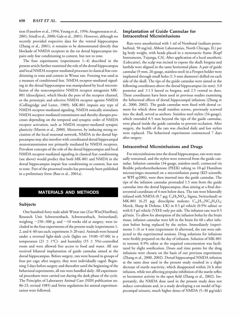
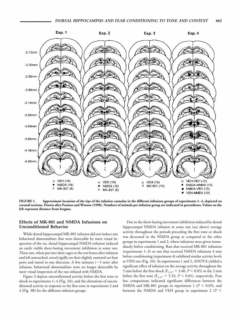
FIGURE 1. Approximate locations of the tips of the infusion cannulae in the different infusions groups of experiments 1–4, depicted oncoronal sections. Drawn after Paxinos and Watson (1998). Numbers of animals per infusion group are indicated in parentheses. Values on theleft represent distance from bregma.
________________ DORSAL HIPPOCAMPUS AND FEAR CONDITIONING TO TONE AND CONTEXT 663

0.0025). Moreover, the difference between VEH and NMDA ratsin experiment 1 closely approached significance (P � 0.050). VEHand MK-801 rats did not differ in experiment 1 (P � 0.50) or 2(P � 0.18). ANOVA of activity during the 5 min before the firstshock in experiment 3 (F1,18 � 0.00, P � 0.97) or the 1 min beforethe first tone in experiment 4 (F1,38 � 1.46, P � 0.23) did notyield a significant effect of the infusion given before conditioning.Analysis of activity levels in the different 1-min blocks of the peri-ods before the first tone or shock (data not shown) did not reveal adecrease of activity over time, which would have reflected habitu-ation of exploratory activity, in any group.
In experiments 2 and 4, in which the tone was the first salientstimulus presented, all infusion groups were more active during thetone presentation than during the immediately preceding period(Fig. 3B), indicating that the tone induced similar behavioralarousal in all groups. For both experiments, ANOVA of the aver-age activity in the 30-s periods both before and during the toneyielded an effect of period (experiment 2: F1,37 � 20.19, P �0.0001; experiment 4: F1,38 � 9.39, P � 0.005) without an inter-action of infusion group and period (experiment 2: F2,37 � 2.60,P � 0.08; experiment 4: F2,38 � 1.56, P � 0.21). Moreover, inboth experiments there was a significant main effect of group,
FIGURE 2. Photomicrographs of coronal sections through the dorsal hippocampus immediately around the infusion sites from rats thatreceived one infusion of vehicle (VEH) (0.5 �l saline), MK-801 (6.25 �g/0.5 �l), or N-methyl-D-aspartate (NMDA) (0.7 �g/0.5 �l). Neuronsare visualized by immunostaining of the neuronal marker protein NeuN. Arrowheads indicate the approximate location of the tips of theinfusion cannulae. Scale bar � 500 �m.
FIGURE 3. Effects of the different infusions on (A) unconditioned activity before the first tone or shock in experiments 1–4, and (B) onthe increase of unconditioned activity in response to the first tone in experiments 2 and 4. In experiments 1–3, infusions were appliedimmediately, in experiment 4, 4 min before behavioral testing. Numbers of animals per infusion group are indicated in parentheses. Presentedvalues are means. Bars � 1 standard error (SE) derived from the appropriate mean square of ANOVA. The SE provides an estimate ofpopulation variance.
664 BAST ET AL.

reflecting movement inhibition in some of the NMDA rats (exper-iment 2: F2,37 � 4.09, P � 0.025; experiment 4: F1,38 � 4.58, P �0.05).
Inspection of the video images from the conditioning sessions ofall four experiments yielded that all infusion groups exhibited sim-ilar vigorous twitching or jumping as immediate response to thefootshock, indicating that NMDA or MK-801 infusions did notaffect shock sensitivity.
Effects of MK-801 and NMDA Infusions onConditioned Freezing
Experiment 1: foreground contextual fearconditioning after dorsal hippocampal NMDA orMK-801 infusion
The freezing data of the conditioning and the context-test ses-sion of experiment 1 are depicted in Figure 4. During condition-ing, freezing resulting from the inescapable footshocks was de-creased in MK-801 as compared with VEH rats until the end of thesession. NMDA rats exhibited similarly low freezing as MK-801rats during the 5-min blocks after the first three shocks, but showedfreezing comparable to the VEH rats in the last two 5-min blocks.Moreover, as compared with the other two groups, NMDA ratsspent more time immobile in the 5-min block before the firstshock, reflecting the short-lasting movement inhibition inducedby the dorsal hippocampal NMDA infusion. ANOVA of freezingthroughout all six 5-min blocks both before and after the shocks inthe conditioning session yielded an effect of infusion (F2,37 � 4.39,P � 0.025) and 5-min block (F5,185 � 23.21, P � 0.0001), as wellas an interaction of these two factors (F10,185 � 3.23, P � 0.001),reflecting that differences between the groups changed throughoutthe session. During the first 5-min block (F2,37 � 5.32, P � 0.01),NMDA rats exhibited increased immobility as compared with theVEH (P � 0.001) and MK-801 groups (P � 0.025), which didnot differ (P � 0.99). In the following three 5-min blocks (F2,37 �
5.81, P � 0.01), average freezing levels were lower in MK-801(P � 0.05) and NMDA (P � 0.005) rats, which did not differ(P � 0.76), than in the VEH group. Finally, in the last two 5-minblocks (F2,37 � 5.65, P � 0.01), freezing was decreased in theMK-801 group as compared with VEH (P � 0.0025) and NMDA(P � 0.05) rats, which no longer differed (P � 0.13).
During the context test, both NMDA and MK-801 rats exhib-ited lower conditioned freezing than the VEH group. ANOVA offreezing values throughout the eight 1-min blocks of the contexttest yielded a significant effect of 1-min block (F7,259 � 5.61, P �0.0001), reflecting a gradual increase of freezing throughout thefirst three 1-min blocks, as well as an effect of infusion (F2,37 �5.12, P � 0.025). Post hoc comparisons demonstrated that aver-age freezing levels throughout the eight 1-min blocks were lower inNMDA than in VEH rats (P � 0.005). There was a strong ten-dency (P � 0.068) for MK-801 rats, which exhibited similarly lowfreezing levels as the NMDA rats (P � 0.50), to show less freezingthan the VEH group.
Experiment 2: fear conditioning to a tone afterdorsal hippocampal NMDA or MK-801 infusion
The freezing data for the different sessions of experiment 2 aredepicted in Figure 5. During conditioning, freezing in response tothe footshocks was decreased in MK-801 as compared with VEHrats until the end of the session. NMDA rats exhibited similarlylow freezing as MK-801 rats during the first half of the session, butshowed freezing comparable to the VEH rats throughout the sec-ond half. Before the first shock, NMDA rats spent more timeimmobile than did the two other groups, reflecting movementinhibition induced by the dorsal hippocampal NMDA infusion.ANOVA of freezing throughout the 11 2-min blocks both beforeand after the tone-shock pairings, as well as of freezing during the10 30-s blocks of tone presentation, yielded an effect of infusion(F2,37 � 4.64, P � 0.025; F2,37 � 12.32, P � 0.0001) and timeblock (F10,370 � 9.85, P � 0.0001; F9,333 � 4.84, P � 0.0001), aswell as an interaction of these two factors (F20,370 � 2.90, P �0.001; F18,333 � 2.46, P � 0.001), reflecting that differences be-tween the groups changed throughout the session. During the first2-min block (F2,37 � 5.32, P � 0.01), NMDA rats spent or tendedto spend, respectively, more time immobile than the VEH (P �0.005) or MK-801 group (P � 0.070), which did not differ (P �0.4). Similar differences appeared to exist throughout the subse-quent 30-s tone presentation of the first tone-shock pairing, al-though ANOVA only yielded a strong tendency toward an effect ofinfusion on the proportion of time spent immobile during thisperiod (F2,37 � 2.99, P � 0.063). During the following five 2-minblocks (F2,37 � 8.08, P � 0.0025), as well as the adjacent 30-s tonepresentations (F2,37 � 12.82, P � 0.0001), of the second to sixthtone-shock pairings, average freezing levels were lower in MK-801(P � 0.0025; P � 0.0001) and NMDA (P � 0.0025; P � 0.001)rats, which did not differ (P � 0.43; P � 0.08), than in VEH rats.Finally, the effect of infusion on average freezing levels during thelast five 2-min blocks (F2,37 � 3.20, P � 0.052) and the last fourtone presentations (F2,37 � 7.96, P � 0.0025) closely approachedsignificance or was significant, respectively. Post hoc comparisons
FIGURE 4. Freezing during conditioning and context-test ses-sions of experiment 1. Vehicle (VEH) (n � 16), N-methyl-D-aspartate(NMDA) (n � 16), or MK-801 (n � 8) was infused into the dorsalhippocampus immediately before conditioning with five unsignaledfootshocks. Mean percentage of time spent freezing is depicted for thesix 5-min blocks both preceding and following the five unsignaledfootshocks during conditioning and for the eight 1-min blocks of thecontext test. Bars � 1 standard error (SE) derived from the appropri-ate mean square of ANOVA.
________________ DORSAL HIPPOCAMPUS AND FEAR CONDITIONING TO TONE AND CONTEXT 665

indicated that average freezing was decreased in the MK-801 groupas compared with VEH (P � 0.05; P � 0.001) and NMDA (P �0.025; P � 0.001) rats, which did not differ anymore (P � 0.93;P � 0.89).
During the context test, all groups exhibited relatively low levelsof freezing (�14%) throughout all eight 1-min blocks, indicatingthat the conditioning procedure did not result in marked condi-tioned fear to the context. Nevertheless, ANOVA yielded an effectof infusion on freezing levels (F2,37 � 3.1, P � 0.05), and post hoccomparisons indicated that, as compared with the VEH group,freezing was or tended to be reduced, respectively, in the NMDA(P � 0.025) and the MK-801 (P � 0.090) group, which exhibitedvery similar freezing (P � 0.77).
During the tone test, conditioned freezing to the tone appearedto be decreased in the NMDA group as compared with both VEHand MK-801 rats. During the three 1-min blocks before toneonset, rats virtually did not exhibit conditioned fear, as evidencedby low levels of immobility (�4%), which did not differ betweenthe infusion groups (main effect of infusion: F2,37 � 1.31, P �0.28; interaction infusion � 1-min block: F4,74 � 4.51, P � 0.60).ANOVA of freezing levels throughout the eight 1-min blocks aftertone onset yielded a trend toward an effect of infusion (F2,37 �
2.67, P � 0.082), indicating higher freezing levels in VEH andMK-801 rats as compared with the NMDA group, a significantmain effect of 1-min block (F10,370 � 18.82, P � 0.0001), and aninteraction of infusion and 1-min block (F10,370 � 2.15, P �0.005). The interaction reflected that, throughout the first two tothree 1-min blocks after tone onset, VEH and MK-801 rats exhib-ited similarly marked freezing (�30%), which then appeared todecrease faster in the VEH than in the MK-801 group toward theend of the session, while NMDA rats exhibited little freezing(�10%) throughout all eight 1-min blocks of tone presentation.
Experiment 3: different effects of dorsalhippocampal MK-801 infusion on fearconditioning to tone and context—a furtherwithin-subject comparison
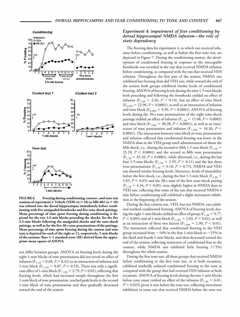
Figure 6 depicts the freezing data for the different sessions ofexperiment 3. During conditioning, freezing in response to thefootshocks was decreased in MK-801 as compared with VEH rats.ANOVA of freezing during the 12 2.5-min blocks both precedingand following the unsignaled shocks and tone-shock pairingsyielded an effect of infusion (F1,18 � 28.10, P � 0.0001), 2.5-minblock (F11,198 � 25.53, P � 0.0001), as well as an interaction ofthese two factors (F11,198 � 6.16, P � 0.0001). The interactionreflected that, while both infusion groups exhibited virtually nofreezing during the two 2.5-min blocks before the first shock,freezing levels increased to a level of �60% in the VEH, but onlyto �20%, in the MK-801 group within the 2.5-min blocks afterthe first two shocks. ANOVA of freezing levels during the 30-stone presentations of the five tone-shock pairings presented in thesecond half of the conditioning session revealed only an effect ofinfusion (F1,18 � 41.13, P � 0.0001), but not an effect of tonepresentation (F4,72 � 0.25, P � 0.91) or an interaction of infusionand tone presentation (F4,72 � 0.33, P � 0.86). Thus, duringconditioning, MK-801 rats exhibited less freezing than VEH rats,in absence, as well as in presence, of the tone.
During the first context test, VEH, but not MK-801 rats, ex-hibited marked conditioned freezing. ANOVA of freezing levelsthroughout the eight 1-min blocks yielded an effect of infusion(F1,18 � 6.23, P � 0.025). Although it appeared that conditionedfreezing in the VEH rats developed gradually throughout the firstthree 1-min blocks, and that toward the end of the session VEHrats exhibited slight extinction of conditioned freezing, ANOVAdid not show an effect of 1-min block (F7,126 � 1.74, P � 0.10) oran interaction of infusion and 1-min block (F7,126 � 1.07, P �0.38). During the second context test, both VEH and MK-801 ratsexhibited similarly low levels of immobility (F1,18 � 0.11, P �0.74). ANOVA on the time spent immobile throughout the eight1-min blocks only yielded an effect of 1-min block (F7,126 � 4.10,P � 0.0005) as immobility levels slightly increased toward the endof the session, probably reflecting a decrease in activity due tohabituation to the context.
In the tone test, MK-801 and VEH rats exhibited similarmarked conditioned freezing to the tone. During the three 1-minblocks before tone onset, rats exhibited virtually no conditionedfear, as evidenced by low levels of immobility (�6%) which did
FIGURE 5. Freezing during conditioning, context-, and tone-testsessions of experiment 2. Vehicle (VEH) (n � 16), N-methyl-D-aspar-tate (NMDA) (n � 16), or MK-801 (n � 8) was infused into the dorsalhippocampus immediately before conditioning with 10 tone-shockpairings. Mean percentage of time spent freezing during conditioningis depicted for the 11 2-min blocks both preceding and following thetone-shock pairings as well as for the 10 30-s tone presentations of thepairings. Mean percentage of time spent freezing during context- andtone test is depicted for each of the eight or 11, respectively, 1-minblocks of the sessions. Bars � 1 standard error (SE) derived from theappropriate mean square of ANOVA.
666 BAST ET AL.
not differ between groups. ANOVA on freezing levels during theeight 1-min blocks of tone presentation did not reveal an effect ofinfusion (F1,18 � 0.68, P � 0.42) or an interaction of infusion and1-min block (F7,126 � 0.87, P � 0.53). There was only a signifi-cant effect of 1-min block (F7,126 � 2.79, P � 0.01), reflecting thatfreezing levels, which had increased steeply throughout the first1-min block of tone presentation, reached peak levels in the second1-min block of tone presentation and then gradually decreasedtoward the end of the session.
Experiment 4: impairment of fear conditioning bydorsal hippocampal NMDA infusion—the role ofstate dependency
The freezing data for experiment 4, in which rats received infu-sions before conditioning, as well as before the first tone test, aredepicted in Figure 7. During the conditioning session, the devel-opment of conditioned freezing in response to the inescapablefootshocks was retarded in the rats that received NMDA infusionbefore conditioning, as compared with the rats that received VEHinfusion. Throughout the first part of the session, NMDA ratsexhibited less freezing than did VEH rats, while toward the end ofthe session both groups exhibited similar levels of conditionedfreezing. ANOVA of freezing levels during the nine 1.5-min blocksboth preceding and following the footshocks yielded no effect ofinfusion (F1,38 � 2.26, P � 0.14), but an effect of time block(F8,304 � 22.96, P � 0.0001), as well as an interaction of infusionand time block (F8,304 � 9.99, P � 0.0001). ANOVA of freezinglevels during the 30-s tone presentations of the eight tone-shockpairings yielded an effect of infusion (F1,38 � 15.08, P � 0.0005)and time block (F7,266 � 38.28, P � 0.0001), as well as an inter-action of tone presentation and infusion (F7,266 � 10.30, P �0.0001). The interaction between time block or tone presentationand infusion reflected that conditioned freezing was lower in theNMDA than in the VEH group until administration of about thefifth shock, i.e., during the second to fifth 1.5 min block (F1,38 �25.10, P � 0.0001) and the second to fifth tone presentation(F1,38 � 45.10, P � 0.0001), while afterward, i.e., during the lastfour 1.5-min blocks (F1,38 � 2.59, P � 0.11) and the last threetone presentations (F1,38 � 0.10, P � 0.75), NMDA and VEHrats showed similar freezing levels. Moreover, levels of immobilitybefore the first shock, i.e., during the first 1.5-min block (F1,38 �5.05, P � 0.05) and the 30-s tone of the first tone-shock pairing(F1,38 � 4.34, P � 0.05), were slightly higher in NMDA than inVEH rats, reflecting that some of the rats that received NMDA 4min before conditioning still exhibited a slight movement inhibi-tion at the beginning of the session.
During the first context test, VEH, but not NMDA, rats exhib-ited marked conditioned freezing. ANOVA of freezing levels dur-ing the eight 1-min blocks yielded an effect of group (F1,38 � 9.77,P � 0.005) and of 1-min block (F7,266 � 2.83, P � 0.01), as wellas an interaction of these two factors (F7,266 � 2.90, P � 0.01).The interaction reflected that conditioned freezing in the VEHgroup increased from �10% in the first 1-min block to �25% inthe third and fourth 1-min blocks, and then decreased toward theend of the session, reflecting extinction of conditioned fear to thecontext, while NMDA rats exhibited little freezing (�5%)throughout the whole session.
During the first tone test, all three groups that received NMDAbefore conditioning or the first tone test, or at both occasions,exhibited markedly reduced conditioned freezing to the tone ascompared with the group that had received VEH infusion at bothoccasions. ANOVA of freezing levels during the two 1-min blocksbefore tone onset yielded an effect of the infusion (F1,36 � 6.01,P � 0.025) given 4 min before the tone test, reflecting movementinhibition in some rats that received NMDA before the tone test
FIGURE 6. Freezing during conditioning, context-, and tone-testsessions of experiment 3. Vehicle (VEH) (n � 10) or MK-801 (n � 10)was infused into the dorsal hippocampus immediately before condi-tioning with five unsignaled footshocks and five tone-shock pairings.Mean percentage of time spent freezing during conditioning is de-picted for the two 2.5-min blocks preceding the shocks, for the five2.5-min blocks following the unsignaled shocks and the tone-shockpairings, as well as for the five 30-s tone presentations of the pairings.Mean percentage of time spent freezing during the context and tonetests is depicted for each of the eight or 11, respectively, 1-min blocksof the sessions. Bars � 1 standard error (SE) derived from the appro-priate mean square of ANOVA.
________________ DORSAL HIPPOCAMPUS AND FEAR CONDITIONING TO TONE AND CONTEXT 667

session. Moreover, there was also an effect of the infusion receivedbefore conditioning (F1,36 � 4.13, P � 0.05), but no interactionbetween the two infusions (F1,36 � 1.87, P � 0.18). This reflectedthat average levels of immobility were higher in rats that had re-ceived VEH before conditioning than in those that had receivedNMDA before conditioning. First, VEH-VEH rats exhibitedhigher freezing than the NMDA-VEH rats in the second 1-minblock, possibly reflecting that in the rats that had received VEHbefore conditioning there was still some fear to the conditioning
context that generalized to the test context. Second, immobilitylevels, reflecting movement inhibition induced by the NMDAinfusion before conditioning, were higher in VEH-NMDA than inNMDA-NMDA rats. This might reflect tolerance to the effects ofNMDA due to the previous NMDA infusion. However, NMDA-induced movement inhibition was only observed in some rats, andit is striking that VEH-NMDA rats exhibited twice as much im-mobility before tone onset in the first tone test as displayed by ratsthat received NMDA before conditioning, before the first shockduring conditioning. Thus, the higher immobility levels in VEH-NMDA as compared with NMDA-NMDA rats before tone onsetin the first tone test are most likely reflecting a sampling error. Forthe eight 1-min blocks of tone presentation, an effect of time(F7,252 � 10.52, P � 0.0001) was the only simple main effectrevealed by ANOVA of freezing. However, all possible interactionsof between-subjects (infusion before conditioning, infusion beforefirst tone test) and repeated-measures (1-min block) factors weresignificant. Most importantly, there was a three-way interaction ofinfusion before conditioning, infusion before first tone test, andtime block (F7,252 � 3.48, P � 0.0025). This reflected that freez-ing in the rats receiving two VEH infusions was higher as com-pared with all other groups during the beginning of the tone pre-sentation, and that this difference was gradually decreasing,indicating extinction of conditioned fear.
Freezing levels throughout the context test 2 and 3 were rela-tively low, indicating extinction of conditioned fear to the context,and did not differ between the groups (all main effects and inter-actions involving infusion before conditioning or first tone test:F � 3.53, P � 0.06). During tone test 2, however, considerableconditioned freezing was still exhibited by the groups that receivedVEH before conditioning. ANOVA of freezing during the eight1-min blocks of tone presentation yielded an interaction of infu-sion before conditioning and 1-min block (F7,252 � 3.92, P �0.0005). This reflected that freezing in the rats which had receivedVEH before conditioning was higher as compared with the othergroups only during the first four 1-min blocks of tone presentation(F1,36 � 7.36, P � 0.025). Although the VEH-NMDA groupappeared to exhibit higher levels of conditioned freezing than theVEH-VEH group, indicating that NMDA infusion before tonetest 1 may have impaired extinction of conditioned fear to the tone,
FIGURE 7. Freezing during conditioning, context-, and tone-testsessions of experiment 4. Conditioning was conducted with eighttone-shock pairings. A first infusion of vehicle (VEH) or N-methyl-D-aspartate (NMDA) was given 4 min before conditioning, resultingin two infusion groups (each n � 20) for conditioning and context test1. A second infusion of VEH or NMDA was given 4 min before tonetest 1, so that four infusion groups (each n � 10), differing withrespect to the combination of infusions received before conditioningand tone test 1, resulted for tone test 1 and the following test sessions.Mean percentage of time spent freezing during conditioning is de-picted for the nine 1.5-min blocks both preceding and following thetone-shock pairings, as well as for the eight 30-s tone presentations ofthe pairings. Mean percentage of time spent freezing during the con-text and tone tests is depicted for each of the eight or 10, respectively,1-min blocks of the sessions. Bars � 1 standard error (SE) derivedfrom the appropriate mean square of ANOVA.
668 BAST ET AL.

ANOVA on freezing during the eight 1-min blocks of tone pre-sentation did not yield significant interactions involving both in-fusions (P � 0.11). During tone test 3, the tone onset still appearedto induce weak conditioned freezing in the two groups that re-ceived VEH infusions before conditioning. ANOVA on freezingduring the eight 1-min blocks of tone presentation yielded aninteraction of infusion before conditioning and 1-min block(F7,252 � 2.86, P � 0.01). In the first 1-min block after tone onset,rats that had received VEH before conditioning exhibited higherconditioned freezing than rats that had received NMDA (F1,36 �4.28, P � 0.05).
DISCUSSION
Altogether, the four experiments of the present study yieldedtwo major findings. Dorsal hippocampal MK-801 infusion beforeconditioning to context or tone resulted in reduced freezing to thecontext, but not the tone, in subsequent test sessions (experiments1–3). Freezing to both context and tone, however, was reduced bydorsal hippocampal NMDA infusion before conditioning (exper-iments 1, 2, and 4).
Dorsal Hippocampal Infusion of MK-801 andNMDA
In view of the neurotoxic potential of MK-801 (Olney et al.,1989) and NMDA (Hajos et al., 1986), we examined infusion-induced neuronal damage, using selective visualization of neuronsby immunostaining of the neuronal marker protein NeuN (Wolfet al., 1996). This examination demonstrated that the neuronaldamage in the dorsal hippocampus was restricted mainly to thecannula tracks and the immediately surrounding areas. Eventhough NMDA may have induced slight additional damage, over-all neuronal damage in the dorsal hippocampus did not differmarkedly between rats infused with VEH, MK-801, or NMDA.That NMDA in the dorsal hippocampus exerts mainly temporaryeffects is further corroborated by the results of experiment 4, whererats infused with NMDA only before tone test 1 exhibited reducedfreezing as compared with VEH rats during tone test 1, but not 2.Moreover, with the small infusion volume (0.5 �l/side) and fineinfusion cannulae (34 gauge) used in the present study, the esti-mated spread of the infused substances, occurring preferentiallydorsally along the external wall of the infusion cannula, is �1 mm(Myers, 1966; Myers et al., 1971; Routtenberg, 1972). Thus, it canbe assumed that differences observed between rats infused withVEH as compared to those infused with MK-801 or NMDA re-flected a temporary alteration of neuronal activity by blockade orstimulation of dorsal hippocampal NMDA receptors.
MK-801 inhibits or shuts off any NMDA receptor-mediatedsignaling. NMDA tonically stimulates NMDA receptor-mediatedmechanisms, disrupting the time and synapse specificity ofNMDA receptor-mediated signaling. Thus, disruption of the samebehavioral process by both MK-801 and NMDA in the dorsalhippocampus indicates that this process depends on time- and
synapse-specific local NMDA receptor-mediated mechanisms,such as synaptic plasticity (Martin et al., 2000). Although MK-801disrupts NMDA receptor-mediated signaling generally, whereasNMDA only interferes with the specificity of this signaling, it ispossible that some processes, like fear conditioning to tone in thepresent study, are spared by dorsal hippocampal MK-801 infusion,but are disrupted by local NMDA infusion. NMDA inducesstrong local neuronal excitation and thereby likely interferes alsowith local coordinated signaling not primarily mediated byNMDA receptors. Furthermore, NMDA may induce aberrantstimulation of dorsal hippocampal efferents, thereby disruptingnormal processing in projection sites of dorsal hippocampal neu-rons. Which of the two possibilities accounts for the effects ofNMDA in the dorsal hippocampus on fear conditioning will bediscussed below.
Conditioned Freezing Resulting From theDifferent Conditioning Procedures
Freezing levels observed during conditioning and test sessions ofexperiments 1–3 in the VEH rats are comparable to those obtainedunder very similar experimental conditions in previous studies(Bast et al., 2001d; Zhang et al., 2001). Freezing during test ses-sions, though marked, reached peak levels of at most 45%. Suchlevels are low enough to largely rule out the possibility of ceilingeffects occluding treatment-induced reductions in conditionedfear. Thus, the specific reduction of freezing during context-, butnot tone-test, sessions in MK-801 rats cannot result from contex-tual conditioning being weaker than conditioning to the tone. Thisis also supported by the fact that MK-801 rats showed reducedfreezing in the context test of experiment 1, but not in the tone testof experiment 2, despite the VEH rats exhibiting similar peak levels(�30%) of freezing in both cases.
Interestingly, freezing during the first tone test in experiment 4(Fig. 7) was stronger than freezing during the tone test in experi-ment 2 (Fig. 5), although rats received more tone shock pairings inexperiment 2. The different levels of conditioned freezing werepossibly due to the longer time span between conditioning andtesting in experiment 4 (7 days) as compared with experiment 2 (1day), suggesting an enhancement of fear memory over periods ofseveral days. While it is well accepted that memory enhances, i.e.,consolidates, over time (McGaugh, 2000), only a few studies havereported proceeding enhancement in the expression of memoryover periods longer than 24 h (cf. Martı et al., 2001). It maytherefore be of general interest for the concept of memory consol-idation to further examine the possibility that consolidation of fearmemory is proceeding over several days.
Unspecific Infusion Effects
Alterations in sensorimotor functions
Observations in the present study did not indicate effects of theinfusions on US or CS processing, as infusion groups did notconsiderably differ in the unconditioned immediate shock re-sponse or the unconditioned activity response to the first tonepresentation. Altered startle reactivity found after dorsal hip-
________________ DORSAL HIPPOCAMPUS AND FEAR CONDITIONING TO TONE AND CONTEXT 669
pocampal infusion of NMDA (Zhang et al., 2002) and MK-801(Zhang et al., 2000) may indicate that these infusions affect theperception or evaluation of US and CS, since alterations in startlereactivity have been related to changes in attentional or emotionalstates (Koch, 1999). Given, however, that startle reactivity wasdecreased by NMDA, but increased by MK-801, in the dorsalhippocampus, the reduced conditioned freezing observed in thepresent study appears not to be linked to the infusion-inducedalterations in startle reactivity.
The measure of unconditioned activity before the first tone orshock at the beginning of conditioning was not altered by MK-801, while NMDA induced short-lasting movement inhibition insome rats. In our previous studies, MK-801 in the dorsal hip-pocampus markedly increased activity for �30 min (Zhang et al.,2000), while NMDA did not alter activity (Zhang et al., 2002) inrats habituated to their environment. This suggests that, duringconditioning in the present study, unconditioned activity was in-creased in the MK-801, but not altered in the NMDA group,except for the first few minutes of the session. High levels of un-conditioned activity at the beginning of the conditioning sessionsprobably masked the increase in activity induced by the dorsalhippocampal MK-801 infusion while favoring detection of theshort-lasting movement inhibition induced by the NMDA infu-sion. Short-lasting movement inhibition may occur after subcon-vulsive stimulation of the dorsal as well as ventral hippocampus(Hallak et al., 1993; Zhang et al., 2001; present study), eventhough, overall, the latter induces hyperactivity (Bast et al., 2001e;Zhang et al., 2002). This may reflect an initial strong disruption ofcoordinated hippocampal electrical activity related to movementinitiation (Leung, 2000). The short-lasting movement inhibition,or the underlying mechanisms, were not linked to the freezingdeficits observed in the present study. Movement inhibition onlyoccurred in some of the rats infused with NMDA, and inspectionof the data revealed freezing deficits were not related to whether ornot a rat exhibited movement inhibition. Reduced freezing hasbeen proposed to reflect, in some cases, hyperactivity interferingwith the performance of the freezing response, for example, in ratswith hippocampal lesions (e.g., Richmond et al., 1999; Gewirtz etal., 2000). In view of the hyperactivity induced by MK-801 in thedorsal hippocampus (Zhang et al., 2000), a performance deficit islikely to account for reduced freezing observed in the presence ofMK-801 in the dorsal hippocampus during conditioning. Thisalso explains why, during conditioning, MK-801 rats exhibitedreduced freezing also during the tone although exhibiting unim-paired conditioned fear to the tone during later testing withoutdrug. Furthermore, this account is consistent with the finding thatfreezing during conditioning was not reduced by the NMDA re-ceptor antagonist APV in the dorsal hippocampus (Young et al.,1994) at a dose not affecting activity (Kawabe et al., 1998). Ratswith NMDA in the dorsal hippocampus are not hyperactive(Zhang et al., 2002) and performed a normal freezing responseafter about four to six shock administrations during conditioning.The latter effect was not due to fading of drug activity given that itwas observed regardless of conditioning lasting �30 min (experi-ments 1 and 2) or only 10 min (experiment 4). Thus, NMDA in
the dorsal hippocampus does not interfere with performance of thefreezing response.
State dependency
Learning can be state dependent, i.e., information learned in aparticular brain state induced by systemic treatment (Overton,1964) or specific manipulations of single brain sites, such as elec-trical stimulation of the dorsal hippocampus (McIntyre et al.,1985), can in some cases only be retrieved with the same brain stateprevailing. If reduced conditioned fear is demonstrated duringtesting in rats conditioned and tested with drugs, the reduction inconditioned fear cannot merely be due to state dependency. Withrespect to dorsal hippocampal MK-801 infusion, which induceshyperactivity and thus is likely to disrupt the performance of freez-ing (see above), this demonstration is not possible using freezing asa measure of conditioned fear (see Bast et al., 2001d; Zhang et al.,2001). However, when tested without drug, rats that received MK-801 infusion before conditioning, only exhibited reduced condi-tioned freezing to context, but normal freezing in response to thetone. This specific reduction in fear to context argues against statedependency, unless fear conditioning to tone and context are dif-ferently state dependent (compare Gale et al., 2001). Moreover, itwas reported that infusions of NMDA receptor antagonists intothe amygdala (Kim and McGaugh, 1992) and even systemic injec-tion of MK-801 (Nakagawa and Iwasaki, 1996) did not inducestate dependency of inhibitory avoidance learning, another form ofclassical aversive conditioning. The virtual absence of conditionedfear during tone test in rats conditioned and tested with NMDA inthe dorsal hippocampus (experiment 4) is a direct demonstrationthat the effects of NMDA in the dorsal hippocampus on fearconditioning do not merely reflect state dependency. A similarpattern of results has been obtained with muscimol infusion intothe amygdala (Helmstetter and Bellgowan, 1994; Muller et al.,1997). Finally, normal conditioned fear during testing was foundin rats that received behaviorally effective infusions of a proteinkinase inhibitor or a dopamine antagonist before testing, but notconditioning, into the amygdala (Goosens et al., 2000; Guarraci etal., 2000); of the local anesthetic bupivacaine before conditioning,but not testing, into the nucleus accumbens (Haralambous andWestbrook, 1999); or of a dopamine agonist or antagonist beforeconditioning, but not testing, into the medial prefrontal cortex(Pezze et al., 2002a; 2003). Thus, even though generalization overdifferent drugs (Castellano and McGaugh, 1990) and brain re-gions (Phillips and LePiane, 1981) has to be made with caution,there is no clear evidence of drug infusions into single brain sitesinducing state dependency of conditioned fear, while several find-ings argue against this possibility.
Specific Infusion Effects on Fear Conditioning
Freezing emerging after the first shock during conditioning iswidely considered to reflect, at least in part, a conditioned fearresponse and, thus, short-term memory of fear (e.g., Kim et al.,1992, 1993; Young et al., 1994; Fanselow, 2000). As discussedabove, reduced freezing observed in MK-801 rats during condi-tioning probably resulted from infusion-induced hyperactivity,
670 BAST ET AL.

and the data of the present study do not therefore allow one todecide whether MK-801 in the dorsal hippocampus impairs for-mation of short-term conditioned fear. The retarded developmentof freezing during conditioning in the NMDA rats, however, prob-ably reflected an interference with the formation of short-term fearmemory.
The reduced freezing during test sessions in rats that had re-ceived dorsal hippocampal MK-801 or NMDA infusions beforeconditioning indicated a genuine impairment of specific memoryprocesses involved in the formation of long-term conditioned fearto either tone or context, or both. The formation of long-termmemory comprises the initial acquisition, short-term consolida-tion, completed within seconds or tens of minutes, and the subse-quent long-term consolidation (Nadel and Moscovitch, 1997; Mc-Gaugh, 2000). Our data do not allow one to decide which of theseveral stages contributing to long-term memory formation thedrug infusions actually affected. The drug infusions may have in-terfered with initial acquisition and short-term consolidation,which is believed to occur within seconds or tens of minutes afterthe acquisition, and, given that the drugs may have been active inthe dorsal hippocampus beyond the conditioning sessions, theearly phase of the subsequent long-term consolidation. Finally,reduced freezing during the tone test in rats that received dorsalhippocampal NMDA infusion only before the tone test (experi-ment 4) indicates a disruption of retrieval/expression of long-termconditioned fear to tone by NMDA in the dorsal hippocampus. Inexperiment 4, rats that had NMDA in the dorsal hippocampusonly during tone test 1 appeared to exhibit increased freezing dur-ing tone test 2, indicating reduced extinction of conditioned fear.Effects of dorsal hippocampal manipulations on extinction of con-ditioned fear may be the subject of future studies.
At the single doses used in the present study, MK-801 (6.25�g/0.5 �l/side) infusion into the dorsal hippocampus only impairedfear conditioning to context, while NMDA (0.7�g/0.5�l/side)infusion impaired fear conditioning to both tone and context. It ispossible that other drug doses would have had other effects. Forexample, while the observed effects of MK-801 indicate that fearconditioning to context is more susceptible to NMDA receptorblockade in the dorsal hippocampus than fear conditioning totone, higher doses of MK-801 may also have affected fear condi-tioning to tone. It is important to note, however, that the MK-801solution used in the present study was nearly saturated and, thus,MK-801 can hardly be infused into the dorsal hippocampus atdoses higher than that used in the present study. In contrast, whilethe effects of NMDA observed in the present study demonstratethat NMDA in the dorsal hippocampus can strongly impair fearconditioning to both tone and context, lower doses of NMDAcould have preferentially interfered with fear conditioning to con-text. Altogether, the infusion effects on fear conditioning observedin the present study are consistent with the notion that fear con-ditioning to context is more susceptible to dorsal hippocampalmanipulations than simple fear conditioning to tone (Anagnost-aras et al., 2001), but that, nevertheless, dorsal hippocampal ma-nipulations may also affect fear conditioning to tone (Maren et al.,1997).
Role of the Dorsal Hippocampus and LocalNMDA Receptor-Mediated Processes in FearConditioning: Comparison With the VentralHippocampus
Fear conditioning to context
Several studies indicated fear conditioning to context to be moresensitive to lesions of the rat dorsal hippocampus than fear condi-tioning to tone (Selden et al., 1991; Kim and Fanselow, 1992;Phillips and LeDoux, 1992, 1994; Anagnostaras et al., 1999a),even though impaired fear conditioning to a tone (Maren et al.,1997) and intact contextual fear conditioning (Phillips and Le-Doux, 1994; Maren et al., 1997; Richmond et al., 1999) were alsofound after such lesions. Moreover, infusion of the competitiveNMDA receptor antagonist APV into the rat dorsal hippocampusresulted in anterograde amnesia of foreground contextual fear con-ditioning, while this manipulation’s effects on fear conditioning toa tone have not been examined (Young et al., 1994). Based on theabove data, NMDA receptor-mediated processes in the rat dorsalhippocampus have been suggested to be required for fear condi-tioning to context, but not tone (Young et al., 1994; Anagnostaraset al., 2001; Gale et al., 2001). This suggestion is confirmed di-rectly by the present finding that blockade of NMDA receptors inthe rat dorsal hippocampus by local infusion of the noncompeti-tive antagonist MK-801 impaired the formation of foreground andbackground contextual fear conditioning, while leaving fear con-ditioning to a tone intact. Similar results were obtained in a recentstudy after infusion of APV into the dorsal hippocampus of mice(Stiedl et al., 2000). In our previous study, MK-801 infusion intothe ventral hippocampus of rats also induced such selective effectson fear conditioning to a context (Zhang et al., 2001). Thus, in thedorsal as well as ventral hippocampus, NMDA receptor signaling isonly required for fear conditioning to context, but not tone. This isconsistent with a specific role of NMDA receptor-mediated syn-aptic plasticity (Martin et al., 2000) in the ventral as well as dorsalhippocampus in contextual fear conditioning, even thoughNMDA receptors may contribute to several aspects of hippocam-pal synaptic transmission (e.g., Rosenblum et al., 1999). In addi-tion to NMDA receptors, acetylcholine receptors in the rat dorsalhippocampus have recently been indicated to be involved in fearconditioning to context, but not tone (Gale et al., 2001; Wallen-stein and Vago, 2001). Interestingly, this has been related to apossible modulation of NMDA receptor-mediated synaptic plas-ticity in the dorsal hippocampus by cholinergic transmission (Galeet al., 2001).
NMDA receptor activation in the dorsal and ventral hippocam-pus may be required solely to form a unified representation of thesingle elements making up a context or also for the associationbetween context and US. Dorsal hippocampal mechanisms havebeen suggested to solely support the formation of a unified contextrepresentation, which afterward becomes independent of the dor-sal hippocampus and is possibly stored in the neocortex. Thissuggestion is supported by the finding that exposure to the condi-tioning context several weeks before lesioning the dorsal hip-pocampus protected rats from anterograde and retrograde amnesia
________________ DORSAL HIPPOCAMPUS AND FEAR CONDITIONING TO TONE AND CONTEXT 671

of contextual fear conditioning (Young et al., 1994; Anagnostaraset al., 2001). Moreover, it is consistent with the dependence ofspatial learning upon the dorsal hippocampus (Moser and Moser,1998) and, in particular, local NMDA receptor-mediated pro-cesses (Morris et al., 1989; Steele and Morris, 1999; Lee andKesner, 2002), given that spatial learning involves the formation ofa unified representation of the environment (Nadel and Willner,1980; Anagnostaras et al., 2001). The ventral hippocampus iscommonly believed to be less important for spatial learning thanthe dorsal hippocampus (Moser and Moser, 1998), and thus it mayalso be less important in forming a context representation. Rather,in contextual fear conditioning, ventral hippocampal processesmay complement the role of the dorsal hippocampus by support-ing formation and, possibly, also further processing and expressionof the context-US association. This would also be plausible basedon the anatomy of the hippocampus (Amaral and Witter, 1995).Although the dorsal hippocampus is well equipped with projec-tions from the sensory cortices, providing contextual information,only the ventral hippocampus has direct connections with extra-hippocampal structures implicated in formation, processing, andexpression of conditioned fear, such as amygdala (e.g., Cahill et al.,1999; Fanselow and LeDoux, 1999), nucleus accumbens (e.g.,Riedel et al., 1997; Haralambus and Westbrook, 1999; Murphy etal., 2000; Pezze et al., 2001b, 2002b), and prefrontal cortex (e.g.,Lacroix et al., 2000; Feenstra et al., 2001; Pezze et al., 2001a,2002a; 2003). For example, it has been proposed that context andUS are associated via synaptic plasticity in the projection from theventral hippocampus to the basolateral amygdala (Maren andFanselow, 1995; Anagnostaras et al., 2001). Information about thecontextual representation formed with participation of dorsal hip-pocampal mechanisms can reach the ventral hippocampus via di-rect intrahippocampal connections between dorsal and ventralhippocampus or via the parahippocampal cortices, in particular theentorhinal cortex.
Fear conditioning to tone
In contrast to the specific impairments in contextual fear condition-ing after blockade of dorsal hippocampal NMDA receptors, tonicstimulation of these receptors by NMDA disrupted formation ofshort-term and long-term fear to both tone and context, as well as theretrieval/expression of long-term fear to the tone. This disruption ofbasic mechanisms of fear conditioning may reflect interference withordered neuronal processing within the dorsal hippocampus or tonicstimulation of dorsal hippocampal projections resulting in interfer-ence with neuronal processing outside the dorsal hippocampus. Forexample, ventral hippocampal NMDA stimulation results in com-plete anterograde amnesia of fear (Zhang et al., 2001), and the effectsof dorsal hippocampal NMDA stimulation may reflect concomitantstimulation of the ventral hippocampus via intrahippocampal connec-tions. This is, however, unlikely, given that dorsal hippocampalNMDA stimulation hardly affects behavioral processes other than fearconditioning (sensorimotor gating, locomotor activity) that are mark-edly altered by ventral hippocampal stimulation (Bast et al., 2001c,e;Zhang et al., 2002). Moreover, the nucleus accumbens core, whichreceives direct dorsal hippocampal projections (Groenewegen et al.,
1987), has been implicated in fear conditioning. However, processingin the nucleus accumbens core may mainly contribute to contextualfear conditioning (see Pezze et al., 2001b). Finally, examination ofFos-protein production after electrical stimulation of the dorsal hip-pocampus suggested that subconvulsive dorsal hippocampal stimula-tion does not propagate out of the dorsal hippocampus (Sato et al.,1998). Thus, the complete anterograde amnesia of fear induced byNMDA in the dorsal hippocampus likely reflects disturbed neuronalprocessing within the dorsal hippocampus. This interpretation is incontrast to prevalent concepts, emphasizing the specific role of thedorsal hippocampus in contextual fear conditioning (Fanselow, 2000;Anagnostaras et al., 2001; Rudy and O’Reilly, 2001). Evidence sup-porting this interpretation was, however, provided by two experi-ments, in which electrolytic as well as cytotoxic lesions of the dorsalhippocampus impaired fear conditioning to a tone (Maren et al.,1997), and by preliminary results indicating deficits in fear condition-ing to tone after temporary inhibition of dorsal hippocampal neuronsby the GABAA agonist muscimol (Bellgowan and Helmstetter, 1995).Connections between dorsal and ventral hippocampus may link dor-sal hippocampal processes to other structures contributing to forma-tion, processing, and expression of conditioned fear to a tone, such asamygdala, nucleus accumbens, and prefrontal cortex (see above). Acontribution of the ventral hippocampus to formation of conditionedfear to tone has clearly been suggested by several recent studies (Maren,1999; Richmond et al., 1999; Bast et al., 2001b,d; Zhang et al., 2001).
Intact fear conditioning after dorsal hippocampallesions
There is an apparent discrepancy between the suggested involve-ment of the dorsal hippocampus in fear conditioning to context and,possibly also tone, and intact fear conditioning found after permanentdorsal hippocampal lesions by several studies (see above). It has longbeen recognized that the loss of function induced by permanent le-sions of a brain structure may be compensated for by other structuresdue to redundancy of neural structures and connections. Conse-quently, a behavioral function may be performed even after perma-nent lesion of a structure that normally serves this function (Bures andBuresova, 1990; Lomber, 1999). Functional compensation has re-cently been proposed to account for intact fear conditioning afterpermanent dorsal hippocampal lesions (Anagnostaras et al., 2001;Bast et al., 2001b). However, it is not clear why compensation afterdorsal hippocampal lesions occurs in some studies, but not in others.Thus, a review of the literature does not yield a clear relation betweena particular experimental parameter (time between lesion and condi-tioning/testing, time between conditioning and testing, conditioningand testing procedures) and the effect of dorsal hippocampal lesionson fear conditioning.
CONCLUSIONS
The present study demonstrates that NMDA receptor-mediatedprocesses in the rat dorsal hippocampus are required for formationof fear to a context, possibly to form a context representation, but
672 BAST ET AL.
not for fear conditioning to tone. Given that NMDA receptor-mediated processes in the dorsal hippocampus are also required forformation of spatial and episodic-like memory (Morris et al., 1989;Steele and Morris, 1999; Lee and Kesner, 2002), these processesmay generally contribute to form representations of more complexrelationships among stimuli, and, thus, also participate in the for-mation of human declarative memory (Eichenbaum, 1996). Theimpairments in formation and retrieval/expression of fear condi-tioning to tone by NMDA in the dorsal hippocampus indicatedthat some processes in the dorsal hippocampus, as in the ventralhippocampus (Maren, 1999; Richmond et al., 1999; Bast et al.,2001b,d; Zhang et al., 2001), may be involved in simple fearconditioning to a tone. This suggestion may be substantiated byfuture studies examining the effects of temporary dorsal hippocam-pal inactivation (e.g., by local tetrodotoxin infusion) on classicalfear conditioning. Altogether, the present data are in line with theview that some hippocampal mechanisms selectively contribute toparticular learning and memory functions while collectively therole of the hippocampus in learning and memory may be moregeneral than previously assumed (see Bast et al., 2001b).
Acknowledgments
This work was supported by grants from the Swiss Federal In-stitute of Technology Zurich. The support of the technical andadministrative staff at the ETH research station Schwerzenbach isgratefully acknowledged, with specific acknowledgments due toJacqueline Kupper, Pascal Guela, Oliver Asprion, and Sepp Tor-lucci for animal care; Liz Weber for histological preparations; PeterSchmid for setup and maintenance of the computerized systemsfor behavioral analysis; Jane Fotheringham for assistance withmanuscript preparation; and Christian Schlatter for help withpreparation of guide cannulae. Thanks are also due to Ben Yee forcritically reading the manuscript and to Marie Pezze, Ana Jongen-Relo, Carmen Sandi, Axel Becker, and Matti Mintz for comments,discussions, and advice that were helpful in accomplishing thepresent study.
REFERENCES
Amaral DG, Witter MP. 1995. Hippocampal formation. In: Paxinos G,editor. The rat nervous system. San Diego, CA: Academic Press. p443–493.
Anagnostaras SG, Maren S, Fanselow MS. 1999a. Temporally gradedretrograde amnesia of contextual fear after hippocampal damage inrats: within-subjects examination. J Neurosci 19:1106–1114.
Anagnostaras SG, Maren S, Sage JR, Goodrich S, Fanselow MS. 1999b.Scopolamine and Pavlovian fear conditioning in rats: dose-effect anal-ysis. Neuropsychopharmacology 21:731–744.
Anagnostaras SG, Gale GD, Fanselow MS. 2001. Hippocampus and con-textual fear conditioning: recent controversies and advances. Hip-pocampus 11:8–17.
Bast T, Zhang W-N, Feldon J. 2001a. Effects of NMDA receptor stimu-lation and blockade in the rat dorsal hippocampus on the formation ofclassical fear conditioning to explicit and contextual cues [abstract].Behav Pharmacol 12(suppl 1):S5.
Bast T, Zhang W-N, Feldon J. 2001b. Hippocampus and classical fearconditioning. Hippocampus 11:828–831.
Bast T, Zhang W-N, Feldon J. 2001c. Hyperactivity, decreased startlereactivity, and disrupted prepulse inhibition following disinhibition ofthe rat ventral hippocampus by the GABAA antagonist picrotoxin.Psychopharmacology 156:225–233.
Bast T, Zhang W-N, Feldon J. 2001d. The ventral hippocampus and fearconditioning in rats: different anterograde amnesias of fear after tetro-dotoxin inactivation and infusion of the GABAA agonist muscimol.Exp Brain Res 139:39–52.
Bast T, Zhang W-N, Heidbreder C, Feldon J. 2001e. Hyperactivity anddisruption of prepulse inhibition induced by N-methyl-D-aspartatestimulation of the ventral hippocampus and the effects of pretreatmentwith haloperidol and clozapine. Neuroscience 103:325–335.
Bellgowan PSF, Helmstetter FJ. 1995. Effects of muscimol applied to thedorsal hippocampus on the acquisition and expression of cued versuscontextual fear conditioning [abstract]. Soc Neurosci Abs 21:1219.
Bures J, Buresova O. 1990. Reversible lesions allow reinterpretation ofsystem levels studies of brain mechanisms of behavior. Concepts Neu-rosci 1:69–89.
Cahill L, Weinberger NM, Roozendaal B, McGaugh JM. 1999. Is theamygdala a locus of “conditioned fear”? Some questions and caveats.Neuron 23:227–228.
Cain DP. 1997. Testing the NMDA, long-term potentiation, and cholin-ergic hypotheses of spatial learning. Neurosci Biobehav Rev 22:181–193.
Castellano C, McGaugh JL. 1990. Effects of post-training bicuculline andmuscimol on retention: lack of state dependency. Behav Neural Biol54:156–164.
Collingridge GL, Lester AJ. 1989. Excitatory amino acid receptors in thevertebrate central nervous system. Pharmacol Rev 40:143–210.
Eichenbaum H. 1996. Is the rodent hippocampus just for place? CurrOpin Neurobiol 6:187–195.
Fanselow MS. 2000. Contextual fear, gestalt memories, and the hip-pocampus. Behav Brain Res 110:73–81.
Fanselow MS, LeDoux JE. 1999. Why we think plasticity underlyingPavlovian fear conditioning occurs in the basolateral amygdala. Neu-ron 23:229–232.
Fanselow MS, Kim JJ, Yipp J, De Oca B. 1994. Differential effects of theN-methyl-D-aspartate antagonist DL-2-amino-5-phosphonovalerateon acquisition of fear of auditory and contextual cues. Behav Neurosci108:235–240.
Feenstra MGP, Vogel M, Botterblom MHA, Joosten RNJMA, de BruinJPC. 2001. Dopamine and noradrenaline efflux in the rat prefrontalcortex after classical aversive conditioning to an auditory cue. EurJ Neurosci 13:1051–1054.
Gale GD, Anagnostaras SG, Fanselow MS. 2001. Cholinergic modula-tion of Pavlovian fear conditioning: effects of intrahippocampal sco-polamine infusion. Hippocampus 11:371–376.
Gewirtz JC, McNish KA, Davis M. 2000. Is the hippocampus necessaryfor contextual fear conditioning. Behav Brain Res 110:83–95.
Goosens KA, Holt W, Maren S. 2000. A role for amygdaloid PKA andPKC in the acquisition of long-term conditional fear memories in rats.Behav Brain Res 114:145–152.
Groenewegen HJ, Vermeulen-Van der Zee E, Te Kortschot A, WitterMP. 1987. Organization of the projections from the subiculum to theventral striatum in the rat: a study using anterograde transport ofPhaseolus vulgaris-leucagglutinin. Neuroscience 23:103–120.
Guarraci FA, Frohardt RJ, Falls WA, Kapp BS. 2000. The effects ofintra-amygdaloid infusions of a D2 dopamine receptor antagonist onPavlovian fear conditioning. Behav Neurosci 114:647–651.
Hajos F, Garthwaite G, Garthwaite J. 1986. Reversible and irreversibleneuronal damage caused by excitatory amino acid analogues in ratcerebellar slices. Neuroscience 18:417–436.
Hallak M, Irtenkauf SM, Janusz CA, Cotton DB. 1993. Stimulation andinhibition of N-methyl-D-aspartate receptors in rats: developing a sei-zure model. Am J Obstet Gynecol 169:695–700.
________________ DORSAL HIPPOCAMPUS AND FEAR CONDITIONING TO TONE AND CONTEXT 673

Haralambous T, Westbrook RF. 1999. An infusion of bupivacaine intothe nucleus accumbens disrupts the acquisition but not the expressionof contextual fear conditioning. Behav Neurosci 5:925–940.
Helmstetter FJ, Bellgowan PS. 1994. Effects of muscimol applied to thebasolateral amygdala on acquisition and expression of contextual fearconditioning in rats. Behav Neurosci 108:105–109.
Inglis FM, Fibiger HC. 1995. Increases in hippocampal and frontal cor-tical acetylcholine release associated with presentation of sensory stim-uli. Neuroscience 66:81–86.
Jongen-Relo AL, Kaufmann S, Feldon J. 2002. A differential involvementof the shell and core subterritories of the nucleus accumbens of rats inattentional processes. Neuroscience 111:95–109.
Kandel ER. 2001. The molecular biology of memory storage: a dialoguebetween genes and synapses. Science 294:1030–1038.
Kawabe K, Ichitani Y, Iwasaki T. 1998. Effects of intrahippocampal AP5treatment on radial-arm maze performance in rats. Brain Res 781:300–306.
Kim JJ, Fanselow MS. 1992. Modality-specific retrograde amnesia of fear.Science 256:675–677.
Kim JJ, Fanselow MS, DeCola JP, Landeira-Fernandez J. 1992. Selectiveimpairment of long-term but not short-term conditional fear by theN-methyl-D-aspartate antagonist APV. Behav Neurosci 106:591–596.
Kim JJ, Rison RA, Fanselow MS. 1993. Effects of amygdala, hippocam-pus, and periaqueductal gray lesions on short- and long-term contex-tual fear. Behav Neurosci 107:1093–1098.
Kim M, McGaugh JL. 1992. Effects of intra-amygdala injections ofNMDA receptor antagonists on acquisition and retention of inhibi-tory avoidance. Brain Res 585:35–48.
Koch M. 1999. The neurobiology of startle. Prog Neurobiol 59:107–128.Lacroix L, Spinelli S, Heidbreder CA, Feldon J. 2000. Differential role of
the medial and lateral prefrontal cortices in fear and anxiety. BehavNeurosci 1119–1130.
Lee I, Kesner RP. 2002. Differential contribution of NMDA receptors inhippocampal subregions to spatial working memory. Nature Neurosci5:162–168.
Leung LS. 2000. Behaviors induced or disrupted by complex partial sei-zures. Neurosci Biobehav Rev 24:763–775.
Lomber SG. 1999. The advantages and limitations of permanent or re-versible deactivation techniques in the assessment of neural function.J Neurosci Methods 86:109–117.
Maren S. 1999. Neurotoxic or electrolytic lesions of the ventral subiculumproduce deficits in the acquisition and expression of Pavlovian fearconditioning in rats. Behav Neurosci 113:283–290.
Maren S, Fanselow MS. 1995. Synaptic plasticity in the basolateral amyg-dala induced by hippocampal formation stimulation in vivo. J Neuro-sci 15:7548–7564.
Maren S, Aharonov G, Fanselow MS. 1997. Neurotoxic lesions of thedorsal hippocampus and Pavlovian fear conditioning in rats. BehavBrain Res 88:261–274.
Martı O, Garcıa A, Velles A, Harbuz MS, Armario A. 2001. Evidence thatsingle exposure to aversive stimuli triggers long-lasting effects in thehypothalamus-pituitary-adrenal axis that consolidate with time. EurJ Neurosci 13:129–136.
Martin SJ, Grimwood PD, Morris RGM. 2000. Synaptic plasticity andmemory: an evaluation of the hypothesis. Annu Rev Neurosci 23:649–711.
McGaugh JL. 2000. Memory—a century of consolidation. Science 287:248–251.
McIntyre DC, Stenstrom RJ, Taylor D, Stokes KA, Edson N. 1985.State-dependent learning following electrical stimulation of the hip-pocampus: intact and split-brain rats. Physiol Behav 34:133–139.
Morris RGM. 2001. Episodic-like memory in animals: psychological cri-teria, neural mechanisms and the value of episodic-like tasks to inves-tigate animal models of neurodegenerative disease. Philos Trans R SocLond B 356:1453–1465.
Morris RGM, Halliwell RF, Bowery N. 1989. Synaptic plasticity andlearning. II. Do different kinds of plasticity underlie different kinds oflearning? Neuropsychologia 27:41–59.
Moser M-B, Moser EI. 1998. Functional differentiation in the hippocam-pus. Hippocampus 8:608–619.
Muller J, Corodimas KP, Fridel Z, LeDoux J. 1997. Functional inactiva-tion of the lateral and basal nuclei of the amygdala by muscimol infu-sion prevents fear conditioning to an explicit conditioned stimulus andto contextual stimuli. Behav Neurosci 111:683–691.
Murphy CA, Pezze M, Feldon J, Heidbreder C. 2000. Differential in-volvement of dopamine in shell and core of the nucleus accumbens inthe expression of latent inhibition to an aversively conditioned stimu-lus. Neuroscience 97:469–477.
Murphy CA, Heidbreder C, Feldon J. 2001. Acute withdrawal from re-peated cocaine treatment enhances latent inhibition of a conditionedfear response. Behav Pharmacol 12:13–23.
Myers RD. 1966. Injection of solutions into cerebral tissue: relation be-tween volume and diffusion. Physiol Behav 1:171–174.
Myers RD, Tytell M, Kawa A, Rudy T. 1971. Micro-injection of 3H-Acetylcholine, 14C-serotonin and 3H-Norepinephrine into the hypo-thalamus of the rat: diffusions into tissue and ventricles. Physiol Behav7:743–751.
Nadel L, Moscovitch M. 1997. Memory consolidation, retrogradeamnesia, and the hippocampal complex. Curr Opin Neurobiol7:217–227.
Nadel L, Willner J. 1980. Context and conditioning: a place for space.Physiol Psychol 8:218–228.
Nakagawa Y, Iwasaki T. 1996. Ethanol-induced state-dependent learningis mediated by 5-hydroxytryptamine3 receptors but not by N-methyl-D-aspartate receptor complex. Brain Res 706:227–232.
Olney JW, Labruyere J, Price MT. 1989. Pathological changes induced incerebrocortical neurons by phencyclidine and related drugs. Science244:1360–1362.
Overton DA. 1964. State-dependent or dissociated learning producedwith pentobarbital. J Comp Physiol Psychol 57:3–12.
Paxinos G, Watson C. 1998. The rat brain in stereotaxic coordinates. SanDiego, CA: Academic Press.
Pezze MA, Bast T, Feldon J. 2001a. Effects of dopamine receptor stimu-lation or blockade in the medial prefrontal cortex on locomotor activ-ity, sensorimotor gating, and conditioned fear [abstract]. Behav Phar-macol 12(suppl 1):S77.
Pezze MA, Heidbreder CA, Feldon J, Murphy CA. 2001b. Selective re-sponding of nucleus accumbens core and shell dopamine to aversivelyconditioned contextual and discrete stimuli. Neuroscience 108:91–102.
Pezze M, Bast T, Feldon J. 2002a. The significance of dopamine trans-mission in the rat medial prefrontal cortex for conditioned fear [ab-stract]. FENS Abstr Vol. 1: A042.21.
Pezze MA, Feldon J, Murphy CA. 2002b. Increased conditioned fearresponse and altered balance of dopamine in the shell and core of thenucleus accumbens during amphetamine withdrawal. Neuropharma-cology 42:633–643.
Pezze MA, Bast F, Feldon J. 2003. The significance of dopamine-trans-mission in the rat medial prefrontal cortex for conditioned fear. CerebCortex (in press).
Phillips AG, LePiane FG. 1981. Differential effects of electrical stimula-tion of amygdala or caudate on inhibitory shock avoidance: a role forstate-dependent learning. Behav Brain Res 2:103–111.
Phillips RG, LeDoux JL. 1992. Differential contribution of amygdala andhippocampus to cued and contextual fear conditioning. Behav Neu-rosci 106:274–285.
Phillips RG, LeDoux JL. 1994. Lesions of the dorsal hippocampal forma-tion interfere with background but not foreground contextual fearconditioning. Learn Mem 1:34–44.
674 BAST ET AL.
Pryce CR, Lehmann J, Feldon J. 1999. Effect of sex on fear conditioningis similar for context and discrete CS in Wistar, Lewis, and Fischer ratstrains. Pharmacol Biochem Behav 64:753–759.
Richmond MA, Murphy CA, Pouzet B, Schmid P, Rawlins JNP, FeldonJ. 1998. A computer controlled analysis of freezing behaviour. J Neu-rosci Methods 86:91–99.
Richmond MA, Yee BK, Pouzet B, Veenman L, Rawlins JNP, Feldon J,Bannerman DM. 1999. Dissociating context and space within thehippocampus: effects of complete, dorsal and ventral excitotoxic le-sions on conditioned freezing and spatial learning. Behav Neurosci113:1189–1203.
Riedel G, Harrington NR, Hall G, Macphail EM. 1997. Nucleus accum-bens lesions impair context, but not cue, conditioning in rats. Neuro-Report 8:2477–2481.
Rosenblum K, Maroun M, Richter-Levin G. 1999. Frequency-dependentinhibition in the dentate gyrus is attenuated by the NMDA receptorblocker MK-801 at doses that do not yet affect long-term potentiation.Hippocampus 9:491–494.
Routtenberg A. 1972. Intracranial chemical injection and behavior: acritical review. Behav Biol 7:601–641.
Rudy JR, O’Reilly RC. 2001. Conjunctive representations, the hip-pocampus, and contextual fear conditioning. Cognitive Affective Be-hav Neurosci 1:66–82.
Selden NRW, Everitt BJ, Jarrard LE, Robbins TW. 1991. Complemen-tary roles for the amygdala and hippocampus in aversive conditioningto explicit and contextual cues. Neuroscience 42:335–350.
Steele RJ, Morris RGM. 1999. Delay-dependent impairment of a match-ing-to-place task with chronic and intrahippocampal infusion of theNMDA-antagonist D-AP5. Hippocampus 9:118–136.
Stiedl O, Birkenfeld K, Palve M, Spiess J. 2000. Impairment of condi-tioned contextual fear of C57BL/6J mice by intracerebral injections ofthe NMDA receptor antagonist APV. Behav Brain Res 116:157–168.
Wallenstein GV, Vago DR. 2001. Intrahippocampal scopolamine impairsboth acquisition and consolidation of contextual fear conditioning.Neurobiol Learn Mem 75:245–252.
Wolf HK, Buslei R, Schmidt-Kastner R, Kulkarni Schmidt-Kastner P,Pietsch T, Wiestler OD, Blumcke I. 1996. NeuN: a useful neuronalmarker for diagnostic histopathology. J Histochem Cytochem44:1167–1171.
Young SL, Bohenek DL, Fanselow MS. 1994. NMDA processes mediateanterograde amnesia of contextual fear conditioning induced by hip-pocampal damage: immunization against amnesia by context preexpo-sure. Behav Neurosci 108:19–29.
Zhang W-N, Bast T, Feldon J. 2000. Microinfusion of the noncompeti-tive N-methyl-D-aspartate antagonist MK-801 (dizocilpine) into thedorsal hippocampus of Wistar rats does not affect latent inhibition andprepulse inhibition but increases startle reaction and locomotor activ-ity. Neuroscience 101:589–599.
Zhang W-N, Bast T, Feldon J. 2001. The ventral hippocampus andfear conditioning in rats: different anterograde amnesias of fearafter infusion of N-methyl-D-aspartate or its noncompetitiveantagonist MK-801 into the ventral hippocampus. Behav Brain Res126:159 –174.
Zhang W-N, Bast T, Feldon J. 2002. Effects of hippocampal N-methyl-D-aspartate infusion on locomotor activity and prepulse inhibition:differences between the dorsal and ventral hippocampus. Behav Neu-rosci 116:72–84.
________________ DORSAL HIPPOCAMPUS AND FEAR CONDITIONING TO TONE AND CONTEXT 675