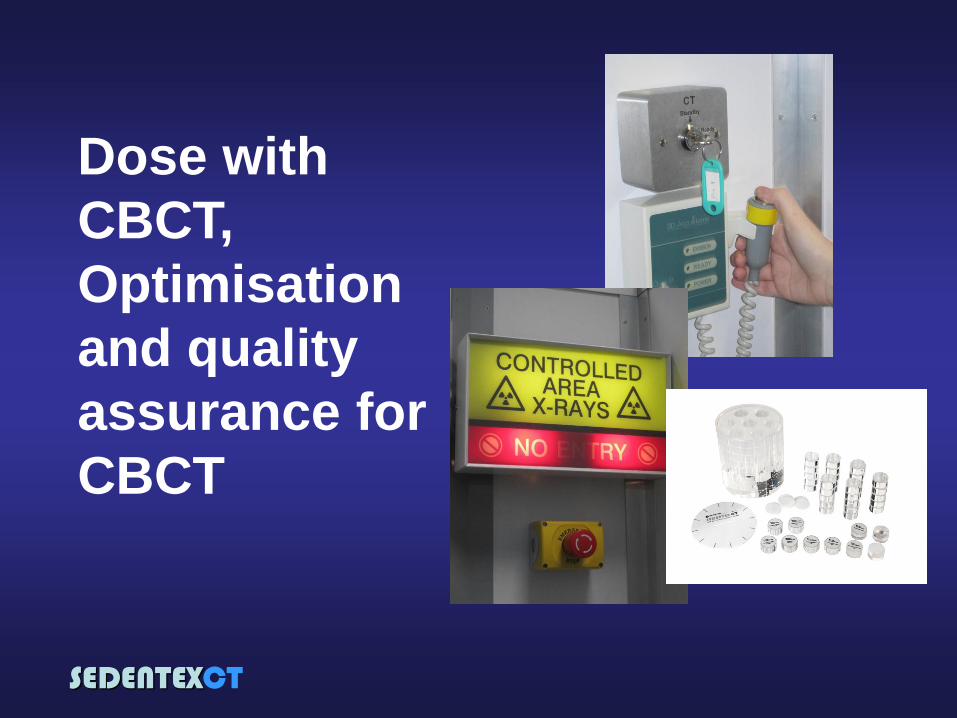
dose with cbct, optimisation and quality assurance for cbct
TRANSCRIPT
SEDENTEXCT
Dose with
CBCT,
Optimisation
and quality
assurance for
CBCT
SEDENTEXCT
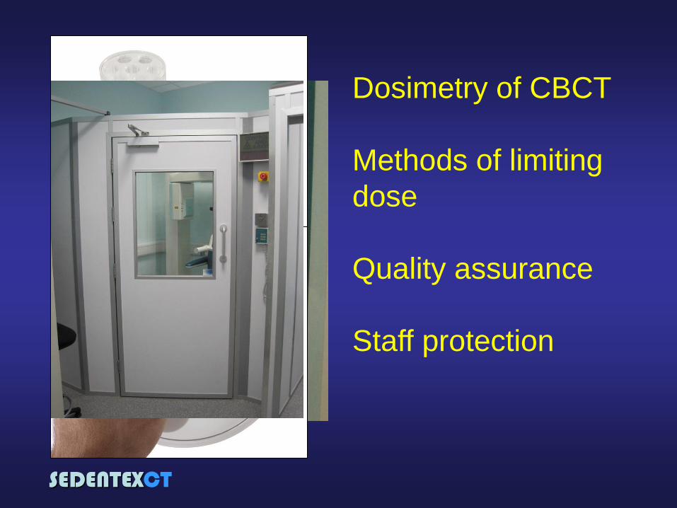
Dosimetry of CBCT
Methods of limiting
dose
Quality assurance
Staff protection
SEDENTEXCT
Learning outcomes
•An understanding of radiation dose associated with CBCT
•An understanding of the CBCT equipment factors that can
be adjusted to change the radiation dose delivered to patients
•A description of a quality assurance programme for CBCT,
including the use of the SEDENTEXCT quality control
phantom
•An understanding of the principles of staff protection
SEDENTEXCT
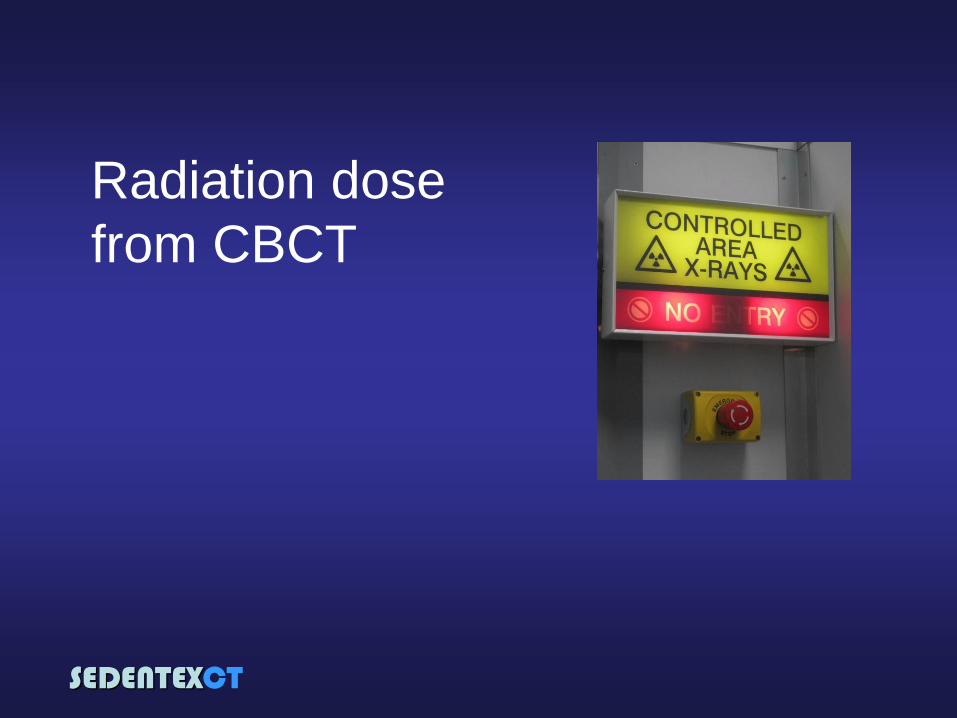
Radiation dose
from CBCT
SEDENTEXCT
SEDENTEXCT
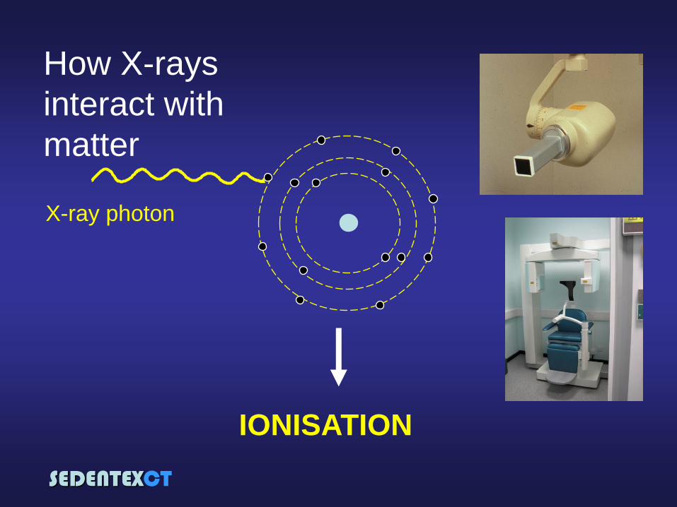
IONISATION
X-ray photon
How X-rays
interact with
matter
SEDENTEXCT
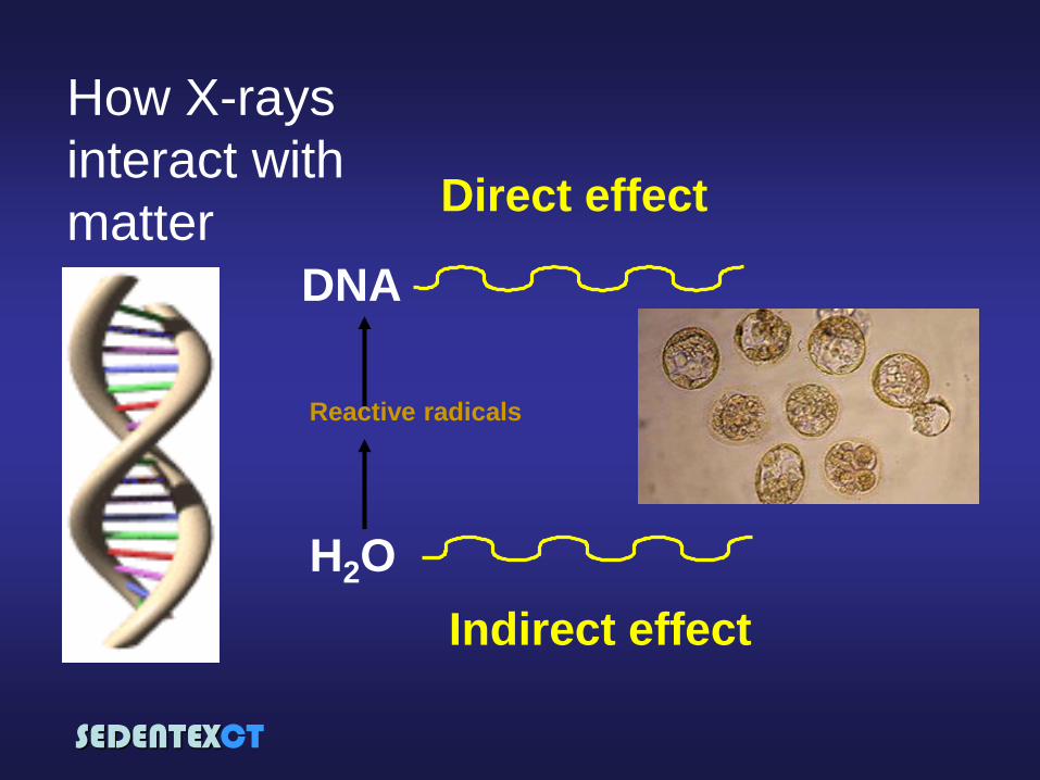
Direct effect
DNA
Indirect effect
H2O
Reactive radicals
How X-rays
interact with
matter
SEDENTEXCT
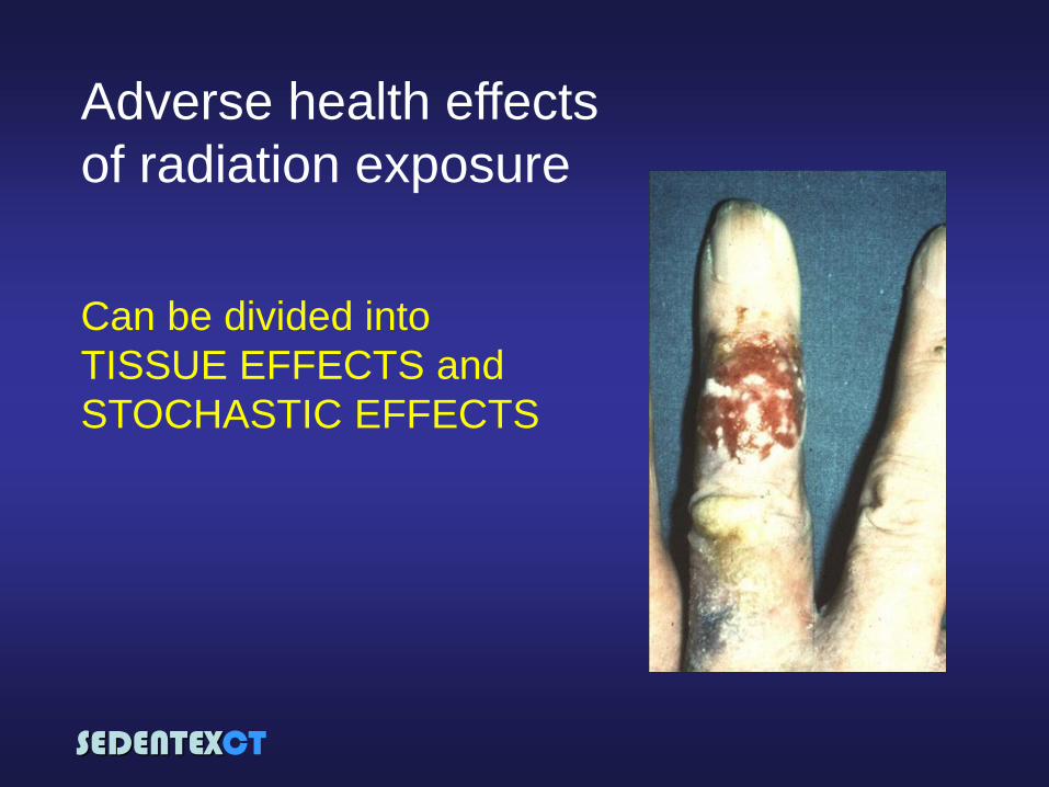
Adverse health effects
of radiation exposure
Can be divided into
TISSUE EFFECTS and
STOCHASTIC EFFECTS
SEDENTEXCT
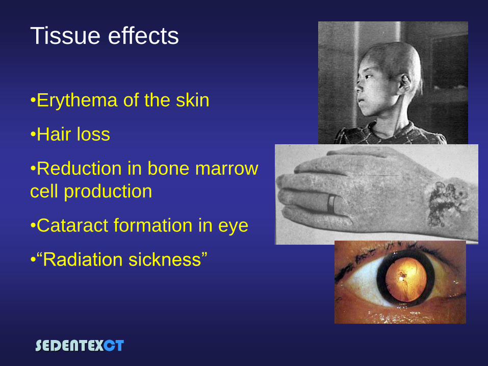
Tissue effects
•Erythema of the skin
•Hair loss
•Reduction in bone marrow
cell production
•Cataract formation in eye
•“Radiation sickness”
SEDENTEXCT
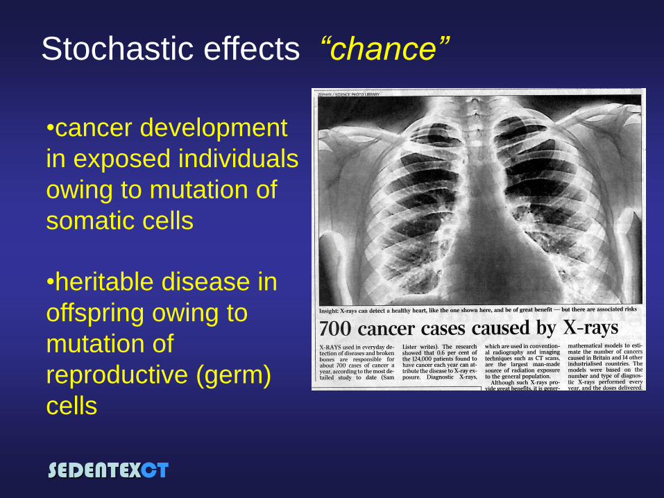
Stochastic effects
•cancer development
in exposed individuals
owing to mutation of
somatic cells
•heritable disease in
offspring owing to
mutation of
reproductive (germ)
cells
“chance”

SEDENTEXCT
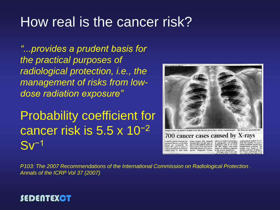
How real is the cancer risk?
International
Commission on
Radiological Protection1
1P103: The 2007 Recommendations of the International Commission on Radiological Protection .
Annals of the ICRP Vol 37 (2007)
Cancer
risk
Radiation dose
“linear no-threshold”
model
100mSv
SEDENTEXCT
Probability coefficient for
cancer risk is 5.5 x 10⁻²
Sv−1
P103: The 2007 Recommendations of the International Commission on Radiological Protection .
Annals of the ICRP Vol 37 (2007)
How real is the cancer risk?
“...provides a prudent basis for
the practical purposes of
radiological protection, i.e., the
management of risks from low-
dose radiation exposure”
SEDENTEXCT
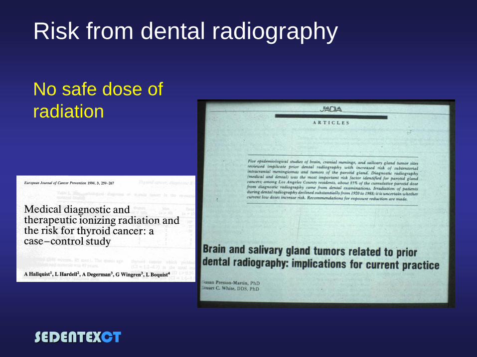
Risk from dental radiography
No safe dose of
radiation
SEDENTEXCT
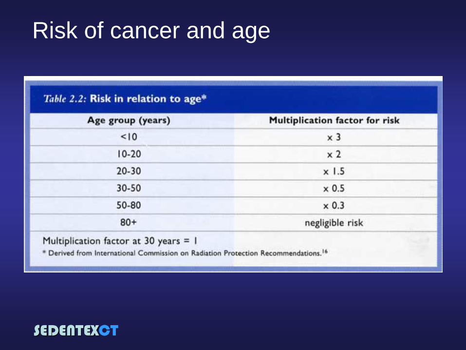
Risk of cancer and age
SEDENTEXCT
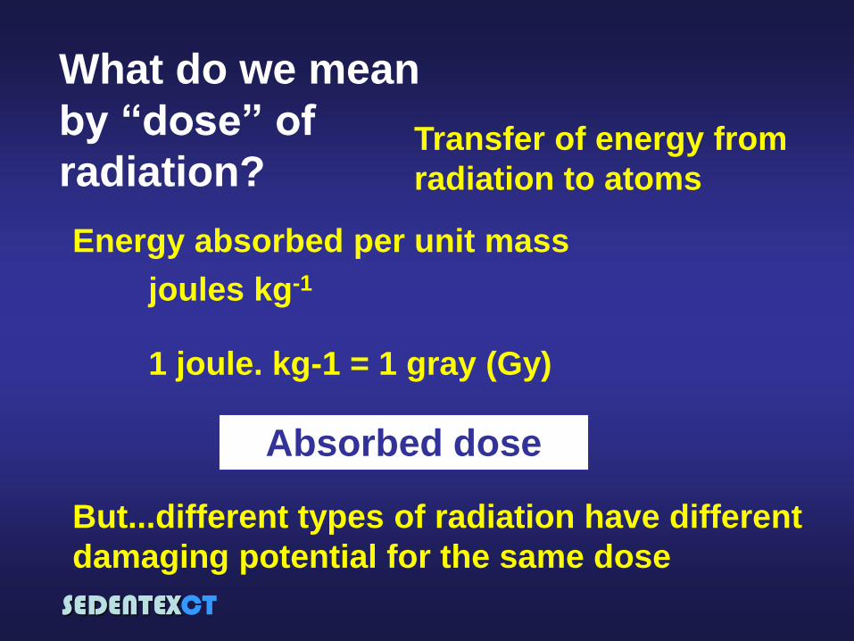
What do we mean
by “dose” of
radiation? Transfer of energy from
radiation to atoms
joules kg-1
1 joule. kg-1 = 1 gray (Gy)
Absorbed dose
But...different types of radiation have different
damaging potential for the same dose
Energy absorbed per unit mass
SEDENTEXCT
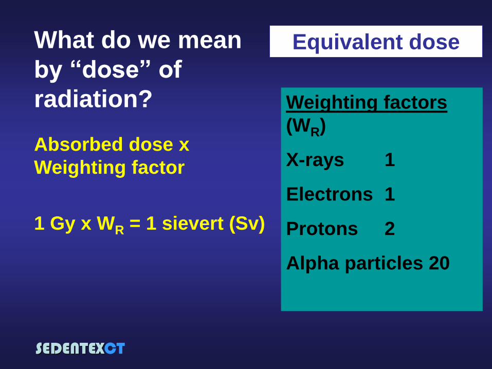
Equivalent dose
Weighting factors
(WR)
X-rays 1
Electrons 1
Protons 2
Alpha particles 20
Absorbed dose x
Weighting factor
1 Gy x WR = 1 sievert (Sv)
What do we mean
by “dose” of
radiation?
SEDENTEXCT
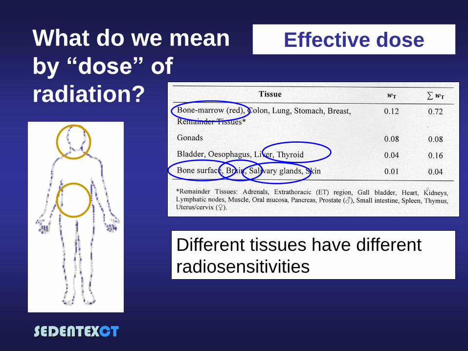
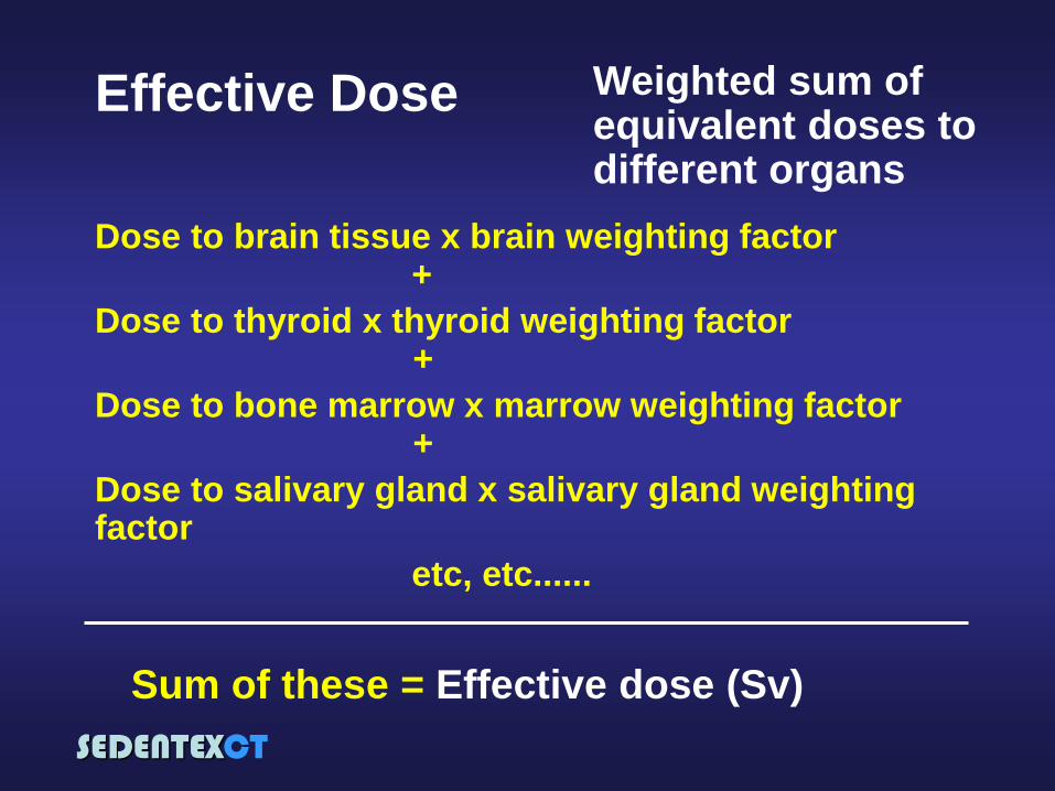
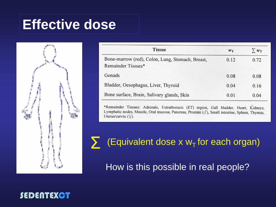
Effective dose
Different tissues have different
radiosensitivities
What do we mean
by “dose” of
radiation?
SEDENTEXCT
Weighted sum of equivalent doses to different organs
Dose to brain tissue x brain weighting factor +
Dose to thyroid x thyroid weighting factor +
Dose to bone marrow x marrow weighting factor +
Dose to salivary gland x salivary gland weighting factor
etc, etc......
Sum of these = Effective dose (Sv)
Effective Dose
SEDENTEXCT
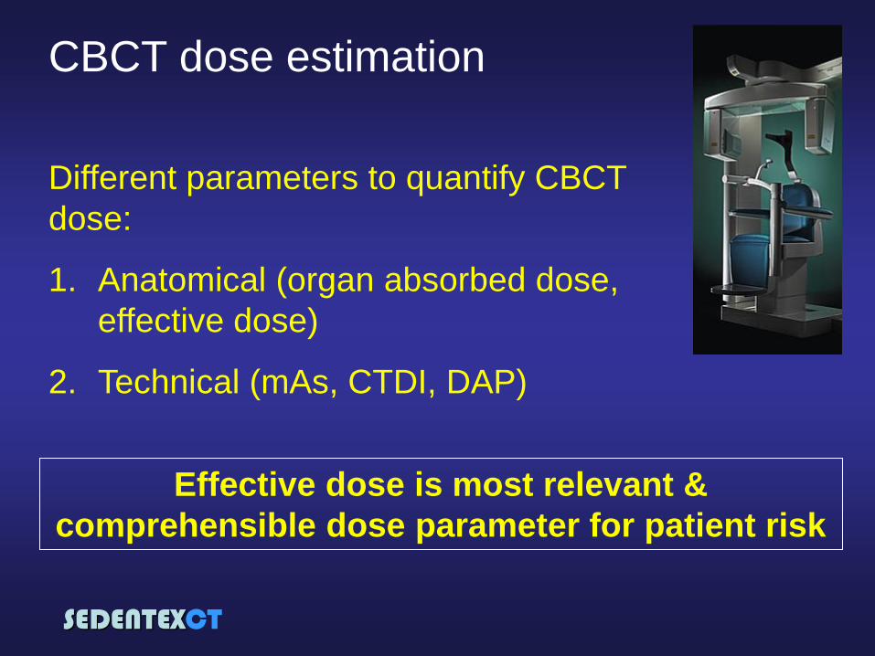
CBCT dose estimation
Different parameters to quantify CBCT
dose:
1. Anatomical (organ absorbed dose,
effective dose)
2. Technical (mAs, CTDI, DAP)
Effective dose is most relevant &
comprehensible dose parameter for patient risk
SEDENTEXCT
Effective dose
(Equivalent dose x wT for each organ) Σ
How is this possible in real people?

SEDENTEXCT
Measuring dose: dosimetry phantoms
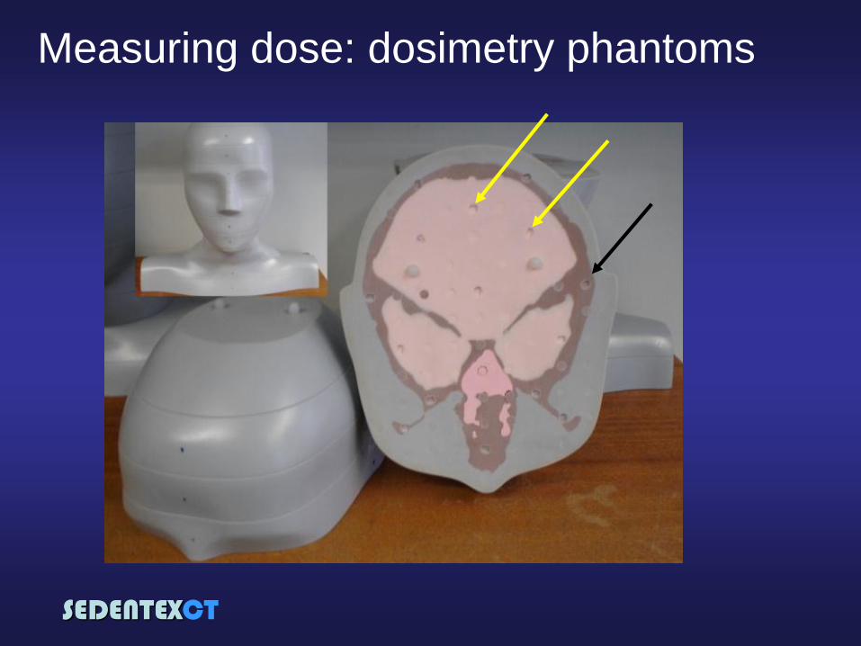
SEDENTEXCT
Measuring dose: dosimetry phantoms
SEDENTEXCT
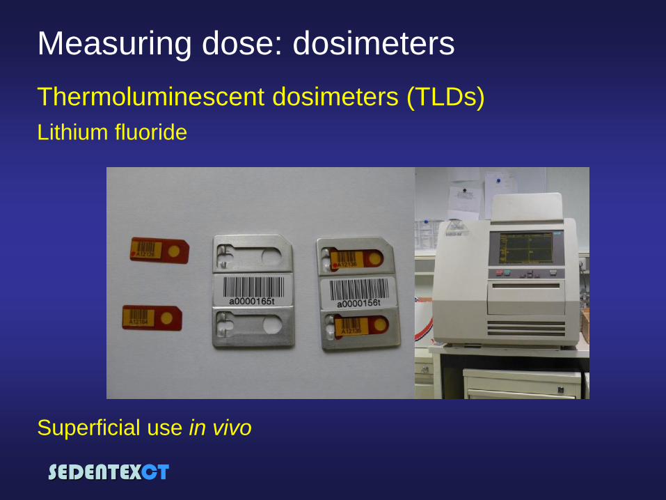
Measuring dose: dosimeters
Thermoluminescent dosimeters (TLDs)
Superficial use in vivo
Lithium fluoride
SEDENTEXCT
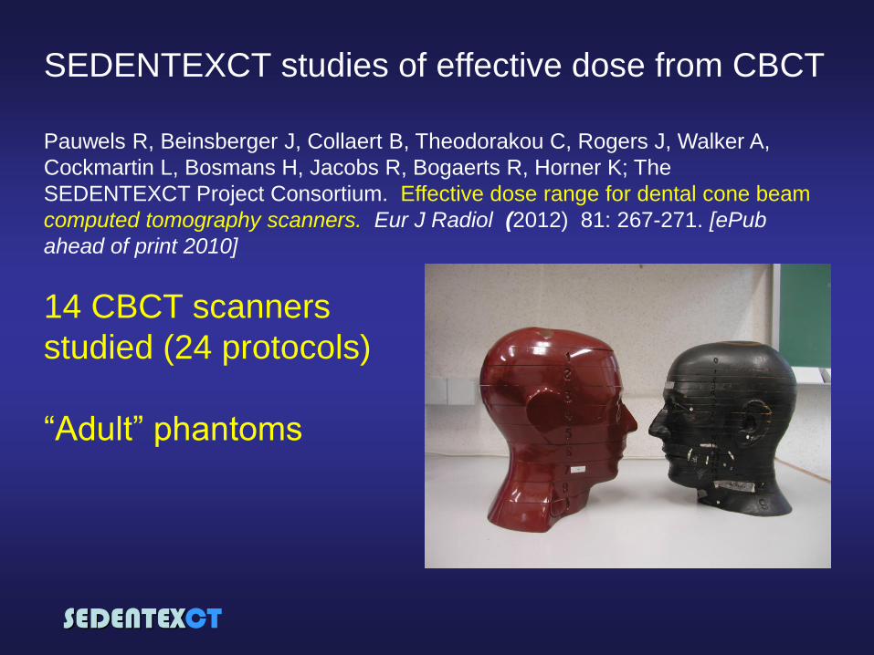
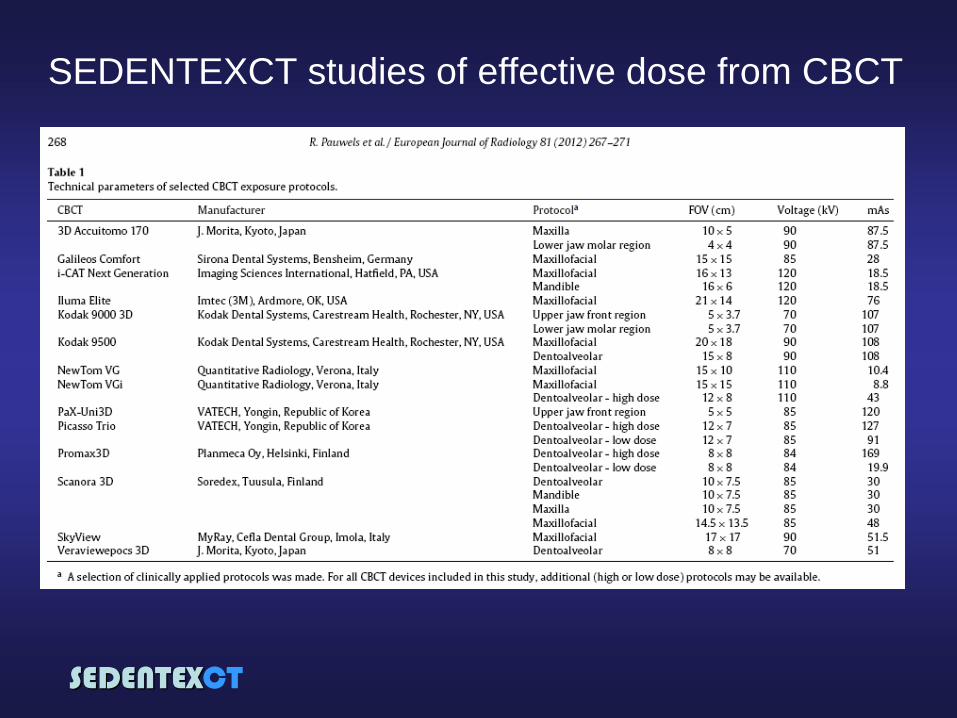
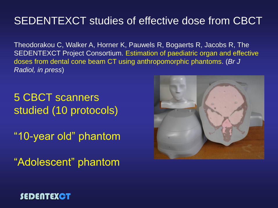
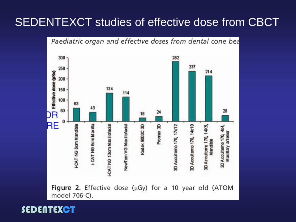
SEDENTEXCT studies of effective dose from CBCT
Pauwels R, Beinsberger J, Collaert B, Theodorakou C, Rogers J, Walker A,
Cockmartin L, Bosmans H, Jacobs R, Bogaerts R, Horner K; The
SEDENTEXCT Project Consortium. Effective dose range for dental cone beam
computed tomography scanners. Eur J Radiol (2012) 81: 267-271. [ePub
ahead of print 2010]
14 CBCT scanners
studied (24 protocols)
“Adult” phantoms
SEDENTEXCT
SEDENTEXCT studies of effective dose from CBCT
SEDENTEXCT
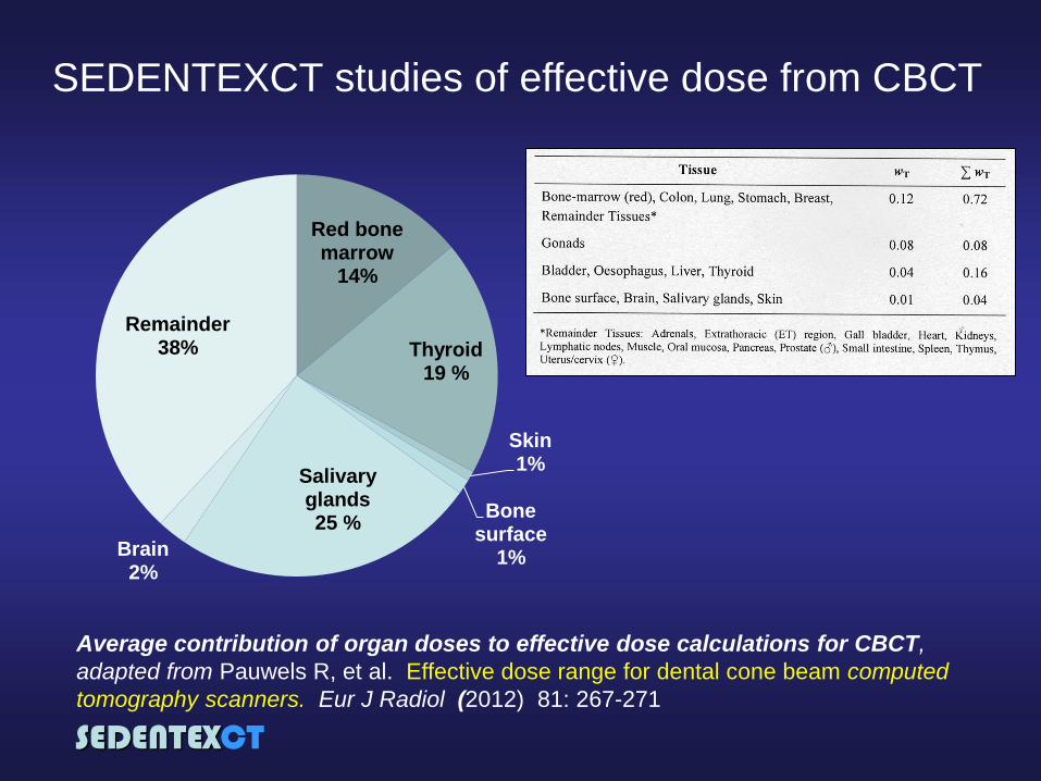
SEDENTEXCT studies of effective dose from CBCT
Red bone marrow
14%
Thyroid 19 %
Skin 1%
Bone surface
1%
Salivary glands 25 %
Brain 2%
Remainder 38%
Average contribution of organ doses to effective dose calculations for CBCT,
adapted from Pauwels R, et al. Effective dose range for dental cone beam computed
tomography scanners. Eur J Radiol (2012) 81: 267-271
SEDENTEXCT
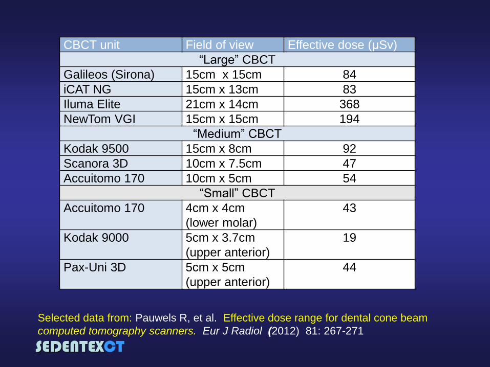
CBCT unit Field of view Effective dose (μSv)
“Large” CBCT
Galileos (Sirona) 15cm x 15cm 84
iCAT NG 15cm x 13cm 83
Iluma Elite 21cm x 14cm 368
NewTom VGI 15cm x 15cm 194
“Medium” CBCT
Kodak 9500 15cm x 8cm 92
Scanora 3D 10cm x 7.5cm 47
Accuitomo 170 10cm x 5cm 54
“Small” CBCT
Accuitomo 170 4cm x 4cm
(lower molar)
43
Kodak 9000 5cm x 3.7cm
(upper anterior)
19
Pax-Uni 3D 5cm x 5cm
(upper anterior)
44
Selected data from: Pauwels R, et al. Effective dose range for dental cone beam
computed tomography scanners. Eur J Radiol (2012) 81: 267-271
SEDENTEXCT
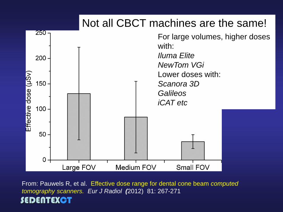
Not all CBCT machines are the same! For large volumes, higher doses
with:
Iluma Elite
NewTom VGi
Lower doses with:
Scanora 3D
Galileos
iCAT etc
From: Pauwels R, et al. Effective dose range for dental cone beam computed
tomography scanners. Eur J Radiol (2012) 81: 267-271
SEDENTEXCT
SEDENTEXCT studies of effective dose from CBCT
Theodorakou C, Walker A, Horner K, Pauwels R, Bogaerts R, Jacobs R, The
SEDENTEXCT Project Consortium. Estimation of paediatric organ and effective
doses from dental cone beam CT using anthropomorphic phantoms. (Br J
Radiol, in press)
5 CBCT scanners
studied (10 protocols)
“10-year old” phantom
“Adolescent” phantom
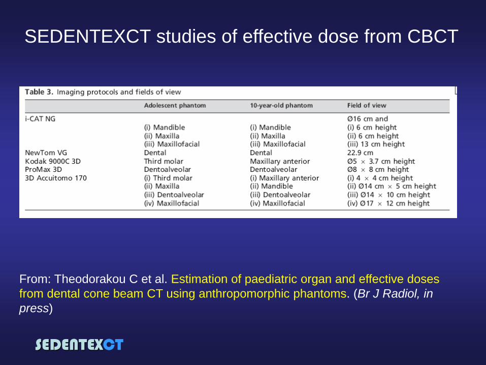
SEDENTEXCT
SEDENTEXCT studies of effective dose from CBCT
From: Theodorakou C et al. Estimation of paediatric organ and effective doses
from dental cone beam CT using anthropomorphic phantoms. (Br J Radiol, in
press)
SEDENTEXCT
SEDENTEXCT studies of effective dose from CBCT
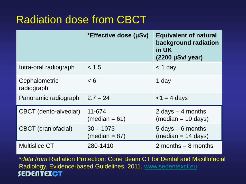
SEDENTEXCT
*Effective dose (μSv) Equivalent of natural
background radiation
in UK
(2200 μSv/ year)
Intra-oral radiograph < 1.5 < 1 day
Cephalometric
radiograph
< 6 1 day
Panoramic radiograph 2.7 – 24 <1 – 4 days
CBCT (dento-alveolar) 11-674
(median = 61)
2 days – 4 months
(median = 10 days)
CBCT (craniofacial) 30 – 1073
(median = 87)
5 days – 6 months
(median = 14 days)
Multislice CT 280-1410 2 months – 8 months
Radiation dose from CBCT
*data from Radiation Protection: Cone Beam CT for Dental and Maxillofacial
Radiology. Evidence-based Guidelines, 2011. www.sedentexct.eu
SEDENTEXCT
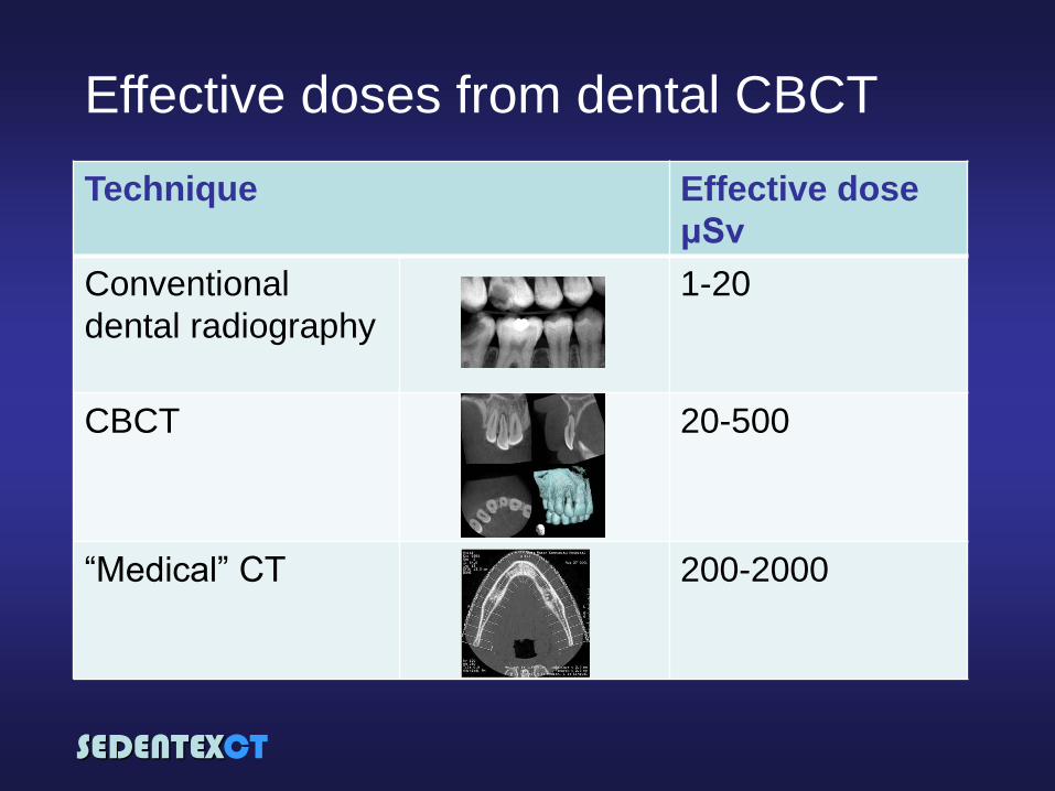
Effective doses from dental CBCT
Technique Effective dose
μSv
Conventional
dental radiography
1-20
CBCT
20-500
“Medical” CT
200-2000
SEDENTEXCT
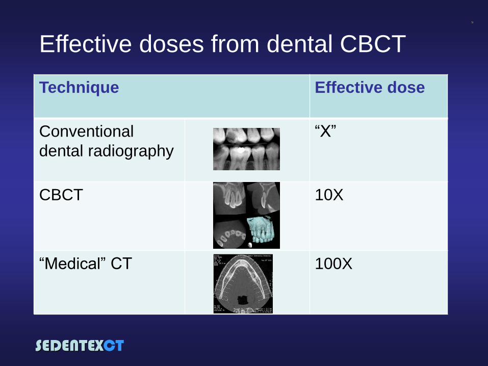
Effective doses from dental CBCT
Technique Effective dose
Conventional
dental radiography
“X”
CBCT
10X
“Medical” CT
100X

SEDENTEXCT
CBCT dose estimation
Different parameters to quantify CBCT dose:
1. Anatomical (organ absorbed dose, effective dose)
2. Technical (mAs, CTDI, DAP)
Dose Area Product (DAP) is most relevant &
comprehensible dose parameter for quality
assurance programmes

SEDENTEXCT
CTDI (Computed tomography dose index)
Designed for
“medical” CT use
Not ideal for CBCT
•Range of field sizes
•Asymmetrical
position of isocentre
•Incomplete rotations
SEDENTEXCT
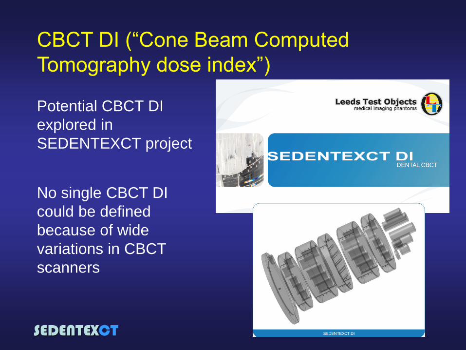
CBCT DI (“Cone Beam Computed
Tomography dose index”)
Potential CBCT DI
explored in
SEDENTEXCT project
No single CBCT DI
could be defined
because of wide
variations in CBCT
scanners
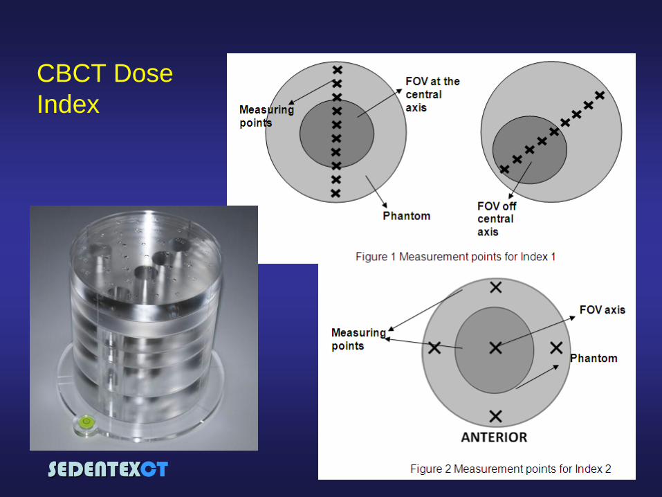
SEDENTEXCT
CBCT Dose
Index
SEDENTEXCT
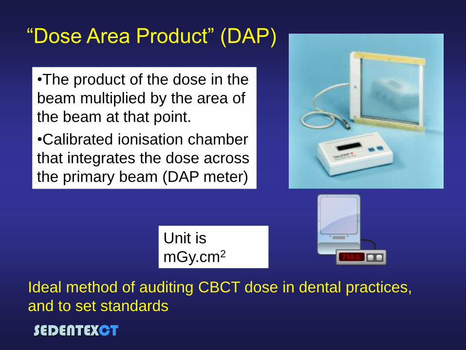
“Dose Area Product” (DAP)
•The product of the dose in the
beam multiplied by the area of
the beam at that point.
•Calibrated ionisation chamber
that integrates the dose across
the primary beam (DAP meter)
Unit is
mGy.cm2
Ideal method of auditing CBCT dose in dental practices,
and to set standards
SEDENTEXCT
Summary “Effective dose” measurements are useful to
consider the risks of cancer associated with X-ray
exposures
Effective doses with CBCT vary enormously....not
all CBCT machines are the same!
Effective dose is not useful for checking individual
dentist’s CBCT equipment. Dose-Area-Product is
currently recommended.
SEDENTEXCT
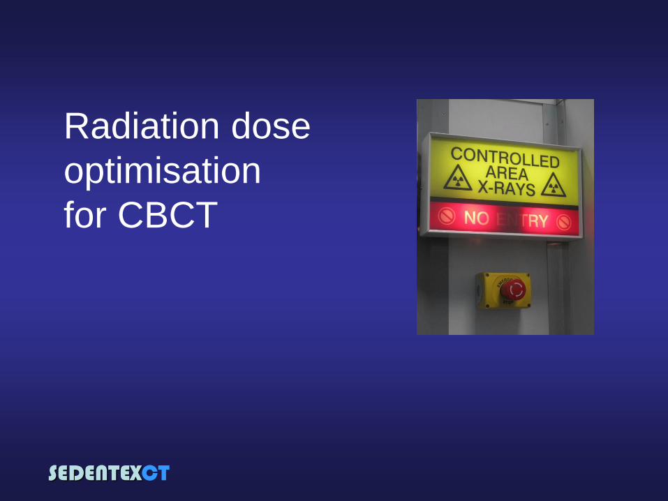
Radiation dose
optimisation
for CBCT
SEDENTEXCT
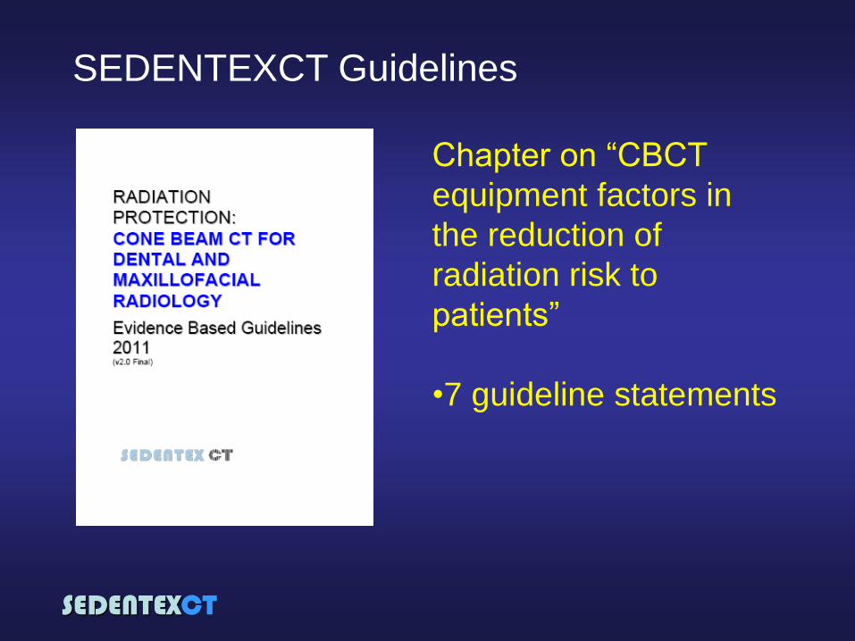
SEDENTEXCT Guidelines
Chapter on “CBCT
equipment factors in
the reduction of
radiation risk to
patients”
•7 guideline statements
SEDENTEXCT
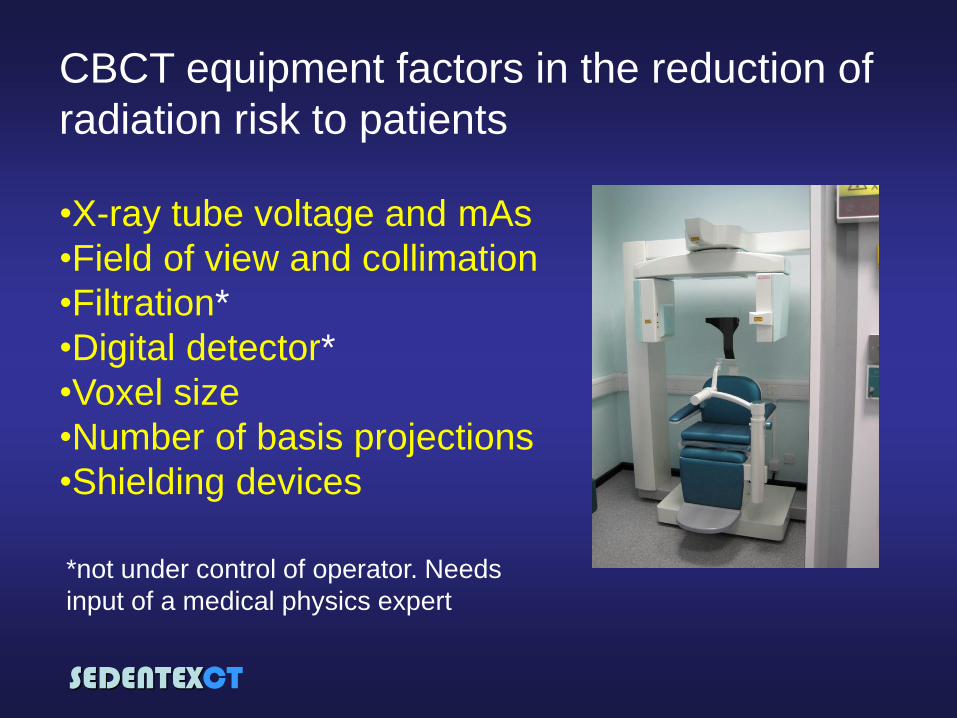
CBCT equipment factors in the reduction of
radiation risk to patients
•X-ray tube voltage and mAs
•Field of view and collimation
•Filtration*
•Digital detector*
•Voxel size
•Number of basis projections
•Shielding devices
*not under control of operator. Needs
input of a medical physics expert
SEDENTEXCT
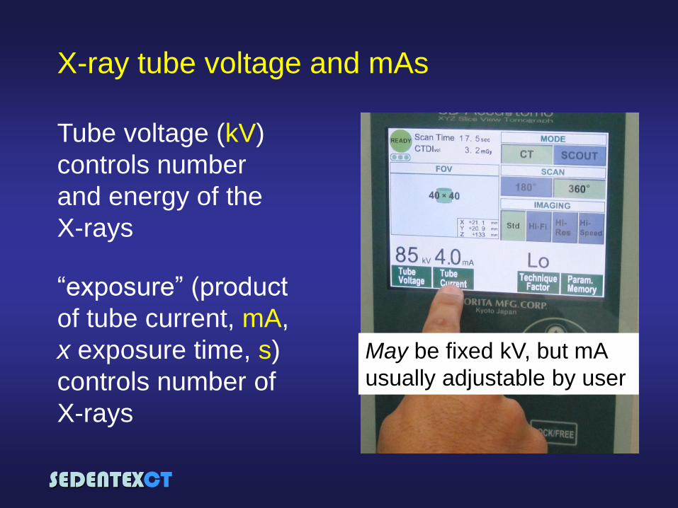
X-ray tube voltage and mAs
Tube voltage (kV)
controls number
and energy of the
X-rays
“exposure” (product
of tube current, mA,
x exposure time, s)
controls number of
X-rays
May be fixed kV, but mA
usually adjustable by user
SEDENTEXCT
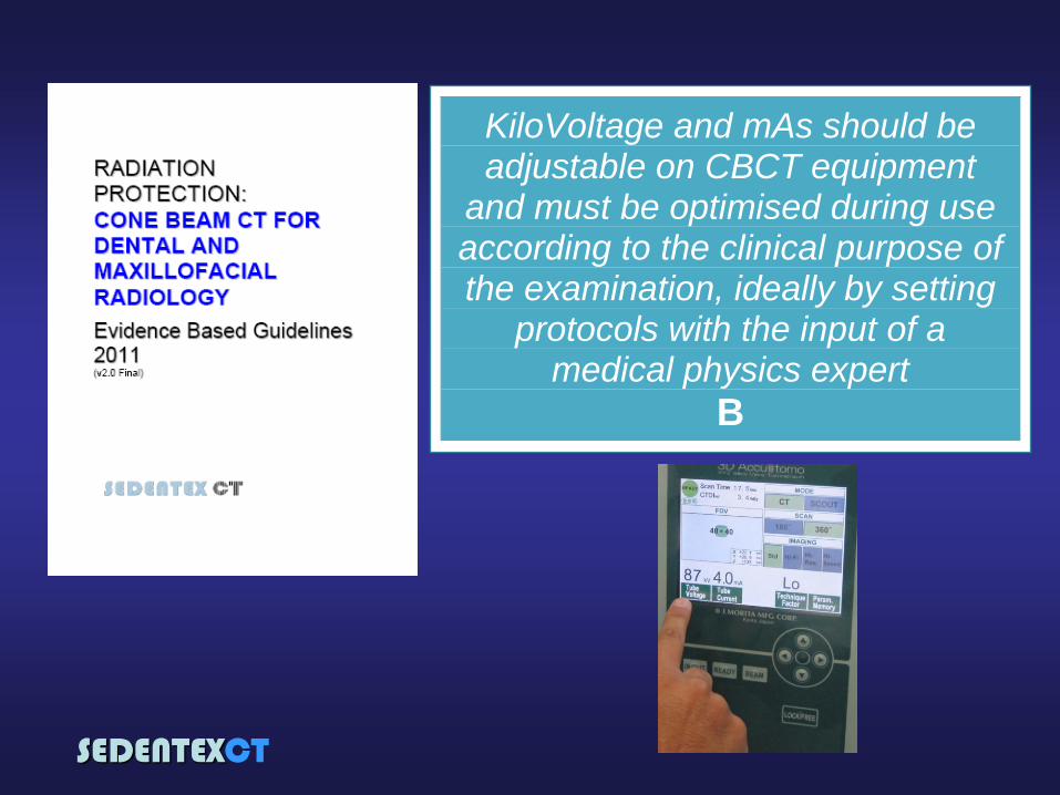
KiloVoltage and mAs should be adjustable on CBCT equipment
and must be optimised during use according to the clinical purpose of the examination, ideally by setting
protocols with the input of a medical physics expert
B
SEDENTEXCT
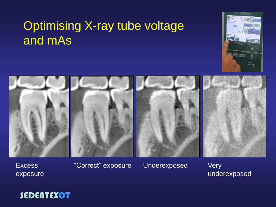
“Correct” exposure Excess
exposure
Underexposed Very
underexposed
Optimising X-ray tube voltage
and mAs
SEDENTEXCT
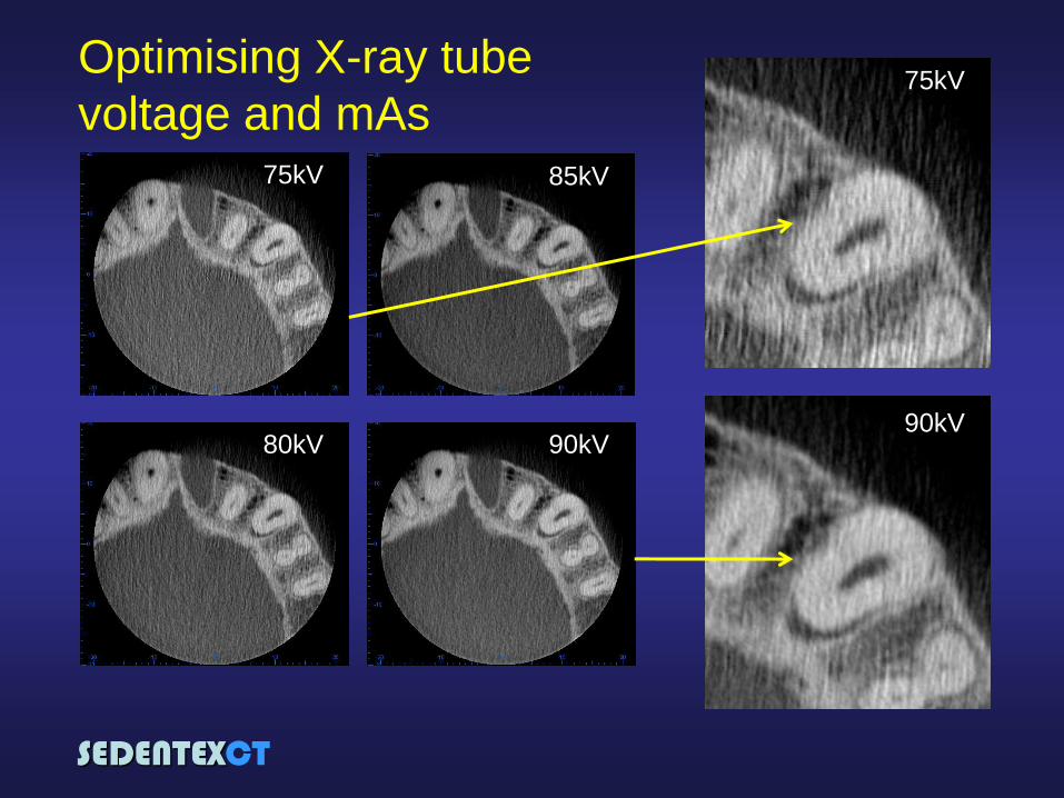
75kV
80kV
85kV
90kV 90kV
75kV Optimising X-ray tube
voltage and mAs
SEDENTEXCT
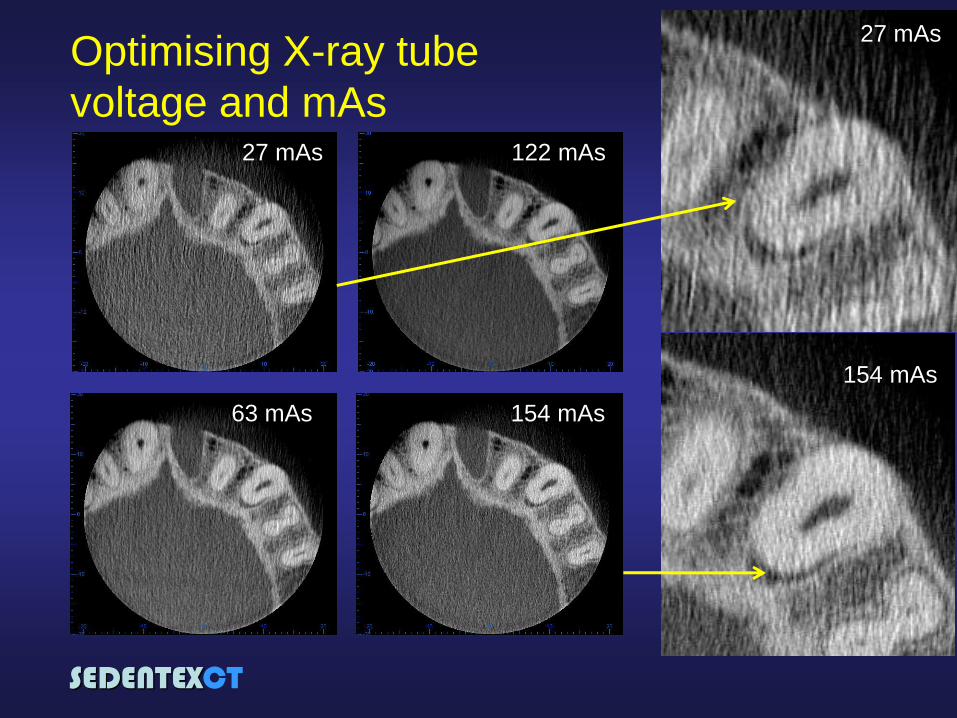
27 mAs
154 mAs
122 mAs
63 mAs
27 mAs
154 mAs
Optimising X-ray tube
voltage and mAs
SEDENTEXCT
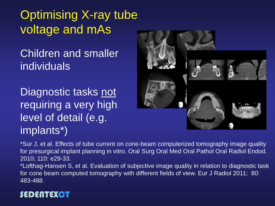
Optimising X-ray tube
voltage and mAs
Children and smaller
individuals
Diagnostic tasks not
requiring a very high
level of detail (e.g.
implants*) *Sur J, et al. Effects of tube current on cone-beam computerized tomography image quality
for presurgical implant planning in vitro. Oral Surg Oral Med Oral Pathol Oral Radiol Endod.
2010; 110: e29-33.
*Lofthag-Hansen S, et al. Evaluation of subjective image quality in relation to diagnostic task
for cone beam computed tomography with different fields of view. Eur J Radiol 2011; 80:
483-488.
SEDENTEXCT
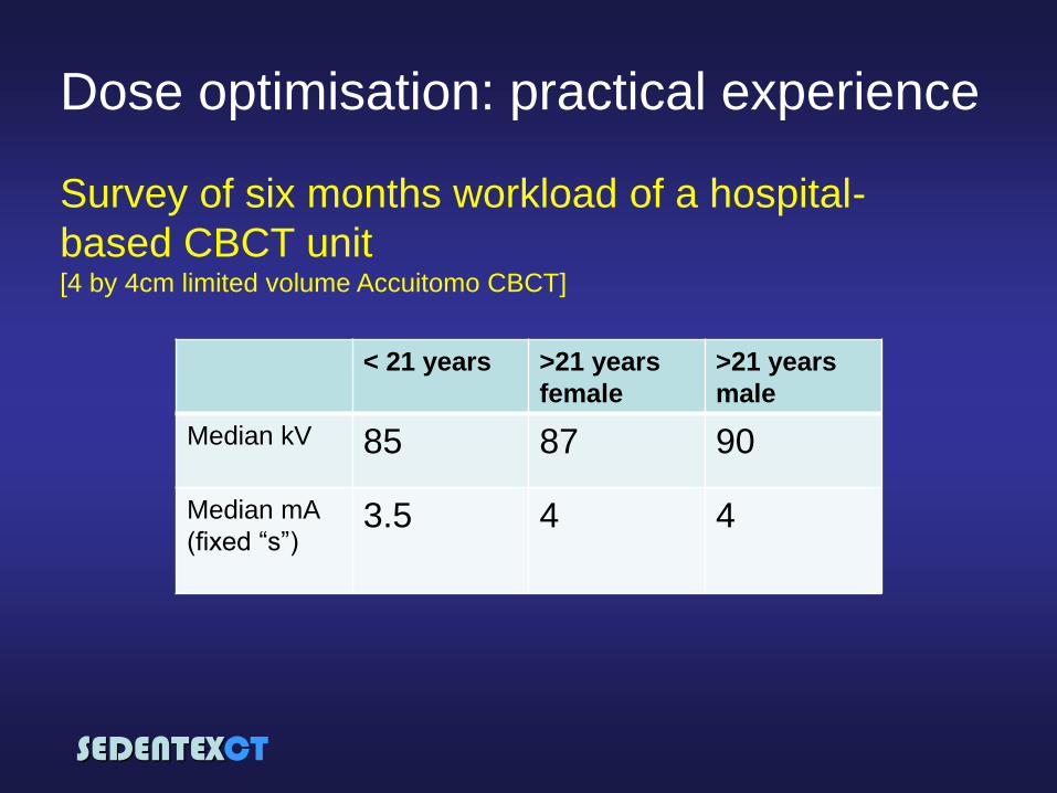
Dose optimisation: practical experience
< 21 years >21 years
female
>21 years
male
Median kV
85 87 90
Median mA
(fixed “s”)
3.5 4 4
Survey of six months workload of a hospital-
based CBCT unit [4 by 4cm limited volume Accuitomo CBCT]
SEDENTEXCT
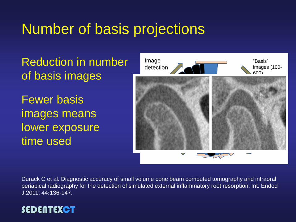
Reduction in number
of basis images
Fewer basis
images means
lower exposure
time used
Number of basis projections
Image
detection“Basis”
images (100-
600)
Durack C et al. Diagnostic accuracy of small volume cone beam computed tomography and intraoral
periapical radiography for the detection of simulated external inflammatory root resorption. Int. Endod
J.2011; 44:136-147.
SEDENTEXCT
Reduction in number
of basis images
Fewer basis
images means
lower exposure
time used
Number of basis projections
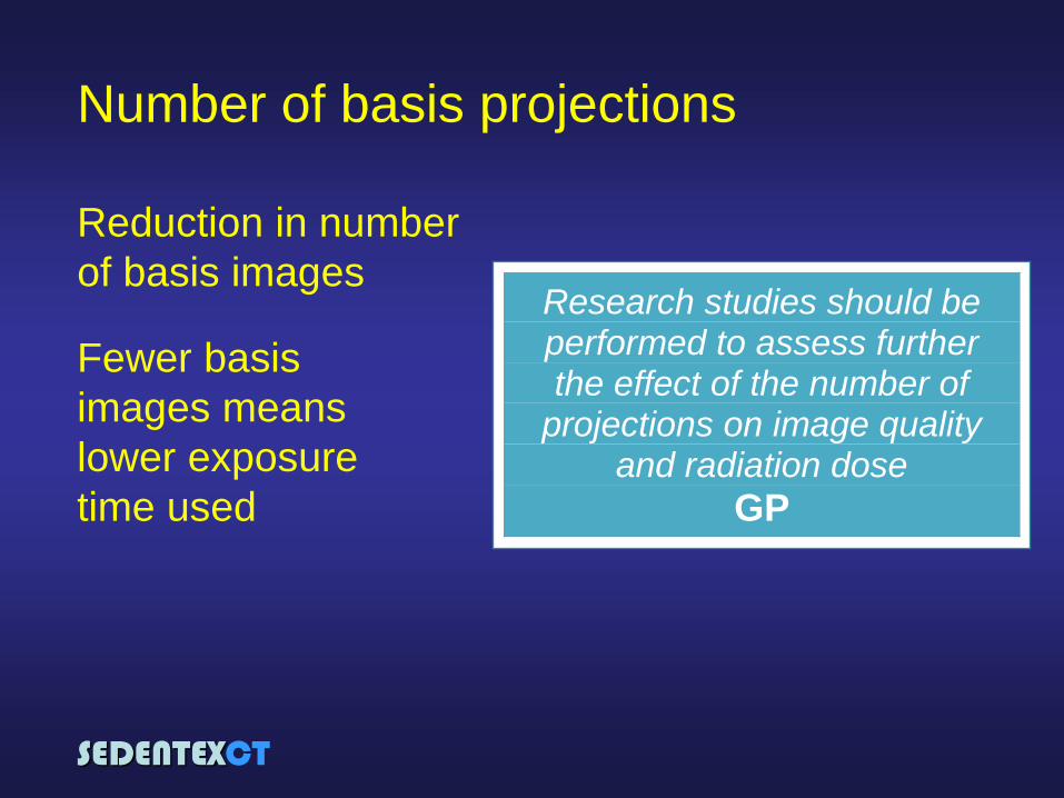
Research studies should be performed to assess further the effect of the number of
projections on image quality and radiation dose
GP
SEDENTEXCT
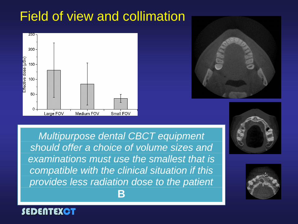
Field of view and collimation
Multipurpose dental CBCT equipment should offer a choice of volume sizes and examinations must use the smallest that is compatible with the clinical situation if this provides less radiation dose to the patient
B
SEDENTEXCT
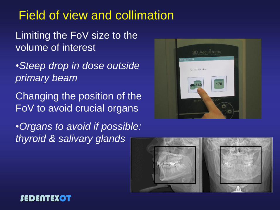
Limiting the FoV size to the
volume of interest
•Steep drop in dose outside
primary beam
Changing the position of the
FoV to avoid crucial organs
•Organs to avoid if possible:
thyroid & salivary glands
Field of view and collimation

SEDENTEXCT
“Resolution” and voxel size
Spatial Resolution is related
to the exposure factors used
“Higher resolution” options
achieved by increased
exposure (mAs or more basis
images)
Multipurpose dental CBCT equipment should offer a choice of voxel sizes and examinations should use the largest voxel size (lowest dose) consistent with
acceptable diagnostic accuracy
C
SEDENTEXCT
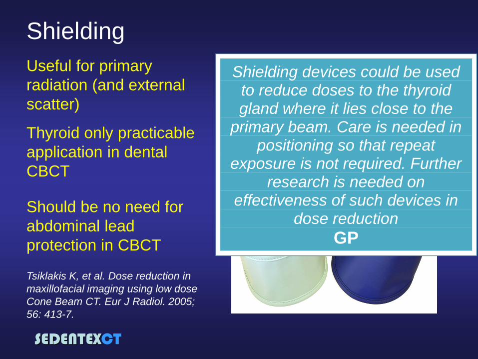
Shielding
Useful for primary
radiation (and external
scatter)
Thyroid only practicable
application in dental
CBCT
Tsiklakis K, et al. Dose reduction in
maxillofacial imaging using low dose
Cone Beam CT. Eur J Radiol. 2005;
56: 413-7.
Should be no need for
abdominal lead
protection in CBCT
Shielding devices could be used to reduce doses to the thyroid gland where it lies close to the
primary beam. Care is needed in positioning so that repeat
exposure is not required. Further research is needed on
effectiveness of such devices in dose reduction
GP GP
SEDENTEXCT
Summary
Several parameters determine the patient dose
associated with a CBCT examination
Some can be adjusted by the operator, some are
fixed by the manufacturer and some can be
changed with the advice of a Medical Physics
Expert
There is always a trade-off between radiation
dose and image quality; the difficult choice is
identifying the compromise......
SEDENTEXCT
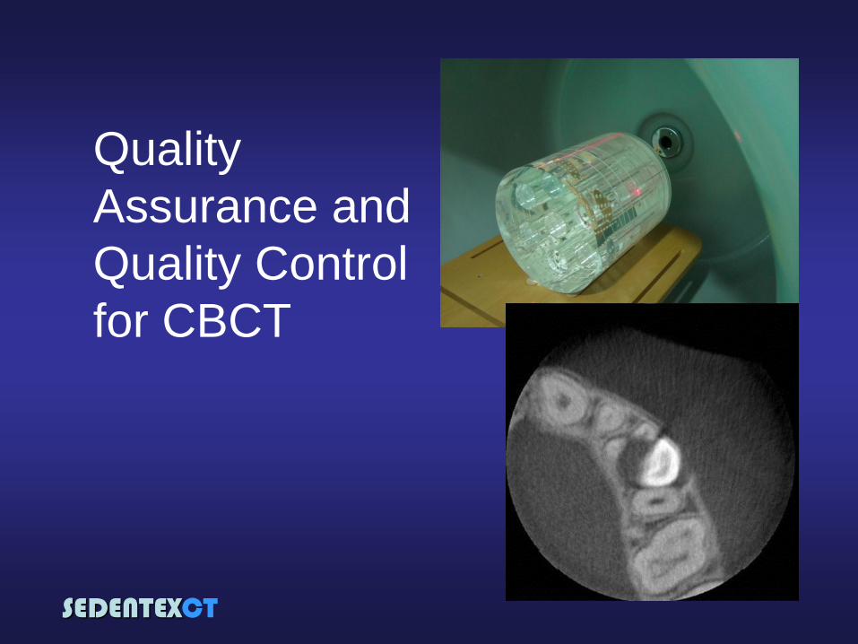
Quality
Assurance and
Quality Control
for CBCT
SEDENTEXCT
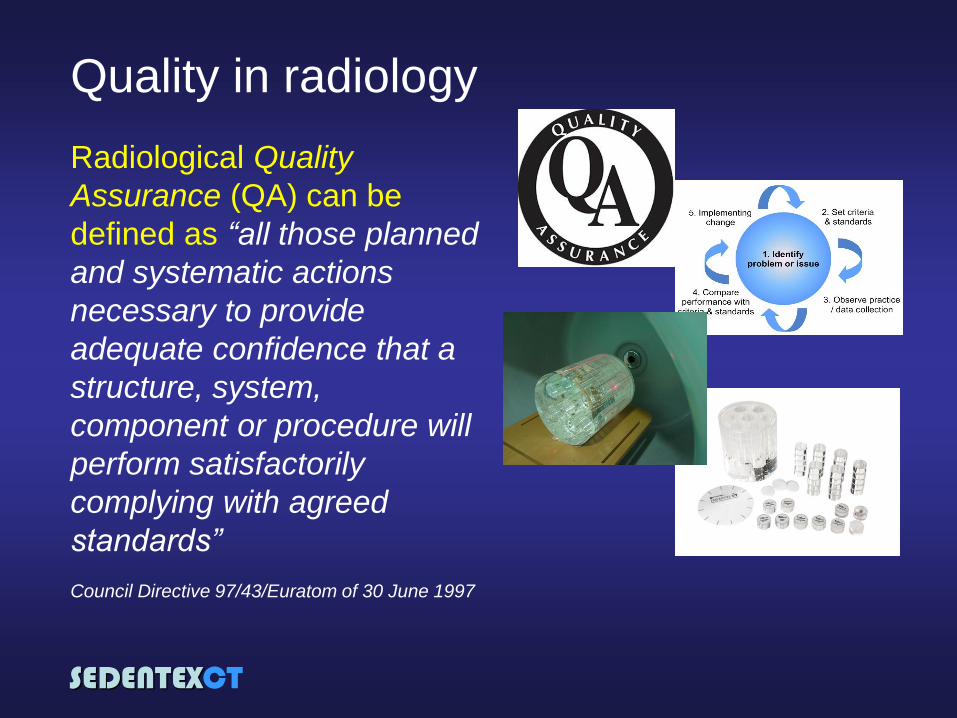
Quality in radiology
Radiological Quality
Assurance (QA) can be
defined as “all those planned
and systematic actions
necessary to provide
adequate confidence that a
structure, system,
component or procedure will
perform satisfactorily
complying with agreed
standards” Council Directive 97/43/Euratom of 30 June 1997
SEDENTEXCT
Quality in radiology
A radiological Quality
Assurance (QA) Programme
is the “framework” or
“organised effort” which is
devised to achieve quality
assurance
Quality
Control
Administrative
procedures to verify
that: •the quality control techniques are performed properly and according to a planned timetable,
•the results of these techniques are evaluated promptly and accurately,
•the necessary corrective measures are taken in response to these results
SEDENTEXCT
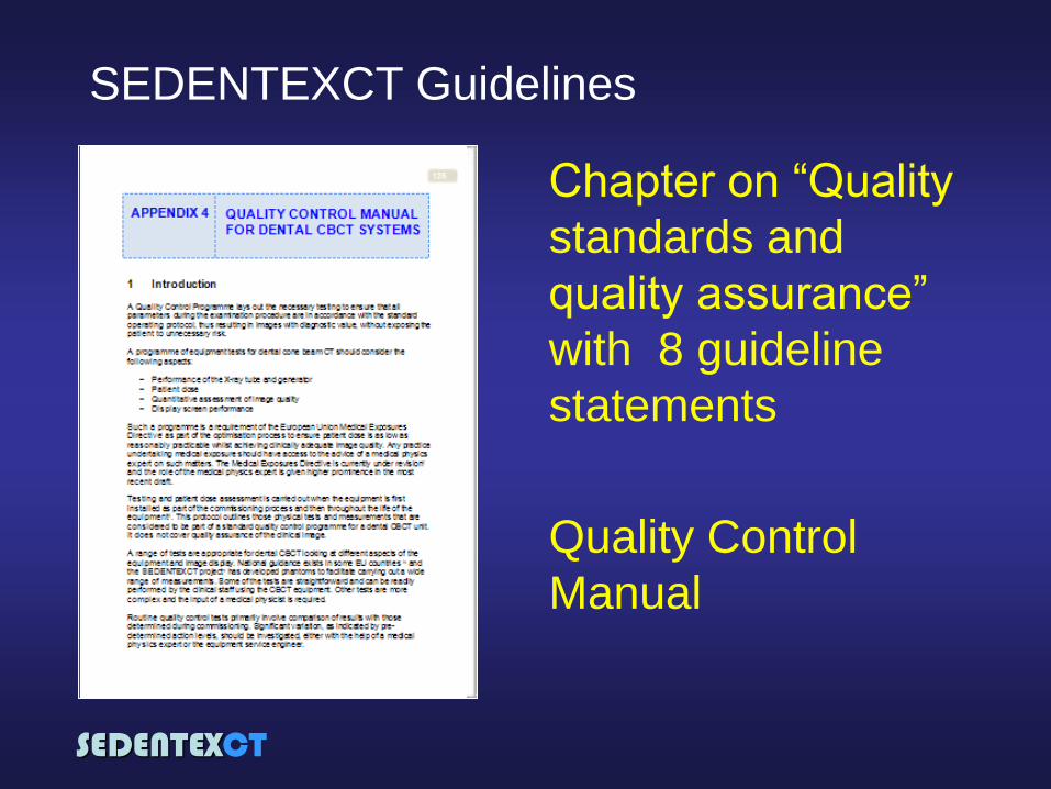
SEDENTEXCT Guidelines
Chapter on “Quality
standards and
quality assurance”
with 8 guideline
statements
Quality Control
Manual
SEDENTEXCT
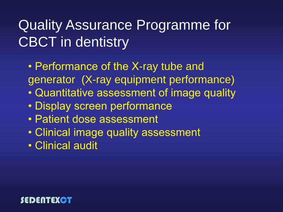
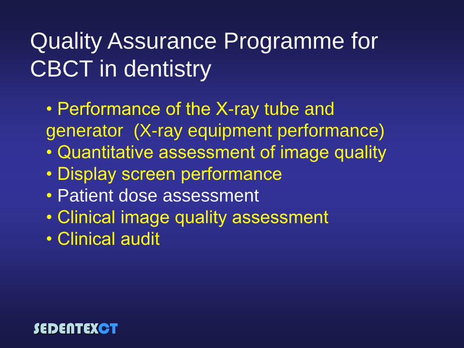
• Performance of the X-ray tube and
generator (X-ray equipment performance)
• Quantitative assessment of image quality
• Display screen performance
• Patient dose assessment
• Clinical image quality assessment
• Clinical audit
Quality Assurance Programme for
CBCT in dentistry
SEDENTEXCT
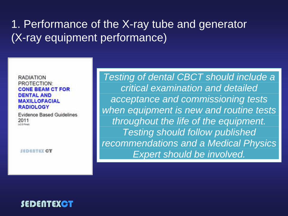
1. Performance of the X-ray tube and generator
(X-ray equipment performance)
Testing of dental CBCT should include a critical examination and detailed
acceptance and commissioning tests when equipment is new and routine tests
throughout the life of the equipment. Testing should follow published
recommendations and a Medical Physics Expert should be involved.
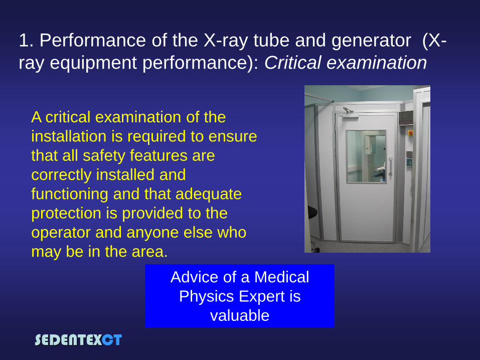
SEDENTEXCT
A critical examination of the
installation is required to ensure
that all safety features are
correctly installed and
functioning and that adequate
protection is provided to the
operator and anyone else who
may be in the area.
1. Performance of the X-ray tube and generator (X-
ray equipment performance): Critical examination
Advice of a Medical
Physics Expert is
valuable
SEDENTEXCT
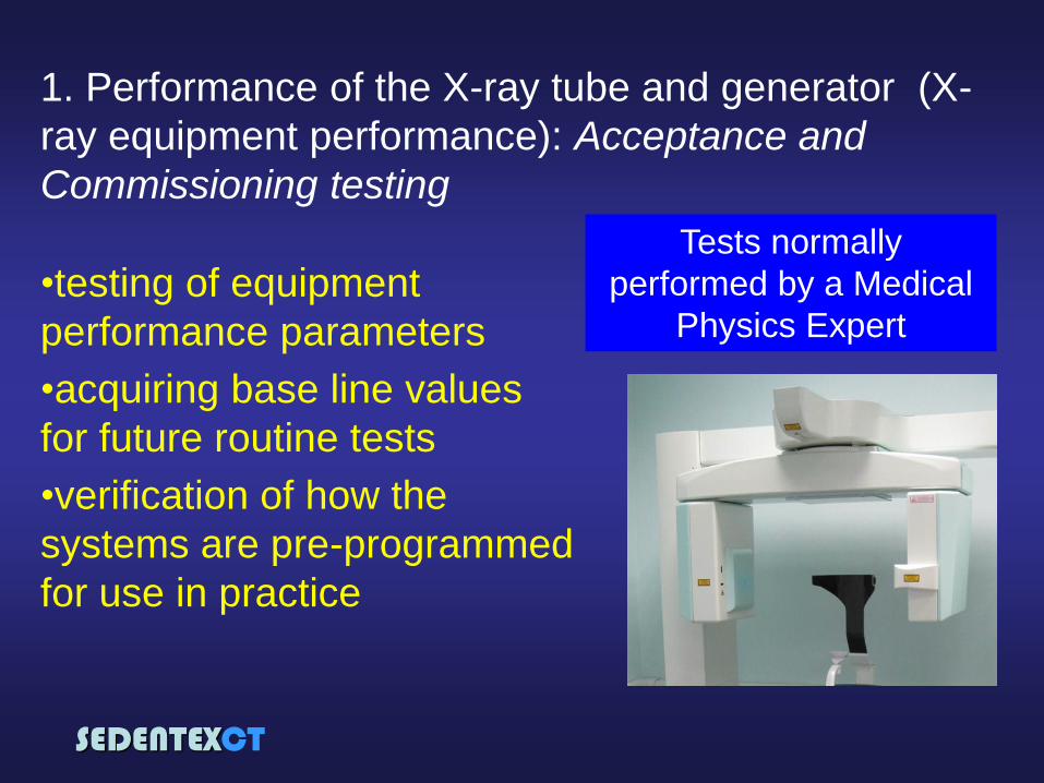
1. Performance of the X-ray tube and generator (X-
ray equipment performance): Acceptance and
Commissioning testing
•testing of equipment
performance parameters
•acquiring base line values
for future routine tests
•verification of how the
systems are pre-programmed
for use in practice
Tests normally
performed by a Medical
Physics Expert
SEDENTEXCT
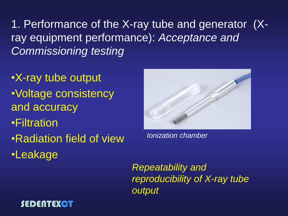
1. Performance of the X-ray tube and generator (X-
ray equipment performance): Acceptance and
Commissioning testing
•X-ray tube output
•Voltage consistency
and accuracy
•Filtration
•Radiation field of view
•Leakage Repeatability and
reproducibility of X-ray tube
output
Ionization chamber
SEDENTEXCT
1. Performance of the X-ray tube and generator (X-
ray equipment performance): Acceptance and
Commissioning testing
•X-ray tube output
•Voltage consistency
and accuracy
•Filtration
•Radiation field of view
•Leakage
Accuracy, repeatability and
reproducibility of kV
kV divider

SEDENTEXCT
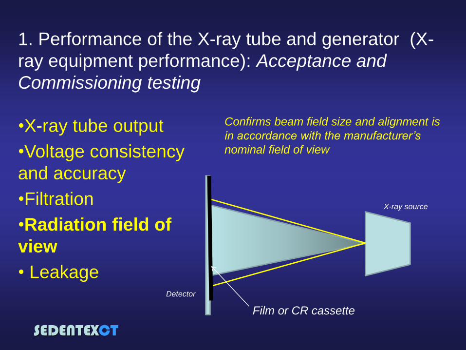
1. Performance of the X-ray tube and generator (X-
ray equipment performance): Acceptance and
Commissioning testing
•X-ray tube output
•Voltage consistency
and accuracy
•Filtration
•Radiation field of view
•Leakage
Can be estimated by measuring the
“Half Value Layer” (the thickness of
aluminium required to reduce the
intensity of the incident X-ray beam by
half )
X-ray source
Detector
Ionisation chamber
Aluminium
SEDENTEXCT
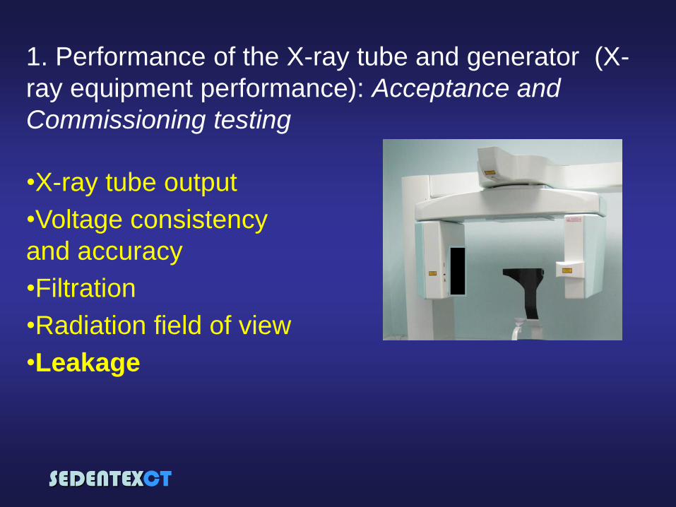
1. Performance of the X-ray tube and generator (X-
ray equipment performance): Acceptance and
Commissioning testing
•X-ray tube output
•Voltage consistency
and accuracy
•Filtration
•Radiation field of
view
• Leakage Detector
X-ray source
Confirms beam field size and alignment is
in accordance with the manufacturer’s
nominal field of view
Film or CR cassette
SEDENTEXCT
1. Performance of the X-ray tube and generator (X-
ray equipment performance): Acceptance and
Commissioning testing
•X-ray tube output
•Voltage consistency
and accuracy
•Filtration
•Radiation field of view
•Leakage
SEDENTEXCT
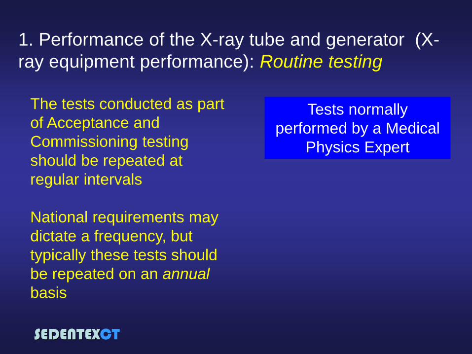
1. Performance of the X-ray tube and generator (X-
ray equipment performance): Routine testing
The tests conducted as part
of Acceptance and
Commissioning testing
should be repeated at
regular intervals
National requirements may
dictate a frequency, but
typically these tests should
be repeated on an annual
basis
Tests normally
performed by a Medical
Physics Expert
SEDENTEXCT
• Performance of the X-ray tube and
generator (X-ray equipment performance)
• Quantitative assessment of image quality
• Display screen performance
• Patient dose assessment
• Clinical image quality assessment
• Clinical audit
Quality Assurance Programme for
CBCT in dentistry
SEDENTEXCT
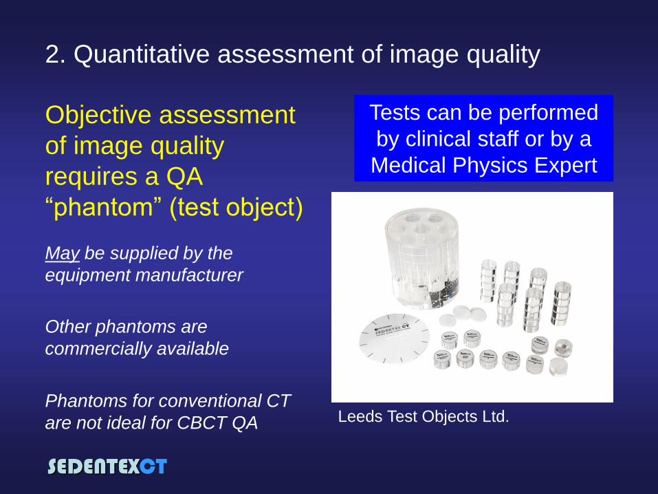
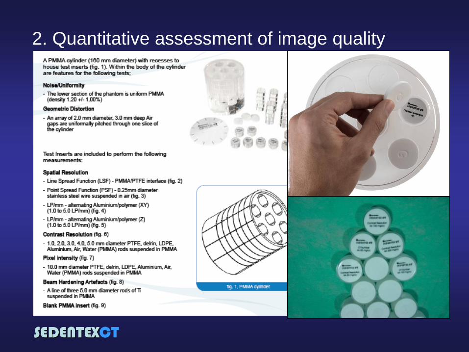
2. Quantitative assessment of image quality
Objective assessment
of image quality
requires a QA
“phantom” (test object)
May be supplied by the
equipment manufacturer
Other phantoms are
commercially available
Tests can be performed
by clinical staff or by a
Medical Physics Expert
Leeds Test Objects Ltd. Phantoms for conventional CT
are not ideal for CBCT QA
SEDENTEXCT
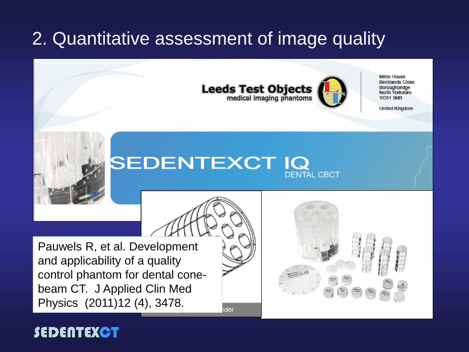
2. Quantitative assessment of image quality
Pauwels R, et al. Development
and applicability of a quality
control phantom for dental cone-
beam CT. J Applied Clin Med
Physics (2011)12 (4), 3478.
SEDENTEXCT
2. Quantitative assessment of image quality
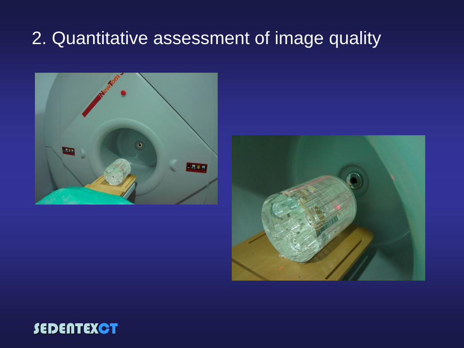
SEDENTEXCT
2. Quantitative assessment of image quality
SEDENTEXCT
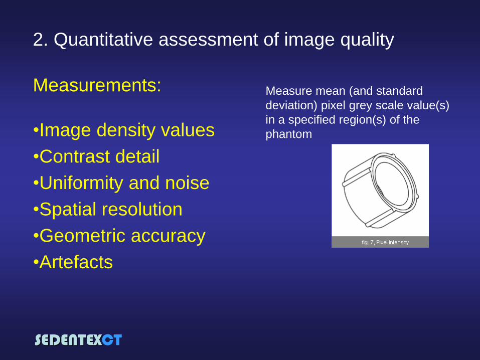
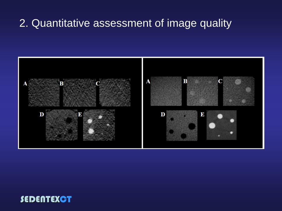
2. Quantitative assessment of image quality
Measurements:
•Image density values
•Contrast detail
•Uniformity and noise
•Spatial resolution
•Geometric accuracy
•Artefacts
Measure mean (and standard
deviation) pixel grey scale value(s)
in a specified region(s) of the
phantom
SEDENTEXCT
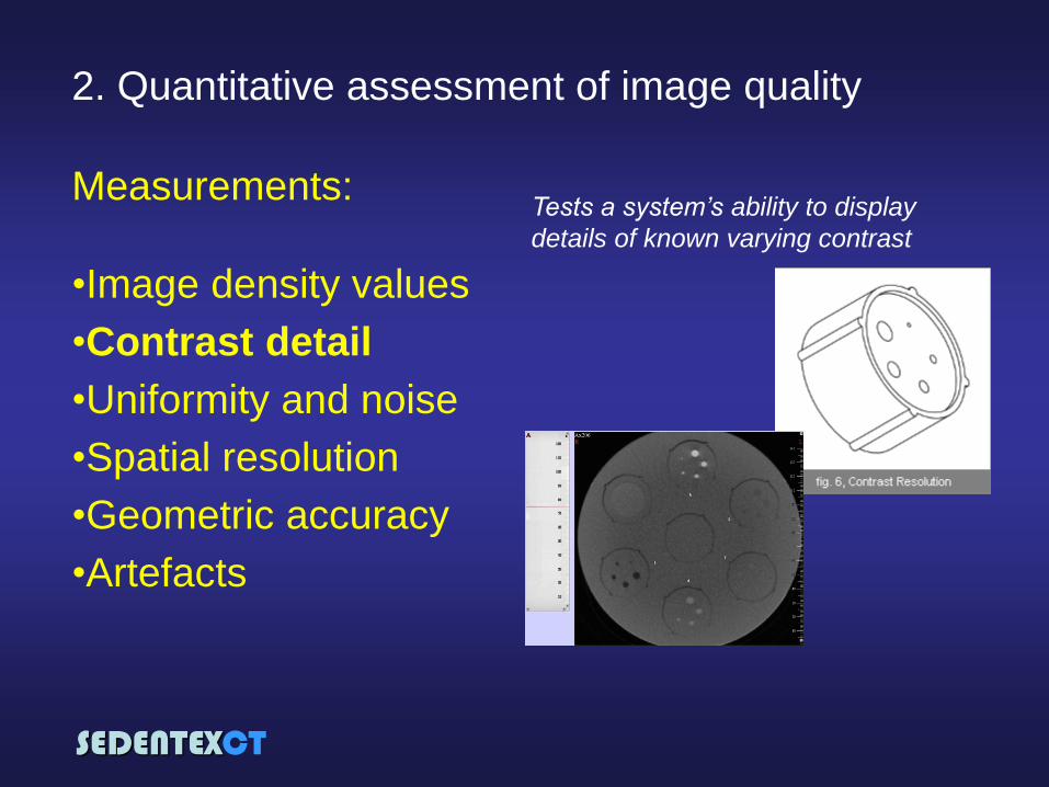
2. Quantitative assessment of image quality
Measurements:
•Image density values
•Contrast detail
•Uniformity and noise
•Spatial resolution
•Geometric accuracy
•Artefacts
Tests a system’s ability to display
details of known varying contrast
SEDENTEXCT
2. Quantitative assessment of image quality
SEDENTEXCT
2. Quantitative assessment of image quality
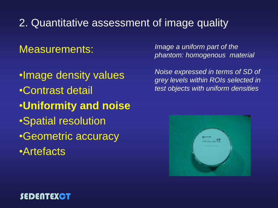
Measurements:
•Image density values
•Contrast detail
•Uniformity and noise
•Spatial resolution
•Geometric accuracy
•Artefacts
Image a uniform part of the
phantom: homogenous material
Noise expressed in terms of SD of
grey levels within ROIs selected in
test objects with uniform densities
SEDENTEXCT
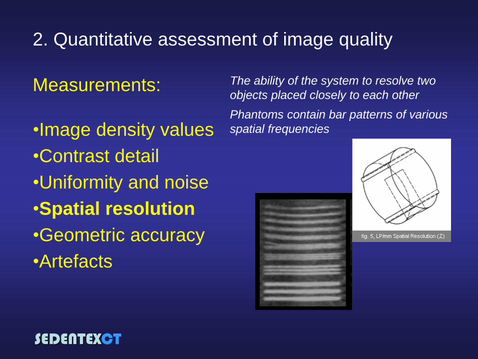
2. Quantitative assessment of image quality
Measurements:
•Image density values
•Contrast detail
•Uniformity and noise
•Spatial resolution
•Geometric accuracy
•Artefacts
The ability of the system to resolve two
objects placed closely to each other
Phantoms contain bar patterns of various
spatial frequencies
SEDENTEXCT
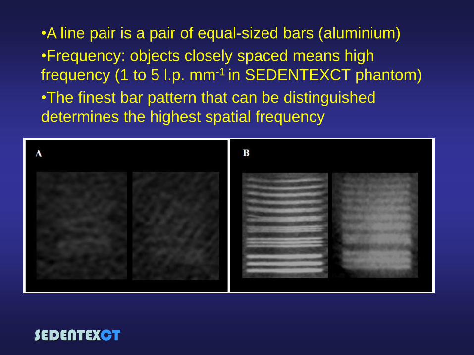
•A line pair is a pair of equal-sized bars (aluminium)
•Frequency: objects closely spaced means high
frequency (1 to 5 l.p. mm-1 in SEDENTEXCT phantom)
•The finest bar pattern that can be distinguished
determines the highest spatial frequency
SEDENTEXCT
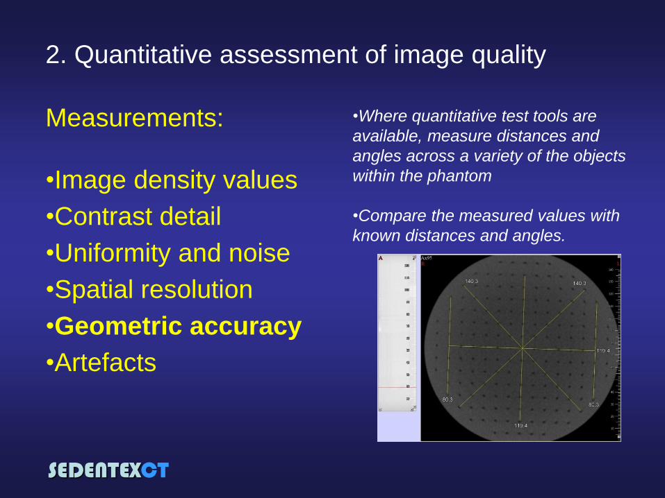
2. Quantitative assessment of image quality
Measurements:
•Image density values
•Contrast detail
•Uniformity and noise
•Spatial resolution
•Geometric accuracy
•Artefacts
•Where quantitative test tools are
available, measure distances and
angles across a variety of the objects
within the phantom
•Compare the measured values with
known distances and angles.

SEDENTEXCT
2. Quantitative assessment of image quality
Measurements:
•Image density values
•Contrast detail
•Uniformity and noise
•Spatial resolution
•Geometric accuracy
•Artefacts
SEDENTEXCT
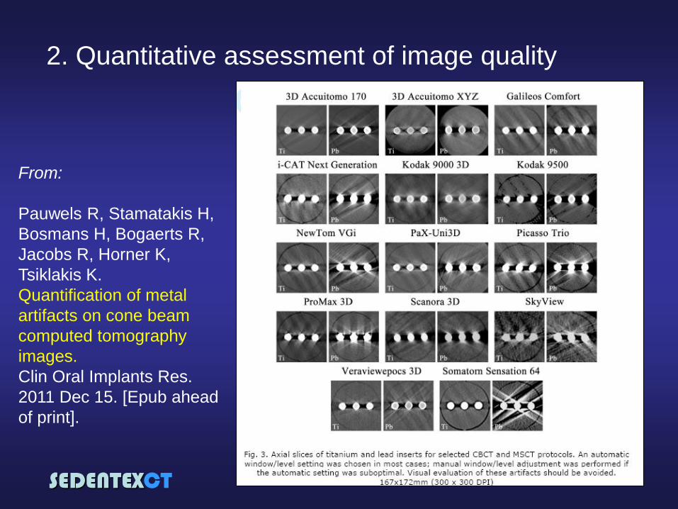
2. Quantitative assessment of image quality
From:
Pauwels R, Stamatakis H,
Bosmans H, Bogaerts R,
Jacobs R, Horner K,
Tsiklakis K.
Quantification of metal
artifacts on cone beam
computed tomography
images.
Clin Oral Implants Res.
2011 Dec 15. [Epub ahead
of print].

SEDENTEXCT
• Performance of the X-ray tube and
generator (X-ray equipment performance)
• Quantitative assessment of image quality
• Display screen performance
• Patient dose assessment
• Clinical image quality assessment
• Clinical audit
Quality Assurance Programme for
CBCT in dentistry
SEDENTEXCT
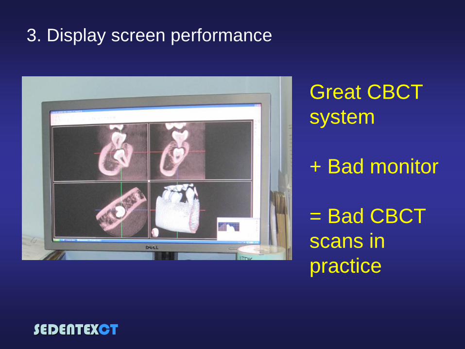
3. Display screen performance
Great CBCT
system
+ Bad monitor
= Bad CBCT
scans in
practice
SEDENTEXCT
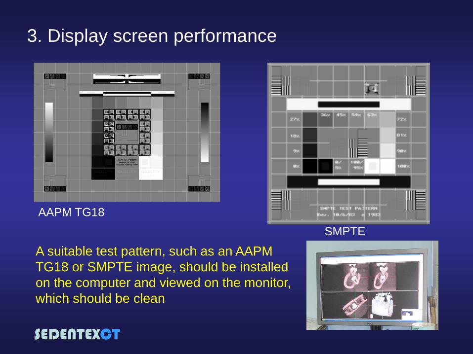
3. Display screen performance
AAPM TG18
SMPTE
A suitable test pattern, such as an AAPM
TG18 or SMPTE image, should be installed
on the computer and viewed on the monitor,
which should be clean
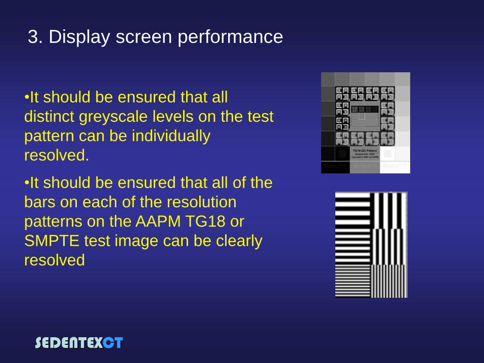
SEDENTEXCT
•It should be ensured that all
distinct greyscale levels on the test
pattern can be individually
resolved.
•It should be ensured that all of the
bars on each of the resolution
patterns on the AAPM TG18 or
SMPTE test image can be clearly
resolved
3. Display screen performance
SEDENTEXCT
• Performance of the X-ray tube and
generator (X-ray equipment performance)
• Quantitative assessment of image quality
• Display screen performance
• Patient dose assessment
• Clinical image quality assessment
• Clinical audit
Quality Assurance Programme for
CBCT in dentistry
SEDENTEXCT
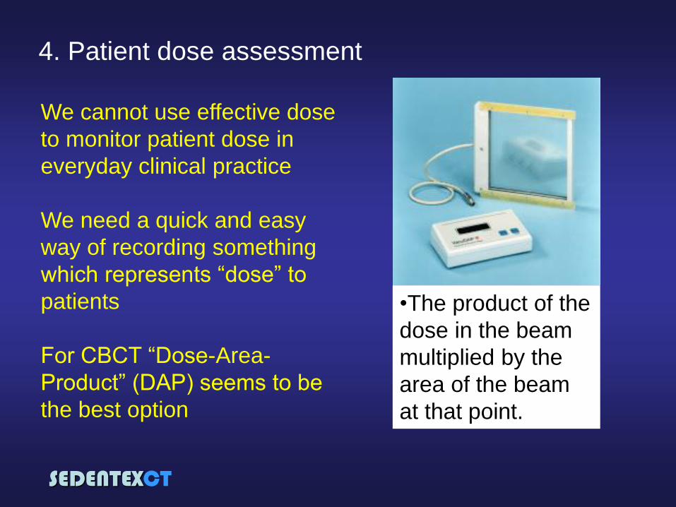
4. Patient dose assessment
We cannot use effective dose
to monitor patient dose in
everyday clinical practice
We need a quick and easy
way of recording something
which represents “dose” to
patients
For CBCT “Dose-Area-
Product” (DAP) seems to be
the best option
•The product of the
dose in the beam
multiplied by the
area of the beam
at that point.
SEDENTEXCT
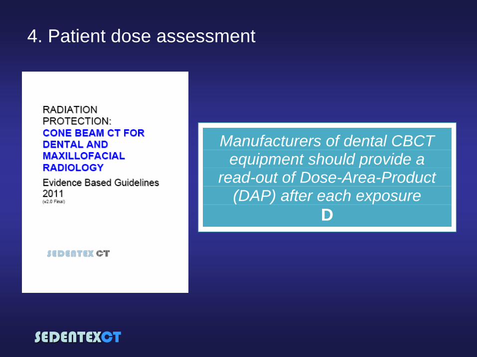
4. Patient dose assessment
Manufacturers of dental CBCT equipment should provide a
read-out of Dose-Area-Product (DAP) after each exposure
D
SEDENTEXCT
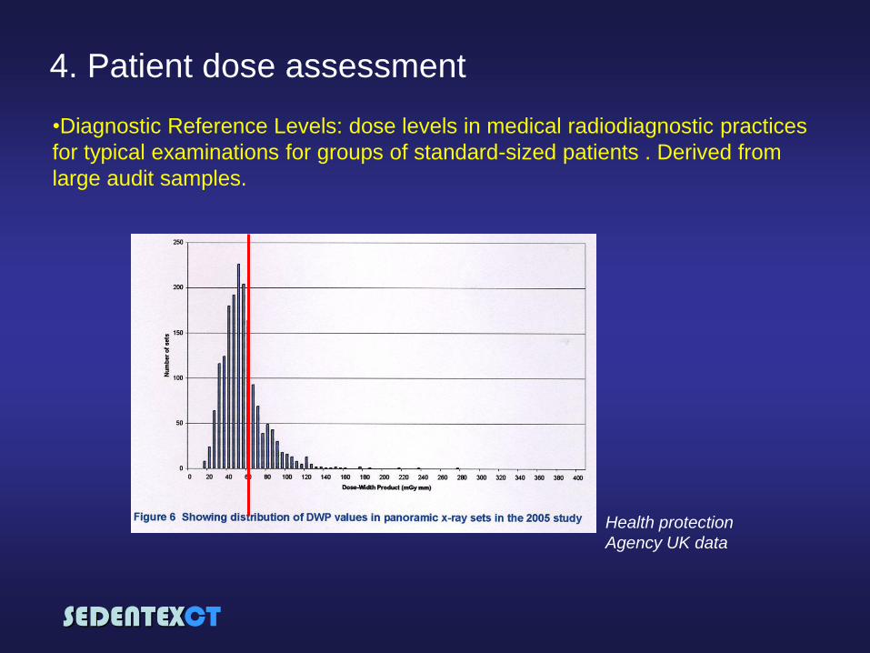
4. Patient dose assessment
•Diagnostic Reference Levels: dose levels in medical radiodiagnostic practices
for typical examinations for groups of standard-sized patients . Derived from
large audit samples.
Health protection
Agency UK data

SEDENTEXCT
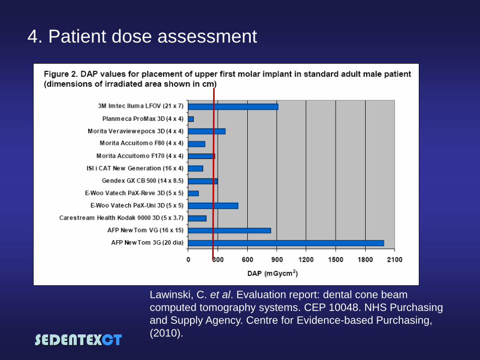
4. Patient dose assessment
•There is insufficient audit data on CBCT to set a formal DRL at the
present time
•Instead, an “achievable dose” has been set as a provisional measure
•Based upon CBCT of an adult patient being assessed for a single
upper molar implant
In the future, an “achievable dose”, and eventually DRL, should also be set for
paediatric CBCT
Until further audit data is published, the panel recommend the adoption of an achievable
Dose Area Product of 250 mGy cm2 for CBCT
imaging for the placement of an upper first molar implant in a standard adult patient
D
SEDENTEXCT
4. Patient dose assessment
Lawinski, C. et al. Evaluation report: dental cone beam
computed tomography systems. CEP 10048. NHS Purchasing
and Supply Agency. Centre for Evidence-based Purchasing,
(2010).

SEDENTEXCT
• Performance of the X-ray tube and
generator (X-ray equipment performance)
• Quantitative assessment of image quality
• Display screen performance
• Patient dose assessment
• Clinical image quality assessment
• Clinical audit
Quality Assurance Programme for
CBCT in dentistry
SEDENTEXCT
Assessment of the clinical quality of images should be a
part of a quality assurance programme for CBCT
GP
5. Clinical image quality assessment
SEDENTEXCT
5. Clinical image quality assessment
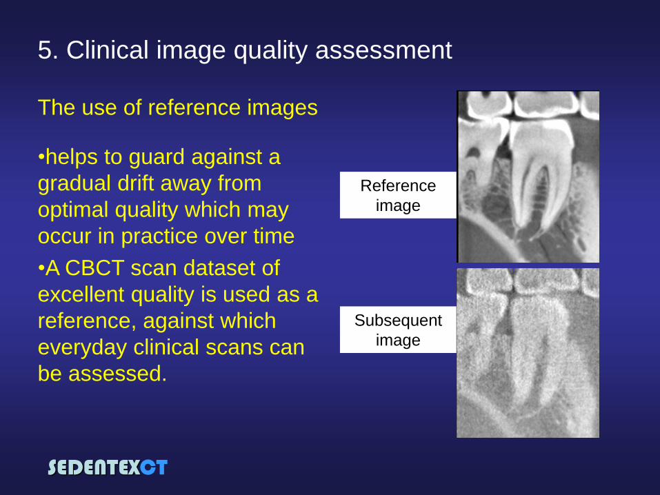
The use of reference images
•helps to guard against a
gradual drift away from
optimal quality which may
occur in practice over time
•A CBCT scan dataset of
excellent quality is used as a
reference, against which
everyday clinical scans can
be assessed.
Reference
image
Subsequent
image

SEDENTEXCT
• Performance of the X-ray tube and
generator (X-ray equipment performance)
• Quantitative assessment of image quality
• Display screen performance
• Patient dose assessment
• Clinical image quality assessment
• Clinical audit
Quality Assurance Programme for
CBCT in dentistry
SEDENTEXCT
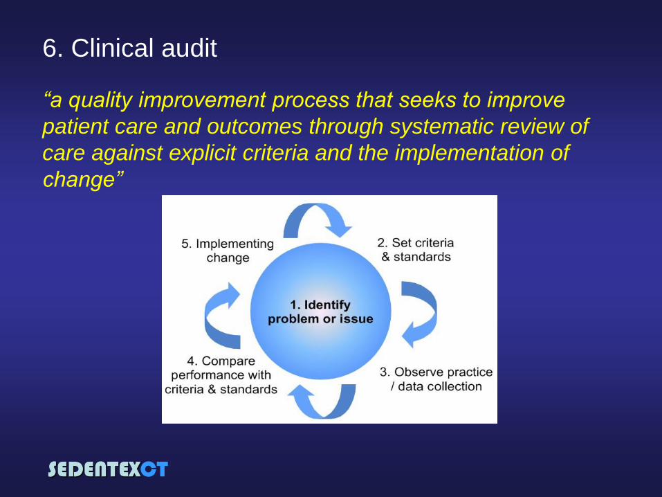
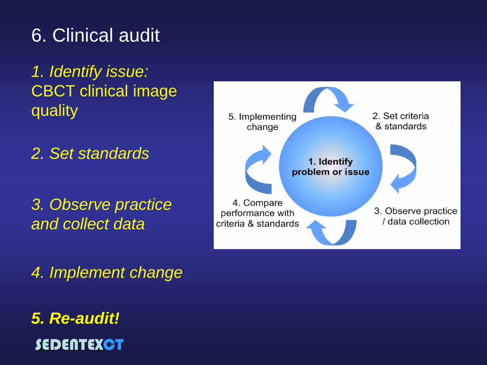
6. Clinical audit
“a quality improvement process that seeks to improve
patient care and outcomes through systematic review of
care against explicit criteria and the implementation of
change”
SEDENTEXCT
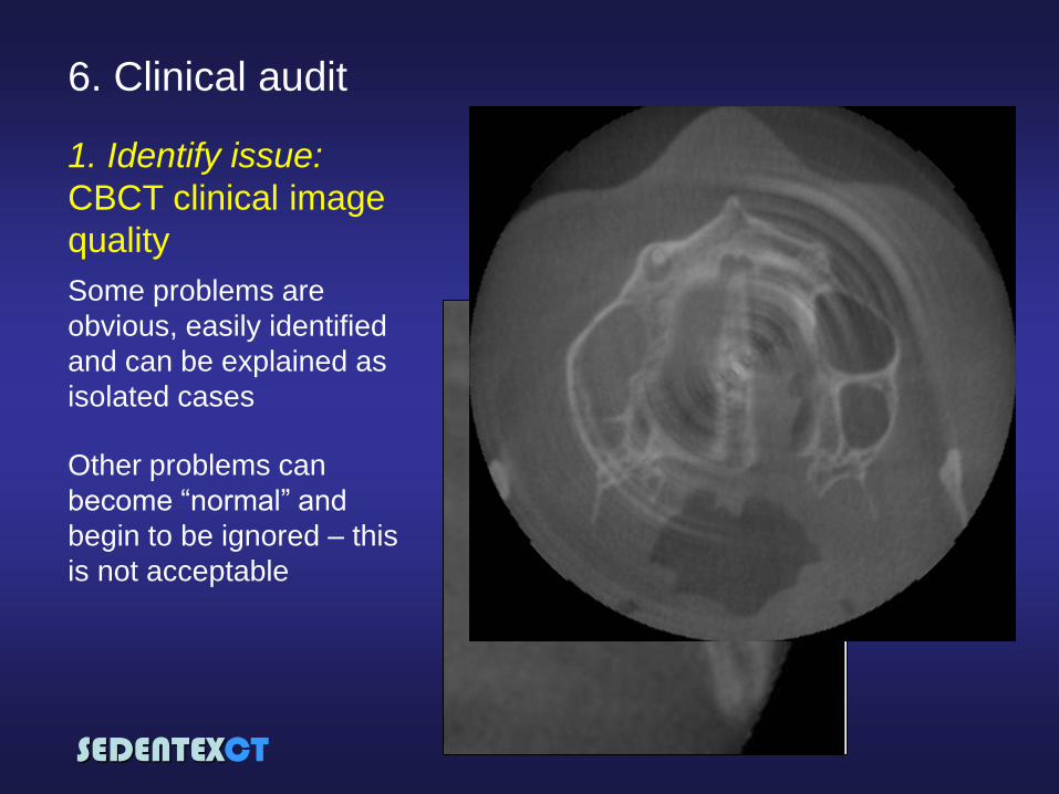
6. Clinical audit
1. Identify issue:
CBCT clinical image
quality
Some problems are
obvious, easily identified
and can be explained as
isolated cases
Other problems can
become “normal” and
begin to be ignored – this
is not acceptable
SEDENTEXCT
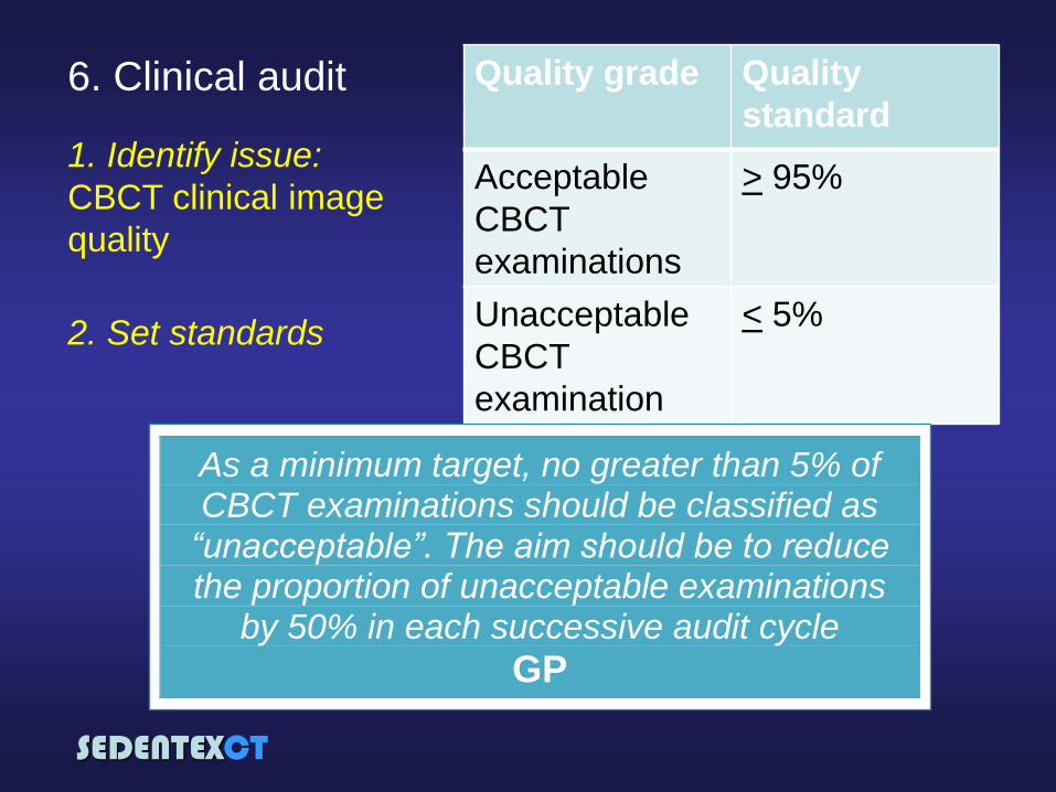
6. Clinical audit
1. Identify issue:
CBCT clinical image
quality
2. Set standards
Quality grade Quality
standard
Acceptable
CBCT
examinations
> 95%
Unacceptable
CBCT
examination
< 5%
As a minimum target, no greater than 5% of CBCT examinations should be classified as
“unacceptable”. The aim should be to reduce the proportion of unacceptable examinations
by 50% in each successive audit cycle
GP
SEDENTEXCT
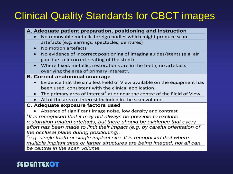
A. Adequate patient preparation, positioning and instruction
No removable metallic foreign bodies which might produce scan artefacts (e.g. earrings, spectacles, dentures)
No motion artefacts
No evidence of incorrect positioning of imaging guides/stents (e.g. air gap due to incorrect seating of the stent)
Where fixed, metallic, restorations are in the teeth, no artefacts overlying the area of primary interest1.
B. Correct anatomical coverage
Evidence that the smallest Field of View available on the equipment has been used, consistent with the clinical application.
The primary area of interest2 at or near the centre of the Field of View.
All of the area of interest included in the scan volume. C. Adequate exposure factors used
Absence of significant image noise, low density and contrast 1It is recognised that it may not always be possible to exclude restoration-related artefacts, but there should be evidence that every effort has been made to limit their impact (e.g. by careful orientation of the occlusal plane during positioning). 2e.g. single tooth or single implant site. It is recognised that where multiple implant sites or larger structures are being imaged, not all can be central in the scan volume.
Clinical Quality Standards for CBCT images
SEDENTEXCT
6. Clinical audit
1. Identify issue:
CBCT clinical image
quality
2. Set standards
3. Observe practice
and collect data
4. Implement change
5. Re-audit!
SEDENTEXCT
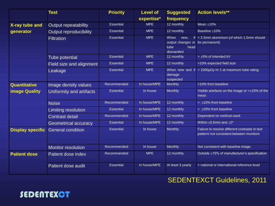
Test Priority Level of
expertise*
Suggested
frequency
Action levels**
X-ray tube and
generator
Output repeatability Essential MPE 12 monthly Mean ±10%
Output reproducibility Essential MPE 12 monthly Baseline ±10%
Filtration Essential MPE When new, if
output changes or
tube head
dismantled
< 2.5mm aluminium (of which 1.5mm should
be permanent)
Tube potential Essential MPE 12 monthly > ±5% of intended kV
Field size and alignment Essential MPE 12 monthly >10% expected field size
Leakage Essential MPE When new and if
damage
suspected
> 1000µGy hr-1 at maximum tube rating.
Quantitative
image Quality
Image density values Recommended In house/MPE Monthly >10% from baseline
Uniformity and artifacts Essential In house Monthly Visible artefacts on the image or >±10% of the
mean
Noise Recommended In house/MPE 12 monthly > ±10% from baseline
Limiting resolution Essential In house/MPE 12 monthly > ±20% from baseline
Contrast detail Recommended In house/MPE 12 monthly Dependent on method used.
Geometrical accuracy Essential In house/MPE 12 monthly Within ±0.5mm and ±2º
Display specific General condition Essential In house Monthly Failure to resolve different contrasts in test
pattern/ not consistent between monitors
Monitor resolution Recommended In house Monthly Not consistent with baseline image
Patient dose Patient dose index Recommended MPE 12 monthly Outside ±15% of manufacturer’s specification
Patient dose audit Essential In house/MPE At least 3 yearly > national or international reference level
SEDENTEXCT Guidelines, 2011
SEDENTEXCT
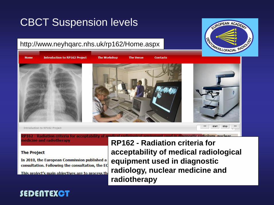
CBCT Suspension levels
RP162 - Radiation criteria for
acceptability of medical radiological
equipment used in diagnostic
radiology, nuclear medicine and
radiotherapy
http://www.neyhqarc.nhs.uk/rp162/Home.aspx
SEDENTEXCT
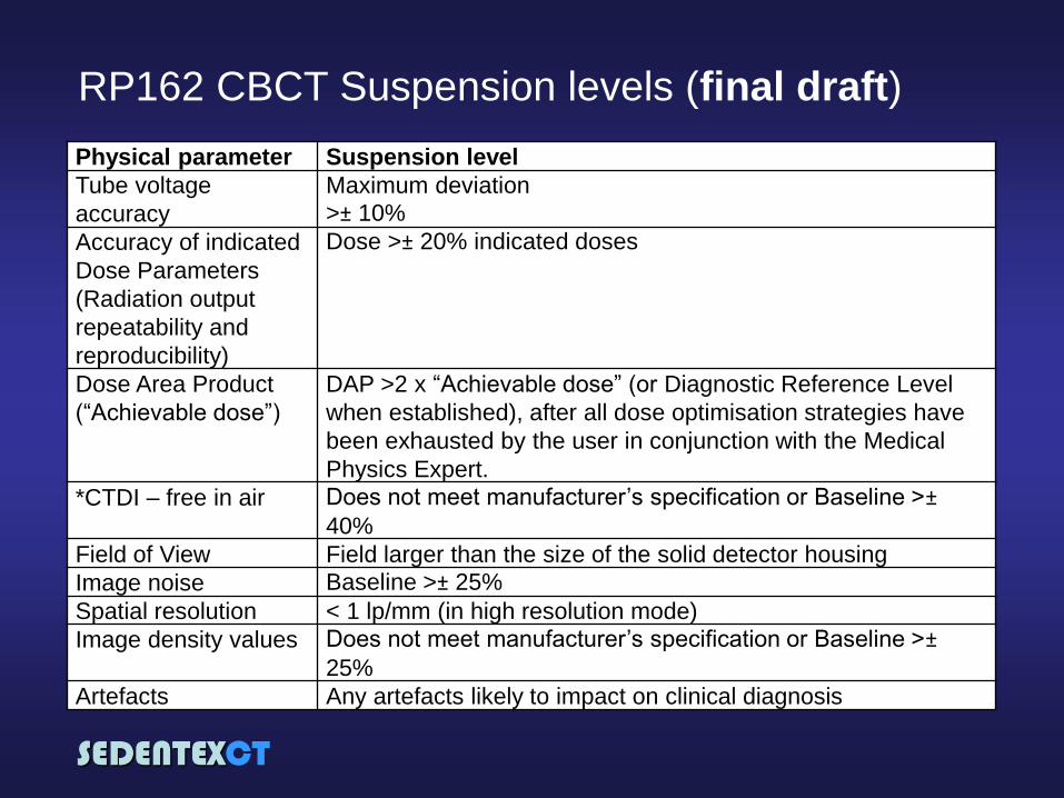
RP162 CBCT Suspension levels (final draft)
Physical parameter Suspension level
Tube voltage
accuracy
Maximum deviation
>± 10%
Accuracy of indicated
Dose Parameters
(Radiation output
repeatability and
reproducibility)
Dose >± 20% indicated doses
Dose Area Product
(“Achievable dose”)
DAP >2 x “Achievable dose” (or Diagnostic Reference Level
when established), after all dose optimisation strategies have
been exhausted by the user in conjunction with the Medical
Physics Expert.
*CTDI – free in air Does not meet manufacturer’s specification or Baseline >±
40%
Field of View Field larger than the size of the solid detector housing
Image noise Baseline >± 25%
Spatial resolution < 1 lp/mm (in high resolution mode)
Image density values Does not meet manufacturer’s specification or Baseline >±
25%
Artefacts Any artefacts likely to impact on clinical diagnosis
SEDENTEXCT
Summary
Quality assurance (QA) is aimed at achieving
acceptable quality at the lowest achievable dose
A QA programme, incorporating a standard series
of quality control tests, is an essential part of this
programme
QA must involve the dentist and a medical
physics expert working as a team
SEDENTEXCT
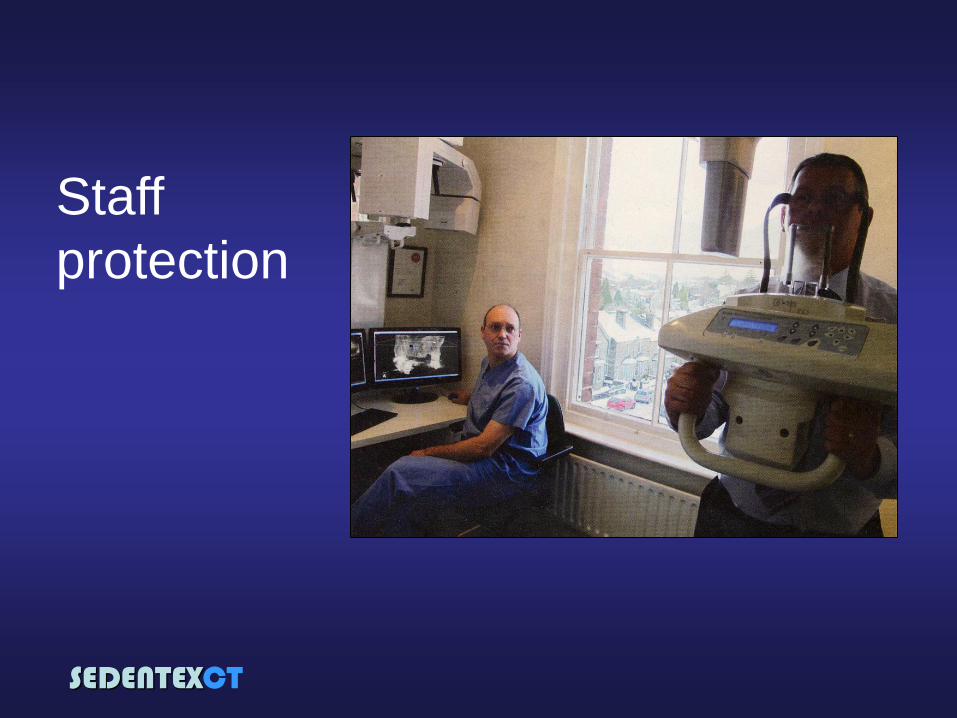
Staff
protection
SEDENTEXCT
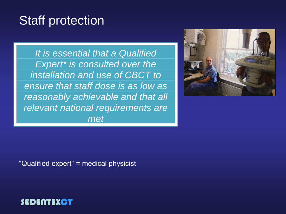
Staff protection
“Qualified expert” = medical physicist
It is essential that a Qualified Expert* is consulted over the
installation and use of CBCT to ensure that staff dose is as low as reasonably achievable and that all relevant national requirements are
met
SEDENTEXCT
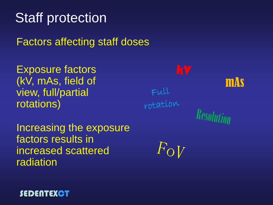
Factors affecting staff doses
Staff protection
Exposure factors (kV, mAs, field of view, full/partial rotations)
Increasing the exposure factors results in increased scattered radiation
mAs
SEDENTEXCT
Factors affecting staff doses
Staff protection
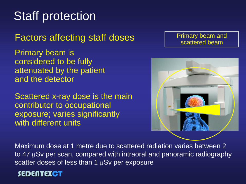
Primary beam and scattered beam
Primary beam is considered to be fully attenuated by the patient and the detector
Scattered x-ray dose is the main contributor to occupational exposure; varies significantly with different units
Maximum dose at 1 metre due to scattered radiation varies between 2
to 47 Sv per scan, compared with intraoral and panoramic radiography
scatter doses of less than 1 Sv per exposure
SEDENTEXCT
Staff protection
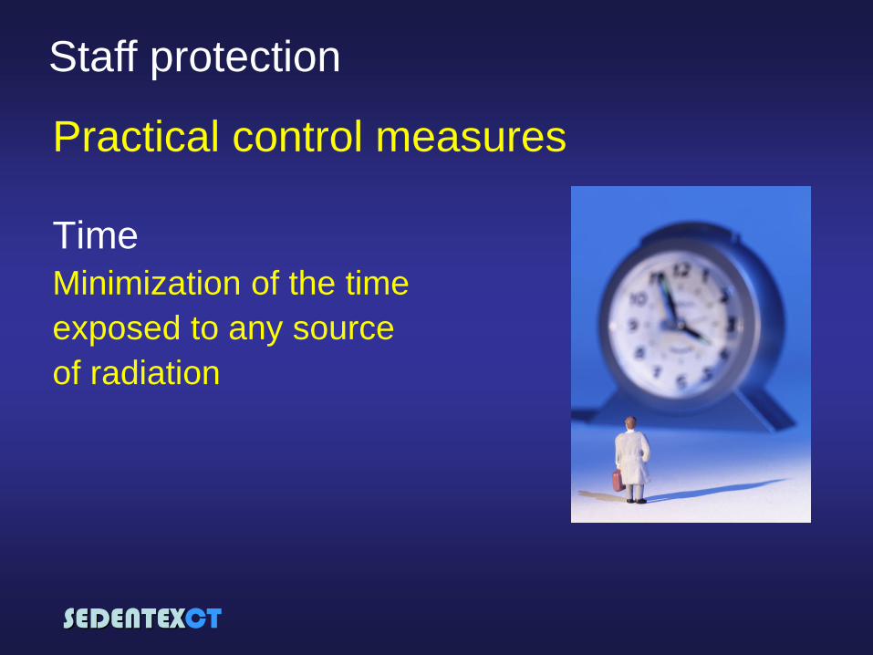
Practical control measures
Time
Minimization of the time
exposed to any source
of radiation
SEDENTEXCT
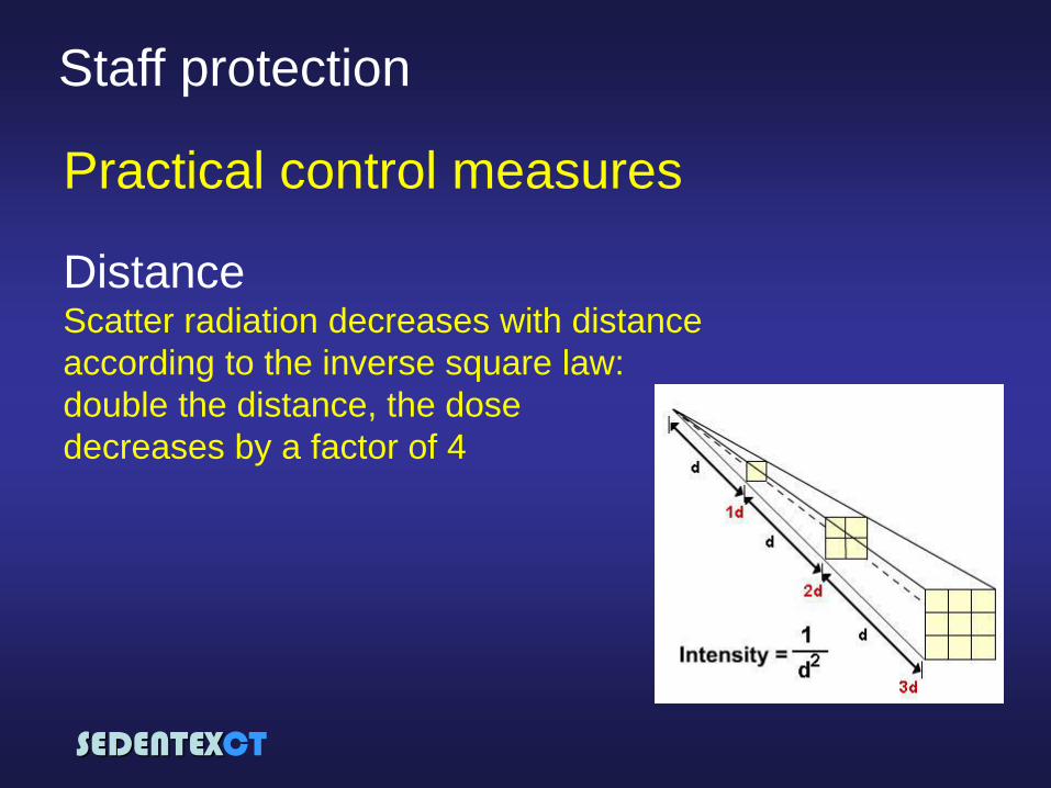
Staff protection
Practical control measures
Distance Scatter radiation decreases with distance
according to the inverse square law:
double the distance, the dose
decreases by a factor of 4
SEDENTEXCT
Staff protection
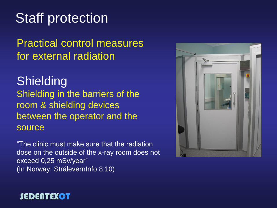
Practical control measures
for external radiation
Shielding Shielding in the barriers of the
room & shielding devices
between the operator and the
source
“The clinic must make sure that the radiation
dose on the outside of the x-ray room does not
exceed 0,25 mSv/year”
(In Norway: StrålevernInfo 8:10)
SEDENTEXCT
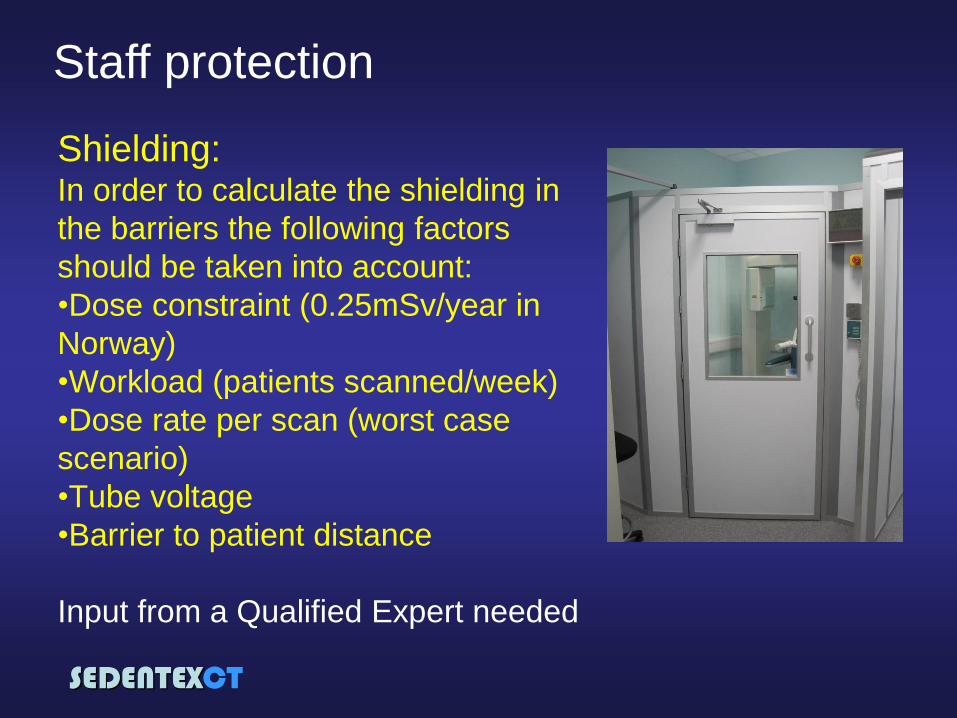
Shielding: In order to calculate the shielding in
the barriers the following factors
should be taken into account:
•Dose constraint (0.25mSv/year in
Norway)
•Workload (patients scanned/week)
•Dose rate per scan (worst case
scenario)
•Tube voltage
•Barrier to patient distance
Input from a Qualified Expert needed
Staff protection
SEDENTEXCT
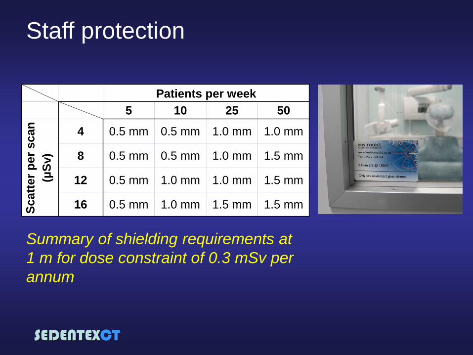
Staff protection
Patients per week
5 10 25 50
Scatt
er
per
scan
(µS
v)
4 0.5 mm 0.5 mm 1.0 mm 1.0 mm
8 0.5 mm 0.5 mm 1.0 mm 1.5 mm
12 0.5 mm 1.0 mm 1.0 mm 1.5 mm
16 0.5 mm 1.0 mm 1.5 mm 1.5 mm
Summary of shielding requirements at
1 m for dose constraint of 0.3 mSv per
annum
SEDENTEXCT
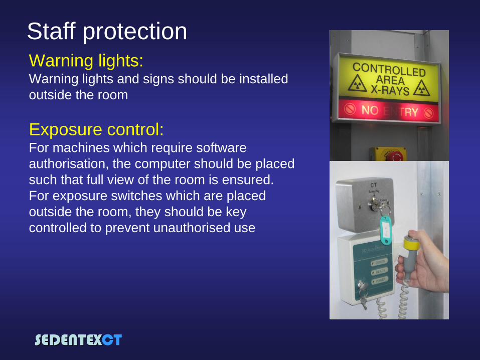
Warning lights: Warning lights and signs should be installed
outside the room
Exposure control: For machines which require software
authorisation, the computer should be placed
such that full view of the room is ensured.
For exposure switches which are placed
outside the room, they should be key
controlled to prevent unauthorised use
Staff protection

SEDENTEXCT
Staff protection
Personal monitoring
• Need for personal monitoring
must be considered in the prior risk
assessment
• If the operating situation is such
that an exposure cannot be initiated
unless the operator is standing
behind a shielded door or window,
occasional monitoring is suggested
• Otherwise, routine monitoring is
recommended
The provision of Personal Monitoring
should be considered
D

SEDENTEXCT
Summary
Staff must have received adequate training
Patient dose optimisation should help with staff dose
optimisation
Policies should be established that eliminate staff
risk from radiation
Shielding is likely to be required with CBCT
installations
INVOLVEMENT OF A MEDICAL PHYSICS EXPERT PRIOR TO EQUIPMENT
INSTALLATION IS ESSENTIAL TO OPTIMISE STAFF DOSE
SEDENTEXCT
SEDENTEXCT:
Guidelines and
evidence-based use of
CBCT
Keith Horner
SEDENTEXCT
Acknowledgement: The research leading to these
results has received funding from the European
Atomic Energy Community’s Seventh Framework
programme FP7/ 2007-2011 under grant agreement
no. 212246 (SEDENTEXCT: Safety and Efficacy of a
New and Emerging Dental X-ray Modality).