double ureter: incidence, types, and its applied
TRANSCRIPT

Received 03/25/2020 Review began 04/04/2020 Review ended 04/13/2020 Published 04/21/2020
© Copyright 2020Arumugam et al. This is an open accessarticle distributed under the terms of theCreative Commons Attribution LicenseCC-BY 4.0., which permits unrestricteduse, distribution, and reproduction in anymedium, provided the original author andsource are credited.
Double Ureter: Incidence, Types, and Its AppliedSignificance—A Cadaveric StudySangeetha Arumugam , Nandha Kumar Subbiah , Arathi Mariappan Senthiappan
1. Anatomy, Katuri Medical College and Hospital, Guntur, IND 2. Anatomy, All India Institute of Medical Sciences,Mangalagiri, IND 3. Anatomy, Chettinad Hospital and Research Institute, Chennai, IND
Corresponding author: Nandha Kumar Subbiah, [email protected]
AbstractIntroductionCongenital ureter anomalies such as double ureters are uncommon developmental anomalies of the renalsystem. An abnormal branching pattern of ureteric bud results in the formation of double ureter. This studyexamined the incidence of double ureter in cadavers of a South Indian population.
MethodsA total of 50 kidney and ureter specimens were carefully dissected out of the posterior abdominal wall andexamined for the presence and subtype of double ureter.
ResultsOf 50 kidneys, three (6%) specimens showed an incomplete double ureter, two on the right kidney and oneon the left. In all three specimens, the double ureter fused at different levels to form a single ureter openinginto the bladder.
ConclusionsThe prevalence of incomplete double ureter is higher in this study compared with that in previous cadavericstudies. Ureteral injuries are a frequent complication of abdominal and pelvic surgeries. Hence, awarenessabout the types and varieties of double ureter will aid radiologists and surgeons in interpreting anddiagnosing urological images and preventing accidental injury while performing surgery.
Categories: Radiology, Urology, AnatomyKeywords: double ureter, ureteroureteric reflux, complete double ureter, incomplete double ureter
IntroductionThe ureters are muscular ducts, around 25-30 cm in length, with narrow lumina. These ureters drain urinefrom the corresponding kidney to the urinary bladder. The ureters have two segments: abdominal and pelvic.The abdominal segments of the ureters adhere closely to the parietal peritoneum and are retroperitonealthroughout their course, and the pelvic segments enter the pelvis by passing over the pelvic brim at thebifurcation of the common iliac arteries before entering the urinary bladder [1]. Congenital anomalies of thekidney and urinary tract, including double ureter, constitute 20% to 30% of all prenatal anomalies. Doubleureter may present as either complete or incomplete/partial duplication [2,3]. Clinically, patients with adouble ureter may be asymptomatic or may present with hematuria or abdominal or flank pain and bepredisposed to ureteral obstruction, ureteroureteric reflux, and recurrent urinary infections [4,5].
Ureteral injury is a common complication of open or laparoscopic surgical procedures involving theabdomen and pelvic region. The occurrence of such ureteral injuries can be prevented by prior imaging of theabdomen and pelvis, as well as examining the ureter. In-depth knowledge of the normal and abnormalpatterns of the ureter is a prerequisite for both radiologists and surgeons to plan any surgical procedure.Many radiologists have reported double ureters after performing excretory urethrograms [6]. However,cadaveric studies on ureteric variations are seldom undertaken. Hence, this cadaveric study was performedto determine the prevalence of double ureters in a South Indian population and to discuss their clinicalimportance.
Materials And MethodsThis study was approved by the Ethics Committee of Katuri Medical College, Guntur, India. Kidneys withintact ureters and bladder were dissected and removed from cadavers of 25 South Indian individuals ofeither sex, following the guidelines outlined by Cunningham’s Manual of Practical Anatomy [7]. Thekidneys, along with the ureters and bladder, were washed thoroughly in running water after removal and
1 2 3
Open Access OriginalArticle DOI: 10.7759/cureus.7760
How to cite this articleArumugam S, Subbiah N, Mariappan Senthiappan A (April 21, 2020) Double Ureter: Incidence, Types, and Its Applied Significance—A CadavericStudy. Cureus 12(4): e7760. DOI 10.7759/cureus.7760

were stored in 10% formalin. Morphologically damaged kidneys were excluded from the study. Kidneys withdouble ureters and their subtypes were described and photographed. The results were tabulated andsummarized in Microsoft Excel and are shown in Table 1.
Ureter (50) Complete Incomplete Percentage
Right ureter (25) 0 02 4%
Left ureter (25) 0 01 2%
TABLE 1: Percentage of complete and incomplete ureters
ResultsUpon examination of 50 kidneys, 47 (94%) had a normal single ureter arising from the renal pelvis andopening into the urinary bladder. The remaining three (6%) specimens showed variations in ureters(Table 1). In one pair of kidneys, the left kidney had double ureters arising from the upper and lower poles ofthe renal pelvis that joined with each other to form a single ureter (Y-shape) distally before opening into theurinary bladder (Figure 1). In the second pair, double ureters emerging from the right kidney were seenfusing midway between the kidney and bladder (Figure 2). In the third specimen, double ureters arising fromthe right kidney formed a single ureter a few centimeters away from the renal pelvis (Figure 3). All doubleureters were unilateral, were incomplete in presentation, and opened through a single ureteric orifice intothe urinary bladder.
2020 Arumugam et al. Cureus 12(4): e7760. DOI 10.7759/cureus.7760 2 of 7

FIGURE 1: Left kidney with an incomplete double ureterRK, right kidney; LK, left kidney
2020 Arumugam et al. Cureus 12(4): e7760. DOI 10.7759/cureus.7760 3 of 7

FIGURE 2: Right kidney with an incomplete double ureterRK, right kidney; LK, left kidney
2020 Arumugam et al. Cureus 12(4): e7760. DOI 10.7759/cureus.7760 4 of 7

FIGURE 3: Right kidney with incomplete double ureter fusing at thelower pole of the kidneyRK, right kidney
DiscussionThe urinary system develops from two sources: metanephric blastema and mesonephric duct. Ureteric budarises as a diverticulum from the caudal end of the mesonephric duct. This diverticulum elongates and laterfuses with the metanephric blastema to form renal pelvis, which further divides into major and minorcalyces [2,4]. Double ureter is caused by abnormalities in the branching pattern of the ureteric bud. In thecase of complete duplication, the ureteral bud arises twice, resulting in a double ureter with a doubleopening into the urinary bladder. In rare cases, one of the ureters can open into sites other than the urinarybladder, such as the vagina, seminal vesicle, urethra, prostate, epididymis, or vas deferens. This condition iscalled ectopic ureter. Incomplete duplication is due to splitting of the ureteric bud anywhere along its courseto its termination into the metanephric blastema. The duplicated ureters unite at a variable distance awayfrom the kidney, and only one ureteric orifice is seen in the bladder on that side. If the ureteric budbifurcates after fusing with metanephric tissue, it results in a double pelvis and double ureter [8].
2020 Arumugam et al. Cureus 12(4): e7760. DOI 10.7759/cureus.7760 5 of 7
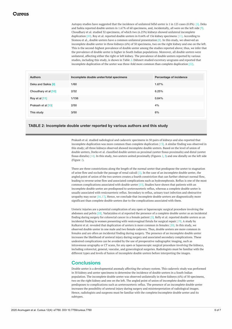
Autopsy studies have suggested that the incidence of unilateral bifid ureter is 1 in 125 cases (0.8%) [1]. Dekaand Saikia reported double ureters in 1.67% of 60 specimens, and, incidentally, all were on the left side [9].Choudhary et al. studied 32 specimens, of which two (6.25%) kidneys showed unilateral incompleteduplication [10]. Roy et al. reported double ureters in 0.64% of 156 kidney specimens [11]. According toSiomou et al., double ureters have a common unilateral presentation [8]. In this study, we observed anincomplete double ureter in three kidneys (6%) of 50 specimens, two on the right kidney and one on the left.This is the second-highest prevalence of double ureter among the studies reported above; thus, we infer thatthe prevalence of double ureter is higher in South Indian populations. Moreover, all double ureters wereunilateral, affecting either the right or left kidney. The prevalence of double ureters reported by variousstudies, including this study, is shown in Table 2. Dähnert studied excretory urograms and reported thatincomplete duplication of the ureter was three-fold more common than complete duplication [12].
Authors Incomplete double ureter/total specimens Percentage of incidence
Deka and Saikia [9] 1/60 1.67%
Choudhary et al [10] 2/32 6.25%
Roy et al [11] 1/156 0.64%
Prakash et al [13] 2/50 4%
This study 3/50 6%
TABLE 2: Incomplete double ureter reported by various authors and this study
Prakash et al. studied radiological and cadaveric specimens in 50 pairs of kidneys and also reported thatincomplete duplication was more common than complete duplication [13]. A similar finding was observed inthis study; all three kidneys observed showed incomplete double ureters. Based on the level of union ofdouble ureters, Dorko et al. classified double ureters as proximal (ureter fissus proximalis) and distal (ureterfissus distalis) [14]. In this study, two ureters united proximally (Figures 2, 3) and one distally on the left side(Figure 1).
There are three constrictions along the length of the normal ureter that predispose the ureter to stagnationof urine flow and occlude the passage of renal calculi [1]. In the case of an incomplete double ureter, theangled point of union of the two ureters creates a fourth constriction that can further obstruct normal flow,leading to reverse urine flow and associated complications such as hydronephrosis. Reflux is one of the mostcommon complications associated with double ureter [15]. Studies have shown that patients with anincomplete double ureter are predisposed to ureteroureteric reflux, whereas a complete double ureter isusually associated with vesicoureteric reflux. Secondary to reflux, urinary tract infection and obstructiveuropathy may occur [16,17]. Hence, we conclude that incomplete double ureters are diagnostically moresignificant than complete double ureters due to the complications associated with them.
Ureteric injuries are a potential complication of any open or laparoscopic surgical procedure involving theabdomen and pelvis [18]. Varlatzidou et al reported the presence of a complete double ureter as an incidentalfinding during surgery for colorectal cancer in a female patient [5]. Kelly et al. reported double ureters as anincidental finding in women presenting with vesicovaginal fistula for surgical repair [19]. A study byKulkarni et al. revealed that duplication of ureters is more common in females [20]. In this study, weobserved double ureter in one male and two female cadavers. Thus, double ureters are more common infemales and are often an incidental finding during surgery. The presence of an incomplete double ureterincreases the likelihood of ureteral injury during surgery and associated secondary complications. Theseundesired complications can be avoided by the use of preoperative radiographic imaging, such asintravenous urography or CT scans, for any open or laparoscopic surgical procedure involving the kidneys,including colorectal, general, vascular, and gynecological surgeries. Radiologists must be familiar with thedifferent types and levels of fusion of incomplete double ureters before interpreting the images.
ConclusionsDouble ureter is a developmental anomaly affecting the urinary system. This cadaveric study was performedin 50 kidney and ureter specimens to determine the incidence of double ureters in a South Indianpopulation. The incomplete double ureter was observed unilaterally in three kidneys (6%) of 50 specimens,two on the right kidney and one on the left. The angled point of union of incomplete double ureterpredisposes to complications such as ureteroureteric reflux. The presence of an incomplete double ureterincreases the possibility of ureteral injury during surgery and misinterpretation of radiological images.Hence, radiologists and surgeons must be familiar with the complete/incomplete double ureter and itssubtypes.
2020 Arumugam et al. Cureus 12(4): e7760. DOI 10.7759/cureus.7760 6 of 7

Additional InformationDisclosuresHuman subjects: Consent was obtained by all participants in this study. Ethics Committee, Katuri MedicalCollege issued approval KMCH/IEC/2019/3/25. Animal subjects: All authors have confirmed that this studydid not involve animal subjects or tissue. Conflicts of interest: In compliance with the ICMJE uniformdisclosure form, all authors declare the following: Payment/services info: All authors have declared that nofinancial support was received from any organization for the submitted work. Financial relationships: Allauthors have declared that they have no financial relationships at present or within the previous three yearswith any organizations that might have an interest in the submitted work. Other relationships: All authorshave declared that there are no other relationships or activities that could appear to have influenced thesubmitted work.
References1. Standring S: Gray's Anatomy: The Anatomical Basis of Clinical Practice. Elsevier, Philadelphia; 2016.2. Campbell MF, Walsh PC: Campbell's urology. Anomalies of the Upper Urinary Tract. Walsh PC, Retik AB,
Vaughan ED Jr, Wein AJ (ed): WB Saunders, Philadelphia; 1992. 2:1376-1381.3. Sadler TW: Langman’s Medical Embryology. Sadler TW (ed): Wolters Kluwer, Philadelphia; 2018.4. Bergman R: Compendium of Human Anatomic Variation: Text, Atlas, and World Literature . Urban &
Schwarzenberg, Baltimore; 1988.5. Varlatzidou A, Zarokosta M, Nikou E, et al.: Complete unilateral ureteral duplication encountered during
intersphincteric resection for low rectal cancer. J Surg Case Rep. 2018, 2018:266. Accessed: April 21, 2020:10.1093/jscr/rjy266
6. Neuman M, Eidelman A, Langer R, Golan A, Bukovsky I, Caspi E: Iatrogenic injuries to the ureter duringgynecologic and obstetric operations. Surg Gynec Obstet. 1991, 173:268-272.
7. Romanes GJ: Cunningham's Manual of Practical Anatomy: Volume Two. Thorax and Abdomen . OxfordUniversity Press, Oxford; 1986.
8. Siomou E, Papadopoulou F, Kollios K, Photopoulos A, Evagelidou E, Androulakakis P, Siamopoulou A:Duplex collecting system diagnosed during the first 6 years of life after a first urinary tract infection: a studyof 63 children. J Urol. 2006, 175:678-682. 10.1016/S0022-5347(05)00184-9
9. Deka B, Saikia R: A study of human cadaveric ureter by simple dissection method . Int J Anat Res. 2016,4:3005-3008. 10.16965/ijar.2016.393
10. Choudhary U, Kumar S, Jee K, Singh A, Bharti P: A cadaveric study on anatomical variations of kidney andureter in India. Int J Res Med Sci. 2017, 5:2358. 10.18203/2320-6012.ijrms20172094
11. Roy M, Singh BR, Gajbe UL, Thute P: Anatomical variations of ureter in central India: a cadaveric study . JDatta Meghe Inst Med Sci Univ. 2017, 12:277-279. 10.4103/jdmimsu.jdmimsu_73_17
12. Dähnert W: Radiology Review Manual . Lippincott Williams & Wilkins, Philadelphia; 1999.10.1007/s003300101096
13. Prakash, Rajini T, Venkatiah J, Bhardwaj AK, Singh DK, Singh G: Double ureter and duplex system: acadaver and radiological study. Urol J. 2011, 8:145-148.
14. Dorko F, Tokarčík J, Výborná E: Congenital malformations of the ureter: anatomical studies . Anat Sci Int.2015, 91:290-294. 10.1007/s12565-015-0296-8
15. Kasat P, Bhosale Y, Muthiyan G: A cadaveric study of variations in the urological system . Int J Anat Res.2018, 6:5686-5694. 10.16965/ijar.2018.311
16. Dinanath P, Ashwini A, Annarao G, Nagaraj S: Bilateral complete duplex renal pelvis and ureters - a casereport. Int J Anat Var. 2011, 4:192-194.
17. Nagpal H, Chauhan R: Unilateral duplex collecting system with incomplete duplication of ureter - a casereport. Int J Res Med Sci. 2017, 5:2254. 10.18203/2320-6012.ijrms20171882
18. Selzman A, Spirnak J: Iatrogenic ureteral injuries: a 20-year experience in treating 165 injuries . J Urol. 1996,155:878-881. 10.1016/s0022-5347(01)66332-8
19. Kelly J, Moghul F, Pisani V: Double ureter at repair of obstetric fistula . Int J Gyne Obstet. 2008, 102:77-78.10.1016/j.ijgo.2008.02.012
20. Kulkarni V, Ramesh BR, Prakash BS: Bilateral bifid ureter with accessory renal artery: a case report . Int J BasMed Sci. 2012, 3:67-70.
2020 Arumugam et al. Cureus 12(4): e7760. DOI 10.7759/cureus.7760 7 of 7