download (3.97 mb )
TRANSCRIPT

Cell Reports, Volume 14
Supplemental Information
Crystal Structure of the Cohesin Gatekeeper Pds5
and in Complex with Kleisin Scc1
Byung-Gil Lee, Maurici B. Roig, Marijke Jansma, Naomi Petela, Jean Metson, KimNasmyth, and Jan Löwe

2
InventoryofSupplementalInformation
SupplementalFigureS1
Electron density map supporting the main finding of the paper, the crystal
structureofPds5(belongstoFigure1A).
SupplementalFiguresS2&S3
MultiplesequencealignmentsshowingtheconservationofScc1bindingtoPds5
andScc3(belongstoFigure1C).
SupplementalFigureS4
Lethality analysis of Pds5 and Scc1 mutants, validating the structure of the
complexofPds5andScc1(belongstoFigure2A&B).
SupplementalFigureS5
ControlexperimentssupportingtheChIP-seqexperimentinFigure2C.
SupplementalFigureS6
Superposition of parts of the Pds5 and previous Scc3 structures, supporting
Figure3C.
SupplementalExperimentalProcedures
ListofStrains
SupplementalReferences
3
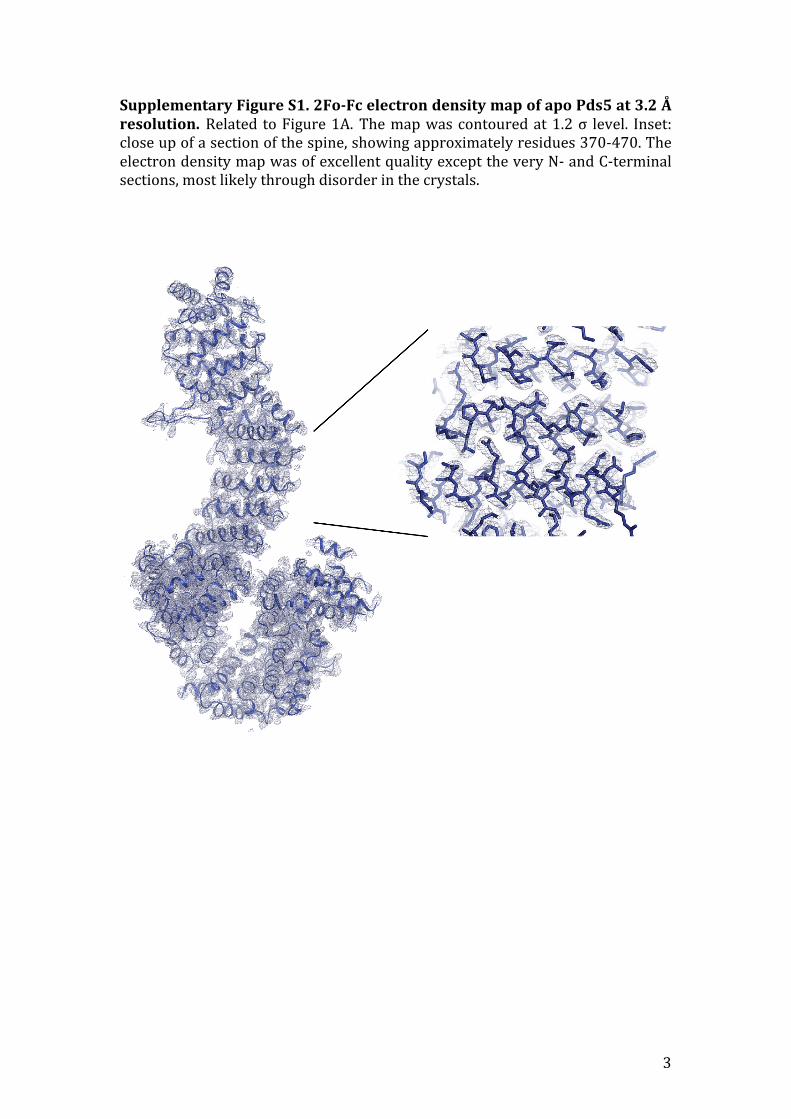
SupplementaryFigureS1.2Fo-FcelectrondensitymapofapoPds5at3.2Åresolution.Related toFigure1A.Themapwascontouredat1.2σ level. Inset:closeupofasectionofthespine,showingapproximatelyresidues370-470.TheelectrondensitymapwasofexcellentqualityexcepttheveryN-andC-terminalsections,mostlikelythroughdisorderinthecrystals.

4
SupplementaryFigureS2.MultiplesequencealignmentshowingconservedPds5 binding regions within Scc1 kleisins. Related to Figure 1C. Up to 10sequencessimilartoeachL.thermotolerans,S.cerevisiae,S.pombe,A.thaliana,H.sapiens andD.melanogaster Scc1 were selected by BLAST (each block evenlysampleddowntoaround30%sequence identity)andsequenceswerealignedbyClustalOmega.Resides120-141areshownforthePds5bindingsiteinScc1,containingmorethanthepeptideco-crystallisedherewithPds5(Ltnumbering;LtScc1 is the first sequence in the alignment.). LtPds5 residues 125-141werebuiltintheco-crystalstructure(Table1,Figure1C,D).

5
SupplementaryFigureS3.MultiplesequencealignmentshowingconservedScc3bindingregionswithinScc1kleisinsfromhumanstoyeast.RelatedtoFigures1Cand3C.500sequencessimilartohumanandyeastScc1wereselectedbyBLASTandallsequences(includingfromL.thermotolerans)werealignedbyClustalOmega.Thealignmentwastruncatedto60sequenceswithoutre-aligning.Resides326-400are shown for the Scc3binding site in Scc1 corresponding tothefragmentco-crystallisedwithSA2/Scc3previously(Ltnumbering;LtScc1isthelastsequenceinthealignment.).NotethathelixBismostlikelymissingfromtheScc1fragmentinyeastsandrelatedorganismsbutotherwisethealignmentindicatesthatthebindingmodeofScc1toScc3isconserved.

6
Supplementary Figure S4.S.cerevisiaePds5(Y458K) growth tests.RelatedtoFigures2A&B.Wild typecells (K699, see listof strains)andcellswith theendogenous PDS5 locus deleted and expressing PDS5 WT (K25118) or pds5(Y458K)(K25126)integratedatthelys2locuswerestreakedonYEPDplatesandincubated at different temperatures. Note that ScPds5 Y458 corresponds toLtPds5Y493(labelledinFigure1D).
7
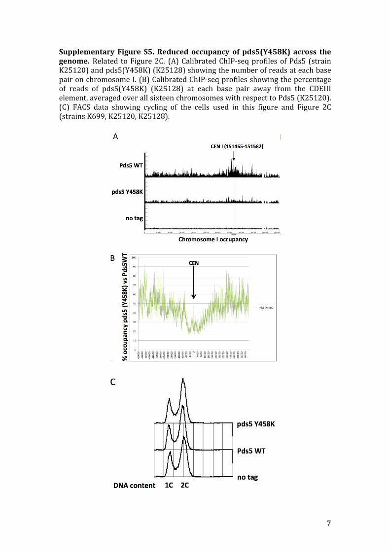
Supplementary Figure S5. Reduced occupancy of pds5(Y458K) across thegenome.Related toFigure2C. (A)CalibratedChIP-seqprofilesofPds5 (strainK25120)andpds5(Y458K)(K25128)showingthenumberofreadsateachbasepaironchromosomeI.(B)CalibratedChIP-seqprofilesshowingthepercentageof reads of pds5(Y458K) (K25128) at each base pair away from the CDEIIIelement,averagedoverallsixteenchromosomeswithrespecttoPds5(K25120).(C) FACS data showing cycling of the cells used in this figure and Figure 2C(strainsK699,K25120,K25128).

8
SupplementaryFigureS6.SuperpositionofLtPds5domainsonpreviouslydetermined Scc3 and SA2 structures. Related to Figure 3C. The LtPds5structurewasdividedinto fourdomains(left) thatwere independentlyalignedin 3D (PyMOL 1.7.6.2 'cealign' and 'align' commands) against the entire Scc3(PDB 4UVK, middle) and SA2 (PDB 4PJU, right) structures. No manualadjustmentsweremade.TwopartsofPds5align reasonablywell against theircorresponding parts in both Scc3 and SA2. The C-terminal domain aligns lesswellanddoesnotsuperimposeatallclosetotheC-terminus.Itshouldbenotedthat the aligning parts show the highest conservation of the canonical HEATrepeatfold.

9
SupplementalExperimentalProcedures
Cloning,overexpressionandpurification
Lachancea thermotolerans CBS6340 Pds5 (LtPds5, NCBI database identifier
XP_002553028.1) was expressed in E. coli using a codon-optimised synthetic
gene(EpochLifescience,TX).Twoconstructswereused,LtPds5-1,aminoacids
35-1221,approx..137kDaandLtPds5-2,aminoacids45-1221,approx.135kDa
(see Table 1). These were cloned into expression vector pHis17 using Gibson
assembly (New England Biolabs, MA), adding the affinity purification tag
LHHHHHH at the C-termini. For overexpression, C41(DE3) cells (Lucigen,WI)
were transformed with the resulting vectors and grown in 2xTY media and
induced with 1 mM IPTG at 16˚C overnight. Cells were harvested and re-
suspended in lysisbuffer (50mMTris-HCl,150mMNaCl,2mMTCEPand5%
(w/v)glycerol,pH8.0),and lysedthroughaConstantSystemscelldisruptorat
25 kPSI in the presence of DNase, RNase and EDTA-free protease inhibitors
(Roche).Thecell lysateswereclarifiedbyultracentrifugationat200,000g ina
Beckman 45 Ti rotor and applied to nickel resin (5 ml HisTrap HP, GE
Healthcare) and eluted with 50-150 mM imidazole in lysis buffer. Fractions
containing Pds5 proteins were further purified using anion exchange
chromatography(5mlHiTrapQFF,GEHealthcare)withgradientsof100-1000
mMNaCl inbuffercontaining50mMTris-HCl,100mMNaCl,2mMTCEP,5%
(w/v) glycerol, pH 8.5. Proteins were concentrated using spin concentrators
(Vivaspin, Satorius, 50 kDa MWCO) and further purified using size exclusion
chromatography(SephacrylS30016/60,GEHealthcare)inbuffercontaining50
mM Tris-HCl, 250 mM NaCl, 5 mM TCEP, pH 7.5. Selenomethionine-labeled
LtPds5proteinswereexpressedusingapublishedfeedbackinhibitionprocedure
(vandenEnt et al., 1999;VanDuyneet al., 1993)andpurifiedusing the same
protocol for the native proteins. All purifications were performed at 4˚C. The
LtScc1 (NCBI database identifier XP_002555756.1) peptide (residues 121-143:
LTNPSQYLLQDAVTEREVLLVPG) design was informed by published results
(Chan et al., 2013). Two otherwise identical, selenomethionine-substituted
mutant peptides, Y127SeMet and L128SeMet, were used to confirm the

10
orientation of Scc1 polypeptide. All peptides were chemically synthesised
(Generon,UK,CambridgePeptides,UK,andGenscript,USA/HongKong).
Crystallisationanddatacollection
Initial crystallisation experiments were carried out using sitting-drop vapour
diffusionwithLMB’s in-househigh-throughput crystallisation facility at 100nl
volumes (Stock et al., 2005). LtPds5-2 protein (amino acids 45-1221) was
crystallisedwithreservoirsolutionscontaining100mMHEPESpH7.5,and1.3-
1.6 M lithium sulphate, and both native and selenomethionine-substituted
crystals were obtained in similar conditions. Diffraction quality crystals were
grownat20˚Cusingconcentrationsof10mg/ml,mixedwithreservoirsolution
0.3 - 0.4 times the protein solution's volume at 1.2 µl volumes. Crystals were
observedafter2-3weeksandwerecryo-protectedbyseriallyincreasinglithium
sulphateconcentrationupto2.2Minthedropandsubsequentflashfreezingin
liquidnitrogen.ForPds5-Scc1co-crystallization,LtPds5-1protein(aminoacids
35-1221)at8mg/mlwasmixedwithLtScc1peptide(aminoacids121-143) in
fivetimesmolarexcess.Complexcrystalswereobtainedwithreservoirsolution
containing 50mM sodium cacodylate pH6.5, 1.4 – 1.6M ammonium sulphate
and5mMmagnesiumacetate,andgrewwithinaweek.Thecrystalswerecryo-
protectedbytransferringtoreservoirsolutionsupplementedwith1.2Msodium
malonateandflashfrozeninliquidnitrogenbeforedatacollection.Co-crystalsof
Pds5and two selenomethionine-substitutedScc1mutantpeptides (Y127SeMet
and L128SeMet) were grown with LtPds5-2 protein under reservoir solution
containing 50 mM sodium cacodylate pH 6.5, 1.4 M lithium sulphate, 40 mM
sodiumcitrate.Thecomplexcrystalswerecryo-protectedbyseriallyincreasing
lithiumsulphateinthedropandflashfrozeninliquidnitrogen.Diffractiondata
werecollectedat100Konbeamlinesi03atDiamondLightSource(Harwell,UK)
andid23eh1attheESRF(Grenoble,France).
Structuredetermination
DiffractiondatawereintegratedandscaledwithXDS(Kabsch,2010)andSCALA
(Winnetal.,2011).PhasingwasdonebySeMetSADcombiningdata fromtwo
separate crystals in order to increase multiplicity and anomalous signal.

11
Selenium positions were identified and SAD phases were calculated using
SHELXC/D/E (Sheldrick, 2008) and PHASER (McCoy et al., 2007). An initial
atomic model was obtained using Crank2 (Skubak and Pannu, 2013), and
manually improved using COOT (Emsley et al., 2010) andMAIN (Turk, 2013).
Forrefinement, thehighresolutionnativeapodataset (Table1)wascorrected
for anisotropy using the UCLA Diffraction Anisotropy Server
(services.mbi.ucla.edu/anisoscale/) (Strong et al., 2006), and the model was
further rebuilt and refined in cycles at 3.2 Å resolution, manually rebuilt as
aboveandrefinedwithREFMAC(Murshudovetal.,1997)andPHENIX(Adamset
al., 2010). TheLtPds5-LtScc1 complex datasetwas evenmore anisotropic and
was also corrected using the UCLA server. Data extended to 3.5 Å in two
directions (Table 1) and 4.5 in the third, leading to an estimate of overall
resolutionof3.6Å.NotethatdatasetstatisticslistedinTable1arethoseofthe
uncorrecteddataat3.6Åresolutionbeforeapplyinganisotropycorrection.The
complex structurewas solvedbymolecular replacementwithPHASER.Due to
overall conformational changes between apo-Pds5 and the Pds5-Scc1 complex
structures, molecular replacement was performed with the apo structure cut
into 4 roughly equal sized subdomains. After improving the atomic model of
Pds5 within the complex structure by manual building and refinement as
described above, strong extra electron density was located and the LtScc1
peptidewas fitted. Modelling of the Scc1 sequence at this low resolutionwas
guidedbytwoadditionalseleniumSADexperiments(detailsofdatanotshown)
usingpeptides containingSeMet residues in twopositions:LtScc1(Y127SeMet)
and LtScc1(L128SeMet). Phases were calculated using ANODE (Thorn and
Sheldrick, 2011) and the refined Pds5 structure in the complex crystals. The
entiremodelwasthenbuiltandrefinedincyclesasfortheapostructure.ForR-
factors and other statistics of the data and models, please refer to Table 1.
Becauseoflowresolution,nowatersorionswereaddedtoanyofthestructures.
FigureswerepreparedusingPYMOL(Schroedinger).Coordinatesandstructure
factorsweredepositedintheProteinDataBank(PDB)withaccessionnumbers
5F0N(apo-LtPds5)and5F0O(LtPds5:LtScc1peptidecomplex).
CellviabilityanalysisofS.cerevisiaePds5andScc1mutants

12
The corresponding residues in S. cerevisiae Pds5 and Scc1 were located by
sequencealignments.MutantversionsofPds5(underitsnativepromoter)were
incorporatedatthelys2locusinheterozygousPDS5/pds5Δdiploidcells(K25105,
seeListofStrains inSupplementaryData).DiploidsweresporulatedonSpoVB
mediaplatesandtetradsdissectedat25°ConYPDmediaplates.Thegenotypeof
the resultinghaploidswasdeterminedby replicaplating, andviable cellswith
only the ectopic copy were additionally tested for temperature sensitivity
streakingcellsonYEPglucoseplatesat25°C,30°Cand37°C(FigureS4).Mutant
versionsofscc1(underitsnativepromoter)wereincorporatedattheleu2locus
in heterozygous SCC1/scc1D diploid cells (K12714) and analysed in a similar
manner.AllmutationswereconfirmedbyDNAsequencing.
Co-immunoprecipitations
Strains were grown in YEPD at 25°C to OD600nm= 0.7 and 70 OD units were
washed in ice-cold PBS and frozen at -80°C. Pellets were thawed and re-
suspended in lysis buffer (50mMTris/HCl, 100mMNaCl, 5mMEDTA, 1mM
DTT,1mMPMSF,RocheCompleteProteaseInhibitors)andlysedinaFastPrep-
24 (MP Biomedicals) disruptor 3 times for 1 min at 5.5 m/s with an equal
volume of acid-washed glass beads (Sigma). Lysates were cleared by
centrifugation at 13 Krpm for 30 min at 4°C and the protein amount of the
supernatant was quantified with a Bradford assay. For cohesion
immuneprecipitation,150µlofwashedanti-HAHighAffinityMatrix(Roche)was
addedtotheclearedlysatesandincubatedovernightat4°Cwhilerotating.The
incubated anti-HAHighAffinityMatrixwaswashed 3 timeswith 1ml of lysis
buffer,re-suspendedin60µlofSDS-PAGEsamplebufferandincubatedat95°C
for10min.10µlofeachsamplewasloadedontoaprecastTris-acetategel(3-8%,
NuPAGE),followedbyWesternblottingandimmunodetectionofthePKepitopes
withanti-PKantibody(AbDSerotec)andtheHAepitopeswithantiHAantibody
(12CA5).
CalibratedChIP-seqanalysis
CalibratedChIP-seqwasperformedasdescribed(Huetal.,2015)usingK23308,
K699,K25120andK25128strainsforthisassay.

13
Pds5sequenceconservationmappingonPds5crystalstructure
500 sequences most similar to L. thermotolerans Pds5 were selected from a
BLAST search and aligned using Clustal Omega (http://clustal.org), before
mappingsequenceconservationateachresiduepositionontothestructurewith
ConSurf(http://consurf.tau.ac.il)(Ashkenazyetal.,2010).

14
ListofStrains:
All yeast strains are derivatives of W303, except K23308.
K699 MATa, ade2-1, trp1-1, can1-100, leu2-3,112,his3-11,15, ura3, GAL, psi+
K12714 MATa/alpha scc1:KanMx / WT
K23308 C.glabrata, MATa, Scc1PK9::NatMX
K24593 MATalpha, scc1:KanMX, Scc1-HA6 in pRS305H
K24595 MATalpha, leu2:Scc1-HA6:leu2
K24958 MATa/alpha, scc1:KanMX / WT, leu2:Scc1(V137K)-HA6:leu2
K25002 MATa, leu2:Scc1(V137K)-HA6:leu2
K25105 MATa/alpha pds5::HIS / WT
K25106 MATa/alpha, pds5::HIS / WT, lys2:Pds5-PK9-HphMX:lys2
K25108 MATa/alpha, pds5::HIS / WT, lys2:Pds5(Y458K)-PK9-HphMX:lys2
K25118 MATa pds5::HIS, lys2:Pds5-PK9-HphMX:lys2
K25120 MATa, lys2:Pds5-PK9-HphMX:lys2
K25126 MATa pds5::HIS, lys2:Pds5(Y458K)-PK9-HphMX:lys2
K25128 MATa, lys2:Pds5(Y458K)-PK9-HphMX:lys2
K25166 MATa/alpha scc1:KanMX / WT, leu2:Scc1-HA6:leu2
K25202 MATa, pds5::HIS, lys2:Pds5-PK9-HphMX:lys2, leu2:Scc1-HA6:leu2
K25204 MATalpha, lys2:Pds5-PK9-HphMX:lys2, scc1:KanMX, leu2:Scc1-HA6:leu2
K25206 MATalpha, pds5::HIS, lys2:Pds5-PK9-HphMX:lys2, leu2:Scc1(V137K)-HA6:leu2
K25210 MATalpha, lys2:Pds5(Y458K)-PK9-HphMX:lys2, scc1:KanMX, leu2:Scc1-HA6:leu2

15
SupplementalReferences
Adams,P.D.,Afonine,P.V.,Bunkoczi,G.,Chen,V.B.,Davis,I.W.,Echols,N.,Headd,J.J.,Hung,L.W.,Kapral,G.J.,Grosse-Kunstleve,R.W.,McCoy,A.J.,Moriarty,N.W.,Oeffner,R.,Read,R. J.,Richardson,D.C.,Richardson, J.S.,Terwilliger,T.C.,andZwart, P. H. (2010). PHENIX: a comprehensive Python-based system formacromolecularstructuresolution.ActaCrystallogrDBiolCrystallogr66,213-221.
Ashkenazy, H., Erez, E., Martz, E., Pupko, T., and Ben-Tal, N. (2010). ConSurf2010: calculating evolutionary conservation in sequence and structure ofproteinsandnucleicacids.NucleicAcidsRes.38,W529-W533.
Chan, K. L., Gligoris, T., Upcher, W., Kato, Y., Shirahige, K., Nasmyth, K., andBeckouet,F.(2013).Pds5promotesandprotectscohesinacetylation.Proc.Natl.Acad.Sci.U.S.A.110,13020-13025.
Emsley, P., Lohkamp, B., Scott, W. G., and Cowtan, K. (2010). Features anddevelopmentofCoot.ActaCrystallogrDBiolCrystallogr66,486-501.
Hu, B., Petela, N., Kurze, A., Chan, K. L., Chapard, C., and Nasmyth, K. (2015).Biologicalchromodynamics:ageneralmethodformeasuringproteinoccupancyacrossthegenomebycalibratingChIP-seq.NucleicAcidsRes.43,e132.
Kabsch,W.(2010).XDS.ActaCrystallogrDBiolCrystallogr66,125-132.
McCoy,A.J.,Grosse-Kunstleve,R.W.,Adams,P.D.,Winn,M.D.,Storoni,L.C.,andRead,R.J.(2007).Phasercrystallographicsoftware.J.Appl.Crystallogr.40,658-674.
Murshudov, G. N., Vagin, A. A., and Dodson, E. J. (1997). Refinement ofmacromolecularstructuresbythemaximum-likelihoodmethod.ActaCrystallogrDBiolCrystallogr53,240-255.
Sheldrick,G.M.(2008).AshorthistoryofSHELX.ActaCrystallogrA64,112-122.
Skubak, P., andPannu,N. S. (2013).Automaticprotein structure solution fromweakX-raydata.NatCommun4,2777.
Stock,D.,Perisic,O.,andLöwe,J.(2005).RoboticnanolitreproteincrystallisationattheMRCLaboratoryofMolecularBiology.ProgBiophysMolBiol88,311-327.
Strong, M., Sawaya, M. R., Wang, S., Phillips, M., Cascio, D., and Eisenberg, D.(2006). Toward the structural genomics of complexes: crystal structure of aPE/PPEproteincomplexfromMycobacteriumtuberculosis.Proc.Natl.Acad.Sci.U.S.A.103,8060-8065.
Thorn, A., and Sheldrick, G. M. (2011). ANODE: anomalous and heavy-atomdensitycalculation.J.Appl.Crystallogr.44,1285-1287.

16
Turk,D.(2013).MAINsoftwarefordensityaveraging,modelbuilding,structurerefinementandvalidation.ActaCrystallogrDBiolCrystallogr69,1342-1357.
van den Ent, F., Lockhart, A., Kendrick-Jones, J., and Löwe, J. (1999). CrystalstructureoftheN-terminaldomainofMukB:aproteininvolvedinchromosomepartitioning.Structure7,1181-1187.
VanDuyne, G. D., Standaert, R. F., Karplus, P. A., Schreiber, S. L., and Clardy, J.(1993).AtomicstructuresofthehumanimmunophilinFKBP-12complexeswithFK506andrapamycin.J.Mol.Biol.229,105-124.
Winn,M.D., Ballard, C. C., Cowtan, K.D., Dodson, E. J., Emsley, P., Evans, P. R.,Keegan,R.M.,Krissinel,E.B.,Leslie,A.G.,McCoy,A.,McNicholas,S.J.,Murshudov,G.N.,Pannu,N.S.,Potterton,E.A.,Powell,H.R.,Read,R.J.,Vagin,A.,andWilson,K. S. (2011). Overview of the CCP4 suite and current developments. ActaCrystallogrDBiolCrystallogr67,235-242.