
3350+OPEN ACCESS BOOKS
108000+INTERNATIONAL
AUTHORS AND EDITORS114+ MILLION
DOWNLOADS
BOOKSDELIVERED TO
151 COUNTRIES
AUTHORS AMONG
TOP 1MOST CITED SCIENTIST
122AUTHORS AND EDITORS
FROM TOP 500 UNIVERSITIES
Selection of our books indexed in theBook Citation Index in Web of Sciencetrade
Core Collection (BKCI)
Chapter from the book Methodological Advances in the Culture Manipulation andUtilization of Embryonic Stem Cells for Basic and Practical ApplicationsDownloaded from httpwwwintechopencombooksmethodological-advances-in-the-culture-manipulation-and-utilization-of-embryonic-stem-cells -for-basic-and-practical-applications
PUBLISHED BY
Worlds largest ScienceTechnology amp Medicine
Open Access book publisher
Interested in publishing with IntechOpenContact us at bookdepartmentintechopencom
3
Effective Derivation and Manipulation of Mouse Embryonic Stem Cells
Peng Zhang Xinglong Wu Pengbo Wang and Xiangyun Li Faculty of Animal Science and Technology
Agricultural University of Hebei
P R China
1 Introduction
Mouse embryonic stem (ES) cells were first isolated in 1981 from the inner cell mass of the blastocyst before implantation within the uterine wall ES cells have the unique property of pluripotency and also of capable of infinite self-renewal They can be maintained in undifferentiated state in culture or be differentiated to multilineage cell types from all three embryonic layers both in vivo and in vitro (Evans amp Kaufman 1981 Martin 1981 Brook amp Gardner 1997) These properties of ES cells have made them an extremely interesting and important tool for basic and applied research especially for the studies of embryogenesis gene function and development However current protocols for mouse ES cell derivation are often very inefficient and require a great deal of specialized training and expertise the potential of mouse ES cells has not yet been fully and systematically exploited There are numerous protocols available for mouse ES cell line derivation from blastocysts and interestingly the effectiveness of derivation of ES cells is largely based on the mouse strain In practice the efficiency of derivation in strains other than 129 does not usually exceed 10 (Bryja et al 2006 Batlle-Morera et al 2008) Moreover some protocols require the use of sophisticated techniques such as isolation of inner cell mass via immunosurgery of intact blastocysts isolation of epiblast cells from implanted the egg cylinderstage embryos or selective ablation of differentiated cells Other variations could include derivation on feeder layers the absence of supporting feeders all together or the use of conditioned media or the use of serum replacement (McWhir et al 1996 Schoonjans et al 2003 Cheng et al 2004 Tesar 2005 Bryja et al 2006 Doungpunta et al 2009) Rho kinase inhibitor Y-27632 and the dissociation reagent Accutase were reported to significantly inhibit apoptosis of human ES cells during passaging (Watanabe et al 2007 Ruchi et al 2008) and since then we have adapted these methods in our mouse ES cell derivation protocol Our data demonstrates that Y-27632 and Accutase increase the efficiency of mouse ES cell derivation and the resultant ES cells retain developmental pluripotency (including stable karyotype surface markers teratoma formation and the ability to undergo germline transmission) In this chapter we describe a simple and efficient protocol for derivation of mouse ES cells and provide details on how to culture and manipulate the resultant cells As compared to other available protocols this method does not require special equipment genetic modification or advanced training other than regular tissue culture and animal handling skills It is our hope that this protocol will allow investigators new to the ES field
wwwintechopencom
Methodological Advances in the Culture Manipulation and Utilization of Embryonic Stem Cells for Basic and Practical Applications
46
to efficiently derive mouse ES cell lines even if they do not have previous experience in this area
2 Materials
21 Mice Mice were purchased from Beijing Vitalriver Laboratory Animal Technology (Beijing China) The mice were housed at 25degC under 50~60 relative humidity with a 12h light12h dark photoperiod (lights on at 0600) until they were required Mice were fed with commercial pelleted food and water ad libitum All experimental protocols and animal handling procedures were reviewed and approved by the Laboratory Animal Care and Use Committee of Hebei Province
22 Equipment Special care should be taken to assure that the following equipment is decontaminated before use and periodically check that the equipment is functioning properly 1 Autoclaves (Boxun Apparatus YXQ-LS-30II Shanghai) 2 Dissecting microscope (Nikon SMZ645) 3 Freezers and refrigerators (Xinfei BCD-213KA Henan) 4 Inverted microscope with 4times 10times and 20times objectives (Olympus IX71) 5 Liquid nitrogen storage tanks (Dongya YDS-50B-125 Sichuan) 6 Micro fusion chamber (gap between electrodes 02mm) (Eppendorf 4308 030003) 7 Microforge (Narishige MF-900) 8 Micromanipulator set (Narishige MM-89) 9 Multiporator (Eppendorf AG 22331) 10 Piezo impact drive system (Prime Tech PMM-150FU) 11 Pipette puller (PN-30) and pipette (B100-75-10) (Sutter Instrument) 12 Sterile horizontal flow hood with UV light (Boxun Apparatus VS-840-1 Shanghai) 13 Table-top centrifuge (Guohua TGL-16 Changzhou) 14 Tissue culture incubator settings 37degC 5 CO2 normal atmospheric concentration
and a saturated aqueous atmosphere (Sanyo MCO-15AC) 15 Water bath Set at 37degC for regular use but can also be used at 56degC to heat inactivate
serum (BHW2 GB11241-89 Beijing) 16 Water purification equipment and medium filtration devices (Pall PL 5123 America)
23 Plasticware and other materials It is recommended that disposable plastic material should be used for all tissue culture work 1 Cell counter 2 Centrifuge tubes (Nunc 15ml and 50ml) 3 Cryovials (Nalgene 18ml) 4 Embryo handling pipette 5 Eppendorf tubes (1ml) 6 Equipment for dissection (razor blades scissors micro dissecting scissors straight and
curved forceps tweezers) 7 Four-well plates (Nunc) 8 Gas burner
wwwintechopencom
Effective Derivation and Manipulation of Mouse Embryonic Stem Cells
47
9 High-quality CO2 (Beijing Oxygen Plant Specialty Gases Institute Company Beijing) 10 Microcapillaries harvard GC100T-10 (Harvard Apparatus LTD) 11 Pasteur pipettes 12 Petri dishes (Nunc 35 60 and 90mm diameter) 13 Repeat pipettman (Eppendorf) 14 Sterile filter with 022microm membrane with low protein binding (Millipore) 15 Syringes (Weigao Shandong 1ml 10ml and 20ml)
24 Reagents and solutions All chemicals reagents and solutions should be cell culture tested or of analytical grade 1 2 2 2-tribromoethyl alcohol (Sigma T48402) 2 2-Mercaptoethanol (Invitrogen 21985-023) 3 70 Ethanol (Hengxing Chemical Preparation Company Tianjin) 4 Acetic acid (Zhongliante Chemical Preparation Company Beijing) 5 Agar (Sigma A1296) 6 Albumin from bovine serum (BSA) (Sigma A3311) 7 D-Glucose (Sigma G8769) 8 Dimethyl sulfoxide (DMSO) (Sigma D2650) 9 Distilled water embryo tested (Invitrogen 15230-162) 10 Dulbeccorsquos Modified Eaglersquos Medium (DMEM) (1times) liquid with L-glutamine 4500
mgL D-glucose without sodium pyruvate (Invitrogen 12430-054) 11 Dulbeccorsquos phosphate buffered saline without Ca2+ and Mg2+ (DPBS) 1times liquid
(Invitrogen 14190-144) 12 ES cell medium DMEM supplemented with 15 fetal bovine serum 1 non-essential
amino acids 1 penicillinstreptomycin 1 L-glutamine 01mM 2-mercaptoethanol 1000 IUml LIF Filter sterilize and store at 4degC up to 3-4 weeks
13 ESGRO Complete Accutase (Millipore SF006) 14 Ethylenediaminetetraacetic acid disodium (EDTA) (Sigma ED2SS) 15 Fetal bovine serum (FBS) (Invitrogen 12483-020) 16 Fluorinert FC-77 (Sigma F4758) 17 Freezing medium 90 serum plus 10 DMSO 18 Fusion buffer 03M mannitol (Sigma M-9546) containing 01mM MgCl2 005mM CaCl2
05mM HEPES and 01 BSA 19 Giemsa stain (SSS Reagent Company Shanghai) 20 GlutaMAX-I Supplement (Invitrogen 35050-061) 21 HEPES sodium salt (Sigma H3784) 22 Human chorionic gonadotrophin (HCG) (Sansheng Ningbo 10000 IU) add 200ml 09
NaCl for 50 IUml aliquots and store at ndash20degC 23 KaryoMAXreg colcemidreg solution liquid (10μgml) in DPBS (Invitrogen 15212-012) 24 KSOM with 12 amino acids glucose and phenol red (Millipore MR-121-D) 25 Leukaemia inhibitory factor (LIF) (Millipore LIF2010) 26 M2 media (Sigma M7167) 27 Methanol (Hengxing Chemical Preparation Company Tianjin) 28 Mineral oil (Sigma M8410) 29 Mitomycin C (Sigma M-4287) 30 Mouse embryonic fibroblast (MEF) medium DMEM supplemented with 10 fetal
bovine serum 1 non-essential amino acids 1 penicillinstreptomycin 1
wwwintechopencom
Methodological Advances in the Culture Manipulation and Utilization of Embryonic Stem Cells for Basic and Practical Applications
48
L-glutamine 01mM 2-mercaptoethanol Filter sterilize and store at 4degC for up to 3-4 weeks
31 Paraformaldehyde (Kermel Tianjin) 32 PenicillinStreptomycin (Invitrogen 15140-122) 33 Pregnant mare serum gonadotrophi (PMSG) (Sansheng Ningbo and 5000 IU) add 100
ml 09 NaCl for 50 IUml aliquots and store at ndash20degC 34 Protease (Sigma P8811) 35 Tert-amyl alcohol (Sigma 240486) 36 Triton X-100 (Sigma T9284) 37 TrypsinndashEDTA (005 with EDTA 4Na) (Invitrogen 25300-054) 38 Y-27632 dihydrochloride monohydrate (Sigma Y0503)
3 Methods
31 Isolation of mouse embryonic fibroblasts 1 Quick and humane sacrifice of pregnant female CD1 mice (E135) by cervical dislocation 2 Dissect out uterine horns and transfer into 90mm dish 3 Carefully remove fetuses from the uterus using forceps and scissors and rinse in DPBS
(see Fig1A)
Fig 1 E135 mouse fetuses (A) and primary mouse embryonic fibroblasts (B)
4 Cut away brain and dark red organs wash with fresh DPBS and remove as much blood clots as possible
5 Place tissue into a 60mm dish Using an elbow iris scissors finely mince the fetuses into 1mm3 pieces
6 Wash with fresh DPBS three times 7 Add 2ml of 005 trypsinEDTA and incubate at 37degC for 5-10min 8 Inactivate trypsinEDTA by adding equal volume of MEF medium Using a syringe
pipette up and down several times to dissociate the tissue clumps into single cells 9 Collect the cell suspensions in a fresh 15ml centrifuge tube and pellet cells by
centrifugation at 200g for 5-10min 10 Carefully take off the supernatant and resuspend cells in 1ml MEF medium 11 Seed cells into approximately two 60mm dishes containing 6ml MEF medium Incubate
dishes in a 37degC incubator with 5 CO2 for approximately 3h
wwwintechopencom
Effective Derivation and Manipulation of Mouse Embryonic Stem Cells
49
12 Change medium after cells attach 13 Allow cells to grow until confluence (repeat step 6-10) Split the cells 13-4 (label these
cells as P1) (see Fig1B) 14 When cells reach confluence repeat step 6-10 Resuspend cells in freezing medium and
freeze each plate in three cryovials 15 Transfer the cryovials to -80degC freezer the next day transfer cells to a liquid nitrogen
storage tank
32 Mouse embryonic fibroblast feeder layer preparation 1 Remove cells from liquid nitrogen and thaw quickly in 37degC H2O bath
2 Transfer cells to a fresh 15ml centrifuge tube and pellet cells by centrifugation at 200g
for 5-10min
3 Carefully take off the supernatant and resuspend cells in 1ml MEF medium
4 Seed cells into 60mm dish containing 6ml MEF medium
5 Allow cells to grow until confluence discard the medium and add 2ml MEF medium
containing 10μgml mitomycin C
6 Incubate dishes in a 37degC incubator with 5 CO2 for 3h
7 Carefully take off the medium and wash four times with DPBS (see Note 1)
8 Add 005 TrypsinEDTA for 1min and then add an equal volume of MEF medium for
inactivation
9 Using mechanical force through pipetting recover the cells from the dish centrifuge
and resuspend the cell pellet in MEF medium
10 Seed 5times105 cells into a 35mm dish containing 2ml MEF medium
11 Put dishes in a 37degC incubator with 5 CO2 until use
33 Isolation of blastocysts 1 Mouse embryonic fibroblast feeder layers should be changed into ES medium 2h prior
to blastocyst isolation
2 Quick and humane sacrifice of pregnant female C57BL6 mice (E35) mated to 129Sv
by cervical dislocation
3 Open the abdominal cavity the uteri are removed by cutting across the cervix and are
cut below the junction with the oviducts
4 Place the uteri in a small volume of M2 medium in a 35mm dish and flush each horn
with M2 medium
5 Collect and wash blastocysts three times in M2 medium
6 Transfer the blastocysts onto the prepared MEF feeder layer and culture at 37degC within
a CO2 incubator
34 Isolation and dissociation of ICM outgrowth 1 After 4 to 5 days gently circle the ICM outgrowths with a finely drawn glass probe
removing the ICM from the surrounding trophoblast cells
2 Take a sterile non-coated Petri dish and add several small drops (30μl) of DPBS and
Accutase (see Note 2)
3 Transfer the ICM outgrowths to the drops of DPBS and then repeat this procedure in
the Accutase drops and incubate at 37degC for 15-20 min
wwwintechopencom
Methodological Advances in the Culture Manipulation and Utilization of Embryonic Stem Cells for Basic and Practical Applications
50
4 Using a P200 pipette and yellow tip transfer the ICM outgrowths to a small drop of ES
cell medium and pipet outgrowths into small cell clumps of 5-10 cells using mechanical
force (see Note 3)
5 Transfer the cell clumps into 4-well plates with fresh MEF feeders and ES cell medium
supplemented with 10μM Y-27632 (see Note 4)
6 After 24h of culture change replace the medium with normal ES cell medium without
Y-27632
7 Change ES medium every other day and observe the cultures After 4 to 6 days ES cell
colonies will appear (see Fig 2)
8 Definitive ES cell colonies are collected and dissociated into individual cells using the
method above for ICM outgrowth dissociation The cells are seeded again in 35mm
dishes with fresh feeder layers This is considered passage 1
Fig 2 Derivation of ES cells from mouse blastocysts A Blastocyst (E35) B Blastocyst attach and spread on MEF feeder layer C Expanded ICM outgrowth ready for the first dissociation D ES cell colonies
35 Culture and passage of ES Cells 1 When ES cell colonies become large and within proximity of other colonies it is
necessary to passage 2 Change fresh ES cell medium 2h prior to passaging
wwwintechopencom
Effective Derivation and Manipulation of Mouse Embryonic Stem Cells
51
3 Aspirate the old medium and wash the cells twice with DPBS 4 Add appropriate amount of 005 trypsinEDTA solution and incubate for 1-2min at
room temperature 5 Once the cell colonies begin to detach carefully remove trypsinEDTA solution and
add 1ml ES cell medium and further dissociate the detached ES colonies into single cell suspension by pipetting several times
6 Adjust the concentration of ES cells and transfer the suspensions into 35mm dishes at a rate of 15
36 Freezing of ES Cells 1 Change fresh ES cell medium 2h prior to freezing 2 Trypsinize cells and harvest as described earlier 3 Collect the cells by centrifugation at 200g for 5min 4 Remove the supernatant and resuspend the pellet in freezing medium 5 Aseptically aliquot the suspension into sterile freezing vials label each vial with the
date and cell typeclone number and place the vials into a thermos cup 6 Freeze the cells overnight at -80degC then transfer to the liquid nitrogen
37 Thawing of ES cells 1 Remove a vial of frozen cells from the liquid nitrogen and transfer to 37degC water bath 2 Transfer cell suspension to a 15ml centrifuge tube and add ES cell medium to 5ml 3 Pellet cells by centrifugation at 200g for 5min 4 Carefully take off the supernatant and resuspend cells in 1ml ES cell medium 5 Plate ES cell suspension onto a prepared feeder layer in a 35mm dish at a suitable
density
38 Karyotyping of ES Cells 1 Add fresh ES cell media containing colcemid at a final concentration of 01μgml to an
exponentially growing ES cell cultures Return to the incubator for 40min 2 Wash slides in fresh fixation solution (31 methanol acetic acid) and then soak them in
ice cold water until ready to use (distilled water plus some ice) It is important for the slides to be both cold and wet when ready for use
3 Wash cells twice with DPBS Completely dissociate colonies into single cell with trypsin Add MEF media and resuspend the cells in a 15ml tube Spin down at 200g for 10min Remove the supernatant
4 Add 1ml 056 KCl dropwise Flick the tube to loose the pellet again to a single cell suspension (no big chucks) Add 9ml of 056 KCl and incubate for 15min
5 Add 2-3 drops of fresh fixation solution to the tube and invert it several times Spin down the cells at 200g for 5min
6 Remove the supernatant Add 1ml fixation solution Flick tube to resuspend pellet add 9ml fixation solution Spin down the cells at 200g for 5min
7 Repeat step 6 three times and resuspend cells in appropriate fixation solution Adjust cell density to 1times106ml
8 Remove slide from the water blot edges to remove excess liquid and drop the cell suspension (dropwise) from at least one foot above the surface of the slide (2-3 dropsslide) Allow the cells spread and then place slides on heat stage (60degC) to speed
wwwintechopencom
Methodological Advances in the Culture Manipulation and Utilization of Embryonic Stem Cells for Basic and Practical Applications
52
up drying) Prepare 3 or 4 slides for each sample The remaining cells can be stored at -20degC for several years
9 Stain slides with Giemsa solution for 15min Rinse slides with running water 10 Photograph chromosome spreads and count (see Fig 3A)
39 Alkaline phosphatase stain Staining for alkaline phosphatase should be performed at room temperature using an alkaline phosphatase detection kit (Boster Bio-Tech AR1023 Wuhan) 1 Aspirate the medium and wash the ES cell cultures twice with DPBS for 1min each 2 Fix ES cells with 4 paraformaldehyde in DPBS at 4degC for 15min 3 Remove paraformaldehyde and add freshly prepared alkaline phosphatase staining
solution 4 Keep reactions in the dark and incubate at room temperature until desired degree of
color development has occurred (This process usually takes 20-40min) 5 Terminate the color reaction by washing with running water 6 Observe the images under an inverted microscope (see Fig 3B)
Fig 3 A Karyotype of ES cells B ES cell colonies with positive alkaline phosphatase activity
310 Immunostaining of ES Cells 1 Seed ES cells on feeder layers in 4- well plate until near confluence
2 Remove media and fix cells with 4 paraformaldehyde for 10min at room temperature
3 Wash with DPBS for three times
4 Add 02 Triton X-100 in DPBS for 5min and then wash with DPBS for three times
5 Add DPBS with 10 FBS for 30min
6 Dilute primary antibodies (rabbit anti-oct-4 polyclonal IgG rabbit anti-nanog
polyclonal IgG rabbit anti-sox-2 polyclonal IgG mouse anti-ssea-1 monoclonal IgG)
(Santa Cruz sc-9081 sc-33760 sc-20088 sc-21702) at 1200 dilution rate in DPBS with
10 FBS
7 Add primary antibodies and incubate cells overnight at 4ordmC refrigerator
8 Wash with DPBS three times
wwwintechopencom
Effective Derivation and Manipulation of Mouse Embryonic Stem Cells
53
9 Dilute second antibodies (donkey anti rabbit IgG-FITC donkey anti mouse IgG-FITC) (Santa Cruz sc-2090 sc-2099) at 1500 dilution rate in DPBS with 10 FBS
10 Add appropriate second antibodies into cells and incubate 2h at room temperate 11 Wash with DPBS three times and observe under an inverted fluorescent microscope
(see Fig 4)
Fig 4 Immunostaining of ES cell pluripotent markers (nanog oct-4 sox-2 ssea-1)
311 Formation of embryoid bodies 1 Trypsinize and dissociate ES cell colonies into single cells 2 Collect by centrifugation and resuspend the cells at a density of 5times105 cellsml in MEF
medium supplemented with 10μM Y-27632 3 Transfer 2ml cell suspension into a 35mm dish coated with 1 agar (see Note 5) 4 Culture overnight at incubator carefully collect cell aggregates and transfer them into a
new 35mm dish coated agar Add 2ml MEF medium without Y-27632 for further culture
5 After 3-5 days many cystic embryoid bodies appear (see Fig5)
312 Formation of teratoma 1 Pretreat ES colonies with 10μM Y-27632 for 1h prior to dissociation 2 Dissociate with Accutase harvest the cells by centrifuge at 200g for 10min and remove
the supernatant
wwwintechopencom
Methodological Advances in the Culture Manipulation and Utilization of Embryonic Stem Cells for Basic and Practical Applications
54
3 Add DPBS and resuspend cells to a final density of 2times106 cellsml 4 Subcutaneously inject 02 ml cell suspension into the CD1 mouse groin (see Note 6) 5 Teratoma will be observed approximately 6 weeks later (see Fig 6)
Fig 5 Cystic embryoid bodies by ES cell suspension culture
Fig 6 Formation of teratoma by injecting ES cells into CD1 mouse groin
313 Production of chimeric mice by aggregation with eight-cell stage embryos 1 Prepare several drops of KSOM culture medium containing 10μM Y-27632 in a 35mm
Petri dish and cover with mineral oil 2 Sterilize a darning needle by dipping in ethanol Make a series of 10 small depressions
in each microdrop by pressing a darning needle into the Petri dish surface (see Note 7) Keep the plate in a 37degC incubator at 5 CO2
3 Oviducts from pregnant female mice (E25) are flushed with M2 to collect eight-cell embryos
4 Place several drops of M2 05 protease and KSOM in 35mm Petri dish (do not mix) 5 Wash the embryos with three drops of protease medium and transfer them into drops
of protease Incubate embryos at 37degC for 5-7min
wwwintechopencom
Effective Derivation and Manipulation of Mouse Embryonic Stem Cells
55
6 Wash embryos three times with KSOM and when zona pellucida begins to disappear transfer them into drops of KSOM
7 Transfer embryos into an aggregation plate (one embryo in each depression) Return plate to the incubator
8 Pull a glass capillary tube using a pipette puller this will be used for ES cell clump preparation
9 Pick ES cell colonies from Petri dish and wash them three times with fresh DPBS 10 Transfer colonies to trypsinEDTA and incubate at 37degC for 3-5min 11 Immediately transfer the colonies into a drop of KSOM and wash them three times 12 Colonies should be dissociated into 15-20 cell clumps using a glass capillary tube and
mechanical force 13 Using a pipette place one clump of ES cells into each depression 14 Return the plate to incubator and culture overnight (see Fig 7) 15 Transfer 8-10 aggregation blastocysts to each uterine horn of pseudopregnant CD1
females (25dpc) The female CD1 mouse will deliver approximately 17 days later
Fig 7 Production of chimeras by aggregation A 8-cell embryos without zona pellucida B C Aggregation depression ES cells and embryos D Aggregate of ES cells and embryo
314 Production of chimeric mice by tetraploid blastocyst injection Production of tetraploid mouse embryos
1 Collect 2-cell embryos (E15) of the CD1 outbred strain by flushing the oviducts and wash them using several drops of M2 medium
wwwintechopencom
Methodological Advances in the Culture Manipulation and Utilization of Embryonic Stem Cells for Basic and Practical Applications
56
2 Transfer them into KSOM drops and keep at incubator until electrofusion 3 Wash 2-cell embryos through several drops of fusion buffer and place 20-30 embryos
into the fusion buffer between the electrodes of the 02mm micro fusion chamber (see Fig 8A)
4 Set Eppendorf Multiporator mode to electrofusion 2V voltage and 15s duration for embryo prealignment 20V voltage 50μs duration and two pulses for fusion
5 Transfer the embryos to the M2 drop and wash them through several drops of M2 medium
6 Transfer the embryos into KSOM drops under mineral oil and culture at 37degC incubator
7 In the next 30-60min choose all properly fused embryos and incubate them in KSOM medium under mineral oil at 37degC 5 CO2 (see Fig 8B)
8 After 24h culture most of embryos should develop to blastocyst stage and are ready to be injected with ES cells
Fig 8 A 2-cell stage embryos aligned in fusion chamber B Fused embryos
Preparation of ES cells for blastocyst injection
1 Replace fresh ES medium 3h prior to injection
2 Pick ES cell colonies and rinse them with DPBS
3 Transfer the colonies into Accutase drops and incubate at 37degC for 5 to 7min
4 Using a P200 pipette transfer the colonies to a small drop of ES medium with 10μM Y-
27632 and break up the colonies into single cell suspension
5 Incubate the single cell suspension with 10μM Y-27632 for 1h at room temperature prior
to injection to remove cell membrane blebs (see Fig 9) (see Note 8)
Microinjection needles preparation using a PN-30 micropipette puller and a MF-900 microforge
1 Turn on the machine and choose appropriate settings For the PN-30 puller use magnet (main) 90 magnet (sub) 30 and heater 80 as the starting values
2 Fix one glass capillary (100mm) on the needle puller 3 Start the pulling process of the needle 4 Remove the needle carefully and check the tip under a stereomicroscope 5 Mount a capillary in the microforge and break this at the required diameter by fusing
the glass onto the glass bead on the microforge and turning off the heat while drawing it away
wwwintechopencom
Effective Derivation and Manipulation of Mouse Embryonic Stem Cells
57
Fig 9 Compare to the control cells without Y-27632 treatment (A) the treatment of Y-27632 make ES cells smooth and soft (B) so that they can easily be picked up by injection pipette
6 For making holding pipets break the holding pipet at a point where the outside
diameter is approx 80μm and the inside diameter approx 60μm and then move the
holding pipet toward the glowing glass bead and the cut will start to melt inward
creating a smoother narrower tip
7 For making injection pipets break the injection pipet at a point where the outside
diameter is approx 15μm and the inside diameter approx 12μm
8 Hold the pipet above the glass bead and heat a point about 5mm from the pipet tip The
pipet will bend under gravity as the glass melts Allow the pipet to bend approx 60deg
Piezo-supported microinjection
1 Prepare dishes for injection using the lids of standard 35mm plastic disposable Petri
dishes
2 Place a 400μl drop of M2 medium on the lid and cover it with mineral oil
3 Fluorinert FC-77 in a 15mm length was back-loaded into an injection pipette Push
Fluorinert FC-77 through the shoulder to near the tip to empty the air in the pipette
4 Attach the injection pipette to the pipette holder of the piezo unit and hang the piezo
unit on the micromanipulator and aspirate 1mm M2 into the pipette to keep ES cells
away from Fluorinert FC-77
5 Attach the holding pipette on the other side of the micromanipulator
6 Transfer embryos and ES cells into the drop of M2 under mineral oil
7 Aspirate several ES cells and use several piezo pulses (eg intensity=3 frequency=3) to
penetrate the zona and trophectoderm layer (see Note 9)
8 Advance the tip of the pipette near the opposite side of the blastocysts from where it is
held by the holding pipette
9 Inject 15-20 ES cells into the blastocoel cavity (see Fig 10A B)
10 Culture injected blastocysts in KSOM medium at 37degC 5 CO2 for 2h and allow them
recovery
11 Transfer 8-10 injected blastocysts to each uterine horn of 25dpc pseudopregnant CD1
femals
12 Recipient mice will give birth after 17 days (see Fig 10C D)
wwwintechopencom
Methodological Advances in the Culture Manipulation and Utilization of Embryonic Stem Cells for Basic and Practical Applications
58
Fig 10 A Piezo-actuated tetraploid blastocyst injection B Injected tetraploid blastocysts C Dead ES mouse pups D Survival ES mouse pups
315 Embryo transfer and Caesarean sections 1 Select a pseudopregnant CD1 female (25dpc) as an embryo recipient 2 Prepare the transfer pipette forceps and scissors etc 3 Anesthetized mice by intraperitoneal injection of Avertin after drug delivery quickly
put the mouse back into the cage (see Note 10) 4 After the mouse is fully anesthetized place the mouse in the prone position on a lid of a
Petri dish Using an absorbent ball with 75 ethanol sterilize the area and shave the animal
5 Make a 05-1cm transverse dorsal incision at the below of the last ribs 6 Uncover incision to find ovarian fat pad pull it out using forceps and hold it using clamps 7 Collect the embryos carefully one by one lined up in the transfer pipette with as little
medium in between as possible 8 Make a hole through the uterine wall into the lumen with a 25G needle The site of this
hole should be near the last loop of the oviduct where it descends into the top of the uterine horn at the UT junction
9 Insert the transfer pipette into a hole and carefully expel the embryos Check the transfer pipette to ensure that all embryos have been transferred 8-10 embryos are usually transferred into each horn
10 When embryos are to be transferred into uteri put the ovary back into the abdominal cavity and suture the incision
wwwintechopencom
Effective Derivation and Manipulation of Mouse Embryonic Stem Cells
59
11 Place the mouse in clean cage Lighting to heat until it regaines consciousness 12 After 17 days the pups will be born
4 Notes
1 Mitomycin C (Sigma M4287) Dissolve the powder into sterile distilled DMEM and adjust the concentration to 10μgml just before use Wash layers several times with DPBS to completely remove any mitomycin C residue from the feeder layers
2 Accutase is a ready to use cell detachment solution of proteolytic and collagenolytic enzymes and it does not contain mammalian or bacterial derived products Cell lines tested for Accutase application includes fibroblasts keratinocytes vascular smooth muscle cells primary chick embryo neuronal cells bone marrow stem cells 293 cells 3T3 cells HeLa cells insect cells human embryonic stem cells and human neural stem cells Moreover Accutase is quite gentle on cells and will not induce cell death if cells are treated a little longer than the optimal time period Thus we use Acutase instead of trypsin for enzymatic dissociation of ICM
3 This is a key step for ES cell isolation Watch the whole dissociation process under a microscope Careful not to pipette with too much force as single cells will not survive Compared to trypsin Accutase treated ICM outgrowths are easier to dissociate into small cell clumps
4 The ROCK inhibitor Y-27632 permits survival of dissociated human embryonic stem cells Similar to human ES cells we find that mouse ICM cells show sensitivity to trypsin They undergo cell death after dissociation and the cloning efficiency of dissociated ICM cells is generally very low Our data indicates that increased cellular adhesion induced by Y-27632 enhances the survival of dissociated ICM cells With more ES-like colonies appearing when the dissociated ICM cells are seeded in medium supplemented with 10 μM Y-27632 for 24h Moreover we have not observed adverse effects of Y-27632 treatment on pluripotency in maintenance culture even after a number of passages
5 Dishes are coated with agar to completely prevent cell attachment This can increase cell aggregation and embryonic body formation In brief add 1g agar into 100ml DPBS and autoclave for 30min Cool to 50degC at room temperature Rapidly cover the internal wall and bottom of 35mm dishes with agar solution Remove the extra solution and place dishes in hood until the remaining agar solidification In addition adding Y-27632 in the embryonic body medium can also enhance suspending cells to aggregate and embryonic body formation
6 Typically severe combined immunodeficient mice are used for making teratoma This strain of mouse is expensive and not easily raised Our data indicates that most mouse strains could be recipient for teratoma production by inguinal subcutaneous injection such as CD1 C57BL6times129Sv
7 Carefully make small depressions using needle and appropriate force in the Petri dish surface Too much force may break the dish and insufficient force will not produce the required depressions which can easily lead to the loss of embryos and ES cells
8 Prior to blastocyst injection the pretreatment of 10μM Y-27632 for 1h at room temperature can alter the membrane architecture of ES cells so that they can be easily collected by injection pipet Otherwise the ES cell surface is covered with blebs (see Fig 10)
9 Within the microinjection pipette ES cells should be kept approximately 100μm away from the pipette tip Damage to ES cells may occur if the cells are very close to the tip when the piezo pulses are applied
wwwintechopencom
Methodological Advances in the Culture Manipulation and Utilization of Embryonic Stem Cells for Basic and Practical Applications
60
10 Avertin anesthetic 125 stocks avertin is prepared by mixing 25g of 2 2 2-tribromoethyl alcohol (Sigma) 5ml of tert-amyl alcohol (Sigma) with 200ml water Avertin is intraperitoneally injected at a dose of 002ml per gram of body weight for anesthetization
5 Acknowledgements
We would like to thank Roman Krawetz (The University of Calgary Canada) for providing his expert and critical reading on the manuscript This work was supported by grants from National Natural Science Foundation of China (No 30871790) and Natural Science Foundation of Hebei (No C2009000600)
6 References
Batlle-Morera L Smith A amp Nichols J (2008) Parameters influencing derivation of embryonic stem cells from murine embryos Genesis 46(12)758-767 ISSN 1526-954X
Brook FA amp Gardner RL (1997) The origin and efficient derivation of embryonic stem cells in the mouse Proc Natl Acad Sci USA 945709-5712 ISSN 0027-8424
Bryja V Bonilla S Cajaacutenek L Parish CL Schwartz CM Luo Y Rao MS amp Arenas E (2006) An efficient method for the derivation of mouse embryonic stem cells Stem Cells 24(4)844-849 ISSN 1066-5099
Cheng J Dutra A Takesono A Garrett-Beal L amp Schwartzberg PL (2004) Improved generation of C57BL6J mouse embryonic stem cells in a defined serum-free media Genesis 39100-104 ISSN 1526-954X
Doungpunta J Santhi A Sathanawongs A Jarujinda Y amp Oranratnachai A (2009) Fivefold increase in derivation rates of mouse embryonic stem cellsafter supplementation of the media with multiple factors Theriogenology 72(2)232-242 ISSN 0093-691X
Evans M J amp Kaufman M H (1981) Establishment in culture of pluripotential cells from mouse embryos Nature 292(5819)154-156 ISSN 0028-0836
Martin G R (1981) Isolation of a pluripotent cell line from early mouse embryos cultured in medium conditioned by teratocarcinoma stem cells Proc Natl Acad Sci USA 787634-7638 ISSN 0027-8424
McWhir J Schnieke AE Ansell R Wallace H Colman A Scott ARamp Kind AJ (1996) Selective ablation of differentiated cells permits isolation of embryonic stem cell lines from murine embryos with a non-permissive genetic background Nat
Genet 14223-226 ISSN 1061-4036 Ruchi B Jacqueline L Min K amp Alexey VT (2008) Efficient propagation of single cells
accutase-dissociated human embryonic stem cells Mol Reprod Dev 75(5)818-827 ISSN 1040-452X
Schoonjans L Kreemers V Danloy S Moreadith RW Laroche Y amp Collen D (2003) Improved generation of germline competent embryonic stem cell lines from inbred mouse strains Stem Cells 21(1)90-97 ISSN 1066-5099
Tesar PJ (2005) Derivation of germ-line-competent embryonic stemcell lines from preblastocyst mouse embryos Proc Natl Acad Sci USA 102(23)8239-8244 ISSN 0027-8424
Watanabe K Ueno M Kamiya D Nishiyama A Matsumura M Wataya T Takahashi JB Nishikawa S Nishikawa S Muguruma K amp Sasai Y (2007) A ROCK inhibitor permits survival of dissociated human embryonic stem cells Nat
Biotechnol 25681-686 ISSN 1087-0156
wwwintechopencom
Methodological Advances in the Culture Manipulation andUtilization of Embryonic Stem Cells for Basic and PracticalApplicationsEdited by Prof Craig Atwood
ISBN 978-953-307-197-8Hard cover 506 pagesPublisher InTechPublished online 26 April 2011Published in print edition April 2011
InTech EuropeUniversity Campus STeP Ri Slavka Krautzeka 83A 51000 Rijeka Croatia Phone +385 (51) 770 447 Fax +385 (51) 686 166wwwintechopencom
InTech ChinaUnit 405 Office Block Hotel Equatorial Shanghai No65 Yan An Road (West) Shanghai 200040 China
Phone +86-21-62489820 Fax +86-21-62489821
Pluripotent stem cells have the potential to revolutionise medicine providing treatment options for a widerange of diseases and conditions that currently lack therapies or cures This book describes methodologicaladvances in the culture and manipulation of embryonic stem cells that will serve to bring this promise topractice
How to referenceIn order to correctly reference this scholarly work feel free to copy and paste the following
Peng Zhang Xinglong Wu Pengbo Wang and Xiangyun Li (2011) Effective Derivation and Manipulation ofMouse Embryonic Stem Cells Methodological Advances in the Culture Manipulation and Utilization ofEmbryonic Stem Cells for Basic and Practical Applications Prof Craig Atwood (Ed) ISBN 978-953-307-197-8 InTech Available from httpwwwintechopencombooksmethodological-advances-in-the-culture-manipulation-and-utilization-of-embryonic-stem-cells-for-basic-and-practical-applicationseffective-derivation-and-manipulation-of-mouse-embryonic-stem-cells
Methodological Advances in the Culture Manipulation and Utilization of Embryonic Stem Cells for Basic and Practical Applications
46
to efficiently derive mouse ES cell lines even if they do not have previous experience in this area
2 Materials
21 Mice Mice were purchased from Beijing Vitalriver Laboratory Animal Technology (Beijing China) The mice were housed at 25degC under 50~60 relative humidity with a 12h light12h dark photoperiod (lights on at 0600) until they were required Mice were fed with commercial pelleted food and water ad libitum All experimental protocols and animal handling procedures were reviewed and approved by the Laboratory Animal Care and Use Committee of Hebei Province
22 Equipment Special care should be taken to assure that the following equipment is decontaminated before use and periodically check that the equipment is functioning properly 1 Autoclaves (Boxun Apparatus YXQ-LS-30II Shanghai) 2 Dissecting microscope (Nikon SMZ645) 3 Freezers and refrigerators (Xinfei BCD-213KA Henan) 4 Inverted microscope with 4times 10times and 20times objectives (Olympus IX71) 5 Liquid nitrogen storage tanks (Dongya YDS-50B-125 Sichuan) 6 Micro fusion chamber (gap between electrodes 02mm) (Eppendorf 4308 030003) 7 Microforge (Narishige MF-900) 8 Micromanipulator set (Narishige MM-89) 9 Multiporator (Eppendorf AG 22331) 10 Piezo impact drive system (Prime Tech PMM-150FU) 11 Pipette puller (PN-30) and pipette (B100-75-10) (Sutter Instrument) 12 Sterile horizontal flow hood with UV light (Boxun Apparatus VS-840-1 Shanghai) 13 Table-top centrifuge (Guohua TGL-16 Changzhou) 14 Tissue culture incubator settings 37degC 5 CO2 normal atmospheric concentration
and a saturated aqueous atmosphere (Sanyo MCO-15AC) 15 Water bath Set at 37degC for regular use but can also be used at 56degC to heat inactivate
serum (BHW2 GB11241-89 Beijing) 16 Water purification equipment and medium filtration devices (Pall PL 5123 America)
23 Plasticware and other materials It is recommended that disposable plastic material should be used for all tissue culture work 1 Cell counter 2 Centrifuge tubes (Nunc 15ml and 50ml) 3 Cryovials (Nalgene 18ml) 4 Embryo handling pipette 5 Eppendorf tubes (1ml) 6 Equipment for dissection (razor blades scissors micro dissecting scissors straight and
curved forceps tweezers) 7 Four-well plates (Nunc) 8 Gas burner
wwwintechopencom
Effective Derivation and Manipulation of Mouse Embryonic Stem Cells
47
9 High-quality CO2 (Beijing Oxygen Plant Specialty Gases Institute Company Beijing) 10 Microcapillaries harvard GC100T-10 (Harvard Apparatus LTD) 11 Pasteur pipettes 12 Petri dishes (Nunc 35 60 and 90mm diameter) 13 Repeat pipettman (Eppendorf) 14 Sterile filter with 022microm membrane with low protein binding (Millipore) 15 Syringes (Weigao Shandong 1ml 10ml and 20ml)
24 Reagents and solutions All chemicals reagents and solutions should be cell culture tested or of analytical grade 1 2 2 2-tribromoethyl alcohol (Sigma T48402) 2 2-Mercaptoethanol (Invitrogen 21985-023) 3 70 Ethanol (Hengxing Chemical Preparation Company Tianjin) 4 Acetic acid (Zhongliante Chemical Preparation Company Beijing) 5 Agar (Sigma A1296) 6 Albumin from bovine serum (BSA) (Sigma A3311) 7 D-Glucose (Sigma G8769) 8 Dimethyl sulfoxide (DMSO) (Sigma D2650) 9 Distilled water embryo tested (Invitrogen 15230-162) 10 Dulbeccorsquos Modified Eaglersquos Medium (DMEM) (1times) liquid with L-glutamine 4500
mgL D-glucose without sodium pyruvate (Invitrogen 12430-054) 11 Dulbeccorsquos phosphate buffered saline without Ca2+ and Mg2+ (DPBS) 1times liquid
(Invitrogen 14190-144) 12 ES cell medium DMEM supplemented with 15 fetal bovine serum 1 non-essential
amino acids 1 penicillinstreptomycin 1 L-glutamine 01mM 2-mercaptoethanol 1000 IUml LIF Filter sterilize and store at 4degC up to 3-4 weeks
13 ESGRO Complete Accutase (Millipore SF006) 14 Ethylenediaminetetraacetic acid disodium (EDTA) (Sigma ED2SS) 15 Fetal bovine serum (FBS) (Invitrogen 12483-020) 16 Fluorinert FC-77 (Sigma F4758) 17 Freezing medium 90 serum plus 10 DMSO 18 Fusion buffer 03M mannitol (Sigma M-9546) containing 01mM MgCl2 005mM CaCl2
05mM HEPES and 01 BSA 19 Giemsa stain (SSS Reagent Company Shanghai) 20 GlutaMAX-I Supplement (Invitrogen 35050-061) 21 HEPES sodium salt (Sigma H3784) 22 Human chorionic gonadotrophin (HCG) (Sansheng Ningbo 10000 IU) add 200ml 09
NaCl for 50 IUml aliquots and store at ndash20degC 23 KaryoMAXreg colcemidreg solution liquid (10μgml) in DPBS (Invitrogen 15212-012) 24 KSOM with 12 amino acids glucose and phenol red (Millipore MR-121-D) 25 Leukaemia inhibitory factor (LIF) (Millipore LIF2010) 26 M2 media (Sigma M7167) 27 Methanol (Hengxing Chemical Preparation Company Tianjin) 28 Mineral oil (Sigma M8410) 29 Mitomycin C (Sigma M-4287) 30 Mouse embryonic fibroblast (MEF) medium DMEM supplemented with 10 fetal
bovine serum 1 non-essential amino acids 1 penicillinstreptomycin 1
wwwintechopencom
Methodological Advances in the Culture Manipulation and Utilization of Embryonic Stem Cells for Basic and Practical Applications
48
L-glutamine 01mM 2-mercaptoethanol Filter sterilize and store at 4degC for up to 3-4 weeks
31 Paraformaldehyde (Kermel Tianjin) 32 PenicillinStreptomycin (Invitrogen 15140-122) 33 Pregnant mare serum gonadotrophi (PMSG) (Sansheng Ningbo and 5000 IU) add 100
ml 09 NaCl for 50 IUml aliquots and store at ndash20degC 34 Protease (Sigma P8811) 35 Tert-amyl alcohol (Sigma 240486) 36 Triton X-100 (Sigma T9284) 37 TrypsinndashEDTA (005 with EDTA 4Na) (Invitrogen 25300-054) 38 Y-27632 dihydrochloride monohydrate (Sigma Y0503)
3 Methods
31 Isolation of mouse embryonic fibroblasts 1 Quick and humane sacrifice of pregnant female CD1 mice (E135) by cervical dislocation 2 Dissect out uterine horns and transfer into 90mm dish 3 Carefully remove fetuses from the uterus using forceps and scissors and rinse in DPBS
(see Fig1A)
Fig 1 E135 mouse fetuses (A) and primary mouse embryonic fibroblasts (B)
4 Cut away brain and dark red organs wash with fresh DPBS and remove as much blood clots as possible
5 Place tissue into a 60mm dish Using an elbow iris scissors finely mince the fetuses into 1mm3 pieces
6 Wash with fresh DPBS three times 7 Add 2ml of 005 trypsinEDTA and incubate at 37degC for 5-10min 8 Inactivate trypsinEDTA by adding equal volume of MEF medium Using a syringe
pipette up and down several times to dissociate the tissue clumps into single cells 9 Collect the cell suspensions in a fresh 15ml centrifuge tube and pellet cells by
centrifugation at 200g for 5-10min 10 Carefully take off the supernatant and resuspend cells in 1ml MEF medium 11 Seed cells into approximately two 60mm dishes containing 6ml MEF medium Incubate
dishes in a 37degC incubator with 5 CO2 for approximately 3h
wwwintechopencom
Effective Derivation and Manipulation of Mouse Embryonic Stem Cells
49
12 Change medium after cells attach 13 Allow cells to grow until confluence (repeat step 6-10) Split the cells 13-4 (label these
cells as P1) (see Fig1B) 14 When cells reach confluence repeat step 6-10 Resuspend cells in freezing medium and
freeze each plate in three cryovials 15 Transfer the cryovials to -80degC freezer the next day transfer cells to a liquid nitrogen
storage tank
32 Mouse embryonic fibroblast feeder layer preparation 1 Remove cells from liquid nitrogen and thaw quickly in 37degC H2O bath
2 Transfer cells to a fresh 15ml centrifuge tube and pellet cells by centrifugation at 200g
for 5-10min
3 Carefully take off the supernatant and resuspend cells in 1ml MEF medium
4 Seed cells into 60mm dish containing 6ml MEF medium
5 Allow cells to grow until confluence discard the medium and add 2ml MEF medium
containing 10μgml mitomycin C
6 Incubate dishes in a 37degC incubator with 5 CO2 for 3h
7 Carefully take off the medium and wash four times with DPBS (see Note 1)
8 Add 005 TrypsinEDTA for 1min and then add an equal volume of MEF medium for
inactivation
9 Using mechanical force through pipetting recover the cells from the dish centrifuge
and resuspend the cell pellet in MEF medium
10 Seed 5times105 cells into a 35mm dish containing 2ml MEF medium
11 Put dishes in a 37degC incubator with 5 CO2 until use
33 Isolation of blastocysts 1 Mouse embryonic fibroblast feeder layers should be changed into ES medium 2h prior
to blastocyst isolation
2 Quick and humane sacrifice of pregnant female C57BL6 mice (E35) mated to 129Sv
by cervical dislocation
3 Open the abdominal cavity the uteri are removed by cutting across the cervix and are
cut below the junction with the oviducts
4 Place the uteri in a small volume of M2 medium in a 35mm dish and flush each horn
with M2 medium
5 Collect and wash blastocysts three times in M2 medium
6 Transfer the blastocysts onto the prepared MEF feeder layer and culture at 37degC within
a CO2 incubator
34 Isolation and dissociation of ICM outgrowth 1 After 4 to 5 days gently circle the ICM outgrowths with a finely drawn glass probe
removing the ICM from the surrounding trophoblast cells
2 Take a sterile non-coated Petri dish and add several small drops (30μl) of DPBS and
Accutase (see Note 2)
3 Transfer the ICM outgrowths to the drops of DPBS and then repeat this procedure in
the Accutase drops and incubate at 37degC for 15-20 min
wwwintechopencom
Methodological Advances in the Culture Manipulation and Utilization of Embryonic Stem Cells for Basic and Practical Applications
50
4 Using a P200 pipette and yellow tip transfer the ICM outgrowths to a small drop of ES
cell medium and pipet outgrowths into small cell clumps of 5-10 cells using mechanical
force (see Note 3)
5 Transfer the cell clumps into 4-well plates with fresh MEF feeders and ES cell medium
supplemented with 10μM Y-27632 (see Note 4)
6 After 24h of culture change replace the medium with normal ES cell medium without
Y-27632
7 Change ES medium every other day and observe the cultures After 4 to 6 days ES cell
colonies will appear (see Fig 2)
8 Definitive ES cell colonies are collected and dissociated into individual cells using the
method above for ICM outgrowth dissociation The cells are seeded again in 35mm
dishes with fresh feeder layers This is considered passage 1
Fig 2 Derivation of ES cells from mouse blastocysts A Blastocyst (E35) B Blastocyst attach and spread on MEF feeder layer C Expanded ICM outgrowth ready for the first dissociation D ES cell colonies
35 Culture and passage of ES Cells 1 When ES cell colonies become large and within proximity of other colonies it is
necessary to passage 2 Change fresh ES cell medium 2h prior to passaging
wwwintechopencom
Effective Derivation and Manipulation of Mouse Embryonic Stem Cells
51
3 Aspirate the old medium and wash the cells twice with DPBS 4 Add appropriate amount of 005 trypsinEDTA solution and incubate for 1-2min at
room temperature 5 Once the cell colonies begin to detach carefully remove trypsinEDTA solution and
add 1ml ES cell medium and further dissociate the detached ES colonies into single cell suspension by pipetting several times
6 Adjust the concentration of ES cells and transfer the suspensions into 35mm dishes at a rate of 15
36 Freezing of ES Cells 1 Change fresh ES cell medium 2h prior to freezing 2 Trypsinize cells and harvest as described earlier 3 Collect the cells by centrifugation at 200g for 5min 4 Remove the supernatant and resuspend the pellet in freezing medium 5 Aseptically aliquot the suspension into sterile freezing vials label each vial with the
date and cell typeclone number and place the vials into a thermos cup 6 Freeze the cells overnight at -80degC then transfer to the liquid nitrogen
37 Thawing of ES cells 1 Remove a vial of frozen cells from the liquid nitrogen and transfer to 37degC water bath 2 Transfer cell suspension to a 15ml centrifuge tube and add ES cell medium to 5ml 3 Pellet cells by centrifugation at 200g for 5min 4 Carefully take off the supernatant and resuspend cells in 1ml ES cell medium 5 Plate ES cell suspension onto a prepared feeder layer in a 35mm dish at a suitable
density
38 Karyotyping of ES Cells 1 Add fresh ES cell media containing colcemid at a final concentration of 01μgml to an
exponentially growing ES cell cultures Return to the incubator for 40min 2 Wash slides in fresh fixation solution (31 methanol acetic acid) and then soak them in
ice cold water until ready to use (distilled water plus some ice) It is important for the slides to be both cold and wet when ready for use
3 Wash cells twice with DPBS Completely dissociate colonies into single cell with trypsin Add MEF media and resuspend the cells in a 15ml tube Spin down at 200g for 10min Remove the supernatant
4 Add 1ml 056 KCl dropwise Flick the tube to loose the pellet again to a single cell suspension (no big chucks) Add 9ml of 056 KCl and incubate for 15min
5 Add 2-3 drops of fresh fixation solution to the tube and invert it several times Spin down the cells at 200g for 5min
6 Remove the supernatant Add 1ml fixation solution Flick tube to resuspend pellet add 9ml fixation solution Spin down the cells at 200g for 5min
7 Repeat step 6 three times and resuspend cells in appropriate fixation solution Adjust cell density to 1times106ml
8 Remove slide from the water blot edges to remove excess liquid and drop the cell suspension (dropwise) from at least one foot above the surface of the slide (2-3 dropsslide) Allow the cells spread and then place slides on heat stage (60degC) to speed
wwwintechopencom
Methodological Advances in the Culture Manipulation and Utilization of Embryonic Stem Cells for Basic and Practical Applications
52
up drying) Prepare 3 or 4 slides for each sample The remaining cells can be stored at -20degC for several years
9 Stain slides with Giemsa solution for 15min Rinse slides with running water 10 Photograph chromosome spreads and count (see Fig 3A)
39 Alkaline phosphatase stain Staining for alkaline phosphatase should be performed at room temperature using an alkaline phosphatase detection kit (Boster Bio-Tech AR1023 Wuhan) 1 Aspirate the medium and wash the ES cell cultures twice with DPBS for 1min each 2 Fix ES cells with 4 paraformaldehyde in DPBS at 4degC for 15min 3 Remove paraformaldehyde and add freshly prepared alkaline phosphatase staining
solution 4 Keep reactions in the dark and incubate at room temperature until desired degree of
color development has occurred (This process usually takes 20-40min) 5 Terminate the color reaction by washing with running water 6 Observe the images under an inverted microscope (see Fig 3B)
Fig 3 A Karyotype of ES cells B ES cell colonies with positive alkaline phosphatase activity
310 Immunostaining of ES Cells 1 Seed ES cells on feeder layers in 4- well plate until near confluence
2 Remove media and fix cells with 4 paraformaldehyde for 10min at room temperature
3 Wash with DPBS for three times
4 Add 02 Triton X-100 in DPBS for 5min and then wash with DPBS for three times
5 Add DPBS with 10 FBS for 30min
6 Dilute primary antibodies (rabbit anti-oct-4 polyclonal IgG rabbit anti-nanog
polyclonal IgG rabbit anti-sox-2 polyclonal IgG mouse anti-ssea-1 monoclonal IgG)
(Santa Cruz sc-9081 sc-33760 sc-20088 sc-21702) at 1200 dilution rate in DPBS with
10 FBS
7 Add primary antibodies and incubate cells overnight at 4ordmC refrigerator
8 Wash with DPBS three times
wwwintechopencom
Effective Derivation and Manipulation of Mouse Embryonic Stem Cells
53
9 Dilute second antibodies (donkey anti rabbit IgG-FITC donkey anti mouse IgG-FITC) (Santa Cruz sc-2090 sc-2099) at 1500 dilution rate in DPBS with 10 FBS
10 Add appropriate second antibodies into cells and incubate 2h at room temperate 11 Wash with DPBS three times and observe under an inverted fluorescent microscope
(see Fig 4)
Fig 4 Immunostaining of ES cell pluripotent markers (nanog oct-4 sox-2 ssea-1)
311 Formation of embryoid bodies 1 Trypsinize and dissociate ES cell colonies into single cells 2 Collect by centrifugation and resuspend the cells at a density of 5times105 cellsml in MEF
medium supplemented with 10μM Y-27632 3 Transfer 2ml cell suspension into a 35mm dish coated with 1 agar (see Note 5) 4 Culture overnight at incubator carefully collect cell aggregates and transfer them into a
new 35mm dish coated agar Add 2ml MEF medium without Y-27632 for further culture
5 After 3-5 days many cystic embryoid bodies appear (see Fig5)
312 Formation of teratoma 1 Pretreat ES colonies with 10μM Y-27632 for 1h prior to dissociation 2 Dissociate with Accutase harvest the cells by centrifuge at 200g for 10min and remove
the supernatant
wwwintechopencom
Methodological Advances in the Culture Manipulation and Utilization of Embryonic Stem Cells for Basic and Practical Applications
54
3 Add DPBS and resuspend cells to a final density of 2times106 cellsml 4 Subcutaneously inject 02 ml cell suspension into the CD1 mouse groin (see Note 6) 5 Teratoma will be observed approximately 6 weeks later (see Fig 6)
Fig 5 Cystic embryoid bodies by ES cell suspension culture
Fig 6 Formation of teratoma by injecting ES cells into CD1 mouse groin
313 Production of chimeric mice by aggregation with eight-cell stage embryos 1 Prepare several drops of KSOM culture medium containing 10μM Y-27632 in a 35mm
Petri dish and cover with mineral oil 2 Sterilize a darning needle by dipping in ethanol Make a series of 10 small depressions
in each microdrop by pressing a darning needle into the Petri dish surface (see Note 7) Keep the plate in a 37degC incubator at 5 CO2
3 Oviducts from pregnant female mice (E25) are flushed with M2 to collect eight-cell embryos
4 Place several drops of M2 05 protease and KSOM in 35mm Petri dish (do not mix) 5 Wash the embryos with three drops of protease medium and transfer them into drops
of protease Incubate embryos at 37degC for 5-7min
wwwintechopencom
Effective Derivation and Manipulation of Mouse Embryonic Stem Cells
55
6 Wash embryos three times with KSOM and when zona pellucida begins to disappear transfer them into drops of KSOM
7 Transfer embryos into an aggregation plate (one embryo in each depression) Return plate to the incubator
8 Pull a glass capillary tube using a pipette puller this will be used for ES cell clump preparation
9 Pick ES cell colonies from Petri dish and wash them three times with fresh DPBS 10 Transfer colonies to trypsinEDTA and incubate at 37degC for 3-5min 11 Immediately transfer the colonies into a drop of KSOM and wash them three times 12 Colonies should be dissociated into 15-20 cell clumps using a glass capillary tube and
mechanical force 13 Using a pipette place one clump of ES cells into each depression 14 Return the plate to incubator and culture overnight (see Fig 7) 15 Transfer 8-10 aggregation blastocysts to each uterine horn of pseudopregnant CD1
females (25dpc) The female CD1 mouse will deliver approximately 17 days later
Fig 7 Production of chimeras by aggregation A 8-cell embryos without zona pellucida B C Aggregation depression ES cells and embryos D Aggregate of ES cells and embryo
314 Production of chimeric mice by tetraploid blastocyst injection Production of tetraploid mouse embryos
1 Collect 2-cell embryos (E15) of the CD1 outbred strain by flushing the oviducts and wash them using several drops of M2 medium
wwwintechopencom
Methodological Advances in the Culture Manipulation and Utilization of Embryonic Stem Cells for Basic and Practical Applications
56
2 Transfer them into KSOM drops and keep at incubator until electrofusion 3 Wash 2-cell embryos through several drops of fusion buffer and place 20-30 embryos
into the fusion buffer between the electrodes of the 02mm micro fusion chamber (see Fig 8A)
4 Set Eppendorf Multiporator mode to electrofusion 2V voltage and 15s duration for embryo prealignment 20V voltage 50μs duration and two pulses for fusion
5 Transfer the embryos to the M2 drop and wash them through several drops of M2 medium
6 Transfer the embryos into KSOM drops under mineral oil and culture at 37degC incubator
7 In the next 30-60min choose all properly fused embryos and incubate them in KSOM medium under mineral oil at 37degC 5 CO2 (see Fig 8B)
8 After 24h culture most of embryos should develop to blastocyst stage and are ready to be injected with ES cells
Fig 8 A 2-cell stage embryos aligned in fusion chamber B Fused embryos
Preparation of ES cells for blastocyst injection
1 Replace fresh ES medium 3h prior to injection
2 Pick ES cell colonies and rinse them with DPBS
3 Transfer the colonies into Accutase drops and incubate at 37degC for 5 to 7min
4 Using a P200 pipette transfer the colonies to a small drop of ES medium with 10μM Y-
27632 and break up the colonies into single cell suspension
5 Incubate the single cell suspension with 10μM Y-27632 for 1h at room temperature prior
to injection to remove cell membrane blebs (see Fig 9) (see Note 8)
Microinjection needles preparation using a PN-30 micropipette puller and a MF-900 microforge
1 Turn on the machine and choose appropriate settings For the PN-30 puller use magnet (main) 90 magnet (sub) 30 and heater 80 as the starting values
2 Fix one glass capillary (100mm) on the needle puller 3 Start the pulling process of the needle 4 Remove the needle carefully and check the tip under a stereomicroscope 5 Mount a capillary in the microforge and break this at the required diameter by fusing
the glass onto the glass bead on the microforge and turning off the heat while drawing it away
wwwintechopencom
Effective Derivation and Manipulation of Mouse Embryonic Stem Cells
57
Fig 9 Compare to the control cells without Y-27632 treatment (A) the treatment of Y-27632 make ES cells smooth and soft (B) so that they can easily be picked up by injection pipette
6 For making holding pipets break the holding pipet at a point where the outside
diameter is approx 80μm and the inside diameter approx 60μm and then move the
holding pipet toward the glowing glass bead and the cut will start to melt inward
creating a smoother narrower tip
7 For making injection pipets break the injection pipet at a point where the outside
diameter is approx 15μm and the inside diameter approx 12μm
8 Hold the pipet above the glass bead and heat a point about 5mm from the pipet tip The
pipet will bend under gravity as the glass melts Allow the pipet to bend approx 60deg
Piezo-supported microinjection
1 Prepare dishes for injection using the lids of standard 35mm plastic disposable Petri
dishes
2 Place a 400μl drop of M2 medium on the lid and cover it with mineral oil
3 Fluorinert FC-77 in a 15mm length was back-loaded into an injection pipette Push
Fluorinert FC-77 through the shoulder to near the tip to empty the air in the pipette
4 Attach the injection pipette to the pipette holder of the piezo unit and hang the piezo
unit on the micromanipulator and aspirate 1mm M2 into the pipette to keep ES cells
away from Fluorinert FC-77
5 Attach the holding pipette on the other side of the micromanipulator
6 Transfer embryos and ES cells into the drop of M2 under mineral oil
7 Aspirate several ES cells and use several piezo pulses (eg intensity=3 frequency=3) to
penetrate the zona and trophectoderm layer (see Note 9)
8 Advance the tip of the pipette near the opposite side of the blastocysts from where it is
held by the holding pipette
9 Inject 15-20 ES cells into the blastocoel cavity (see Fig 10A B)
10 Culture injected blastocysts in KSOM medium at 37degC 5 CO2 for 2h and allow them
recovery
11 Transfer 8-10 injected blastocysts to each uterine horn of 25dpc pseudopregnant CD1
femals
12 Recipient mice will give birth after 17 days (see Fig 10C D)
wwwintechopencom
Methodological Advances in the Culture Manipulation and Utilization of Embryonic Stem Cells for Basic and Practical Applications
58
Fig 10 A Piezo-actuated tetraploid blastocyst injection B Injected tetraploid blastocysts C Dead ES mouse pups D Survival ES mouse pups
315 Embryo transfer and Caesarean sections 1 Select a pseudopregnant CD1 female (25dpc) as an embryo recipient 2 Prepare the transfer pipette forceps and scissors etc 3 Anesthetized mice by intraperitoneal injection of Avertin after drug delivery quickly
put the mouse back into the cage (see Note 10) 4 After the mouse is fully anesthetized place the mouse in the prone position on a lid of a
Petri dish Using an absorbent ball with 75 ethanol sterilize the area and shave the animal
5 Make a 05-1cm transverse dorsal incision at the below of the last ribs 6 Uncover incision to find ovarian fat pad pull it out using forceps and hold it using clamps 7 Collect the embryos carefully one by one lined up in the transfer pipette with as little
medium in between as possible 8 Make a hole through the uterine wall into the lumen with a 25G needle The site of this
hole should be near the last loop of the oviduct where it descends into the top of the uterine horn at the UT junction
9 Insert the transfer pipette into a hole and carefully expel the embryos Check the transfer pipette to ensure that all embryos have been transferred 8-10 embryos are usually transferred into each horn
10 When embryos are to be transferred into uteri put the ovary back into the abdominal cavity and suture the incision
wwwintechopencom
Effective Derivation and Manipulation of Mouse Embryonic Stem Cells
59
11 Place the mouse in clean cage Lighting to heat until it regaines consciousness 12 After 17 days the pups will be born
4 Notes
1 Mitomycin C (Sigma M4287) Dissolve the powder into sterile distilled DMEM and adjust the concentration to 10μgml just before use Wash layers several times with DPBS to completely remove any mitomycin C residue from the feeder layers
2 Accutase is a ready to use cell detachment solution of proteolytic and collagenolytic enzymes and it does not contain mammalian or bacterial derived products Cell lines tested for Accutase application includes fibroblasts keratinocytes vascular smooth muscle cells primary chick embryo neuronal cells bone marrow stem cells 293 cells 3T3 cells HeLa cells insect cells human embryonic stem cells and human neural stem cells Moreover Accutase is quite gentle on cells and will not induce cell death if cells are treated a little longer than the optimal time period Thus we use Acutase instead of trypsin for enzymatic dissociation of ICM
3 This is a key step for ES cell isolation Watch the whole dissociation process under a microscope Careful not to pipette with too much force as single cells will not survive Compared to trypsin Accutase treated ICM outgrowths are easier to dissociate into small cell clumps
4 The ROCK inhibitor Y-27632 permits survival of dissociated human embryonic stem cells Similar to human ES cells we find that mouse ICM cells show sensitivity to trypsin They undergo cell death after dissociation and the cloning efficiency of dissociated ICM cells is generally very low Our data indicates that increased cellular adhesion induced by Y-27632 enhances the survival of dissociated ICM cells With more ES-like colonies appearing when the dissociated ICM cells are seeded in medium supplemented with 10 μM Y-27632 for 24h Moreover we have not observed adverse effects of Y-27632 treatment on pluripotency in maintenance culture even after a number of passages
5 Dishes are coated with agar to completely prevent cell attachment This can increase cell aggregation and embryonic body formation In brief add 1g agar into 100ml DPBS and autoclave for 30min Cool to 50degC at room temperature Rapidly cover the internal wall and bottom of 35mm dishes with agar solution Remove the extra solution and place dishes in hood until the remaining agar solidification In addition adding Y-27632 in the embryonic body medium can also enhance suspending cells to aggregate and embryonic body formation
6 Typically severe combined immunodeficient mice are used for making teratoma This strain of mouse is expensive and not easily raised Our data indicates that most mouse strains could be recipient for teratoma production by inguinal subcutaneous injection such as CD1 C57BL6times129Sv
7 Carefully make small depressions using needle and appropriate force in the Petri dish surface Too much force may break the dish and insufficient force will not produce the required depressions which can easily lead to the loss of embryos and ES cells
8 Prior to blastocyst injection the pretreatment of 10μM Y-27632 for 1h at room temperature can alter the membrane architecture of ES cells so that they can be easily collected by injection pipet Otherwise the ES cell surface is covered with blebs (see Fig 10)
9 Within the microinjection pipette ES cells should be kept approximately 100μm away from the pipette tip Damage to ES cells may occur if the cells are very close to the tip when the piezo pulses are applied
wwwintechopencom
Methodological Advances in the Culture Manipulation and Utilization of Embryonic Stem Cells for Basic and Practical Applications
60
10 Avertin anesthetic 125 stocks avertin is prepared by mixing 25g of 2 2 2-tribromoethyl alcohol (Sigma) 5ml of tert-amyl alcohol (Sigma) with 200ml water Avertin is intraperitoneally injected at a dose of 002ml per gram of body weight for anesthetization
5 Acknowledgements
We would like to thank Roman Krawetz (The University of Calgary Canada) for providing his expert and critical reading on the manuscript This work was supported by grants from National Natural Science Foundation of China (No 30871790) and Natural Science Foundation of Hebei (No C2009000600)
6 References
Batlle-Morera L Smith A amp Nichols J (2008) Parameters influencing derivation of embryonic stem cells from murine embryos Genesis 46(12)758-767 ISSN 1526-954X
Brook FA amp Gardner RL (1997) The origin and efficient derivation of embryonic stem cells in the mouse Proc Natl Acad Sci USA 945709-5712 ISSN 0027-8424
Bryja V Bonilla S Cajaacutenek L Parish CL Schwartz CM Luo Y Rao MS amp Arenas E (2006) An efficient method for the derivation of mouse embryonic stem cells Stem Cells 24(4)844-849 ISSN 1066-5099
Cheng J Dutra A Takesono A Garrett-Beal L amp Schwartzberg PL (2004) Improved generation of C57BL6J mouse embryonic stem cells in a defined serum-free media Genesis 39100-104 ISSN 1526-954X
Doungpunta J Santhi A Sathanawongs A Jarujinda Y amp Oranratnachai A (2009) Fivefold increase in derivation rates of mouse embryonic stem cellsafter supplementation of the media with multiple factors Theriogenology 72(2)232-242 ISSN 0093-691X
Evans M J amp Kaufman M H (1981) Establishment in culture of pluripotential cells from mouse embryos Nature 292(5819)154-156 ISSN 0028-0836
Martin G R (1981) Isolation of a pluripotent cell line from early mouse embryos cultured in medium conditioned by teratocarcinoma stem cells Proc Natl Acad Sci USA 787634-7638 ISSN 0027-8424
McWhir J Schnieke AE Ansell R Wallace H Colman A Scott ARamp Kind AJ (1996) Selective ablation of differentiated cells permits isolation of embryonic stem cell lines from murine embryos with a non-permissive genetic background Nat
Genet 14223-226 ISSN 1061-4036 Ruchi B Jacqueline L Min K amp Alexey VT (2008) Efficient propagation of single cells
accutase-dissociated human embryonic stem cells Mol Reprod Dev 75(5)818-827 ISSN 1040-452X
Schoonjans L Kreemers V Danloy S Moreadith RW Laroche Y amp Collen D (2003) Improved generation of germline competent embryonic stem cell lines from inbred mouse strains Stem Cells 21(1)90-97 ISSN 1066-5099
Tesar PJ (2005) Derivation of germ-line-competent embryonic stemcell lines from preblastocyst mouse embryos Proc Natl Acad Sci USA 102(23)8239-8244 ISSN 0027-8424
Watanabe K Ueno M Kamiya D Nishiyama A Matsumura M Wataya T Takahashi JB Nishikawa S Nishikawa S Muguruma K amp Sasai Y (2007) A ROCK inhibitor permits survival of dissociated human embryonic stem cells Nat
Biotechnol 25681-686 ISSN 1087-0156
wwwintechopencom
Methodological Advances in the Culture Manipulation andUtilization of Embryonic Stem Cells for Basic and PracticalApplicationsEdited by Prof Craig Atwood
ISBN 978-953-307-197-8Hard cover 506 pagesPublisher InTechPublished online 26 April 2011Published in print edition April 2011
InTech EuropeUniversity Campus STeP Ri Slavka Krautzeka 83A 51000 Rijeka Croatia Phone +385 (51) 770 447 Fax +385 (51) 686 166wwwintechopencom
InTech ChinaUnit 405 Office Block Hotel Equatorial Shanghai No65 Yan An Road (West) Shanghai 200040 China
Phone +86-21-62489820 Fax +86-21-62489821
Pluripotent stem cells have the potential to revolutionise medicine providing treatment options for a widerange of diseases and conditions that currently lack therapies or cures This book describes methodologicaladvances in the culture and manipulation of embryonic stem cells that will serve to bring this promise topractice
How to referenceIn order to correctly reference this scholarly work feel free to copy and paste the following
Peng Zhang Xinglong Wu Pengbo Wang and Xiangyun Li (2011) Effective Derivation and Manipulation ofMouse Embryonic Stem Cells Methodological Advances in the Culture Manipulation and Utilization ofEmbryonic Stem Cells for Basic and Practical Applications Prof Craig Atwood (Ed) ISBN 978-953-307-197-8 InTech Available from httpwwwintechopencombooksmethodological-advances-in-the-culture-manipulation-and-utilization-of-embryonic-stem-cells-for-basic-and-practical-applicationseffective-derivation-and-manipulation-of-mouse-embryonic-stem-cells

Effective Derivation and Manipulation of Mouse Embryonic Stem Cells
47
9 High-quality CO2 (Beijing Oxygen Plant Specialty Gases Institute Company Beijing) 10 Microcapillaries harvard GC100T-10 (Harvard Apparatus LTD) 11 Pasteur pipettes 12 Petri dishes (Nunc 35 60 and 90mm diameter) 13 Repeat pipettman (Eppendorf) 14 Sterile filter with 022microm membrane with low protein binding (Millipore) 15 Syringes (Weigao Shandong 1ml 10ml and 20ml)
24 Reagents and solutions All chemicals reagents and solutions should be cell culture tested or of analytical grade 1 2 2 2-tribromoethyl alcohol (Sigma T48402) 2 2-Mercaptoethanol (Invitrogen 21985-023) 3 70 Ethanol (Hengxing Chemical Preparation Company Tianjin) 4 Acetic acid (Zhongliante Chemical Preparation Company Beijing) 5 Agar (Sigma A1296) 6 Albumin from bovine serum (BSA) (Sigma A3311) 7 D-Glucose (Sigma G8769) 8 Dimethyl sulfoxide (DMSO) (Sigma D2650) 9 Distilled water embryo tested (Invitrogen 15230-162) 10 Dulbeccorsquos Modified Eaglersquos Medium (DMEM) (1times) liquid with L-glutamine 4500
mgL D-glucose without sodium pyruvate (Invitrogen 12430-054) 11 Dulbeccorsquos phosphate buffered saline without Ca2+ and Mg2+ (DPBS) 1times liquid
(Invitrogen 14190-144) 12 ES cell medium DMEM supplemented with 15 fetal bovine serum 1 non-essential
amino acids 1 penicillinstreptomycin 1 L-glutamine 01mM 2-mercaptoethanol 1000 IUml LIF Filter sterilize and store at 4degC up to 3-4 weeks
13 ESGRO Complete Accutase (Millipore SF006) 14 Ethylenediaminetetraacetic acid disodium (EDTA) (Sigma ED2SS) 15 Fetal bovine serum (FBS) (Invitrogen 12483-020) 16 Fluorinert FC-77 (Sigma F4758) 17 Freezing medium 90 serum plus 10 DMSO 18 Fusion buffer 03M mannitol (Sigma M-9546) containing 01mM MgCl2 005mM CaCl2
05mM HEPES and 01 BSA 19 Giemsa stain (SSS Reagent Company Shanghai) 20 GlutaMAX-I Supplement (Invitrogen 35050-061) 21 HEPES sodium salt (Sigma H3784) 22 Human chorionic gonadotrophin (HCG) (Sansheng Ningbo 10000 IU) add 200ml 09
NaCl for 50 IUml aliquots and store at ndash20degC 23 KaryoMAXreg colcemidreg solution liquid (10μgml) in DPBS (Invitrogen 15212-012) 24 KSOM with 12 amino acids glucose and phenol red (Millipore MR-121-D) 25 Leukaemia inhibitory factor (LIF) (Millipore LIF2010) 26 M2 media (Sigma M7167) 27 Methanol (Hengxing Chemical Preparation Company Tianjin) 28 Mineral oil (Sigma M8410) 29 Mitomycin C (Sigma M-4287) 30 Mouse embryonic fibroblast (MEF) medium DMEM supplemented with 10 fetal
bovine serum 1 non-essential amino acids 1 penicillinstreptomycin 1
wwwintechopencom
Methodological Advances in the Culture Manipulation and Utilization of Embryonic Stem Cells for Basic and Practical Applications
48
L-glutamine 01mM 2-mercaptoethanol Filter sterilize and store at 4degC for up to 3-4 weeks
31 Paraformaldehyde (Kermel Tianjin) 32 PenicillinStreptomycin (Invitrogen 15140-122) 33 Pregnant mare serum gonadotrophi (PMSG) (Sansheng Ningbo and 5000 IU) add 100
ml 09 NaCl for 50 IUml aliquots and store at ndash20degC 34 Protease (Sigma P8811) 35 Tert-amyl alcohol (Sigma 240486) 36 Triton X-100 (Sigma T9284) 37 TrypsinndashEDTA (005 with EDTA 4Na) (Invitrogen 25300-054) 38 Y-27632 dihydrochloride monohydrate (Sigma Y0503)
3 Methods
31 Isolation of mouse embryonic fibroblasts 1 Quick and humane sacrifice of pregnant female CD1 mice (E135) by cervical dislocation 2 Dissect out uterine horns and transfer into 90mm dish 3 Carefully remove fetuses from the uterus using forceps and scissors and rinse in DPBS
(see Fig1A)
Fig 1 E135 mouse fetuses (A) and primary mouse embryonic fibroblasts (B)
4 Cut away brain and dark red organs wash with fresh DPBS and remove as much blood clots as possible
5 Place tissue into a 60mm dish Using an elbow iris scissors finely mince the fetuses into 1mm3 pieces
6 Wash with fresh DPBS three times 7 Add 2ml of 005 trypsinEDTA and incubate at 37degC for 5-10min 8 Inactivate trypsinEDTA by adding equal volume of MEF medium Using a syringe
pipette up and down several times to dissociate the tissue clumps into single cells 9 Collect the cell suspensions in a fresh 15ml centrifuge tube and pellet cells by
centrifugation at 200g for 5-10min 10 Carefully take off the supernatant and resuspend cells in 1ml MEF medium 11 Seed cells into approximately two 60mm dishes containing 6ml MEF medium Incubate
dishes in a 37degC incubator with 5 CO2 for approximately 3h
wwwintechopencom
Effective Derivation and Manipulation of Mouse Embryonic Stem Cells
49
12 Change medium after cells attach 13 Allow cells to grow until confluence (repeat step 6-10) Split the cells 13-4 (label these
cells as P1) (see Fig1B) 14 When cells reach confluence repeat step 6-10 Resuspend cells in freezing medium and
freeze each plate in three cryovials 15 Transfer the cryovials to -80degC freezer the next day transfer cells to a liquid nitrogen
storage tank
32 Mouse embryonic fibroblast feeder layer preparation 1 Remove cells from liquid nitrogen and thaw quickly in 37degC H2O bath
2 Transfer cells to a fresh 15ml centrifuge tube and pellet cells by centrifugation at 200g
for 5-10min
3 Carefully take off the supernatant and resuspend cells in 1ml MEF medium
4 Seed cells into 60mm dish containing 6ml MEF medium
5 Allow cells to grow until confluence discard the medium and add 2ml MEF medium
containing 10μgml mitomycin C
6 Incubate dishes in a 37degC incubator with 5 CO2 for 3h
7 Carefully take off the medium and wash four times with DPBS (see Note 1)
8 Add 005 TrypsinEDTA for 1min and then add an equal volume of MEF medium for
inactivation
9 Using mechanical force through pipetting recover the cells from the dish centrifuge
and resuspend the cell pellet in MEF medium
10 Seed 5times105 cells into a 35mm dish containing 2ml MEF medium
11 Put dishes in a 37degC incubator with 5 CO2 until use
33 Isolation of blastocysts 1 Mouse embryonic fibroblast feeder layers should be changed into ES medium 2h prior
to blastocyst isolation
2 Quick and humane sacrifice of pregnant female C57BL6 mice (E35) mated to 129Sv
by cervical dislocation
3 Open the abdominal cavity the uteri are removed by cutting across the cervix and are
cut below the junction with the oviducts
4 Place the uteri in a small volume of M2 medium in a 35mm dish and flush each horn
with M2 medium
5 Collect and wash blastocysts three times in M2 medium
6 Transfer the blastocysts onto the prepared MEF feeder layer and culture at 37degC within
a CO2 incubator
34 Isolation and dissociation of ICM outgrowth 1 After 4 to 5 days gently circle the ICM outgrowths with a finely drawn glass probe
removing the ICM from the surrounding trophoblast cells
2 Take a sterile non-coated Petri dish and add several small drops (30μl) of DPBS and
Accutase (see Note 2)
3 Transfer the ICM outgrowths to the drops of DPBS and then repeat this procedure in
the Accutase drops and incubate at 37degC for 15-20 min
wwwintechopencom
Methodological Advances in the Culture Manipulation and Utilization of Embryonic Stem Cells for Basic and Practical Applications
50
4 Using a P200 pipette and yellow tip transfer the ICM outgrowths to a small drop of ES
cell medium and pipet outgrowths into small cell clumps of 5-10 cells using mechanical
force (see Note 3)
5 Transfer the cell clumps into 4-well plates with fresh MEF feeders and ES cell medium
supplemented with 10μM Y-27632 (see Note 4)
6 After 24h of culture change replace the medium with normal ES cell medium without
Y-27632
7 Change ES medium every other day and observe the cultures After 4 to 6 days ES cell
colonies will appear (see Fig 2)
8 Definitive ES cell colonies are collected and dissociated into individual cells using the
method above for ICM outgrowth dissociation The cells are seeded again in 35mm
dishes with fresh feeder layers This is considered passage 1
Fig 2 Derivation of ES cells from mouse blastocysts A Blastocyst (E35) B Blastocyst attach and spread on MEF feeder layer C Expanded ICM outgrowth ready for the first dissociation D ES cell colonies
35 Culture and passage of ES Cells 1 When ES cell colonies become large and within proximity of other colonies it is
necessary to passage 2 Change fresh ES cell medium 2h prior to passaging
wwwintechopencom
Effective Derivation and Manipulation of Mouse Embryonic Stem Cells
51
3 Aspirate the old medium and wash the cells twice with DPBS 4 Add appropriate amount of 005 trypsinEDTA solution and incubate for 1-2min at
room temperature 5 Once the cell colonies begin to detach carefully remove trypsinEDTA solution and
add 1ml ES cell medium and further dissociate the detached ES colonies into single cell suspension by pipetting several times
6 Adjust the concentration of ES cells and transfer the suspensions into 35mm dishes at a rate of 15
36 Freezing of ES Cells 1 Change fresh ES cell medium 2h prior to freezing 2 Trypsinize cells and harvest as described earlier 3 Collect the cells by centrifugation at 200g for 5min 4 Remove the supernatant and resuspend the pellet in freezing medium 5 Aseptically aliquot the suspension into sterile freezing vials label each vial with the
date and cell typeclone number and place the vials into a thermos cup 6 Freeze the cells overnight at -80degC then transfer to the liquid nitrogen
37 Thawing of ES cells 1 Remove a vial of frozen cells from the liquid nitrogen and transfer to 37degC water bath 2 Transfer cell suspension to a 15ml centrifuge tube and add ES cell medium to 5ml 3 Pellet cells by centrifugation at 200g for 5min 4 Carefully take off the supernatant and resuspend cells in 1ml ES cell medium 5 Plate ES cell suspension onto a prepared feeder layer in a 35mm dish at a suitable
density
38 Karyotyping of ES Cells 1 Add fresh ES cell media containing colcemid at a final concentration of 01μgml to an
exponentially growing ES cell cultures Return to the incubator for 40min 2 Wash slides in fresh fixation solution (31 methanol acetic acid) and then soak them in
ice cold water until ready to use (distilled water plus some ice) It is important for the slides to be both cold and wet when ready for use
3 Wash cells twice with DPBS Completely dissociate colonies into single cell with trypsin Add MEF media and resuspend the cells in a 15ml tube Spin down at 200g for 10min Remove the supernatant
4 Add 1ml 056 KCl dropwise Flick the tube to loose the pellet again to a single cell suspension (no big chucks) Add 9ml of 056 KCl and incubate for 15min
5 Add 2-3 drops of fresh fixation solution to the tube and invert it several times Spin down the cells at 200g for 5min
6 Remove the supernatant Add 1ml fixation solution Flick tube to resuspend pellet add 9ml fixation solution Spin down the cells at 200g for 5min
7 Repeat step 6 three times and resuspend cells in appropriate fixation solution Adjust cell density to 1times106ml
8 Remove slide from the water blot edges to remove excess liquid and drop the cell suspension (dropwise) from at least one foot above the surface of the slide (2-3 dropsslide) Allow the cells spread and then place slides on heat stage (60degC) to speed
wwwintechopencom
Methodological Advances in the Culture Manipulation and Utilization of Embryonic Stem Cells for Basic and Practical Applications
52
up drying) Prepare 3 or 4 slides for each sample The remaining cells can be stored at -20degC for several years
9 Stain slides with Giemsa solution for 15min Rinse slides with running water 10 Photograph chromosome spreads and count (see Fig 3A)
39 Alkaline phosphatase stain Staining for alkaline phosphatase should be performed at room temperature using an alkaline phosphatase detection kit (Boster Bio-Tech AR1023 Wuhan) 1 Aspirate the medium and wash the ES cell cultures twice with DPBS for 1min each 2 Fix ES cells with 4 paraformaldehyde in DPBS at 4degC for 15min 3 Remove paraformaldehyde and add freshly prepared alkaline phosphatase staining
solution 4 Keep reactions in the dark and incubate at room temperature until desired degree of
color development has occurred (This process usually takes 20-40min) 5 Terminate the color reaction by washing with running water 6 Observe the images under an inverted microscope (see Fig 3B)
Fig 3 A Karyotype of ES cells B ES cell colonies with positive alkaline phosphatase activity
310 Immunostaining of ES Cells 1 Seed ES cells on feeder layers in 4- well plate until near confluence
2 Remove media and fix cells with 4 paraformaldehyde for 10min at room temperature
3 Wash with DPBS for three times
4 Add 02 Triton X-100 in DPBS for 5min and then wash with DPBS for three times
5 Add DPBS with 10 FBS for 30min
6 Dilute primary antibodies (rabbit anti-oct-4 polyclonal IgG rabbit anti-nanog
polyclonal IgG rabbit anti-sox-2 polyclonal IgG mouse anti-ssea-1 monoclonal IgG)
(Santa Cruz sc-9081 sc-33760 sc-20088 sc-21702) at 1200 dilution rate in DPBS with
10 FBS
7 Add primary antibodies and incubate cells overnight at 4ordmC refrigerator
8 Wash with DPBS three times
wwwintechopencom
Effective Derivation and Manipulation of Mouse Embryonic Stem Cells
53
9 Dilute second antibodies (donkey anti rabbit IgG-FITC donkey anti mouse IgG-FITC) (Santa Cruz sc-2090 sc-2099) at 1500 dilution rate in DPBS with 10 FBS
10 Add appropriate second antibodies into cells and incubate 2h at room temperate 11 Wash with DPBS three times and observe under an inverted fluorescent microscope
(see Fig 4)
Fig 4 Immunostaining of ES cell pluripotent markers (nanog oct-4 sox-2 ssea-1)
311 Formation of embryoid bodies 1 Trypsinize and dissociate ES cell colonies into single cells 2 Collect by centrifugation and resuspend the cells at a density of 5times105 cellsml in MEF
medium supplemented with 10μM Y-27632 3 Transfer 2ml cell suspension into a 35mm dish coated with 1 agar (see Note 5) 4 Culture overnight at incubator carefully collect cell aggregates and transfer them into a
new 35mm dish coated agar Add 2ml MEF medium without Y-27632 for further culture
5 After 3-5 days many cystic embryoid bodies appear (see Fig5)
312 Formation of teratoma 1 Pretreat ES colonies with 10μM Y-27632 for 1h prior to dissociation 2 Dissociate with Accutase harvest the cells by centrifuge at 200g for 10min and remove
the supernatant
wwwintechopencom
Methodological Advances in the Culture Manipulation and Utilization of Embryonic Stem Cells for Basic and Practical Applications
54
3 Add DPBS and resuspend cells to a final density of 2times106 cellsml 4 Subcutaneously inject 02 ml cell suspension into the CD1 mouse groin (see Note 6) 5 Teratoma will be observed approximately 6 weeks later (see Fig 6)
Fig 5 Cystic embryoid bodies by ES cell suspension culture
Fig 6 Formation of teratoma by injecting ES cells into CD1 mouse groin
313 Production of chimeric mice by aggregation with eight-cell stage embryos 1 Prepare several drops of KSOM culture medium containing 10μM Y-27632 in a 35mm
Petri dish and cover with mineral oil 2 Sterilize a darning needle by dipping in ethanol Make a series of 10 small depressions
in each microdrop by pressing a darning needle into the Petri dish surface (see Note 7) Keep the plate in a 37degC incubator at 5 CO2
3 Oviducts from pregnant female mice (E25) are flushed with M2 to collect eight-cell embryos
4 Place several drops of M2 05 protease and KSOM in 35mm Petri dish (do not mix) 5 Wash the embryos with three drops of protease medium and transfer them into drops
of protease Incubate embryos at 37degC for 5-7min
wwwintechopencom
Effective Derivation and Manipulation of Mouse Embryonic Stem Cells
55
6 Wash embryos three times with KSOM and when zona pellucida begins to disappear transfer them into drops of KSOM
7 Transfer embryos into an aggregation plate (one embryo in each depression) Return plate to the incubator
8 Pull a glass capillary tube using a pipette puller this will be used for ES cell clump preparation
9 Pick ES cell colonies from Petri dish and wash them three times with fresh DPBS 10 Transfer colonies to trypsinEDTA and incubate at 37degC for 3-5min 11 Immediately transfer the colonies into a drop of KSOM and wash them three times 12 Colonies should be dissociated into 15-20 cell clumps using a glass capillary tube and
mechanical force 13 Using a pipette place one clump of ES cells into each depression 14 Return the plate to incubator and culture overnight (see Fig 7) 15 Transfer 8-10 aggregation blastocysts to each uterine horn of pseudopregnant CD1
females (25dpc) The female CD1 mouse will deliver approximately 17 days later
Fig 7 Production of chimeras by aggregation A 8-cell embryos without zona pellucida B C Aggregation depression ES cells and embryos D Aggregate of ES cells and embryo
314 Production of chimeric mice by tetraploid blastocyst injection Production of tetraploid mouse embryos
1 Collect 2-cell embryos (E15) of the CD1 outbred strain by flushing the oviducts and wash them using several drops of M2 medium
wwwintechopencom
Methodological Advances in the Culture Manipulation and Utilization of Embryonic Stem Cells for Basic and Practical Applications
56
2 Transfer them into KSOM drops and keep at incubator until electrofusion 3 Wash 2-cell embryos through several drops of fusion buffer and place 20-30 embryos
into the fusion buffer between the electrodes of the 02mm micro fusion chamber (see Fig 8A)
4 Set Eppendorf Multiporator mode to electrofusion 2V voltage and 15s duration for embryo prealignment 20V voltage 50μs duration and two pulses for fusion
5 Transfer the embryos to the M2 drop and wash them through several drops of M2 medium
6 Transfer the embryos into KSOM drops under mineral oil and culture at 37degC incubator
7 In the next 30-60min choose all properly fused embryos and incubate them in KSOM medium under mineral oil at 37degC 5 CO2 (see Fig 8B)
8 After 24h culture most of embryos should develop to blastocyst stage and are ready to be injected with ES cells
Fig 8 A 2-cell stage embryos aligned in fusion chamber B Fused embryos
Preparation of ES cells for blastocyst injection
1 Replace fresh ES medium 3h prior to injection
2 Pick ES cell colonies and rinse them with DPBS
3 Transfer the colonies into Accutase drops and incubate at 37degC for 5 to 7min
4 Using a P200 pipette transfer the colonies to a small drop of ES medium with 10μM Y-
27632 and break up the colonies into single cell suspension
5 Incubate the single cell suspension with 10μM Y-27632 for 1h at room temperature prior
to injection to remove cell membrane blebs (see Fig 9) (see Note 8)
Microinjection needles preparation using a PN-30 micropipette puller and a MF-900 microforge
1 Turn on the machine and choose appropriate settings For the PN-30 puller use magnet (main) 90 magnet (sub) 30 and heater 80 as the starting values
2 Fix one glass capillary (100mm) on the needle puller 3 Start the pulling process of the needle 4 Remove the needle carefully and check the tip under a stereomicroscope 5 Mount a capillary in the microforge and break this at the required diameter by fusing
the glass onto the glass bead on the microforge and turning off the heat while drawing it away
wwwintechopencom
Effective Derivation and Manipulation of Mouse Embryonic Stem Cells
57
Fig 9 Compare to the control cells without Y-27632 treatment (A) the treatment of Y-27632 make ES cells smooth and soft (B) so that they can easily be picked up by injection pipette
6 For making holding pipets break the holding pipet at a point where the outside
diameter is approx 80μm and the inside diameter approx 60μm and then move the
holding pipet toward the glowing glass bead and the cut will start to melt inward
creating a smoother narrower tip
7 For making injection pipets break the injection pipet at a point where the outside
diameter is approx 15μm and the inside diameter approx 12μm
8 Hold the pipet above the glass bead and heat a point about 5mm from the pipet tip The
pipet will bend under gravity as the glass melts Allow the pipet to bend approx 60deg
Piezo-supported microinjection
1 Prepare dishes for injection using the lids of standard 35mm plastic disposable Petri
dishes
2 Place a 400μl drop of M2 medium on the lid and cover it with mineral oil
3 Fluorinert FC-77 in a 15mm length was back-loaded into an injection pipette Push
Fluorinert FC-77 through the shoulder to near the tip to empty the air in the pipette
4 Attach the injection pipette to the pipette holder of the piezo unit and hang the piezo
unit on the micromanipulator and aspirate 1mm M2 into the pipette to keep ES cells
away from Fluorinert FC-77
5 Attach the holding pipette on the other side of the micromanipulator
6 Transfer embryos and ES cells into the drop of M2 under mineral oil
7 Aspirate several ES cells and use several piezo pulses (eg intensity=3 frequency=3) to
penetrate the zona and trophectoderm layer (see Note 9)
8 Advance the tip of the pipette near the opposite side of the blastocysts from where it is
held by the holding pipette
9 Inject 15-20 ES cells into the blastocoel cavity (see Fig 10A B)
10 Culture injected blastocysts in KSOM medium at 37degC 5 CO2 for 2h and allow them
recovery
11 Transfer 8-10 injected blastocysts to each uterine horn of 25dpc pseudopregnant CD1
femals
12 Recipient mice will give birth after 17 days (see Fig 10C D)
wwwintechopencom
Methodological Advances in the Culture Manipulation and Utilization of Embryonic Stem Cells for Basic and Practical Applications
58
Fig 10 A Piezo-actuated tetraploid blastocyst injection B Injected tetraploid blastocysts C Dead ES mouse pups D Survival ES mouse pups
315 Embryo transfer and Caesarean sections 1 Select a pseudopregnant CD1 female (25dpc) as an embryo recipient 2 Prepare the transfer pipette forceps and scissors etc 3 Anesthetized mice by intraperitoneal injection of Avertin after drug delivery quickly
put the mouse back into the cage (see Note 10) 4 After the mouse is fully anesthetized place the mouse in the prone position on a lid of a
Petri dish Using an absorbent ball with 75 ethanol sterilize the area and shave the animal
5 Make a 05-1cm transverse dorsal incision at the below of the last ribs 6 Uncover incision to find ovarian fat pad pull it out using forceps and hold it using clamps 7 Collect the embryos carefully one by one lined up in the transfer pipette with as little
medium in between as possible 8 Make a hole through the uterine wall into the lumen with a 25G needle The site of this
hole should be near the last loop of the oviduct where it descends into the top of the uterine horn at the UT junction
9 Insert the transfer pipette into a hole and carefully expel the embryos Check the transfer pipette to ensure that all embryos have been transferred 8-10 embryos are usually transferred into each horn
10 When embryos are to be transferred into uteri put the ovary back into the abdominal cavity and suture the incision
wwwintechopencom
Effective Derivation and Manipulation of Mouse Embryonic Stem Cells
59
11 Place the mouse in clean cage Lighting to heat until it regaines consciousness 12 After 17 days the pups will be born
4 Notes
1 Mitomycin C (Sigma M4287) Dissolve the powder into sterile distilled DMEM and adjust the concentration to 10μgml just before use Wash layers several times with DPBS to completely remove any mitomycin C residue from the feeder layers
2 Accutase is a ready to use cell detachment solution of proteolytic and collagenolytic enzymes and it does not contain mammalian or bacterial derived products Cell lines tested for Accutase application includes fibroblasts keratinocytes vascular smooth muscle cells primary chick embryo neuronal cells bone marrow stem cells 293 cells 3T3 cells HeLa cells insect cells human embryonic stem cells and human neural stem cells Moreover Accutase is quite gentle on cells and will not induce cell death if cells are treated a little longer than the optimal time period Thus we use Acutase instead of trypsin for enzymatic dissociation of ICM
3 This is a key step for ES cell isolation Watch the whole dissociation process under a microscope Careful not to pipette with too much force as single cells will not survive Compared to trypsin Accutase treated ICM outgrowths are easier to dissociate into small cell clumps
4 The ROCK inhibitor Y-27632 permits survival of dissociated human embryonic stem cells Similar to human ES cells we find that mouse ICM cells show sensitivity to trypsin They undergo cell death after dissociation and the cloning efficiency of dissociated ICM cells is generally very low Our data indicates that increased cellular adhesion induced by Y-27632 enhances the survival of dissociated ICM cells With more ES-like colonies appearing when the dissociated ICM cells are seeded in medium supplemented with 10 μM Y-27632 for 24h Moreover we have not observed adverse effects of Y-27632 treatment on pluripotency in maintenance culture even after a number of passages
5 Dishes are coated with agar to completely prevent cell attachment This can increase cell aggregation and embryonic body formation In brief add 1g agar into 100ml DPBS and autoclave for 30min Cool to 50degC at room temperature Rapidly cover the internal wall and bottom of 35mm dishes with agar solution Remove the extra solution and place dishes in hood until the remaining agar solidification In addition adding Y-27632 in the embryonic body medium can also enhance suspending cells to aggregate and embryonic body formation
6 Typically severe combined immunodeficient mice are used for making teratoma This strain of mouse is expensive and not easily raised Our data indicates that most mouse strains could be recipient for teratoma production by inguinal subcutaneous injection such as CD1 C57BL6times129Sv
7 Carefully make small depressions using needle and appropriate force in the Petri dish surface Too much force may break the dish and insufficient force will not produce the required depressions which can easily lead to the loss of embryos and ES cells
8 Prior to blastocyst injection the pretreatment of 10μM Y-27632 for 1h at room temperature can alter the membrane architecture of ES cells so that they can be easily collected by injection pipet Otherwise the ES cell surface is covered with blebs (see Fig 10)
9 Within the microinjection pipette ES cells should be kept approximately 100μm away from the pipette tip Damage to ES cells may occur if the cells are very close to the tip when the piezo pulses are applied
wwwintechopencom
Methodological Advances in the Culture Manipulation and Utilization of Embryonic Stem Cells for Basic and Practical Applications
60
10 Avertin anesthetic 125 stocks avertin is prepared by mixing 25g of 2 2 2-tribromoethyl alcohol (Sigma) 5ml of tert-amyl alcohol (Sigma) with 200ml water Avertin is intraperitoneally injected at a dose of 002ml per gram of body weight for anesthetization
5 Acknowledgements
We would like to thank Roman Krawetz (The University of Calgary Canada) for providing his expert and critical reading on the manuscript This work was supported by grants from National Natural Science Foundation of China (No 30871790) and Natural Science Foundation of Hebei (No C2009000600)
6 References
Batlle-Morera L Smith A amp Nichols J (2008) Parameters influencing derivation of embryonic stem cells from murine embryos Genesis 46(12)758-767 ISSN 1526-954X
Brook FA amp Gardner RL (1997) The origin and efficient derivation of embryonic stem cells in the mouse Proc Natl Acad Sci USA 945709-5712 ISSN 0027-8424
Bryja V Bonilla S Cajaacutenek L Parish CL Schwartz CM Luo Y Rao MS amp Arenas E (2006) An efficient method for the derivation of mouse embryonic stem cells Stem Cells 24(4)844-849 ISSN 1066-5099
Cheng J Dutra A Takesono A Garrett-Beal L amp Schwartzberg PL (2004) Improved generation of C57BL6J mouse embryonic stem cells in a defined serum-free media Genesis 39100-104 ISSN 1526-954X
Doungpunta J Santhi A Sathanawongs A Jarujinda Y amp Oranratnachai A (2009) Fivefold increase in derivation rates of mouse embryonic stem cellsafter supplementation of the media with multiple factors Theriogenology 72(2)232-242 ISSN 0093-691X
Evans M J amp Kaufman M H (1981) Establishment in culture of pluripotential cells from mouse embryos Nature 292(5819)154-156 ISSN 0028-0836
Martin G R (1981) Isolation of a pluripotent cell line from early mouse embryos cultured in medium conditioned by teratocarcinoma stem cells Proc Natl Acad Sci USA 787634-7638 ISSN 0027-8424
McWhir J Schnieke AE Ansell R Wallace H Colman A Scott ARamp Kind AJ (1996) Selective ablation of differentiated cells permits isolation of embryonic stem cell lines from murine embryos with a non-permissive genetic background Nat
Genet 14223-226 ISSN 1061-4036 Ruchi B Jacqueline L Min K amp Alexey VT (2008) Efficient propagation of single cells
accutase-dissociated human embryonic stem cells Mol Reprod Dev 75(5)818-827 ISSN 1040-452X
Schoonjans L Kreemers V Danloy S Moreadith RW Laroche Y amp Collen D (2003) Improved generation of germline competent embryonic stem cell lines from inbred mouse strains Stem Cells 21(1)90-97 ISSN 1066-5099
Tesar PJ (2005) Derivation of germ-line-competent embryonic stemcell lines from preblastocyst mouse embryos Proc Natl Acad Sci USA 102(23)8239-8244 ISSN 0027-8424
Watanabe K Ueno M Kamiya D Nishiyama A Matsumura M Wataya T Takahashi JB Nishikawa S Nishikawa S Muguruma K amp Sasai Y (2007) A ROCK inhibitor permits survival of dissociated human embryonic stem cells Nat
Biotechnol 25681-686 ISSN 1087-0156
wwwintechopencom
Methodological Advances in the Culture Manipulation andUtilization of Embryonic Stem Cells for Basic and PracticalApplicationsEdited by Prof Craig Atwood
ISBN 978-953-307-197-8Hard cover 506 pagesPublisher InTechPublished online 26 April 2011Published in print edition April 2011
InTech EuropeUniversity Campus STeP Ri Slavka Krautzeka 83A 51000 Rijeka Croatia Phone +385 (51) 770 447 Fax +385 (51) 686 166wwwintechopencom
InTech ChinaUnit 405 Office Block Hotel Equatorial Shanghai No65 Yan An Road (West) Shanghai 200040 China
Phone +86-21-62489820 Fax +86-21-62489821
Pluripotent stem cells have the potential to revolutionise medicine providing treatment options for a widerange of diseases and conditions that currently lack therapies or cures This book describes methodologicaladvances in the culture and manipulation of embryonic stem cells that will serve to bring this promise topractice
How to referenceIn order to correctly reference this scholarly work feel free to copy and paste the following
Peng Zhang Xinglong Wu Pengbo Wang and Xiangyun Li (2011) Effective Derivation and Manipulation ofMouse Embryonic Stem Cells Methodological Advances in the Culture Manipulation and Utilization ofEmbryonic Stem Cells for Basic and Practical Applications Prof Craig Atwood (Ed) ISBN 978-953-307-197-8 InTech Available from httpwwwintechopencombooksmethodological-advances-in-the-culture-manipulation-and-utilization-of-embryonic-stem-cells-for-basic-and-practical-applicationseffective-derivation-and-manipulation-of-mouse-embryonic-stem-cells
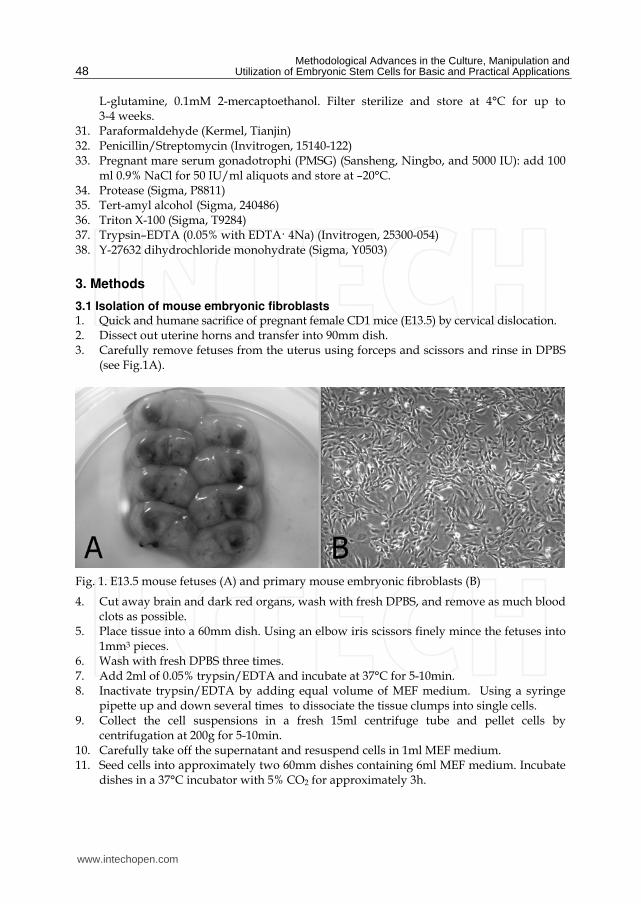
Methodological Advances in the Culture Manipulation and Utilization of Embryonic Stem Cells for Basic and Practical Applications
48
L-glutamine 01mM 2-mercaptoethanol Filter sterilize and store at 4degC for up to 3-4 weeks
31 Paraformaldehyde (Kermel Tianjin) 32 PenicillinStreptomycin (Invitrogen 15140-122) 33 Pregnant mare serum gonadotrophi (PMSG) (Sansheng Ningbo and 5000 IU) add 100
ml 09 NaCl for 50 IUml aliquots and store at ndash20degC 34 Protease (Sigma P8811) 35 Tert-amyl alcohol (Sigma 240486) 36 Triton X-100 (Sigma T9284) 37 TrypsinndashEDTA (005 with EDTA 4Na) (Invitrogen 25300-054) 38 Y-27632 dihydrochloride monohydrate (Sigma Y0503)
3 Methods
31 Isolation of mouse embryonic fibroblasts 1 Quick and humane sacrifice of pregnant female CD1 mice (E135) by cervical dislocation 2 Dissect out uterine horns and transfer into 90mm dish 3 Carefully remove fetuses from the uterus using forceps and scissors and rinse in DPBS
(see Fig1A)
Fig 1 E135 mouse fetuses (A) and primary mouse embryonic fibroblasts (B)
4 Cut away brain and dark red organs wash with fresh DPBS and remove as much blood clots as possible
5 Place tissue into a 60mm dish Using an elbow iris scissors finely mince the fetuses into 1mm3 pieces
6 Wash with fresh DPBS three times 7 Add 2ml of 005 trypsinEDTA and incubate at 37degC for 5-10min 8 Inactivate trypsinEDTA by adding equal volume of MEF medium Using a syringe
pipette up and down several times to dissociate the tissue clumps into single cells 9 Collect the cell suspensions in a fresh 15ml centrifuge tube and pellet cells by
centrifugation at 200g for 5-10min 10 Carefully take off the supernatant and resuspend cells in 1ml MEF medium 11 Seed cells into approximately two 60mm dishes containing 6ml MEF medium Incubate
dishes in a 37degC incubator with 5 CO2 for approximately 3h
wwwintechopencom
Effective Derivation and Manipulation of Mouse Embryonic Stem Cells
49
12 Change medium after cells attach 13 Allow cells to grow until confluence (repeat step 6-10) Split the cells 13-4 (label these
cells as P1) (see Fig1B) 14 When cells reach confluence repeat step 6-10 Resuspend cells in freezing medium and
freeze each plate in three cryovials 15 Transfer the cryovials to -80degC freezer the next day transfer cells to a liquid nitrogen
storage tank
32 Mouse embryonic fibroblast feeder layer preparation 1 Remove cells from liquid nitrogen and thaw quickly in 37degC H2O bath
2 Transfer cells to a fresh 15ml centrifuge tube and pellet cells by centrifugation at 200g
for 5-10min
3 Carefully take off the supernatant and resuspend cells in 1ml MEF medium
4 Seed cells into 60mm dish containing 6ml MEF medium
5 Allow cells to grow until confluence discard the medium and add 2ml MEF medium
containing 10μgml mitomycin C
6 Incubate dishes in a 37degC incubator with 5 CO2 for 3h
7 Carefully take off the medium and wash four times with DPBS (see Note 1)
8 Add 005 TrypsinEDTA for 1min and then add an equal volume of MEF medium for
inactivation
9 Using mechanical force through pipetting recover the cells from the dish centrifuge
and resuspend the cell pellet in MEF medium
10 Seed 5times105 cells into a 35mm dish containing 2ml MEF medium
11 Put dishes in a 37degC incubator with 5 CO2 until use
33 Isolation of blastocysts 1 Mouse embryonic fibroblast feeder layers should be changed into ES medium 2h prior
to blastocyst isolation
2 Quick and humane sacrifice of pregnant female C57BL6 mice (E35) mated to 129Sv
by cervical dislocation
3 Open the abdominal cavity the uteri are removed by cutting across the cervix and are
cut below the junction with the oviducts
4 Place the uteri in a small volume of M2 medium in a 35mm dish and flush each horn
with M2 medium
5 Collect and wash blastocysts three times in M2 medium
6 Transfer the blastocysts onto the prepared MEF feeder layer and culture at 37degC within
a CO2 incubator
34 Isolation and dissociation of ICM outgrowth 1 After 4 to 5 days gently circle the ICM outgrowths with a finely drawn glass probe
removing the ICM from the surrounding trophoblast cells
2 Take a sterile non-coated Petri dish and add several small drops (30μl) of DPBS and
Accutase (see Note 2)
3 Transfer the ICM outgrowths to the drops of DPBS and then repeat this procedure in
the Accutase drops and incubate at 37degC for 15-20 min
wwwintechopencom
Methodological Advances in the Culture Manipulation and Utilization of Embryonic Stem Cells for Basic and Practical Applications
50
4 Using a P200 pipette and yellow tip transfer the ICM outgrowths to a small drop of ES
cell medium and pipet outgrowths into small cell clumps of 5-10 cells using mechanical
force (see Note 3)
5 Transfer the cell clumps into 4-well plates with fresh MEF feeders and ES cell medium
supplemented with 10μM Y-27632 (see Note 4)
6 After 24h of culture change replace the medium with normal ES cell medium without
Y-27632
7 Change ES medium every other day and observe the cultures After 4 to 6 days ES cell
colonies will appear (see Fig 2)
8 Definitive ES cell colonies are collected and dissociated into individual cells using the
method above for ICM outgrowth dissociation The cells are seeded again in 35mm
dishes with fresh feeder layers This is considered passage 1
Fig 2 Derivation of ES cells from mouse blastocysts A Blastocyst (E35) B Blastocyst attach and spread on MEF feeder layer C Expanded ICM outgrowth ready for the first dissociation D ES cell colonies
35 Culture and passage of ES Cells 1 When ES cell colonies become large and within proximity of other colonies it is
necessary to passage 2 Change fresh ES cell medium 2h prior to passaging
wwwintechopencom
Effective Derivation and Manipulation of Mouse Embryonic Stem Cells
51
3 Aspirate the old medium and wash the cells twice with DPBS 4 Add appropriate amount of 005 trypsinEDTA solution and incubate for 1-2min at
room temperature 5 Once the cell colonies begin to detach carefully remove trypsinEDTA solution and
add 1ml ES cell medium and further dissociate the detached ES colonies into single cell suspension by pipetting several times
6 Adjust the concentration of ES cells and transfer the suspensions into 35mm dishes at a rate of 15
36 Freezing of ES Cells 1 Change fresh ES cell medium 2h prior to freezing 2 Trypsinize cells and harvest as described earlier 3 Collect the cells by centrifugation at 200g for 5min 4 Remove the supernatant and resuspend the pellet in freezing medium 5 Aseptically aliquot the suspension into sterile freezing vials label each vial with the
date and cell typeclone number and place the vials into a thermos cup 6 Freeze the cells overnight at -80degC then transfer to the liquid nitrogen
37 Thawing of ES cells 1 Remove a vial of frozen cells from the liquid nitrogen and transfer to 37degC water bath 2 Transfer cell suspension to a 15ml centrifuge tube and add ES cell medium to 5ml 3 Pellet cells by centrifugation at 200g for 5min 4 Carefully take off the supernatant and resuspend cells in 1ml ES cell medium 5 Plate ES cell suspension onto a prepared feeder layer in a 35mm dish at a suitable
density
38 Karyotyping of ES Cells 1 Add fresh ES cell media containing colcemid at a final concentration of 01μgml to an
exponentially growing ES cell cultures Return to the incubator for 40min 2 Wash slides in fresh fixation solution (31 methanol acetic acid) and then soak them in
ice cold water until ready to use (distilled water plus some ice) It is important for the slides to be both cold and wet when ready for use
3 Wash cells twice with DPBS Completely dissociate colonies into single cell with trypsin Add MEF media and resuspend the cells in a 15ml tube Spin down at 200g for 10min Remove the supernatant
4 Add 1ml 056 KCl dropwise Flick the tube to loose the pellet again to a single cell suspension (no big chucks) Add 9ml of 056 KCl and incubate for 15min
5 Add 2-3 drops of fresh fixation solution to the tube and invert it several times Spin down the cells at 200g for 5min
6 Remove the supernatant Add 1ml fixation solution Flick tube to resuspend pellet add 9ml fixation solution Spin down the cells at 200g for 5min
7 Repeat step 6 three times and resuspend cells in appropriate fixation solution Adjust cell density to 1times106ml
8 Remove slide from the water blot edges to remove excess liquid and drop the cell suspension (dropwise) from at least one foot above the surface of the slide (2-3 dropsslide) Allow the cells spread and then place slides on heat stage (60degC) to speed
wwwintechopencom
Methodological Advances in the Culture Manipulation and Utilization of Embryonic Stem Cells for Basic and Practical Applications
52
up drying) Prepare 3 or 4 slides for each sample The remaining cells can be stored at -20degC for several years
9 Stain slides with Giemsa solution for 15min Rinse slides with running water 10 Photograph chromosome spreads and count (see Fig 3A)
39 Alkaline phosphatase stain Staining for alkaline phosphatase should be performed at room temperature using an alkaline phosphatase detection kit (Boster Bio-Tech AR1023 Wuhan) 1 Aspirate the medium and wash the ES cell cultures twice with DPBS for 1min each 2 Fix ES cells with 4 paraformaldehyde in DPBS at 4degC for 15min 3 Remove paraformaldehyde and add freshly prepared alkaline phosphatase staining
solution 4 Keep reactions in the dark and incubate at room temperature until desired degree of
color development has occurred (This process usually takes 20-40min) 5 Terminate the color reaction by washing with running water 6 Observe the images under an inverted microscope (see Fig 3B)
Fig 3 A Karyotype of ES cells B ES cell colonies with positive alkaline phosphatase activity
310 Immunostaining of ES Cells 1 Seed ES cells on feeder layers in 4- well plate until near confluence
2 Remove media and fix cells with 4 paraformaldehyde for 10min at room temperature
3 Wash with DPBS for three times
4 Add 02 Triton X-100 in DPBS for 5min and then wash with DPBS for three times
5 Add DPBS with 10 FBS for 30min
6 Dilute primary antibodies (rabbit anti-oct-4 polyclonal IgG rabbit anti-nanog
polyclonal IgG rabbit anti-sox-2 polyclonal IgG mouse anti-ssea-1 monoclonal IgG)
(Santa Cruz sc-9081 sc-33760 sc-20088 sc-21702) at 1200 dilution rate in DPBS with
10 FBS
7 Add primary antibodies and incubate cells overnight at 4ordmC refrigerator
8 Wash with DPBS three times
wwwintechopencom
Effective Derivation and Manipulation of Mouse Embryonic Stem Cells
53
9 Dilute second antibodies (donkey anti rabbit IgG-FITC donkey anti mouse IgG-FITC) (Santa Cruz sc-2090 sc-2099) at 1500 dilution rate in DPBS with 10 FBS
10 Add appropriate second antibodies into cells and incubate 2h at room temperate 11 Wash with DPBS three times and observe under an inverted fluorescent microscope
(see Fig 4)
Fig 4 Immunostaining of ES cell pluripotent markers (nanog oct-4 sox-2 ssea-1)
311 Formation of embryoid bodies 1 Trypsinize and dissociate ES cell colonies into single cells 2 Collect by centrifugation and resuspend the cells at a density of 5times105 cellsml in MEF
medium supplemented with 10μM Y-27632 3 Transfer 2ml cell suspension into a 35mm dish coated with 1 agar (see Note 5) 4 Culture overnight at incubator carefully collect cell aggregates and transfer them into a
new 35mm dish coated agar Add 2ml MEF medium without Y-27632 for further culture
5 After 3-5 days many cystic embryoid bodies appear (see Fig5)
312 Formation of teratoma 1 Pretreat ES colonies with 10μM Y-27632 for 1h prior to dissociation 2 Dissociate with Accutase harvest the cells by centrifuge at 200g for 10min and remove
the supernatant
wwwintechopencom
Methodological Advances in the Culture Manipulation and Utilization of Embryonic Stem Cells for Basic and Practical Applications
54
3 Add DPBS and resuspend cells to a final density of 2times106 cellsml 4 Subcutaneously inject 02 ml cell suspension into the CD1 mouse groin (see Note 6) 5 Teratoma will be observed approximately 6 weeks later (see Fig 6)
Fig 5 Cystic embryoid bodies by ES cell suspension culture
Fig 6 Formation of teratoma by injecting ES cells into CD1 mouse groin
313 Production of chimeric mice by aggregation with eight-cell stage embryos 1 Prepare several drops of KSOM culture medium containing 10μM Y-27632 in a 35mm
Petri dish and cover with mineral oil 2 Sterilize a darning needle by dipping in ethanol Make a series of 10 small depressions
in each microdrop by pressing a darning needle into the Petri dish surface (see Note 7) Keep the plate in a 37degC incubator at 5 CO2
3 Oviducts from pregnant female mice (E25) are flushed with M2 to collect eight-cell embryos
4 Place several drops of M2 05 protease and KSOM in 35mm Petri dish (do not mix) 5 Wash the embryos with three drops of protease medium and transfer them into drops
of protease Incubate embryos at 37degC for 5-7min
wwwintechopencom
Effective Derivation and Manipulation of Mouse Embryonic Stem Cells
55
6 Wash embryos three times with KSOM and when zona pellucida begins to disappear transfer them into drops of KSOM
7 Transfer embryos into an aggregation plate (one embryo in each depression) Return plate to the incubator
8 Pull a glass capillary tube using a pipette puller this will be used for ES cell clump preparation
9 Pick ES cell colonies from Petri dish and wash them three times with fresh DPBS 10 Transfer colonies to trypsinEDTA and incubate at 37degC for 3-5min 11 Immediately transfer the colonies into a drop of KSOM and wash them three times 12 Colonies should be dissociated into 15-20 cell clumps using a glass capillary tube and
mechanical force 13 Using a pipette place one clump of ES cells into each depression 14 Return the plate to incubator and culture overnight (see Fig 7) 15 Transfer 8-10 aggregation blastocysts to each uterine horn of pseudopregnant CD1
females (25dpc) The female CD1 mouse will deliver approximately 17 days later
Fig 7 Production of chimeras by aggregation A 8-cell embryos without zona pellucida B C Aggregation depression ES cells and embryos D Aggregate of ES cells and embryo
314 Production of chimeric mice by tetraploid blastocyst injection Production of tetraploid mouse embryos
1 Collect 2-cell embryos (E15) of the CD1 outbred strain by flushing the oviducts and wash them using several drops of M2 medium
wwwintechopencom
Methodological Advances in the Culture Manipulation and Utilization of Embryonic Stem Cells for Basic and Practical Applications
56
2 Transfer them into KSOM drops and keep at incubator until electrofusion 3 Wash 2-cell embryos through several drops of fusion buffer and place 20-30 embryos
into the fusion buffer between the electrodes of the 02mm micro fusion chamber (see Fig 8A)
4 Set Eppendorf Multiporator mode to electrofusion 2V voltage and 15s duration for embryo prealignment 20V voltage 50μs duration and two pulses for fusion
5 Transfer the embryos to the M2 drop and wash them through several drops of M2 medium
6 Transfer the embryos into KSOM drops under mineral oil and culture at 37degC incubator
7 In the next 30-60min choose all properly fused embryos and incubate them in KSOM medium under mineral oil at 37degC 5 CO2 (see Fig 8B)
8 After 24h culture most of embryos should develop to blastocyst stage and are ready to be injected with ES cells
Fig 8 A 2-cell stage embryos aligned in fusion chamber B Fused embryos
Preparation of ES cells for blastocyst injection
1 Replace fresh ES medium 3h prior to injection
2 Pick ES cell colonies and rinse them with DPBS
3 Transfer the colonies into Accutase drops and incubate at 37degC for 5 to 7min
4 Using a P200 pipette transfer the colonies to a small drop of ES medium with 10μM Y-
27632 and break up the colonies into single cell suspension
5 Incubate the single cell suspension with 10μM Y-27632 for 1h at room temperature prior
to injection to remove cell membrane blebs (see Fig 9) (see Note 8)
Microinjection needles preparation using a PN-30 micropipette puller and a MF-900 microforge
1 Turn on the machine and choose appropriate settings For the PN-30 puller use magnet (main) 90 magnet (sub) 30 and heater 80 as the starting values
2 Fix one glass capillary (100mm) on the needle puller 3 Start the pulling process of the needle 4 Remove the needle carefully and check the tip under a stereomicroscope 5 Mount a capillary in the microforge and break this at the required diameter by fusing
the glass onto the glass bead on the microforge and turning off the heat while drawing it away
wwwintechopencom
Effective Derivation and Manipulation of Mouse Embryonic Stem Cells
57
Fig 9 Compare to the control cells without Y-27632 treatment (A) the treatment of Y-27632 make ES cells smooth and soft (B) so that they can easily be picked up by injection pipette
6 For making holding pipets break the holding pipet at a point where the outside
diameter is approx 80μm and the inside diameter approx 60μm and then move the
holding pipet toward the glowing glass bead and the cut will start to melt inward
creating a smoother narrower tip
7 For making injection pipets break the injection pipet at a point where the outside
diameter is approx 15μm and the inside diameter approx 12μm
8 Hold the pipet above the glass bead and heat a point about 5mm from the pipet tip The
pipet will bend under gravity as the glass melts Allow the pipet to bend approx 60deg
Piezo-supported microinjection
1 Prepare dishes for injection using the lids of standard 35mm plastic disposable Petri
dishes
2 Place a 400μl drop of M2 medium on the lid and cover it with mineral oil
3 Fluorinert FC-77 in a 15mm length was back-loaded into an injection pipette Push
Fluorinert FC-77 through the shoulder to near the tip to empty the air in the pipette
4 Attach the injection pipette to the pipette holder of the piezo unit and hang the piezo
unit on the micromanipulator and aspirate 1mm M2 into the pipette to keep ES cells
away from Fluorinert FC-77
5 Attach the holding pipette on the other side of the micromanipulator
6 Transfer embryos and ES cells into the drop of M2 under mineral oil
7 Aspirate several ES cells and use several piezo pulses (eg intensity=3 frequency=3) to
penetrate the zona and trophectoderm layer (see Note 9)
8 Advance the tip of the pipette near the opposite side of the blastocysts from where it is
held by the holding pipette
9 Inject 15-20 ES cells into the blastocoel cavity (see Fig 10A B)
10 Culture injected blastocysts in KSOM medium at 37degC 5 CO2 for 2h and allow them
recovery
11 Transfer 8-10 injected blastocysts to each uterine horn of 25dpc pseudopregnant CD1
femals
12 Recipient mice will give birth after 17 days (see Fig 10C D)
wwwintechopencom
Methodological Advances in the Culture Manipulation and Utilization of Embryonic Stem Cells for Basic and Practical Applications
58
Fig 10 A Piezo-actuated tetraploid blastocyst injection B Injected tetraploid blastocysts C Dead ES mouse pups D Survival ES mouse pups
315 Embryo transfer and Caesarean sections 1 Select a pseudopregnant CD1 female (25dpc) as an embryo recipient 2 Prepare the transfer pipette forceps and scissors etc 3 Anesthetized mice by intraperitoneal injection of Avertin after drug delivery quickly
put the mouse back into the cage (see Note 10) 4 After the mouse is fully anesthetized place the mouse in the prone position on a lid of a
Petri dish Using an absorbent ball with 75 ethanol sterilize the area and shave the animal
5 Make a 05-1cm transverse dorsal incision at the below of the last ribs 6 Uncover incision to find ovarian fat pad pull it out using forceps and hold it using clamps 7 Collect the embryos carefully one by one lined up in the transfer pipette with as little
medium in between as possible 8 Make a hole through the uterine wall into the lumen with a 25G needle The site of this
hole should be near the last loop of the oviduct where it descends into the top of the uterine horn at the UT junction
9 Insert the transfer pipette into a hole and carefully expel the embryos Check the transfer pipette to ensure that all embryos have been transferred 8-10 embryos are usually transferred into each horn
10 When embryos are to be transferred into uteri put the ovary back into the abdominal cavity and suture the incision
wwwintechopencom
Effective Derivation and Manipulation of Mouse Embryonic Stem Cells
59
11 Place the mouse in clean cage Lighting to heat until it regaines consciousness 12 After 17 days the pups will be born
4 Notes
1 Mitomycin C (Sigma M4287) Dissolve the powder into sterile distilled DMEM and adjust the concentration to 10μgml just before use Wash layers several times with DPBS to completely remove any mitomycin C residue from the feeder layers
2 Accutase is a ready to use cell detachment solution of proteolytic and collagenolytic enzymes and it does not contain mammalian or bacterial derived products Cell lines tested for Accutase application includes fibroblasts keratinocytes vascular smooth muscle cells primary chick embryo neuronal cells bone marrow stem cells 293 cells 3T3 cells HeLa cells insect cells human embryonic stem cells and human neural stem cells Moreover Accutase is quite gentle on cells and will not induce cell death if cells are treated a little longer than the optimal time period Thus we use Acutase instead of trypsin for enzymatic dissociation of ICM
3 This is a key step for ES cell isolation Watch the whole dissociation process under a microscope Careful not to pipette with too much force as single cells will not survive Compared to trypsin Accutase treated ICM outgrowths are easier to dissociate into small cell clumps
4 The ROCK inhibitor Y-27632 permits survival of dissociated human embryonic stem cells Similar to human ES cells we find that mouse ICM cells show sensitivity to trypsin They undergo cell death after dissociation and the cloning efficiency of dissociated ICM cells is generally very low Our data indicates that increased cellular adhesion induced by Y-27632 enhances the survival of dissociated ICM cells With more ES-like colonies appearing when the dissociated ICM cells are seeded in medium supplemented with 10 μM Y-27632 for 24h Moreover we have not observed adverse effects of Y-27632 treatment on pluripotency in maintenance culture even after a number of passages
5 Dishes are coated with agar to completely prevent cell attachment This can increase cell aggregation and embryonic body formation In brief add 1g agar into 100ml DPBS and autoclave for 30min Cool to 50degC at room temperature Rapidly cover the internal wall and bottom of 35mm dishes with agar solution Remove the extra solution and place dishes in hood until the remaining agar solidification In addition adding Y-27632 in the embryonic body medium can also enhance suspending cells to aggregate and embryonic body formation
6 Typically severe combined immunodeficient mice are used for making teratoma This strain of mouse is expensive and not easily raised Our data indicates that most mouse strains could be recipient for teratoma production by inguinal subcutaneous injection such as CD1 C57BL6times129Sv
7 Carefully make small depressions using needle and appropriate force in the Petri dish surface Too much force may break the dish and insufficient force will not produce the required depressions which can easily lead to the loss of embryos and ES cells
8 Prior to blastocyst injection the pretreatment of 10μM Y-27632 for 1h at room temperature can alter the membrane architecture of ES cells so that they can be easily collected by injection pipet Otherwise the ES cell surface is covered with blebs (see Fig 10)
9 Within the microinjection pipette ES cells should be kept approximately 100μm away from the pipette tip Damage to ES cells may occur if the cells are very close to the tip when the piezo pulses are applied
wwwintechopencom
Methodological Advances in the Culture Manipulation and Utilization of Embryonic Stem Cells for Basic and Practical Applications
60
10 Avertin anesthetic 125 stocks avertin is prepared by mixing 25g of 2 2 2-tribromoethyl alcohol (Sigma) 5ml of tert-amyl alcohol (Sigma) with 200ml water Avertin is intraperitoneally injected at a dose of 002ml per gram of body weight for anesthetization
5 Acknowledgements
We would like to thank Roman Krawetz (The University of Calgary Canada) for providing his expert and critical reading on the manuscript This work was supported by grants from National Natural Science Foundation of China (No 30871790) and Natural Science Foundation of Hebei (No C2009000600)
6 References
Batlle-Morera L Smith A amp Nichols J (2008) Parameters influencing derivation of embryonic stem cells from murine embryos Genesis 46(12)758-767 ISSN 1526-954X
Brook FA amp Gardner RL (1997) The origin and efficient derivation of embryonic stem cells in the mouse Proc Natl Acad Sci USA 945709-5712 ISSN 0027-8424
Bryja V Bonilla S Cajaacutenek L Parish CL Schwartz CM Luo Y Rao MS amp Arenas E (2006) An efficient method for the derivation of mouse embryonic stem cells Stem Cells 24(4)844-849 ISSN 1066-5099
Cheng J Dutra A Takesono A Garrett-Beal L amp Schwartzberg PL (2004) Improved generation of C57BL6J mouse embryonic stem cells in a defined serum-free media Genesis 39100-104 ISSN 1526-954X
Doungpunta J Santhi A Sathanawongs A Jarujinda Y amp Oranratnachai A (2009) Fivefold increase in derivation rates of mouse embryonic stem cellsafter supplementation of the media with multiple factors Theriogenology 72(2)232-242 ISSN 0093-691X
Evans M J amp Kaufman M H (1981) Establishment in culture of pluripotential cells from mouse embryos Nature 292(5819)154-156 ISSN 0028-0836
Martin G R (1981) Isolation of a pluripotent cell line from early mouse embryos cultured in medium conditioned by teratocarcinoma stem cells Proc Natl Acad Sci USA 787634-7638 ISSN 0027-8424
McWhir J Schnieke AE Ansell R Wallace H Colman A Scott ARamp Kind AJ (1996) Selective ablation of differentiated cells permits isolation of embryonic stem cell lines from murine embryos with a non-permissive genetic background Nat
Genet 14223-226 ISSN 1061-4036 Ruchi B Jacqueline L Min K amp Alexey VT (2008) Efficient propagation of single cells
accutase-dissociated human embryonic stem cells Mol Reprod Dev 75(5)818-827 ISSN 1040-452X
Schoonjans L Kreemers V Danloy S Moreadith RW Laroche Y amp Collen D (2003) Improved generation of germline competent embryonic stem cell lines from inbred mouse strains Stem Cells 21(1)90-97 ISSN 1066-5099
Tesar PJ (2005) Derivation of germ-line-competent embryonic stemcell lines from preblastocyst mouse embryos Proc Natl Acad Sci USA 102(23)8239-8244 ISSN 0027-8424
Watanabe K Ueno M Kamiya D Nishiyama A Matsumura M Wataya T Takahashi JB Nishikawa S Nishikawa S Muguruma K amp Sasai Y (2007) A ROCK inhibitor permits survival of dissociated human embryonic stem cells Nat
Biotechnol 25681-686 ISSN 1087-0156
wwwintechopencom
Methodological Advances in the Culture Manipulation andUtilization of Embryonic Stem Cells for Basic and PracticalApplicationsEdited by Prof Craig Atwood
ISBN 978-953-307-197-8Hard cover 506 pagesPublisher InTechPublished online 26 April 2011Published in print edition April 2011
InTech EuropeUniversity Campus STeP Ri Slavka Krautzeka 83A 51000 Rijeka Croatia Phone +385 (51) 770 447 Fax +385 (51) 686 166wwwintechopencom
InTech ChinaUnit 405 Office Block Hotel Equatorial Shanghai No65 Yan An Road (West) Shanghai 200040 China
Phone +86-21-62489820 Fax +86-21-62489821
Pluripotent stem cells have the potential to revolutionise medicine providing treatment options for a widerange of diseases and conditions that currently lack therapies or cures This book describes methodologicaladvances in the culture and manipulation of embryonic stem cells that will serve to bring this promise topractice
How to referenceIn order to correctly reference this scholarly work feel free to copy and paste the following
Peng Zhang Xinglong Wu Pengbo Wang and Xiangyun Li (2011) Effective Derivation and Manipulation ofMouse Embryonic Stem Cells Methodological Advances in the Culture Manipulation and Utilization ofEmbryonic Stem Cells for Basic and Practical Applications Prof Craig Atwood (Ed) ISBN 978-953-307-197-8 InTech Available from httpwwwintechopencombooksmethodological-advances-in-the-culture-manipulation-and-utilization-of-embryonic-stem-cells-for-basic-and-practical-applicationseffective-derivation-and-manipulation-of-mouse-embryonic-stem-cells

Effective Derivation and Manipulation of Mouse Embryonic Stem Cells
49
12 Change medium after cells attach 13 Allow cells to grow until confluence (repeat step 6-10) Split the cells 13-4 (label these
cells as P1) (see Fig1B) 14 When cells reach confluence repeat step 6-10 Resuspend cells in freezing medium and
freeze each plate in three cryovials 15 Transfer the cryovials to -80degC freezer the next day transfer cells to a liquid nitrogen
storage tank
32 Mouse embryonic fibroblast feeder layer preparation 1 Remove cells from liquid nitrogen and thaw quickly in 37degC H2O bath
2 Transfer cells to a fresh 15ml centrifuge tube and pellet cells by centrifugation at 200g
for 5-10min
3 Carefully take off the supernatant and resuspend cells in 1ml MEF medium
4 Seed cells into 60mm dish containing 6ml MEF medium
5 Allow cells to grow until confluence discard the medium and add 2ml MEF medium
containing 10μgml mitomycin C
6 Incubate dishes in a 37degC incubator with 5 CO2 for 3h
7 Carefully take off the medium and wash four times with DPBS (see Note 1)
8 Add 005 TrypsinEDTA for 1min and then add an equal volume of MEF medium for
inactivation
9 Using mechanical force through pipetting recover the cells from the dish centrifuge
and resuspend the cell pellet in MEF medium
10 Seed 5times105 cells into a 35mm dish containing 2ml MEF medium
11 Put dishes in a 37degC incubator with 5 CO2 until use
33 Isolation of blastocysts 1 Mouse embryonic fibroblast feeder layers should be changed into ES medium 2h prior
to blastocyst isolation
2 Quick and humane sacrifice of pregnant female C57BL6 mice (E35) mated to 129Sv
by cervical dislocation
3 Open the abdominal cavity the uteri are removed by cutting across the cervix and are
cut below the junction with the oviducts
4 Place the uteri in a small volume of M2 medium in a 35mm dish and flush each horn
with M2 medium
5 Collect and wash blastocysts three times in M2 medium
6 Transfer the blastocysts onto the prepared MEF feeder layer and culture at 37degC within
a CO2 incubator
34 Isolation and dissociation of ICM outgrowth 1 After 4 to 5 days gently circle the ICM outgrowths with a finely drawn glass probe
removing the ICM from the surrounding trophoblast cells
2 Take a sterile non-coated Petri dish and add several small drops (30μl) of DPBS and
Accutase (see Note 2)
3 Transfer the ICM outgrowths to the drops of DPBS and then repeat this procedure in
the Accutase drops and incubate at 37degC for 15-20 min
wwwintechopencom
Methodological Advances in the Culture Manipulation and Utilization of Embryonic Stem Cells for Basic and Practical Applications
50
4 Using a P200 pipette and yellow tip transfer the ICM outgrowths to a small drop of ES
cell medium and pipet outgrowths into small cell clumps of 5-10 cells using mechanical
force (see Note 3)
5 Transfer the cell clumps into 4-well plates with fresh MEF feeders and ES cell medium
supplemented with 10μM Y-27632 (see Note 4)
6 After 24h of culture change replace the medium with normal ES cell medium without
Y-27632
7 Change ES medium every other day and observe the cultures After 4 to 6 days ES cell
colonies will appear (see Fig 2)
8 Definitive ES cell colonies are collected and dissociated into individual cells using the
method above for ICM outgrowth dissociation The cells are seeded again in 35mm
dishes with fresh feeder layers This is considered passage 1
Fig 2 Derivation of ES cells from mouse blastocysts A Blastocyst (E35) B Blastocyst attach and spread on MEF feeder layer C Expanded ICM outgrowth ready for the first dissociation D ES cell colonies
35 Culture and passage of ES Cells 1 When ES cell colonies become large and within proximity of other colonies it is
necessary to passage 2 Change fresh ES cell medium 2h prior to passaging
wwwintechopencom
Effective Derivation and Manipulation of Mouse Embryonic Stem Cells
51
3 Aspirate the old medium and wash the cells twice with DPBS 4 Add appropriate amount of 005 trypsinEDTA solution and incubate for 1-2min at
room temperature 5 Once the cell colonies begin to detach carefully remove trypsinEDTA solution and
add 1ml ES cell medium and further dissociate the detached ES colonies into single cell suspension by pipetting several times
6 Adjust the concentration of ES cells and transfer the suspensions into 35mm dishes at a rate of 15
36 Freezing of ES Cells 1 Change fresh ES cell medium 2h prior to freezing 2 Trypsinize cells and harvest as described earlier 3 Collect the cells by centrifugation at 200g for 5min 4 Remove the supernatant and resuspend the pellet in freezing medium 5 Aseptically aliquot the suspension into sterile freezing vials label each vial with the
date and cell typeclone number and place the vials into a thermos cup 6 Freeze the cells overnight at -80degC then transfer to the liquid nitrogen
37 Thawing of ES cells 1 Remove a vial of frozen cells from the liquid nitrogen and transfer to 37degC water bath 2 Transfer cell suspension to a 15ml centrifuge tube and add ES cell medium to 5ml 3 Pellet cells by centrifugation at 200g for 5min 4 Carefully take off the supernatant and resuspend cells in 1ml ES cell medium 5 Plate ES cell suspension onto a prepared feeder layer in a 35mm dish at a suitable
density
38 Karyotyping of ES Cells 1 Add fresh ES cell media containing colcemid at a final concentration of 01μgml to an
exponentially growing ES cell cultures Return to the incubator for 40min 2 Wash slides in fresh fixation solution (31 methanol acetic acid) and then soak them in
ice cold water until ready to use (distilled water plus some ice) It is important for the slides to be both cold and wet when ready for use
3 Wash cells twice with DPBS Completely dissociate colonies into single cell with trypsin Add MEF media and resuspend the cells in a 15ml tube Spin down at 200g for 10min Remove the supernatant
4 Add 1ml 056 KCl dropwise Flick the tube to loose the pellet again to a single cell suspension (no big chucks) Add 9ml of 056 KCl and incubate for 15min
5 Add 2-3 drops of fresh fixation solution to the tube and invert it several times Spin down the cells at 200g for 5min
6 Remove the supernatant Add 1ml fixation solution Flick tube to resuspend pellet add 9ml fixation solution Spin down the cells at 200g for 5min
7 Repeat step 6 three times and resuspend cells in appropriate fixation solution Adjust cell density to 1times106ml
8 Remove slide from the water blot edges to remove excess liquid and drop the cell suspension (dropwise) from at least one foot above the surface of the slide (2-3 dropsslide) Allow the cells spread and then place slides on heat stage (60degC) to speed
wwwintechopencom
Methodological Advances in the Culture Manipulation and Utilization of Embryonic Stem Cells for Basic and Practical Applications
52
up drying) Prepare 3 or 4 slides for each sample The remaining cells can be stored at -20degC for several years
9 Stain slides with Giemsa solution for 15min Rinse slides with running water 10 Photograph chromosome spreads and count (see Fig 3A)
39 Alkaline phosphatase stain Staining for alkaline phosphatase should be performed at room temperature using an alkaline phosphatase detection kit (Boster Bio-Tech AR1023 Wuhan) 1 Aspirate the medium and wash the ES cell cultures twice with DPBS for 1min each 2 Fix ES cells with 4 paraformaldehyde in DPBS at 4degC for 15min 3 Remove paraformaldehyde and add freshly prepared alkaline phosphatase staining
solution 4 Keep reactions in the dark and incubate at room temperature until desired degree of
color development has occurred (This process usually takes 20-40min) 5 Terminate the color reaction by washing with running water 6 Observe the images under an inverted microscope (see Fig 3B)
Fig 3 A Karyotype of ES cells B ES cell colonies with positive alkaline phosphatase activity
310 Immunostaining of ES Cells 1 Seed ES cells on feeder layers in 4- well plate until near confluence
2 Remove media and fix cells with 4 paraformaldehyde for 10min at room temperature
3 Wash with DPBS for three times
4 Add 02 Triton X-100 in DPBS for 5min and then wash with DPBS for three times
5 Add DPBS with 10 FBS for 30min
6 Dilute primary antibodies (rabbit anti-oct-4 polyclonal IgG rabbit anti-nanog
polyclonal IgG rabbit anti-sox-2 polyclonal IgG mouse anti-ssea-1 monoclonal IgG)
(Santa Cruz sc-9081 sc-33760 sc-20088 sc-21702) at 1200 dilution rate in DPBS with
10 FBS
7 Add primary antibodies and incubate cells overnight at 4ordmC refrigerator
8 Wash with DPBS three times
wwwintechopencom
Effective Derivation and Manipulation of Mouse Embryonic Stem Cells
53
9 Dilute second antibodies (donkey anti rabbit IgG-FITC donkey anti mouse IgG-FITC) (Santa Cruz sc-2090 sc-2099) at 1500 dilution rate in DPBS with 10 FBS
10 Add appropriate second antibodies into cells and incubate 2h at room temperate 11 Wash with DPBS three times and observe under an inverted fluorescent microscope
(see Fig 4)
Fig 4 Immunostaining of ES cell pluripotent markers (nanog oct-4 sox-2 ssea-1)
311 Formation of embryoid bodies 1 Trypsinize and dissociate ES cell colonies into single cells 2 Collect by centrifugation and resuspend the cells at a density of 5times105 cellsml in MEF
medium supplemented with 10μM Y-27632 3 Transfer 2ml cell suspension into a 35mm dish coated with 1 agar (see Note 5) 4 Culture overnight at incubator carefully collect cell aggregates and transfer them into a
new 35mm dish coated agar Add 2ml MEF medium without Y-27632 for further culture
5 After 3-5 days many cystic embryoid bodies appear (see Fig5)
312 Formation of teratoma 1 Pretreat ES colonies with 10μM Y-27632 for 1h prior to dissociation 2 Dissociate with Accutase harvest the cells by centrifuge at 200g for 10min and remove
the supernatant
wwwintechopencom
Methodological Advances in the Culture Manipulation and Utilization of Embryonic Stem Cells for Basic and Practical Applications
54
3 Add DPBS and resuspend cells to a final density of 2times106 cellsml 4 Subcutaneously inject 02 ml cell suspension into the CD1 mouse groin (see Note 6) 5 Teratoma will be observed approximately 6 weeks later (see Fig 6)
Fig 5 Cystic embryoid bodies by ES cell suspension culture
Fig 6 Formation of teratoma by injecting ES cells into CD1 mouse groin
313 Production of chimeric mice by aggregation with eight-cell stage embryos 1 Prepare several drops of KSOM culture medium containing 10μM Y-27632 in a 35mm
Petri dish and cover with mineral oil 2 Sterilize a darning needle by dipping in ethanol Make a series of 10 small depressions
in each microdrop by pressing a darning needle into the Petri dish surface (see Note 7) Keep the plate in a 37degC incubator at 5 CO2
3 Oviducts from pregnant female mice (E25) are flushed with M2 to collect eight-cell embryos
4 Place several drops of M2 05 protease and KSOM in 35mm Petri dish (do not mix) 5 Wash the embryos with three drops of protease medium and transfer them into drops
of protease Incubate embryos at 37degC for 5-7min
wwwintechopencom
Effective Derivation and Manipulation of Mouse Embryonic Stem Cells
55
6 Wash embryos three times with KSOM and when zona pellucida begins to disappear transfer them into drops of KSOM
7 Transfer embryos into an aggregation plate (one embryo in each depression) Return plate to the incubator
8 Pull a glass capillary tube using a pipette puller this will be used for ES cell clump preparation
9 Pick ES cell colonies from Petri dish and wash them three times with fresh DPBS 10 Transfer colonies to trypsinEDTA and incubate at 37degC for 3-5min 11 Immediately transfer the colonies into a drop of KSOM and wash them three times 12 Colonies should be dissociated into 15-20 cell clumps using a glass capillary tube and
mechanical force 13 Using a pipette place one clump of ES cells into each depression 14 Return the plate to incubator and culture overnight (see Fig 7) 15 Transfer 8-10 aggregation blastocysts to each uterine horn of pseudopregnant CD1
females (25dpc) The female CD1 mouse will deliver approximately 17 days later
Fig 7 Production of chimeras by aggregation A 8-cell embryos without zona pellucida B C Aggregation depression ES cells and embryos D Aggregate of ES cells and embryo
314 Production of chimeric mice by tetraploid blastocyst injection Production of tetraploid mouse embryos
1 Collect 2-cell embryos (E15) of the CD1 outbred strain by flushing the oviducts and wash them using several drops of M2 medium
wwwintechopencom
Methodological Advances in the Culture Manipulation and Utilization of Embryonic Stem Cells for Basic and Practical Applications
56
2 Transfer them into KSOM drops and keep at incubator until electrofusion 3 Wash 2-cell embryos through several drops of fusion buffer and place 20-30 embryos
into the fusion buffer between the electrodes of the 02mm micro fusion chamber (see Fig 8A)
4 Set Eppendorf Multiporator mode to electrofusion 2V voltage and 15s duration for embryo prealignment 20V voltage 50μs duration and two pulses for fusion
5 Transfer the embryos to the M2 drop and wash them through several drops of M2 medium
6 Transfer the embryos into KSOM drops under mineral oil and culture at 37degC incubator
7 In the next 30-60min choose all properly fused embryos and incubate them in KSOM medium under mineral oil at 37degC 5 CO2 (see Fig 8B)
8 After 24h culture most of embryos should develop to blastocyst stage and are ready to be injected with ES cells
Fig 8 A 2-cell stage embryos aligned in fusion chamber B Fused embryos
Preparation of ES cells for blastocyst injection
1 Replace fresh ES medium 3h prior to injection
2 Pick ES cell colonies and rinse them with DPBS
3 Transfer the colonies into Accutase drops and incubate at 37degC for 5 to 7min
4 Using a P200 pipette transfer the colonies to a small drop of ES medium with 10μM Y-
27632 and break up the colonies into single cell suspension
5 Incubate the single cell suspension with 10μM Y-27632 for 1h at room temperature prior
to injection to remove cell membrane blebs (see Fig 9) (see Note 8)
Microinjection needles preparation using a PN-30 micropipette puller and a MF-900 microforge
1 Turn on the machine and choose appropriate settings For the PN-30 puller use magnet (main) 90 magnet (sub) 30 and heater 80 as the starting values
2 Fix one glass capillary (100mm) on the needle puller 3 Start the pulling process of the needle 4 Remove the needle carefully and check the tip under a stereomicroscope 5 Mount a capillary in the microforge and break this at the required diameter by fusing
the glass onto the glass bead on the microforge and turning off the heat while drawing it away
wwwintechopencom
Effective Derivation and Manipulation of Mouse Embryonic Stem Cells
57
Fig 9 Compare to the control cells without Y-27632 treatment (A) the treatment of Y-27632 make ES cells smooth and soft (B) so that they can easily be picked up by injection pipette
6 For making holding pipets break the holding pipet at a point where the outside
diameter is approx 80μm and the inside diameter approx 60μm and then move the
holding pipet toward the glowing glass bead and the cut will start to melt inward
creating a smoother narrower tip
7 For making injection pipets break the injection pipet at a point where the outside
diameter is approx 15μm and the inside diameter approx 12μm
8 Hold the pipet above the glass bead and heat a point about 5mm from the pipet tip The
pipet will bend under gravity as the glass melts Allow the pipet to bend approx 60deg
Piezo-supported microinjection
1 Prepare dishes for injection using the lids of standard 35mm plastic disposable Petri
dishes
2 Place a 400μl drop of M2 medium on the lid and cover it with mineral oil
3 Fluorinert FC-77 in a 15mm length was back-loaded into an injection pipette Push
Fluorinert FC-77 through the shoulder to near the tip to empty the air in the pipette
4 Attach the injection pipette to the pipette holder of the piezo unit and hang the piezo
unit on the micromanipulator and aspirate 1mm M2 into the pipette to keep ES cells
away from Fluorinert FC-77
5 Attach the holding pipette on the other side of the micromanipulator
6 Transfer embryos and ES cells into the drop of M2 under mineral oil
7 Aspirate several ES cells and use several piezo pulses (eg intensity=3 frequency=3) to
penetrate the zona and trophectoderm layer (see Note 9)
8 Advance the tip of the pipette near the opposite side of the blastocysts from where it is
held by the holding pipette
9 Inject 15-20 ES cells into the blastocoel cavity (see Fig 10A B)
10 Culture injected blastocysts in KSOM medium at 37degC 5 CO2 for 2h and allow them
recovery
11 Transfer 8-10 injected blastocysts to each uterine horn of 25dpc pseudopregnant CD1
femals
12 Recipient mice will give birth after 17 days (see Fig 10C D)
wwwintechopencom
Methodological Advances in the Culture Manipulation and Utilization of Embryonic Stem Cells for Basic and Practical Applications
58
Fig 10 A Piezo-actuated tetraploid blastocyst injection B Injected tetraploid blastocysts C Dead ES mouse pups D Survival ES mouse pups
315 Embryo transfer and Caesarean sections 1 Select a pseudopregnant CD1 female (25dpc) as an embryo recipient 2 Prepare the transfer pipette forceps and scissors etc 3 Anesthetized mice by intraperitoneal injection of Avertin after drug delivery quickly
put the mouse back into the cage (see Note 10) 4 After the mouse is fully anesthetized place the mouse in the prone position on a lid of a
Petri dish Using an absorbent ball with 75 ethanol sterilize the area and shave the animal
5 Make a 05-1cm transverse dorsal incision at the below of the last ribs 6 Uncover incision to find ovarian fat pad pull it out using forceps and hold it using clamps 7 Collect the embryos carefully one by one lined up in the transfer pipette with as little
medium in between as possible 8 Make a hole through the uterine wall into the lumen with a 25G needle The site of this
hole should be near the last loop of the oviduct where it descends into the top of the uterine horn at the UT junction
9 Insert the transfer pipette into a hole and carefully expel the embryos Check the transfer pipette to ensure that all embryos have been transferred 8-10 embryos are usually transferred into each horn
10 When embryos are to be transferred into uteri put the ovary back into the abdominal cavity and suture the incision
wwwintechopencom
Effective Derivation and Manipulation of Mouse Embryonic Stem Cells
59
11 Place the mouse in clean cage Lighting to heat until it regaines consciousness 12 After 17 days the pups will be born
4 Notes
1 Mitomycin C (Sigma M4287) Dissolve the powder into sterile distilled DMEM and adjust the concentration to 10μgml just before use Wash layers several times with DPBS to completely remove any mitomycin C residue from the feeder layers
2 Accutase is a ready to use cell detachment solution of proteolytic and collagenolytic enzymes and it does not contain mammalian or bacterial derived products Cell lines tested for Accutase application includes fibroblasts keratinocytes vascular smooth muscle cells primary chick embryo neuronal cells bone marrow stem cells 293 cells 3T3 cells HeLa cells insect cells human embryonic stem cells and human neural stem cells Moreover Accutase is quite gentle on cells and will not induce cell death if cells are treated a little longer than the optimal time period Thus we use Acutase instead of trypsin for enzymatic dissociation of ICM
3 This is a key step for ES cell isolation Watch the whole dissociation process under a microscope Careful not to pipette with too much force as single cells will not survive Compared to trypsin Accutase treated ICM outgrowths are easier to dissociate into small cell clumps
4 The ROCK inhibitor Y-27632 permits survival of dissociated human embryonic stem cells Similar to human ES cells we find that mouse ICM cells show sensitivity to trypsin They undergo cell death after dissociation and the cloning efficiency of dissociated ICM cells is generally very low Our data indicates that increased cellular adhesion induced by Y-27632 enhances the survival of dissociated ICM cells With more ES-like colonies appearing when the dissociated ICM cells are seeded in medium supplemented with 10 μM Y-27632 for 24h Moreover we have not observed adverse effects of Y-27632 treatment on pluripotency in maintenance culture even after a number of passages
5 Dishes are coated with agar to completely prevent cell attachment This can increase cell aggregation and embryonic body formation In brief add 1g agar into 100ml DPBS and autoclave for 30min Cool to 50degC at room temperature Rapidly cover the internal wall and bottom of 35mm dishes with agar solution Remove the extra solution and place dishes in hood until the remaining agar solidification In addition adding Y-27632 in the embryonic body medium can also enhance suspending cells to aggregate and embryonic body formation
6 Typically severe combined immunodeficient mice are used for making teratoma This strain of mouse is expensive and not easily raised Our data indicates that most mouse strains could be recipient for teratoma production by inguinal subcutaneous injection such as CD1 C57BL6times129Sv
7 Carefully make small depressions using needle and appropriate force in the Petri dish surface Too much force may break the dish and insufficient force will not produce the required depressions which can easily lead to the loss of embryos and ES cells
8 Prior to blastocyst injection the pretreatment of 10μM Y-27632 for 1h at room temperature can alter the membrane architecture of ES cells so that they can be easily collected by injection pipet Otherwise the ES cell surface is covered with blebs (see Fig 10)
9 Within the microinjection pipette ES cells should be kept approximately 100μm away from the pipette tip Damage to ES cells may occur if the cells are very close to the tip when the piezo pulses are applied
wwwintechopencom
Methodological Advances in the Culture Manipulation and Utilization of Embryonic Stem Cells for Basic and Practical Applications
60
10 Avertin anesthetic 125 stocks avertin is prepared by mixing 25g of 2 2 2-tribromoethyl alcohol (Sigma) 5ml of tert-amyl alcohol (Sigma) with 200ml water Avertin is intraperitoneally injected at a dose of 002ml per gram of body weight for anesthetization
5 Acknowledgements
We would like to thank Roman Krawetz (The University of Calgary Canada) for providing his expert and critical reading on the manuscript This work was supported by grants from National Natural Science Foundation of China (No 30871790) and Natural Science Foundation of Hebei (No C2009000600)
6 References
Batlle-Morera L Smith A amp Nichols J (2008) Parameters influencing derivation of embryonic stem cells from murine embryos Genesis 46(12)758-767 ISSN 1526-954X
Brook FA amp Gardner RL (1997) The origin and efficient derivation of embryonic stem cells in the mouse Proc Natl Acad Sci USA 945709-5712 ISSN 0027-8424
Bryja V Bonilla S Cajaacutenek L Parish CL Schwartz CM Luo Y Rao MS amp Arenas E (2006) An efficient method for the derivation of mouse embryonic stem cells Stem Cells 24(4)844-849 ISSN 1066-5099
Cheng J Dutra A Takesono A Garrett-Beal L amp Schwartzberg PL (2004) Improved generation of C57BL6J mouse embryonic stem cells in a defined serum-free media Genesis 39100-104 ISSN 1526-954X
Doungpunta J Santhi A Sathanawongs A Jarujinda Y amp Oranratnachai A (2009) Fivefold increase in derivation rates of mouse embryonic stem cellsafter supplementation of the media with multiple factors Theriogenology 72(2)232-242 ISSN 0093-691X
Evans M J amp Kaufman M H (1981) Establishment in culture of pluripotential cells from mouse embryos Nature 292(5819)154-156 ISSN 0028-0836
Martin G R (1981) Isolation of a pluripotent cell line from early mouse embryos cultured in medium conditioned by teratocarcinoma stem cells Proc Natl Acad Sci USA 787634-7638 ISSN 0027-8424
McWhir J Schnieke AE Ansell R Wallace H Colman A Scott ARamp Kind AJ (1996) Selective ablation of differentiated cells permits isolation of embryonic stem cell lines from murine embryos with a non-permissive genetic background Nat
Genet 14223-226 ISSN 1061-4036 Ruchi B Jacqueline L Min K amp Alexey VT (2008) Efficient propagation of single cells
accutase-dissociated human embryonic stem cells Mol Reprod Dev 75(5)818-827 ISSN 1040-452X
Schoonjans L Kreemers V Danloy S Moreadith RW Laroche Y amp Collen D (2003) Improved generation of germline competent embryonic stem cell lines from inbred mouse strains Stem Cells 21(1)90-97 ISSN 1066-5099
Tesar PJ (2005) Derivation of germ-line-competent embryonic stemcell lines from preblastocyst mouse embryos Proc Natl Acad Sci USA 102(23)8239-8244 ISSN 0027-8424
Watanabe K Ueno M Kamiya D Nishiyama A Matsumura M Wataya T Takahashi JB Nishikawa S Nishikawa S Muguruma K amp Sasai Y (2007) A ROCK inhibitor permits survival of dissociated human embryonic stem cells Nat
Biotechnol 25681-686 ISSN 1087-0156
wwwintechopencom
Methodological Advances in the Culture Manipulation andUtilization of Embryonic Stem Cells for Basic and PracticalApplicationsEdited by Prof Craig Atwood
ISBN 978-953-307-197-8Hard cover 506 pagesPublisher InTechPublished online 26 April 2011Published in print edition April 2011
InTech EuropeUniversity Campus STeP Ri Slavka Krautzeka 83A 51000 Rijeka Croatia Phone +385 (51) 770 447 Fax +385 (51) 686 166wwwintechopencom
InTech ChinaUnit 405 Office Block Hotel Equatorial Shanghai No65 Yan An Road (West) Shanghai 200040 China
Phone +86-21-62489820 Fax +86-21-62489821
Pluripotent stem cells have the potential to revolutionise medicine providing treatment options for a widerange of diseases and conditions that currently lack therapies or cures This book describes methodologicaladvances in the culture and manipulation of embryonic stem cells that will serve to bring this promise topractice
How to referenceIn order to correctly reference this scholarly work feel free to copy and paste the following
Peng Zhang Xinglong Wu Pengbo Wang and Xiangyun Li (2011) Effective Derivation and Manipulation ofMouse Embryonic Stem Cells Methodological Advances in the Culture Manipulation and Utilization ofEmbryonic Stem Cells for Basic and Practical Applications Prof Craig Atwood (Ed) ISBN 978-953-307-197-8 InTech Available from httpwwwintechopencombooksmethodological-advances-in-the-culture-manipulation-and-utilization-of-embryonic-stem-cells-for-basic-and-practical-applicationseffective-derivation-and-manipulation-of-mouse-embryonic-stem-cells
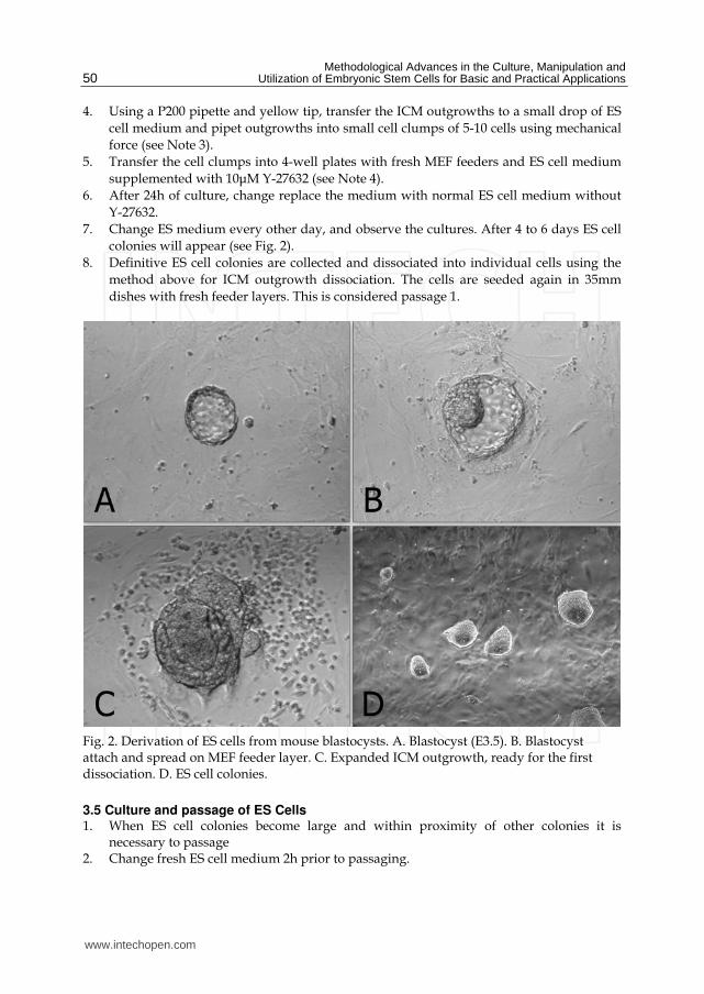
Methodological Advances in the Culture Manipulation and Utilization of Embryonic Stem Cells for Basic and Practical Applications
50
4 Using a P200 pipette and yellow tip transfer the ICM outgrowths to a small drop of ES
cell medium and pipet outgrowths into small cell clumps of 5-10 cells using mechanical
force (see Note 3)
5 Transfer the cell clumps into 4-well plates with fresh MEF feeders and ES cell medium
supplemented with 10μM Y-27632 (see Note 4)
6 After 24h of culture change replace the medium with normal ES cell medium without
Y-27632
7 Change ES medium every other day and observe the cultures After 4 to 6 days ES cell
colonies will appear (see Fig 2)
8 Definitive ES cell colonies are collected and dissociated into individual cells using the
method above for ICM outgrowth dissociation The cells are seeded again in 35mm
dishes with fresh feeder layers This is considered passage 1
Fig 2 Derivation of ES cells from mouse blastocysts A Blastocyst (E35) B Blastocyst attach and spread on MEF feeder layer C Expanded ICM outgrowth ready for the first dissociation D ES cell colonies
35 Culture and passage of ES Cells 1 When ES cell colonies become large and within proximity of other colonies it is
necessary to passage 2 Change fresh ES cell medium 2h prior to passaging
wwwintechopencom
Effective Derivation and Manipulation of Mouse Embryonic Stem Cells
51
3 Aspirate the old medium and wash the cells twice with DPBS 4 Add appropriate amount of 005 trypsinEDTA solution and incubate for 1-2min at
room temperature 5 Once the cell colonies begin to detach carefully remove trypsinEDTA solution and
add 1ml ES cell medium and further dissociate the detached ES colonies into single cell suspension by pipetting several times
6 Adjust the concentration of ES cells and transfer the suspensions into 35mm dishes at a rate of 15
36 Freezing of ES Cells 1 Change fresh ES cell medium 2h prior to freezing 2 Trypsinize cells and harvest as described earlier 3 Collect the cells by centrifugation at 200g for 5min 4 Remove the supernatant and resuspend the pellet in freezing medium 5 Aseptically aliquot the suspension into sterile freezing vials label each vial with the
date and cell typeclone number and place the vials into a thermos cup 6 Freeze the cells overnight at -80degC then transfer to the liquid nitrogen
37 Thawing of ES cells 1 Remove a vial of frozen cells from the liquid nitrogen and transfer to 37degC water bath 2 Transfer cell suspension to a 15ml centrifuge tube and add ES cell medium to 5ml 3 Pellet cells by centrifugation at 200g for 5min 4 Carefully take off the supernatant and resuspend cells in 1ml ES cell medium 5 Plate ES cell suspension onto a prepared feeder layer in a 35mm dish at a suitable
density
38 Karyotyping of ES Cells 1 Add fresh ES cell media containing colcemid at a final concentration of 01μgml to an
exponentially growing ES cell cultures Return to the incubator for 40min 2 Wash slides in fresh fixation solution (31 methanol acetic acid) and then soak them in
ice cold water until ready to use (distilled water plus some ice) It is important for the slides to be both cold and wet when ready for use
3 Wash cells twice with DPBS Completely dissociate colonies into single cell with trypsin Add MEF media and resuspend the cells in a 15ml tube Spin down at 200g for 10min Remove the supernatant
4 Add 1ml 056 KCl dropwise Flick the tube to loose the pellet again to a single cell suspension (no big chucks) Add 9ml of 056 KCl and incubate for 15min
5 Add 2-3 drops of fresh fixation solution to the tube and invert it several times Spin down the cells at 200g for 5min
6 Remove the supernatant Add 1ml fixation solution Flick tube to resuspend pellet add 9ml fixation solution Spin down the cells at 200g for 5min
7 Repeat step 6 three times and resuspend cells in appropriate fixation solution Adjust cell density to 1times106ml
8 Remove slide from the water blot edges to remove excess liquid and drop the cell suspension (dropwise) from at least one foot above the surface of the slide (2-3 dropsslide) Allow the cells spread and then place slides on heat stage (60degC) to speed
wwwintechopencom
Methodological Advances in the Culture Manipulation and Utilization of Embryonic Stem Cells for Basic and Practical Applications
52
up drying) Prepare 3 or 4 slides for each sample The remaining cells can be stored at -20degC for several years
9 Stain slides with Giemsa solution for 15min Rinse slides with running water 10 Photograph chromosome spreads and count (see Fig 3A)
39 Alkaline phosphatase stain Staining for alkaline phosphatase should be performed at room temperature using an alkaline phosphatase detection kit (Boster Bio-Tech AR1023 Wuhan) 1 Aspirate the medium and wash the ES cell cultures twice with DPBS for 1min each 2 Fix ES cells with 4 paraformaldehyde in DPBS at 4degC for 15min 3 Remove paraformaldehyde and add freshly prepared alkaline phosphatase staining
solution 4 Keep reactions in the dark and incubate at room temperature until desired degree of
color development has occurred (This process usually takes 20-40min) 5 Terminate the color reaction by washing with running water 6 Observe the images under an inverted microscope (see Fig 3B)
Fig 3 A Karyotype of ES cells B ES cell colonies with positive alkaline phosphatase activity
310 Immunostaining of ES Cells 1 Seed ES cells on feeder layers in 4- well plate until near confluence
2 Remove media and fix cells with 4 paraformaldehyde for 10min at room temperature
3 Wash with DPBS for three times
4 Add 02 Triton X-100 in DPBS for 5min and then wash with DPBS for three times
5 Add DPBS with 10 FBS for 30min
6 Dilute primary antibodies (rabbit anti-oct-4 polyclonal IgG rabbit anti-nanog
polyclonal IgG rabbit anti-sox-2 polyclonal IgG mouse anti-ssea-1 monoclonal IgG)
(Santa Cruz sc-9081 sc-33760 sc-20088 sc-21702) at 1200 dilution rate in DPBS with
10 FBS
7 Add primary antibodies and incubate cells overnight at 4ordmC refrigerator
8 Wash with DPBS three times
wwwintechopencom
Effective Derivation and Manipulation of Mouse Embryonic Stem Cells
53
9 Dilute second antibodies (donkey anti rabbit IgG-FITC donkey anti mouse IgG-FITC) (Santa Cruz sc-2090 sc-2099) at 1500 dilution rate in DPBS with 10 FBS
10 Add appropriate second antibodies into cells and incubate 2h at room temperate 11 Wash with DPBS three times and observe under an inverted fluorescent microscope
(see Fig 4)
Fig 4 Immunostaining of ES cell pluripotent markers (nanog oct-4 sox-2 ssea-1)
311 Formation of embryoid bodies 1 Trypsinize and dissociate ES cell colonies into single cells 2 Collect by centrifugation and resuspend the cells at a density of 5times105 cellsml in MEF
medium supplemented with 10μM Y-27632 3 Transfer 2ml cell suspension into a 35mm dish coated with 1 agar (see Note 5) 4 Culture overnight at incubator carefully collect cell aggregates and transfer them into a
new 35mm dish coated agar Add 2ml MEF medium without Y-27632 for further culture
5 After 3-5 days many cystic embryoid bodies appear (see Fig5)
312 Formation of teratoma 1 Pretreat ES colonies with 10μM Y-27632 for 1h prior to dissociation 2 Dissociate with Accutase harvest the cells by centrifuge at 200g for 10min and remove
the supernatant
wwwintechopencom
Methodological Advances in the Culture Manipulation and Utilization of Embryonic Stem Cells for Basic and Practical Applications
54
3 Add DPBS and resuspend cells to a final density of 2times106 cellsml 4 Subcutaneously inject 02 ml cell suspension into the CD1 mouse groin (see Note 6) 5 Teratoma will be observed approximately 6 weeks later (see Fig 6)
Fig 5 Cystic embryoid bodies by ES cell suspension culture
Fig 6 Formation of teratoma by injecting ES cells into CD1 mouse groin
313 Production of chimeric mice by aggregation with eight-cell stage embryos 1 Prepare several drops of KSOM culture medium containing 10μM Y-27632 in a 35mm
Petri dish and cover with mineral oil 2 Sterilize a darning needle by dipping in ethanol Make a series of 10 small depressions
in each microdrop by pressing a darning needle into the Petri dish surface (see Note 7) Keep the plate in a 37degC incubator at 5 CO2
3 Oviducts from pregnant female mice (E25) are flushed with M2 to collect eight-cell embryos
4 Place several drops of M2 05 protease and KSOM in 35mm Petri dish (do not mix) 5 Wash the embryos with three drops of protease medium and transfer them into drops
of protease Incubate embryos at 37degC for 5-7min
wwwintechopencom
Effective Derivation and Manipulation of Mouse Embryonic Stem Cells
55
6 Wash embryos three times with KSOM and when zona pellucida begins to disappear transfer them into drops of KSOM
7 Transfer embryos into an aggregation plate (one embryo in each depression) Return plate to the incubator
8 Pull a glass capillary tube using a pipette puller this will be used for ES cell clump preparation
9 Pick ES cell colonies from Petri dish and wash them three times with fresh DPBS 10 Transfer colonies to trypsinEDTA and incubate at 37degC for 3-5min 11 Immediately transfer the colonies into a drop of KSOM and wash them three times 12 Colonies should be dissociated into 15-20 cell clumps using a glass capillary tube and
mechanical force 13 Using a pipette place one clump of ES cells into each depression 14 Return the plate to incubator and culture overnight (see Fig 7) 15 Transfer 8-10 aggregation blastocysts to each uterine horn of pseudopregnant CD1
females (25dpc) The female CD1 mouse will deliver approximately 17 days later
Fig 7 Production of chimeras by aggregation A 8-cell embryos without zona pellucida B C Aggregation depression ES cells and embryos D Aggregate of ES cells and embryo
314 Production of chimeric mice by tetraploid blastocyst injection Production of tetraploid mouse embryos
1 Collect 2-cell embryos (E15) of the CD1 outbred strain by flushing the oviducts and wash them using several drops of M2 medium
wwwintechopencom
Methodological Advances in the Culture Manipulation and Utilization of Embryonic Stem Cells for Basic and Practical Applications
56
2 Transfer them into KSOM drops and keep at incubator until electrofusion 3 Wash 2-cell embryos through several drops of fusion buffer and place 20-30 embryos
into the fusion buffer between the electrodes of the 02mm micro fusion chamber (see Fig 8A)
4 Set Eppendorf Multiporator mode to electrofusion 2V voltage and 15s duration for embryo prealignment 20V voltage 50μs duration and two pulses for fusion
5 Transfer the embryos to the M2 drop and wash them through several drops of M2 medium
6 Transfer the embryos into KSOM drops under mineral oil and culture at 37degC incubator
7 In the next 30-60min choose all properly fused embryos and incubate them in KSOM medium under mineral oil at 37degC 5 CO2 (see Fig 8B)
8 After 24h culture most of embryos should develop to blastocyst stage and are ready to be injected with ES cells
Fig 8 A 2-cell stage embryos aligned in fusion chamber B Fused embryos
Preparation of ES cells for blastocyst injection
1 Replace fresh ES medium 3h prior to injection
2 Pick ES cell colonies and rinse them with DPBS
3 Transfer the colonies into Accutase drops and incubate at 37degC for 5 to 7min
4 Using a P200 pipette transfer the colonies to a small drop of ES medium with 10μM Y-
27632 and break up the colonies into single cell suspension
5 Incubate the single cell suspension with 10μM Y-27632 for 1h at room temperature prior
to injection to remove cell membrane blebs (see Fig 9) (see Note 8)
Microinjection needles preparation using a PN-30 micropipette puller and a MF-900 microforge
1 Turn on the machine and choose appropriate settings For the PN-30 puller use magnet (main) 90 magnet (sub) 30 and heater 80 as the starting values
2 Fix one glass capillary (100mm) on the needle puller 3 Start the pulling process of the needle 4 Remove the needle carefully and check the tip under a stereomicroscope 5 Mount a capillary in the microforge and break this at the required diameter by fusing
the glass onto the glass bead on the microforge and turning off the heat while drawing it away
wwwintechopencom
Effective Derivation and Manipulation of Mouse Embryonic Stem Cells
57
Fig 9 Compare to the control cells without Y-27632 treatment (A) the treatment of Y-27632 make ES cells smooth and soft (B) so that they can easily be picked up by injection pipette
6 For making holding pipets break the holding pipet at a point where the outside
diameter is approx 80μm and the inside diameter approx 60μm and then move the
holding pipet toward the glowing glass bead and the cut will start to melt inward
creating a smoother narrower tip
7 For making injection pipets break the injection pipet at a point where the outside
diameter is approx 15μm and the inside diameter approx 12μm
8 Hold the pipet above the glass bead and heat a point about 5mm from the pipet tip The
pipet will bend under gravity as the glass melts Allow the pipet to bend approx 60deg
Piezo-supported microinjection
1 Prepare dishes for injection using the lids of standard 35mm plastic disposable Petri
dishes
2 Place a 400μl drop of M2 medium on the lid and cover it with mineral oil
3 Fluorinert FC-77 in a 15mm length was back-loaded into an injection pipette Push
Fluorinert FC-77 through the shoulder to near the tip to empty the air in the pipette
4 Attach the injection pipette to the pipette holder of the piezo unit and hang the piezo
unit on the micromanipulator and aspirate 1mm M2 into the pipette to keep ES cells
away from Fluorinert FC-77
5 Attach the holding pipette on the other side of the micromanipulator
6 Transfer embryos and ES cells into the drop of M2 under mineral oil
7 Aspirate several ES cells and use several piezo pulses (eg intensity=3 frequency=3) to
penetrate the zona and trophectoderm layer (see Note 9)
8 Advance the tip of the pipette near the opposite side of the blastocysts from where it is
held by the holding pipette
9 Inject 15-20 ES cells into the blastocoel cavity (see Fig 10A B)
10 Culture injected blastocysts in KSOM medium at 37degC 5 CO2 for 2h and allow them
recovery
11 Transfer 8-10 injected blastocysts to each uterine horn of 25dpc pseudopregnant CD1
femals
12 Recipient mice will give birth after 17 days (see Fig 10C D)
wwwintechopencom
Methodological Advances in the Culture Manipulation and Utilization of Embryonic Stem Cells for Basic and Practical Applications
58
Fig 10 A Piezo-actuated tetraploid blastocyst injection B Injected tetraploid blastocysts C Dead ES mouse pups D Survival ES mouse pups
315 Embryo transfer and Caesarean sections 1 Select a pseudopregnant CD1 female (25dpc) as an embryo recipient 2 Prepare the transfer pipette forceps and scissors etc 3 Anesthetized mice by intraperitoneal injection of Avertin after drug delivery quickly
put the mouse back into the cage (see Note 10) 4 After the mouse is fully anesthetized place the mouse in the prone position on a lid of a
Petri dish Using an absorbent ball with 75 ethanol sterilize the area and shave the animal
5 Make a 05-1cm transverse dorsal incision at the below of the last ribs 6 Uncover incision to find ovarian fat pad pull it out using forceps and hold it using clamps 7 Collect the embryos carefully one by one lined up in the transfer pipette with as little
medium in between as possible 8 Make a hole through the uterine wall into the lumen with a 25G needle The site of this
hole should be near the last loop of the oviduct where it descends into the top of the uterine horn at the UT junction
9 Insert the transfer pipette into a hole and carefully expel the embryos Check the transfer pipette to ensure that all embryos have been transferred 8-10 embryos are usually transferred into each horn
10 When embryos are to be transferred into uteri put the ovary back into the abdominal cavity and suture the incision
wwwintechopencom
Effective Derivation and Manipulation of Mouse Embryonic Stem Cells
59
11 Place the mouse in clean cage Lighting to heat until it regaines consciousness 12 After 17 days the pups will be born
4 Notes
1 Mitomycin C (Sigma M4287) Dissolve the powder into sterile distilled DMEM and adjust the concentration to 10μgml just before use Wash layers several times with DPBS to completely remove any mitomycin C residue from the feeder layers
2 Accutase is a ready to use cell detachment solution of proteolytic and collagenolytic enzymes and it does not contain mammalian or bacterial derived products Cell lines tested for Accutase application includes fibroblasts keratinocytes vascular smooth muscle cells primary chick embryo neuronal cells bone marrow stem cells 293 cells 3T3 cells HeLa cells insect cells human embryonic stem cells and human neural stem cells Moreover Accutase is quite gentle on cells and will not induce cell death if cells are treated a little longer than the optimal time period Thus we use Acutase instead of trypsin for enzymatic dissociation of ICM
3 This is a key step for ES cell isolation Watch the whole dissociation process under a microscope Careful not to pipette with too much force as single cells will not survive Compared to trypsin Accutase treated ICM outgrowths are easier to dissociate into small cell clumps
4 The ROCK inhibitor Y-27632 permits survival of dissociated human embryonic stem cells Similar to human ES cells we find that mouse ICM cells show sensitivity to trypsin They undergo cell death after dissociation and the cloning efficiency of dissociated ICM cells is generally very low Our data indicates that increased cellular adhesion induced by Y-27632 enhances the survival of dissociated ICM cells With more ES-like colonies appearing when the dissociated ICM cells are seeded in medium supplemented with 10 μM Y-27632 for 24h Moreover we have not observed adverse effects of Y-27632 treatment on pluripotency in maintenance culture even after a number of passages
5 Dishes are coated with agar to completely prevent cell attachment This can increase cell aggregation and embryonic body formation In brief add 1g agar into 100ml DPBS and autoclave for 30min Cool to 50degC at room temperature Rapidly cover the internal wall and bottom of 35mm dishes with agar solution Remove the extra solution and place dishes in hood until the remaining agar solidification In addition adding Y-27632 in the embryonic body medium can also enhance suspending cells to aggregate and embryonic body formation
6 Typically severe combined immunodeficient mice are used for making teratoma This strain of mouse is expensive and not easily raised Our data indicates that most mouse strains could be recipient for teratoma production by inguinal subcutaneous injection such as CD1 C57BL6times129Sv
7 Carefully make small depressions using needle and appropriate force in the Petri dish surface Too much force may break the dish and insufficient force will not produce the required depressions which can easily lead to the loss of embryos and ES cells
8 Prior to blastocyst injection the pretreatment of 10μM Y-27632 for 1h at room temperature can alter the membrane architecture of ES cells so that they can be easily collected by injection pipet Otherwise the ES cell surface is covered with blebs (see Fig 10)
9 Within the microinjection pipette ES cells should be kept approximately 100μm away from the pipette tip Damage to ES cells may occur if the cells are very close to the tip when the piezo pulses are applied
wwwintechopencom
Methodological Advances in the Culture Manipulation and Utilization of Embryonic Stem Cells for Basic and Practical Applications
60
10 Avertin anesthetic 125 stocks avertin is prepared by mixing 25g of 2 2 2-tribromoethyl alcohol (Sigma) 5ml of tert-amyl alcohol (Sigma) with 200ml water Avertin is intraperitoneally injected at a dose of 002ml per gram of body weight for anesthetization
5 Acknowledgements
We would like to thank Roman Krawetz (The University of Calgary Canada) for providing his expert and critical reading on the manuscript This work was supported by grants from National Natural Science Foundation of China (No 30871790) and Natural Science Foundation of Hebei (No C2009000600)
6 References
Batlle-Morera L Smith A amp Nichols J (2008) Parameters influencing derivation of embryonic stem cells from murine embryos Genesis 46(12)758-767 ISSN 1526-954X
Brook FA amp Gardner RL (1997) The origin and efficient derivation of embryonic stem cells in the mouse Proc Natl Acad Sci USA 945709-5712 ISSN 0027-8424
Bryja V Bonilla S Cajaacutenek L Parish CL Schwartz CM Luo Y Rao MS amp Arenas E (2006) An efficient method for the derivation of mouse embryonic stem cells Stem Cells 24(4)844-849 ISSN 1066-5099
Cheng J Dutra A Takesono A Garrett-Beal L amp Schwartzberg PL (2004) Improved generation of C57BL6J mouse embryonic stem cells in a defined serum-free media Genesis 39100-104 ISSN 1526-954X
Doungpunta J Santhi A Sathanawongs A Jarujinda Y amp Oranratnachai A (2009) Fivefold increase in derivation rates of mouse embryonic stem cellsafter supplementation of the media with multiple factors Theriogenology 72(2)232-242 ISSN 0093-691X
Evans M J amp Kaufman M H (1981) Establishment in culture of pluripotential cells from mouse embryos Nature 292(5819)154-156 ISSN 0028-0836
Martin G R (1981) Isolation of a pluripotent cell line from early mouse embryos cultured in medium conditioned by teratocarcinoma stem cells Proc Natl Acad Sci USA 787634-7638 ISSN 0027-8424
McWhir J Schnieke AE Ansell R Wallace H Colman A Scott ARamp Kind AJ (1996) Selective ablation of differentiated cells permits isolation of embryonic stem cell lines from murine embryos with a non-permissive genetic background Nat
Genet 14223-226 ISSN 1061-4036 Ruchi B Jacqueline L Min K amp Alexey VT (2008) Efficient propagation of single cells
accutase-dissociated human embryonic stem cells Mol Reprod Dev 75(5)818-827 ISSN 1040-452X
Schoonjans L Kreemers V Danloy S Moreadith RW Laroche Y amp Collen D (2003) Improved generation of germline competent embryonic stem cell lines from inbred mouse strains Stem Cells 21(1)90-97 ISSN 1066-5099
Tesar PJ (2005) Derivation of germ-line-competent embryonic stemcell lines from preblastocyst mouse embryos Proc Natl Acad Sci USA 102(23)8239-8244 ISSN 0027-8424
Watanabe K Ueno M Kamiya D Nishiyama A Matsumura M Wataya T Takahashi JB Nishikawa S Nishikawa S Muguruma K amp Sasai Y (2007) A ROCK inhibitor permits survival of dissociated human embryonic stem cells Nat
Biotechnol 25681-686 ISSN 1087-0156
wwwintechopencom
Methodological Advances in the Culture Manipulation andUtilization of Embryonic Stem Cells for Basic and PracticalApplicationsEdited by Prof Craig Atwood
ISBN 978-953-307-197-8Hard cover 506 pagesPublisher InTechPublished online 26 April 2011Published in print edition April 2011
InTech EuropeUniversity Campus STeP Ri Slavka Krautzeka 83A 51000 Rijeka Croatia Phone +385 (51) 770 447 Fax +385 (51) 686 166wwwintechopencom
InTech ChinaUnit 405 Office Block Hotel Equatorial Shanghai No65 Yan An Road (West) Shanghai 200040 China
Phone +86-21-62489820 Fax +86-21-62489821
Pluripotent stem cells have the potential to revolutionise medicine providing treatment options for a widerange of diseases and conditions that currently lack therapies or cures This book describes methodologicaladvances in the culture and manipulation of embryonic stem cells that will serve to bring this promise topractice
How to referenceIn order to correctly reference this scholarly work feel free to copy and paste the following
Peng Zhang Xinglong Wu Pengbo Wang and Xiangyun Li (2011) Effective Derivation and Manipulation ofMouse Embryonic Stem Cells Methodological Advances in the Culture Manipulation and Utilization ofEmbryonic Stem Cells for Basic and Practical Applications Prof Craig Atwood (Ed) ISBN 978-953-307-197-8 InTech Available from httpwwwintechopencombooksmethodological-advances-in-the-culture-manipulation-and-utilization-of-embryonic-stem-cells-for-basic-and-practical-applicationseffective-derivation-and-manipulation-of-mouse-embryonic-stem-cells

Effective Derivation and Manipulation of Mouse Embryonic Stem Cells
51
3 Aspirate the old medium and wash the cells twice with DPBS 4 Add appropriate amount of 005 trypsinEDTA solution and incubate for 1-2min at
room temperature 5 Once the cell colonies begin to detach carefully remove trypsinEDTA solution and
add 1ml ES cell medium and further dissociate the detached ES colonies into single cell suspension by pipetting several times
6 Adjust the concentration of ES cells and transfer the suspensions into 35mm dishes at a rate of 15
36 Freezing of ES Cells 1 Change fresh ES cell medium 2h prior to freezing 2 Trypsinize cells and harvest as described earlier 3 Collect the cells by centrifugation at 200g for 5min 4 Remove the supernatant and resuspend the pellet in freezing medium 5 Aseptically aliquot the suspension into sterile freezing vials label each vial with the
date and cell typeclone number and place the vials into a thermos cup 6 Freeze the cells overnight at -80degC then transfer to the liquid nitrogen
37 Thawing of ES cells 1 Remove a vial of frozen cells from the liquid nitrogen and transfer to 37degC water bath 2 Transfer cell suspension to a 15ml centrifuge tube and add ES cell medium to 5ml 3 Pellet cells by centrifugation at 200g for 5min 4 Carefully take off the supernatant and resuspend cells in 1ml ES cell medium 5 Plate ES cell suspension onto a prepared feeder layer in a 35mm dish at a suitable
density
38 Karyotyping of ES Cells 1 Add fresh ES cell media containing colcemid at a final concentration of 01μgml to an
exponentially growing ES cell cultures Return to the incubator for 40min 2 Wash slides in fresh fixation solution (31 methanol acetic acid) and then soak them in
ice cold water until ready to use (distilled water plus some ice) It is important for the slides to be both cold and wet when ready for use
3 Wash cells twice with DPBS Completely dissociate colonies into single cell with trypsin Add MEF media and resuspend the cells in a 15ml tube Spin down at 200g for 10min Remove the supernatant
4 Add 1ml 056 KCl dropwise Flick the tube to loose the pellet again to a single cell suspension (no big chucks) Add 9ml of 056 KCl and incubate for 15min
5 Add 2-3 drops of fresh fixation solution to the tube and invert it several times Spin down the cells at 200g for 5min
6 Remove the supernatant Add 1ml fixation solution Flick tube to resuspend pellet add 9ml fixation solution Spin down the cells at 200g for 5min
7 Repeat step 6 three times and resuspend cells in appropriate fixation solution Adjust cell density to 1times106ml
8 Remove slide from the water blot edges to remove excess liquid and drop the cell suspension (dropwise) from at least one foot above the surface of the slide (2-3 dropsslide) Allow the cells spread and then place slides on heat stage (60degC) to speed
wwwintechopencom
Methodological Advances in the Culture Manipulation and Utilization of Embryonic Stem Cells for Basic and Practical Applications
52
up drying) Prepare 3 or 4 slides for each sample The remaining cells can be stored at -20degC for several years
9 Stain slides with Giemsa solution for 15min Rinse slides with running water 10 Photograph chromosome spreads and count (see Fig 3A)
39 Alkaline phosphatase stain Staining for alkaline phosphatase should be performed at room temperature using an alkaline phosphatase detection kit (Boster Bio-Tech AR1023 Wuhan) 1 Aspirate the medium and wash the ES cell cultures twice with DPBS for 1min each 2 Fix ES cells with 4 paraformaldehyde in DPBS at 4degC for 15min 3 Remove paraformaldehyde and add freshly prepared alkaline phosphatase staining
solution 4 Keep reactions in the dark and incubate at room temperature until desired degree of
color development has occurred (This process usually takes 20-40min) 5 Terminate the color reaction by washing with running water 6 Observe the images under an inverted microscope (see Fig 3B)
Fig 3 A Karyotype of ES cells B ES cell colonies with positive alkaline phosphatase activity
310 Immunostaining of ES Cells 1 Seed ES cells on feeder layers in 4- well plate until near confluence
2 Remove media and fix cells with 4 paraformaldehyde for 10min at room temperature
3 Wash with DPBS for three times
4 Add 02 Triton X-100 in DPBS for 5min and then wash with DPBS for three times
5 Add DPBS with 10 FBS for 30min
6 Dilute primary antibodies (rabbit anti-oct-4 polyclonal IgG rabbit anti-nanog
polyclonal IgG rabbit anti-sox-2 polyclonal IgG mouse anti-ssea-1 monoclonal IgG)
(Santa Cruz sc-9081 sc-33760 sc-20088 sc-21702) at 1200 dilution rate in DPBS with
10 FBS
7 Add primary antibodies and incubate cells overnight at 4ordmC refrigerator
8 Wash with DPBS three times
wwwintechopencom
Effective Derivation and Manipulation of Mouse Embryonic Stem Cells
53
9 Dilute second antibodies (donkey anti rabbit IgG-FITC donkey anti mouse IgG-FITC) (Santa Cruz sc-2090 sc-2099) at 1500 dilution rate in DPBS with 10 FBS
10 Add appropriate second antibodies into cells and incubate 2h at room temperate 11 Wash with DPBS three times and observe under an inverted fluorescent microscope
(see Fig 4)
Fig 4 Immunostaining of ES cell pluripotent markers (nanog oct-4 sox-2 ssea-1)
311 Formation of embryoid bodies 1 Trypsinize and dissociate ES cell colonies into single cells 2 Collect by centrifugation and resuspend the cells at a density of 5times105 cellsml in MEF
medium supplemented with 10μM Y-27632 3 Transfer 2ml cell suspension into a 35mm dish coated with 1 agar (see Note 5) 4 Culture overnight at incubator carefully collect cell aggregates and transfer them into a
new 35mm dish coated agar Add 2ml MEF medium without Y-27632 for further culture
5 After 3-5 days many cystic embryoid bodies appear (see Fig5)
312 Formation of teratoma 1 Pretreat ES colonies with 10μM Y-27632 for 1h prior to dissociation 2 Dissociate with Accutase harvest the cells by centrifuge at 200g for 10min and remove
the supernatant
wwwintechopencom
Methodological Advances in the Culture Manipulation and Utilization of Embryonic Stem Cells for Basic and Practical Applications
54
3 Add DPBS and resuspend cells to a final density of 2times106 cellsml 4 Subcutaneously inject 02 ml cell suspension into the CD1 mouse groin (see Note 6) 5 Teratoma will be observed approximately 6 weeks later (see Fig 6)
Fig 5 Cystic embryoid bodies by ES cell suspension culture
Fig 6 Formation of teratoma by injecting ES cells into CD1 mouse groin
313 Production of chimeric mice by aggregation with eight-cell stage embryos 1 Prepare several drops of KSOM culture medium containing 10μM Y-27632 in a 35mm
Petri dish and cover with mineral oil 2 Sterilize a darning needle by dipping in ethanol Make a series of 10 small depressions
in each microdrop by pressing a darning needle into the Petri dish surface (see Note 7) Keep the plate in a 37degC incubator at 5 CO2
3 Oviducts from pregnant female mice (E25) are flushed with M2 to collect eight-cell embryos
4 Place several drops of M2 05 protease and KSOM in 35mm Petri dish (do not mix) 5 Wash the embryos with three drops of protease medium and transfer them into drops
of protease Incubate embryos at 37degC for 5-7min
wwwintechopencom
Effective Derivation and Manipulation of Mouse Embryonic Stem Cells
55
6 Wash embryos three times with KSOM and when zona pellucida begins to disappear transfer them into drops of KSOM
7 Transfer embryos into an aggregation plate (one embryo in each depression) Return plate to the incubator
8 Pull a glass capillary tube using a pipette puller this will be used for ES cell clump preparation
9 Pick ES cell colonies from Petri dish and wash them three times with fresh DPBS 10 Transfer colonies to trypsinEDTA and incubate at 37degC for 3-5min 11 Immediately transfer the colonies into a drop of KSOM and wash them three times 12 Colonies should be dissociated into 15-20 cell clumps using a glass capillary tube and
mechanical force 13 Using a pipette place one clump of ES cells into each depression 14 Return the plate to incubator and culture overnight (see Fig 7) 15 Transfer 8-10 aggregation blastocysts to each uterine horn of pseudopregnant CD1
females (25dpc) The female CD1 mouse will deliver approximately 17 days later
Fig 7 Production of chimeras by aggregation A 8-cell embryos without zona pellucida B C Aggregation depression ES cells and embryos D Aggregate of ES cells and embryo
314 Production of chimeric mice by tetraploid blastocyst injection Production of tetraploid mouse embryos
1 Collect 2-cell embryos (E15) of the CD1 outbred strain by flushing the oviducts and wash them using several drops of M2 medium
wwwintechopencom
Methodological Advances in the Culture Manipulation and Utilization of Embryonic Stem Cells for Basic and Practical Applications
56
2 Transfer them into KSOM drops and keep at incubator until electrofusion 3 Wash 2-cell embryos through several drops of fusion buffer and place 20-30 embryos
into the fusion buffer between the electrodes of the 02mm micro fusion chamber (see Fig 8A)
4 Set Eppendorf Multiporator mode to electrofusion 2V voltage and 15s duration for embryo prealignment 20V voltage 50μs duration and two pulses for fusion
5 Transfer the embryos to the M2 drop and wash them through several drops of M2 medium
6 Transfer the embryos into KSOM drops under mineral oil and culture at 37degC incubator
7 In the next 30-60min choose all properly fused embryos and incubate them in KSOM medium under mineral oil at 37degC 5 CO2 (see Fig 8B)
8 After 24h culture most of embryos should develop to blastocyst stage and are ready to be injected with ES cells
Fig 8 A 2-cell stage embryos aligned in fusion chamber B Fused embryos
Preparation of ES cells for blastocyst injection
1 Replace fresh ES medium 3h prior to injection
2 Pick ES cell colonies and rinse them with DPBS
3 Transfer the colonies into Accutase drops and incubate at 37degC for 5 to 7min
4 Using a P200 pipette transfer the colonies to a small drop of ES medium with 10μM Y-
27632 and break up the colonies into single cell suspension
5 Incubate the single cell suspension with 10μM Y-27632 for 1h at room temperature prior
to injection to remove cell membrane blebs (see Fig 9) (see Note 8)
Microinjection needles preparation using a PN-30 micropipette puller and a MF-900 microforge
1 Turn on the machine and choose appropriate settings For the PN-30 puller use magnet (main) 90 magnet (sub) 30 and heater 80 as the starting values
2 Fix one glass capillary (100mm) on the needle puller 3 Start the pulling process of the needle 4 Remove the needle carefully and check the tip under a stereomicroscope 5 Mount a capillary in the microforge and break this at the required diameter by fusing
the glass onto the glass bead on the microforge and turning off the heat while drawing it away
wwwintechopencom
Effective Derivation and Manipulation of Mouse Embryonic Stem Cells
57
Fig 9 Compare to the control cells without Y-27632 treatment (A) the treatment of Y-27632 make ES cells smooth and soft (B) so that they can easily be picked up by injection pipette
6 For making holding pipets break the holding pipet at a point where the outside
diameter is approx 80μm and the inside diameter approx 60μm and then move the
holding pipet toward the glowing glass bead and the cut will start to melt inward
creating a smoother narrower tip
7 For making injection pipets break the injection pipet at a point where the outside
diameter is approx 15μm and the inside diameter approx 12μm
8 Hold the pipet above the glass bead and heat a point about 5mm from the pipet tip The
pipet will bend under gravity as the glass melts Allow the pipet to bend approx 60deg
Piezo-supported microinjection
1 Prepare dishes for injection using the lids of standard 35mm plastic disposable Petri
dishes
2 Place a 400μl drop of M2 medium on the lid and cover it with mineral oil
3 Fluorinert FC-77 in a 15mm length was back-loaded into an injection pipette Push
Fluorinert FC-77 through the shoulder to near the tip to empty the air in the pipette
4 Attach the injection pipette to the pipette holder of the piezo unit and hang the piezo
unit on the micromanipulator and aspirate 1mm M2 into the pipette to keep ES cells
away from Fluorinert FC-77
5 Attach the holding pipette on the other side of the micromanipulator
6 Transfer embryos and ES cells into the drop of M2 under mineral oil
7 Aspirate several ES cells and use several piezo pulses (eg intensity=3 frequency=3) to
penetrate the zona and trophectoderm layer (see Note 9)
8 Advance the tip of the pipette near the opposite side of the blastocysts from where it is
held by the holding pipette
9 Inject 15-20 ES cells into the blastocoel cavity (see Fig 10A B)
10 Culture injected blastocysts in KSOM medium at 37degC 5 CO2 for 2h and allow them
recovery
11 Transfer 8-10 injected blastocysts to each uterine horn of 25dpc pseudopregnant CD1
femals
12 Recipient mice will give birth after 17 days (see Fig 10C D)
wwwintechopencom
Methodological Advances in the Culture Manipulation and Utilization of Embryonic Stem Cells for Basic and Practical Applications
58
Fig 10 A Piezo-actuated tetraploid blastocyst injection B Injected tetraploid blastocysts C Dead ES mouse pups D Survival ES mouse pups
315 Embryo transfer and Caesarean sections 1 Select a pseudopregnant CD1 female (25dpc) as an embryo recipient 2 Prepare the transfer pipette forceps and scissors etc 3 Anesthetized mice by intraperitoneal injection of Avertin after drug delivery quickly
put the mouse back into the cage (see Note 10) 4 After the mouse is fully anesthetized place the mouse in the prone position on a lid of a
Petri dish Using an absorbent ball with 75 ethanol sterilize the area and shave the animal
5 Make a 05-1cm transverse dorsal incision at the below of the last ribs 6 Uncover incision to find ovarian fat pad pull it out using forceps and hold it using clamps 7 Collect the embryos carefully one by one lined up in the transfer pipette with as little
medium in between as possible 8 Make a hole through the uterine wall into the lumen with a 25G needle The site of this
hole should be near the last loop of the oviduct where it descends into the top of the uterine horn at the UT junction
9 Insert the transfer pipette into a hole and carefully expel the embryos Check the transfer pipette to ensure that all embryos have been transferred 8-10 embryos are usually transferred into each horn
10 When embryos are to be transferred into uteri put the ovary back into the abdominal cavity and suture the incision
wwwintechopencom
Effective Derivation and Manipulation of Mouse Embryonic Stem Cells
59
11 Place the mouse in clean cage Lighting to heat until it regaines consciousness 12 After 17 days the pups will be born
4 Notes
1 Mitomycin C (Sigma M4287) Dissolve the powder into sterile distilled DMEM and adjust the concentration to 10μgml just before use Wash layers several times with DPBS to completely remove any mitomycin C residue from the feeder layers
2 Accutase is a ready to use cell detachment solution of proteolytic and collagenolytic enzymes and it does not contain mammalian or bacterial derived products Cell lines tested for Accutase application includes fibroblasts keratinocytes vascular smooth muscle cells primary chick embryo neuronal cells bone marrow stem cells 293 cells 3T3 cells HeLa cells insect cells human embryonic stem cells and human neural stem cells Moreover Accutase is quite gentle on cells and will not induce cell death if cells are treated a little longer than the optimal time period Thus we use Acutase instead of trypsin for enzymatic dissociation of ICM
3 This is a key step for ES cell isolation Watch the whole dissociation process under a microscope Careful not to pipette with too much force as single cells will not survive Compared to trypsin Accutase treated ICM outgrowths are easier to dissociate into small cell clumps
4 The ROCK inhibitor Y-27632 permits survival of dissociated human embryonic stem cells Similar to human ES cells we find that mouse ICM cells show sensitivity to trypsin They undergo cell death after dissociation and the cloning efficiency of dissociated ICM cells is generally very low Our data indicates that increased cellular adhesion induced by Y-27632 enhances the survival of dissociated ICM cells With more ES-like colonies appearing when the dissociated ICM cells are seeded in medium supplemented with 10 μM Y-27632 for 24h Moreover we have not observed adverse effects of Y-27632 treatment on pluripotency in maintenance culture even after a number of passages
5 Dishes are coated with agar to completely prevent cell attachment This can increase cell aggregation and embryonic body formation In brief add 1g agar into 100ml DPBS and autoclave for 30min Cool to 50degC at room temperature Rapidly cover the internal wall and bottom of 35mm dishes with agar solution Remove the extra solution and place dishes in hood until the remaining agar solidification In addition adding Y-27632 in the embryonic body medium can also enhance suspending cells to aggregate and embryonic body formation
6 Typically severe combined immunodeficient mice are used for making teratoma This strain of mouse is expensive and not easily raised Our data indicates that most mouse strains could be recipient for teratoma production by inguinal subcutaneous injection such as CD1 C57BL6times129Sv
7 Carefully make small depressions using needle and appropriate force in the Petri dish surface Too much force may break the dish and insufficient force will not produce the required depressions which can easily lead to the loss of embryos and ES cells
8 Prior to blastocyst injection the pretreatment of 10μM Y-27632 for 1h at room temperature can alter the membrane architecture of ES cells so that they can be easily collected by injection pipet Otherwise the ES cell surface is covered with blebs (see Fig 10)
9 Within the microinjection pipette ES cells should be kept approximately 100μm away from the pipette tip Damage to ES cells may occur if the cells are very close to the tip when the piezo pulses are applied
wwwintechopencom
Methodological Advances in the Culture Manipulation and Utilization of Embryonic Stem Cells for Basic and Practical Applications
60
10 Avertin anesthetic 125 stocks avertin is prepared by mixing 25g of 2 2 2-tribromoethyl alcohol (Sigma) 5ml of tert-amyl alcohol (Sigma) with 200ml water Avertin is intraperitoneally injected at a dose of 002ml per gram of body weight for anesthetization
5 Acknowledgements
We would like to thank Roman Krawetz (The University of Calgary Canada) for providing his expert and critical reading on the manuscript This work was supported by grants from National Natural Science Foundation of China (No 30871790) and Natural Science Foundation of Hebei (No C2009000600)
6 References
Batlle-Morera L Smith A amp Nichols J (2008) Parameters influencing derivation of embryonic stem cells from murine embryos Genesis 46(12)758-767 ISSN 1526-954X
Brook FA amp Gardner RL (1997) The origin and efficient derivation of embryonic stem cells in the mouse Proc Natl Acad Sci USA 945709-5712 ISSN 0027-8424
Bryja V Bonilla S Cajaacutenek L Parish CL Schwartz CM Luo Y Rao MS amp Arenas E (2006) An efficient method for the derivation of mouse embryonic stem cells Stem Cells 24(4)844-849 ISSN 1066-5099
Cheng J Dutra A Takesono A Garrett-Beal L amp Schwartzberg PL (2004) Improved generation of C57BL6J mouse embryonic stem cells in a defined serum-free media Genesis 39100-104 ISSN 1526-954X
Doungpunta J Santhi A Sathanawongs A Jarujinda Y amp Oranratnachai A (2009) Fivefold increase in derivation rates of mouse embryonic stem cellsafter supplementation of the media with multiple factors Theriogenology 72(2)232-242 ISSN 0093-691X
Evans M J amp Kaufman M H (1981) Establishment in culture of pluripotential cells from mouse embryos Nature 292(5819)154-156 ISSN 0028-0836
Martin G R (1981) Isolation of a pluripotent cell line from early mouse embryos cultured in medium conditioned by teratocarcinoma stem cells Proc Natl Acad Sci USA 787634-7638 ISSN 0027-8424
McWhir J Schnieke AE Ansell R Wallace H Colman A Scott ARamp Kind AJ (1996) Selective ablation of differentiated cells permits isolation of embryonic stem cell lines from murine embryos with a non-permissive genetic background Nat
Genet 14223-226 ISSN 1061-4036 Ruchi B Jacqueline L Min K amp Alexey VT (2008) Efficient propagation of single cells
accutase-dissociated human embryonic stem cells Mol Reprod Dev 75(5)818-827 ISSN 1040-452X
Schoonjans L Kreemers V Danloy S Moreadith RW Laroche Y amp Collen D (2003) Improved generation of germline competent embryonic stem cell lines from inbred mouse strains Stem Cells 21(1)90-97 ISSN 1066-5099
Tesar PJ (2005) Derivation of germ-line-competent embryonic stemcell lines from preblastocyst mouse embryos Proc Natl Acad Sci USA 102(23)8239-8244 ISSN 0027-8424
Watanabe K Ueno M Kamiya D Nishiyama A Matsumura M Wataya T Takahashi JB Nishikawa S Nishikawa S Muguruma K amp Sasai Y (2007) A ROCK inhibitor permits survival of dissociated human embryonic stem cells Nat
Biotechnol 25681-686 ISSN 1087-0156
wwwintechopencom
Methodological Advances in the Culture Manipulation andUtilization of Embryonic Stem Cells for Basic and PracticalApplicationsEdited by Prof Craig Atwood
ISBN 978-953-307-197-8Hard cover 506 pagesPublisher InTechPublished online 26 April 2011Published in print edition April 2011
InTech EuropeUniversity Campus STeP Ri Slavka Krautzeka 83A 51000 Rijeka Croatia Phone +385 (51) 770 447 Fax +385 (51) 686 166wwwintechopencom
InTech ChinaUnit 405 Office Block Hotel Equatorial Shanghai No65 Yan An Road (West) Shanghai 200040 China
Phone +86-21-62489820 Fax +86-21-62489821
Pluripotent stem cells have the potential to revolutionise medicine providing treatment options for a widerange of diseases and conditions that currently lack therapies or cures This book describes methodologicaladvances in the culture and manipulation of embryonic stem cells that will serve to bring this promise topractice
How to referenceIn order to correctly reference this scholarly work feel free to copy and paste the following
Peng Zhang Xinglong Wu Pengbo Wang and Xiangyun Li (2011) Effective Derivation and Manipulation ofMouse Embryonic Stem Cells Methodological Advances in the Culture Manipulation and Utilization ofEmbryonic Stem Cells for Basic and Practical Applications Prof Craig Atwood (Ed) ISBN 978-953-307-197-8 InTech Available from httpwwwintechopencombooksmethodological-advances-in-the-culture-manipulation-and-utilization-of-embryonic-stem-cells-for-basic-and-practical-applicationseffective-derivation-and-manipulation-of-mouse-embryonic-stem-cells
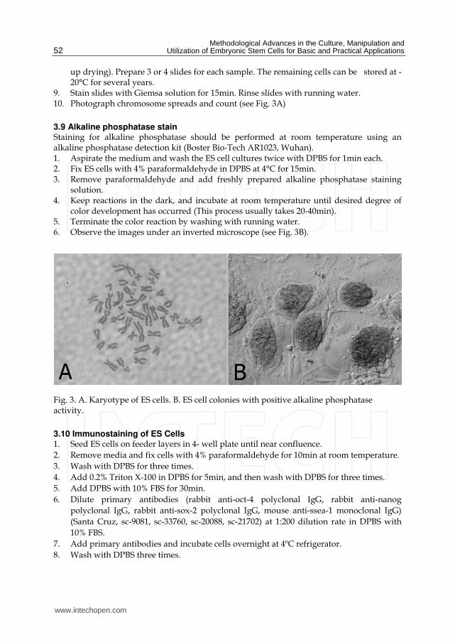
Methodological Advances in the Culture Manipulation and Utilization of Embryonic Stem Cells for Basic and Practical Applications
52
up drying) Prepare 3 or 4 slides for each sample The remaining cells can be stored at -20degC for several years
9 Stain slides with Giemsa solution for 15min Rinse slides with running water 10 Photograph chromosome spreads and count (see Fig 3A)
39 Alkaline phosphatase stain Staining for alkaline phosphatase should be performed at room temperature using an alkaline phosphatase detection kit (Boster Bio-Tech AR1023 Wuhan) 1 Aspirate the medium and wash the ES cell cultures twice with DPBS for 1min each 2 Fix ES cells with 4 paraformaldehyde in DPBS at 4degC for 15min 3 Remove paraformaldehyde and add freshly prepared alkaline phosphatase staining
solution 4 Keep reactions in the dark and incubate at room temperature until desired degree of
color development has occurred (This process usually takes 20-40min) 5 Terminate the color reaction by washing with running water 6 Observe the images under an inverted microscope (see Fig 3B)
Fig 3 A Karyotype of ES cells B ES cell colonies with positive alkaline phosphatase activity
310 Immunostaining of ES Cells 1 Seed ES cells on feeder layers in 4- well plate until near confluence
2 Remove media and fix cells with 4 paraformaldehyde for 10min at room temperature
3 Wash with DPBS for three times
4 Add 02 Triton X-100 in DPBS for 5min and then wash with DPBS for three times
5 Add DPBS with 10 FBS for 30min
6 Dilute primary antibodies (rabbit anti-oct-4 polyclonal IgG rabbit anti-nanog
polyclonal IgG rabbit anti-sox-2 polyclonal IgG mouse anti-ssea-1 monoclonal IgG)
(Santa Cruz sc-9081 sc-33760 sc-20088 sc-21702) at 1200 dilution rate in DPBS with
10 FBS
7 Add primary antibodies and incubate cells overnight at 4ordmC refrigerator
8 Wash with DPBS three times
wwwintechopencom
Effective Derivation and Manipulation of Mouse Embryonic Stem Cells
53
9 Dilute second antibodies (donkey anti rabbit IgG-FITC donkey anti mouse IgG-FITC) (Santa Cruz sc-2090 sc-2099) at 1500 dilution rate in DPBS with 10 FBS
10 Add appropriate second antibodies into cells and incubate 2h at room temperate 11 Wash with DPBS three times and observe under an inverted fluorescent microscope
(see Fig 4)
Fig 4 Immunostaining of ES cell pluripotent markers (nanog oct-4 sox-2 ssea-1)
311 Formation of embryoid bodies 1 Trypsinize and dissociate ES cell colonies into single cells 2 Collect by centrifugation and resuspend the cells at a density of 5times105 cellsml in MEF
medium supplemented with 10μM Y-27632 3 Transfer 2ml cell suspension into a 35mm dish coated with 1 agar (see Note 5) 4 Culture overnight at incubator carefully collect cell aggregates and transfer them into a
new 35mm dish coated agar Add 2ml MEF medium without Y-27632 for further culture
5 After 3-5 days many cystic embryoid bodies appear (see Fig5)
312 Formation of teratoma 1 Pretreat ES colonies with 10μM Y-27632 for 1h prior to dissociation 2 Dissociate with Accutase harvest the cells by centrifuge at 200g for 10min and remove
the supernatant
wwwintechopencom
Methodological Advances in the Culture Manipulation and Utilization of Embryonic Stem Cells for Basic and Practical Applications
54
3 Add DPBS and resuspend cells to a final density of 2times106 cellsml 4 Subcutaneously inject 02 ml cell suspension into the CD1 mouse groin (see Note 6) 5 Teratoma will be observed approximately 6 weeks later (see Fig 6)
Fig 5 Cystic embryoid bodies by ES cell suspension culture
Fig 6 Formation of teratoma by injecting ES cells into CD1 mouse groin
313 Production of chimeric mice by aggregation with eight-cell stage embryos 1 Prepare several drops of KSOM culture medium containing 10μM Y-27632 in a 35mm
Petri dish and cover with mineral oil 2 Sterilize a darning needle by dipping in ethanol Make a series of 10 small depressions
in each microdrop by pressing a darning needle into the Petri dish surface (see Note 7) Keep the plate in a 37degC incubator at 5 CO2
3 Oviducts from pregnant female mice (E25) are flushed with M2 to collect eight-cell embryos
4 Place several drops of M2 05 protease and KSOM in 35mm Petri dish (do not mix) 5 Wash the embryos with three drops of protease medium and transfer them into drops
of protease Incubate embryos at 37degC for 5-7min
wwwintechopencom
Effective Derivation and Manipulation of Mouse Embryonic Stem Cells
55
6 Wash embryos three times with KSOM and when zona pellucida begins to disappear transfer them into drops of KSOM
7 Transfer embryos into an aggregation plate (one embryo in each depression) Return plate to the incubator
8 Pull a glass capillary tube using a pipette puller this will be used for ES cell clump preparation
9 Pick ES cell colonies from Petri dish and wash them three times with fresh DPBS 10 Transfer colonies to trypsinEDTA and incubate at 37degC for 3-5min 11 Immediately transfer the colonies into a drop of KSOM and wash them three times 12 Colonies should be dissociated into 15-20 cell clumps using a glass capillary tube and
mechanical force 13 Using a pipette place one clump of ES cells into each depression 14 Return the plate to incubator and culture overnight (see Fig 7) 15 Transfer 8-10 aggregation blastocysts to each uterine horn of pseudopregnant CD1
females (25dpc) The female CD1 mouse will deliver approximately 17 days later
Fig 7 Production of chimeras by aggregation A 8-cell embryos without zona pellucida B C Aggregation depression ES cells and embryos D Aggregate of ES cells and embryo
314 Production of chimeric mice by tetraploid blastocyst injection Production of tetraploid mouse embryos
1 Collect 2-cell embryos (E15) of the CD1 outbred strain by flushing the oviducts and wash them using several drops of M2 medium
wwwintechopencom
Methodological Advances in the Culture Manipulation and Utilization of Embryonic Stem Cells for Basic and Practical Applications
56
2 Transfer them into KSOM drops and keep at incubator until electrofusion 3 Wash 2-cell embryos through several drops of fusion buffer and place 20-30 embryos
into the fusion buffer between the electrodes of the 02mm micro fusion chamber (see Fig 8A)
4 Set Eppendorf Multiporator mode to electrofusion 2V voltage and 15s duration for embryo prealignment 20V voltage 50μs duration and two pulses for fusion
5 Transfer the embryos to the M2 drop and wash them through several drops of M2 medium
6 Transfer the embryos into KSOM drops under mineral oil and culture at 37degC incubator
7 In the next 30-60min choose all properly fused embryos and incubate them in KSOM medium under mineral oil at 37degC 5 CO2 (see Fig 8B)
8 After 24h culture most of embryos should develop to blastocyst stage and are ready to be injected with ES cells
Fig 8 A 2-cell stage embryos aligned in fusion chamber B Fused embryos
Preparation of ES cells for blastocyst injection
1 Replace fresh ES medium 3h prior to injection
2 Pick ES cell colonies and rinse them with DPBS
3 Transfer the colonies into Accutase drops and incubate at 37degC for 5 to 7min
4 Using a P200 pipette transfer the colonies to a small drop of ES medium with 10μM Y-
27632 and break up the colonies into single cell suspension
5 Incubate the single cell suspension with 10μM Y-27632 for 1h at room temperature prior
to injection to remove cell membrane blebs (see Fig 9) (see Note 8)
Microinjection needles preparation using a PN-30 micropipette puller and a MF-900 microforge
1 Turn on the machine and choose appropriate settings For the PN-30 puller use magnet (main) 90 magnet (sub) 30 and heater 80 as the starting values
2 Fix one glass capillary (100mm) on the needle puller 3 Start the pulling process of the needle 4 Remove the needle carefully and check the tip under a stereomicroscope 5 Mount a capillary in the microforge and break this at the required diameter by fusing
the glass onto the glass bead on the microforge and turning off the heat while drawing it away
wwwintechopencom
Effective Derivation and Manipulation of Mouse Embryonic Stem Cells
57
Fig 9 Compare to the control cells without Y-27632 treatment (A) the treatment of Y-27632 make ES cells smooth and soft (B) so that they can easily be picked up by injection pipette
6 For making holding pipets break the holding pipet at a point where the outside
diameter is approx 80μm and the inside diameter approx 60μm and then move the
holding pipet toward the glowing glass bead and the cut will start to melt inward
creating a smoother narrower tip
7 For making injection pipets break the injection pipet at a point where the outside
diameter is approx 15μm and the inside diameter approx 12μm
8 Hold the pipet above the glass bead and heat a point about 5mm from the pipet tip The
pipet will bend under gravity as the glass melts Allow the pipet to bend approx 60deg
Piezo-supported microinjection
1 Prepare dishes for injection using the lids of standard 35mm plastic disposable Petri
dishes
2 Place a 400μl drop of M2 medium on the lid and cover it with mineral oil
3 Fluorinert FC-77 in a 15mm length was back-loaded into an injection pipette Push
Fluorinert FC-77 through the shoulder to near the tip to empty the air in the pipette
4 Attach the injection pipette to the pipette holder of the piezo unit and hang the piezo
unit on the micromanipulator and aspirate 1mm M2 into the pipette to keep ES cells
away from Fluorinert FC-77
5 Attach the holding pipette on the other side of the micromanipulator
6 Transfer embryos and ES cells into the drop of M2 under mineral oil
7 Aspirate several ES cells and use several piezo pulses (eg intensity=3 frequency=3) to
penetrate the zona and trophectoderm layer (see Note 9)
8 Advance the tip of the pipette near the opposite side of the blastocysts from where it is
held by the holding pipette
9 Inject 15-20 ES cells into the blastocoel cavity (see Fig 10A B)
10 Culture injected blastocysts in KSOM medium at 37degC 5 CO2 for 2h and allow them
recovery
11 Transfer 8-10 injected blastocysts to each uterine horn of 25dpc pseudopregnant CD1
femals
12 Recipient mice will give birth after 17 days (see Fig 10C D)
wwwintechopencom
Methodological Advances in the Culture Manipulation and Utilization of Embryonic Stem Cells for Basic and Practical Applications
58
Fig 10 A Piezo-actuated tetraploid blastocyst injection B Injected tetraploid blastocysts C Dead ES mouse pups D Survival ES mouse pups
315 Embryo transfer and Caesarean sections 1 Select a pseudopregnant CD1 female (25dpc) as an embryo recipient 2 Prepare the transfer pipette forceps and scissors etc 3 Anesthetized mice by intraperitoneal injection of Avertin after drug delivery quickly
put the mouse back into the cage (see Note 10) 4 After the mouse is fully anesthetized place the mouse in the prone position on a lid of a
Petri dish Using an absorbent ball with 75 ethanol sterilize the area and shave the animal
5 Make a 05-1cm transverse dorsal incision at the below of the last ribs 6 Uncover incision to find ovarian fat pad pull it out using forceps and hold it using clamps 7 Collect the embryos carefully one by one lined up in the transfer pipette with as little
medium in between as possible 8 Make a hole through the uterine wall into the lumen with a 25G needle The site of this
hole should be near the last loop of the oviduct where it descends into the top of the uterine horn at the UT junction
9 Insert the transfer pipette into a hole and carefully expel the embryos Check the transfer pipette to ensure that all embryos have been transferred 8-10 embryos are usually transferred into each horn
10 When embryos are to be transferred into uteri put the ovary back into the abdominal cavity and suture the incision
wwwintechopencom
Effective Derivation and Manipulation of Mouse Embryonic Stem Cells
59
11 Place the mouse in clean cage Lighting to heat until it regaines consciousness 12 After 17 days the pups will be born
4 Notes
1 Mitomycin C (Sigma M4287) Dissolve the powder into sterile distilled DMEM and adjust the concentration to 10μgml just before use Wash layers several times with DPBS to completely remove any mitomycin C residue from the feeder layers
2 Accutase is a ready to use cell detachment solution of proteolytic and collagenolytic enzymes and it does not contain mammalian or bacterial derived products Cell lines tested for Accutase application includes fibroblasts keratinocytes vascular smooth muscle cells primary chick embryo neuronal cells bone marrow stem cells 293 cells 3T3 cells HeLa cells insect cells human embryonic stem cells and human neural stem cells Moreover Accutase is quite gentle on cells and will not induce cell death if cells are treated a little longer than the optimal time period Thus we use Acutase instead of trypsin for enzymatic dissociation of ICM
3 This is a key step for ES cell isolation Watch the whole dissociation process under a microscope Careful not to pipette with too much force as single cells will not survive Compared to trypsin Accutase treated ICM outgrowths are easier to dissociate into small cell clumps
4 The ROCK inhibitor Y-27632 permits survival of dissociated human embryonic stem cells Similar to human ES cells we find that mouse ICM cells show sensitivity to trypsin They undergo cell death after dissociation and the cloning efficiency of dissociated ICM cells is generally very low Our data indicates that increased cellular adhesion induced by Y-27632 enhances the survival of dissociated ICM cells With more ES-like colonies appearing when the dissociated ICM cells are seeded in medium supplemented with 10 μM Y-27632 for 24h Moreover we have not observed adverse effects of Y-27632 treatment on pluripotency in maintenance culture even after a number of passages
5 Dishes are coated with agar to completely prevent cell attachment This can increase cell aggregation and embryonic body formation In brief add 1g agar into 100ml DPBS and autoclave for 30min Cool to 50degC at room temperature Rapidly cover the internal wall and bottom of 35mm dishes with agar solution Remove the extra solution and place dishes in hood until the remaining agar solidification In addition adding Y-27632 in the embryonic body medium can also enhance suspending cells to aggregate and embryonic body formation
6 Typically severe combined immunodeficient mice are used for making teratoma This strain of mouse is expensive and not easily raised Our data indicates that most mouse strains could be recipient for teratoma production by inguinal subcutaneous injection such as CD1 C57BL6times129Sv
7 Carefully make small depressions using needle and appropriate force in the Petri dish surface Too much force may break the dish and insufficient force will not produce the required depressions which can easily lead to the loss of embryos and ES cells
8 Prior to blastocyst injection the pretreatment of 10μM Y-27632 for 1h at room temperature can alter the membrane architecture of ES cells so that they can be easily collected by injection pipet Otherwise the ES cell surface is covered with blebs (see Fig 10)
9 Within the microinjection pipette ES cells should be kept approximately 100μm away from the pipette tip Damage to ES cells may occur if the cells are very close to the tip when the piezo pulses are applied
wwwintechopencom
Methodological Advances in the Culture Manipulation and Utilization of Embryonic Stem Cells for Basic and Practical Applications
60
10 Avertin anesthetic 125 stocks avertin is prepared by mixing 25g of 2 2 2-tribromoethyl alcohol (Sigma) 5ml of tert-amyl alcohol (Sigma) with 200ml water Avertin is intraperitoneally injected at a dose of 002ml per gram of body weight for anesthetization
5 Acknowledgements
We would like to thank Roman Krawetz (The University of Calgary Canada) for providing his expert and critical reading on the manuscript This work was supported by grants from National Natural Science Foundation of China (No 30871790) and Natural Science Foundation of Hebei (No C2009000600)
6 References
Batlle-Morera L Smith A amp Nichols J (2008) Parameters influencing derivation of embryonic stem cells from murine embryos Genesis 46(12)758-767 ISSN 1526-954X
Brook FA amp Gardner RL (1997) The origin and efficient derivation of embryonic stem cells in the mouse Proc Natl Acad Sci USA 945709-5712 ISSN 0027-8424
Bryja V Bonilla S Cajaacutenek L Parish CL Schwartz CM Luo Y Rao MS amp Arenas E (2006) An efficient method for the derivation of mouse embryonic stem cells Stem Cells 24(4)844-849 ISSN 1066-5099
Cheng J Dutra A Takesono A Garrett-Beal L amp Schwartzberg PL (2004) Improved generation of C57BL6J mouse embryonic stem cells in a defined serum-free media Genesis 39100-104 ISSN 1526-954X
Doungpunta J Santhi A Sathanawongs A Jarujinda Y amp Oranratnachai A (2009) Fivefold increase in derivation rates of mouse embryonic stem cellsafter supplementation of the media with multiple factors Theriogenology 72(2)232-242 ISSN 0093-691X
Evans M J amp Kaufman M H (1981) Establishment in culture of pluripotential cells from mouse embryos Nature 292(5819)154-156 ISSN 0028-0836
Martin G R (1981) Isolation of a pluripotent cell line from early mouse embryos cultured in medium conditioned by teratocarcinoma stem cells Proc Natl Acad Sci USA 787634-7638 ISSN 0027-8424
McWhir J Schnieke AE Ansell R Wallace H Colman A Scott ARamp Kind AJ (1996) Selective ablation of differentiated cells permits isolation of embryonic stem cell lines from murine embryos with a non-permissive genetic background Nat
Genet 14223-226 ISSN 1061-4036 Ruchi B Jacqueline L Min K amp Alexey VT (2008) Efficient propagation of single cells
accutase-dissociated human embryonic stem cells Mol Reprod Dev 75(5)818-827 ISSN 1040-452X
Schoonjans L Kreemers V Danloy S Moreadith RW Laroche Y amp Collen D (2003) Improved generation of germline competent embryonic stem cell lines from inbred mouse strains Stem Cells 21(1)90-97 ISSN 1066-5099
Tesar PJ (2005) Derivation of germ-line-competent embryonic stemcell lines from preblastocyst mouse embryos Proc Natl Acad Sci USA 102(23)8239-8244 ISSN 0027-8424
Watanabe K Ueno M Kamiya D Nishiyama A Matsumura M Wataya T Takahashi JB Nishikawa S Nishikawa S Muguruma K amp Sasai Y (2007) A ROCK inhibitor permits survival of dissociated human embryonic stem cells Nat
Biotechnol 25681-686 ISSN 1087-0156
wwwintechopencom
Methodological Advances in the Culture Manipulation andUtilization of Embryonic Stem Cells for Basic and PracticalApplicationsEdited by Prof Craig Atwood
ISBN 978-953-307-197-8Hard cover 506 pagesPublisher InTechPublished online 26 April 2011Published in print edition April 2011
InTech EuropeUniversity Campus STeP Ri Slavka Krautzeka 83A 51000 Rijeka Croatia Phone +385 (51) 770 447 Fax +385 (51) 686 166wwwintechopencom
InTech ChinaUnit 405 Office Block Hotel Equatorial Shanghai No65 Yan An Road (West) Shanghai 200040 China
Phone +86-21-62489820 Fax +86-21-62489821
Pluripotent stem cells have the potential to revolutionise medicine providing treatment options for a widerange of diseases and conditions that currently lack therapies or cures This book describes methodologicaladvances in the culture and manipulation of embryonic stem cells that will serve to bring this promise topractice
How to referenceIn order to correctly reference this scholarly work feel free to copy and paste the following
Peng Zhang Xinglong Wu Pengbo Wang and Xiangyun Li (2011) Effective Derivation and Manipulation ofMouse Embryonic Stem Cells Methodological Advances in the Culture Manipulation and Utilization ofEmbryonic Stem Cells for Basic and Practical Applications Prof Craig Atwood (Ed) ISBN 978-953-307-197-8 InTech Available from httpwwwintechopencombooksmethodological-advances-in-the-culture-manipulation-and-utilization-of-embryonic-stem-cells-for-basic-and-practical-applicationseffective-derivation-and-manipulation-of-mouse-embryonic-stem-cells
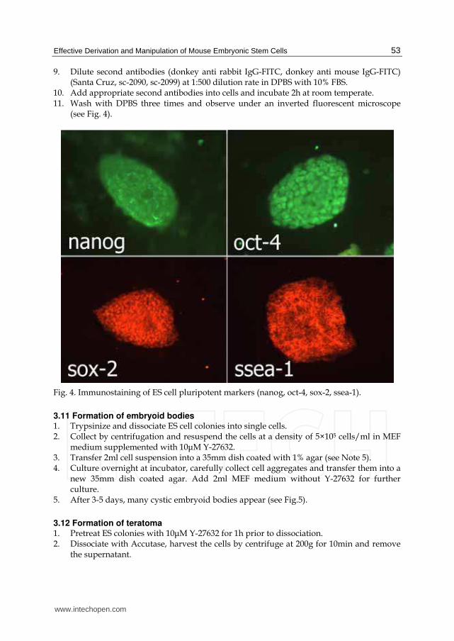
Effective Derivation and Manipulation of Mouse Embryonic Stem Cells
53
9 Dilute second antibodies (donkey anti rabbit IgG-FITC donkey anti mouse IgG-FITC) (Santa Cruz sc-2090 sc-2099) at 1500 dilution rate in DPBS with 10 FBS
10 Add appropriate second antibodies into cells and incubate 2h at room temperate 11 Wash with DPBS three times and observe under an inverted fluorescent microscope
(see Fig 4)
Fig 4 Immunostaining of ES cell pluripotent markers (nanog oct-4 sox-2 ssea-1)
311 Formation of embryoid bodies 1 Trypsinize and dissociate ES cell colonies into single cells 2 Collect by centrifugation and resuspend the cells at a density of 5times105 cellsml in MEF
medium supplemented with 10μM Y-27632 3 Transfer 2ml cell suspension into a 35mm dish coated with 1 agar (see Note 5) 4 Culture overnight at incubator carefully collect cell aggregates and transfer them into a
new 35mm dish coated agar Add 2ml MEF medium without Y-27632 for further culture
5 After 3-5 days many cystic embryoid bodies appear (see Fig5)
312 Formation of teratoma 1 Pretreat ES colonies with 10μM Y-27632 for 1h prior to dissociation 2 Dissociate with Accutase harvest the cells by centrifuge at 200g for 10min and remove
the supernatant
wwwintechopencom
Methodological Advances in the Culture Manipulation and Utilization of Embryonic Stem Cells for Basic and Practical Applications
54
3 Add DPBS and resuspend cells to a final density of 2times106 cellsml 4 Subcutaneously inject 02 ml cell suspension into the CD1 mouse groin (see Note 6) 5 Teratoma will be observed approximately 6 weeks later (see Fig 6)
Fig 5 Cystic embryoid bodies by ES cell suspension culture
Fig 6 Formation of teratoma by injecting ES cells into CD1 mouse groin
313 Production of chimeric mice by aggregation with eight-cell stage embryos 1 Prepare several drops of KSOM culture medium containing 10μM Y-27632 in a 35mm
Petri dish and cover with mineral oil 2 Sterilize a darning needle by dipping in ethanol Make a series of 10 small depressions
in each microdrop by pressing a darning needle into the Petri dish surface (see Note 7) Keep the plate in a 37degC incubator at 5 CO2
3 Oviducts from pregnant female mice (E25) are flushed with M2 to collect eight-cell embryos
4 Place several drops of M2 05 protease and KSOM in 35mm Petri dish (do not mix) 5 Wash the embryos with three drops of protease medium and transfer them into drops
of protease Incubate embryos at 37degC for 5-7min
wwwintechopencom
Effective Derivation and Manipulation of Mouse Embryonic Stem Cells
55
6 Wash embryos three times with KSOM and when zona pellucida begins to disappear transfer them into drops of KSOM
7 Transfer embryos into an aggregation plate (one embryo in each depression) Return plate to the incubator
8 Pull a glass capillary tube using a pipette puller this will be used for ES cell clump preparation
9 Pick ES cell colonies from Petri dish and wash them three times with fresh DPBS 10 Transfer colonies to trypsinEDTA and incubate at 37degC for 3-5min 11 Immediately transfer the colonies into a drop of KSOM and wash them three times 12 Colonies should be dissociated into 15-20 cell clumps using a glass capillary tube and
mechanical force 13 Using a pipette place one clump of ES cells into each depression 14 Return the plate to incubator and culture overnight (see Fig 7) 15 Transfer 8-10 aggregation blastocysts to each uterine horn of pseudopregnant CD1
females (25dpc) The female CD1 mouse will deliver approximately 17 days later
Fig 7 Production of chimeras by aggregation A 8-cell embryos without zona pellucida B C Aggregation depression ES cells and embryos D Aggregate of ES cells and embryo
314 Production of chimeric mice by tetraploid blastocyst injection Production of tetraploid mouse embryos
1 Collect 2-cell embryos (E15) of the CD1 outbred strain by flushing the oviducts and wash them using several drops of M2 medium
wwwintechopencom
Methodological Advances in the Culture Manipulation and Utilization of Embryonic Stem Cells for Basic and Practical Applications
56
2 Transfer them into KSOM drops and keep at incubator until electrofusion 3 Wash 2-cell embryos through several drops of fusion buffer and place 20-30 embryos
into the fusion buffer between the electrodes of the 02mm micro fusion chamber (see Fig 8A)
4 Set Eppendorf Multiporator mode to electrofusion 2V voltage and 15s duration for embryo prealignment 20V voltage 50μs duration and two pulses for fusion
5 Transfer the embryos to the M2 drop and wash them through several drops of M2 medium
6 Transfer the embryos into KSOM drops under mineral oil and culture at 37degC incubator
7 In the next 30-60min choose all properly fused embryos and incubate them in KSOM medium under mineral oil at 37degC 5 CO2 (see Fig 8B)
8 After 24h culture most of embryos should develop to blastocyst stage and are ready to be injected with ES cells
Fig 8 A 2-cell stage embryos aligned in fusion chamber B Fused embryos
Preparation of ES cells for blastocyst injection
1 Replace fresh ES medium 3h prior to injection
2 Pick ES cell colonies and rinse them with DPBS
3 Transfer the colonies into Accutase drops and incubate at 37degC for 5 to 7min
4 Using a P200 pipette transfer the colonies to a small drop of ES medium with 10μM Y-
27632 and break up the colonies into single cell suspension
5 Incubate the single cell suspension with 10μM Y-27632 for 1h at room temperature prior
to injection to remove cell membrane blebs (see Fig 9) (see Note 8)
Microinjection needles preparation using a PN-30 micropipette puller and a MF-900 microforge
1 Turn on the machine and choose appropriate settings For the PN-30 puller use magnet (main) 90 magnet (sub) 30 and heater 80 as the starting values
2 Fix one glass capillary (100mm) on the needle puller 3 Start the pulling process of the needle 4 Remove the needle carefully and check the tip under a stereomicroscope 5 Mount a capillary in the microforge and break this at the required diameter by fusing
the glass onto the glass bead on the microforge and turning off the heat while drawing it away
wwwintechopencom
Effective Derivation and Manipulation of Mouse Embryonic Stem Cells
57
Fig 9 Compare to the control cells without Y-27632 treatment (A) the treatment of Y-27632 make ES cells smooth and soft (B) so that they can easily be picked up by injection pipette
6 For making holding pipets break the holding pipet at a point where the outside
diameter is approx 80μm and the inside diameter approx 60μm and then move the
holding pipet toward the glowing glass bead and the cut will start to melt inward
creating a smoother narrower tip
7 For making injection pipets break the injection pipet at a point where the outside
diameter is approx 15μm and the inside diameter approx 12μm
8 Hold the pipet above the glass bead and heat a point about 5mm from the pipet tip The
pipet will bend under gravity as the glass melts Allow the pipet to bend approx 60deg
Piezo-supported microinjection
1 Prepare dishes for injection using the lids of standard 35mm plastic disposable Petri
dishes
2 Place a 400μl drop of M2 medium on the lid and cover it with mineral oil
3 Fluorinert FC-77 in a 15mm length was back-loaded into an injection pipette Push
Fluorinert FC-77 through the shoulder to near the tip to empty the air in the pipette
4 Attach the injection pipette to the pipette holder of the piezo unit and hang the piezo
unit on the micromanipulator and aspirate 1mm M2 into the pipette to keep ES cells
away from Fluorinert FC-77
5 Attach the holding pipette on the other side of the micromanipulator
6 Transfer embryos and ES cells into the drop of M2 under mineral oil
7 Aspirate several ES cells and use several piezo pulses (eg intensity=3 frequency=3) to
penetrate the zona and trophectoderm layer (see Note 9)
8 Advance the tip of the pipette near the opposite side of the blastocysts from where it is
held by the holding pipette
9 Inject 15-20 ES cells into the blastocoel cavity (see Fig 10A B)
10 Culture injected blastocysts in KSOM medium at 37degC 5 CO2 for 2h and allow them
recovery
11 Transfer 8-10 injected blastocysts to each uterine horn of 25dpc pseudopregnant CD1
femals
12 Recipient mice will give birth after 17 days (see Fig 10C D)
wwwintechopencom
Methodological Advances in the Culture Manipulation and Utilization of Embryonic Stem Cells for Basic and Practical Applications
58
Fig 10 A Piezo-actuated tetraploid blastocyst injection B Injected tetraploid blastocysts C Dead ES mouse pups D Survival ES mouse pups
315 Embryo transfer and Caesarean sections 1 Select a pseudopregnant CD1 female (25dpc) as an embryo recipient 2 Prepare the transfer pipette forceps and scissors etc 3 Anesthetized mice by intraperitoneal injection of Avertin after drug delivery quickly
put the mouse back into the cage (see Note 10) 4 After the mouse is fully anesthetized place the mouse in the prone position on a lid of a
Petri dish Using an absorbent ball with 75 ethanol sterilize the area and shave the animal
5 Make a 05-1cm transverse dorsal incision at the below of the last ribs 6 Uncover incision to find ovarian fat pad pull it out using forceps and hold it using clamps 7 Collect the embryos carefully one by one lined up in the transfer pipette with as little
medium in between as possible 8 Make a hole through the uterine wall into the lumen with a 25G needle The site of this
hole should be near the last loop of the oviduct where it descends into the top of the uterine horn at the UT junction
9 Insert the transfer pipette into a hole and carefully expel the embryos Check the transfer pipette to ensure that all embryos have been transferred 8-10 embryos are usually transferred into each horn
10 When embryos are to be transferred into uteri put the ovary back into the abdominal cavity and suture the incision
wwwintechopencom
Effective Derivation and Manipulation of Mouse Embryonic Stem Cells
59
11 Place the mouse in clean cage Lighting to heat until it regaines consciousness 12 After 17 days the pups will be born
4 Notes
1 Mitomycin C (Sigma M4287) Dissolve the powder into sterile distilled DMEM and adjust the concentration to 10μgml just before use Wash layers several times with DPBS to completely remove any mitomycin C residue from the feeder layers
2 Accutase is a ready to use cell detachment solution of proteolytic and collagenolytic enzymes and it does not contain mammalian or bacterial derived products Cell lines tested for Accutase application includes fibroblasts keratinocytes vascular smooth muscle cells primary chick embryo neuronal cells bone marrow stem cells 293 cells 3T3 cells HeLa cells insect cells human embryonic stem cells and human neural stem cells Moreover Accutase is quite gentle on cells and will not induce cell death if cells are treated a little longer than the optimal time period Thus we use Acutase instead of trypsin for enzymatic dissociation of ICM
3 This is a key step for ES cell isolation Watch the whole dissociation process under a microscope Careful not to pipette with too much force as single cells will not survive Compared to trypsin Accutase treated ICM outgrowths are easier to dissociate into small cell clumps
4 The ROCK inhibitor Y-27632 permits survival of dissociated human embryonic stem cells Similar to human ES cells we find that mouse ICM cells show sensitivity to trypsin They undergo cell death after dissociation and the cloning efficiency of dissociated ICM cells is generally very low Our data indicates that increased cellular adhesion induced by Y-27632 enhances the survival of dissociated ICM cells With more ES-like colonies appearing when the dissociated ICM cells are seeded in medium supplemented with 10 μM Y-27632 for 24h Moreover we have not observed adverse effects of Y-27632 treatment on pluripotency in maintenance culture even after a number of passages
5 Dishes are coated with agar to completely prevent cell attachment This can increase cell aggregation and embryonic body formation In brief add 1g agar into 100ml DPBS and autoclave for 30min Cool to 50degC at room temperature Rapidly cover the internal wall and bottom of 35mm dishes with agar solution Remove the extra solution and place dishes in hood until the remaining agar solidification In addition adding Y-27632 in the embryonic body medium can also enhance suspending cells to aggregate and embryonic body formation
6 Typically severe combined immunodeficient mice are used for making teratoma This strain of mouse is expensive and not easily raised Our data indicates that most mouse strains could be recipient for teratoma production by inguinal subcutaneous injection such as CD1 C57BL6times129Sv
7 Carefully make small depressions using needle and appropriate force in the Petri dish surface Too much force may break the dish and insufficient force will not produce the required depressions which can easily lead to the loss of embryos and ES cells
8 Prior to blastocyst injection the pretreatment of 10μM Y-27632 for 1h at room temperature can alter the membrane architecture of ES cells so that they can be easily collected by injection pipet Otherwise the ES cell surface is covered with blebs (see Fig 10)
9 Within the microinjection pipette ES cells should be kept approximately 100μm away from the pipette tip Damage to ES cells may occur if the cells are very close to the tip when the piezo pulses are applied
wwwintechopencom
Methodological Advances in the Culture Manipulation and Utilization of Embryonic Stem Cells for Basic and Practical Applications
60
10 Avertin anesthetic 125 stocks avertin is prepared by mixing 25g of 2 2 2-tribromoethyl alcohol (Sigma) 5ml of tert-amyl alcohol (Sigma) with 200ml water Avertin is intraperitoneally injected at a dose of 002ml per gram of body weight for anesthetization
5 Acknowledgements
We would like to thank Roman Krawetz (The University of Calgary Canada) for providing his expert and critical reading on the manuscript This work was supported by grants from National Natural Science Foundation of China (No 30871790) and Natural Science Foundation of Hebei (No C2009000600)
6 References
Batlle-Morera L Smith A amp Nichols J (2008) Parameters influencing derivation of embryonic stem cells from murine embryos Genesis 46(12)758-767 ISSN 1526-954X
Brook FA amp Gardner RL (1997) The origin and efficient derivation of embryonic stem cells in the mouse Proc Natl Acad Sci USA 945709-5712 ISSN 0027-8424
Bryja V Bonilla S Cajaacutenek L Parish CL Schwartz CM Luo Y Rao MS amp Arenas E (2006) An efficient method for the derivation of mouse embryonic stem cells Stem Cells 24(4)844-849 ISSN 1066-5099
Cheng J Dutra A Takesono A Garrett-Beal L amp Schwartzberg PL (2004) Improved generation of C57BL6J mouse embryonic stem cells in a defined serum-free media Genesis 39100-104 ISSN 1526-954X
Doungpunta J Santhi A Sathanawongs A Jarujinda Y amp Oranratnachai A (2009) Fivefold increase in derivation rates of mouse embryonic stem cellsafter supplementation of the media with multiple factors Theriogenology 72(2)232-242 ISSN 0093-691X
Evans M J amp Kaufman M H (1981) Establishment in culture of pluripotential cells from mouse embryos Nature 292(5819)154-156 ISSN 0028-0836
Martin G R (1981) Isolation of a pluripotent cell line from early mouse embryos cultured in medium conditioned by teratocarcinoma stem cells Proc Natl Acad Sci USA 787634-7638 ISSN 0027-8424
McWhir J Schnieke AE Ansell R Wallace H Colman A Scott ARamp Kind AJ (1996) Selective ablation of differentiated cells permits isolation of embryonic stem cell lines from murine embryos with a non-permissive genetic background Nat
Genet 14223-226 ISSN 1061-4036 Ruchi B Jacqueline L Min K amp Alexey VT (2008) Efficient propagation of single cells
accutase-dissociated human embryonic stem cells Mol Reprod Dev 75(5)818-827 ISSN 1040-452X
Schoonjans L Kreemers V Danloy S Moreadith RW Laroche Y amp Collen D (2003) Improved generation of germline competent embryonic stem cell lines from inbred mouse strains Stem Cells 21(1)90-97 ISSN 1066-5099
Tesar PJ (2005) Derivation of germ-line-competent embryonic stemcell lines from preblastocyst mouse embryos Proc Natl Acad Sci USA 102(23)8239-8244 ISSN 0027-8424
Watanabe K Ueno M Kamiya D Nishiyama A Matsumura M Wataya T Takahashi JB Nishikawa S Nishikawa S Muguruma K amp Sasai Y (2007) A ROCK inhibitor permits survival of dissociated human embryonic stem cells Nat
Biotechnol 25681-686 ISSN 1087-0156
wwwintechopencom
Methodological Advances in the Culture Manipulation andUtilization of Embryonic Stem Cells for Basic and PracticalApplicationsEdited by Prof Craig Atwood
ISBN 978-953-307-197-8Hard cover 506 pagesPublisher InTechPublished online 26 April 2011Published in print edition April 2011
InTech EuropeUniversity Campus STeP Ri Slavka Krautzeka 83A 51000 Rijeka Croatia Phone +385 (51) 770 447 Fax +385 (51) 686 166wwwintechopencom
InTech ChinaUnit 405 Office Block Hotel Equatorial Shanghai No65 Yan An Road (West) Shanghai 200040 China
Phone +86-21-62489820 Fax +86-21-62489821
Pluripotent stem cells have the potential to revolutionise medicine providing treatment options for a widerange of diseases and conditions that currently lack therapies or cures This book describes methodologicaladvances in the culture and manipulation of embryonic stem cells that will serve to bring this promise topractice
How to referenceIn order to correctly reference this scholarly work feel free to copy and paste the following
Peng Zhang Xinglong Wu Pengbo Wang and Xiangyun Li (2011) Effective Derivation and Manipulation ofMouse Embryonic Stem Cells Methodological Advances in the Culture Manipulation and Utilization ofEmbryonic Stem Cells for Basic and Practical Applications Prof Craig Atwood (Ed) ISBN 978-953-307-197-8 InTech Available from httpwwwintechopencombooksmethodological-advances-in-the-culture-manipulation-and-utilization-of-embryonic-stem-cells-for-basic-and-practical-applicationseffective-derivation-and-manipulation-of-mouse-embryonic-stem-cells
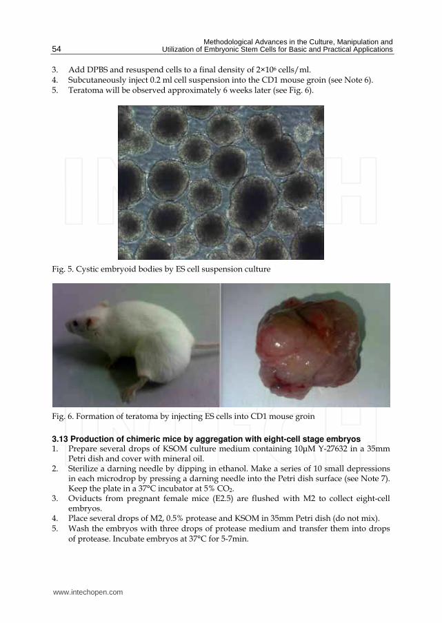
Methodological Advances in the Culture Manipulation and Utilization of Embryonic Stem Cells for Basic and Practical Applications
54
3 Add DPBS and resuspend cells to a final density of 2times106 cellsml 4 Subcutaneously inject 02 ml cell suspension into the CD1 mouse groin (see Note 6) 5 Teratoma will be observed approximately 6 weeks later (see Fig 6)
Fig 5 Cystic embryoid bodies by ES cell suspension culture
Fig 6 Formation of teratoma by injecting ES cells into CD1 mouse groin
313 Production of chimeric mice by aggregation with eight-cell stage embryos 1 Prepare several drops of KSOM culture medium containing 10μM Y-27632 in a 35mm
Petri dish and cover with mineral oil 2 Sterilize a darning needle by dipping in ethanol Make a series of 10 small depressions
in each microdrop by pressing a darning needle into the Petri dish surface (see Note 7) Keep the plate in a 37degC incubator at 5 CO2
3 Oviducts from pregnant female mice (E25) are flushed with M2 to collect eight-cell embryos
4 Place several drops of M2 05 protease and KSOM in 35mm Petri dish (do not mix) 5 Wash the embryos with three drops of protease medium and transfer them into drops
of protease Incubate embryos at 37degC for 5-7min
wwwintechopencom
Effective Derivation and Manipulation of Mouse Embryonic Stem Cells
55
6 Wash embryos three times with KSOM and when zona pellucida begins to disappear transfer them into drops of KSOM
7 Transfer embryos into an aggregation plate (one embryo in each depression) Return plate to the incubator
8 Pull a glass capillary tube using a pipette puller this will be used for ES cell clump preparation
9 Pick ES cell colonies from Petri dish and wash them three times with fresh DPBS 10 Transfer colonies to trypsinEDTA and incubate at 37degC for 3-5min 11 Immediately transfer the colonies into a drop of KSOM and wash them three times 12 Colonies should be dissociated into 15-20 cell clumps using a glass capillary tube and
mechanical force 13 Using a pipette place one clump of ES cells into each depression 14 Return the plate to incubator and culture overnight (see Fig 7) 15 Transfer 8-10 aggregation blastocysts to each uterine horn of pseudopregnant CD1
females (25dpc) The female CD1 mouse will deliver approximately 17 days later
Fig 7 Production of chimeras by aggregation A 8-cell embryos without zona pellucida B C Aggregation depression ES cells and embryos D Aggregate of ES cells and embryo
314 Production of chimeric mice by tetraploid blastocyst injection Production of tetraploid mouse embryos
1 Collect 2-cell embryos (E15) of the CD1 outbred strain by flushing the oviducts and wash them using several drops of M2 medium
wwwintechopencom
Methodological Advances in the Culture Manipulation and Utilization of Embryonic Stem Cells for Basic and Practical Applications
56
2 Transfer them into KSOM drops and keep at incubator until electrofusion 3 Wash 2-cell embryos through several drops of fusion buffer and place 20-30 embryos
into the fusion buffer between the electrodes of the 02mm micro fusion chamber (see Fig 8A)
4 Set Eppendorf Multiporator mode to electrofusion 2V voltage and 15s duration for embryo prealignment 20V voltage 50μs duration and two pulses for fusion
5 Transfer the embryos to the M2 drop and wash them through several drops of M2 medium
6 Transfer the embryos into KSOM drops under mineral oil and culture at 37degC incubator
7 In the next 30-60min choose all properly fused embryos and incubate them in KSOM medium under mineral oil at 37degC 5 CO2 (see Fig 8B)
8 After 24h culture most of embryos should develop to blastocyst stage and are ready to be injected with ES cells
Fig 8 A 2-cell stage embryos aligned in fusion chamber B Fused embryos
Preparation of ES cells for blastocyst injection
1 Replace fresh ES medium 3h prior to injection
2 Pick ES cell colonies and rinse them with DPBS
3 Transfer the colonies into Accutase drops and incubate at 37degC for 5 to 7min
4 Using a P200 pipette transfer the colonies to a small drop of ES medium with 10μM Y-
27632 and break up the colonies into single cell suspension
5 Incubate the single cell suspension with 10μM Y-27632 for 1h at room temperature prior
to injection to remove cell membrane blebs (see Fig 9) (see Note 8)
Microinjection needles preparation using a PN-30 micropipette puller and a MF-900 microforge
1 Turn on the machine and choose appropriate settings For the PN-30 puller use magnet (main) 90 magnet (sub) 30 and heater 80 as the starting values
2 Fix one glass capillary (100mm) on the needle puller 3 Start the pulling process of the needle 4 Remove the needle carefully and check the tip under a stereomicroscope 5 Mount a capillary in the microforge and break this at the required diameter by fusing
the glass onto the glass bead on the microforge and turning off the heat while drawing it away
wwwintechopencom
Effective Derivation and Manipulation of Mouse Embryonic Stem Cells
57
Fig 9 Compare to the control cells without Y-27632 treatment (A) the treatment of Y-27632 make ES cells smooth and soft (B) so that they can easily be picked up by injection pipette
6 For making holding pipets break the holding pipet at a point where the outside
diameter is approx 80μm and the inside diameter approx 60μm and then move the
holding pipet toward the glowing glass bead and the cut will start to melt inward
creating a smoother narrower tip
7 For making injection pipets break the injection pipet at a point where the outside
diameter is approx 15μm and the inside diameter approx 12μm
8 Hold the pipet above the glass bead and heat a point about 5mm from the pipet tip The
pipet will bend under gravity as the glass melts Allow the pipet to bend approx 60deg
Piezo-supported microinjection
1 Prepare dishes for injection using the lids of standard 35mm plastic disposable Petri
dishes
2 Place a 400μl drop of M2 medium on the lid and cover it with mineral oil
3 Fluorinert FC-77 in a 15mm length was back-loaded into an injection pipette Push
Fluorinert FC-77 through the shoulder to near the tip to empty the air in the pipette
4 Attach the injection pipette to the pipette holder of the piezo unit and hang the piezo
unit on the micromanipulator and aspirate 1mm M2 into the pipette to keep ES cells
away from Fluorinert FC-77
5 Attach the holding pipette on the other side of the micromanipulator
6 Transfer embryos and ES cells into the drop of M2 under mineral oil
7 Aspirate several ES cells and use several piezo pulses (eg intensity=3 frequency=3) to
penetrate the zona and trophectoderm layer (see Note 9)
8 Advance the tip of the pipette near the opposite side of the blastocysts from where it is
held by the holding pipette
9 Inject 15-20 ES cells into the blastocoel cavity (see Fig 10A B)
10 Culture injected blastocysts in KSOM medium at 37degC 5 CO2 for 2h and allow them
recovery
11 Transfer 8-10 injected blastocysts to each uterine horn of 25dpc pseudopregnant CD1
femals
12 Recipient mice will give birth after 17 days (see Fig 10C D)
wwwintechopencom
Methodological Advances in the Culture Manipulation and Utilization of Embryonic Stem Cells for Basic and Practical Applications
58
Fig 10 A Piezo-actuated tetraploid blastocyst injection B Injected tetraploid blastocysts C Dead ES mouse pups D Survival ES mouse pups
315 Embryo transfer and Caesarean sections 1 Select a pseudopregnant CD1 female (25dpc) as an embryo recipient 2 Prepare the transfer pipette forceps and scissors etc 3 Anesthetized mice by intraperitoneal injection of Avertin after drug delivery quickly
put the mouse back into the cage (see Note 10) 4 After the mouse is fully anesthetized place the mouse in the prone position on a lid of a
Petri dish Using an absorbent ball with 75 ethanol sterilize the area and shave the animal
5 Make a 05-1cm transverse dorsal incision at the below of the last ribs 6 Uncover incision to find ovarian fat pad pull it out using forceps and hold it using clamps 7 Collect the embryos carefully one by one lined up in the transfer pipette with as little
medium in between as possible 8 Make a hole through the uterine wall into the lumen with a 25G needle The site of this
hole should be near the last loop of the oviduct where it descends into the top of the uterine horn at the UT junction
9 Insert the transfer pipette into a hole and carefully expel the embryos Check the transfer pipette to ensure that all embryos have been transferred 8-10 embryos are usually transferred into each horn
10 When embryos are to be transferred into uteri put the ovary back into the abdominal cavity and suture the incision
wwwintechopencom
Effective Derivation and Manipulation of Mouse Embryonic Stem Cells
59
11 Place the mouse in clean cage Lighting to heat until it regaines consciousness 12 After 17 days the pups will be born
4 Notes
1 Mitomycin C (Sigma M4287) Dissolve the powder into sterile distilled DMEM and adjust the concentration to 10μgml just before use Wash layers several times with DPBS to completely remove any mitomycin C residue from the feeder layers
2 Accutase is a ready to use cell detachment solution of proteolytic and collagenolytic enzymes and it does not contain mammalian or bacterial derived products Cell lines tested for Accutase application includes fibroblasts keratinocytes vascular smooth muscle cells primary chick embryo neuronal cells bone marrow stem cells 293 cells 3T3 cells HeLa cells insect cells human embryonic stem cells and human neural stem cells Moreover Accutase is quite gentle on cells and will not induce cell death if cells are treated a little longer than the optimal time period Thus we use Acutase instead of trypsin for enzymatic dissociation of ICM
3 This is a key step for ES cell isolation Watch the whole dissociation process under a microscope Careful not to pipette with too much force as single cells will not survive Compared to trypsin Accutase treated ICM outgrowths are easier to dissociate into small cell clumps
4 The ROCK inhibitor Y-27632 permits survival of dissociated human embryonic stem cells Similar to human ES cells we find that mouse ICM cells show sensitivity to trypsin They undergo cell death after dissociation and the cloning efficiency of dissociated ICM cells is generally very low Our data indicates that increased cellular adhesion induced by Y-27632 enhances the survival of dissociated ICM cells With more ES-like colonies appearing when the dissociated ICM cells are seeded in medium supplemented with 10 μM Y-27632 for 24h Moreover we have not observed adverse effects of Y-27632 treatment on pluripotency in maintenance culture even after a number of passages
5 Dishes are coated with agar to completely prevent cell attachment This can increase cell aggregation and embryonic body formation In brief add 1g agar into 100ml DPBS and autoclave for 30min Cool to 50degC at room temperature Rapidly cover the internal wall and bottom of 35mm dishes with agar solution Remove the extra solution and place dishes in hood until the remaining agar solidification In addition adding Y-27632 in the embryonic body medium can also enhance suspending cells to aggregate and embryonic body formation
6 Typically severe combined immunodeficient mice are used for making teratoma This strain of mouse is expensive and not easily raised Our data indicates that most mouse strains could be recipient for teratoma production by inguinal subcutaneous injection such as CD1 C57BL6times129Sv
7 Carefully make small depressions using needle and appropriate force in the Petri dish surface Too much force may break the dish and insufficient force will not produce the required depressions which can easily lead to the loss of embryos and ES cells
8 Prior to blastocyst injection the pretreatment of 10μM Y-27632 for 1h at room temperature can alter the membrane architecture of ES cells so that they can be easily collected by injection pipet Otherwise the ES cell surface is covered with blebs (see Fig 10)
9 Within the microinjection pipette ES cells should be kept approximately 100μm away from the pipette tip Damage to ES cells may occur if the cells are very close to the tip when the piezo pulses are applied
wwwintechopencom
Methodological Advances in the Culture Manipulation and Utilization of Embryonic Stem Cells for Basic and Practical Applications
60
10 Avertin anesthetic 125 stocks avertin is prepared by mixing 25g of 2 2 2-tribromoethyl alcohol (Sigma) 5ml of tert-amyl alcohol (Sigma) with 200ml water Avertin is intraperitoneally injected at a dose of 002ml per gram of body weight for anesthetization
5 Acknowledgements
We would like to thank Roman Krawetz (The University of Calgary Canada) for providing his expert and critical reading on the manuscript This work was supported by grants from National Natural Science Foundation of China (No 30871790) and Natural Science Foundation of Hebei (No C2009000600)
6 References
Batlle-Morera L Smith A amp Nichols J (2008) Parameters influencing derivation of embryonic stem cells from murine embryos Genesis 46(12)758-767 ISSN 1526-954X
Brook FA amp Gardner RL (1997) The origin and efficient derivation of embryonic stem cells in the mouse Proc Natl Acad Sci USA 945709-5712 ISSN 0027-8424
Bryja V Bonilla S Cajaacutenek L Parish CL Schwartz CM Luo Y Rao MS amp Arenas E (2006) An efficient method for the derivation of mouse embryonic stem cells Stem Cells 24(4)844-849 ISSN 1066-5099
Cheng J Dutra A Takesono A Garrett-Beal L amp Schwartzberg PL (2004) Improved generation of C57BL6J mouse embryonic stem cells in a defined serum-free media Genesis 39100-104 ISSN 1526-954X
Doungpunta J Santhi A Sathanawongs A Jarujinda Y amp Oranratnachai A (2009) Fivefold increase in derivation rates of mouse embryonic stem cellsafter supplementation of the media with multiple factors Theriogenology 72(2)232-242 ISSN 0093-691X
Evans M J amp Kaufman M H (1981) Establishment in culture of pluripotential cells from mouse embryos Nature 292(5819)154-156 ISSN 0028-0836
Martin G R (1981) Isolation of a pluripotent cell line from early mouse embryos cultured in medium conditioned by teratocarcinoma stem cells Proc Natl Acad Sci USA 787634-7638 ISSN 0027-8424
McWhir J Schnieke AE Ansell R Wallace H Colman A Scott ARamp Kind AJ (1996) Selective ablation of differentiated cells permits isolation of embryonic stem cell lines from murine embryos with a non-permissive genetic background Nat
Genet 14223-226 ISSN 1061-4036 Ruchi B Jacqueline L Min K amp Alexey VT (2008) Efficient propagation of single cells
accutase-dissociated human embryonic stem cells Mol Reprod Dev 75(5)818-827 ISSN 1040-452X
Schoonjans L Kreemers V Danloy S Moreadith RW Laroche Y amp Collen D (2003) Improved generation of germline competent embryonic stem cell lines from inbred mouse strains Stem Cells 21(1)90-97 ISSN 1066-5099
Tesar PJ (2005) Derivation of germ-line-competent embryonic stemcell lines from preblastocyst mouse embryos Proc Natl Acad Sci USA 102(23)8239-8244 ISSN 0027-8424
Watanabe K Ueno M Kamiya D Nishiyama A Matsumura M Wataya T Takahashi JB Nishikawa S Nishikawa S Muguruma K amp Sasai Y (2007) A ROCK inhibitor permits survival of dissociated human embryonic stem cells Nat
Biotechnol 25681-686 ISSN 1087-0156
wwwintechopencom
Methodological Advances in the Culture Manipulation andUtilization of Embryonic Stem Cells for Basic and PracticalApplicationsEdited by Prof Craig Atwood
ISBN 978-953-307-197-8Hard cover 506 pagesPublisher InTechPublished online 26 April 2011Published in print edition April 2011
InTech EuropeUniversity Campus STeP Ri Slavka Krautzeka 83A 51000 Rijeka Croatia Phone +385 (51) 770 447 Fax +385 (51) 686 166wwwintechopencom
InTech ChinaUnit 405 Office Block Hotel Equatorial Shanghai No65 Yan An Road (West) Shanghai 200040 China
Phone +86-21-62489820 Fax +86-21-62489821
Pluripotent stem cells have the potential to revolutionise medicine providing treatment options for a widerange of diseases and conditions that currently lack therapies or cures This book describes methodologicaladvances in the culture and manipulation of embryonic stem cells that will serve to bring this promise topractice
How to referenceIn order to correctly reference this scholarly work feel free to copy and paste the following
Peng Zhang Xinglong Wu Pengbo Wang and Xiangyun Li (2011) Effective Derivation and Manipulation ofMouse Embryonic Stem Cells Methodological Advances in the Culture Manipulation and Utilization ofEmbryonic Stem Cells for Basic and Practical Applications Prof Craig Atwood (Ed) ISBN 978-953-307-197-8 InTech Available from httpwwwintechopencombooksmethodological-advances-in-the-culture-manipulation-and-utilization-of-embryonic-stem-cells-for-basic-and-practical-applicationseffective-derivation-and-manipulation-of-mouse-embryonic-stem-cells
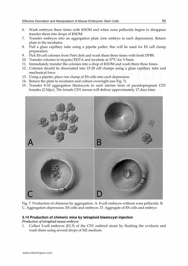
Effective Derivation and Manipulation of Mouse Embryonic Stem Cells
55
6 Wash embryos three times with KSOM and when zona pellucida begins to disappear transfer them into drops of KSOM
7 Transfer embryos into an aggregation plate (one embryo in each depression) Return plate to the incubator
8 Pull a glass capillary tube using a pipette puller this will be used for ES cell clump preparation
9 Pick ES cell colonies from Petri dish and wash them three times with fresh DPBS 10 Transfer colonies to trypsinEDTA and incubate at 37degC for 3-5min 11 Immediately transfer the colonies into a drop of KSOM and wash them three times 12 Colonies should be dissociated into 15-20 cell clumps using a glass capillary tube and
mechanical force 13 Using a pipette place one clump of ES cells into each depression 14 Return the plate to incubator and culture overnight (see Fig 7) 15 Transfer 8-10 aggregation blastocysts to each uterine horn of pseudopregnant CD1
females (25dpc) The female CD1 mouse will deliver approximately 17 days later
Fig 7 Production of chimeras by aggregation A 8-cell embryos without zona pellucida B C Aggregation depression ES cells and embryos D Aggregate of ES cells and embryo
314 Production of chimeric mice by tetraploid blastocyst injection Production of tetraploid mouse embryos
1 Collect 2-cell embryos (E15) of the CD1 outbred strain by flushing the oviducts and wash them using several drops of M2 medium
wwwintechopencom
Methodological Advances in the Culture Manipulation and Utilization of Embryonic Stem Cells for Basic and Practical Applications
56
2 Transfer them into KSOM drops and keep at incubator until electrofusion 3 Wash 2-cell embryos through several drops of fusion buffer and place 20-30 embryos
into the fusion buffer between the electrodes of the 02mm micro fusion chamber (see Fig 8A)
4 Set Eppendorf Multiporator mode to electrofusion 2V voltage and 15s duration for embryo prealignment 20V voltage 50μs duration and two pulses for fusion
5 Transfer the embryos to the M2 drop and wash them through several drops of M2 medium
6 Transfer the embryos into KSOM drops under mineral oil and culture at 37degC incubator
7 In the next 30-60min choose all properly fused embryos and incubate them in KSOM medium under mineral oil at 37degC 5 CO2 (see Fig 8B)
8 After 24h culture most of embryos should develop to blastocyst stage and are ready to be injected with ES cells
Fig 8 A 2-cell stage embryos aligned in fusion chamber B Fused embryos
Preparation of ES cells for blastocyst injection
1 Replace fresh ES medium 3h prior to injection
2 Pick ES cell colonies and rinse them with DPBS
3 Transfer the colonies into Accutase drops and incubate at 37degC for 5 to 7min
4 Using a P200 pipette transfer the colonies to a small drop of ES medium with 10μM Y-
27632 and break up the colonies into single cell suspension
5 Incubate the single cell suspension with 10μM Y-27632 for 1h at room temperature prior
to injection to remove cell membrane blebs (see Fig 9) (see Note 8)
Microinjection needles preparation using a PN-30 micropipette puller and a MF-900 microforge
1 Turn on the machine and choose appropriate settings For the PN-30 puller use magnet (main) 90 magnet (sub) 30 and heater 80 as the starting values
2 Fix one glass capillary (100mm) on the needle puller 3 Start the pulling process of the needle 4 Remove the needle carefully and check the tip under a stereomicroscope 5 Mount a capillary in the microforge and break this at the required diameter by fusing
the glass onto the glass bead on the microforge and turning off the heat while drawing it away
wwwintechopencom
Effective Derivation and Manipulation of Mouse Embryonic Stem Cells
57
Fig 9 Compare to the control cells without Y-27632 treatment (A) the treatment of Y-27632 make ES cells smooth and soft (B) so that they can easily be picked up by injection pipette
6 For making holding pipets break the holding pipet at a point where the outside
diameter is approx 80μm and the inside diameter approx 60μm and then move the
holding pipet toward the glowing glass bead and the cut will start to melt inward
creating a smoother narrower tip
7 For making injection pipets break the injection pipet at a point where the outside
diameter is approx 15μm and the inside diameter approx 12μm
8 Hold the pipet above the glass bead and heat a point about 5mm from the pipet tip The
pipet will bend under gravity as the glass melts Allow the pipet to bend approx 60deg
Piezo-supported microinjection
1 Prepare dishes for injection using the lids of standard 35mm plastic disposable Petri
dishes
2 Place a 400μl drop of M2 medium on the lid and cover it with mineral oil
3 Fluorinert FC-77 in a 15mm length was back-loaded into an injection pipette Push
Fluorinert FC-77 through the shoulder to near the tip to empty the air in the pipette
4 Attach the injection pipette to the pipette holder of the piezo unit and hang the piezo
unit on the micromanipulator and aspirate 1mm M2 into the pipette to keep ES cells
away from Fluorinert FC-77
5 Attach the holding pipette on the other side of the micromanipulator
6 Transfer embryos and ES cells into the drop of M2 under mineral oil
7 Aspirate several ES cells and use several piezo pulses (eg intensity=3 frequency=3) to
penetrate the zona and trophectoderm layer (see Note 9)
8 Advance the tip of the pipette near the opposite side of the blastocysts from where it is
held by the holding pipette
9 Inject 15-20 ES cells into the blastocoel cavity (see Fig 10A B)
10 Culture injected blastocysts in KSOM medium at 37degC 5 CO2 for 2h and allow them
recovery
11 Transfer 8-10 injected blastocysts to each uterine horn of 25dpc pseudopregnant CD1
femals
12 Recipient mice will give birth after 17 days (see Fig 10C D)
wwwintechopencom
Methodological Advances in the Culture Manipulation and Utilization of Embryonic Stem Cells for Basic and Practical Applications
58
Fig 10 A Piezo-actuated tetraploid blastocyst injection B Injected tetraploid blastocysts C Dead ES mouse pups D Survival ES mouse pups
315 Embryo transfer and Caesarean sections 1 Select a pseudopregnant CD1 female (25dpc) as an embryo recipient 2 Prepare the transfer pipette forceps and scissors etc 3 Anesthetized mice by intraperitoneal injection of Avertin after drug delivery quickly
put the mouse back into the cage (see Note 10) 4 After the mouse is fully anesthetized place the mouse in the prone position on a lid of a
Petri dish Using an absorbent ball with 75 ethanol sterilize the area and shave the animal
5 Make a 05-1cm transverse dorsal incision at the below of the last ribs 6 Uncover incision to find ovarian fat pad pull it out using forceps and hold it using clamps 7 Collect the embryos carefully one by one lined up in the transfer pipette with as little
medium in between as possible 8 Make a hole through the uterine wall into the lumen with a 25G needle The site of this
hole should be near the last loop of the oviduct where it descends into the top of the uterine horn at the UT junction
9 Insert the transfer pipette into a hole and carefully expel the embryos Check the transfer pipette to ensure that all embryos have been transferred 8-10 embryos are usually transferred into each horn
10 When embryos are to be transferred into uteri put the ovary back into the abdominal cavity and suture the incision
wwwintechopencom
Effective Derivation and Manipulation of Mouse Embryonic Stem Cells
59
11 Place the mouse in clean cage Lighting to heat until it regaines consciousness 12 After 17 days the pups will be born
4 Notes
1 Mitomycin C (Sigma M4287) Dissolve the powder into sterile distilled DMEM and adjust the concentration to 10μgml just before use Wash layers several times with DPBS to completely remove any mitomycin C residue from the feeder layers
2 Accutase is a ready to use cell detachment solution of proteolytic and collagenolytic enzymes and it does not contain mammalian or bacterial derived products Cell lines tested for Accutase application includes fibroblasts keratinocytes vascular smooth muscle cells primary chick embryo neuronal cells bone marrow stem cells 293 cells 3T3 cells HeLa cells insect cells human embryonic stem cells and human neural stem cells Moreover Accutase is quite gentle on cells and will not induce cell death if cells are treated a little longer than the optimal time period Thus we use Acutase instead of trypsin for enzymatic dissociation of ICM
3 This is a key step for ES cell isolation Watch the whole dissociation process under a microscope Careful not to pipette with too much force as single cells will not survive Compared to trypsin Accutase treated ICM outgrowths are easier to dissociate into small cell clumps
4 The ROCK inhibitor Y-27632 permits survival of dissociated human embryonic stem cells Similar to human ES cells we find that mouse ICM cells show sensitivity to trypsin They undergo cell death after dissociation and the cloning efficiency of dissociated ICM cells is generally very low Our data indicates that increased cellular adhesion induced by Y-27632 enhances the survival of dissociated ICM cells With more ES-like colonies appearing when the dissociated ICM cells are seeded in medium supplemented with 10 μM Y-27632 for 24h Moreover we have not observed adverse effects of Y-27632 treatment on pluripotency in maintenance culture even after a number of passages
5 Dishes are coated with agar to completely prevent cell attachment This can increase cell aggregation and embryonic body formation In brief add 1g agar into 100ml DPBS and autoclave for 30min Cool to 50degC at room temperature Rapidly cover the internal wall and bottom of 35mm dishes with agar solution Remove the extra solution and place dishes in hood until the remaining agar solidification In addition adding Y-27632 in the embryonic body medium can also enhance suspending cells to aggregate and embryonic body formation
6 Typically severe combined immunodeficient mice are used for making teratoma This strain of mouse is expensive and not easily raised Our data indicates that most mouse strains could be recipient for teratoma production by inguinal subcutaneous injection such as CD1 C57BL6times129Sv
7 Carefully make small depressions using needle and appropriate force in the Petri dish surface Too much force may break the dish and insufficient force will not produce the required depressions which can easily lead to the loss of embryos and ES cells
8 Prior to blastocyst injection the pretreatment of 10μM Y-27632 for 1h at room temperature can alter the membrane architecture of ES cells so that they can be easily collected by injection pipet Otherwise the ES cell surface is covered with blebs (see Fig 10)
9 Within the microinjection pipette ES cells should be kept approximately 100μm away from the pipette tip Damage to ES cells may occur if the cells are very close to the tip when the piezo pulses are applied
wwwintechopencom
Methodological Advances in the Culture Manipulation and Utilization of Embryonic Stem Cells for Basic and Practical Applications
60
10 Avertin anesthetic 125 stocks avertin is prepared by mixing 25g of 2 2 2-tribromoethyl alcohol (Sigma) 5ml of tert-amyl alcohol (Sigma) with 200ml water Avertin is intraperitoneally injected at a dose of 002ml per gram of body weight for anesthetization
5 Acknowledgements
We would like to thank Roman Krawetz (The University of Calgary Canada) for providing his expert and critical reading on the manuscript This work was supported by grants from National Natural Science Foundation of China (No 30871790) and Natural Science Foundation of Hebei (No C2009000600)
6 References
Batlle-Morera L Smith A amp Nichols J (2008) Parameters influencing derivation of embryonic stem cells from murine embryos Genesis 46(12)758-767 ISSN 1526-954X
Brook FA amp Gardner RL (1997) The origin and efficient derivation of embryonic stem cells in the mouse Proc Natl Acad Sci USA 945709-5712 ISSN 0027-8424
Bryja V Bonilla S Cajaacutenek L Parish CL Schwartz CM Luo Y Rao MS amp Arenas E (2006) An efficient method for the derivation of mouse embryonic stem cells Stem Cells 24(4)844-849 ISSN 1066-5099
Cheng J Dutra A Takesono A Garrett-Beal L amp Schwartzberg PL (2004) Improved generation of C57BL6J mouse embryonic stem cells in a defined serum-free media Genesis 39100-104 ISSN 1526-954X
Doungpunta J Santhi A Sathanawongs A Jarujinda Y amp Oranratnachai A (2009) Fivefold increase in derivation rates of mouse embryonic stem cellsafter supplementation of the media with multiple factors Theriogenology 72(2)232-242 ISSN 0093-691X
Evans M J amp Kaufman M H (1981) Establishment in culture of pluripotential cells from mouse embryos Nature 292(5819)154-156 ISSN 0028-0836
Martin G R (1981) Isolation of a pluripotent cell line from early mouse embryos cultured in medium conditioned by teratocarcinoma stem cells Proc Natl Acad Sci USA 787634-7638 ISSN 0027-8424
McWhir J Schnieke AE Ansell R Wallace H Colman A Scott ARamp Kind AJ (1996) Selective ablation of differentiated cells permits isolation of embryonic stem cell lines from murine embryos with a non-permissive genetic background Nat
Genet 14223-226 ISSN 1061-4036 Ruchi B Jacqueline L Min K amp Alexey VT (2008) Efficient propagation of single cells
accutase-dissociated human embryonic stem cells Mol Reprod Dev 75(5)818-827 ISSN 1040-452X
Schoonjans L Kreemers V Danloy S Moreadith RW Laroche Y amp Collen D (2003) Improved generation of germline competent embryonic stem cell lines from inbred mouse strains Stem Cells 21(1)90-97 ISSN 1066-5099
Tesar PJ (2005) Derivation of germ-line-competent embryonic stemcell lines from preblastocyst mouse embryos Proc Natl Acad Sci USA 102(23)8239-8244 ISSN 0027-8424
Watanabe K Ueno M Kamiya D Nishiyama A Matsumura M Wataya T Takahashi JB Nishikawa S Nishikawa S Muguruma K amp Sasai Y (2007) A ROCK inhibitor permits survival of dissociated human embryonic stem cells Nat
Biotechnol 25681-686 ISSN 1087-0156
wwwintechopencom
Methodological Advances in the Culture Manipulation andUtilization of Embryonic Stem Cells for Basic and PracticalApplicationsEdited by Prof Craig Atwood
ISBN 978-953-307-197-8Hard cover 506 pagesPublisher InTechPublished online 26 April 2011Published in print edition April 2011
InTech EuropeUniversity Campus STeP Ri Slavka Krautzeka 83A 51000 Rijeka Croatia Phone +385 (51) 770 447 Fax +385 (51) 686 166wwwintechopencom
InTech ChinaUnit 405 Office Block Hotel Equatorial Shanghai No65 Yan An Road (West) Shanghai 200040 China
Phone +86-21-62489820 Fax +86-21-62489821
Pluripotent stem cells have the potential to revolutionise medicine providing treatment options for a widerange of diseases and conditions that currently lack therapies or cures This book describes methodologicaladvances in the culture and manipulation of embryonic stem cells that will serve to bring this promise topractice
How to referenceIn order to correctly reference this scholarly work feel free to copy and paste the following
Peng Zhang Xinglong Wu Pengbo Wang and Xiangyun Li (2011) Effective Derivation and Manipulation ofMouse Embryonic Stem Cells Methodological Advances in the Culture Manipulation and Utilization ofEmbryonic Stem Cells for Basic and Practical Applications Prof Craig Atwood (Ed) ISBN 978-953-307-197-8 InTech Available from httpwwwintechopencombooksmethodological-advances-in-the-culture-manipulation-and-utilization-of-embryonic-stem-cells-for-basic-and-practical-applicationseffective-derivation-and-manipulation-of-mouse-embryonic-stem-cells
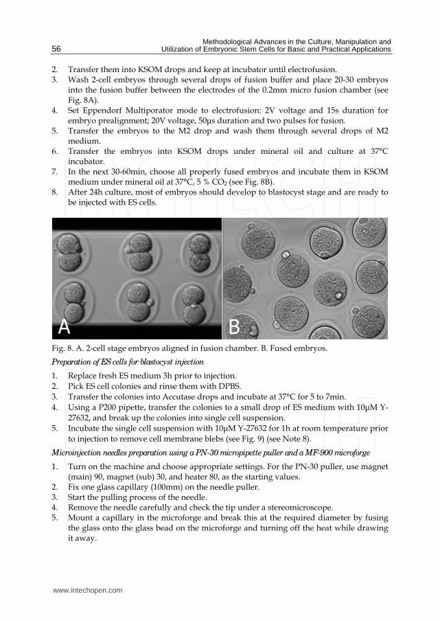
Methodological Advances in the Culture Manipulation and Utilization of Embryonic Stem Cells for Basic and Practical Applications
56
2 Transfer them into KSOM drops and keep at incubator until electrofusion 3 Wash 2-cell embryos through several drops of fusion buffer and place 20-30 embryos
into the fusion buffer between the electrodes of the 02mm micro fusion chamber (see Fig 8A)
4 Set Eppendorf Multiporator mode to electrofusion 2V voltage and 15s duration for embryo prealignment 20V voltage 50μs duration and two pulses for fusion
5 Transfer the embryos to the M2 drop and wash them through several drops of M2 medium
6 Transfer the embryos into KSOM drops under mineral oil and culture at 37degC incubator
7 In the next 30-60min choose all properly fused embryos and incubate them in KSOM medium under mineral oil at 37degC 5 CO2 (see Fig 8B)
8 After 24h culture most of embryos should develop to blastocyst stage and are ready to be injected with ES cells
Fig 8 A 2-cell stage embryos aligned in fusion chamber B Fused embryos
Preparation of ES cells for blastocyst injection
1 Replace fresh ES medium 3h prior to injection
2 Pick ES cell colonies and rinse them with DPBS
3 Transfer the colonies into Accutase drops and incubate at 37degC for 5 to 7min
4 Using a P200 pipette transfer the colonies to a small drop of ES medium with 10μM Y-
27632 and break up the colonies into single cell suspension
5 Incubate the single cell suspension with 10μM Y-27632 for 1h at room temperature prior
to injection to remove cell membrane blebs (see Fig 9) (see Note 8)
Microinjection needles preparation using a PN-30 micropipette puller and a MF-900 microforge
1 Turn on the machine and choose appropriate settings For the PN-30 puller use magnet (main) 90 magnet (sub) 30 and heater 80 as the starting values
2 Fix one glass capillary (100mm) on the needle puller 3 Start the pulling process of the needle 4 Remove the needle carefully and check the tip under a stereomicroscope 5 Mount a capillary in the microforge and break this at the required diameter by fusing
the glass onto the glass bead on the microforge and turning off the heat while drawing it away
wwwintechopencom
Effective Derivation and Manipulation of Mouse Embryonic Stem Cells
57
Fig 9 Compare to the control cells without Y-27632 treatment (A) the treatment of Y-27632 make ES cells smooth and soft (B) so that they can easily be picked up by injection pipette
6 For making holding pipets break the holding pipet at a point where the outside
diameter is approx 80μm and the inside diameter approx 60μm and then move the
holding pipet toward the glowing glass bead and the cut will start to melt inward
creating a smoother narrower tip
7 For making injection pipets break the injection pipet at a point where the outside
diameter is approx 15μm and the inside diameter approx 12μm
8 Hold the pipet above the glass bead and heat a point about 5mm from the pipet tip The
pipet will bend under gravity as the glass melts Allow the pipet to bend approx 60deg
Piezo-supported microinjection
1 Prepare dishes for injection using the lids of standard 35mm plastic disposable Petri
dishes
2 Place a 400μl drop of M2 medium on the lid and cover it with mineral oil
3 Fluorinert FC-77 in a 15mm length was back-loaded into an injection pipette Push
Fluorinert FC-77 through the shoulder to near the tip to empty the air in the pipette
4 Attach the injection pipette to the pipette holder of the piezo unit and hang the piezo
unit on the micromanipulator and aspirate 1mm M2 into the pipette to keep ES cells
away from Fluorinert FC-77
5 Attach the holding pipette on the other side of the micromanipulator
6 Transfer embryos and ES cells into the drop of M2 under mineral oil
7 Aspirate several ES cells and use several piezo pulses (eg intensity=3 frequency=3) to
penetrate the zona and trophectoderm layer (see Note 9)
8 Advance the tip of the pipette near the opposite side of the blastocysts from where it is
held by the holding pipette
9 Inject 15-20 ES cells into the blastocoel cavity (see Fig 10A B)
10 Culture injected blastocysts in KSOM medium at 37degC 5 CO2 for 2h and allow them
recovery
11 Transfer 8-10 injected blastocysts to each uterine horn of 25dpc pseudopregnant CD1
femals
12 Recipient mice will give birth after 17 days (see Fig 10C D)
wwwintechopencom
Methodological Advances in the Culture Manipulation and Utilization of Embryonic Stem Cells for Basic and Practical Applications
58
Fig 10 A Piezo-actuated tetraploid blastocyst injection B Injected tetraploid blastocysts C Dead ES mouse pups D Survival ES mouse pups
315 Embryo transfer and Caesarean sections 1 Select a pseudopregnant CD1 female (25dpc) as an embryo recipient 2 Prepare the transfer pipette forceps and scissors etc 3 Anesthetized mice by intraperitoneal injection of Avertin after drug delivery quickly
put the mouse back into the cage (see Note 10) 4 After the mouse is fully anesthetized place the mouse in the prone position on a lid of a
Petri dish Using an absorbent ball with 75 ethanol sterilize the area and shave the animal
5 Make a 05-1cm transverse dorsal incision at the below of the last ribs 6 Uncover incision to find ovarian fat pad pull it out using forceps and hold it using clamps 7 Collect the embryos carefully one by one lined up in the transfer pipette with as little
medium in between as possible 8 Make a hole through the uterine wall into the lumen with a 25G needle The site of this
hole should be near the last loop of the oviduct where it descends into the top of the uterine horn at the UT junction
9 Insert the transfer pipette into a hole and carefully expel the embryos Check the transfer pipette to ensure that all embryos have been transferred 8-10 embryos are usually transferred into each horn
10 When embryos are to be transferred into uteri put the ovary back into the abdominal cavity and suture the incision
wwwintechopencom
Effective Derivation and Manipulation of Mouse Embryonic Stem Cells
59
11 Place the mouse in clean cage Lighting to heat until it regaines consciousness 12 After 17 days the pups will be born
4 Notes
1 Mitomycin C (Sigma M4287) Dissolve the powder into sterile distilled DMEM and adjust the concentration to 10μgml just before use Wash layers several times with DPBS to completely remove any mitomycin C residue from the feeder layers
2 Accutase is a ready to use cell detachment solution of proteolytic and collagenolytic enzymes and it does not contain mammalian or bacterial derived products Cell lines tested for Accutase application includes fibroblasts keratinocytes vascular smooth muscle cells primary chick embryo neuronal cells bone marrow stem cells 293 cells 3T3 cells HeLa cells insect cells human embryonic stem cells and human neural stem cells Moreover Accutase is quite gentle on cells and will not induce cell death if cells are treated a little longer than the optimal time period Thus we use Acutase instead of trypsin for enzymatic dissociation of ICM
3 This is a key step for ES cell isolation Watch the whole dissociation process under a microscope Careful not to pipette with too much force as single cells will not survive Compared to trypsin Accutase treated ICM outgrowths are easier to dissociate into small cell clumps
4 The ROCK inhibitor Y-27632 permits survival of dissociated human embryonic stem cells Similar to human ES cells we find that mouse ICM cells show sensitivity to trypsin They undergo cell death after dissociation and the cloning efficiency of dissociated ICM cells is generally very low Our data indicates that increased cellular adhesion induced by Y-27632 enhances the survival of dissociated ICM cells With more ES-like colonies appearing when the dissociated ICM cells are seeded in medium supplemented with 10 μM Y-27632 for 24h Moreover we have not observed adverse effects of Y-27632 treatment on pluripotency in maintenance culture even after a number of passages
5 Dishes are coated with agar to completely prevent cell attachment This can increase cell aggregation and embryonic body formation In brief add 1g agar into 100ml DPBS and autoclave for 30min Cool to 50degC at room temperature Rapidly cover the internal wall and bottom of 35mm dishes with agar solution Remove the extra solution and place dishes in hood until the remaining agar solidification In addition adding Y-27632 in the embryonic body medium can also enhance suspending cells to aggregate and embryonic body formation
6 Typically severe combined immunodeficient mice are used for making teratoma This strain of mouse is expensive and not easily raised Our data indicates that most mouse strains could be recipient for teratoma production by inguinal subcutaneous injection such as CD1 C57BL6times129Sv
7 Carefully make small depressions using needle and appropriate force in the Petri dish surface Too much force may break the dish and insufficient force will not produce the required depressions which can easily lead to the loss of embryos and ES cells
8 Prior to blastocyst injection the pretreatment of 10μM Y-27632 for 1h at room temperature can alter the membrane architecture of ES cells so that they can be easily collected by injection pipet Otherwise the ES cell surface is covered with blebs (see Fig 10)
9 Within the microinjection pipette ES cells should be kept approximately 100μm away from the pipette tip Damage to ES cells may occur if the cells are very close to the tip when the piezo pulses are applied
wwwintechopencom
Methodological Advances in the Culture Manipulation and Utilization of Embryonic Stem Cells for Basic and Practical Applications
60
10 Avertin anesthetic 125 stocks avertin is prepared by mixing 25g of 2 2 2-tribromoethyl alcohol (Sigma) 5ml of tert-amyl alcohol (Sigma) with 200ml water Avertin is intraperitoneally injected at a dose of 002ml per gram of body weight for anesthetization
5 Acknowledgements
We would like to thank Roman Krawetz (The University of Calgary Canada) for providing his expert and critical reading on the manuscript This work was supported by grants from National Natural Science Foundation of China (No 30871790) and Natural Science Foundation of Hebei (No C2009000600)
6 References
Batlle-Morera L Smith A amp Nichols J (2008) Parameters influencing derivation of embryonic stem cells from murine embryos Genesis 46(12)758-767 ISSN 1526-954X
Brook FA amp Gardner RL (1997) The origin and efficient derivation of embryonic stem cells in the mouse Proc Natl Acad Sci USA 945709-5712 ISSN 0027-8424
Bryja V Bonilla S Cajaacutenek L Parish CL Schwartz CM Luo Y Rao MS amp Arenas E (2006) An efficient method for the derivation of mouse embryonic stem cells Stem Cells 24(4)844-849 ISSN 1066-5099
Cheng J Dutra A Takesono A Garrett-Beal L amp Schwartzberg PL (2004) Improved generation of C57BL6J mouse embryonic stem cells in a defined serum-free media Genesis 39100-104 ISSN 1526-954X
Doungpunta J Santhi A Sathanawongs A Jarujinda Y amp Oranratnachai A (2009) Fivefold increase in derivation rates of mouse embryonic stem cellsafter supplementation of the media with multiple factors Theriogenology 72(2)232-242 ISSN 0093-691X
Evans M J amp Kaufman M H (1981) Establishment in culture of pluripotential cells from mouse embryos Nature 292(5819)154-156 ISSN 0028-0836
Martin G R (1981) Isolation of a pluripotent cell line from early mouse embryos cultured in medium conditioned by teratocarcinoma stem cells Proc Natl Acad Sci USA 787634-7638 ISSN 0027-8424
McWhir J Schnieke AE Ansell R Wallace H Colman A Scott ARamp Kind AJ (1996) Selective ablation of differentiated cells permits isolation of embryonic stem cell lines from murine embryos with a non-permissive genetic background Nat
Genet 14223-226 ISSN 1061-4036 Ruchi B Jacqueline L Min K amp Alexey VT (2008) Efficient propagation of single cells
accutase-dissociated human embryonic stem cells Mol Reprod Dev 75(5)818-827 ISSN 1040-452X
Schoonjans L Kreemers V Danloy S Moreadith RW Laroche Y amp Collen D (2003) Improved generation of germline competent embryonic stem cell lines from inbred mouse strains Stem Cells 21(1)90-97 ISSN 1066-5099
Tesar PJ (2005) Derivation of germ-line-competent embryonic stemcell lines from preblastocyst mouse embryos Proc Natl Acad Sci USA 102(23)8239-8244 ISSN 0027-8424
Watanabe K Ueno M Kamiya D Nishiyama A Matsumura M Wataya T Takahashi JB Nishikawa S Nishikawa S Muguruma K amp Sasai Y (2007) A ROCK inhibitor permits survival of dissociated human embryonic stem cells Nat
Biotechnol 25681-686 ISSN 1087-0156
wwwintechopencom
Methodological Advances in the Culture Manipulation andUtilization of Embryonic Stem Cells for Basic and PracticalApplicationsEdited by Prof Craig Atwood
ISBN 978-953-307-197-8Hard cover 506 pagesPublisher InTechPublished online 26 April 2011Published in print edition April 2011
InTech EuropeUniversity Campus STeP Ri Slavka Krautzeka 83A 51000 Rijeka Croatia Phone +385 (51) 770 447 Fax +385 (51) 686 166wwwintechopencom
InTech ChinaUnit 405 Office Block Hotel Equatorial Shanghai No65 Yan An Road (West) Shanghai 200040 China
Phone +86-21-62489820 Fax +86-21-62489821
Pluripotent stem cells have the potential to revolutionise medicine providing treatment options for a widerange of diseases and conditions that currently lack therapies or cures This book describes methodologicaladvances in the culture and manipulation of embryonic stem cells that will serve to bring this promise topractice
How to referenceIn order to correctly reference this scholarly work feel free to copy and paste the following
Peng Zhang Xinglong Wu Pengbo Wang and Xiangyun Li (2011) Effective Derivation and Manipulation ofMouse Embryonic Stem Cells Methodological Advances in the Culture Manipulation and Utilization ofEmbryonic Stem Cells for Basic and Practical Applications Prof Craig Atwood (Ed) ISBN 978-953-307-197-8 InTech Available from httpwwwintechopencombooksmethodological-advances-in-the-culture-manipulation-and-utilization-of-embryonic-stem-cells-for-basic-and-practical-applicationseffective-derivation-and-manipulation-of-mouse-embryonic-stem-cells

Effective Derivation and Manipulation of Mouse Embryonic Stem Cells
57
Fig 9 Compare to the control cells without Y-27632 treatment (A) the treatment of Y-27632 make ES cells smooth and soft (B) so that they can easily be picked up by injection pipette
6 For making holding pipets break the holding pipet at a point where the outside
diameter is approx 80μm and the inside diameter approx 60μm and then move the
holding pipet toward the glowing glass bead and the cut will start to melt inward
creating a smoother narrower tip
7 For making injection pipets break the injection pipet at a point where the outside
diameter is approx 15μm and the inside diameter approx 12μm
8 Hold the pipet above the glass bead and heat a point about 5mm from the pipet tip The
pipet will bend under gravity as the glass melts Allow the pipet to bend approx 60deg
Piezo-supported microinjection
1 Prepare dishes for injection using the lids of standard 35mm plastic disposable Petri
dishes
2 Place a 400μl drop of M2 medium on the lid and cover it with mineral oil
3 Fluorinert FC-77 in a 15mm length was back-loaded into an injection pipette Push
Fluorinert FC-77 through the shoulder to near the tip to empty the air in the pipette
4 Attach the injection pipette to the pipette holder of the piezo unit and hang the piezo
unit on the micromanipulator and aspirate 1mm M2 into the pipette to keep ES cells
away from Fluorinert FC-77
5 Attach the holding pipette on the other side of the micromanipulator
6 Transfer embryos and ES cells into the drop of M2 under mineral oil
7 Aspirate several ES cells and use several piezo pulses (eg intensity=3 frequency=3) to
penetrate the zona and trophectoderm layer (see Note 9)
8 Advance the tip of the pipette near the opposite side of the blastocysts from where it is
held by the holding pipette
9 Inject 15-20 ES cells into the blastocoel cavity (see Fig 10A B)
10 Culture injected blastocysts in KSOM medium at 37degC 5 CO2 for 2h and allow them
recovery
11 Transfer 8-10 injected blastocysts to each uterine horn of 25dpc pseudopregnant CD1
femals
12 Recipient mice will give birth after 17 days (see Fig 10C D)
wwwintechopencom
Methodological Advances in the Culture Manipulation and Utilization of Embryonic Stem Cells for Basic and Practical Applications
58
Fig 10 A Piezo-actuated tetraploid blastocyst injection B Injected tetraploid blastocysts C Dead ES mouse pups D Survival ES mouse pups
315 Embryo transfer and Caesarean sections 1 Select a pseudopregnant CD1 female (25dpc) as an embryo recipient 2 Prepare the transfer pipette forceps and scissors etc 3 Anesthetized mice by intraperitoneal injection of Avertin after drug delivery quickly
put the mouse back into the cage (see Note 10) 4 After the mouse is fully anesthetized place the mouse in the prone position on a lid of a
Petri dish Using an absorbent ball with 75 ethanol sterilize the area and shave the animal
5 Make a 05-1cm transverse dorsal incision at the below of the last ribs 6 Uncover incision to find ovarian fat pad pull it out using forceps and hold it using clamps 7 Collect the embryos carefully one by one lined up in the transfer pipette with as little
medium in between as possible 8 Make a hole through the uterine wall into the lumen with a 25G needle The site of this
hole should be near the last loop of the oviduct where it descends into the top of the uterine horn at the UT junction
9 Insert the transfer pipette into a hole and carefully expel the embryos Check the transfer pipette to ensure that all embryos have been transferred 8-10 embryos are usually transferred into each horn
10 When embryos are to be transferred into uteri put the ovary back into the abdominal cavity and suture the incision
wwwintechopencom
Effective Derivation and Manipulation of Mouse Embryonic Stem Cells
59
11 Place the mouse in clean cage Lighting to heat until it regaines consciousness 12 After 17 days the pups will be born
4 Notes
1 Mitomycin C (Sigma M4287) Dissolve the powder into sterile distilled DMEM and adjust the concentration to 10μgml just before use Wash layers several times with DPBS to completely remove any mitomycin C residue from the feeder layers
2 Accutase is a ready to use cell detachment solution of proteolytic and collagenolytic enzymes and it does not contain mammalian or bacterial derived products Cell lines tested for Accutase application includes fibroblasts keratinocytes vascular smooth muscle cells primary chick embryo neuronal cells bone marrow stem cells 293 cells 3T3 cells HeLa cells insect cells human embryonic stem cells and human neural stem cells Moreover Accutase is quite gentle on cells and will not induce cell death if cells are treated a little longer than the optimal time period Thus we use Acutase instead of trypsin for enzymatic dissociation of ICM
3 This is a key step for ES cell isolation Watch the whole dissociation process under a microscope Careful not to pipette with too much force as single cells will not survive Compared to trypsin Accutase treated ICM outgrowths are easier to dissociate into small cell clumps
4 The ROCK inhibitor Y-27632 permits survival of dissociated human embryonic stem cells Similar to human ES cells we find that mouse ICM cells show sensitivity to trypsin They undergo cell death after dissociation and the cloning efficiency of dissociated ICM cells is generally very low Our data indicates that increased cellular adhesion induced by Y-27632 enhances the survival of dissociated ICM cells With more ES-like colonies appearing when the dissociated ICM cells are seeded in medium supplemented with 10 μM Y-27632 for 24h Moreover we have not observed adverse effects of Y-27632 treatment on pluripotency in maintenance culture even after a number of passages
5 Dishes are coated with agar to completely prevent cell attachment This can increase cell aggregation and embryonic body formation In brief add 1g agar into 100ml DPBS and autoclave for 30min Cool to 50degC at room temperature Rapidly cover the internal wall and bottom of 35mm dishes with agar solution Remove the extra solution and place dishes in hood until the remaining agar solidification In addition adding Y-27632 in the embryonic body medium can also enhance suspending cells to aggregate and embryonic body formation
6 Typically severe combined immunodeficient mice are used for making teratoma This strain of mouse is expensive and not easily raised Our data indicates that most mouse strains could be recipient for teratoma production by inguinal subcutaneous injection such as CD1 C57BL6times129Sv
7 Carefully make small depressions using needle and appropriate force in the Petri dish surface Too much force may break the dish and insufficient force will not produce the required depressions which can easily lead to the loss of embryos and ES cells
8 Prior to blastocyst injection the pretreatment of 10μM Y-27632 for 1h at room temperature can alter the membrane architecture of ES cells so that they can be easily collected by injection pipet Otherwise the ES cell surface is covered with blebs (see Fig 10)
9 Within the microinjection pipette ES cells should be kept approximately 100μm away from the pipette tip Damage to ES cells may occur if the cells are very close to the tip when the piezo pulses are applied
wwwintechopencom
Methodological Advances in the Culture Manipulation and Utilization of Embryonic Stem Cells for Basic and Practical Applications
60
10 Avertin anesthetic 125 stocks avertin is prepared by mixing 25g of 2 2 2-tribromoethyl alcohol (Sigma) 5ml of tert-amyl alcohol (Sigma) with 200ml water Avertin is intraperitoneally injected at a dose of 002ml per gram of body weight for anesthetization
5 Acknowledgements
We would like to thank Roman Krawetz (The University of Calgary Canada) for providing his expert and critical reading on the manuscript This work was supported by grants from National Natural Science Foundation of China (No 30871790) and Natural Science Foundation of Hebei (No C2009000600)
6 References
Batlle-Morera L Smith A amp Nichols J (2008) Parameters influencing derivation of embryonic stem cells from murine embryos Genesis 46(12)758-767 ISSN 1526-954X
Brook FA amp Gardner RL (1997) The origin and efficient derivation of embryonic stem cells in the mouse Proc Natl Acad Sci USA 945709-5712 ISSN 0027-8424
Bryja V Bonilla S Cajaacutenek L Parish CL Schwartz CM Luo Y Rao MS amp Arenas E (2006) An efficient method for the derivation of mouse embryonic stem cells Stem Cells 24(4)844-849 ISSN 1066-5099
Cheng J Dutra A Takesono A Garrett-Beal L amp Schwartzberg PL (2004) Improved generation of C57BL6J mouse embryonic stem cells in a defined serum-free media Genesis 39100-104 ISSN 1526-954X
Doungpunta J Santhi A Sathanawongs A Jarujinda Y amp Oranratnachai A (2009) Fivefold increase in derivation rates of mouse embryonic stem cellsafter supplementation of the media with multiple factors Theriogenology 72(2)232-242 ISSN 0093-691X
Evans M J amp Kaufman M H (1981) Establishment in culture of pluripotential cells from mouse embryos Nature 292(5819)154-156 ISSN 0028-0836
Martin G R (1981) Isolation of a pluripotent cell line from early mouse embryos cultured in medium conditioned by teratocarcinoma stem cells Proc Natl Acad Sci USA 787634-7638 ISSN 0027-8424
McWhir J Schnieke AE Ansell R Wallace H Colman A Scott ARamp Kind AJ (1996) Selective ablation of differentiated cells permits isolation of embryonic stem cell lines from murine embryos with a non-permissive genetic background Nat
Genet 14223-226 ISSN 1061-4036 Ruchi B Jacqueline L Min K amp Alexey VT (2008) Efficient propagation of single cells
accutase-dissociated human embryonic stem cells Mol Reprod Dev 75(5)818-827 ISSN 1040-452X
Schoonjans L Kreemers V Danloy S Moreadith RW Laroche Y amp Collen D (2003) Improved generation of germline competent embryonic stem cell lines from inbred mouse strains Stem Cells 21(1)90-97 ISSN 1066-5099
Tesar PJ (2005) Derivation of germ-line-competent embryonic stemcell lines from preblastocyst mouse embryos Proc Natl Acad Sci USA 102(23)8239-8244 ISSN 0027-8424
Watanabe K Ueno M Kamiya D Nishiyama A Matsumura M Wataya T Takahashi JB Nishikawa S Nishikawa S Muguruma K amp Sasai Y (2007) A ROCK inhibitor permits survival of dissociated human embryonic stem cells Nat
Biotechnol 25681-686 ISSN 1087-0156
wwwintechopencom
Methodological Advances in the Culture Manipulation andUtilization of Embryonic Stem Cells for Basic and PracticalApplicationsEdited by Prof Craig Atwood
ISBN 978-953-307-197-8Hard cover 506 pagesPublisher InTechPublished online 26 April 2011Published in print edition April 2011
InTech EuropeUniversity Campus STeP Ri Slavka Krautzeka 83A 51000 Rijeka Croatia Phone +385 (51) 770 447 Fax +385 (51) 686 166wwwintechopencom
InTech ChinaUnit 405 Office Block Hotel Equatorial Shanghai No65 Yan An Road (West) Shanghai 200040 China
Phone +86-21-62489820 Fax +86-21-62489821
Pluripotent stem cells have the potential to revolutionise medicine providing treatment options for a widerange of diseases and conditions that currently lack therapies or cures This book describes methodologicaladvances in the culture and manipulation of embryonic stem cells that will serve to bring this promise topractice
How to referenceIn order to correctly reference this scholarly work feel free to copy and paste the following
Peng Zhang Xinglong Wu Pengbo Wang and Xiangyun Li (2011) Effective Derivation and Manipulation ofMouse Embryonic Stem Cells Methodological Advances in the Culture Manipulation and Utilization ofEmbryonic Stem Cells for Basic and Practical Applications Prof Craig Atwood (Ed) ISBN 978-953-307-197-8 InTech Available from httpwwwintechopencombooksmethodological-advances-in-the-culture-manipulation-and-utilization-of-embryonic-stem-cells-for-basic-and-practical-applicationseffective-derivation-and-manipulation-of-mouse-embryonic-stem-cells
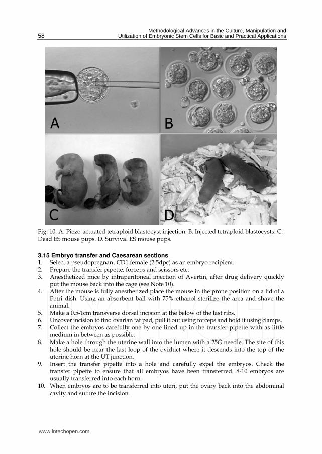
Methodological Advances in the Culture Manipulation and Utilization of Embryonic Stem Cells for Basic and Practical Applications
58
Fig 10 A Piezo-actuated tetraploid blastocyst injection B Injected tetraploid blastocysts C Dead ES mouse pups D Survival ES mouse pups
315 Embryo transfer and Caesarean sections 1 Select a pseudopregnant CD1 female (25dpc) as an embryo recipient 2 Prepare the transfer pipette forceps and scissors etc 3 Anesthetized mice by intraperitoneal injection of Avertin after drug delivery quickly
put the mouse back into the cage (see Note 10) 4 After the mouse is fully anesthetized place the mouse in the prone position on a lid of a
Petri dish Using an absorbent ball with 75 ethanol sterilize the area and shave the animal
5 Make a 05-1cm transverse dorsal incision at the below of the last ribs 6 Uncover incision to find ovarian fat pad pull it out using forceps and hold it using clamps 7 Collect the embryos carefully one by one lined up in the transfer pipette with as little
medium in between as possible 8 Make a hole through the uterine wall into the lumen with a 25G needle The site of this
hole should be near the last loop of the oviduct where it descends into the top of the uterine horn at the UT junction
9 Insert the transfer pipette into a hole and carefully expel the embryos Check the transfer pipette to ensure that all embryos have been transferred 8-10 embryos are usually transferred into each horn
10 When embryos are to be transferred into uteri put the ovary back into the abdominal cavity and suture the incision
wwwintechopencom
Effective Derivation and Manipulation of Mouse Embryonic Stem Cells
59
11 Place the mouse in clean cage Lighting to heat until it regaines consciousness 12 After 17 days the pups will be born
4 Notes
1 Mitomycin C (Sigma M4287) Dissolve the powder into sterile distilled DMEM and adjust the concentration to 10μgml just before use Wash layers several times with DPBS to completely remove any mitomycin C residue from the feeder layers
2 Accutase is a ready to use cell detachment solution of proteolytic and collagenolytic enzymes and it does not contain mammalian or bacterial derived products Cell lines tested for Accutase application includes fibroblasts keratinocytes vascular smooth muscle cells primary chick embryo neuronal cells bone marrow stem cells 293 cells 3T3 cells HeLa cells insect cells human embryonic stem cells and human neural stem cells Moreover Accutase is quite gentle on cells and will not induce cell death if cells are treated a little longer than the optimal time period Thus we use Acutase instead of trypsin for enzymatic dissociation of ICM
3 This is a key step for ES cell isolation Watch the whole dissociation process under a microscope Careful not to pipette with too much force as single cells will not survive Compared to trypsin Accutase treated ICM outgrowths are easier to dissociate into small cell clumps
4 The ROCK inhibitor Y-27632 permits survival of dissociated human embryonic stem cells Similar to human ES cells we find that mouse ICM cells show sensitivity to trypsin They undergo cell death after dissociation and the cloning efficiency of dissociated ICM cells is generally very low Our data indicates that increased cellular adhesion induced by Y-27632 enhances the survival of dissociated ICM cells With more ES-like colonies appearing when the dissociated ICM cells are seeded in medium supplemented with 10 μM Y-27632 for 24h Moreover we have not observed adverse effects of Y-27632 treatment on pluripotency in maintenance culture even after a number of passages
5 Dishes are coated with agar to completely prevent cell attachment This can increase cell aggregation and embryonic body formation In brief add 1g agar into 100ml DPBS and autoclave for 30min Cool to 50degC at room temperature Rapidly cover the internal wall and bottom of 35mm dishes with agar solution Remove the extra solution and place dishes in hood until the remaining agar solidification In addition adding Y-27632 in the embryonic body medium can also enhance suspending cells to aggregate and embryonic body formation
6 Typically severe combined immunodeficient mice are used for making teratoma This strain of mouse is expensive and not easily raised Our data indicates that most mouse strains could be recipient for teratoma production by inguinal subcutaneous injection such as CD1 C57BL6times129Sv
7 Carefully make small depressions using needle and appropriate force in the Petri dish surface Too much force may break the dish and insufficient force will not produce the required depressions which can easily lead to the loss of embryos and ES cells
8 Prior to blastocyst injection the pretreatment of 10μM Y-27632 for 1h at room temperature can alter the membrane architecture of ES cells so that they can be easily collected by injection pipet Otherwise the ES cell surface is covered with blebs (see Fig 10)
9 Within the microinjection pipette ES cells should be kept approximately 100μm away from the pipette tip Damage to ES cells may occur if the cells are very close to the tip when the piezo pulses are applied
wwwintechopencom
Methodological Advances in the Culture Manipulation and Utilization of Embryonic Stem Cells for Basic and Practical Applications
60
10 Avertin anesthetic 125 stocks avertin is prepared by mixing 25g of 2 2 2-tribromoethyl alcohol (Sigma) 5ml of tert-amyl alcohol (Sigma) with 200ml water Avertin is intraperitoneally injected at a dose of 002ml per gram of body weight for anesthetization
5 Acknowledgements
We would like to thank Roman Krawetz (The University of Calgary Canada) for providing his expert and critical reading on the manuscript This work was supported by grants from National Natural Science Foundation of China (No 30871790) and Natural Science Foundation of Hebei (No C2009000600)
6 References
Batlle-Morera L Smith A amp Nichols J (2008) Parameters influencing derivation of embryonic stem cells from murine embryos Genesis 46(12)758-767 ISSN 1526-954X
Brook FA amp Gardner RL (1997) The origin and efficient derivation of embryonic stem cells in the mouse Proc Natl Acad Sci USA 945709-5712 ISSN 0027-8424
Bryja V Bonilla S Cajaacutenek L Parish CL Schwartz CM Luo Y Rao MS amp Arenas E (2006) An efficient method for the derivation of mouse embryonic stem cells Stem Cells 24(4)844-849 ISSN 1066-5099
Cheng J Dutra A Takesono A Garrett-Beal L amp Schwartzberg PL (2004) Improved generation of C57BL6J mouse embryonic stem cells in a defined serum-free media Genesis 39100-104 ISSN 1526-954X
Doungpunta J Santhi A Sathanawongs A Jarujinda Y amp Oranratnachai A (2009) Fivefold increase in derivation rates of mouse embryonic stem cellsafter supplementation of the media with multiple factors Theriogenology 72(2)232-242 ISSN 0093-691X
Evans M J amp Kaufman M H (1981) Establishment in culture of pluripotential cells from mouse embryos Nature 292(5819)154-156 ISSN 0028-0836
Martin G R (1981) Isolation of a pluripotent cell line from early mouse embryos cultured in medium conditioned by teratocarcinoma stem cells Proc Natl Acad Sci USA 787634-7638 ISSN 0027-8424
McWhir J Schnieke AE Ansell R Wallace H Colman A Scott ARamp Kind AJ (1996) Selective ablation of differentiated cells permits isolation of embryonic stem cell lines from murine embryos with a non-permissive genetic background Nat
Genet 14223-226 ISSN 1061-4036 Ruchi B Jacqueline L Min K amp Alexey VT (2008) Efficient propagation of single cells
accutase-dissociated human embryonic stem cells Mol Reprod Dev 75(5)818-827 ISSN 1040-452X
Schoonjans L Kreemers V Danloy S Moreadith RW Laroche Y amp Collen D (2003) Improved generation of germline competent embryonic stem cell lines from inbred mouse strains Stem Cells 21(1)90-97 ISSN 1066-5099
Tesar PJ (2005) Derivation of germ-line-competent embryonic stemcell lines from preblastocyst mouse embryos Proc Natl Acad Sci USA 102(23)8239-8244 ISSN 0027-8424
Watanabe K Ueno M Kamiya D Nishiyama A Matsumura M Wataya T Takahashi JB Nishikawa S Nishikawa S Muguruma K amp Sasai Y (2007) A ROCK inhibitor permits survival of dissociated human embryonic stem cells Nat
Biotechnol 25681-686 ISSN 1087-0156
wwwintechopencom
Methodological Advances in the Culture Manipulation andUtilization of Embryonic Stem Cells for Basic and PracticalApplicationsEdited by Prof Craig Atwood
ISBN 978-953-307-197-8Hard cover 506 pagesPublisher InTechPublished online 26 April 2011Published in print edition April 2011
InTech EuropeUniversity Campus STeP Ri Slavka Krautzeka 83A 51000 Rijeka Croatia Phone +385 (51) 770 447 Fax +385 (51) 686 166wwwintechopencom
InTech ChinaUnit 405 Office Block Hotel Equatorial Shanghai No65 Yan An Road (West) Shanghai 200040 China
Phone +86-21-62489820 Fax +86-21-62489821
Pluripotent stem cells have the potential to revolutionise medicine providing treatment options for a widerange of diseases and conditions that currently lack therapies or cures This book describes methodologicaladvances in the culture and manipulation of embryonic stem cells that will serve to bring this promise topractice
How to referenceIn order to correctly reference this scholarly work feel free to copy and paste the following
Peng Zhang Xinglong Wu Pengbo Wang and Xiangyun Li (2011) Effective Derivation and Manipulation ofMouse Embryonic Stem Cells Methodological Advances in the Culture Manipulation and Utilization ofEmbryonic Stem Cells for Basic and Practical Applications Prof Craig Atwood (Ed) ISBN 978-953-307-197-8 InTech Available from httpwwwintechopencombooksmethodological-advances-in-the-culture-manipulation-and-utilization-of-embryonic-stem-cells-for-basic-and-practical-applicationseffective-derivation-and-manipulation-of-mouse-embryonic-stem-cells


